Investigation of Therapeutics Benefits of Dioscorea ...
14
Available online www.jocpr.com Journal of Chemical and Pharmaceutical Research, 2017, 9(6):313-326 Research Article ISSN : 0975-7384 CODEN(USA) : JCPRC5 313 Investigation of Therapeutics Benefits of Dioscorea bulbifera in Breast Cancer Rashmi C Anandpara * and Pravin R Tirgar Department of Pharmacology, School of Pharmacy, RK University, Rajkot, Gujarat, India _____________________________________________________________________________ ABSTRACT Aim: To investigate the therapeutics benefits of Dioscorea bulbifera in breast cancer. Materials and Methods: Extract of Dioscorea bulbifera prepared by using Soxhlet extraction method. The various experimental models were performed as, In silico Docking Study and Pass Online study, In vivoNMU induce breast cancer in female wistar rats for 30 days, In vitro MDA-MB 231 breast cancer cell line study and hemolytic activity. All studies were compared with standard drug Tamoxifen. Results: Molecular docking studies showed higher binding affinity and more number of interactions of Diosbulbin B with respective receptors as compared with the standard like Doxorubicin, Fluvistrant, Mefepristone, Onapristone. In vitro NMU induced cancer model revealed that the estrogen and progesterone levels decreases in blood serum in test group as compared to disease control group and standard group after 30 days of study. In vitro MDAMB 231 cell treated with Dioscoreabulbifera (10, 50, 100, 200, 500 mg/ml) showed significant decreased in cell count and cell viability, which indicates cytotoxic effect of hydro alcoholic extract of Dioscorea bulbifera. Conclusions: From the findings of the performed models we can conclude that hydroalcoholic extract of Dioscorea bulbifera shows anti-cancer activity which can work by aromatase inhibiting activity as it reduce estrange and progesterone levels and might be useful as a better and safer herbal alternate of breast cancer. Keywords: Anti cancer activity; Breast cancer; Dioscorea bulbifera; NMU induced breast cancer model; Cell line study; Hemolytic activity; In silico docking study _____________________________________________________________________________ INTRODUCTION Cancer is a general term used to refer to a condition where the body’s cells begin to grow and reproduce in an uncontrollable way. These cells can then invade and destroy healthy tissue, including organs. Cancer sometimes begins in one part of the body before spreading to other parts. Cancer is a common condition and a serious health related problem. More than one in three people can develop some form of cancer during their lifetime. There are more than hundred different types of cancers are there but the most common types of the cancers are Breast cancer, Cervical cancer, Prostate cancer, Lung cancer, Mouth cancer [1]. There ‘’ are different types of breast cancers such as ductal carcinoma, infiltrating ductal carcinoma, lobular carcinoma, invasive lobular carcinoma, inflammatory breast cancer, etc. Early “ breast cancer often does not cause symptoms. Therefore regular breast exams and mammograms are important, so cancers that don't have symptoms may be found ” earlier. Various ‘’ types of the treatments were used for breast cancer such as hormone therapy, radiation therapy, chemotherapy, targeted drug therapy, mammography, ultrasound, etc. [2] Targeted therapy drugs that targets the specifically targeted genes and makes some kind of changes in genes and helps in cancer cells. ’’ For eg. Drugs that target HER2, including trastuzumab(Herceptin), pertuzumab(Perjeta), lapatinib(Tykerb), PARP Inhibitors, Angiogenesis drugs [3]. The ‘’ use of the plant-derived natural products for medicinal benefits has played an important role in almost all the people on earth. In Indian system of medicine (ISM), Ayurveda, Siddha, Unani and Homeopathic system provide health care for large part of the population, ISM is vas and its review revealed several plants which can be used for the drug development [4]. Since 1961, several anticancer drugs have been made available on the market that trace their
Transcript of Investigation of Therapeutics Benefits of Dioscorea ...

Available online www.jocpr.com
Journal of Chemical and Pharmaceutical Research, 2017, 9(6):313-326
Research Article ISSN : 0975-7384
CODEN(USA) : JCPRC5
313
Investigation of Therapeutics Benefits of Dioscorea bulbifera in Breast Cancer
Rashmi C Anandpara* and Pravin R Tirgar
Department of Pharmacology, School of Pharmacy, RK University, Rajkot, Gujarat, India
_____________________________________________________________________________
ABSTRACT
Aim: To investigate the therapeutics benefits of Dioscorea bulbifera in breast cancer. Materials and Methods:
Extract of Dioscorea bulbifera prepared by using Soxhlet extraction method. The various experimental models were
performed as, In silico Docking Study and Pass Online study, In vivoNMU induce breast cancer in female wistar
rats for 30 days, In vitro MDA-MB 231 breast cancer cell line study and hemolytic activity. All studies were
compared with standard drug Tamoxifen. Results: Molecular docking studies showed higher binding affinity and
more number of interactions of Diosbulbin B with respective receptors as compared with the standard like
Doxorubicin, Fluvistrant, Mefepristone, Onapristone. In vitro NMU induced cancer model revealed that the
estrogen and progesterone levels decreases in blood serum in test group as compared to disease control group and
standard group after 30 days of study. In vitro MDAMB 231 cell treated with Dioscoreabulbifera (10, 50, 100, 200,
500 mg/ml) showed significant decreased in cell count and cell viability, which indicates cytotoxic effect of hydro
alcoholic extract of Dioscorea bulbifera. Conclusions: From the findings of the performed models we can conclude
that hydroalcoholic extract of Dioscorea bulbifera shows anti-cancer activity which can work by aromatase
inhibiting activity as it reduce estrange and progesterone levels and might be useful as a better and safer herbal
alternate of breast cancer.
Keywords: Anti cancer activity; Breast cancer; Dioscorea bulbifera; NMU induced breast cancer model; Cell line
study; Hemolytic activity; In silico docking study
_____________________________________________________________________________
INTRODUCTION
Cancer is a general term used to refer to a condition where the body’s cells begin to grow and reproduce in an
uncontrollable way. These cells can then invade and destroy healthy tissue, including organs. Cancer sometimes
begins in one part of the body before spreading to other parts. Cancer is a common condition and a serious health
related problem. More than one in three people can develop some form of cancer during their lifetime. There are
more than hundred different types of cancers are there but the most common types of the cancers are Breast cancer,
Cervical cancer, Prostate cancer, Lung cancer, Mouth cancer [1]. There ‘’are different types of breast cancers such as
ductal carcinoma, infiltrating ductal carcinoma, lobular carcinoma, invasive lobular carcinoma, inflammatory breast
cancer, etc. Early “breast cancer often does not cause symptoms. Therefore regular breast exams and mammograms
are important, so cancers that don't have symptoms may be found” earlier. Various
‘’types of the treatments were
used for breast cancer such as hormone therapy, radiation therapy, chemotherapy, targeted drug therapy,
mammography, ultrasound, etc. [2] Targeted therapy drugs that targets the specifically targeted genes and makes
some kind of changes in genes and helps in cancer cells.’’ For eg. Drugs that target HER2, including
trastuzumab(Herceptin), pertuzumab(Perjeta), lapatinib(Tykerb), PARP Inhibitors, Angiogenesis drugs [3]. The ‘’use
of the plant-derived natural products for medicinal benefits has played an important role in almost all the people on
earth. In Indian system of medicine (ISM), Ayurveda, Siddha, Unani and Homeopathic system provide health care
for large part of the population, ISM is vas and its review revealed several plants which can be used for the drug
development [4]. Since 1961, several anticancer drugs have been made available on the market that trace their

RC Anandpara and PR Tirgar J. Chem. Pharm. Res., 2017, 9(6):313-326 _____________________________________________________________________________________________
314
origins to plants such as Taxol(Paclitaxel) [4], Navelbine [4], and Gemzar(Gemcitabine) [5], Oncovin(Vincristine)
[6] etc are allopathic drugs used in cancer disease but have serious side effects and also they are very costly as
compared to herbal medicines and also have very less side effects so we choose herbal drug for the management of
cancer disease” i.e. Dioscorea bulbifera(Varahikand). Dioscorea bulbifera
“is one of the unique medicinal plants
among 600 species in the family Dioscoreaceae which has found its importance in traditional medicine throughout
the world. Aglycons of its steroid saponins like diosgenin have gained attention as precursors in the synthesis of sex
hormones, cardiatonic glucosides, fertility control compounds, corticosteroids and anabolic” agents [7]. It
“also
possess the various activities like anti-tumor, anti-diabetic, anti-inflammatory, gastroprotective, anti-oxidant, etc
types of” activity [8]. Dioscorea bulbifera contains Diosbulbin B as chemical constituent which is used in synthesis
of corticosteroids which is responsible for management of breast cancer [9]. Research herb possess the anti-oxidant
property by acting on free radicals like DPPH and OH ions so it can be used in management of cancer disease
[10,11].Mixture of different parts of the plants i.e leaf, roots and bulbs were used in this study for the management
of breast cancer.
It is reported that Dioscorea bulbifera possess the anti tumor activity so it can be used in management of breast
cancer [8]. To complete this investigation of Dioscorea bulbifera various experimental models were used, such as In
Silico method (Docking study) from that we can come to know about the binding affinity of the drug towards the
respective receptors, In vitro model (Hemolytic Assay) from that we can come to know about the dose of the drug as
well the cytotoxic effect of the drug, In vivo (N methyl nitrosoureinduce model), from that we can come to know
about the pharmacological effects of the drug.
MATERIALS AND METHODS
Collection and authentification
The plant used in this study was acquired from local area near Porbandar city. Authenticity of this plant was
confirmed by Mrs. Trupti Marakna, Assistant Professor, School of Science, RK University by comparing their
morphological characters with the description mentioned in different standard texts.
Pharmacognostic studies
Morphological charecterization of crude herb was done and compared with available literature.
Extraction (Figure 1)
Figure 1: Method performed for extraction
Extraction of Diosbulbin B from Dioscorea bulbifera was done by Soxhlet extraction (continuous hot extraction)
process. Plant was collected from local area near Porbandar region and was dried in open atmosphere. Pulverisation
of different parts of plants was prepared. Weighed powder was placed in thimble and measured amount of extract
solvent (70%V/V methanol) was added. Evaporation of extract solvent will be done using water bath. % yield will
be measure.
Experimental models
In silico model
Pass online study: Structure of Diosbulbin B was downloaded from website https://pubchem.ncbi.nlm.nih.gov/ and
saved as pdf file format. This file format does not open in PASS online software. Therefore, it was converted to .mol

RC Anandpara and PR Tirgar J. Chem. Pharm. Res., 2017, 9(6):313-326 _____________________________________________________________________________________________
315
file format by using OpenBable GUI 2.3.2 software. This file was uploaded in PASS online software and predicted
biological activity profile for Diosbulbin B was obtained.
Docking protocol: For “docking, ligands and proteins are required in .pdbqt file format. They were prepared for
docking using AutoDock Tool 1.5.6 (ATD). Also, grid box sizes were decided using the same programme. Finally,
molecular docking was done using AutoDock Vina 1.0 and further steps for docking were followed as” under
[12,13].
In vitro model
Hemolytic assay [14]:
Blood was collected by retroorbital method from animals in tube containing anti-coagulant. Blood solution was
prepared by washing six times with tris buffered solution (TBS), containing 6.05 gm tris HCL, 8.76 gm NaCl in 800
ml distilled water and pH 7.6 of solution was adjusted by 1M HCl. Following last wash, red blood cells (RBC) were
diluted to 1/10 of their volume with TBS. The assay was performed by mixing 0.3 ml of RBC solution with 1.2 ml
of crude extract; 1.2 ml of distilled water was set as positive control and 1.2 ml of TBS as a negative control. The
mixture were kept vortexed, left for 2h at room temperature, and then centrifuged at 4000 RPM for 10 min at 40°C.
Absorbance of supernants were measured at 541 nm in UV visible spectrophotometer. The percentage hemolysis of
each fraction was calculated using expression below (Figure 2).
Hemolytic activity =
Figure 2: Remi Centrifuge
Cell line Study [15]:
Approximately 10,000 cells were plated per well in a 96 well plate and the plate was incubated at 37 degree Celsius
in a CO2 incubator. Thereafter the cells were treated with the different concentrations of the compound. Each
concentration was tested in triplicates. The plate was then incubated at 37 degree Celsius in a CO2 incubator.
Thereafter, the cell viability upon 48 hours of drug treatment was assessed using MTT assay.
20 microliter of 5mg/ml MTT solution were added to each well after 46 hours of drug exposure and the plate were
incubated at 37 degree Celsius in a CO2 incubator for 2 hours.
The reduction of MTT by the viable cells leads to the formation of formazon. The medium were removed from the
wells and 100 microliter of DMSO was added to dissolve the formazon crystals and absorbance of the dissolved
formazon was taken at 570 nm using plate reader. Percent cell viability was calculated by equating the absorbance of
the control well to 100 percent viability performed in Perd University, Ahmedabad.
In vivo model
N- methyl nitrosourea (NMU) induce cancer model [16]:
Female wistar rats at 21 days of age were selected for experiment. NMU (50 mg/kg i.p.) were injected on 1, 7, 14,
21 days alternately through left and right abdominal wall. The MNU was always dissolved immediately before use
in 0.9 % NaCl adjusted to pH 4 with acetic acid. The solubility of NMU in water at room temperature was 1.4 %
(w/v). The experiment was terminated on the 30th day of the animals’ age. At the end of 30 days of study blood
were collected from animals by retroorbital method and serum was separated by centrifugation. Then serum was
subjected for the analysis of estrogen and progesterone level by ARCHITECT i system.

RC Anandpara and PR Tirgar J. Chem. Pharm. Res., 2017, 9(6):313-326 _____________________________________________________________________________________________
316
Biochemical parameters:
Serum: Estrogen, Progesterone
Assay Procedure for estrogen: The system automatically performs the following actions:
Dispenses 80µL of sample and 75µL of Ancillary Pack Reagent into a cuvette and incubates for 4.5 minutes at
37°C. Dispenses 75µL of Lite Reagent and incubates for 2.75 minutes at 37°C.
Dispenses 100µL of Solid Phase Reagent with 25µL of Ancillary Well Reagent and incubates for 5.5 minutes at
37°C. Separates, aspirates, and washes the cuvette with wash 1 and dispenses 300 µL of Acid Reagent and Base
Reagent each to initiate the chemiluminescent reaction.
Calculation of results:
The system reports serum estradiol results in pg/mL (common units) or pmol/L (SI units), depending on the units
defined when setting up the assay. Conversion formula is 1 pg/mL = 3.67 pmol/L
Assay procedure for progesterone:
Load the ARCHITECT Progesterone Reagent Kit on the ARCHITECTi System. Verify that all necessary assay
reagents are present. Ensure that septums are present on all reagent bottles. The minimum sample cup volume is
calculated by the system and is printed on the Order list report. No more than 10 replicates may be sampled from the
same sample cup. To minimize the effects of evaporation verify adequate sample cup volume is present prior to
running the test. If using primary or aliquot tubes, use the sample gauge to ensure sufficient patient specimen is
present. ARCHITECT Progesterone Calibrators and Controls must be mixed thoroughly by low speed vortex or
inversion prior to use. To obtain the recommended volume requirements for the ARCHITECT Progesterone
Calibrators and Controls, dispense a minimum of 200 µL of each calibrator or a minimum of 150 µL of each control
into each respective sample cup. Load samples and press run.
List of chemical and instruments needed: Instruments Equipments
Heating mental Micro pipette and micro tips
Cooling centrifuge EDTA tubes
Weighing balance Eppendorf
Spectrophotometer Capillary
ARCHITECTi System Small plastic beakers
CO2 incubator Tuberculin syringe
Plate Reader Oral feeding needle
Laminar flow hood Dissection Box
Chemicals:
Water, methanol, diethyl ether, DMSO etc. 5.6 Statistical Analysis Method To checking the significance of data,
following statistical tests were performed;
ANOVA: to see variability within all the groups.
Tuckey’s test: for the same purpose mentioned in above test.
INSTAT software: to derive all the statistical terms like Standard Error of Mean (SEM),
ANOVA, P-value, Degree of freedom, Standard deviation, etc
RESULTS
Pharmacognostic evaluation of Hydroalcoholic extract of Dioscorea bulbifera
Extract of crude drug prepared by Soxhlet extraction method using methanol and water as solvent. After completion
of three cycles solvent in RBF is evaporated and 27.37% W/W yield is obtained having dark brown colour and
semisolid consistency (Table 1).
Table 1: Pharmacognostic evaluation of extracts of Dioscorea bulbifera
Extract % W/W yield Colour Consistency
Hydroalcoholic extract of Dioscorea bulbifera 27.37% Dark Brown Semi solid
Phytochemical screening of hydroalcoholic extract of Dioscorea bulbifera
The extracts obtained from Soxhlet were subjected to various chemical tests to determine the presence of
phytochemical constituents like favanoids, terpenoids, steroid, saponin, cardiac glycosides. The result is reported
below (Table 2)

RC Anandpara and PR Tirgar J. Chem. Pharm. Res., 2017, 9(6):313-326 _____________________________________________________________________________________________
317
Table 2: Phytochemical screening of extracts of Dioscorea bulbifera
Type of extract Name of Compound Dioscorea bulbifera
Hydroalcoholic extract
Cholesterol -
Alkaloid -
Flavanoid +++
Terpenoid +++
Cardiac glycoside ++
Steroid ++
Saponin +
+ indicates the presence of phytochemical constituents; - indicates the presence of phytochemical constituents
Pass online study for activity prediction:
Predicted pharmacological activities of diosbulbin B using Pass online software revealed that Diosbulbin B might be
active (Pa) 93.6 % as BRAF expression inhibitor, 82.6 % apoptosis inducer, 74.2% as antineoplastic, 59% TNF
expression inhibitors, 48% chemopreventive as cancer treatment. (Table 3)
Table 3: Activity determination Study of Dioscorea bulbifera using PASS Online Software
Pa Pi Activity
0,936 0,001 BRAF expression inhibitor
0,826 0,006 Apoptosis agonist
0,742 0,019 Antineoplastic
0,590 0,014 TNF expression inhibitor
0,569 0,017 AR expression inhibitors
0,480 0,016 Chemopreventive
Pa – Probability of activation; Pi – Probability of inactivation
In silico study- Docking study of Dioscorea bulbifera:
From docking study of dioscorea bulbifera, we come to know about the binding affinity of drug towards the
respective receptor. If drug is having more binding affinity then it requires less energy for binding. From the below
result of binding affinity of Diosbulbin B with the estrogen protein/receptor, there was significant decrease in
binding affinity as compared to Doxorubicin as standard drug. There was no any kind of significant difference of
binding affinity of Diosbulbin B as compared to Fluvestrant as standard drug (Table 4) (Figures 3 and 4).
Table 4: Docking study of Diosbulbin B with estrogen receptor
Mode Estrogen
Diosbulbin B Doxorubicin Temoxifene Fluvestrant
1 -12.4 -9.5 -7.3 -9.9
2 -12 -9.3 -7.1 -9.3
3 -11.1 -9 -7 -9.3
4 -10.4 -8.9 -7 -9.3
5 -10.2 -8.7 -6.9 -9
6 -10.2 -8.6 -6.8 -9
7 -10.2 -8.5 -6.7 -9
8 -10.1 -8.4 -6.7 -9
9 -10.1 -8.4 -6.6 -9
Binding affinity of Diosbulbin B decreases with the progesterone receptor as compared to Doxorubicin,
Mefepristone and Onapristone (Table 5).
Table 5: Docking study of Diosbulbin B with progesterone receptor
Mode Progesterone
Diosbulbin B Doxorubicin Mefepristone Onapristone
1 -12.3 -11.1 -10.3 -10.6
2 -11.8 -10.7 -10 -9.7
3 -11.8 -10.3 -9.9 -9.2
4 -11.8 -10.3 -9.8 -9.2
5 -11.6 -10.1 -9.8 -9.1
6 -11.5 -9.9 -9.6 -9.1
7 -11.5 -9.8 -9.5 -9.1
8 -11.4 -9.7 -9.4 -9
9 -11.9 -9.6 -9.3 -8.9
RC Anandpara and PR Tirgar J. Chem. Pharm. Res., 2017, 9(6):313-326 _____________________________________________________________________________________________
318
Figure 3: Binding affinity of Diosbulbin B with estrogen receptor
Figure 4: Binding affinity of Diosbulbin B with progesterone receptor
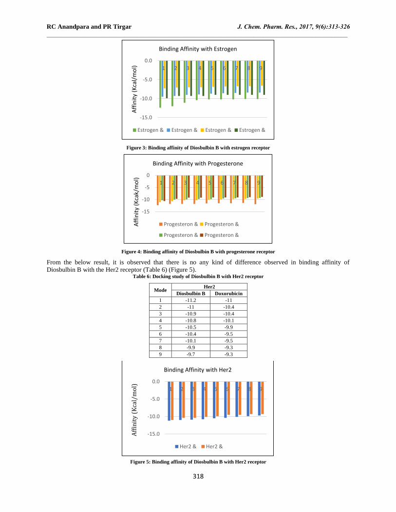
From the below result, it is observed that there is no any kind of difference observed in binding affinity of
Diosbulbin B with the Her2 receptor (Table 6) (Figure 5). Table 6: Docking study of Diosbulbin B with Her2 receptor
Mode Her2
Diosbulbin B Doxorubicin
1 -11.2 -11
2 -11 -10.4
3 -10.9 -10.4
4 -10.8 -10.1
5 -10.5 -9.9
6 -10.4 -9.5
7 -10.1 -9.5
8 -9.9 -9.3
9 -9.7 -9.3
Figure 5: Binding affinity of Diosbulbin B with Her2 receptor
-15.0
-10.0
-5.0
0.0
1 2 3 4 5 6 7 8 9
Aff
init
y (K
cal/
mo
l)
Binding Affinity with Estrogen
Estrogen & Estrogen & Estrogen & Estrogen &
-15
-10
-5
0
1 2 3 4 5 6 7 8 9
Aff
init
y (K
cak/
mo
l)
Binding Affinity with Progesterone
Progesteron & Progesteron &
Progesteron & Progesteron &
-15.0
-10.0
-5.0
0.0
1 2 3 4 5 6 7 8 9
Aff
init
y (
Kca
l/m
ol)
Binding Affinity with Her2
Her2 & Her2 &

RC Anandpara and PR Tirgar J. Chem. Pharm. Res., 2017, 9(6):313-326 _____________________________________________________________________________________________
319
From the below result, it is observed that Diosbulbin B on aromatase having same binding affinity as compared to
Exemestane and Testolactone. There was decrease in binding affinity of Diosbulbin B on aromatase as compared to
Doxorubicin and Letrozole (Table 7) (Figure 6).
Table 7: Docking study of Diosbulbin B with aromatase
Mode Aromatase
Diosbulbin B Doxorubicin Exemestane Letrozole Testolactone
1 -11.4 -10.4 -11.2 -7.7 -11.5
2 -10.7 -10.2 -10.6 -7.6 -10.7
3 -10.7 -10 -10.6 -7.6 -10.5
4 -10.5 -9.9 -10.5 -7.4 -10.4
5 -10.4 -9.8 -10.4 -7.4 -10.4
6 -10.2 -9.8 -10.3 -7.3 -10.4
7 -10.1 -9.8 -10.2 -7.3 -10.1
8 -10 -9.8 -9.9 -7.2 -9.9
9 -10 -9.8 -9.6 -7.2 -9.9
Figure 6: Binding affinity of Diosbulbin B with aromatase
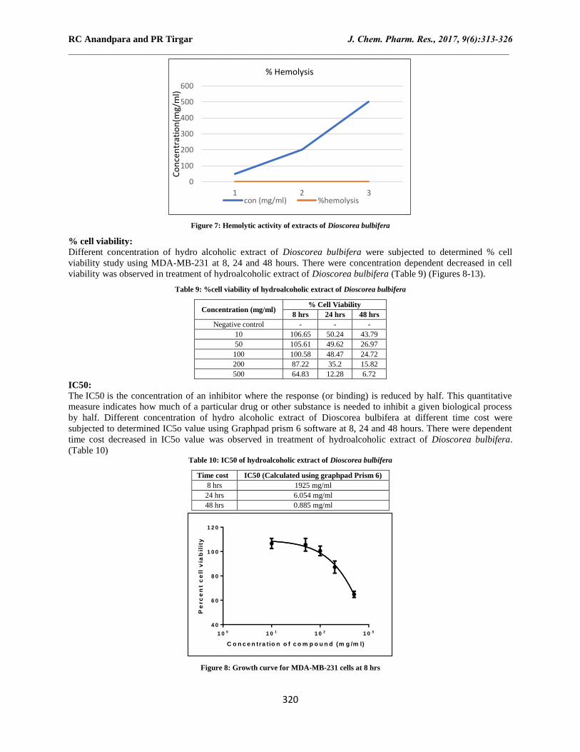
Effects of hydroalcoholic extract of Dioscorea bulbifera on cell viability (Hemolytic activity):
Haemolytic activity of extract was done to know the cytotoxic effect of Dioscorea bulbifera, by collecting blood
from animals by retroorbital method. Serum was separated by cooling centrifuge and different solution of drug was
prepared and absorbance of sample was taken by UV-visible spectrophotometer. As the absorbance of sample
increases there are more chances of haemolysis, which indicates cytotoxic activity of test samples of different
concentration (Table 8).
Table 8: Hemolytic activity of extracts of Dioscorea bulbifera
Concentration(mg/ml) Absorbance (λmax 541 nm) % haemolysis
50 1.954 36%
200 2.573 54%
500 3.552 82%
Effects of Hydroalcoholic extract of Dioscorea bulbifera on MDAMB 231 breast cancer cell line:
The breast cancer cell lines MDA-MB-231 were used to evaluate cytotoxic effects of Hydroalcoholic extract of
Dioscorea bulbifera. MDA-MB-231 cancer cell line is helpful to understand important role in control of growth and
proliferation of breast tumor cells. MDA-MB-231 cancer cell may serve as a model for native human breast cancer
cell line as they express high estrogen receptors and adenosine receptor. Overexpression of these receptors results in
activation of Adenosine A2B receptors mediate a stimulation of adenylyl cyclase, but are in addition coupled to a
Ca2+ signal. The Ca2+ response requires activation of phospholipase C and appears to be mediated via Gq/11
pathway. Further work will be required to understand the role of the A2B adenosine receptors in tumor growth and
development (Figure 7).
-15.0
-10.0
-5.0
0.0
1 2 3 4 5 6 7 8 9
Aff
init
y (K
cal/
mo
l)
Binding Affinity with Aromatase
Aromatase & Aromatase & Aromatase &
Aromatase & Aromatase &
RC Anandpara and PR Tirgar J. Chem. Pharm. Res., 2017, 9(6):313-326 _____________________________________________________________________________________________
320
Figure 7: Hemolytic activity of extracts of Dioscorea bulbifera
% cell viability:
Different concentration of hydro alcoholic extract of Dioscorea bulbifera were subjected to determined % cell
viability study using MDA-MB-231 at 8, 24 and 48 hours. There were concentration dependent decreased in cell
viability was observed in treatment of hydroalcoholic extract of Dioscorea bulbifera (Table 9) (Figures 8-13).
Table 9: %cell viability of hydroalcoholic extract of Dioscorea bulbifera
Concentration (mg/ml) % Cell Viability
8 hrs 24 hrs 48 hrs
Negative control - - -
10 106.65 50.24 43.79
50 105.61 49.62 26.97
100 100.58 48.47 24.72
200 87.22 35.2 15.82
500 64.83 12.28 6.72
IC50:
The IC50 is the concentration of an inhibitor where the response (or binding) is reduced by half. This quantitative
measure indicates how much of a particular drug or other substance is needed to inhibit a given biological process
by half. Different concentration of hydro alcoholic extract of Dioscorea bulbifera at different time cost were
subjected to determined IC5o value using Graphpad prism 6 software at 8, 24 and 48 hours. There were dependent
time cost decreased in IC5o value was observed in treatment of hydroalcoholic extract of Dioscorea bulbifera.
(Table 10) Table 10: IC50 of hydroalcoholic extract of Dioscorea bulbifera
Time cost IC50 (Calculated using graphpad Prism 6)
8 hrs 1925 mg/ml
24 hrs 6.054 mg/ml
48 hrs 0.885 mg/ml
Figure 8: Growth curve for MDA-MB-231 cells at 8 hrs
0
100
200
300
400
500
600
1 2 3
Co
nce
ntr
atio
n(m
g/m
l)
% Hemolysis
con (mg/ml) %hemolysis
C o n c e n tra tio n o f c o m p o u n d (m g /m l)
Pe
rc
en
t c
ell
via
bil
ity
1 0 0 1 0 1 1 0 2 1 0 3
4 0
6 0
8 0
1 0 0
1 2 0

RC Anandpara and PR Tirgar J. Chem. Pharm. Res., 2017, 9(6):313-326 _____________________________________________________________________________________________
321
Figure 9: Photomicrograph of MDA-MB-231 cells at 8 hrs
Figure 10: Growth curve for MDA-MB-231 cells at 24 hrs
C o n c e n tra tio n o f c o m p o u n d (m g /m l)
Pe
rc
en
t c
ell
via
bil
ity
1 0 0 1 0 1 1 0 2 1 0 3
0
2 0
4 0
6 0

RC Anandpara and PR Tirgar J. Chem. Pharm. Res., 2017, 9(6):313-326 _____________________________________________________________________________________________
322
Figure 11: Photomicrograph of MDA-MB-231 cells at 24 hrs
Figure 12: Growth curve for MDA-MB-231 cells at 48 hrs
C o n c e n tra tio n o f c o m p o u n d (m g /m l)
Pe
rc
en
t c
ell
via
bil
ity
1 0 0 1 0 1 1 0 2 1 0 3
0
1 0
2 0
3 0
4 0
5 0

RC Anandpara and PR Tirgar J. Chem. Pharm. Res., 2017, 9(6):313-326 _____________________________________________________________________________________________
323
Figure 13: Photomicrograph of MDA-MB-231 cells at 48 hrs
Effects of Hydroalcoholic extract of Dioscorea bulbifera on Estrogen level:
Estrogens are considered to play a major role in promoting the proliferation of both the normal and the neoplastic
breast epithelium. Increase estrogen levels in body indicate that there are more chances of breast cancer in human.
There was a significant increase in estrogen level in disease control group (74.54 ± 3.10) as compared to normal
control group (41.36 ± 1.11). There was significant decrease in estrogen level in standard Tamoxifen(20mg/kg)
group (58.76 ± 2.40), HEDB (200mg/kg) group (57.17 ± 2.32), HEDB (500mg/kg) group (59.49 ± 1.64) as
compared to disease control group (Tables 11 and 12) (Figure 14).
Table 11: Beneficial effect of Dioscorea bulbifera on Estrogen levels at 15th day in NMU induce breast cancer Wistar female rats
Groups Estrogen level on 15th day (pg/ml) Average ± SEM
Normal 42.07 40.01 41.01 43.03 40.04 42.05 41.36 ± 1.11
Disease 75.02 79.52 71.47 73.03 77.36 70.89 74.54 ± 3.10#
Standard Tamoxifen (20 mg/kg) 55.25 58.23 59.63 63.24 57.58 58.67 58.76 ± 2.40*
HEDB (200mg/kg) 58.02 56.32 59.36 53.27 60.23 55.85 57.17 ± 2.32**
HEDB (500mg/kg) 59.32 61.32 57.56 57.17 60.34 61.23 59.49 ± 1.64***
# - Significant increase in estrogen level in disease control group as compared to normal control group; ** indicate significant decreased in estrogen level in standard treated group as compared to diseases control group (level of significance p<0.001≈significant.); *** indicate
significant decreased in estrogen level in treatment group as compared to diseases control group (level of significance p<0.001≈highly significant).

RC Anandpara and PR Tirgar J. Chem. Pharm. Res., 2017, 9(6):313-326 _____________________________________________________________________________________________
324
Table 12: Beneficial effect of Dioscorea bulbifera on Estrogen levels after 30 days in NMU induce breast cancer Wistar female rats
Groups Estrogen level after 30 days (pg/ml) Average ± SEM
Normal 42.07 40.01 41.01 43.03 40.04 42.05 41.36 ± 1.11
Disease 95.03 99.05 97.05 96.03 98.04 95.05 96.70 ± 1.49#
Standard Tamoxifen (20 mg/kg) 48.07 47.01 50.23 55.55 49.04 49.09 49.83 ± 2.74**
HEDB (200mg/kg) 45.77 47.93 49.06 48.55 49.04 47.09 47.90 ± 1.17***
HEDB (500mg/kg) 44.77 43.06 41.66 42.55 45.04 44.09 43.52 ± 1.21***
# - Significant increase in estrogen level in disease control group as compared to normal control group; ** indicate significant decreased in estrogen level in standard treated group as compared to diseases control group (level of significance p<0.001≈significant.); *** indicate
significant decreased in estrogen level in treatment group as compared to diseases control group (level of significance p<0.001≈highly
significant.)
Figure 14: Estrogen levels in hydroalcoholic extract of Dioscorea bulbifera
n=6 and results were shown as mean ± SEM; Control group received distilled water; Disease control group received NMU (50mg/kg, i.p.) in
respective group; Standard control group received Tamoxifen (20mg/kg, p.o.) in respective group; Test (T1) group received HEDB(200mg/kg,
p.o.) (Hydro alcoholic extracts of Dioscorea bulbifera); Test (T2) group received HEDB (500mg/kg, p.o.) (Hydro alcoholic extracts of Dioscorea
bulbifera)
# - Significant increased in estrogen level in disease control group as compared to normal control group; ** indicate significant decreased in
estrogen level in standard treated group as compared to diseases control group (level of significance p<0.001≈significant.); *** indicate significant decreased in estrogen level in treatment group as compared to diseases control group (level of significance p<0.001≈highly
significant.)
Effects of hydroalcoholic extract of Dioscorea bulbifera on progesterone level:
Progesterone is an ovarian steroid hormone that is essential for normal breast development during puberty and in
preparation for lactation and breastfeeding. Progesterone decreases the cell proliferation. As the level of
progesterone increases there are more chances of breast cancer. There was significant increase in progesterone levels
in disease control group (36.62 ± 1.12#) as compared to normal group (9.68 ± 0.37). There was significant decrease
in progesterone level in standard group (21.08 ± 2.41**), HEDB (200 mg/kg) group (20.14 ± 1.75***) and HEDB
(500 mg/kg) group (19.08 ± 2.45***) as compared to disease control group (36.62 ± 1.12#). (Tables 13 and 4)
(Figure 15) Table 13: Beneficial effect of Dioscorea bulbifera on Progesterone levels at 15th day in NMU induced breast cancer Wistar female rats
Groups Progesterone level on 15th day (pg/ml) Average ± SEM
Normal 9.2 10.3 9.5 9.7 10 9.4 9.68 ± 0.37
Disease 38.23 36.3 34.6 36.7 37.5 36.4 36.62 ± 1.12#
Standard Tamoxifen (20 mg/kg) 19.4 22.5 20.5 25.7 20 18.4 21.08 ± 2.41**
HEDB (200mg/kg) 18.36 20.38 23.25 19.23 21.32 18.32 20.14 ± 1.75***
HEDB (500mg/kg) 15.3 20.32 16.35 19.65 22.32 20.54 19.08 ± 2.45***
# - Significant increase in progesterone level in disease control group as compared to normal control group; ** indicate significant decreased in
progesterone level in standard treated group as compared to diseases control group (level of significance p<0.001≈significant.); *** indicate significant decreased in progesterone level in treatment group as compared to diseases control group (level of significance p<0.001≈highly
significant)
Table 14: Beneficial effect of Dioscorea bulbifera on Progesterone levels after 30 days in NMU induce breast cancer Wistar female rats
Groups Progesterone level after 30 days (pg/ml) Average ± SEM
Normal 9.2 10.3 9.5 9.7 10 9.4 9.68 ± 0.37
Disease >40 38.2 37.3 39.2 37.5 38.6 38.16 ± 0.70#
Standard Tamoxifen (20 mg/kg) 9.4 9.5 9.5 11.7 10 9.4 9.91 ± 0.82**
HEDB (200mg/kg) 9.4 9.5 9.5 9.2 10 9.4 9.5 ± 0.24***
HEDB (500mg/kg) 9.3 10.5 9.5 9.7 10 9.4 9.73 ± 0.41***

RC Anandpara and PR Tirgar J. Chem. Pharm. Res., 2017, 9(6):313-326 _____________________________________________________________________________________________
325
# - Significant increase in progesterone level in disease control group as compared to normal control group; ** indicate significant decreased in
progesterone level in standard treated group as compared to diseases control group (level of significance p<0.001≈significant.); *** indicate
significant decreased in progesterone level in treatment group as compared to diseases control group (level of significance p<0.001≈highly significant.)
Figure 15: Progesterone levels in hydroalcoholic extract of Dioscorea bulbifera
n=6 and results were shown as mean ± SEM; Control group received distilled water; Disease control group received NMU(50mg/kg) in respective group; Standard control group received Tamoxifen (20mg/kg) in respective group; Treatment (1) control group received
HEDB(200mg/kg); Treatment (2) control group received HEDB(500mg/kg)
# - Significant increase in progesterone level in disease control group as compared to normal control group; ** indicate significant decreased in
progesterone level in standard treated group as compared to diseases control group (level of significance p<0.001≈significant.); *** indicate
significant decreased in progesterone level in treatment group as compared to diseases control group (level of significance p<0.001≈highly significant)
DISCUSSION AND CONCLUSION
In silico study of Diosbulbin B was carried out to know the binding affinity of Diosbulbin B with the respective
receptors/proteins. The receptors/proteins involved in study are estrogen, progesterone, Her2, Aromatase. In vitro
Haemolytic activity of extract was done to know the cytotoxic effect of Dioscorea bulbifera, as the absorbance of
sample increases there are more chances of haemolysis, which indicates cytotoxic activity of test samples of
different concentration. Different concentration of hydro alcoholic extract of Dioscorea bulbifera were subjected to
determined % cell viability study using MDA-MB-231 cell line at 8, 24 and 48 hours. There were concentration
dependent decreased in cell viability was observed in treatment of hydroalcoholic extract of Dioscorea bulbifera.
The results of NMU induce breast cancer revealed that, Estrogens are considered to play a major role in promoting
the proliferation of both the normal and the neoplastic breast epithelium. Increased estrogen levels in body indicate,
that there are more chances of breast cancer in human. There was a significant increase in estrogen level in disease
control group (74.54 ± 3.10 pg/ml) as compared to normal control group (41.36 ± 1.11 pg/ml). There was significant
decrease in estrogen level in standard Tamoxifen(20mg/kg) group (58.76 ± 2.40, pg/ml), HEDB (200mg/kg) group
(57.17 ± 2.32, pg/ml), HEDB (500mg/kg) group (59.49 ± 1.64, pg/ml) as compared to disease control group. As the
level of progesterone increases there are more chances of breast cancer. There was significant increase in
progesterone levels in disease control group (36.62 ± 1.12#,
pg/ml) as compared to normal group (9.68 ± 0.37,
pg/ml). There was significant decrease in progesterone level in standard group (21.08 ± 2.41**), HEDB (200 mg/kg)
group (20.14 ± 1.75*** pg/ml) and HEDB (500 mg/kg) group (19.08 ± 2.45*** pg/ml) as compared to disease
control group (36.62 ± 1.12# pg/ml). Thus at the end of 30 days trial, results indicated that Dioscorea bulbifera was
effective in breast cancer management by decreasing estrogen and progesterone levels in blood serum and also
decreases the cell viability of MDA-MB 231 cells as concentration and time points increases. These data indicted
hydroalcoholic extract of Dioscorea bulbifera has therapeutic effect in breast cancer by working on adenosine
receptor.
ACKNOWLEDGEMENT
There are always moments when you look back in time, recall and reminisce things about past. Writing this
acknowledgement is one of those very few moments. First of all I thank to my parents who is like a rejuvenation
source for me, for always being ready to cope with any trouble around me what so ever, in spite of having no
scientific background. I specially thank my fellow classmates’ here- Rashmi Anandpara, for always being one step

RC Anandpara and PR Tirgar J. Chem. Pharm. Res., 2017, 9(6):313-326 _____________________________________________________________________________________________
326
ahead from the other students, which provides easy way for learning all subjects during exam especially cology
subject which she links whole subject with routine life throughout course! She helped tirelessly through this project
and played troubleshooter for me. And also give moral support to me. And Jaydeep Limbasiya for help me in animal
handling and also in writing thesis and giving this document a professional touch which was hard task for me! He is
a person to rely upon and to call whenever needed, unconditionally. And also give moral support to me. I
congratulate School of Pharmacy RK University for having such an enlightened teaching staff in form of Mr. Tejas
Ganatra and Dr. Pravin Tirgar who guided us in this difficult journey. I would like to thanks SHTC trust honor of
school of pharmacy for providing good facility as well as academic environment. Lastly I would like thanks to all
my classmates viz., Raval Chirag, Uttam Khatri, Rakesh Parmar, Jigar Pansara, Brijesh Charavda and all to make
this course such a joyful for me. Thanks to all ………..
REFERENCES
[1] https://www.nidirect.gov./ uk, accessed on 27/11/2015
[2] JR Balentine; MC Stöppler. Quick Guide Breast Cancer Diagnosis, and Treatment.
[3] http://www.cancer.org/, accessed on 27/11/2015.
[4] HA Mavani. Evaluation of antiarthritic activity of polyherbal formulation ‘RIPARE” in experimental rats.
Uka Tarsadia University, 2013.
[5] https://www.breastcancercare.org./uk, accessed on 27/11/2015
[6] http://chemocare.com/chemotherapy, accessed on 27/11/2015
[7] L Ćulafić, K Šavikin-Fodulović, D Grubišić, M Nešković. Dioscorea balcanica Košanin and D. caucasica
Lipsky: In Vitro Culture and Production of Diosgenin, In: Medicinal and Aromatic Plants XI, Springer
Berlin Heidelberg, 1999. 85-104.
[8] TK Lim. Edible medicinal and non-medicinal plants. New York, NY, USA, Springer; 2012.
[9] S Ghosh; VS Parihar; P More; DD Dhavale; BA Chopade. Med Chem. 2015, 5(4), 154-159.
[10] http://www.globinmed.com/index.php?option=com_content&view=article&id=32&Itemid=118, accessed
on 27/11/2015
[11] X Chen; SH Wu; XB Zeng; XW Jiang; XP Chen; J Yuan; BH Lu; J Li. Afr J Tradition Complement Altern
Med. 2013, 10(5), 261-266.
[12] S Parasuraman; R Raveendran; B Vijayakumar; D Velmurugan; S Balamurugan. Indian J Pharmacol. 2012
44(2), 197.
[13] PK Thakur; J Kumar; D Ray; F Anjum; MI Hassan. J Nat Sci Biol Med. 2013, 4(1), 51-56.
[14] DB Alencar; AA Melo; GC Silva; RL Lima; K Pires-Cavalcante; RF Carneiro; AS Rabelo; OV Sousa; RH
Vieira; FA Viana; AH Sampaio. An Acad Bras Cienc. 2015, 87(2), 1113-1123.
[15] VC Lin; AS Eng; NE Hen; EH Ng; SH Chowdhury. Clin Cancer Res. 2001, 7(9), 2880-2886.
[16] HJ Thompson; JN McGinley; K Rothhammer; M Singh. Carcinogenesis. 1995, 16(10), 2407-2412.