Investigation of the Soret effect in copper nanoparticle colloids by self-phase modulation and...
9
This article was downloaded by: [Fordham University] On: 01 April 2013, At: 10:08 Publisher: Taylor & Francis Informa Ltd Registered in England and Wales Registered Number: 1072954 Registered office: Mortimer House, 37-41 Mortimer Street, London W1T 3JH, UK Journal of Modern Optics Publication details, including instructions for authors and subscription information: http://www.tandfonline.com/loi/tmop20 Investigation of the Soret effect in copper nanoparticle colloids by self-phase modulation and Z-scan Rouhollah Karimzadeh a , Nastaran Mansour a & Javid Zamiranvari b a Department of Physics, Shahid Beheshti University, Evin, Tehran-19839, Iran b Department of Physics, Shahid Rajaee Teacher Training University, Lavizan, Tehran 16788, Iran Version of record first published: 29 Jan 2013. To cite this article: Rouhollah Karimzadeh , Nastaran Mansour & Javid Zamiranvari (2013): Investigation of the Soret effect in copper nanoparticle colloids by self-phase modulation and Z-scan, Journal of Modern Optics, 60:2, 163-170 To link to this article: http://dx.doi.org/10.1080/09500340.2013.765050 PLEASE SCROLL DOWN FOR ARTICLE Full terms and conditions of use: http://www.tandfonline.com/page/terms-and-conditions This article may be used for research, teaching, and private study purposes. Any substantial or systematic reproduction, redistribution, reselling, loan, sub-licensing, systematic supply, or distribution in any form to anyone is expressly forbidden. The publisher does not give any warranty express or implied or make any representation that the contents will be complete or accurate or up to date. The accuracy of any instructions, formulae, and drug doses should be independently verified with primary sources. The publisher shall not be liable for any loss, actions, claims, proceedings, demand, or costs or damages whatsoever or howsoever caused arising directly or indirectly in connection with or arising out of the use of this material.
Transcript of Investigation of the Soret effect in copper nanoparticle colloids by self-phase modulation and...

This article was downloaded by: [Fordham University]On: 01 April 2013, At: 10:08Publisher: Taylor & FrancisInforma Ltd Registered in England and Wales Registered Number: 1072954 Registered office: Mortimer House,37-41 Mortimer Street, London W1T 3JH, UK
Journal of Modern OpticsPublication details, including instructions for authors and subscription information:http://www.tandfonline.com/loi/tmop20
Investigation of the Soret effect in copper nanoparticlecolloids by self-phase modulation and Z-scanRouhollah Karimzadeh a , Nastaran Mansour a & Javid Zamiranvari ba Department of Physics, Shahid Beheshti University, Evin, Tehran-19839, Iranb Department of Physics, Shahid Rajaee Teacher Training University, Lavizan, Tehran 16788,IranVersion of record first published: 29 Jan 2013.
To cite this article: Rouhollah Karimzadeh , Nastaran Mansour & Javid Zamiranvari (2013): Investigation of the Soret effect incopper nanoparticle colloids by self-phase modulation and Z-scan, Journal of Modern Optics, 60:2, 163-170
To link to this article: http://dx.doi.org/10.1080/09500340.2013.765050
PLEASE SCROLL DOWN FOR ARTICLE
Full terms and conditions of use: http://www.tandfonline.com/page/terms-and-conditions
This article may be used for research, teaching, and private study purposes. Any substantial or systematicreproduction, redistribution, reselling, loan, sub-licensing, systematic supply, or distribution in any form toanyone is expressly forbidden.
The publisher does not give any warranty express or implied or make any representation that the contentswill be complete or accurate or up to date. The accuracy of any instructions, formulae, and drug doses shouldbe independently verified with primary sources. The publisher shall not be liable for any loss, actions, claims,proceedings, demand, or costs or damages whatsoever or howsoever caused arising directly or indirectly inconnection with or arising out of the use of this material.

Investigation of the Soret effect in copper nanoparticle colloids by self-phase modulation andZ-scan
Rouhollah Karimzadeha*, Nastaran Mansoura and Javid Zamiranvarib
aDepartment of Physics, Shahid Beheshti University, Evin, Tehran-19839, Iran; bDepartment of Physics, Shahid Rajaee TeacherTraining University, Lavizan, Tehran 16788, Iran
(Received 25 July 2012; final version received 3 January 2013)
A theoretical investigation is reported on the photo-thermal response of copper nanoparticle colloids in the presence ofthermal and mass diffusion processes. The absorbed laser beam energy is converted into heat and generates a tempera-ture gradient, which causes nanoparticle concentration variation in the fluid. Transverse spatial redistribution of thenanoparticles induces changes in absorption and refraction of the medium. Experimentally, the photo-thermal responsewas determined by measuring absorption and refraction changes of the nanoparticle colloids using the far-field diffrac-tion patterns and Z-scan technique. This allowed evaluation of the Soret coefficient of the nanoparticle colloids andexcellent agreement is found in measuring this coefficient using both the far-field diffraction pattern and Z-scantechnique.
Keywords: copper nanoparticle colloid; Soret effect; thermal nonlinear phase shift; nonlinear refractive index; absorptioncoefficient
1. Introduction
In transmission of a laser beam through nanoparticlessuspended in transparent liquids, a number of nonlinearoptical responses have been observed. For example, asmall absorption of the particles results in a laser beaminduced thermal gradient. The first effect of the tempera-ture changes is a refractive index variation, whichbehaves like a diverging or converging lens dependingon the sign of the thermo-optical coefficient [1]. Thesecond one is a nanoparticle concentration changethrough a thermo-diffusion process related to the Soretcoefficient [2]. A number of optical experiments havebeen reported in the literature for studying the Soreteffect [3–10]. Giglio and Vendramini [4] suggested thatthe thermal lens effect could be used as a propertechnique for studying this effect. Alves et al. [9] usedthe Z-scan technique to measure the Soret coefficient ofthe nanoparticle suspensions. They generalized the con-cept of a thermal lens to a material lens as a result ofthe coupling of a concentration variation with the opticalproperties of the medium. In their model, the refractiveindex change due to the induced thermal and concentra-tion variations in the medium was treated as ideal thinlenses with the parabolic approximation [1]. Theypresented the closed Z-scan formalism based on thethermal and concentration thin lens effects. Tabiryan
et al. studied absorption changes and lensing effectcaused by the thermo-diffusion of the magnetic colloidsin the laser beam path [11]. They showed that a lineardescription of the thermal interaction of the laser beamswith complex media is not adequate in many cases, andmay lead to nonphysical conclusions about changes inthe temperature of a colloid containing absorbing parti-cles. In their analysis, it was shown that the redistribu-tion of the concentration of the nanoparticles andtemperature of the sample induce a transverse modula-tion of the refraction and absorption coefficients, whichleads to the formation of the far-field diffraction ringpatterns. They revealed a measurement technique forSoret coefficient by using the far-field diffraction ringpatterns.
In this paper, we will present a theoretical formalismwhich allows us to analyze the behavior of a Gaussianlaser beam propagating through a nanoparticle colloidalsuspension when thermal and concentration processes aretaken into account. The model is backed up by experi-ments on copper nanoparticles (CuNPs) suspended inpolysiloxane oil. It will be shown how the Soret coeffi-cient can be determined from the diffraction ring patternsof the transmitted beam and closed Z-scan experiments.It has been found that the simulation results are in agree-ment with the experimental data.
*Corresponding author. Email: [email protected]
Journal of Modern Optics, 2013Vol. 60, No. 2, 163–170, http://dx.doi.org/10.1080/09500340.2013.765050
� 2013 Taylor & Francis
Dow
nloa
ded
by [
Ford
ham
Uni
vers
ity]
at 1
0:08
01
Apr
il 20
13

2. Experimental setup
The CuNP colloids were fabricated by nanosecondpulsed laser ablation of a highly pure copper plate inpolysiloxane oil with a laser fluence of about 8 J/cm2 for2 h. The details of fabrication procedure of the CuNPshave been given in [12]. Scanning electron microscopy(SEM) has shown the formation of spherical coppernanoparticles with average size about 90 ± 40 nm (seeFigure 1). The thermal nonlinear optical properties of theprepared CuNPs were studied by diffraction ring patternsand the Z-scan measurement technique using low-powerlaser irradiation (100 mW) at a wavelength of 532 nm.A similar optical geometry to that given in [12] was usedfor the Z-scan measurements. To observe and record thefar-field diffraction ring patterns, a screen was mounted
in front of the cell (400 cm apart from the sample cell)and the images were recorded by a digital camera.
3. Results and discussion
The typical change of the far-field diffraction ring pat-terns of the applied laser beam at different power propa-gated in the CuNP colloids is shown in Figure 2. Asindicated in this figure, by increasing the laser beampower, the size of the diffracted beam pattern and thenumber of optical rings is increased. In these patterns,the intensity profile is symmetric about the central posi-tion. It has been reported that the asymmetric diffractionrings illustrate the occurrence of a convection process[13,14]. So, the symmetric patterns indicate that the con-vection effect is not important.
Figure 3 shows the power dependence of the numberof the diffraction rings. The number of rings was mea-sured by using the profile of the intensity pattern in thedifferent diameter directions (see, for example, Figure 4).It can be seen that for the higher incident laser intensity,the measured ring number is a nonlinear function of thelaser power. These results illustrate that the presence ofthe Soret effect enhances the thermal nonlinear refractionresponse and as a result induces a self-phase modulationeffect in the laser beam propagation.
In order to investigate the key physical mechanismsthat contribute to the thermal nonlinear optical responseof the CuNPs dispersed in the polysiloxane oil, closedaperture Z-scan experiments were performed at a laserwavelength of 532 nm. The experimental data wererecorded by gradually moving the sample along the axisFigure 1. SEM image of the CuNP in polysiloxane oil.
Figure 2. Images of the far-field diffraction pattern of the laser beam passing through the CuNP colloidal sample at different laserpower.
164 R. Karimzadeh et al.
Dow
nloa
ded
by [
Ford
ham
Uni
vers
ity]
at 1
0:08
01
Apr
il 20
13

of propagation (z-axis) of a focused Gaussian beamthrough its focal plane and measuring the transmissionof the sample for each z-position. Figure 5 shows thenormalized transmission of the Z-scan measurements asa function of distance from the focus of the Gaussianbeam for the colloids at different applied laser power(the data are normalized to the linear transmittancewhich measured far from the lens focus). The Z-scanresults of the CuNP colloids exhibit an asymmetric peakfollowed by valley, typical of negative nonlinearity forthe refractive index. As discussed in [12], the experimen-tal results of the closed Z-scan measurement of thephoto-thermal response cannot be explained with theSheik–Bahae formalism [15]. In this formalism, it isassumed that the radiation light is locally interacting withthe sample and the susceptibility of the medium is afunction of only the local intensity. But in this case, the
focused CW laser beam is absorbed by the sample andimmediately gives rise to local heating followed by theprocess of the heat and mass diffusion effects in themedium. The heat and mass diffusion induces a spatialtemperature and concentration distribution in the samplewhich is significantly different from the applied Gaussianlaser intensity. As a result, a nonlocal interactionbetween the radiation light and the sample must be con-sidered for analyzing the experimental results of theclosed Z-scan measurement.
When a focused Gaussian laser beam propagatesthrough the materials, the energy absorbed by the mediais converted into heat and creates a spatial temperatureprofile, Tðr; tÞ, which is obtained from the solution ofthe heat equation
@T
@t¼ DthDT þ aI
qcP; ð1Þ
where Iðr; tÞ is the laser intensity, a is the absorptioncoefficient, Dth ¼ K=qcP is the thermal diffusivity, K isthe thermal conductivity, q is the density and cP is thespecific heat of the sample. In solution with nanoparticlecolloids, the temperature gradient induces migration ofnanoparticles in the thermal gradient. This tendency ofparticles to diffuse under the influence of a temperaturegradient may result in the buildup of a concentration gra-dient. Redistribution of the nanoparticles within the non-uniform temperature profile changes the absorption and,hence, the heat insertion into the sample. The concentra-tion dependence of the absorption coefficient is essen-tially important for liquid systems containing stronglyabsorbing mobile centers such as nanoparticles. Thermaldiffusion in a binary system is studied by an extensionof Fick’s second law of diffusion [16]:
Figure 3. The number of diffraction rings as a function oflaser beam power.
Figure 4. The far-field intensity profile of the 100 mW laserbeam passing though the CuNP colloidal sample.
Figure 5. Experimental results of the normalized closedaperture Z-scan measurements for the CuNPs dispersed in thepolysiloxane oil at different applied laser power.
Journal of Modern Optics 165
Dow
nloa
ded
by [
Ford
ham
Uni
vers
ity]
at 1
0:08
01
Apr
il 20
13

@c
@t¼ D�cþ DTr½cð1� cÞrT � ð2Þ
where cðr; tÞ is the concentration of nanoparticles, D isthe mass diffusion coefficient, and DT is the thermal dif-fusion coefficient. In the steady state when the mass fluxvanishes, the concentration gradient is given by
rc ¼ �STcð1� cÞrT ; ð3Þ
where ST ¼ DT=D is the Soret coefficient measured inK-1. The Soret effect plays a crucial role in determiningthe profile of the distribution of the absorbing particles.This dependence provides feedback, which can be bothpositive and negative, as determined by the sign of theSoret coefficient. The thermal effect is responsible forlocal refraction changes, while the Soret effect can inprinciple induce either absorption or refraction variations.So, Equations (1) and (2) are coupled because of theconcentration dependence of the absorption coefficientaðr; tÞ ¼ a0cðr; tÞ (a0 is the absorption coefficient perunit concentration). Tabiryan et al. discussed the qualita-tive features of the equilibrium state of the concentrationdistribution over the sample in the absence of the massflux [11]. They assumed the relationship between theconcentration profiles and temperature gradient asfollows
dc0
dr¼ �ST
dT
dr; ð4Þ
where c0ðrÞ ¼ cðrÞ=c0 � 1. They introduced the Soretfeedback by linking the absorption coefficient, and hencethe source term in the heat equation (Equation (1)), tothe concentration of the absorbers. They derived the con-centration and temperature profile in the sample by tak-ing into account the concentration dependence of theabsorption coefficient. However, they did not considerthe nonlocal thermal response in their derived formulas.As reported in [17,18], the nonlinear thermal phase shiftis wider than the incident laser intensity. Then, they con-sidered different dependence for the thermal response ofthe sample without regarding the magnitude of the on-axis nonlinear phase shift. By obtaining the nonlocalthermal response of the colloid sample, the thermal andas a result concentration profile is given by
c0ðrÞ ¼ � ge�r2=x2
1þ ge�r2=x2 ð5Þ
and
TðrÞ ¼ 1
ST
ge�r2=x2
1þ ge�r2=x2 ; ð6Þ
where g ¼ a0STPpqcPDth
is the magnitude of the nonlinear
Soret feedback, a0 is the absorption coefficient per unitconcentration, P is the laser beam power and x is theradius of the laser beam. Variation in the concentrationof the particles and temperature of the liquid yields vari-ations of the absorption coefficient and refractive indexof the sample:
aðrÞ ¼ a0ð1þ c0ðrÞÞ ð7Þand
Dn ¼ dn
dTDT þ dn
dcDc; ð8Þ
where dn=dT is the thermo-optical coefficient and dn=dcis the concentration dependence of the refractive index.Then the profile of the absorption coefficient and nonlin-ear phase shift can be obtained by
aðrÞ ¼ a01þ ge�r2=x2 ð9Þ
and
D/ ¼ �2pLk
c0dn
dc� 1
ST
dn
dT
� �g
1þ gð1þ gÞe�r2=x2
1þ ge�r2=x2 ;
ð10Þwhere k is the laser wavelength and L is the samplelength. It is reported that the number of the bright ringsobserved in the far-field characterizes the maximal phaseshift, D/max, induced by the radiation in the nonlinearmedium [19]. According to the results reported in [19],every new bright ring in the diffraction pattern intensityprofile appears as the optical wave phase increases byvalue being a multiple of 2p. Thus the approximate num-
ber of bright rings N is determined by N ¼ ½j�umaxj�2p ,
where the angle brackets ½ � denote the integer part of avalue. So we can estimate the number of the bright ringsas a function of the laser beam power by
N ¼ L
kc0dn
dc� 1
ST
dn
dT
� �g
1þ g
��������: ð11Þ
By fitting the experimental results of the number of thediffraction rings as a function of the laser beam powerfor CuNP colloids (see Figure 3) with Equation (11)(solid curve in Figure 6) and using the measured valuesof the concentration dependent of the refractive index,absorption and thermo-optical coefficient, the Soret coef-ficient can be determined.
As reported in [12], the absorption and thermo-opti-cal coefficient of the colloid can be measured by thetransmission and closed Z-scan techniques in the low
166 R. Karimzadeh et al.
Dow
nloa
ded
by [
Ford
ham
Uni
vers
ity]
at 1
0:08
01
Apr
il 20
13

laser power region. In order to measure the concentrationdependence of the refractive index, five different concen-trations of the colloids were prepared and their refractiveindex measured by the refractometry technique based onFresnel diffraction from a phase wedge [20]. The variousCuNP colloids with different concentration are denotedby a, b, c, d and e with the copper nanoparticle concen-trations of 0:014, 0:028, 0:057, 0:082 and 0:117 kg/m3,respectively. The typical fringes pattern of the sampleswith different concentration were obtained by illuminat-ing a BK7 wedge perpendicularly by an expanded laserbeam and recording the fringes by a CCD camera (seeFigure 7). Figure 8 presents a plot of the measuredrefractive index of the colloidal samples versus nanopar-ticle concentration. From this figure, the concentrationdependence of the refractive index can be extracted,which is measured as 0.051 m3/kg. Now, by using the
extracted fit parameters, the Soret coefficient, ST , isobtained as �0.043 K-1.
The far-field diffraction ring patterns can be calcu-lated on the basis of the Fresnel–Kirchhoff diffractionintegral. Assuming that the complex amplitude of thelight field at the entrance of the sample medium is givenby
Uðr; t; z ¼ 0Þ ¼ U0 exp � r2
x2
� �exp �ik
r2
2R
� �; ð12Þ
where U0 ¼ffiffiffiffiffiffi2Ppx2
qis the on axis amplitude of the applied
laser beam, k is the free space wave-number and R is theradius of the wave-front at corresponding position (theorigin of the coordinate system is taken at the beam-waist position), the complex amplitude of the laser beamat exit plane of the sample is given by
Uðr; t; z ¼ LÞ ¼ U0 expð�a0cðrÞLÞ exp � r2
x2
� �
� exp �ikr2
2R
� �exp �iD/ðr; tÞð Þ: ð13Þ
So, the complex amplitude and the corresponding farfield intensity profile of the laser beam at the detectorplane (located at the far field) are given by
Uðx; tÞ ¼ U0ipx2
0
kdexpðikdÞ
Z 1
0
expð�a0cðrÞLÞ
� exp � r2
x2
� �exp �i
r2
x2z=z0 þ Duðr; tÞ
� �� �
� J0k
dxr
� �rdr ð14Þ
and
Figure 6. Theoretical fit of the experimental results of thenumber of diffraction rings as a function of the laser beampower using Equation (11).
Figure 7. The fringe patterns of the expanded laser lightdiffracted from the BK7 wedge immersed in various sampleswith different nanoparticle concentrations.
Figure 8. The refractive index of the CuNP colloids withrespect to the nanoparticle concentration.
Journal of Modern Optics 167
Dow
nloa
ded
by [
Ford
ham
Uni
vers
ity]
at 1
0:08
01
Apr
il 20
13

Iðx; tÞ ¼ U0ipx2
0
kdexpðikdÞ
Z 1
0
exp �a0cðrÞLð Þ����� exp � r2
x2
� �exp �i
r2
x2z=z0 þ D/ðr; tÞ
� �� �:
� J0k
dxr
� �rdr
����2
; ð15Þ
where d is the distance between the position of thedetector and the exit plane of the sample. Then, thecorresponding far-field on-axis transmittance as function
of the sample position, z, at the output detector planewhich is used for the Z-scan experiment is given by
Tðz=z0; tÞ ¼ 2ð1� iz=z0Þx2
Z 1
0
expð�a0cðrÞLÞ exp � r2
x2
� ������ exp �i
r2
x2z=z0 þ Duðr; tÞ
� �� �rdr
����2
: ð16Þ
Using Equation (15), we can simulate the diffraction ringpatterns of the Gaussian laser beam with different power
Figure 9. Calculated far-field intensity distribution of the laser beam passing through the CuNP colloid at different laser beam powerof 35, 55, 75 and 100 mW.
168 R. Karimzadeh et al.
Dow
nloa
ded
by [
Ford
ham
Uni
vers
ity]
at 1
0:08
01
Apr
il 20
13
propagating through the CuNP colloidal sample. In thesimulation, the sample properties and the laser beamparameters are considered to be those in the experiment.The calculated results of the far-field intensity distribu-tion at a distance d = 4 m from the optical cell areshown in Figure 9. The intensity value in every figure isnormalized to its maximum. These figures show that byincreasing the peak power, the number of the bright dif-fraction rings and the interval between the rings from thepattern center to the outer most area are graduallyincreased. It is found that the experimental results of thediffraction ring patterns are in good agreement with thepresented analysis.
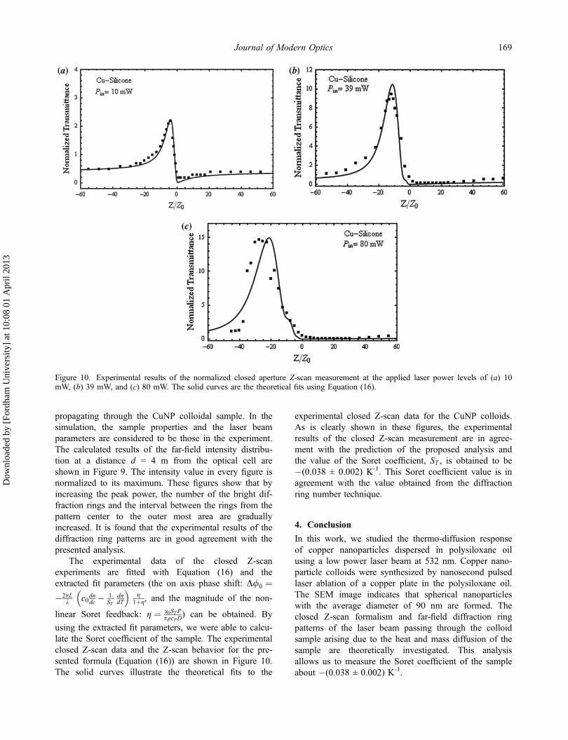
The experimental data of the closed Z-scanexperiments are fitted with Equation (16) and theextracted fit parameters (the on axis phase shift: D/0 ¼�2pL
k c0dndc � 1ST
dndT
� g
1þg, and the magnitude of the non-
linear Soret feedback: g ¼ a0STPpqcPD
) can be obtained. By
using the extracted fit parameters, we were able to calcu-late the Soret coefficient of the sample. The experimentalclosed Z-scan data and the Z-scan behavior for the pre-sented formula (Equation (16)) are shown in Figure 10.The solid curves illustrate the theoretical fits to the
experimental closed Z-scan data for the CuNP colloids.As is clearly shown in these figures, the experimentalresults of the closed Z-scan measurement are in agree-ment with the prediction of the proposed analysis andthe value of the Soret coefficient, ST , is obtained to be�(0.038 ± 0.002) K-1. This Soret coefficient value is inagreement with the value obtained from the diffractionring number technique.
4. Conclusion
In this work, we studied the thermo-diffusion responseof copper nanoparticles dispersed in polysiloxane oilusing a low power laser beam at 532 nm. Copper nano-particle colloids were synthesized by nanosecond pulsedlaser ablation of a copper plate in the polysiloxane oil.The SEM image indicates that spherical nanoparticleswith the average diameter of 90 nm are formed. Theclosed Z-scan formalism and far-field diffraction ringpatterns of the laser beam passing through the colloidsample arising due to the heat and mass diffusion of thesample are theoretically investigated. This analysisallows us to measure the Soret coefficient of the sampleabout �(0.038 ± 0.002) K-1.
Figure 10. Experimental results of the normalized closed aperture Z-scan measurement at the applied laser power levels of (a) 10mW, (b) 39 mW, and (c) 80 mW. The solid curves are the theoretical fits using Equation (16).
Journal of Modern Optics 169
Dow
nloa
ded
by [
Ford
ham
Uni
vers
ity]
at 1
0:08
01
Apr
il 20
13

References[1] Gordon, J.P.; Leite, R.C.C.; Moore, R.S.; Porto, S.P.S.;
Whinnery, J.R. J. Appl. Phys. 1965, 36, 3–8.[2] Giglio, M.; Vendramini, A. Phys. Rev. Lett. 1975, 34,
561–564.[3] Srinivasan, S.; Ziad Saghir, M. Int. J. Therm. Sci. 2011,
50, 1125–1137.[4] Giglio, M.; Vendramini, A. Appl. Phys. Lett. 1974, 25,
555–557.[5] Arnaud, N.; Georges, J. Spectrochim. Acta, Part A 2004,
60, 1817–1823.[6] Rusconi, R.; Isa, L.; Piazza, R.J. Opt. Soc. Am. B 2004,
21, 605–616.[7] Polyakov, P.; Wiegand, S. Phys. Chem. Chem. Phys.
2009, 11, 846–871.[8] Cabrera, H.; Sira, E.; Rahm, K.; Garcia-Sucre, M. Appl.
Phys. Lett. 2009, 94, 051103.[9] Alves, S.; Bourdon, A.; Figueiredo Neto, A.M. J. Opt.
Soc. Am. B 2003, 20, 713–718.[10] Soga, D.; Alves, S.; Campos, A.; Tourinho, F.A.;
Depeyrot, J.; Figueiredo Neto, A.M. J. Opt. Soc. Am. B2007, 24, 49–55.
[11] Tabiryan, N.V.; Lue, W. Phys. Rev. E: Stat. Phys., Plasmas,Fluids, Relat. Interdiscip. Top. 1998, 57, 4431–4440.
[12] Zamiranvari, J.; Karimzadeh, R.; Mansour, N. J. Opt.(Bristol, U.K.) 2010, 12, 035212.
[13] Akhmanov, S.A.; Krindach, D.P.; Migulin, A.V.; Sukhorukov,A.P.; Khokhlov, R.V. IEEE J. Quantum Electron. 1968, 4,568–575.
[14] Karimzadeh, R. J. Opt. (Bristol, U.K.) 2012, 14, 095701.[15] Sheik-Bahae, M.; Said, A.A.; Wei, T.H.; Hagan, D.J.; Van
Stryland, E.W. IEEE J. Quantum Electron. 1990, 26,760–769.
[16] Köhler, K.; Müler, M.J. Chem. Phys. 1995, 103,4367–4370.
[17] Garcia Ramirez, E.V.; Arroyo Carrasco, M.L.; MendezOtero, M.M.; Chavez Cerda, S.; Itorbe Castillo, M.D.Opt. Express 2010, 18, 22067–22079.
[18] Garcia Ramirez, E.V.; Arroyo Carrasco, M.L.; MendezOtero, M.M.; Reynoseo Lara, E.; Chavez Cerda, S.; ItorbeCastillo, M.D. J. Opt. (Bristol, U.K.) 2011, 13, 085203.
[19] Deng, L.; He, K.; Zhou, T.; Li, C. J. Opt. A: Pure Appl.Opt. 2005, 7, 409–415.
[20] Tavassoly, M.T.; Saber, A. Opt. Lett. 2010, 35,3679–3681.
170 R. Karimzadeh et al.
Dow
nloa
ded
by [
Ford
ham
Uni
vers
ity]
at 1
0:08
01
Apr
il 20
13