INVESTIGATION OF HELICOBACTER PYLORI INFECTION … · INVESTIGATION OF HELICOBACTER PYLORI...
6
INTRODUCTION Helicobacter pylori is fastidious, small, curved, microaerophilic and highly motile Gram negative bac- teria. It is about 2.5 - 5.0 mm long and 0.5 - 1.0 mm wide with 4-6 unipolar sheathed flagella [1]. H. py- lori is identified on the basis of colony morphology (translucent colonies varying in size from barely de- tectable with the naked eye to approximately 3 mm); H. pylori are Gram-negative, curved rods that are ure- ase, catalase and oxidase positive. The addition of tetrazolium salts aids in the identification of H. pylori colonies cultured on agar media [2]. H. pylori is the etiologic agent of a variety of gastrointestinal disor- ders including chronic active gastritis, peptic ulcer and gastric cancer [3]. It is clear that virtually all H. pylori infected subjects develop local and systemic immune response against this organism [4], as with most bacterial infections, H. pylori stimulates an im- mune response and circulating antibodies appear in the peripheral blood stream. This is the basis for se- rology tests to diagnose H. pylori [5]. It has been shown that the immune response to H. pylori at the mucosal level is predominantly of the IgA type, while the systemic immune antibody response essentially consists of the IgG class [6]. Immunoglobulins of the IgM class do not appear to differ between H. pylori positive and negative patients [7]. Performance varies significantly between diffe- rent commercial serologic kits, with the highest ex- ceeding 90% in sensitivity and specificity and the lo- 240 ABSTRACT Helicobacter pylori (H. pylori) is the etiologic agent of a variety of gastrointestinal disorders. The rationale of the current study is to evaluate six enzyme immunoassays for detection of anti-H. pylori immunoglobulin G (IgG) and IgA in Jordanian patients. Biopsy specimens and blood sam- ples were obtained from patients underwent the endoscopy unit at Al-Bashir hospital in Jordan. The serum samples were investigated for the presence of anti- H. pylori IgG and IgA antibodies in patients with positive H. pylori biopsy samples. The results showed that IgG utilizing kits are more sensitive than of IgA kits and the IgA kits are more specific than of that IgG kits. Moreover, the biop- sy is seemingly the gold standard for diagnosis of H. pylori is followed by H. pylori culture on bru- cella agar medium. An imperfect relation between the presence of H. pylori infection and the anti- body response was existed that could be explained either because of the unsatisfactory sensitivi- ties and specificities of the commercial kits used or because of weak immunological response in our patients to H. pylori antigens. Collectively, the H. pylori diagnosis that depends on the detec- tion of anti- H. pylori antibodies in the hospital setting and in the screening programs should con- sider another test for confirmation the initial diagnosis. INVESTIGATION OF HELICOBACTER PYLORI INFECTION IN JORDANIAN PATIENTS USING SIX ENZYME IMMUNOASSAYS FOR IMMUNOGLOBULIN G (IGG) AND IGA TESTING Hasan Abusini 1 , Khaled Abuelteen 2 , Ali Elkarmi 2 , Faris Q. Alenzi 3 , Mahmoud Lotfy 1,4 1) Department of Applied Medical Sciences, Jouf University, Qurayat, Saudi Arabia; 2) Department of Biology and Biotechnology, Faculty of Science, Hashemite University, Jordan; 3) Department of Clinical Laboratory Sciences, College of Applied Medical Sciences, King Saud University, Al-Kharaj, Saudi Arabia; 4) Molecular and Cellular Biology Department, Genetic Engineering and Biotechnology Research Institute, Minufiya University, Sadat City, Minufiya, Egypt Key words: Helicobacter pylori, IgA, IgG, Jordan Conflict of interest: Non Declared * Corresponding author: Dr. Mahmoud Lotfy - Current Address: Department of Applied Medical Sciences, Jouf University, Qurayat, P.O. 1300, Saudi Arabia. Permanent Address: Molecular and Cellular Biology Department, Genetic Engineering and Biotechnology Research Institute, Minufiya University, Sadat City, P.O 22857-79, Egypt. E-mail: [email protected] Roum Arch Microbiol Immunol. 2009; 68(4): 240-245
Transcript of INVESTIGATION OF HELICOBACTER PYLORI INFECTION … · INVESTIGATION OF HELICOBACTER PYLORI...

INTRODUCTION
Helicobacter pylori is fastidious, small, curved, microaerophilic and highly motile Gram negative bac-teria. It is about 2.5 - 5.0 µm long and 0.5 - 1.0 µm wide with 4-6 unipolar sheathed flagella [1]. H. py-lori is identified on the basis of colony morphology (translucent colonies varying in size from barely de-tectable with the naked eye to approximately 3 mm);H. pylori are Gram-negative, curved rods that are ure-ase, catalase and oxidase positive. The addition of tetrazolium salts aids in the identification of H. pylori colonies cultured on agar media [2]. H. pylori is the etiologic agent of a variety of gastrointestinal disor-ders including chronic active gastritis, peptic ulcer and gastric cancer [3]. It is clear that virtually all H.
pylori infected subjects develop local and systemic immune response against this organism [4], as with most bacterial infections, H. pylori stimulates an im-mune response and circulating antibodies appear in the peripheral blood stream. This is the basis for se-rology tests to diagnose H. pylori [5]. It has been shown that the immune response to H. pylori at the mucosal level is predominantly of the IgA type, while the systemic immune antibody response essentially consists of the IgG class [6]. Immunoglobulins of the IgM class do not appear to differ between H. pylori positive and negative patients [7].
Performance varies significantly between diffe-rent commercial serologic kits, with the highest ex-ceeding 90% in sensitivity and specificity and the lo-
240
ABSTRACTHelicobacter pylori (H. pylori) is the etiologic agent of a variety of gastrointestinal disorders. The rationale of the current study is to evaluate six enzyme immunoassays for detection of anti-H. pylori immunoglobulin G (IgG) and IgA in Jordanian patients. Biopsy specimens and blood sam-ples were obtained from patients underwent the endoscopy unit at Al-Bashir hospital in Jordan. The serum samples were investigated for the presence of anti- H. pylori IgG and IgA antibodies in patients with positive H. pylori biopsy samples. The results showed that IgG utilizing kits are more sensitive than of IgA kits and the IgA kits are more specific than of that IgG kits. Moreover, the biop-sy is seemingly the gold standard for diagnosis of H. pylori is followed by H. pylori culture on bru-cella agar medium. An imperfect relation between the presence of H. pylori infection and the anti-body response was existed that could be explained either because of the unsatisfactory sensitivi-ties and specificities of the commercial kits used or because of weak immunological response in our patients to H. pylori antigens. Collectively, the H. pylori diagnosis that depends on the detec-tion of anti- H.pylori antibodies in the hospital setting and in the screening programs should con-sider another test for confirmation the initial diagnosis.
INVESTIGATION OF HELICOBACTER PYLORI INFECTION IN JORDANIAN PATIENTS USING SIX ENZYME IMMUNOASSAYS
FOR IMMUNOGLOBULIN G (IGG) AND IGA TESTING
Hasan Abusini1, Khaled Abuelteen2, Ali Elkarmi2, Faris Q. Alenzi3, Mahmoud Lotfy1,4
1)Department of Applied Medical Sciences, Jouf University, Qurayat, Saudi Arabia; 2)Department of Biology and Biotechnology, Faculty of Science, Hashemite University, Jordan; 3)Department of Clinical Laboratory Sciences,
College of Applied Medical Sciences, King Saud University, Al-Kharaj, Saudi Arabia; 4)Molecular and Cellular Biology Department, Genetic Engineering and Biotechnology Research Institute, Minufiya University, Sadat City, Minufiya, Egypt
Key words: Helicobacter pylori, IgA, IgG, Jordan
Conflict of interest: Non Declared
* Corresponding author: Dr. Mahmoud Lotfy - Current Address: Department of Applied Medical Sciences, Jouf University, Qurayat, P.O. 1300, Saudi Arabia. Permanent Address: Molecular and Cellular Biology Department, Genetic Engineering and Biotechnology Research Institute, Minufiya University, Sadat City, P.O 22857-79, Egypt. E-mail: [email protected]
Roum Arch Microbiol Immunol. 2009; 68(4): 240-245

west having less than 70% in sensitivity and less than 50% in specificity [8]. The purpose of this study was to evaluate six commercially available ELISA kits for detection of anti-H. pylori IgG and IgA in patients attending the Gastroenterology unit at Al-Bashir hos-pital in Jordan. Moreover, to assess the culture on dif-ferent media for detecting H. pylori isolates.
MATERIALS AND METHODS
PatientsThe study group included 71 individuals aged
13 to 83 years (37.7±1.5 years) who underwent en-doscopy at the Gastroenterology unit. The study pro-tocol respected the most recent Declaration of Hel-sinki [9], and all of the patients gave consent to the use of their sera and clinical data for research purposes after being informed about the nature of the study.
MediaBrucella medium (HiMedia Laboratories, Mum-
bai, India), Columbia medium (Biomark, India) and Brain Heart Infusion (BHI) medium (Liofil, Italy) were prepared in accordance with the manufacturer’s in-structions. The antibiotic supplement in each media are identical; each medium contains 10 mg of van-comycin per liter, 2.5 IU of polymyxin B per liter, 5 mg of trimethoprim per liter, 2 mg of amphotericin B per liter and 5 mg of cefsulodin per liter. All media were further supplemented with whole blood (5%) and 40 mg of 2,3,5-triphenyltetrazolium chloride (Sigma, USA) per liter.
Biopsy SpecimensDuring upper endoscopy, gastric mucosal biop-
sy was taken for histopathology, formalin fixed, cut into five µm sections, stained with hematoxylin and eosin and viewed for the presence of H. pylori (Fig.1). A positive result was considered indicative of active H. pylori infection as reported by us earlier[10]. Additional two biopsies one from the antrum and the other from the corpus were taken for micro-biological studies as shown below.
The biopsy specimens were collected into tubes containing three ml of brucella broth supplemented with 0.5% (w/v) bovine serum albumin. The tubes were transported to the laboratory on an ice box in less than three hours.
Isolation and IdentificationThe biopsy specimens were finely minced with
a sterile scalpel blade in one ml of brucella broth in
Helicobacter pylori in Jordanian Patients
241
sterile conditions. One drop of the tissue homoge-nate was inoculated onto each of the three selective media plates. After incubation at 37°C under mi-croaerophilic conditions (10% CO2, 5% O2, and 98%humidity) for 10 days. Isolates were identified as H. pylori on the basis of colonial morphology, in addi-tion to the negativity for Gram staining, nitrate reduc-tion, glucose and sucrose fermentation and indole production, moreover, the positivity for catalase, oxi-dase, and urease tests.
Enzyme Immunoassays:Venous blood samples (5ml) were obtained from
each patient at the time of endoscopy. The serum was separated, divided into aliquots, and stored at -20°C. All sera were tested with six ELISA kits: Anti- Heli-cobacter pylori (Anti-Hp) IgG ELISA (BioCheck Inc. USA), Anti-Hp IgA ELISA (BioCheck Inc. USA), Anti-Hp IgG ELISA (Biotest, Germany), Anti-Hp IgA ELISA (Biotest, Germany), Anti-Hp ELISA IgG (Euroimmun, Germany), and Anti-Hp ELISA IgA (Euroimmun, Ger-many). The assays were performed in accordance with the manufacturers’ instructions.
Statistical AnalysisNominal data were analyzed using chi-square
test and P values < 0.05 were considered statistically significant. Data were tabulated and analyzed using the SPSS 17 statistical package (SPSS Inc. Chicago,IL). The sensitivity, specificity and accuracy were cal-culated as described by us earlier [11].
Figure 1: Shows a section from the corpus of an infected patient. M: Mucus secreting cells. HP: H. pylori (X 1000).
Roum Arch Microbiol Immunol. 2009; 68(4): 240-245

RESULTS
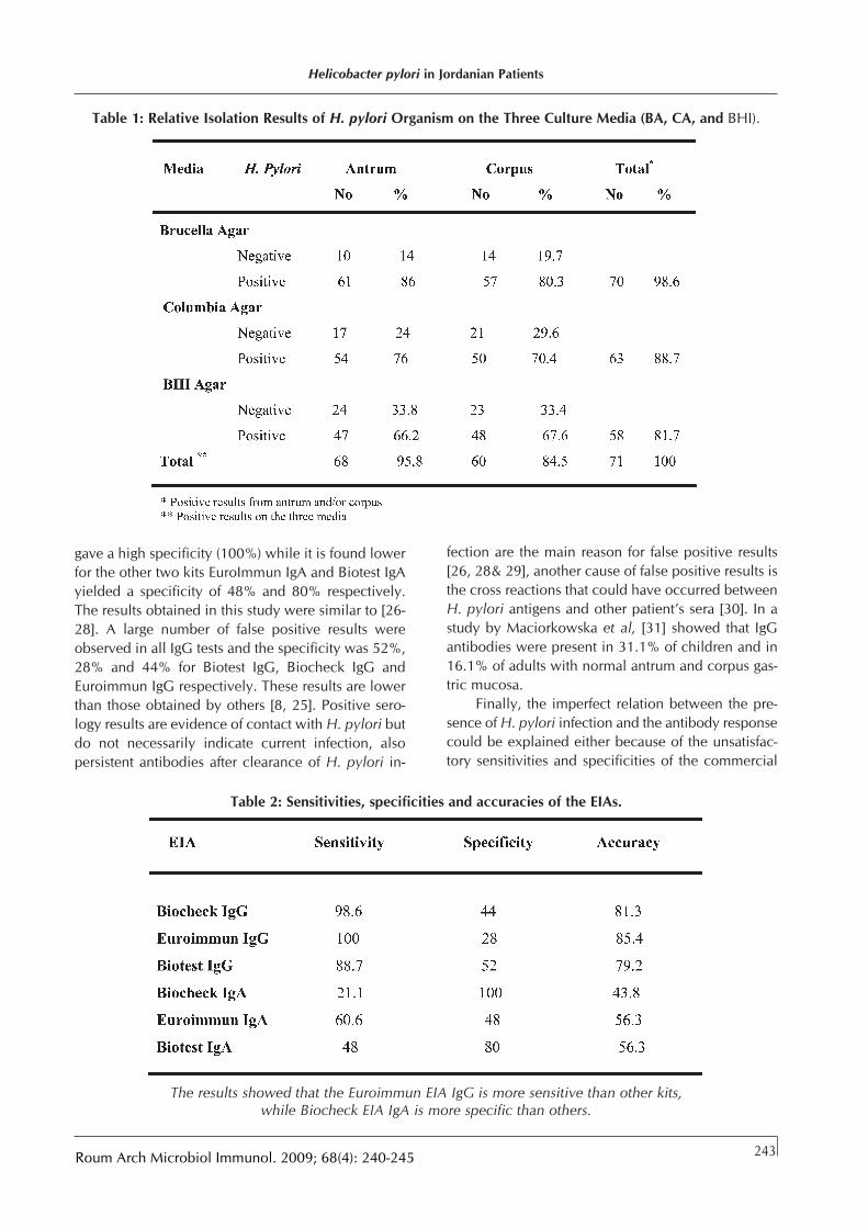
CultureBrucella agar medium gave the highest isolation
rate, Out of 71 positive biopsy specimens; isolation of H. pylori was achieved in 61 antrum specimens (86%). On the other hand, 57 out of 71 corpus biop-sy specimens (80.3%) were culture positive. The num-ber of patients from whom H. pylori was isolated from either the corpus or antrum specimens was 70 out of 71 infected, thus H. pylori was recovered in98.6% of the patients infected. Using Columbia agar the organism was recovered from 54 antral speci-mens (76%). On the other hand when corpus speci-mens were cultured the recovery was achieved from 50 specimens (70.4%). The total number of patients from whom H. pylori was isolated either from corpus or antrum specimens was 63, thus H. pylori was recovered in 88.7% of the patients infected. BHI gave the lowest isolation rate for H. pylori. Out of 71 pos-itive antral biopsy specimens, the organism was iso-lated only from 47 specimens (66.2%). In the oppo-site direction when corpus specimens were cultured, the number of specimens retreat the organism was 48 specimens (67.6%) (table 1).
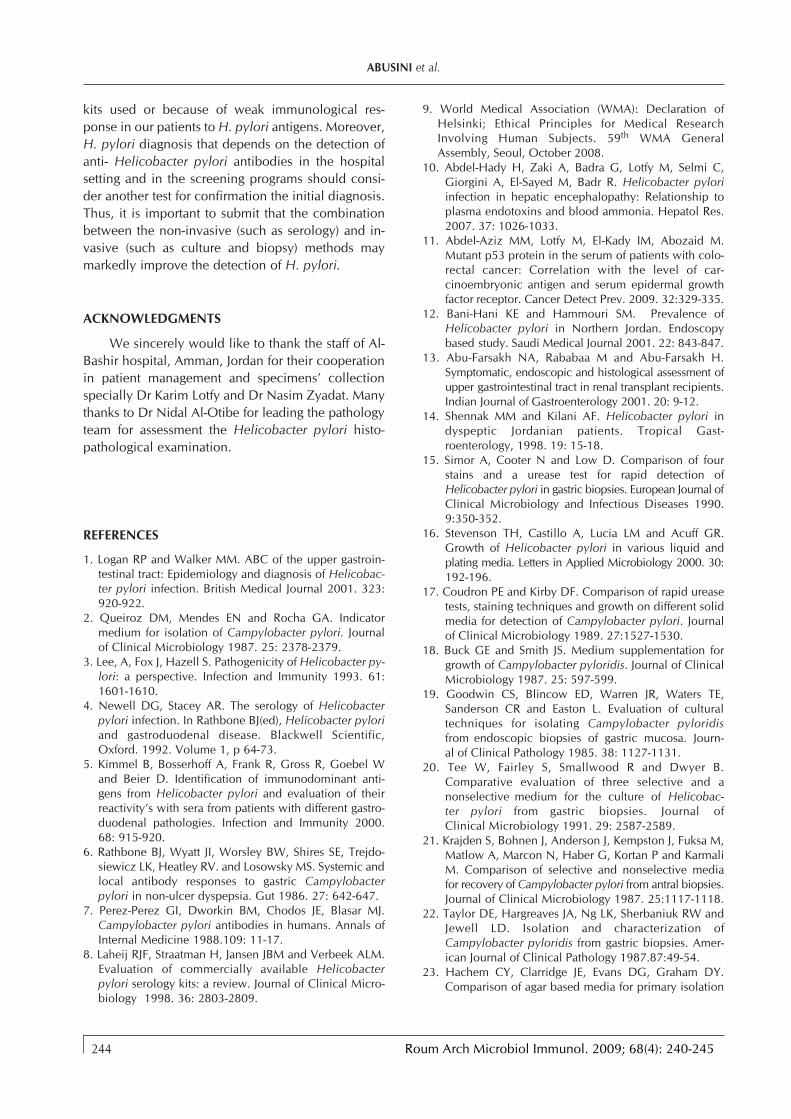
Enzyme Linked Immunosorbent Assay (ELISA)The serum samples were investigated for the
presence of anti- H. pylori IgG and IgA antibodies in patients with positive H. pylori biopsy samples. The results showed that IgG utilizing kits are more sensi-tive than of IgA kits and the IgA kits are more specif-ic than of that IgG kits. Euroimmun IgG kit is the most sensitive one, on the hand, BioCheck IgA is the most specific kit. The sensitivities, specificities and accura-cies of the six kits are illustrated in table (2).
DISCUSSION
H. pylori infection is prevalent in Jordan as evi-denced by several investigators [12-14]. In the current study, we evaluated seventy one positive H. pylori infected Jordanian patients with three different cul-ture media and six EIA assays for anti-H. pylori anti-bodies (IgG and IgA). It is well known that gastric mucosal biopsy for H. pylori is the standard gold for diagnosing that infection. Several histological stains, one of them hematoxylin and eosin, was compared to culture and the results revealed that all stains showed high specificity ranging from 97 to 100% [15].
A wide variety of solid media have been used by many investigators for the isolation of H. pylori [16-19]. In this study, different results were obtained by
using three selective media for isolation of H. pylori. Brucella agar was considered the best medium and showed the highest H. pylori recovery rate (98.6%), while Columbia and BHI agar produce a lower recov-ery rate, 88.7% and 81.7% respectively. The use of selective media has been recommended in order to improve the culture isolation of H. pylori from gastric biopsy specimens and to prevent bacterial over-growth by contaminants [20, 21].
These media (Brucella, Columbia and BHI Agar) were used successfully by many investigators [18,22]. In a study by Hachem et al., [23] proved that BHI agar was the best one among the media tested and gave 96% recovery rate. While the recovery rate was 78% with trypticase soy agar, 64% with egg yolk agar and 32% with Columbia blood agar-cyclodextrin. In this study, Brucella agar was the most accurate of the media evaluated for culture isolation of H. pylori from gastric biopsy specimens. The inclusion of 2,3,5-triphenyltetrazolium chloride gives H. pylori colonies a specific golden yellow pigment which helps in their identification, therefore reducing the false identification of H. pylori. It is worth to mention that all plates were incubated in an atmosphere of 10% CO2 and 98% relative humidity for up to 10 days to provide excellent growth conditions. Tee et al., [20] stated that selective medium provides heav-ier growth than non selective one. Their results shows that Skirrow’s medium gave the highest isola-tion rate while growth on Dent’s and modified Glupezyniski media were equal and less better than the first one. Chocolate agar yielded a 76% positivity rate. The addition of blood not only enhances the growth of H. pylori, but also facilitated to distinguishH. pylori colonies from those of other bacteria; colonies of H. pylori appear distinctly clear [17].
In the present study, 100% of H. pylori infected patients had a positive test result with Euroimmun IgG. On the other hand the other two kits, Biocheck IgG and Biotest IgG identified 98.6% and 88.7% respectively. On the other hand 60.6% of H. pylori infected patients had a positive test result with Euroimmun IgA. Only 21.1% and 48% of the infect-ed patients were identified by Biocheck IgA and Biotest IgA kits respectively. The sensitivities of the three IgA based tests in comparison to IgG antibodies confirm the results of previous studies. Best et al, [24] report a sensitivity of only 50% for IgA, while a sen-sitivity of 100% was found for IgG in the same cohort in the same study. Also Kindermann et al, [25] sup-port these results and found that the sensitivities were <50% for the IgA kits and varied markedly between the IgG kits between 70.7-92.7%. Biocheck IgA kit
242
ABUSINI et al.
Roum Arch Microbiol Immunol. 2009; 68(4): 240-245
gave a high specificity (100%) while it is found lower for the other two kits EuroImmun IgA and Biotest IgA yielded a specificity of 48% and 80% respectively. The results obtained in this study were similar to [26-28]. A large number of false positive results were observed in all IgG tests and the specificity was 52%, 28% and 44% for Biotest IgG, Biocheck IgG and Euroimmun IgG respectively. These results are lower than those obtained by others [8, 25]. Positive sero-logy results are evidence of contact with H. pylori but do not necessarily indicate current infection, also persistent antibodies after clearance of H. pylori in-
fection are the main reason for false positive results [26, 28& 29], another cause of false positive results is the cross reactions that could have occurred betweenH. pylori antigens and other patient’s sera [30]. In a study by Maciorkowska et al, [31] showed that IgG antibodies were present in 31.1% of children and in16.1% of adults with normal antrum and corpus gas-tric mucosa.
Finally, the imperfect relation between the pre-sence of H. pylori infection and the antibody response could be explained either because of the unsatisfac-tory sensitivities and specificities of the commercial
Helicobacter pylori in Jordanian Patients
243
Table 1: Relative Isolation Results of H. pylori Organism on the Three Culture Media (BA, CA, and BHI).
Table 2: Sensitivities, specificities and accuracies of the EIAs.
The results showed that the Euroimmun EIA IgG is more sensitive than other kits, while Biocheck EIA IgA is more specific than others.
Roum Arch Microbiol Immunol. 2009; 68(4): 240-245
kits used or because of weak immunological res-ponse in our patients to H. pylori antigens. Moreover,H. pylori diagnosis that depends on the detection of anti- Helicobacter pylori antibodies in the hospital setting and in the screening programs should consi-der another test for confirmation the initial diagnosis. Thus, it is important to submit that the combination between the non-invasive (such as serology) and in-vasive (such as culture and biopsy) methods may markedly improve the detection of H. pylori.
ACKNOWLEDGMENTS
We sincerely would like to thank the staff of Al-Bashir hospital, Amman, Jordan for their cooperation in patient management and specimens’ collection specially Dr Karim Lotfy and Dr Nasim Zyadat. Many thanks to Dr Nidal Al-Otibe for leading the pathology team for assessment the Helicobacter pylori histo-pathological examination.
REFERENCES
1. Logan RP and Walker MM. ABC of the upper gastroin-testinal tract: Epidemiology and diagnosis of Helicobac-ter pylori infection. British Medical Journal 2001. 323: 920-922.
2. Queiroz DM, Mendes EN and Rocha GA. Indicator medium for isolation of Campylobacter pylori. Journal of Clinical Microbiology 1987. 25: 2378-2379.
3. Lee, A, Fox J, Hazell S. Pathogenicity of Helicobacter py-lori: a perspective. Infection and Immunity 1993. 61: 1601-1610.
4. Newell DG, Stacey AR. The serology of Helicobacter pylori infection. In Rathbone BJ(ed), Helicobacter pylori and gastroduodenal disease. Blackwell Scientific, Oxford. 1992. Volume 1, p 64-73.
5. Kimmel B, Bosserhoff A, Frank R, Gross R, Goebel W and Beier D. Identification of immunodominant anti-gens from Helicobacter pylori and evaluation of their reactivity’s with sera from patients with different gastro-duodenal pathologies. Infection and Immunity 2000. 68: 915-920.
6. Rathbone BJ, Wyatt JI, Worsley BW, Shires SE, Trejdo-siewicz LK, Heatley RV. and Losowsky MS. Systemic and local antibody responses to gastric Campylobacter pylori in non-ulcer dyspepsia. Gut 1986. 27: 642-647.
7. Perez-Perez GI, Dworkin BM, Chodos JE, Blasar MJ. Campylobacter pylori antibodies in humans. Annals of Internal Medicine 1988.109: 11-17.
8. Laheij RJF, Straatman H, Jansen JBM and Verbeek ALM. Evaluation of commercially available Helicobacter pylori serology kits: a review. Journal of Clinical Micro-biology 1998. 36: 2803-2809.
9. World Medical Association (WMA): Declaration of Helsinki; Ethical Principles for Medical Research Involving Human Subjects. 59th WMA General Assembly, Seoul, October 2008.
10. Abdel-Hady H, Zaki A, Badra G, Lotfy M, Selmi C, Giorgini A, El-Sayed M, Badr R. Helicobacter pylori infection in hepatic encephalopathy: Relationship to plasma endotoxins and blood ammonia. Hepatol Res. 2007. 37: 1026-1033.
11. Abdel-Aziz MM, Lotfy M, El-Kady IM, Abozaid M. Mutant p53 protein in the serum of patients with colo-rectal cancer: Correlation with the level of car-cinoembryonic antigen and serum epidermal growth factor receptor. Cancer Detect Prev. 2009. 32:329-335.
12. Bani-Hani KE and Hammouri SM. Prevalence of Helicobacter pylori in Northern Jordan. Endoscopy based study. Saudi Medical Journal 2001. 22: 843-847.
13. Abu-Farsakh NA, Rababaa M and Abu-Farsakh H. Symptomatic, endoscopic and histological assessment of upper gastrointestinal tract in renal transplant recipients. Indian Journal of Gastroenterology 2001. 20: 9-12.
14. Shennak MM and Kilani AF. Helicobacter pylori in dyspeptic Jordanian patients. Tropical Gast-roenterology, 1998. 19: 15-18.
15. Simor A, Cooter N and Low D. Comparison of four stains and a urease test for rapid detection of Helicobacter pylori in gastric biopsies. European Journal of Clinical Microbiology and Infectious Diseases 1990. 9:350-352.
16. Stevenson TH, Castillo A, Lucia LM and Acuff GR. Growth of Helicobacter pylori in various liquid and plating media. Letters in Applied Microbiology 2000. 30: 192-196.
17. Coudron PE and Kirby DF. Comparison of rapid urease tests, staining techniques and growth on different solid media for detection of Campylobacter pylori. Journal of Clinical Microbiology 1989. 27:1527-1530.
18. Buck GE and Smith JS. Medium supplementation for growth of Campylobacter pyloridis. Journal of Clinical Microbiology 1987. 25: 597-599.
19. Goodwin CS, Blincow ED, Warren JR, Waters TE, Sanderson CR and Easton L. Evaluation of cultural techniques for isolating Campylobacter pyloridis from endoscopic biopsies of gastric mucosa. Journ-al of Clinical Pathology 1985. 38: 1127-1131.
20. Tee W, Fairley S, Smallwood R and Dwyer B. Comparative evaluation of three selective and a nonselective medium for the culture of Helicobac-ter pylori from gastric biopsies. Journal of Clinical Microbiology 1991. 29: 2587-2589.
21. Krajden S, Bohnen J, Anderson J, Kempston J, Fuksa M, Matlow A, Marcon N, Haber G, Kortan P and KarmaliM. Comparison of selective and nonselective media for recovery of Campylobacter pylori from antral biopsies. Journal of Clinical Microbiology 1987. 25:1117-1118.
22. Taylor DE, Hargreaves JA, Ng LK, Sherbaniuk RW and Jewell LD. Isolation and characterization of Campylobacter pyloridis from gastric biopsies. Amer-ican Journal of Clinical Pathology 1987.87:49-54.
23. Hachem CY, Clarridge JE, Evans DG, Graham DY. Comparison of agar based media for primary isolation
244
ABUSINI et al.
Roum Arch Microbiol Immunol. 2009; 68(4): 240-245
of Helicobacter pylori. Journal of Clinical Pathology 1995. 48: 714-716.
24. Best LM, Veldhuyzen van Zanten SJ, Sherman PM, Bezanson GS. Serological detection of Helicobacter pylori antibodies in children and their parents. Journal of Clinical Microbiology 1994. 32: 1193-1196.
25. Kindermann A, Konstantopoulos N, Lehn N, Demmelmair H, Koletzko S. Evaluation of two commercial enzyme immunoassays, testing immunoglobulin G (IgG) and IgA responses, for diagnosis of Helicobacter pylori infection in children. Journal of Clinical Microbiology 2001. 39: 3591-3596.
26. Glupezynski Y. Microbiological and serological diagnostic tests for Helicobacter pylori: an overview. Acta Gastroenterology Belgica 1998. 61: 321-326.
27. Meijer BC, Thijs JC, Kleibeuker JH, Van Zwet A, Berrelkamp R. Evaluation of eight enzyme immunoassays for detection of immunoglobulin G against Helicobacter pylori. Journal of Clinical Microbiology 1997. 35: 292-294.
28. Thijs JC, van Zwet AA, Thijs WJ, Oey HB, Karrenbeld A, Stellaard F, Luijt DS, Meyer BC and Kleibeuker JH. Diagnostic tests for Helicobacter pylori: A prospective evaluation of their accuracy, without selecting a single test as the gold standard. American Journal of Gas-troenterology 1996. 91: 2125-2129.
29. Cutler AF and Prasad VM. Long-term follow-up of Heli-cobacter pylori serology after successful eradication. American Journal of Gastroenterology 1996. 91: 85-88.
30. Goossens H, Glupczynski Y, Burette A, Borre C, ButzlerJ. Evaluation of a commercially available second-gen-eration immunoglobulin G immunoassay for detection of Helicobacter pylori infection. Journal of Clinical Microbiology 1992.30: 176-180.
31. Maciorkowska E, kaczmarski M, Kemona A, Kondej-Muszynska K and Gocal M. Comparative evaluation of gastric mucosa morphological changes in children and adults with positive IgG antibodies to Helicobacter pylori. Rocz Akad Med Bialymst 2003. 48: 100-104.
Helicobacter pylori in Jordanian Patients
245Roum Arch Microbiol Immunol. 2009; 68(4): 240-245