INVESTIGATION OF 1450 NM INFRARED LIGHT FOR...
44
INVESTIGATION OF 1450 NM INFRARED LIGHT FOR CLINICAL NERVE STIMULATION By Zane Christopher Ricks Thesis Submitted to the Faculty of the Graduate School of Vanderbilt University in partial fulfillment of the requirements for the degree of MASTER OF SCIENCE in Biomedical Engineering December, 2014 Nashville, Tennessee Approved: Professor Anita Mahadevan-Jansen Professor E. Duco Jansen
Transcript of INVESTIGATION OF 1450 NM INFRARED LIGHT FOR...

INVESTIGATION OF 1450 NM INFRARED LIGHT FOR CLINICAL NERVE
STIMULATION
By
Zane Christopher Ricks
Thesis
Submitted to the Faculty of the
Graduate School of Vanderbilt University
in partial fulfillment of the requirements
for the degree of
MASTER OF SCIENCE
in
Biomedical Engineering
December, 2014
Nashville, Tennessee
Approved:
Professor Anita Mahadevan-Jansen
Professor E. Duco Jansen
ii
ACKNOWLEDGEMENTS
I would like to acknowledge the support of the W. M. Keck Foundation Free Electron
Laser Center as well as Lockheed-Martin (Funding info.), in addition to the Translational
Pathology Shared Research Core at Vanderbilt University. I would also like to extend my thanks
to Jonathan Cayce, a tremendous source of support in the completion of this work.

iii
TABLE OF CONTENTS
Page
ACKNOWLEDGEMENTS ............................................................................................................ ii
LIST OF FIGURES AND TABLES.............................................................................................. iv
Chapter
I. INTRODUCTION ...............................................................................................................1
Overview of the Peripheral Nervous System ...........................................................1
Electrical Neural Stimulation and Recording ..........................................................3
Infrared Neural Stimulation and its Current Applications .......................................5
Motivation ................................................................................................................7
Hypothesis and Objectives .......................................................................................8
Works Cited ...........................................................................................................10
II. INVESTIGATION OF 1450 NM INFRARED LIGHT FOR CLINICAL NERVE
STIMULATION ................................................................................................................12
Introduction ............................................................................................................12
Methods..................................................................................................................16
Results ....................................................................................................................20
Discussion ..............................................................................................................28
Works Cited ...........................................................................................................33
III. FUTURE DIRECTIONS ...................................................................................................36

iv
LIST OF FIGURES
I.1 Illustration of the peripheral nervous system .............................................................1
I.2 Anatomy of a neural cell body ...................................................................................2
I.3 Electrical recording of single electrical stimulation ...................................................4
I.4 Light absorption properties of water ..........................................................................8
II.1 Schematic of surgical preparation ............................................................................17
II.2 Stimulation thresholds for 1450 nm and 1875 nm light ...........................................21
II.3 Stimulation thresholds between different spot sizes and wavelengths ....................23
II.4 Toluidine-blue stained irradiated nerve slices ..........................................................25
II.5 Nerve responses before and after damage to electrical and optical stimuli .............27
LIST OF TABLES
II.1 Threshold radiant exposures per wavelength and spot size ......................................22
1
CHAPTER 1
INTRODUCTION
Overview of the Peripheral Nervous System
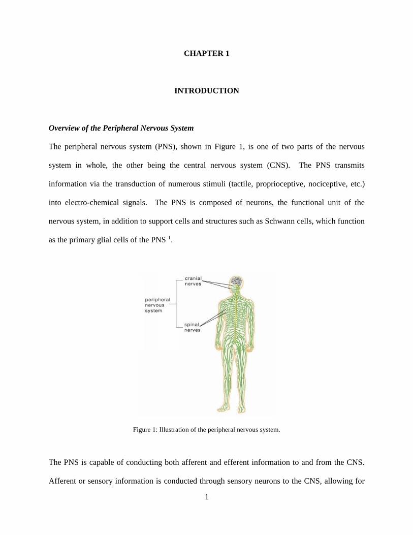
The peripheral nervous system (PNS), shown in Figure 1, is one of two parts of the nervous
system in whole, the other being the central nervous system (CNS). The PNS transmits
information via the transduction of numerous stimuli (tactile, proprioceptive, nociceptive, etc.)
into electro-chemical signals. The PNS is composed of neurons, the functional unit of the
nervous system, in addition to support cells and structures such as Schwann cells, which function
as the primary glial cells of the PNS 1.
Figure 1: Illustration of the peripheral nervous system.
The PNS is capable of conducting both afferent and efferent information to and from the CNS.
Afferent or sensory information is conducted through sensory neurons to the CNS, allowing for

2
the perception of inputs from outside stimuli. Efferent or motor information is conducted from
the CNS through motor neurons, allowing for innervation and subsequent locomotion of smooth
and skeletal muscle. These two types of neurons, motor and sensory, are coupled together to
form nerves which originate directly from the spinal cord and brain. The autonomic nervous
system (ANS) is a specialized part of the PNS which is responsible for reflex actions, motor
movements triggered in response to stimuli irrespective of CNS input 1.
The neuron, the functional unit of the nervous system, contains two processes, a dendrite and an
axon, which are connected to a cell body, as shown in Figure 2.
Figure 2: Anatomy of a neural cell body.
Axons terminate in a synapse, which is utilized in signal communication via the release of a
neurotransmitter. This neurotransmitter binds to a receptor protein on the postsynaptic
membrane of the receiving neuron, causing the opening of an ion channel. Negatively or
positively charged ions flood into the cell via this open ion channel, causing respectively either
depolarization or hyperpolarization of the membrane. Depolarization at a synapse evokes a

3
transient electro-chemical signal which travels down the dendrite and cell body by opening
voltage-gated ion channels through depolarization of subsequent sections of the membrane2.
Should this signal reach the axon hillock, identified in Figure 3, without a loss of significant
amount of energy, an action potential will result, propagating towards the next synapse.
The conduction velocity of such a signal is, in general, directly related to myelination, axon
diameter, and the distance between the nodes of Ranvier of the myelin sheath. A-type fibers,
which are myelinated and larger (2-20 μm) have conduction velocities varying between 12-120
m/s. B-type fibers, myelinated fibers with smaller diameters (<3 µm) than those of A-type fibers
have conduction velocities ranging from 3-15 m/s. C-type fibers, which are even smaller (0.3-
1.3 µs) and unmyelinated, conduct only between 0.5-2.3 m/s 3. In myelinated fibers, nodes of
Ranvier increase conduction velocities by allowing saltatory conduction, a sort of leap of
depolarization waves between nodes.
Electrical Neural Stimulation and Recording
The gold standard for neurophysiological study has traditionally been centered around electrical
methods. An electrode is placed on or adjacent to a neuronal target and a small current is applied,
depolarizing the membrane of the neuron and evoking an action potential. This signal then
propagates down the axon towards additional synapses or a target tissue, causing a measurable
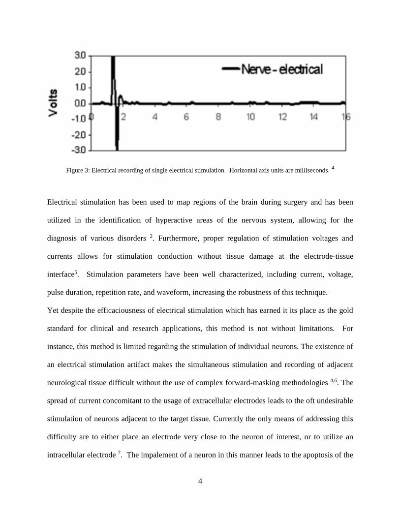
response such as a motor action. An example of an electrical stimulation and response is shown
in Figure 3. Electrical stimulation has been utilized in the stimulation of multiple tissue types,
including skeletal muscle, cardiac muscle in pacemaking applications, and deep brain regions for
the treatment of neuropathies such as Parkinson’s disease and Tourette’s syndrome.
4
Figure 3: Electrical recording of single electrical stimulation. Horizontal axis units are milliseconds. 4
Electrical stimulation has been used to map regions of the brain during surgery and has been
utilized in the identification of hyperactive areas of the nervous system, allowing for the
diagnosis of various disorders 2. Furthermore, proper regulation of stimulation voltages and
currents allows for stimulation conduction without tissue damage at the electrode-tissue
interface5. Stimulation parameters have been well characterized, including current, voltage,
pulse duration, repetition rate, and waveform, increasing the robustness of this technique.
Yet despite the efficaciousness of electrical stimulation which has earned it its place as the gold
standard for clinical and research applications, this method is not without limitations. For
instance, this method is limited regarding the stimulation of individual neurons. The existence of
an electrical stimulation artifact makes the simultaneous stimulation and recording of adjacent
neurological tissue difficult without the use of complex forward-masking methodologies 4,6. The
spread of current concomitant to the usage of extracellular electrodes leads to the oft undesirable
stimulation of neurons adjacent to the target tissue. Currently the only means of addressing this
difficulty are to either place an electrode very close to the neuron of interest, or to utilize an
intracellular electrode 7. The impalement of a neuron in this manner leads to the apoptosis of the
5
cell upon electrode removal8. Due to these limitations in traditional electrophysiological
techniques, which lead to a lack of spatial precision, a number of modalities have been
developed to address them. Infrared neural stimulation methods are among them.
Infrared Neural Stimulation and its Current Applications
Infrared neural stimulation (INS), the use of pulsed infrared light to stimulate an action potential,
has seen a growth in interest in recent years in the realm of neurophysiological study in both the
central and peripheral nervous systems 4,9–11. This increase in interest stems from a number of
advantages inherent to INS. INS has a high degree of spatial acuity, allowing for the selective
stimulation of a small number of neurological targets. Furthermore, depending on application, it
is also a potential contact-free method of stimulation, requiring no physical contact between
target tissue and the stimulation source. This overcomes one of the limitations present in other
methods of stimulation, such as the necessity of physical contact and often intrusion of an
electrode in order to selectively stimulate an individual neuron or axon, which oft introduces
trauma to the tissue being studied. Another beneficial aspect of INS is the absence of a
stimulation artifact. In many applications wherein electrophysiological recordings are performed
for the observation of a response to a stimulus, there is a stimulation artifact, a presence of the
electrical stimulus in the recording, that is unavoidable due to the fact that the stimulation
modality occurs in the same domain as the recording technique. When recordings are made
adjacent to a site of stimulation, the neurophysiological response is contaminated by the
electrical field used for stimulation. This has prompted the development of numerous techniques
for the reduction of this artifact. INS is not affected by these restrictions. This is because INS, as
it is currently understood, functions through the introduction of a thermal gradient, rather than
6
through an electrical current 12–15. As INS avoids these limitations, it may serve as a viable
adjunct to electrical methods of neurophysiological study.
As an optical modality of neural stimulation, parallels may be drawn between INS and the field
of optogenetics. Optogenetics is a neuromodulation technique that takes advantage of the
introduction of exogenous light-sensitive proteins into target tissue, allowing for selective
stimulation of only those tissues expressing these proteins when exposed to the proper
wavelength of light16. Although optogenetics has seen tremendous success and utility in the field
of neurophysiological study, its applications are limited by the need for exogenous factors, which
confounds introduction into a clinical setting. INS does not require the use of exogenous factors
or other modification of target tissues, thereby avoiding this limitation.
INS has seen use in a number of applications including, though not limited to, cochlear
stimulation 17–19, somatosensory cortex stimulation 20, visual cortex stimulation in non-human
primates 21, cardiomyocyte pacemaking 22, and stimulation of the peripheral nervous system 23.
There has also been ongoing study into its viability in the identification of damaged nerves
during dorsal root rhizotomy procedures. However, questions remain extant about the viability of
INS as a tool for neurophysiological assessment.
Motivation
INS has a proven robustness in neurophysiological applications, but there have been roadblocks
impeding its introduction into the clinic. One challenge facing this clinical push is maintaining
repeatability and constancy of stimulation within a safe window of stimulation. Variability has
been observed in the threshold of radiant exposure needed to stimulate neural targets, which calls
to question the issue of repeatability, with reproducibility of stimulation observed to be as low as

7
23% 24. There are methods to address this, such as continuous monitoring of neural responses
and adjustment of stimulation parameters as necessary, but these require a level of attention that
is not feasible in a prolonged study and would not be acceptable in a clinical application.
Furthermore, INS does an excellent job of stimulating small regions of interest, but clinical
applications such as nerve monitoring require a more ubiquitous stimulation of tissue. Increasing
laser power and/or spot size is a potential solution to both of these questions; if it can be done
safely.
Increasing radiant exposures to levels above stimulation threshold includes a concomitant
concern for thermal damage with higher energy pulses. There is threshold of safe radiant
exposures beyond which damage is much more likely to occur. Comparing stimulation
thresholds and safety thresholds yields a safety ratio, a metric of how much additional energy can
be used without risk of damage. Wavelengths used in earlier studies had safety ratios on the
order of 2:1 25, which is insufficient for a clinical application. Improving this safety ratio is the
primary means of facilitating the translation of INS into the clinical setting.

8
Hypothesis and Objectives
A majority of previous INS work has utilized the 2120 and 1875 nm wavelengths. While these
studies have demonstrated that INS is capable of evoking neural responses, there has been an
omnipresent question of the repeatability and safety of INS in generating these responses.
Previous safety ratios between damage and stimulation have been on the order of 2:1, which is
insufficient for a clinical setting. Repeatability has also been an issue, with stimulation
thresholds varying within an animal on a temporal basis, leading to reproducibility below 25%.
This leads to far too much ambiguity: the functionality of a nerve cannot be accurately assessed
if there is uncertainty as to whether or not a proper stimulus has been applied to it. It must be
noted, however, that in actual application all lasers produce a wavelength range, rather than a
single individual wavelength. The previously utilized 1875 nm light is located on a slope for
water absorption (Figure 4), the primary chromophore for INS; it is possible that variance in the
actual wavelength generated by the laser leads to an increase or decrease in heat generation.
Figure 4: Light absorption properties of water.

9
By this assumption it may mean that a wavelength centered on a relative peak, wherein slight
variance in wavelength does not lead to a large variance in absorption, will produce a more
constant and reproducible stimulation. Light sources in the 1.4-1.5 µm range are also more
readily available than 1.8-1.9 µm sources due to the telecom industry, meaning a more
ubiquitous availability of sources of optical stimulus.
There are three main objectives to this work aimed at improving the performance of INS and
demonstrating its clinical relevancy. The first objective is to determine if differing wavelengths
of similar absorptive characteristics exhibit differences in stimulation and damage thresholds and
resulting safety thresholds. If a difference is found, this would indicate a potential avenue of
exploration for the improvement of INS safety and efficacy. The second objective is to
determine if laser spot size and spatial beam profile has an effect on stimulation thresholds. This
objective will elucidate whether or not larger spot sizes may be used for more ubiquitous
stimulation, and if the energy may be more evenly distributed for a mitigation of damage risks.
The final objective is to assess whether or not INS may serve as an adjunct to electrical methods
in the clinic in the assessment of nerve damage. It is believed that electrical signal may be
conducted through, or more accurately, around damaged nerves, while INS-evoked signals will
not.
This thesis focuses on the improvement of INS safety ratios, and on demonstrating its clinical
application and relevancy. The results from this thesis will guide future studies and ease the
transition of INS towards its use as a diagnostic tool in the clinical setting.
10
Works Cited
1. Hall, J. E. Guyton and Hall Textbook of Medical Physiology. Physiology 1091 (2010).
2. Plonsey, R. & Barr, R. C. Bioelectricity: A quantitative approach. Bioelectr. A Quant.
Approach 1–528 (2007). doi:10.1007/978-0-387-48865-3
3. Barrett, K. E. & Ganong, W. F. Ganong’s review of medical physiology. Ophthalmic Surg.
17, 499–501 (2010).
4. Wells, J., Kao, C., Jansen, E. D., Konrad, P. & Mahadevan-Jansen, A. Application of
infrared light for in vivo neural stimulation. J. Biomed. Opt. 10, 64003–64012 (2005).
5. Schuettler, M., Franke, M., Krueger, T. B. & Stieglitz, T. A voltage-controlled current
source with regulated electrode bias-voltage for safe neural stimulation. J. Neurosci.
Methods 171, 248–252 (2008).
6. Wells, J., Konrad, P., Kao, C., Jansen, E. D. & Mahadevan-Jansen, A. Pulsed laser versus
electrical energy for peripheral nerve stimulation. J. Neurosci. Methods 163, 326–337
(2007).
7. Starr, P. A. et al. Implantation of deep brain stimulators into subthalmic nucleus: technical
approach and magnetic imaging-verified electrode locations. J. Neurosurg. 97, 370–387
(2002).
8. Civillico, E. F. & Contreras, D. Comparison of responses to electrical stimulation and
whisker deflection using two different voltage-sensitive dyes in mouse barrel cortex in
vivo. J. Membr. Biol. 208, 171–182 (2005).
9. Chernov, M. M. et al. Material considerations for optical interfacing to the nervous system.
MRS Bull. 37, 599–605 (2012).
10. Tozburun, S., Lagoda, G. A., Burnett, A. L. & Fried, N. M. Gaussian versus flat-top
spatial beam profiles for optical stimulation of the prostate nerves. in Bios (Kollias, N. et
al.) 75484W–75484W–6 (International Society for Optics and Photonics, 2010).
doi:10.1117/12.852994
11. Wells, J. et al. Optical stimulation of neural tissue in vivo. Opt. Lett. 30, 504–506 (2005).
12. Thompson, A. C., Wade, S. a, Cadusch, P. J., Brown, W. G. a & Stoddart, P. R. Modeling
of the temporal effects of heating during infrared neural stimulation. J. Biomed. Opt. 18,
035004 (2013).
11
13. Liljemalm, R., Nyberg, T. & Von Holst, H. Heating during infrared neural stimulation.
Lasers Surg. Med. 45, 469–481 (2013).
14. Thompson, A. C., Wade, S. A., Brown, W. G. A. & Stoddart, P. R. Modeling of light
absorption in tissue during infrared neural stimulation. J. Biomed. Opt. 17, 075002 (2012).
15. Shapiro, M. G., Homma, K., Villarreal, S., Richter, C.-P. & Bezanilla, F. Infrared light
excites cells by changing their electrical capacitance. Nat. Commun. 3, 736 (2012).
16. Deisseroth, K. Optogenetics. Nat. Methods 8, 26–29 (2011).
17. Matic, A. I. et al. Behavioral and Electrophysiological Responses Evoked by Chronic
Infrared Neural Stimulation of the Cochlea. PLoS One 8, 10 (2013).
18. Moreno, L. E. et al. Infrared neural stimulation: beam path in the guinea pig cochlea. Hear.
Res. 282, 289–302 (2011).
19. Richter, C.-P. et al. Spread of cochlear excitation during stimulation with pulsed infrared
radiation: inferior colliculus measurements. J. Neural Eng. 8, 056006 (2011).
20. Cayce, J. M., Friedman, R. M., Jansen, E. D., Mahavaden-Jansen, A. & Roe, A. W. Pulsed
infrared light alters neural activity in rat somatosensory cortex< i> in vivo</i>.
Neuroimage 57, 155–166 (2011).
21. Cayce, J. M. et al. Infrared neural stimulation of primary visual cortex in non-human
primates. Neuroimage 84, 181–190 (2014).
22. Jenkins, M. W. et al. Optical pacing of the embryonic heart. Nat. Photonics 4, 623–626
(2010).
23. Wells, J. et al. Biophysical mechanisms of transient optical stimulation of peripheral nerve.
Biophys. J. 93, 2567–2580 (2007).
24. Duke, A. R., Lu, H., Jenkins, M. W., Chiel, H. J. & Jansen, E. D. Spatial and temporal
variability in response to hybrid electro-optical stimulation. J. Neural Eng. 9, 036003
(2012).
25. Wells, J. D. et al. Optically mediated nerve stimulation: Identification of injury thresholds.
Lasers Surg. Med. 39, 513–26 (2007).
12
CHAPTER 2
INVESTIGATION OF 1450 NM INFRARED LIGHT FOR CLINICAL NERVE
STIMULATION
Introduction
Neural stimulation, the process of instigating action potentials through an external energy source in
central and peripheral neurons, is a technique of import and utility in myriad neuroscientific fields.
From neurophysiological study and nerve mapping, to nerve monitoring and functional restoration,
the ability to perturb neurological systems has led to a greater understanding of the arrangement of
the central and peripheral nervous systems. This understanding has allowed for treatments that
address functional disorders such as Parkinson’s disease, Tourette’s syndrome, and the restoration of
hearing to name a few.
The gold standard for study and treatment in the nervous system is electrical stimulation; however,
this method is limited in the stimulation of individual neurons. The existence of an electrical
stimulation artifact makes the simultaneous stimulation and recording of adjacent neurological tissue
difficult without the use of complex forward-masking methodologies1,2. The spread of current
concomitant to the usage of extracellular electrodes leads to the oft undesirable stimulation of
neurons adjacent to the target tissue. Currently the only means of addressing this difficulty are to
either place an electrode very close to the neuron of interest, or to utilize an intracellular electrode3.
However, electrical methods are not the only means of stimulating action potentials. Mechanical
methods are regularly employed in applications of tactile stimulation, such as two-point
discrimination tests 4. Magnetic fields have been utilized as a non-invasive means of transcranial
13
stimulation of the brain via the electromagnetic induction of depolarizing electrical currents,
although this method is limited in its spatial resolution 5,6. Chemical means of neural stimulation
have also been explored via the alteration of ion concentrations in in vitro experiments, though the
delivery of chemicals of interest and maintenance of ion concentrations in vivo is an extant challenge.
Yet none of these methods sufficiently address the challenge of spatial selectivity, requiring
exploration of alternative means of stimulation. Optical methods of stimulation, which utilize light
as a mode of stimulus, address the challenge of spatial selectivity.
Optical stimulation improves spatial selectivity, a necessary feature in the stimulation of discrete
neurological targets, as it only perturbs those regions of tissue that are illuminated. Optical methods
include neurotransmitter uncaging, optogenetics, and infrared stimulation (INS). Neurotransmitter
uncaging utilizes the activation of exogenous neurotransmitters with two-photon illumination
methods, and may be seen as unification of light and chemically mediated modes of stimulation 7.
Optogenetics takes advantage of the selective upregulation of light-sensitive channels such as
channelrhodopsins using adeno-associated viruses, and allows for specific site activation based on
protein uptake 8. However, both of these methods require the introduction of exogenous factors,
which impedes the path to clinical translation.
INS is a thermally mediated method of optical stimulation which requires no prior preparation of
target tissue outside of exposure. It has been shown to perturb target tissue through a temperature
gradient attributed to an alteration of membrane capacitance 9,10. INS utilizes pulsed or continuous
wave infrared laser light in the stimulation of neural targets, and has been shown viable in the
stimulation of cardiomyocytes 11, hair cells of the cochlea 12,13,14, the somatosensory and visual
cortex 15,16, the sciatic nerve 1,2,17, the cavernous nerve 18,19, and the facial nerve of the peripheral
nervous system 20. This diversity of applications, coupled with its spatial selectivity and absence of
14
exogenous factors, demonstrate the potential utility of INS in not only neurophysiological study, but
in clinical applications as well. However, certain questions and challenges impede this clinical
translation.
One challenge facing this clinical push is maintaining repeatability and constancy of stimulation
within a safe window of stimulation. Variability has been observed in the threshold of radiant
exposure needed to stimulate neural targets, which calls to question the issue of repeatability, with
reproducibility of stimulation observed to be as low as 23% 21. There are methods to address this,
such as continuous monitoring of neural responses and adjustment of stimulation parameters as
necessary, but these require a level of attention that is not feasible in a prolonged study and would
not be acceptable in a clinical application. Furthermore, although INS does an excellent job of
stimulating small regions of interest, clinical applications such as nerve monitoring require a more
ubiquitous stimulation of tissue; it is unknown if a change in beam size or shape will affect
repeatability. Increasing laser power is a potential solution to issue of repeatability; if it can be done
safely.
Increasing radiant exposures to levels above stimulation threshold includes a concomitant concern
for thermal damage with higher energy pulses. There is threshold of safe radiant exposures beyond
which damage is much more likely to occur. Comparing stimulation thresholds and safety
thresholds yields a safety ratio, a metric of how much additional energy can be used without risk of
damage. Wavelengths used in earlier studies had safety ratios on the order of 2:12, which is not ideal
for a clinical application. This prompted the investigation of a new wavelength, 1450 nm, for the
purposes of improving this safety ratio of stimulation and ultimately repeatability. The purpose of
choosing this wavelength is its similar absorptive properties in water when compared to 1875 nm
light.
15
We investigated other factors in addition to wavelength, with the aim of improving INS safety
thresholds and repeatability for the purpose of clinical translation. We addressed whether or not a
change in radiant exposure is necessary to reliably stimulate a neurological target when a laser spot
size is varied in the interest of increasing or decreasing the region of stimulation. This is of
importance when considering the variations in size and locations of neurological targets. We also
investigated beam shape and the question as to whether or not a change in the distribution of energy
across a laser spot will alter the thresholds necessary for activation. Previous research utilized a
Gaussian beam profile, which creates a center region of increased heat generation. A flat-top beam
profile features a uniform light distribution as compared to the traditional Gaussian profile. If
thresholds between the two profiles are identical, the more even distribution of energy of the flat-top
profile could mean a mitigation of thermal damage risks.
Finally we performed a study utilizing clinical hardware and software based on observations in the
outlined experiments, in order to further assess the usefulness of INS in a clinical application. We
made a comparison of stimulation modalities, INS and electrical, in healthy and damaged nerves,
with recordings taken distal to the site of injury using the NIM 2.0 nerve monitoring system. The
differing modes of stimulation and absence of an artifact in optical stimulation revealed INS to be a
potential adjunct to electrical in the identification of damaged nerves; a necessary task for nerve
monitoring.
The findings of this research illuminate a number of means of improving stimulation repeatability
and increasing safety margins. Here we demonstrate that 1450 nm light enables safe and repeatable
stimulation at multiple nerve locations, something not feasible with wavelengths used in earlier
research. By demonstrating safety and repeatability, we have ultimately facilitated the clinical
translation of INS.

16
Methods
All experiments were conducted at the Vanderbilt University W.M. Keck Free Electron Laser Center
and Vanderbilt Biomedical Optics Laboratory following approval by the Institution of Animal Care
and Use Committee.
Rat preparation
In vivo sciatic nerve experiments were performed using male Sprague-Dawley rats (n = 33) (250-350
g), purchased from Charles River labs. Animals received anesthetization with continuously inhaled
isoflurane. A rectal probe and heating pad (catalog# 40-90-8, FHC, Bowdoin, ME) maintained a
target body temperature for the rat of 36-37° C for the duration of experiments. Animals rested on a
polycarbonate platform and fitted with a nose cone for continual isoflurane anesthesia; front limbs
were taped into place to secure the animal’s position. Both hind limbs were shaved, and the dorsal
surface of the foot was taped to the edge of the platform. The length of the hind paw was kept
perpendicular to the working surface. An incision was made in the skin starting above the Achilles
tendon and proceeding towards the vertebral column. The skin was separated from the underlying
tissue to expose the biceps femoris. The biceps femoris was then cut and divided proximal to the
Achilles tendon, allowing identification of the sciatic nerve. The incision continued towards the
vertebral column; upon completion, towel clamps separated the biceps femoris and keep the main
trunk of the sciatic nerve exposed (Figure 1). The epineurium was then dissected away from the
sciatic nerve.
17
Figure 1: Schematic of surgical preparation, including laser, fiber positioning over sciatic nerve, and electrode placement in soleus
muscle. Inset shows Gaussian and flat-top laser beam profiles utilized in study.
Optical stimulation
Silver electrodes were inserted into the soleus muscle for the recording of EMG signals, using the
DigiData 1440A Data Acquisition (Molecular Devices) system and Axograph software. Input signals
were amplified by 1000, and filtered through a 100-1000 Hz band pass filter.
Optical stimulation was delivered using two types of fiber optic probes. Polished bare fibers (Ocean
Optics, NA = 2.2) provided a Gaussian beam profile, with spot size adjusted by distance of the fiber
tip to the site of stimulation; spot sizes were measured using the knife-edge method22, which
provided spot sizes accurate within ±50 µm. A custom-made fiber optic probe, manufactured to
generate a 1 mm spot size for the wavelengths utilized at distances up to 5 mm from the probe tip,
provided a flat-top beam profile. The probes were connected to one of two tunable pulsed diode
lasers of different wavelengths (Lockheed Martin Aculight), then positioned and held in place using
18
a micromanipulator (World Precision Instruments) for these experiments. Two wavelengths (λ1 =
1450 nm λ2 = 1875 nm) were utilized in this study. Repetition rate was kept constant at 2 Hz; diode
power and pulse widths were adjusted to vary radiant exposure during experiments.
For each experimental procedure, animal sciatic nerves received stimulation in 10 second trains for a
total of 20 pulses per train (2 Hz), and EMG responses were recorded. Stimulation threshold was
defined as the radiant exposure at which 50% activation of a targeted nerve occurred. Stimulation
was said to have occurred when a visual change (~0.2 mV) was observed in the baseline signal of
the recording electrodes.
Wavelength Comparison
Animal studies (n = 6) to discern general differences between 1450 nm and 1875 nm light were
conducted to establish an optimal wavelength for repeatable and safe laser excitation of neural tissue.
Identical trials were performed for each wavelength, beginning with an assessment of the stimulation
threshold for an 800 µm Gaussian spot size centered on tibial branch of the sciatic nerve. Additional
experiments included a spot size comparison (n = 6) within and across wavelengths for 500, 1000,
and 1500 µm spot sizes. Within these experiments, the effect of beam profile was also assessed at
the 1000 µm spot size diameter. The experimental protocol followed the methods described
previously, with 10 second trains of 20 pulses employed and EMG responses measured using
AxoGraph software. Comparisons were made using unpaired t-test analyses.
Histological damage study
Animals (n = 12) were prepared via the surgical protocol described above. A 400 µm optical fiber
(NA = 0.22; Ocean Optics) delivered optical stimulation at a distance of 1.5 mm from the sciatic
nerve, creating a spot size of approximately 500 µm. Spot size was measured prior to experiments.

19
The two tunable pulsed diode lasers described in the previous experiment (λ1 = 1450 nm, λ2 = 1875
nm) were used in this study. A train of 20 pulses was triggered using AxoGraph software and a
Digidata 1440A Data Acquisition system at a repetition rate of 2 Hz. Radiant exposures of
approximately 6.00 J/cm2 served as positive controls. A negative control was created by positioning
the fiber similarly to other trials, but omitting the step of irradiation. Three additional spots varied
between the following radiant exposure values: one times 1875 nm threshold, two times 1875 nm
threshold, and three times 1875 nm threshold. These three radiant exposures were used for both 1450
nm and 1875 nm trials. There were five trial spots in total proceeding axially along each nerve, with
a distance of at least 1 mm between each.
Positive control spots were marked with black marking dye (Thermo Scientific), negative control
spots were marked with yellow marking dye (Thermo Scientific), and all other irradiated spots were
marked with blue tissue stain (Delasco). After stimulation, nerves were immediately excised from
the hind leg and placed in 4% paraformaldehyde for fixation. Nerves fixed for a period of at least 48
hours and were then taken to a histologist for paraffin embedding, slicing, and toluidine blue staining.
Toluidine blue was chosen for its functionality as a myelin stain.
Nerve monitoring study
Animals (n=9) were prepared via the surgical preparation described above, with omission of
epineurium removal. Muscular action potentials were measured using the NIM 2.0 Nerve
Monitoring System (Medtronic) in three locations: the gastrocnemius, the soleus, and the anterior
tibialis. Nerves received optical stimulation from the 1.0 mm flat-top probe from Aculight, at radiant
exposures ranging between 1.4-1.6 J/cm2, and monophasic electrical pulses using the NIM 2.0 probe
at amperages ranging between 0.03-0.10 mA. Nerves were stimulated at 2 Hz optically and 4 Hz
electrically. Stimulations were repeated on the nerve after a 15 minute period to confirm continued
20
functionality, and then the nerve was cauterized. Following cauterization, the stimulation protocol
described above was employed, and signals were recorded distal to the site of injury.
Results
Effect of wavelength on stimulus response
Stimulation trains were introduced in the rat sciatic nerve using 1450 nm and 1875 nm light. The
percentage of stimuli that instigated muscular action potentials were compared to a given radiant
exposure per animal (Figure 2).
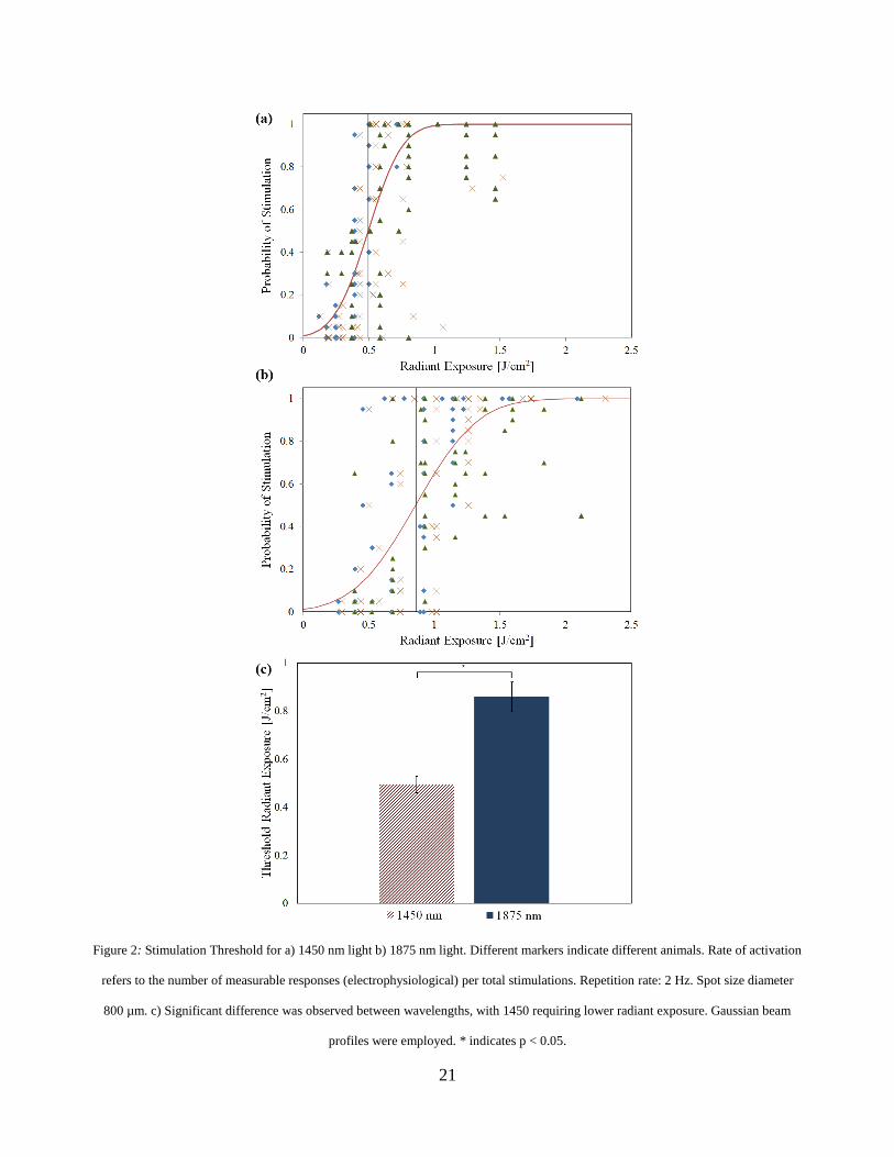
21
Figure 2: Stimulation Threshold for a) 1450 nm light b) 1875 nm light. Different markers indicate different animals. Rate of activation
refers to the number of measurable responses (electrophysiological) per total stimulations. Repetition rate: 2 Hz. Spot size diameter
800 µm. c) Significant difference was observed between wavelengths, with 1450 requiring lower radiant exposure. Gaussian beam
profiles were employed. * indicates p < 0.05.

22
It was observed that between animals the stimulation threshold for 1450 nm light demonstrated less
variation between animals than 1875 nm light in the sciatic nerve. There is an intermediate range
where a given radiant exposure’s probability of instigating an action potential varies between 0 and 1,
that was present in 5 of the 6 animals. The 1450 nm light exhibited a statistically significant lower
stimulation threshold (0.493 ± .084 J/cm2) in comparison to 1875 nm (0.858 ± 0.084 J/cm2) (n = 6
animals, p < 0.05) (Table 1) (Figure 3).
Spot Size [µm]
1450 nm
[J/cm2]
1875 nm
[J/cm2]
500 0.717 ± .067 0.652 ± 0.045
800 0.493 ± .084 0.858 ± 0.084
1000 0.472 ± .110 0.694 ± 0.097
1000 (Flat Top) 0.589 ± 0.030 0.772 ± 0.054
1500 0.563 ± .034 0.995 ± 0.083
Table 1: Threshold radiant exposures per wavelength and spot size.

23
Figure 3: Stimulation thresholds between different spot sizes and wavelengths. The inset contains a comparison between wavelengths
for the 1000 µm flat-top probe. * indicates p < 0.05.
Effect of Beam Profile and Spot Size on Stimulation Threshold
Gaussian and flat-top beam profiles were compared in order to assess the effect of energy
distribution in a laser spot. Comparisons between spot sizes (500, 800, 1000, and 1500 µm
diameters), wavelengths (1450 and 1875 nm), and beam profile appear in Figure 3. Threshold
radiant exposures according to spot size and beam profile are given in Table 1. Although statistically
significant differences (p < 0.05) in thresholds were observed between 500 µm and 1500 µm
Gaussian spot sizes within wavelengths, there was no prominent trend observed when comparing
1450 nm and 1875 nm light. An increase of stimulation threshold was concomitant with increasing

24
spot size for 1875 nm light, but this pattern of behavior was not observed for 1450 nm light. There
was no statistically significant difference in stimulation threshold between flat-top and Gaussian
beam profiles of equal diameter for either 1450 nm or 1875 nm light (p>0.05).
Histology
A histological analysis was performed on excised nerves following acute stimulation within the main
trunk of the sciatic nerve for the purpose of assessing and quantifying damage within neural tissue
following irradiation. Comparisons between a negative control, a positive control, and approximately
three times the 1875 nm threshold are shown in Figure 4.

25
Figure 4: Toluidine-blue stained irradiated nerve slices. Histological images taken at 60x (a) Negative control, no stimulation, (b)
positive control: 6 J/cm2 1450 nm light, (c) 3.08 J/cm2 irradiation at 1450 nm(d) 3.15 J/cm2 irradiation at 1875 nm. Ellipse indicates
region of damage.

26
Damaging levels of radiant exposure (6 J/cm2) produced disturbances in the myelin of the peripheral
nerve, with the greatest disruptions present in the center of the irradiated area; axonal striations were
absent in the positive control (Figure 4b). Radiant exposures up to three times that of 1875 nm light
threshold did not produce observable damage for 1450 nm or 1875 nm light. In Figure 4c and 4d, the
integrity of the myelin is preserved, without the rupturing extant in Figure 4b.
Nerve monitoring study
Both optical and electrical stimuli were able to perturb a measurable response in the gastrocnemius,
soleus, and anterior tibialis muscles as shown in the comparison given in Figure 5. Electrically
evoked responses were greater than those evoked optically by an order of ten within healthy nerves.
After nerves were cauterized, only electrical stimulation evoked a measurable signal distal to the site
of injury, due to current spread.

27
Figure 5: Response comparison before and after damage, all four horizontal axes: 10 s. (a) vertical axis range: 2000 μV, (b) vertical
axis range: 50 μV, (c) vertical axis range: 200 μV, (d) vertical axis range: 50 μV.
28
Discussion
Infrared light has a track record of success in the stimulation of numerous physiological targets,
proving a spatially selective means of perturbing action potentials without the need for exogenous
factors. It is readily paired with multiple means of neural monitoring such as electrical recordings,
calcium staining and fluorescence imaging 15, intrinsic imaging 23, and fMRI 24, a difficult avenue for
electrical stimulation. However, although INS has been refined as a tool of neurophysiological study,
there have been challenges that required address, including a difficulty in maintaining repeatability
in experiments while maintaining radiant exposure values within a safe window. Previously explored
methods addressing this include hybrid methods of stimulation which combine below-threshold
optical and electrical stimuli which, when combined, cause activation of a neurological target 17,25.
Yet even within these methods a temporal effect has been observed while using 1875 nm light,
requiring continual monitoring and adjustment of laser parameters. This hinders repeatability and is
an inconvenience that eliminates clinical utility. Increasing laser power relative to threshold has been
a hitherto unavailable option. This paper accomplishes this goal through the parameterization and
application of a previously unexplored wavelength of light in the field of optical stimulation.
Here we provide another means of maintaining safe radiant exposure levels with increased
repeatability through the use of a new wavelength of light: 1450 nm. We found statistically
significant reductions in the threshold radiant exposure when using 1450 nm light for all but one spot
parameter as compared to 1875 nm. Our histological findings did not demonstrate a reduction in
damage thresholds, indicating an increase in the margin of safety through the use of this wavelength
of light. This work also highlights the efficacy of flat-top beam profiles as compared to the
previously utilized Gaussian profile, showing no statistically significant difference between the two.
Spot size was also investigated and found to have a minimal effect on threshold radiant exposure,

29
meaning a more ubiquitous irradiation of target tissue is viable without increasing radiant exposure
to damaging levels. This work was supported through the demonstration of infrared light’s utility in
clinical applications in a nerve monitoring scenario, wherein INS showed a greater propensity for the
identification of damaged nerves.
The viability of 1450 nm light in INS applications is evident when performance is compared to that
of 1875 nm light. Stimulation thresholds are reduced in a statistically significant degree for 1450 nm
light, with repeatable stimulations more easily attained at lower values of radiant exposure and
identical repetition rates. This reduction in threshold means a mitigation in the risk of thermal
damage. It should be noted that 1450 nm and 1875 nm light have similar absorptive properties in
water 26. However, 1450 nm light is on a relative peak on the water absorption curve, while 1875 nm
light is on a prominent slope. This fact, coupled with the reality of the broadband nature of the diode
laser light, may account for the increased repeatability demonstrated by 1450 nm light. Although
there was an apparent direct relationship between spot size and stimulation threshold within the 1875
nm wavelength, there was no such relationship observed for 1450 nm light. The question as to why a
potential relationship between spot size and threshold appeared for the 1875 nm light may be
addressed by the positioning of the fiber itself, and the type and percentage of axonal tissue
irradiated as the spot size was increased. The sciatic nerve is well-organized, with sensory and motor
axons occupying different areas of it 27. It is possible that as spot size was adjusted, the encompassed
area did not fully include motor axons, the tissue type responsible for the magnitude of measured
responses.
The flat-top beam profile exhibited no significant difference in stimulation threshold for a 1000 µm
size when compared to the Gaussian profile. This is in contrast to previous literature in which a flat-
top profile required lower radiant exposure 28, though this could be attributed to differences in
30
experimental protocol. There was a decrease in stimulation threshold variance for the flat-top beam
profile as compared to the Gaussian profile, which may be attributed to the collimation of the light
emitted from the probe. The beam is collimated to a 1 mm diameter spot size at distances up to 5
mm away, reducing the variation in spot size that occurs concomitantly with variations in probe tip
distance from the site of stimulation with bare fibers. A Gaussian profile does not have an even
distribution of energy; rather there is a maximum of energy deposition located within the center of
the laser spot. Subsequently, there appears to be an increased risk of thermal damage at higher values
of radiant exposure centered at the site of irradiation. A majority of damage was centered within 100
µm for a 500 μm spot size. The flat-top profile’s distribution of energy is much more uniform as
compared to that of the Gaussian profile. The elimination of a hot spot could lead to a reduction in
the risk of thermal damage at similar values of radiant exposure for a given wavelength, although
this would be best determined by additional histological analysis. The functionality of the flat-top
probe demonstrated in the comparison and nerve monitoring studies agrees with results presented by
Tozburun et al., wherein the rat cavernous nerve was stimulated successfully and efficaciously with
a 1 mm spot size flat-top beam28. This finding presents an added clinical viability in the elimination
of spot size variation when the probe is held by a practicing physician.
The histological analysis performed revealed no acute thermal damage for either 1450 nm or 1875
nm light at radiant exposures up to 3.1 J/cm2, the maximum value of radiant exposure used below
the positive control. This is an encouraging result in particular for the 1450 nm light, representing
the potential for safe stimulation at greater than five times its threshold for stimulation. This is a
marked improvement over the 2:1 safety ratio observed in earlier work2. A 5:1 safety ratio allows for
the use of larger radiant exposures to safely stimulate neural tissue, increasing the repeatability
during the course of a procedure: an important requirement for clinical application, although it is

31
important to note that nerves were excised and fixed within 30 minutes after irradiation, and any
chronic effects of infrared stimulation were not evaluated within the scope of this work.
The histological results and those detailed prior spurred the exploration of INS as a nerve monitoring
modality in this work. Clinical monitoring hardware and software was utilized in the nerve
monitoring study, and no additional preparation of the nerve was performed outside of exposure. The
purpose of these conditions was to represent the clinical setting as faithfully as possible. It was
theorized that a larger spot size could be used for a more ubiquitous irradiation of the sciatic nerve
with no significant increase in radiant exposure for the course of the nerve monitoring experiments.
This assumption was based on the results of the previously discussed spot size experiments. The flat-
top profile was selected for the nerve monitoring experiments due to its comparable performance to
the Gaussian profile, and the collimated nature of the probe. The combination of the large spot size
and beam profile enabled the uniform irradiation and stimulation of multiple motor axons, evoking
muscular action potentials in the gastrocnemius, soleus, and anterior tibialis. Electric stimulation
perturbed responses approximately one order of magnitude higher than that of INS, which can be
attributed to the spread of current through the nerve. It is believed to be this current spread which
also produced a measurable signal distal to sites of neural damage, a signal which was absent when
INS was applied. Electrical stimulation produced a false positive for functionality in a damaged
nerve whereas INS did not. This is due to the nature by which INS evokes action potentials.
The mechanism of INS is believed to be a temperature-rate dependent membrane capacitance change
at the site of irradiation. Shapiro et al. demonstrated this phenomenon through computational
modeling and irradiation of protein-free lipid bilayers 9. Liu et al. demonstrated similar mechanisms
through rapid heat shocks of cardiomyocytes10. A capacitance change is not the only theory that
exists to explain the mechanism of INS, however. Additional theories suggest the activation of heat-
32
sensitive channels such as the TRPV1 species17, or the alteration of calcium dynamics in organelles
such as the mitochondria and endoplasmic reticulum29. It is possible that the mechanism of INS is a
gestalt of all of these phenomena. What is known is that this mode of stimulation is highly localized,
as demonstrated in previous electrophysiological and histological study of the cochlea13,14, and in
imaging studies of the cortex15,23. Only action potentials conduct along a nerve with INS, as opposed
to the addition of current that give rise to the stimulation artifact seen with electrical methods.
We have evaluated a new wavelength of light for the efficient and repeatable stimulation of neurons,
and we’ve further established INS parameters regarding the threshold of stimulation. The results of
this work establish 1450 nm light as a more efficacious wavelength as compared to the previously
utilized 1875 nm light, with lower stimulation thresholds and increased repeatability. Furthermore
we’ve demonstrated that the flat-top beam profile is comparable in performance to the Gaussian
beam profile, which yields another means of reducing the risk of non-uniform thermal damage
during experimental procedures. Through these results we’ve demonstrated a safer and more
efficacious means of performing INS, increasing its utility to researchers and its ease of transition to
the clinical setting. There it can increase the quality of care through the identification and assessment
of nerve health.
Acknowledgements
We acknowledge the support of the W. M. Keck Foundation Free Electron Laser Center as well as
Lockheed-Martin (Funding info.), in addition to the Translational Pathology Shared Research Core
at Vanderbilt University.

33
Works Cited
1. Wells J, Kao C. Application of infrared light for in vivo neural stimulation. J Biomed Opt.
2005;10(6):064003. doi:10.1117/1.2121772.
2. Wells JD, Thomsen S, Whitaker P, et al. Optically mediated nerve stimulation: Identification
of injury thresholds. Lasers Surg Med. 2007;39(6):513-26. doi:10.1002/lsm.20522.
3. Starr PA, Christine CW, Theodosopoulos P V, et al. Implantation of deep brain stimulators
into subthalmic nucleus: technical approach and magnetic imaging-verified electrode
locations. J Neurosurg. 2002;97(2):370-387.
4. Stevens JC, Patterson MQ. Dimensions of spatial acuity in the touch sense: changes over the
life span. Somatosens Mot Res. 1995;12(1):29-47. Available at:
http://www.ncbi.nlm.nih.gov/pubmed/7571941.
5. Barker AT. An introduction to the basic principles of magnetic nerve stimulation. J Clin
Neurophysiol. 1991;8(1):26-37.
6. Hallett M. Transcranial magnetic stimulation and the human brain. Nature.
2000;406(6792):147-150. Available at: http://dx.doi.org/10.1038/35018000.
7. Shoham S, O’Connor DH, Sarkisov D V, Wang SS. Rapid neurotransmitter uncaging in
spatially defined patterns. Nat Methods. 2005;2(11):837-843. doi:10.1038/nmeth793.
8. Deisseroth K. Optogenetics. Nat Methods. 2011;8(1):26-29. doi:10.1038/NMETH.F.324.
9. Shapiro MG, Homma K, Villarreal S, Richter C-P, Bezanilla F. Infrared light excites cells by
changing their electrical capacitance. Nat Commun. 2012;3:736.
10. Liu Q, Frerck MJ, Holman H a, Jorgensen EM, Rabbitt RD. Exciting cell membranes with a
blustering heat shock. Biophys J. 2014;106(8):1570-7. doi:10.1016/j.bpj.2014.03.008.
11. Jenkins MW, Duke AR, Gu S, et al. Optical pacing of the embryonic heart. Nat Photonics.
2010;4(9):623-626.
12. Matic AI, Robinson AM, Young HK, et al. Behavioral and Electrophysiological Responses
Evoked by Chronic Infrared Neural Stimulation of the Cochlea. PLoS One. 2013;8(3):10.
doi:10.1371/journal.pone.0058189.
13. Richter C-P, Rajguru SM, Matic AI, et al. Spread of cochlear excitation during stimulation
with pulsed infrared radiation: inferior colliculus measurements. J Neural Eng.
2011;8(5):056006. doi:10.1088/1741-2560/8/5/056006.
34
14. Moreno LE, Rajguru SM, Matic AI, et al. Infrared neural stimulation: beam path in the guinea
pig cochlea. Hear Res. 2011;282(1-2):289-302. doi:10.1016/j.heares.2011.06.006.
15. Cayce JM, Friedman RM, Jansen ED, Mahavaden-Jansen A, Roe AW. Pulsed infrared light
alters neural activity in rat somatosensory cortex< i> in vivo</i>. Neuroimage.
2011;57(1):155-166.
16. Cayce JM, Kao CC, Malphrus JD, Konrad PE, Mahadevan-Jansen A, Jansen ED. Infrared
neural stimulation of thalamocortical brain slices. Sel Top Quantum Electron IEEE J.
2010;16(3):565-572.
17. Peterson EJ, Tyler DJ. Motor neuron activation in peripheral nerves using infrared neural
stimulation. J Neural Eng. 2014;11(1):016001. doi:10.1088/1741-2560/11/1/016001.
18. Tozburun S, Stahl CD, Hutchens TC, Lagoda G a, Burnett AL, Fried NM. Continuous-wave
infrared subsurface optical stimulation of the rat prostate cavernous nerves using a 1490-nm
diode laser. Urology. 2013;82(4):969-73. doi:10.1016/j.urology.2013.06.031.
19. Tozburun S, Lagoda G a, Burnett AL, Fried NM. Subsurface near-infrared laser stimulation of
the periprostatic cavernous nerves. J Biophotonics. 2012;5(10):793-800.
doi:10.1002/jbio.201100134.
20. Teudt IU, Nevel AE, Izzo AD, Walsh JT, Richter C-P. Optical stimulation of the facial nerve:
a new monitoring technique? Laryngoscope. 2007;117(9):1641-7.
doi:10.1097/MLG.0b013e318074ec00.
21. Duke AR, Lu H, Jenkins MW, Chiel HJ, Jansen ED. Spatial and temporal variability in
response to hybrid electro-optical stimulation. J Neural Eng. 2012;9(3):036003.
doi:10.1088/1741-2560/9/3/036003.
22. Khosrofian JM, Garetz BA. Measurement of a Gaussian laser beam diameter through the
direct inversion of knife-edge data. Appl Opt. 1983;22(21):3406. doi:10.1364/AO.22.003406.
23. Cayce JM, Friedman RM, Chen G, Jansen ED, Mahadevan-Jansen A, Roe AW. Infrared
neural stimulation of primary visual cortex in non-human primates. Neuroimage.
2014;84:181-190.
24. Chen G, Cayce JM, Friedman RM, et al. Functional tract tracing in non-human primates using
pulsed infrared laser light with optical imaging and fMRI. Soc Neurosci Abstr Viewer Itiner
Plan. 2012;42.
25. Duke AR, Lu H, Jenkins MW, Chiel HJ, Jansen ED. Spatial and temporal variability in
response to hybrid electro-optical stimulation. J Neural Eng. 2012;9(3):036003.
doi:10.1088/1741-2560/9/3/036003.
35
26. Hale GM, Querry MR. Optical Constants of Water in the 200-nm to 200-microm Wavelength
Region. Appl Opt. 1973;12(3):555-63. doi:10.1364/AO.12.000555.
27. Badia J, Pascual-Font A, Vivó M, Udina E, Navarro X. Topographical distribution of motor
fascicles in the sciatic-tibial nerve of the rat. Muscle Nerve. 2010;42(2):192-201.
doi:10.1002/mus.21652.
28. Tozburun S, Lagoda GA, Burnett AL, Fried NM. Gaussian versus flat-top spatial beam
profiles for optical stimulation of the prostate nerves. In: Kollias N, Choi B, Zeng H, et al.,
eds. BiOS. International Society for Optics and Photonics; 2010:75484W-75484W-6.
doi:10.1117/12.852994.
29. Paviolo C, Haycock JW, Cadusch PJ, McArthur SL, Stoddart PR. Laser exposure of gold
nanorods can induce intracellular calcium transients. J Biophotonics. 2013.
doi:10.1002/jbio.201300043.
36
CHAPTER III
FUTURE DIRECTIONS
We have evaluated a new wavelength of light for the efficient and repeatable stimulation of
neurons, and we’ve further established INS parameters regarding the threshold of stimulation.
The results of this work establish 1450 nm light as a more efficacious wavelength as compared to
the previously utilized 1875 nm light, with lower stimulation thresholds and increased
repeatability. Furthermore we’ve demonstrated that the flat-top beam profile is comparable in
performance to the Gaussian beam profile, which yields another means of reducing the risk of
non-uniform thermal damage during experimental procedures. Through these results we’ve
demonstrated a safer and more efficacious means of performing INS, increasing its utility to
researchers and its ease of transition to the clinical setting. There it can increase the quality of
care through the identification and assessment of nerve health.
However, addressing one set of questions leads to additional ones. The efficaciousness of 1450
nm light has been proven, and it is theorized that it benefits from its location a relative peak of
absorption. It is possible that other wavelengths centered on similar peaks might exhibit a
similar constancy in performance, but there is the question about the coefficient of absorption
itself, particularly in terms of penetration depth. It is worth exploring whether or not
wavelengths with a greater penetration depth would allow for stimulation of deeper regions of
interest. One potential means of exploring this would be an application similar to two photon
uncaging, wherein two light paths are set to converge on a region of interest, combining to
activate an exogenous neurotransmitter 1. Using INS, perhaps a similar effect could be achieved
without the use of an exogenous substance.
37
Another question exists regarding the long-term effects of INS. The histological assays
performed in this study did not account for chronic usage of INS, and a study exploring the
effects of such an application would provide valuable information about the safety and efficacy
of INS.
The move towards clinical applications is necessarily gated by the completion of the
aforementioned studies; however, this does not impede exploration of additional utilizations of
INS. The spatial resolution of INS has already been shown to be efficacious in the stimulation of
the cochlea 2–4. This spatial resolution may further serve in the development of prostheses which
restore not only the form and motility of a lost limb, but encode tactile and proprioceptive
sensory information as well. Previous work has revealed what appears to be an inhibitory effect
on neural signal conduction, which has significant implications for treatment of neuropathy5.
INS can potentially serve as a treatment for chronic pain, spastic paraplegia, and other conditions
in which quiescence of nerves is required. Of course, elucidating and differentiating the
phenomena by which INS either activates or inhibits neural activity warrants attention.
We have observed that 1450 nm light can stimulate the peripheral nervous system reliably, so it
is worth assessing its utility in the central nervous system (CNS). In prior work 1875 nm was
used to activate CNS tissue in both in vitro6 and in vivo7–9 applications. The use of 1450 nm may
facilitate an INS means of cortical mapping, or even in treatments of neuropathies currently
addressed by electrical methods such as deep brain stimulation (DBS). The latter comes with the
caveats that the amount of light delivered, the metric by which stimulation scales, is limited by
the geometry of the fiber that transports it. Development of novel geometries may be necessary
to facilitate optimal light delivery while mitigating the trauma of introducing the fiber into
cortical tissue; such considerations and developed have already been made for optogenetic

38
applications 10. The formation of glial scar tissue has been shown to impede conduction of
electrical stimuli, and this is a relevant issue for optical stimuli as well 11–14. This potential
impedance should be the subject of rigorous study to determine the efficacy of INS in DBS
applications.
The limitations of INS should be considered in the planning of future work, but by no means
should its potential be dismissed. This work demonstrates that INS can be done safely and
reliably, and that it provides a unique utility absent from other methods of neurological
stimulation. The possibilities for INS are limited only by the imagination.

39
Works Cited
1. Shoham, S., O’Connor, D. H., Sarkisov, D. V & Wang, S. S. Rapid neurotransmitter
uncaging in spatially defined patterns. Nat Methods 2, 837–843 (2005).
2. Richter, C.-P. et al. Spread of cochlear excitation during stimulation with pulsed infrared
radiation: inferior colliculus measurements. J. Neural Eng. 8, 056006 (2011).
3. Matic, A. I. et al. Behavioral and Electrophysiological Responses Evoked by Chronic
Infrared Neural Stimulation of the Cochlea. PLoS One 8, 10 (2013).
4. Moreno, L. E. et al. Infrared neural stimulation: beam path in the guinea pig cochlea. Hear.
Res. 282, 289–302 (2011).
5. Duke, A. R., Lu, H., Jenkins, M. W., Chiel, H. J. & Jansen, E. D. Spatial and temporal
variability in response to hybrid electro-optical stimulation. J. Neural Eng. 9, 036003
(2012).
6. Cayce, J. M. et al. Infrared neural stimulation of thalamocortical brain slices. Sel. Top.
Quantum Electron. IEEE J. 16, 565–572 (2010).
7. Cayce, J. M., Friedman, R. M., Jansen, E. D., Mahavaden-Jansen, A. & Roe, A. W. Pulsed
infrared light alters neural activity in rat somatosensory cortex< i> in vivo</i>.
Neuroimage 57, 155–166 (2011).
8. Cayce, J. M. et al. Infrared neural stimulation of primary visual cortex in non-human
primates. Neuroimage 84, 181–190 (2014).
9. Chen, G. et al. Functional tract tracing in non-human primates using pulsed infrared laser
light with optical imaging and fMRI. Soc. Neurosci. Abstr. Viewer Itiner. Plan. 42, (2012).
10. Zorzos, A. N., Boyden, E. S. & Fonstad, C. G. Multiwaveguide implantable probe for
light delivery to sets of distributed brain targets. Opt. Lett. 35, 4133–4135 (2010).
11. Logan, A., Green, J., Hunter, A., Jackson, R. & Berry, M. Inhibition of glial scarring in
the injured rat brain by a recombinant human monoclonal antibody to transforming growth
factor-beta2. Eur. J. Neurosci. 11, 2367–2374 (1999).
12. Peković, S. et al. Downregulation of glial scarring after brain injury: The effect of purine
nucleoside analogue ribavirin. in Ann. N. Y. Acad. Sci. 1048, 296–310 (2005).
13. Polikov, V. S., Block, M. L., Fellous, J. M., Hong, J. S. & Reichert, W. M. In vitro model
of glial scarring around neuroelectrodes chronically implanted in the CNS. Biomaterials
27, 5368–5376 (2006).
40
14. Tamura, K. et al. A glass-coated tungsten microelectrode enclosing optical fibers for
optogenetic exploration in primate deep brain structures. J. Neurosci. Methods 211, 49–57
(2012).