Distinct mechanisms underlie pattern formation in the skin and skin ...
Investigating the mechanisms that may underlie thereduction in contrast sensitivity during dynamicaccommodation
Department of Vision Sciences, Glasgow CaledonianUniversity, Cowcaddens Road, Glasgow, UKSven Mucke
Department of Vision Sciences, Glasgow CaledonianUniversity, Cowcaddens Road, Glasgow, UKVelitchko Manahilov
Department of Vision Sciences, Glasgow CaledonianUniversity, Cowcaddens Road, Glasgow, UKNiall C. Strang
Department of Vision Sciences, Glasgow CaledonianUniversity, Cowcaddens Road, Glasgow, UKDirk Seidel
Department of Vision Sciences, Glasgow CaledonianUniversity, Cowcaddens Road, Glasgow, UKLyle S. Gray
Department of Vision Sciences, Glasgow CaledonianUniversity, Cowcaddens Road, Glasgow, UKUma Shahani
Head and eye movements, together with ocular accommodation enable us to explore our visual environment. The stabilityof this environment is maintained during saccadic and vergence eye movements due to reduced contrast sensitivity to lowspatial frequency information. Our recent work has revealed a new type of selective reduction of contrast sensitivity to highspatial frequency patterns during the fast phase of dynamic accommodation responses compared with steady-stateaccommodation. Here were report data which show a strong correlation between the effects of reduced contrast sensitivityduring dynamic accommodation and velocity of accommodation responses, elicited by ramp changes in accommodativedemand. The results were accounted for by a contrast gain control model of a cortical mechanism for contrast detectionduring dynamic ocular accommodation. Sensitivity, however, was not altered during attempted accommodation responsesin the absence of crystalline-lens changes due to cycloplegia. These findings suggest that contrast sensitivity reductionduring dynamic accommodation may be a consequence of cortical inhibition driven by proprioceptive-like signals originatingwithin the ciliary muscle, rather than by corollary discharge signals elicited simultaneously with the motor command to theciliary muscle.
Keywords: accommodation, accommodative suppression, computational modeling, contrast sensitivity, eye movements,proprioception
Citation: Mucke, S., Manahilov, V., Strang, N. C., Seidel, D., Gray, L. S., & Shahani, U. (2010). Investigating themechanisms that may underlie the reduction in contrast sensitivity during dynamic accommodation. Journal ofVision, 10(5):5, 1–14, http://journalofvision.org/content/10/5/5, doi:10.1167/10.5.5.
Introduction
We perceive the external world as a continuous three-dimensional visual scene in which objects are stable andclear. Our ability to process spatially detailed visualinformation is restricted to a small part of the retina, thefovea. Detailed processing of objects at different spatiallocations in the visual scene is achieved by a combinationof ocular movements: saccades place the retinal image ofinterest on the fovea; vergence eye movements minimize
retinal disparity to maintain binocular single vision; andocular accommodation changes lens power to producesharp retinal images. During these oculomotor movementsremarkable transformations of the retinal representation ofthe external world occur, and yet, we generally perceive asequence of sharp snapshots of different objects at differ-ent positions in the visual scene.For hundreds of years, scientists have questioned why
we do not perceive degraded images among the snapshots.The object of much of this work has been to investigatethe neural mechanisms involved in maintaining stable
Journal of Vision (2010) 10(5):5, 1–14 http://journalofvision.org/content/10/5/5 1
doi: 10 .1167 /10 .5 .5 Received July 31, 2009; published May 6, 2010 ISSN 1534-7362 * ARVO
visual perception during eye movements (for a review seeWurtz, 2008). Reduced sensitivity of visual informationprocessing during saccadic eye movement is selective forlow-spatial frequency information and, as a result,alleviates anomalous motion perception, whereas contrastdetection thresholds for high-spatial frequency patternsand detection thresholds for iso-luminant color stimuliremain normal (Burr, Morrone, & Ross, 1994, Latour,1962, Volkmann, 1986). This suggests that the sensitivityof the magnocellular pathway is being selectively reducedduring saccadic eye movements (Burr et al., 1994).Saccadic suppression does not seem to remove visualinformation from awareness however, as a recent studyfound in which low spatial frequency informationremained accessible to the visual system throughout thesaccadic eye movement (Watson & Krekelberg, 2009).Spatial selectivity of reduced sensitivity during vergenceeye movements has not been explored in detail, but studieshave shown that vergence eye movements attenuatesensitivity to uniform flash stimuli, which represent lowspatial frequency information (Manning, 1986, Manning &Riggs, 1984).Recently, we have found reduced contrast sensitivity to
patterns of higher spatial frequency during dynamicaccommodation step responses (Mucke, Manahilov,Strang, Seidel, & Gray, 2008). Contrast sensitivityreduction during dynamic accommodation shows spatial-frequency selectivity, which differs from that of saccadicsuppression and is not related to the predicted reduction incontrast sensitivity produced by optical factors (Fernandez& Artal, 2005). Contrast sensitivity to patterns of higherspatial frequency was reduced significantly during dynamicaccommodation compared with steady-state accommoda-tion, whilst sensitivity to patterns of lower spatial fre-quency was not altered. This effect was maximal when theaccommodation step response reached its peak velocity(350–400 ms after the accommodation target onset), withunaltered sensitivity being found around the starting(200 ms) and end points (500–600 ms) of the response.Neural mechanisms underlying reduced contrast sensi-
tivity during saccadic eye movements have been attributedmainly to extra-retinal signals, such as corollary dischargesignals (Sperry, 1950) elicited simultaneously with themotor command to the extra-ocular muscles. These out-flow signals precede the physical muscle response andcause maximal reduction in sensitivity around theresponse onset (for review see Wurtz, 2008). Suchcorollary discharge signals are also believed to beavailable for accommodation (Rosenbluth & Allman,2002). However, the late onset of contrast sensitivityreduction during dynamic accommodation, as comparedto saccadic suppression, suggests a different neural pathway.While afferents from mechanoreceptors within extra-ocular muscles have been rejected for saccadic suppres-sion, because of their delayed availability (Wurtz, 2008),such neural input from the ciliary muscle could arrive
in time to reduce contrast sensitivity during dynamicaccommodation. The existence of mechanoreceptorswithin the ciliary muscle has only recently been estab-lished (Flugel-Koch, Neuhuber, Kaufman, & Lutjen-Drecoll, 2009).The aim of this study was to explore whether a
proprioceptive-like (inflow) mechanism, a corollary dis-charge (outflow) mechanism or both contribute to thereduced sensitivity during dynamic accommodation. First,we investigated the correlation between the magnitude ofcontrast sensitivity reduction during dynamic accommo-dation and accommodation velocity for ramp changes inaccommodative demand. The obtained results wereaccounted for by a contrast gain control model of acortical mechanism for detecting patterns using divisivenormalization, with the magnitude of contrast sensitivityreduction being proportional to the absolute value ofaccommodation velocity. The velocity of accommodationresponses reflects the change in refractive power of thecrystalline eye lens as a consequence of the change inciliary muscle tone. Given that contrast sensitivity isreduced during the fast phase of the accommodationresponse and the magnitude of this effect is proportionalto accommodation velocity, one could suggest thatcontrast sensitivity reduction during dynamic accommo-dation is determined by a cortical inhibitory mechanismcontrolled by proprioceptive-like feedback produced bymechanoreceptors of the ciliary muscle.This hypothesis, however, does not reject the role of
corollary discharge in regulating the observed reduction incontrast sensitivity. The outflow mechanism should reducecontrast sensitivity mainly during the onset of dynamicaccommodation responses (following the logic of corol-lary discharge signals, reviewed in Wurtz, 2008), but itmight also affect pattern detection during the fastaccommodation phase. In order to test these possibilities,we examined whether contrast sensitivity was reducedwhen the ciliary muscle is paralyzed. We assumed that theproprioceptive inputs from within the paralyzed ciliarymuscle would be eliminated in a similar way as theproprioceptive inputs from paralyzed extra-ocular musclesare greatly reduced or absent (for reviews see Donaldson,2000 and Ruskell, 1999). Additionally, we assumed thatattempted dynamic accommodation to a far-to-nearaccommodation target with paralyzed ciliary musclesshould have elicited a corollary discharge signal togetherwith the motor command (Rosenbluth & Allman, 2002;Sperry, 1950). If similar reduction in contrast sensitivityoccurred during dynamic accommodation attempts, wecould suggest that the corollary discharge from the motorcommand elicited in the midbrain areas would be itssource. Alternatively, lack of contrast sensitivity reductionwould support the hypothesis that proprioceptive-likesignals from within the ciliary muscle would perhapscause the loss in contrast sensitivity. If, however, contrastsensitivity reduction occurred with reduced magnitude,
Journal of Vision (2010) 10(5):5, 1–14 Mucke et al. 2
then both corollary discharge and proprioceptive-likesignals could be regarded as factors contributing to theloss in contrast sensitivity.
Methods
Subjects
Four subjects, three naıve to the aims of the experimentand one of the authors, participated with informed consentin this study. All participants were young emmetropes(aged 19–34) with good ocular and general health, visionof 6/6 or better and a near point of accommodation closerthan 30 cm. The latter was measured during eyeexamination and confirmed during practice runs of theexperiment.
Apparatus
Accommodation step responses were induced bymounting an accommodation target on a linear actuator(Movopart M 55, Tollo linear AB, Kristianstad, Sweden),powered by a servo drive and run by a NextMove ESBmotion controller. The system had a non-linearity of0.39% and maximum speed of 5 meters per second, whichallowed the accommodation target to travel from 1 m to33 cm in about 180 ms. The actuator was programmed toperform a non-linear movement to generate a constantaccommodation target speed in dioptres per second (D/s).Initiation of movement of the accommodation target wasrandomized to suppress any temporal frequency pattern.Ocular accommodation was monitored and recorded with
a modified infra-red open-field autorefractor (Shin-NipponSRW-5000) and data acquisition software (LabView 7)which allowed a sampling rate of 60 Hz, well above theNyquist frequency for accommodation (Denieul, 1982),with a resolution of G0.001 D (Wolffsohn, Gilmartin,Mallen, & Tsujimura, 2001). Continuous accommodationrecordings were smoothed with a low-pass filter at 10 Hz,and blinks removed using an algorithm described pre-viously by Day, Strang, Seidel, Gray, and Mallen (2006).However, blinks were rare during trials, which lastedbetween 3 and 5 seconds depending on the random timedelay for initiation of the accommodation target move-ment. Accommodation velocity was calculated for eachsubject and each target speed individually, and used todetermine the onset of the dynamic accommodationresponse (Suryakumar, Meyers, Irving, & Bobier, 2007).The experiment took place in a dimmed room to allowfor pupil sizes 93 mm. A smaller pupil would result insignal loss from the autorefractor. Head and eye move-ments were minimized by using a chin and forehead restand the eye under test was carefully aligned with the
accommodation target and center of the test monitor. Pupilsizes were monitored and recorded in Experiment 2utilizing an EyeLink I video eye tracker with a samplingrate of 250 Hz (SR Research Ltd., Mississauga, Ontario,Canada). Recordings were smoothed offline with a sliding100 ms window.
Visual stimuli
The test stimulus was generated by a Pentium 3computer on a 19W RGB monitor (Vision Master Pro450, Iiyama Electronics America, Inc.) at a screenresolution of 1024 � 768 pixels and a frame rate of 75Hz. The stimulus was displayed using a 256-color look-uptable and a 12-bit gray-scale resolution obtained by acustom video summation device (Pelli & Zhang, 1991).The distance between the screen (angular size of 19 �14.4 deg) and the subject was 1 m. The mean luminancewas 30 cd/m2. The monitor’s gamma non-linearity wascorrected carefully using an OptiCal photometer (Cam-bridge Research System Ltd.) interfaced to the PC. The90% decay of the monitor phosphor signal (using whitecolor and an image formed by 1 line) was about 1 ms.The test stimulus was a 4-deg diameter circular patch of
a horizontal square-wave grating with a fundamentalfrequency of 9 c/deg. The effects of spatial transients atthe stimulus edges were reduced by dumping the contrastof the outer area (0.5 deg) with a linear function. Indynamic conditions, the test stimulus was presented atdifferent time lags (to an accuracy of 1 screen frame),which were triggered by the onset of the accommodationtarget movement. Stimulus duration was about 5 ms (1/3screen frame of 13.3 ms plus phosphor decay) for themajority of conditions and about 18 ms (11/3 screenframes plus phosphor decay) for the highest defocuscondition.
Experimental design
This study compared contrast sensitivity measuresbetween conditions with steady-state and dynamic accom-modation. For the static condition (steady-state accom-modation), subjects were instructed to focus monocularly(dominant eye) on a Snellen E (angular letter size 10 arcminat 1 m) at five individual distances ranging from 1 m to33 cm, whilst the fellow eye was occluded. For the dynamiccondition, two-dioptre (1–3 D) far-to-near accommodationramp responses with different velocities were induced. Avalid dynamic accommodation response was performedafter a reasonable reaction time (È200 ms), with a con-sistent dioptrical increase, and with an amplitude 91 D.Individual continuous recordings of 100 accommodationresponses following each target speed (10, 3, 2, 1 D/s) wereobtained prior to the experiment and averaged. From the
Journal of Vision (2010) 10(5):5, 1–14 Mucke et al. 3
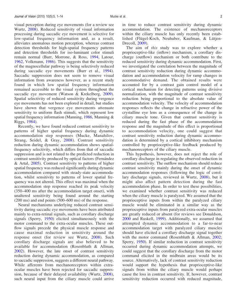
mean accommodation response profile of the 10 D/s targetspeed individual accommodation velocity profiles werecomputed by differentiating the averaged accommodationresponses using a 2-point-difference algorithm, and sub-sequently smoothing the data, using a 100 ms window.Three points of interest (B, C, D) were chosen aroundaccommodation peak velocity, one point (A) beforeaccommodation response onset, and a further point (E)near the final steady-state response level (Figure 1). Eachpoint of interest in the dynamic condition had a time lag,which had a distinct accommodation response level. Thefive accommodation response levels (A–E) were convertedinto five static viewing distances between 1 m (1 D) and33 cm (3 D) (Figure 1). Hence, accommodation ampli-tudes were always matched between static and dynamicconditions. Viewing conditions following accommodationtarget speeds of 3, 2 and 1 D/s were also matched withthe same accommodation response levels (Figure 1), andeach accommodation target speed was given its separateset of five dynamic time lags. Due to the individualaccommodation dynamics of the four subjects, time lagsand accommodation response levels differed slightlyamongst subjects, and are therefore shown as arbitrarypoints of interest A–E (Figure 1).During the first experimental session for each partic-
ipant, the accommodation target was positioned at 1 mand ten measures of steady-state accommodation weretaken. The same procedure was repeated for 33 cm,calibrating the continuous recording of dynamic accom-modation. In dynamic mode the accommodation targetwas moving at four different speeds (1, 2, 3 and 10 D/s)from 1 m to 33 cm. Participants were instructed to “focuscarefully on the accommodation object” (Stark &Atchison,
1994), and to regain focus as fast as possible after theinitiation of an accommodation ramp response.The test stimulus was presented randomly 2 deg above
or below the accommodation target, for the duration of 5ms (points of interest A–D), or 18 ms (point of interest E,Figure 1) on the monitor at 1 m distance. As the subjectaccommodated from 1 m to 33 cm the test stimulusbecame increasingly blurred. This expected reduction incontrast sensitivity made it necessary to use a longer teststimulus duration at the highest level of defocus. Partic-ipants used mouse buttons to indicate the perceivedposition of the grating. Negative feedback was given formismatches. Detection thresholds were measured using astaircase method and a two-alternative-forced-choiceprocedure designed to determine 79% correct responses(Levitt, 1971). Each staircase started at a supra-thresholdcontrast level of the test stimulus with a contrast step of0.2 log units. After each staircase reversal, the step sizewas halved and this process continued until the step sizebecame 0.05 log units. The subsequent six staircasereversals were collected and the threshold measure wasthe geometric mean of these estimates. If the subject wasnot able to accommodate accurately (extremely shortreaction time, slow or inconsistent response, low responseamplitude), or blinked, the experimenter would reject thetrial from the staircase procedure.Within one experimental session contrast thresholds
were measured during one static and four dynamicconditions of equal accommodation response level, fol-lowing different accommodation target speeds. Dynamicaccommodation was monitored throughout the wholeexperiment. Approximately six sessions were necessaryto collect all data for each participant, including practice
Figure 1. Examples of dynamic accommodation ramp responses following target speeds of 10, 3, 2 and 1 D/s as a function of time afterthe accommodation target onset. Horizontal, dashed lines mark equal accommodation response levels across all dynamicaccommodation ramp responses elicited during the experiment, and also connect the time domain via accommodation responseamplitude with static viewing distances for steady-state accommodation. Points of interest (A–E) chosen from the 10 D/s response profile[1� pre-ramp (200 ms), 3� fast phase (300, 350, 400 ms), 1� slow phase (500 ms for LM, CM; 600 ms for KP, SM)] are highlightedacross all dynamic conditions.
Journal of Vision (2010) 10(5):5, 1–14 Mucke et al. 4
runs, and initial dynamic accommodation recordings at allfour target speeds for reaction and response time assess-ment. One session lasted approximately one hour includ-ing small breaks between trials.
Results
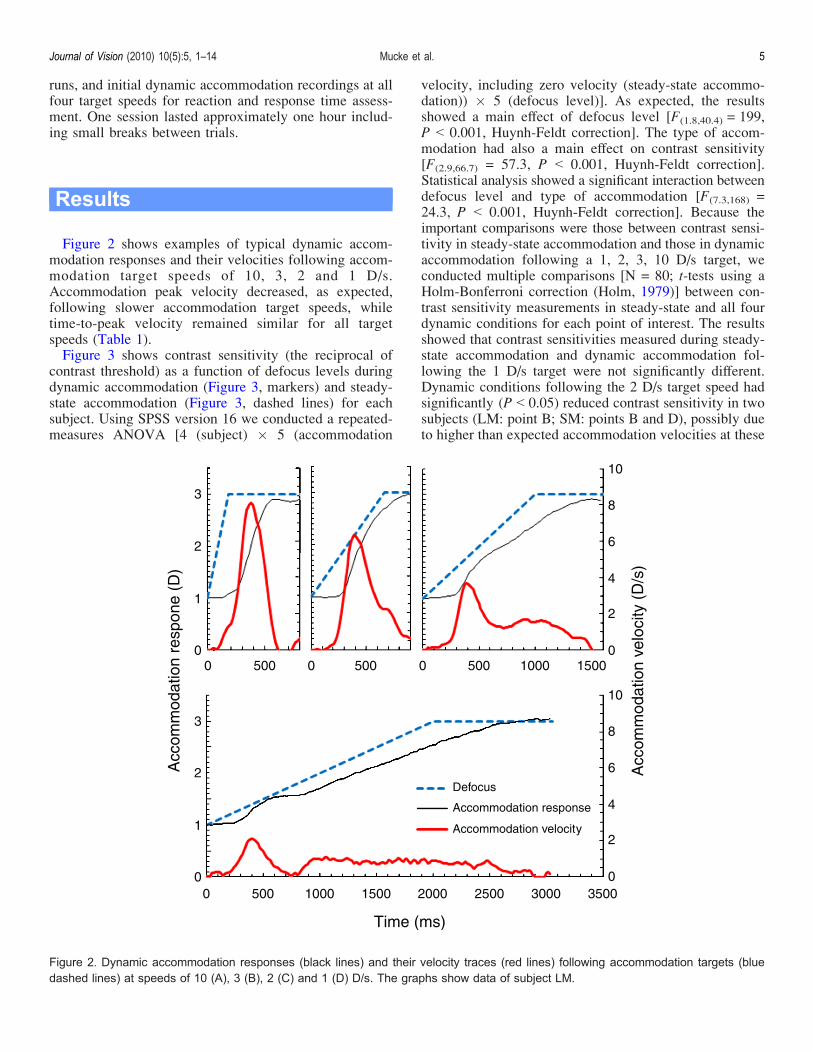
Figure 2 shows examples of typical dynamic accom-modation responses and their velocities following accom-modation target speeds of 10, 3, 2 and 1 D/s.Accommodation peak velocity decreased, as expected,following slower accommodation target speeds, whiletime-to-peak velocity remained similar for all targetspeeds (Table 1).Figure 3 shows contrast sensitivity (the reciprocal of
contrast threshold) as a function of defocus levels duringdynamic accommodation (Figure 3, markers) and steady-state accommodation (Figure 3, dashed lines) for eachsubject. Using SPSS version 16 we conducted a repeated-measures ANOVA [4 (subject) � 5 (accommodation
velocity, including zero velocity (steady-state accommo-dation)) � 5 (defocus level)]. As expected, the resultsshowed a main effect of defocus level [F(1.8,40.4) = 199,P G 0.001, Huynh-Feldt correction]. The type of accom-modation had also a main effect on contrast sensitivity[F(2.9,66.7) = 57.3, P G 0.001, Huynh-Feldt correction].Statistical analysis showed a significant interaction betweendefocus level and type of accommodation [F(7.3,168) =24.3, P G 0.001, Huynh-Feldt correction]. Because theimportant comparisons were those between contrast sensi-tivity in steady-state accommodation and those in dynamicaccommodation following a 1, 2, 3, 10 D/s target, weconducted multiple comparisons [N = 80; t-tests using aHolm-Bonferroni correction (Holm, 1979)] between con-trast sensitivity measurements in steady-state and all fourdynamic conditions for each point of interest. The resultsshowed that contrast sensitivities measured during steady-state accommodation and dynamic accommodation fol-lowing the 1 D/s target were not significantly different.Dynamic conditions following the 2 D/s target speed hadsignificantly (P G 0.05) reduced contrast sensitivity in twosubjects (LM: point B; SM: points B and D), possibly dueto higher than expected accommodation velocities at these
Figure 2. Dynamic accommodation responses (black lines) and their velocity traces (red lines) following accommodation targets (bluedashed lines) at speeds of 10 (A), 3 (B), 2 (C) and 1 (D) D/s. The graphs show data of subject LM.
Journal of Vision (2010) 10(5):5, 1–14 Mucke et al. 5
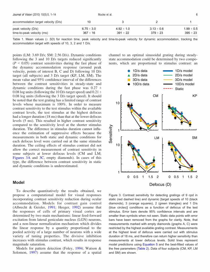
points (LM: 3.69 D/s; SM: 2.56 D/s). Dynamic conditionsfollowing the 3 and 10 D/s targets reduced significantly(P G 0.05) contrast sensitivities during the fast phase ofthe dynamic accommodation response (around peakvelocity, points of interest B, C and D) following 10 D/starget (all subjects) and 3 D/s target (KP, LM, SM). Themean value and 95% confidence interval of the differencesbetween the contrast sensitivities in steady-state anddynamic conditions during the fast phase was 0.27 T0.08 log units (following the 10 D/s target speed) and 0.21 T0.08 log units (following the 3 D/s target speed). It shouldbe noted that the test grating has a limited range of contrastlevels whose maximum is 100%. In order to measurecontrast sensitivity to the test stimulus within the availablecontrast levels, the test stimulus at the highest defocushad a longer duration (18 ms) than that at the lower defocuslevels (5 ms). This resulted in higher contrast sensitivitycompared to the sensitivity level at the shorter stimulusduration. The difference in stimulus duration cannot influ-ence the estimation of suppressive effects because themeasurements in both static and dynamic conditions foreach defocus level were carried out at the same stimulusduration. The ceiling effects of stimulus contrast did notallow the correct measurement of contrast sensitivity insome subjects at lower defocus levels (CM and LM;Figures 3A and 3C, empty diamonds). In cases of thistype, the difference between contrast sensitivity in staticand dynamic conditions is underestimated.
Model
To describe quantitatively the results obtained, wepropose a computational model for visual responsesincorporating contrast sensitivity reduction during ocularaccommodation. Models for contrast gain control(Albrecht & Geisler, 1991; Heeger, 1992) assume thatthe responses of cells of primary visual cortex aredetermined by two main mechanisms: linear feed-forwardexcitation from lateral geniculate nucleus (LGN) neurons,and a non-linear normalization mechanism which dividesthe linear response by a quantity proportional to thepooled activity of a large number of neurons with a widevariety of tuning properties. The divisive inhibitionincreases with stimulus contrast, which results in responsemagnitude saturation.Models for pattern detection (Foley, 1994; Watson &
Solomon, 1997) assume that the response of a spatial
channel to an optimal sinusoidal grating during steady-state accommodation could be determined by two compo-nents, which are proportional to stimulus contrast: an
Figure 3. Contrast sensitivity for detecting gratings of 9 cpd instatic (red dashed line) and dynamic [target speeds of 10 (blackdiamonds), 3 (orange squares), 2 (green triangles) and 1 D/s(blue circles)] conditions as a function of defocus of the teststimulus. Error bars denote 95% confidence intervals and aresmaller than symbols when not seen. Static data points with errorbars have been removed from the graphs for clarity. Note, thatmeasurements marked with empty diamonds (graphs A, C) wererestricted by the highest available grating contrast. Measurementsat the highest level of defocus were carried out with stimulusduration of 18 ms, and therefore can return higher sensitivity thanmeasurements at lower defocus levels. Solid lines representmodel predictions using Equation 5 and the best-fitted values ofthe free parameters (Table 2). Data of four subjects (CM, KP, LMand SM) are shown.
accommodation target velocity (D/s) 10 3 2 1
peak velocity (D/s) 6.75 T 3.0 4.92 T 1.0 3.13 T 0.6 1.99 T 0.5time-to-peak velocity (ms) 367 T 16 391 T 22 379 T 23 395 T 23
Table 1. Mean values (T SD) for reaction time, peak velocity and time-to-peak velocity for dynamic accommodation, tracking theaccommodation target with speeds of 10, 3, 2 and 1 D/s.
Journal of Vision (2010) 10(5):5, 1–14 Mucke et al. 6
excitatory component (aCp) and a divisive inhibitorycomponent (bCp). The response magnitude (Rstat) couldbe written as follows:
Rstat ¼ aCps
1þ bCps; ð1Þ
where a and b are constants and p is a power coefficient.We propose that during dynamic accommodation, the
response (Rdyn) of the same spatial channel would have anadditional inhibitory component whose strength is propor-tional to the velocity of the dynamic accommodationresponse when it exceeds a threshold value of V0 asfollows:
Rdyn ¼ aCpd
1þ bCpd þ m kVkj V0½ �pþ
; ð2Þ
where V is the velocity of the dynamic accommodationresponse, V0 is a parameter representing a thresholdvelocity, [ªVª j V0]+ = ªVª j V0 if ªVª 9 V0, else = 0,m is a coefficient. The contrast sensitivity reductiondepends on the absolute value of the accommodationvelocity, because in a previous study (Mucke et al., 2008)we found that both far-to-near (positive velocity) and near-to-far (negative velocity) accommodation step responsesreduced sensitivity to high spatial frequency information.At threshold, the responses approach a fixed level.
Therefore, the responses in both conditions could beregarded as equal:
aCpd
1þ bCpd þ m kVkj V0½ �pþ
¼ aCps
1þ bCps: ð3Þ
Equation 3 can be simplified as follows:
Cd ¼ Cs 1þ m kVkj V0½ �pþ� �1=p
: ð4Þ
Noting that sensitivity is inversely proportional to thresh-old contrast, Equation 4 can be re-written as follows:
Sd ¼ Ss
1þ m kVkj V0½ �pþ� �1=p : ð5Þ
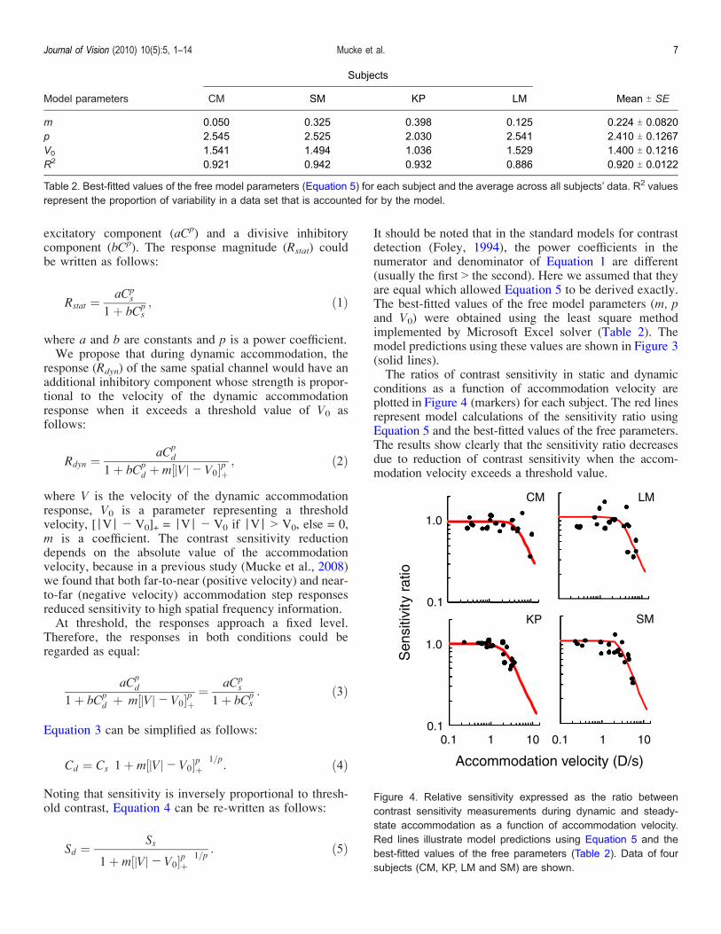
It should be noted that in the standard models for contrastdetection (Foley, 1994), the power coefficients in thenumerator and denominator of Equation 1 are different(usually the first 9 the second). Here we assumed that theyare equal which allowed Equation 5 to be derived exactly.The best-fitted values of the free model parameters (m, pand V0) were obtained using the least square methodimplemented by Microsoft Excel solver (Table 2). Themodel predictions using these values are shown in Figure 3(solid lines).The ratios of contrast sensitivity in static and dynamic
conditions as a function of accommodation velocity areplotted in Figure 4 (markers) for each subject. The red linesrepresent model calculations of the sensitivity ratio usingEquation 5 and the best-fitted values of the free parameters.The results show clearly that the sensitivity ratio decreasesdue to reduction of contrast sensitivity when the accom-modation velocity exceeds a threshold value.
Model parameters
Subjects
Mean T SECM SM KP LM
m 0.050 0.325 0.398 0.125 0.224 T 0.0820p 2.545 2.525 2.030 2.541 2.410 T 0.1267V0 1.541 1.494 1.036 1.529 1.400 T 0.1216R2 0.921 0.942 0.932 0.886 0.920 T 0.0122
Table 2. Best-fitted values of the free model parameters (Equation 5) for each subject and the average across all subjects’ data. R2 valuesrepresent the proportion of variability in a data set that is accounted for by the model.
Figure 4. Relative sensitivity expressed as the ratio betweencontrast sensitivity measurements during dynamic and steady-state accommodation as a function of accommodation velocity.Red lines illustrate model predictions using Equation 5 and thebest-fitted values of the free parameters (Table 2). Data of foursubjects (CM, KP, LM and SM) are shown.
Journal of Vision (2010) 10(5):5, 1–14 Mucke et al. 7
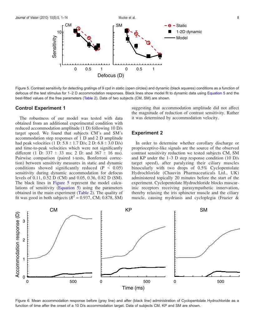
Control Experiment 1
The robustness of our model was tested with dataobtained from an additional experimental condition withreduced accommodation amplitude (1 D) following 10 D/starget speed. We found that subjects CM’s and SM’saccommodation step responses of 1 D and 2 D amplitudehad peak velocities (1 D: 5.8 T 1.7 D/s; 2 D: 6.8 T 3.0 D/s)and time-to-peak velocities which were not significantlydifferent (1 D: 337 T 33 ms; 2 D: and 367 T 16 ms).Pairwise comparison (paired t-tests, Bonferroni correc-tion) between sensitivity measures in static and dynamicconditions showed significantly reduced (P G 0.05)sensitivity during dynamic accommodation for defocuslevels of 0.11, 0.52 D (CM) and 0.05, 0.36, 0.82 D (SM).The black lines in Figure 5 represent the model calcu-lations of sensitivity (Equation 5) using the parametersobtained in the main experiment (Table 2). The quality offit was good in both subjects (R2 = 0.937, CM; 0.878, SM)
suggesting that accommodation amplitude did not affectthe magnitude of reduction of contrast sensitivity. Ratherit was determined by accommodation velocity.
Experiment 2
In order to determine whether corollary discharge orproprioceptive-like signals are the source of the observedcontrast sensitivity reduction we tested subjects CM, SMand KP under the 1–3 D step response condition (10 D/starget speed), after paralyzing their ciliary musclesbinocularly with two drops of 0.5% CyclopentolateHydrochloride (Chauvin Pharmaceuticals Ltd., UK)administered topically 20 minutes before the start of theexperiment. Cyclopentolate Hydrochloride blocks muscar-inic receptors receiving parasympathetic innervation,thereby relaxing the iris sphincter muscle and the ciliarymuscle, causing mydriasis and cycloplegia (Frazier &
Figure 5. Contrast sensitivity for detecting gratings of 9 cpd in static (open circles) and dynamic (black squares) conditions as a function ofdefocus of the test stimulus for 1–2 D accommodation responses. Black lines show model fit to dynamic data using Equation 5 and thebest-fitted values of the free parameters (Table 2). Data of two subjects (CM, SM) are shown.
Figure 6. Mean accommodation response before (gray line) and after (black line) administration of Cyclopentolate Hydrochloride as afunction of time after the onset of a 10 D/s accommodation target. Data of subjects CM, KP and SM are shown.
Journal of Vision (2010) 10(5):5, 1–14 Mucke et al. 8
Jaanus, 2008). The residual accommodation responseamplitude of around 0.5 D (Figure 6) lagged considerablybehind the accommodative demand of 3 D. We assumedthat by paralyzing the ciliary muscle proprioceptive-likefeedback from within the ciliary muscle would be greatlyreduced and may not suffice to cause a reduction ofcontrast sensitivity.There were no significant (paired t-test, Bonferroni
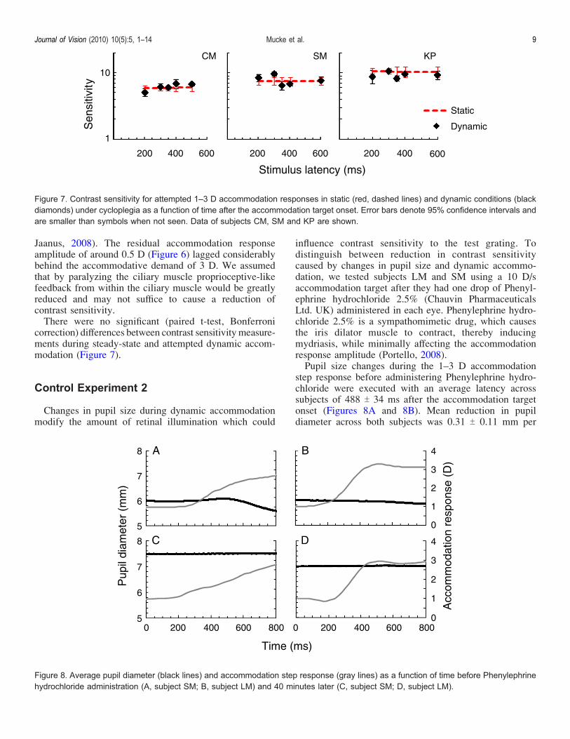
correction) differences between contrast sensitivity measure-ments during steady-state and attempted dynamic accom-modation (Figure 7).
Control Experiment 2
Changes in pupil size during dynamic accommodationmodify the amount of retinal illumination which could
influence contrast sensitivity to the test grating. Todistinguish between reduction in contrast sensitivitycaused by changes in pupil size and dynamic accommo-dation, we tested subjects LM and SM using a 10 D/saccommodation target after they had one drop of Phenyl-ephrine hydrochloride 2.5% (Chauvin PharmaceuticalsLtd. UK) administered in each eye. Phenylephrine hydro-chloride 2.5% is a sympathomimetic drug, which causesthe iris dilator muscle to contract, thereby inducingmydriasis, while minimally affecting the accommodationresponse amplitude (Portello, 2008).Pupil size changes during the 1–3 D accommodation
step response before administering Phenylephrine hydro-chloride were executed with an average latency acrosssubjects of 488 T 34 ms after the accommodation targetonset (Figures 8A and 8B). Mean reduction in pupildiameter across both subjects was 0.31 T 0.11 mm per
Figure 7. Contrast sensitivity for attempted 1–3 D accommodation responses in static (red, dashed lines) and dynamic conditions (blackdiamonds) under cycloplegia as a function of time after the accommodation target onset. Error bars denote 95% confidence intervals andare smaller than symbols when not seen. Data of subjects CM, SM and KP are shown.
Figure 8. Average pupil diameter (black lines) and accommodation step response (gray lines) as a function of time before Phenylephrinehydrochloride administration (A, subject SM; B, subject LM) and 40 minutes later (C, subject SM; D, subject LM).
Journal of Vision (2010) 10(5):5, 1–14 Mucke et al. 9
dioptre accommodation response. Following administra-tion, pupil diameters remained larger than 7 mm in bothsubjects. No changes in pupil diameter were observedduring dynamic accommodation responses after admin-istration of Phenylephrine hydrochloride (Figures 8Cand 8D). Phenylephrine hydrochloride did not significantly(paired t-test) affect accommodation velocity in the twosubjects.In order to ensure constant pupil-sizes a 5 mm artificial
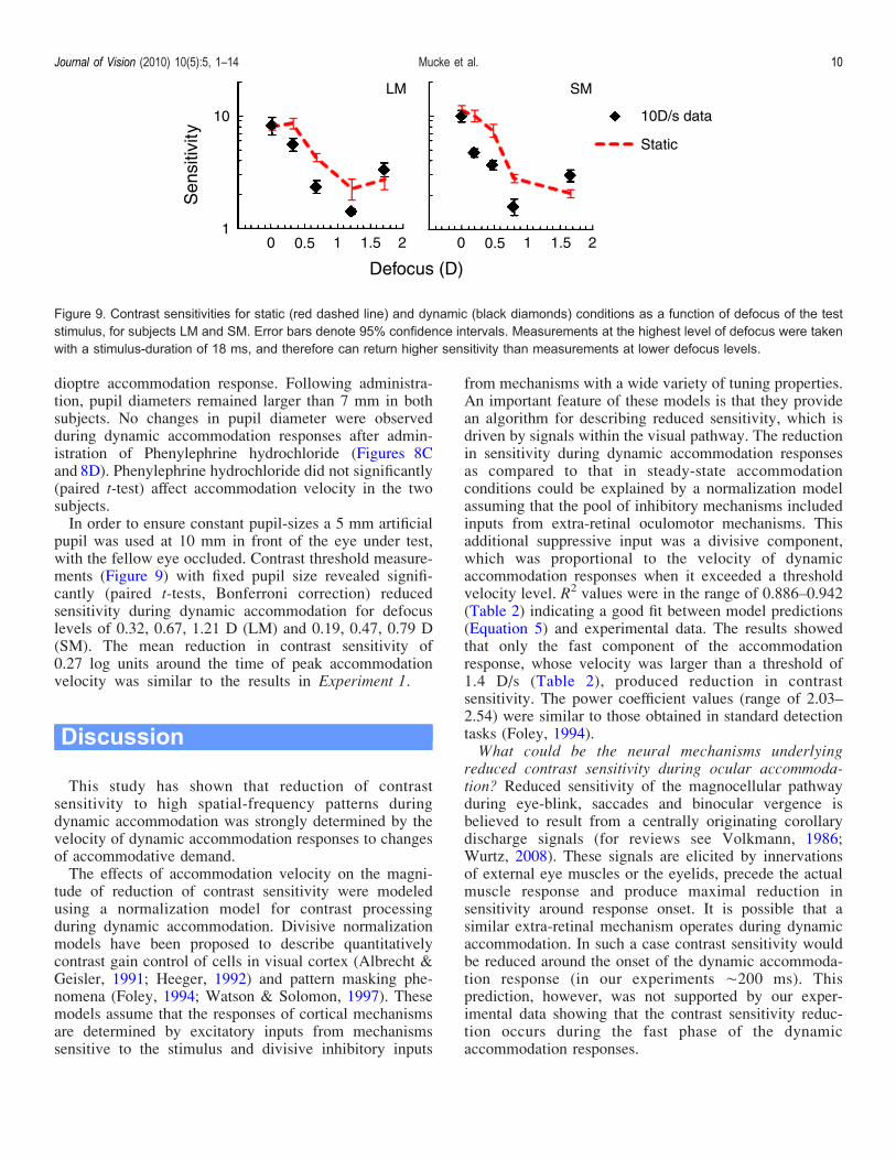
pupil was used at 10 mm in front of the eye under test,with the fellow eye occluded. Contrast threshold measure-ments (Figure 9) with fixed pupil size revealed signifi-cantly (paired t-tests, Bonferroni correction) reducedsensitivity during dynamic accommodation for defocuslevels of 0.32, 0.67, 1.21 D (LM) and 0.19, 0.47, 0.79 D(SM). The mean reduction in contrast sensitivity of0.27 log units around the time of peak accommodationvelocity was similar to the results in Experiment 1.
Discussion
This study has shown that reduction of contrastsensitivity to high spatial-frequency patterns duringdynamic accommodation was strongly determined by thevelocity of dynamic accommodation responses to changesof accommodative demand.The effects of accommodation velocity on the magni-
tude of reduction of contrast sensitivity were modeledusing a normalization model for contrast processingduring dynamic accommodation. Divisive normalizationmodels have been proposed to describe quantitativelycontrast gain control of cells in visual cortex (Albrecht &Geisler, 1991; Heeger, 1992) and pattern masking phe-nomena (Foley, 1994; Watson & Solomon, 1997). Thesemodels assume that the responses of cortical mechanismsare determined by excitatory inputs from mechanismssensitive to the stimulus and divisive inhibitory inputs
from mechanisms with a wide variety of tuning properties.An important feature of these models is that they providean algorithm for describing reduced sensitivity, which isdriven by signals within the visual pathway. The reductionin sensitivity during dynamic accommodation responsesas compared to that in steady-state accommodationconditions could be explained by a normalization modelassuming that the pool of inhibitory mechanisms includedinputs from extra-retinal oculomotor mechanisms. Thisadditional suppressive input was a divisive component,which was proportional to the velocity of dynamicaccommodation responses when it exceeded a thresholdvelocity level. R2 values were in the range of 0.886–0.942(Table 2) indicating a good fit between model predictions(Equation 5) and experimental data. The results showedthat only the fast component of the accommodationresponse, whose velocity was larger than a threshold of1.4 D/s (Table 2), produced reduction in contrastsensitivity. The power coefficient values (range of 2.03–2.54) were similar to those obtained in standard detectiontasks (Foley, 1994).What could be the neural mechanisms underlying
reduced contrast sensitivity during ocular accommoda-tion? Reduced sensitivity of the magnocellular pathwayduring eye-blink, saccades and binocular vergence isbelieved to result from a centrally originating corollarydischarge signals (for reviews see Volkmann, 1986;Wurtz, 2008). These signals are elicited by innervationsof external eye muscles or the eyelids, precede the actualmuscle response and produce maximal reduction insensitivity around response onset. It is possible that asimilar extra-retinal mechanism operates during dynamicaccommodation. In such a case contrast sensitivity wouldbe reduced around the onset of the dynamic accommoda-tion response (in our experiments È200 ms). Thisprediction, however, was not supported by our exper-imental data showing that the contrast sensitivity reduc-tion occurs during the fast phase of the dynamicaccommodation responses.
Figure 9. Contrast sensitivities for static (red dashed line) and dynamic (black diamonds) conditions as a function of defocus of the teststimulus, for subjects LM and SM. Error bars denote 95% confidence intervals. Measurements at the highest level of defocus were takenwith a stimulus-duration of 18 ms, and therefore can return higher sensitivity than measurements at lower defocus levels.
Journal of Vision (2010) 10(5):5, 1–14 Mucke et al. 10
The correlation between the effects of reduction of visualsensitivity and the magnitude of the accommodationvelocity could be due to extra-retinal signals originatingwithin the ciliary muscle. Free sensory nerve endingswithin the ciliary muscles have previously been associatedwith a mechanoreceptor function (Agababow, 1893;Ishikawa, 1962; Tamm & Lutjen-Drecoll, 1996), but onlya recent study has revealed that proprioceptors are presentin ciliary muscles (Flugel-Koch et al., 2009). We proposethat these proprioceptors are able to provide extra-retinalsignals which determine the reduction in contrast sensi-tivity during dynamic accommodation. We assumed thatsignals from proprioceptors could be eliminated byparalyzing the ciliary muscles having in mind thatproprioceptive inputs from paralyzed extra-ocular musclesare greatly reduced or absent (for reviews see Donaldson,2000 and Ruskell, 1999). The results showed that contrastsensitivity during dynamic accommodation remainedunaffected. Since we encouraged subjects to executeaccommodation responses, we assumed that the attemptedaccommodation which was accompanied by convergenceeye movements of the occluded eye should have elicited acorollary discharge signal. Under these assumptions, if thereduction of contrast sensitivity was caused by thiscorollary discharge signal, reduced contrast sensitivitywould have been observed in Experiment 2. Our propo-sition about the underlying neural mechanisms of reducedcontrast sensitivity during dynamic accommodation pointsto encourage further neuro-anatomical research into feed-back loops within the accommodation plant and itscontroller, as well as additional brain imaging studiesproviding independent evidence for the observed reduc-tion of contrast sensitivity.Are there other factors that could contribute to the
observed reduction in contrast sensitivity to high spatialfrequency patterns during dynamic accommodation?Dynamic accommodation is one of three components ofthe near response (Weber, 1851). Vergence eye move-ments and changes in pupil diameter generally accompanydynamic accommodation. For a far-to-near accommoda-tion response convergence and pupil constriction can beobserved (Weber, 1851). Convergence eye movements inthe occluded eye do not influence sensitivity to highspatial frequency patterns (Mucke et al., 2008; Supple-mental Data). The changes in pupil diameter beforeinstilling Phenyelephrine Hydrochloride in ControlExperiment 2 are similar to previously reported values(0.48 mm/D, Kasthurirangan & Glasser, 2005; 0.26 mm/D,Charman & Radhakrishnan, 2009, 0.25 mm/D, Schaeffel,Wilhelm, & Zrenner, 1993). The pupil response latencies(490 ms) measured in Control Experiment 2 are at thehigher end of previously reported values (187–500 ms,Hunter, Milton, Ludtke, Wilhelm, & Wilhelm, 2000;Kasthurirangan & Glasser, 2005), which makes it unlikelythat pupil size changes will have contributed to the lossof contrast sensitivity in our experiments. Furthermore,
contrast sensitivity reduction persists even when theoptical effects of pupil size changes are abolished.The observed loss of contrast sensitivity during
dynamic accommodation could be due to temporalchanges in retinal image quality during dynamic accom-modation. The maintenance of contrast sensitivity withcycloplegia could be related to the lack of such temporalvariations of retinal image quality. In our experimentsobservers viewed a uniform field at 1 m that was notaffected by changes of retinal image blur. Blur would onlyreduce the contrast of the test stimulus. However, the teststimulus had a very short duration, within which retinalimage defocus changes little. Therefore, temporal changesof retinal image blur in these experiments might not haveplayed a significant role in determining sensitivity to thetest stimuli.We also tested whether attention could have an effect on
contrast sensitivity measurements at longer test stimuluspresentation lags as compared to short presentation lags.Three subjects (DS, MD and SM) were presented with astatic accommodation target at an individual test distance[67 cm (DS), 64 cm (MD), 68 cm (SM)], matching the350 ms dynamic condition. Under this condition, sensi-tivity was maximally reduced during accommodation stepresponses. The test stimulus was then presented at variouspresentation lags (200, 300, 350, 400 and 800 ms) andcontrast sensitivity was measured in this static condition(data are not shown). There was no significant differencein contrast sensitivity for the five measurements atdifferent presentation time lags. Hence, attention doesnot seem to affect contrast sensitivity during longer teststimulus presentation lags as compared to short presenta-tion lags.What is the role of contrast sensitivity reduction during
dynamic accommodation in visual information process-ing? When a target moves from far to near its retinalimage becomes blurred. Blur is the principal stimulusavailable from the retinal image to produce small (G1.5 D)dynamic accommodation responses (Fincham, 1951;Schor, Alexander, Cormack, & Stevenson, 1992). Forlarge target movements (92 D) additional cues toaccommodation become available, including proximity(Schor et al., 1992). Large dynamic accommodationresponses have been modeled with two distinct phases: afast phase during the initial part of the response, and aslow phase during the final response stage (Hung &Ciuffreda, 1988; Khosroyani & Hung, 2002; Schor &Bharadwaj, 2006). The fast phase is executed on the basisof proximal information gathered prior to the onset of theresponse and does not use visual feedback. The slowphase, on the other hand, relies on visual feedback fromthe target enabling the accommodative system to focusaccurately on the target and maintain steady-state accom-modation. High spatial frequency information within thetarget is believed to play a vital role in focusing on objects(Charman & Tucker, 1977; Heath, 1956; Kotulak &
Journal of Vision (2010) 10(5):5, 1–14 Mucke et al. 11
Schor, 1987). 3 D visual scenes contain various objects atdifferent viewing distances which act as distracters for theobject we fixate upon. Our ability to focus on objects ofinterest could be severely impaired because patterns fromdistracters may mask high-frequency selective spatialchannels processing the accommodation target (Mitov,Vassilev, & Manahilov, 1981, for a review see Breitmeyer& Ogmen, 2006). Hence, reduction of sensitivity to higherspatial frequencies during the fast phase of the accommo-dation responses could be the mechanism which helps us toperceive sharp targets of interest at the final accommoda-tion stage. Additionally, the clarity of the accommodationtarget at the final stage of dynamic accommodation couldbe enhanced by effects which are similar to previouslyreported effects of blur adaptation on image sharpness(Webster, Georgeson, & Websster, 2002). Retinal imagedefocus and reduced contrast sensitivity during dynamicaccommodation blur visual objects lying between the farand near accommodation targets producing blur adaptionwhich may enhance the clarity of the accommodationtarget at the final stage of the dynamic accommodationresponse. This suggestion requires experimental verifica-tion, which we are planning to carry out in future studies.
Conclusions
The present results show that the reduction of contrastsensitivity to stimuli of higher spatial frequencies duringdynamic ocular accommodation depends on accommoda-tion response velocity. Sensitivity, however, was notaltered during attempted accommodation responses inthe absence of crystalline-lens changes due to cycloplegia.These findings suggest that sensitivity reduction may be aconsequence of cortical inhibition driven by propriocep-tive-like signals originating within the ciliary muscle. Acomputational model, based on an inhibitory componentwith an extra-retinal origin, has been developed whichexplains adequately the detection of spatial patternsduring dynamic ocular accommodation. The model couldbe implemented in a holistic computational model ofhuman vision for incorporation in economical artificialnetwork systems for robots to provide efficient perfor-mance in 3 D environments.
Acknowledgments
We would like to thank James Wolffsohn for providingsoftware for recording dynamic accommodationresponses. Further, we would like to thank the anonymousreviewers for their critical comments and suggestions forControl Experiment 2.
Commercial relationships: none.Corresponding author: Sven Mucke.Email: [email protected]: Cowcaddens Road, Glascow, G4 0BA, UK.
References
Agababow, A. (1893). Die Innervation des Ciliarkorpers.Anatomischer Anzeiger, 8, 555–561.
Albrecht, D. G., & Geisler, W. S. (1991). Motionselectivity and the contrast-response function ofsimple cells in the visual cortex. Visual Neuroscience,7, 531–546. [PubMed]
Breitmeyer, B., & Ogmen, H. (2006). Visual masking:Time slices through conscious and unconscious vision(2nd ed.). Oxford: Oxford Physics Series.
Burr, D. C., Morrone, M. C., & Ross, J. (1994). Selectivesuppression of the magnocellular visual pathwayduring saccadic eye movements. Nature, 371, 511–513.[PubMed]
Charman, W. N., & Radhakrishnan, H. (2009). Accom-modation, pupil diameter and myopia. Ophthalmic &Physiological Optics, 29, 72–79. [PubMed]
Charman, W. N., & Tucker, J. (1977). Dependence ofaccommodation response on the spatial frequencyspectrum of the observed object. Vision Research, 17,129–139. [PubMed]
Day, M., Strang, N. C., Seidel, D., Gray, L. S., & Mallen,E. A. (2006). Refractive group differences in accom-modation microfluctuations with changing accommo-dation stimulus. Ophthalmic & Physiological Optics,26, 88–96. [PubMed]
Denieul, P. (1982). Effects of stimulus vergence on meanaccommodation response, microfluctuations ofaccommodation and optical quality of the humaneye. Vision Research, 22, 561–569. [PubMed]
Donaldson, I. M. (2000). The functions of the proprio-ceptors of the eye muscles. Philosophical Trans-actions of the Royal Society B: Biological Sciences,355, 1685–1754. [PubMed] [Article]
Fernandez, E. J., & Artal, P. (2005). Study on the effectsof monochromatic aberrations in the accommodationresponse by using adaptive optics. Journal of theOptical Society of America A, Optics, Image Science,and Vision, 22, 1732–1738. [PubMed]
Fincham, E. F. (1951). The accommodation reflex and itsstimulus. British Journal of Ophthalmology, 35,381–393. [PubMed]
Flugel-Koch, C. M., Neuhuber, W., Kaufman, P. L., &Lutjen-Drecoll, E. (2009). Morphological indicationfor proprioception in the human ciliary muscle.
Journal of Vision (2010) 10(5):5, 1–14 Mucke et al. 12
Investigative Ophthalmology and Visual Science, 50,5529–5536.
Foley, J. M. (1994). Human luminance pattern-visionmechanisms: Masking experiments require a newmodel. Journal of the Optical Society of America A,Optics, Image Science, and Vision, 11, 1710–1719.[PubMed]
Frazier, M., & Jaanus, S. D. (2008). Cycloplegics. In J. D.Bartlett & S. D. Jaanus (Eds.), Clinical ocular pharma-cology (5th ed., pp. 130–133). Boston: Butterworth-Heinemann.
Heath, G. G. (1956). The influence of visual acuity onaccommodative responses of the eye. AmericanJournal of Optometry and Archives of AmericanAcademy of Optometry, 33, 513–524. [PubMed]
Heeger, D. J. (1992). Normalization of cell responses incat striate cortex. Visual Neuroscience, 9, 181–197.[PubMed]
Holm, S. (1979). A simple sequentially rejective multipletest procedure. Scandinavian Journal of Statistics, 6,65–70.
Hung, G. K., & Ciuffreda, K. J. (1988). Dual-modebehaviour in the human accommodation system.Ophthalmic & Physiological Optics, 8, 327–332.[PubMed]
Hunter, J. D., Milton, J. G., Ludtke, H., Wilhelm, B., &Wilhelm, H. (2000). Spontaneous fluctuations inpupil size are not triggered by lens accommodation.Vision Research, 40, 567–573. [PubMed]
Ishikawa, T. (1962). Fine structure of the human cillarymuscle. Investigative Ophthalmology, 1, 587–608.
Kasthurirangan, S., & Glasser, A. (2005). Characteristicsof pupil responses during far-to-near and near-to-faraccommodation. Ophthalmic & Physiological Optics,25, 328–339. [PubMed]
Khosroyani, M., & Hung, G. K. (2002). A dual-modedynamic model of the human accommodation system.Bulletin of Mathematical Biology, 64, 285–299.[PubMed]
Kotulak, J. C., & Schor, C. M. (1987). The effects ofoptical vergence, contrast, and luminance on theaccommodative response to spatially bandpass filteredtargets. Vision Research, 27, 1797–1806. [PubMed]
Latour, P. L. (1962). Visual threshold during eye move-ments. Vision Research, 2, 261–262.
Levitt, H. (1971). Transformed up-down methods inpsychoacoustics. Journal of the Acoustical Societyof America, 49, 467–477. [PubMed]
Manning, K. A. (1986). Eye-movement-dependent loss invision and its time course during vergence. Journal ofNeuroscience, 6, 1976–1982. [PubMed] [Article]
Manning, K. A., & Riggs, L. A. (1984). Vergence eyemovements and visual suppression. Vision Research,24, 521–526. [PubMed]
Mitov, D., Vassilev, A., & Manahilov, V. (1981).Transient and sustained masking. Perception &Psychophysics, 30, 205–210.
Mucke, S., Manahilov, V., Strang, N. C., Seidel, D., &Gray, L. S. (2008). New type of perceptual suppres-sion during dynamic ocular accommodation. CurrentBiology, 18, R555–R556. [PubMed]
Pelli, D. G., & Zhang, L. (1991). Accurate control ofcontrast on microcomputer displays. Vision Research,31, 1337–1350. [PubMed]
Portello, J. K. (2008). Mydriatics and mydriolytics. InJ. D. Bartlett & S. D. Jaanus (Eds.), Clinicalocular pharmacology (5th ed., pp. 114–117). Boston:Butterworth-Heinemann.
Rosenbluth, D., & Allman, J. M. (2002). The effect ofgaze angle and fixation distance on the responses ofneurons in V1, V2, and V4. Neuron, 33, 143–149.[PubMed]
Ruskell, G. L. (1999). Extraocular muscle proprioceptorsand proprioception. Progress in Retinal and EyeResearch, 18, 269–291. [PubMed]
Schaeffel, F., Wilhelm, H., & Zrenner, E. (1993). Inter-individual variability in the dynamics of naturalaccommodation in humans: Relation to age andrefractive errors. The Journal of Physiology, 461,301–320. [PubMed] [Article]
Schor, C. M., Alexander, J., Cormack, L., & Stevenson, S.(1992). Negative feedback control model of proximalconvergence and accommodation. Ophthalmic &Physiological Optics, 12, 307–318. [PubMed]
Schor, C. M., & Bharadwaj, S. R. (2006). Pulse-stepmodels of control strategies for dynamic ocularaccommodation and disaccommodation. VisionResearch, 46, 242–258. [PubMed]
Sperry, R. W. (1950). Neural basis of the spontaneousoptokinetic response produced by visual inversion.Journal of Comparative and Physiological Psychology,43, 482–489. [PubMed]
Stark, L. R., & Atchison, D. A. (1994). Subject instruc-tions and methods of target presentation in accom-modation research. Investigative Ophthalmology andVisual Science, 35, 528–537. [PubMed]
Suryakumar, R., Meyers, J. P., Irving, E. L., & Bobier,W. R.(2007). Application of video-based technology for thesimultaneous measurement of accommodation andvergence. Vision Research, 47, 260–268. [PubMed]
Tamm, E. R., & Lutjen-Drecoll, E. (1996). Ciliary body.Microscopy Research and Technique, 33, 390–439.[PubMed]
Journal of Vision (2010) 10(5):5, 1–14 Mucke et al. 13
Volkmann, F. C. (1986). Human visual suppression.Vision Research, 26, 1401–1416. [PubMed]
Watson, A. B., & Solomon, J. A. (1997). Model of visualcontrast gain control and pattern masking. Journal ofthe Optical Society of America A, Optics, ImageScience, and Vision, 14, 2379–2391. [PubMed]
Watson, T. L., & Krekelberg, B. (2009). The relationshipbetween saccadic suppression and perceptual stability.Current Biology, 19, 1040–1043. [PubMed]
Weber, E. W. (1851) in Donders, F.C. (1864). Anomaliesof accommodation and refraction of the eyes(pp. 573–575). London: New Sydenham Society.
Webster, M. A., Georgeson, M. A., & Websster, S. M.(2002). Neural adjustment to image blur. NatureNeuroscience, 5, 839–840. [PubMed]
Wolffsohn, J. S., Gilmartin, B., Mallen, E. A., &Tsujimura, S. (2001). Continuous recording ofaccommodation and pupil size using the Shin-NipponSRW-5000 autorefractor. Ophthalmic & Physiolog-ical Optics, 21, 108–113. [PubMed]
Wurtz, R. H. (2008). Neuronal mechanisms of visualstability. Vision Research, 48, 2070–2089. [PubMed]
Journal of Vision (2010) 10(5):5, 1–14 Mucke et al. 14