Investigating the effects of nanomaterials on the...
35
Investigating the effects of nanomaterials on the environment – current knowledge and future research needs Teresa F Fernandes Napier University Edinburgh, UK [email protected]
Transcript of Investigating the effects of nanomaterials on the...

Investigating the effects of nanomaterials on the
environment– current knowledge and future
research needs
Teresa F Fernandes
Napier UniversityEdinburgh, UK

Ecotoxicology- toxicology integration
The main findings of the toxicology can be broken down into two general areas:
(i) Physical and chemical characteristicsSize, surface area, dimensions, solubility (biopersistence, durability), aggregation/clumping, contaminants, composition.
(ii) Toxicological mechanisms Free radical and reactive oxygen species production, oxidative stress, inflammation, toxicokinetics
(absorption, distribution, metabolism and excretion).
Since more toxicological studies have been completed, the information gained from the toxicology can be used to inform ecotoxicology.

Nanomaterials – A risk in the environment?
Risk = Hazard x Exposure(Toxicity)

Routes of release and hence exposure
The Royal Society and Royal Academy of Engineers (2004)
PristineNP
Incorporated into a formulation
Mixed with waste
products
Interacting with
biological systems
Interacting with
environmental chemicals/
species

Navarro et al 2008
More stable
Aggregation
Decrease uptake
Increase uptake
Interactions of NM in the environment

Steevens et al 2008


Still life with nanoparticlesFor the first time, researchers have captured an image of nanoparticles inside a whole, live organism. Nanoparticles have been photographed previously in cells in vitro, but this image, which was presented at the Society of Environmental Toxicology and Chemistry Europe meeting in May by Teresa Fernandes of Napier University (U.K.), captures the tiny particles inside a daphnid or water flea (Daphnia magna).
http://pubs.acs.org/subscribe/journals/esthag/40/i14/html/071506news3.html
Teresa Fernandes, copyright Napier University (U.K.)
Environmental Science and Technology, Vol 40, Issue 14 (2006)

Effects of nanoparticles on different species
Microorganisms
Plants
Invertebrates
Vertebrates
Terrestrial Aquatic
Which species should we study – which are most likely to be exposed?

Effects on microbes
• A range of studies taken from the literature• Over 30 papers or so published to date• Materials studied include: fullerenes, CNT, metals,
metal oxides, quantum dots• Bactericide, viricide, reactive oxygen species
production, oxidative damage, cell membrane damage, inhibits grow (via interference with energy metabolism), cytotoxic
• Range of target species, nanomaterials and endpoints still narrow
Klaine et al (2008) Env. Toxicology and Chemistry

DNA
ROS
Disruption of
membrane/
membrane potential
Damage DNA
Produce reactive
oxygen species (ROS)
Oxidize/damage
proteins
Interrupt electron
transport/respiration
Protein
Protein
e -
e -
CYP
Release hazardous
constituents, e.g., metals, ions+
Cd 2+
Cu 2+-
DNA
ROS
Disruption of
membrane/
membrane potential
Damage DNA
Produce reactive
oxygen species (ROS)
Oxidize/damage
proteins
Interrupt electron
transport/respiration
Protein
Protein
e -
e -
CYP
Release hazardous
constituents, e.g., metals, ions+
Cd 2+
Ag +-
Possible mechanisms of nanomaterial toxicity to bacteria. Different nanomaterials may cause toxicity via one or more of these mechanisms.
Klaine et al (2008) Env. Toxicology and Chemistry

Oberdorster E. 2004 Environ. Health Persp. 112; 1058-1062.
Particles C60 suspended in tetrahydrofuran (THF; heterocyclic organic compound, (CH2)4O).Final suspension contained 30-100nm aggregates.
Species Juvenile largemouth bass
Protocol 48h 0.5 and 1.0 ppm
Vertebrates (1)
Results Increased lipid peroxidation in brain could be due to:•Partitioning of C60 into lipid-rich environments.•Poor antioxidant defence of neural tissue
THF used – Therefore difficult to interpret

A note on particle preparationBrant et al. 2005 Environ.Sci.Technol. 2005 6343-6351
Aim: To investigate the behaviour of colloidal C60 preparedA. Using the organic solvent THFB. Stirred in water for several weeks
(proposed to be more indicative of natural environment)
Results:Both procedures generate n-C60 with negative charge, but more charged in THF.THF remains within n-C60 cluster.
Discussion:n-C60 acquires charge from organic solvents and by
hydrolysis.Possible to disperse n-C60 without a solvent.Experiments using THF need to be re-interpreted.


Does THF affect toxicity?
Henry et al (2007) Attributing Effects of Aqueous C60 Nano-Aggregates to Tetrahydrofuran Decomposition Products in Larval Zebrafish by Assessment of Gene Expression. Environmental Health Perspectives, 115(7)
Zebrafish exposed C60with/without THF
Higher mortality on C60-THF treatment. Gene expression studies indicated most differences found in THF-C60and most of these were similarly expressed in fish exposed to THF-water.Toxic effects were linked to a THF-degradation product, (γ-butyrolactone) rather then to C60 (GS-MS, tox studies)

Vertebrates (2)Smith et al 2007 Aquatic Toxicology 82: 94-109
Particles Single Walled Carbon Nanotubes (SWCNT)1.1nm diameter x 5-30 μm length[SDS (sodium dodecyl sulphate) and sonication]0, 0.1, 0.25, 0.5mg/L up to 10 days
Species Fish - Rainbow Trout
Results Dose dependent:• Rise in ventilation rate, gill pathologies and mucus • Lipid peroxidation in gill, brain and liver• Increased gill and liver glutathione (due to low oxygen-
induced stress in gills?)• Brain pathology• Aggressive behaviour

Smith et al (2007)
The surface of a rainbow trout gill showing how single-wall carbon nanotubes (in black) collect and stick to the mucus coat on the gill surface. Secreted fish mucus rapidly aggregated previouslydispersed SWCNT on the surface of the gills (fish from 0.5 mg l−1 SWCNT treatment);
Phase contrast photograph of a mucus smear (magnification ×40) showing aggregates of nanoparticles associated with the mucoproteins.
Effect of Single-Walled Carbon Nanotubes (SWCNT) to rainbow trout

Vertebrates (3) – polystyrene beads
Kashiwada 2006Environ. Health Persp. 114: 1697
Particles Polystyrene (latex beads) 39.4 – 42000nm
Species Fish – Japanese Medaka
Results Egg – all absorbed into chorion, 474nm highest bioavailability39.4nm shifted into yolk and gall bladder during embryonic development
Adult - 39.4nm accumulated in gills and intestineAlso detected in brain, testis, liver and blood

Marine macroalgaeNielsen et al Nanotoxicology (2008 )
Particles: CB 14nm diameter (Degussa Printex 90)0.1, 1, 10 and 100 μg/mlDynamic Light Scattering charaterisation
Organism: Macroalgae Fucus serratus
Gametes Fertilization Body axis orientation
Germination
Time 0 1h 16h 24h 5d After Fertilization
Rhizoid elongation

Macroalgae treated with carbon nanopartices
10 µm
Spermatozoid adsorbedto CB aggregate
Free swimmingspermatozoid
Physical restriction?
Nielsen et al 2008

Macroalgae treated with carbon nanopartices
Cell membraneCell wall
CB nanoparticles
Inside
Outside1 µm
Do NPs penetrate the cell wall?
Transmission electron microscopy

Result: Carbon nanoparticles may influence Fucus embryos development, for example by affecting:
• Sperm frequency• Orientation of the body axis • Germination and rhizoid elongation (?)
Macroalgae treated with carbon nanoparticesNielsen et al 2008, Nanotoxicology

Control 0.1mg/L
Daphnia magna
25nm TiO2 for 48h
Aquatic Invertebrates
Control 0.1mg/L
14nm CB for 48h
Fernandes et al (2007)
P. Rosenkranz

Comparing the effects of 14 nm and 260 nm CB
0100200300400500600700
Day 1
Day 3
Day 5
Day 7
Day 9
Day 11
Day 13
Day 15
Day 17
Day 19
Day 21
Exposure Time
Cum
ulat
ive
Offs
prin
g %
Control 0.01 mg/l 0.1 mg/l 1 mg/l 5 mg/l 10 mg/l Control 0.1 mg/l 0.5 mg/l 1 mg/l 2 mg/l
050
100150200250300350
Day 1 Day 2 Day 3 Day 4
Exposure time
Cum
ulat
ive
mou
lting
%
0
20
40
60
80
100
Mor
talit
y %
0
50
100
150
200
250
300
350
Day 1 Day 2 Day 3 Day 4
Exposure time
Cum
ulat
ive
mou
lting
%
0
20
40
60
80
100
Mor
talit
y %
0100200300400500600700
Day 1
Day 3
Day 5
Day 7
Day 9
Day 11
Day 13
Day 15
Day 17
Day 19
Day 21
Exposure Time
Cum
ulat
ive
Offs
prin
g %
Fig.2: Mortality (line) and cumulative moult (column) after treatment with 14nm (top) and 260nm carbon black (bottom) in an acute, 96h exposure
Fig.3: Cumulative offspring after treatment with 14nm (top) and 260nm carbon black in a chronic, 21 day exposure
14 nm 14 nm
260 nm 260 nm
Increased mortality
Decreased moulting
Decreased reproductio
n output
P. Rosenkranz

Connecting ecotoxicology and toxicology of water-borne NP:
• Exposure of primary producers
• Exposure of invertebrates
• Exposure of fish
• Uptake into higher animals and humans
• Transport through gastro-intestinal barriers
• Effects of NP in hepatocytes
Assessing effects of silver NP
B. GeiserJoint Environment and Human Health Programme (UK) Collaboration: Univ. Exeter, Birmingham, Bristol

Study Approach
System1. Primary producers
(Pseudokirchneriellasubcapitata)
2. Hepatocytes:Human (C3A), trout (Oncorhynchus mykiss) (primary)
3. Invertebrate - D. magna4. Fish – carp (Cyprinus
carpio)
Characterisation of particles in respective media or water (ongoing):concentration, aggregation, solubility
Assess transport through gastro-intestinal barriers
Endpoints1. Productivity, esterase
activity
2. Cytotoxicity (membrane integrity assessment)
3. Mortality, growth, moulting
4. Bioavailability

What have we learned?• Nano-Ag (35 nm) more toxic than bulk-Ag (0.6µm-
1.6 mm)• Nano-Ag can be accumulated in organs (from fish
studies)• Ingestion is likely the main route of particle uptake
in carp; transport through epithelium?• Surface area may not be the key metric when
expressing toxicity in nano-Ag

Effects on aquatic organisms• A range of studies taken from the literature• About 30 papers or so published to date – large output in 2007/08
(although some as reviews)• Rapid uptake of NM; but also excretion?• Wide range of results observed• Indication of higher toxicity associated with exposures to nano, as
opposed to micro sized materials• Role of preparation method?• Effects of metals – what is the role of dissolution ?
• Still unclear about mechanistic effects• Very few studies on marine systems• Range of target species, nanomaterials and endpoints still narrow
Klaine et al (2008) Env. Tox and Chem.

R.D. Holbrook, K.E. Murphy, J.B. Morrow and K.D. Cole. Trophic transfer of nanoparticles in a simplified invertebrate
food chain. Nature Nanotechnology, June 2008
Photomicrograph of ciliate Tetrahymena pyriformis (l.) during cell division with accumulated quantum dots (CdSe core and ZnS shell) appearing red and close up photomicrograph of rotifer Brachionus calyciflorus that preys on it (r., whole organism seen in upper left corner) with quantum dots assimilated from ingested ciliates appearing red. (Credit: NIST)
Escherichia coli Tetrahymena pyriformis Brachionus calyciflorus
Food chain effects?

Interaction with other chemicals?A
lga
Dap
hnia
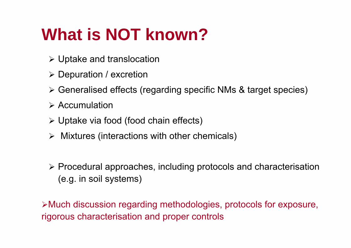
What is NOT known?Uptake and translocation
Depuration / excretion
Generalised effects (regarding specific NMs & target species)
Accumulation
Uptake via food (food chain effects)
Mixtures (interactions with other chemicals)
Procedural approaches, including protocols and characterisation (e.g. in soil systems)
Much discussion regarding methodologies, protocols for exposure,rigorous characterisation and proper controls

Strategy
• Determine environmental exposure
• Investigate routes of exposure in key species
• Investigate mechanisms of action of toxic effects
• Assess bioaccumulation potential
• Interaction with stressors
• Investigate range of particles (material, size, physical-chemical properties -REFERENCE)
• Characterise• Agree standard
methodologies for exposure
• Characterise (in exposure medium)
• Assess env. fate
Effects of OM, Salinity, pHEffects on soil/sediments

The way forward
• Prioritise nanomaterial categories/groups• Use of reference materials• Agree standard methodologies for exposure• Link cause-effect (link properties to effects
– e.g. Kow?)• Assess environmental fate – transfer across
environmental compartments• Life-cycle assessment

AcknowledgementsNapier UniversityProfessor Vicki Stone – toxicologistProfessor Nick Christofi - microbiologistDr David Brown - toxicologistDr Hanne Nielsen – Fucus serratus (DNRI)Birgit Gaiser – D. magna, fish and human cells (NERC)Philipp Rosenkranz – Daphnia magna (CSL/DEFRA)Iain Reid – Lymnaea stagnalis (Napier)Jon Mullinger – Lumbriculus variegatus (Unilever)CSLDr Qasim Chaudhry
SnIRC colleaguesCollaboratorsProfessor Charles Tyler – Exeter University (NERC)Dr Jamie Lead – Birmingham University (NERC)Dr Mark Jepson – Bristol University (NERC)NanoNet (NERC, UK)NanoImpactNet (FP7)
SCENIHR colleagues
Joint Environment and Human Health Programme (UK)

This paper was produced for a meeting organized by Health & Consumer Protection DG and represents the views of its author on thesubject. These views have not been adopted or in any way approved by the Commission and should not be relied upon as a statement of the Commission's or Health & Consumer Protection DG's views. The European Commission does not guarantee the accuracy of the dataincluded in this paper, nor does it accept responsibility for any use made thereof.