Introduction to Toxicology
63
Introduction to Toxicology Tahar AbdAlaziz Suliman MD, PhD Clinical Toxicology & Neurology (Pediatric)
-
Upload
taharsuliman -
Category
Documents
-
view
42 -
download
2
description
Introduction To Toxicology - PowerPoint PPT Presentation
Transcript of Introduction to Toxicology

Introduction to ToxicologyIntroduction to ToxicologyTahar AbdAlaziz Suliman MD, PhDClinical Toxicology & Neurology (Pediatric)
Tahar AbdAlaziz Suliman MD, PhDClinical Toxicology & Neurology (Pediatric)
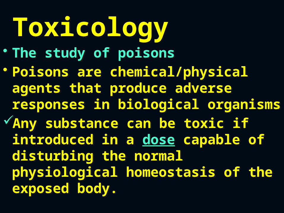
Toxicology Toxicology • The study of poisons
• Poisons are chemical/physical agents that produce adverse responses in biological organisms
Any substance can be toxic if introduced in a dose capable of disturbing the normal physiological homeostasis of the exposed body.
• The study of poisons• Poisons are chemical/physical agents that produce adverse responses in biological organisms
Any substance can be toxic if introduced in a dose capable of disturbing the normal physiological homeostasis of the exposed body.
OTHER TERMSOTHER TERMS
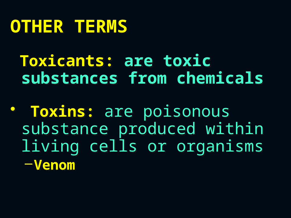
Toxicants: are toxic substances from chemicals
• Toxins: are poisonous substance produced within living cells or organisms–Venom
Toxicants: are toxic substances from chemicals
• Toxins: are poisonous substance produced within living cells or organisms–Venom
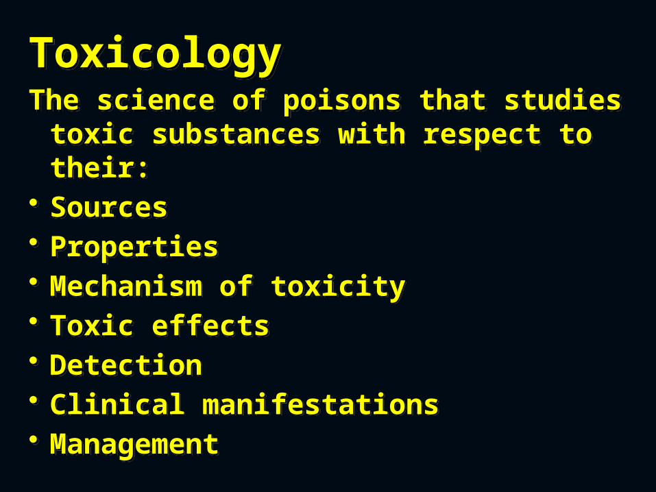
ToxicologyThe science of poisons that studies
toxic substances with respect to their:
• Sources• Properties• Mechanism of toxicity• Toxic effects• Detection• Clinical manifestations• Management
ToxicologyThe science of poisons that studies
toxic substances with respect to their:
• Sources• Properties• Mechanism of toxicity• Toxic effects• Detection• Clinical manifestations• Management
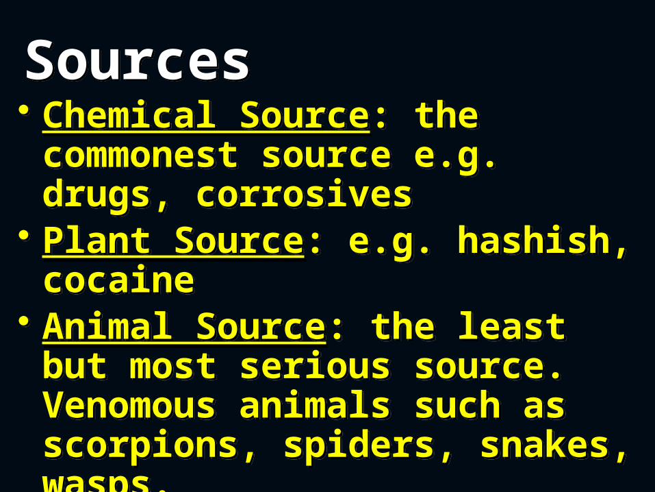
Sources Sources • Chemical Source: the commonest source e.g. drugs, corrosives
• Plant Source: e.g. hashish, cocaine
• Animal Source: the least but most serious source. Venomous animals such as scorpions, spiders, snakes, wasps.
• Chemical Source: the commonest source e.g. drugs, corrosives
• Plant Source: e.g. hashish, cocaine
• Animal Source: the least but most serious source. Venomous animals such as scorpions, spiders, snakes, wasps.
Venomous & Poisonous AnimalsVenomous & Poisonous Animals
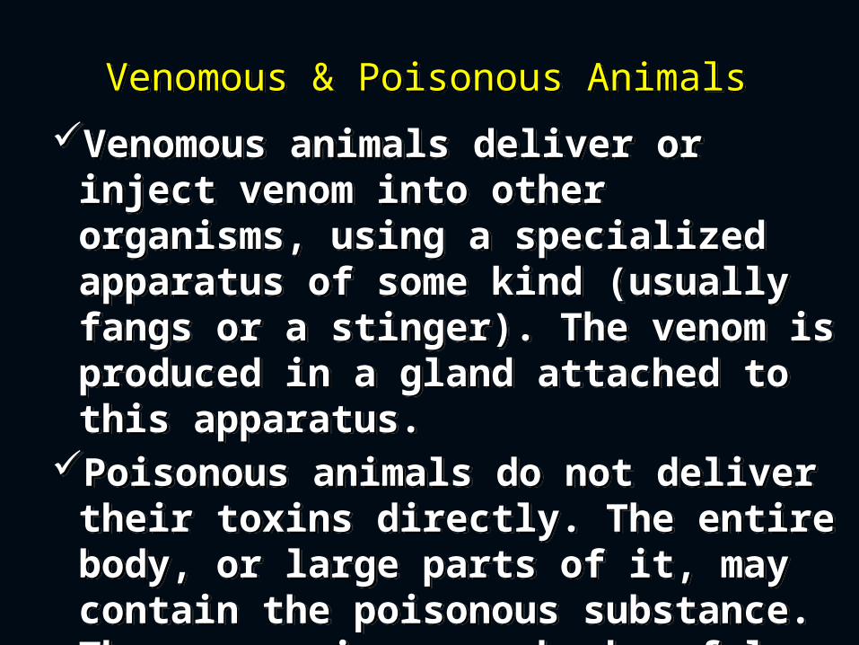
Venomous animals deliver or inject venom into other organisms, using a specialized apparatus of some kind (usually fangs or a stinger). The venom is produced in a gland attached to this apparatus.
Poisonous animals do not deliver their toxins directly. The entire body, or large parts of it, may contain the poisonous substance. These organisms may be harmful when eaten or touched.
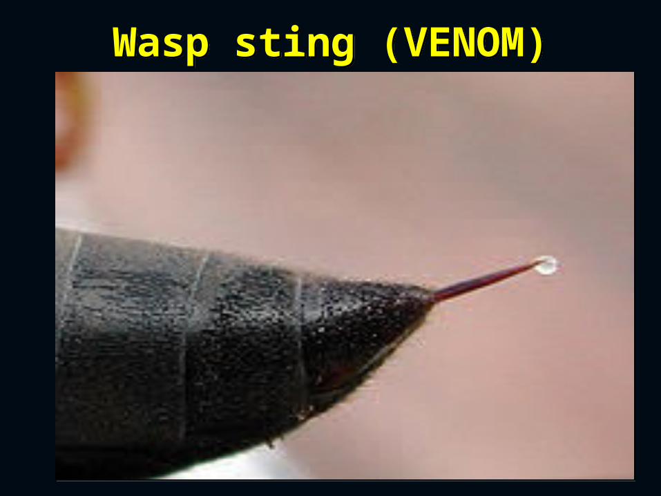
Wasp sting (VENOM)Wasp sting (VENOM)

Androctonus bicolorAndroctonus bicolor

Cerastes viperaCerastes vipera
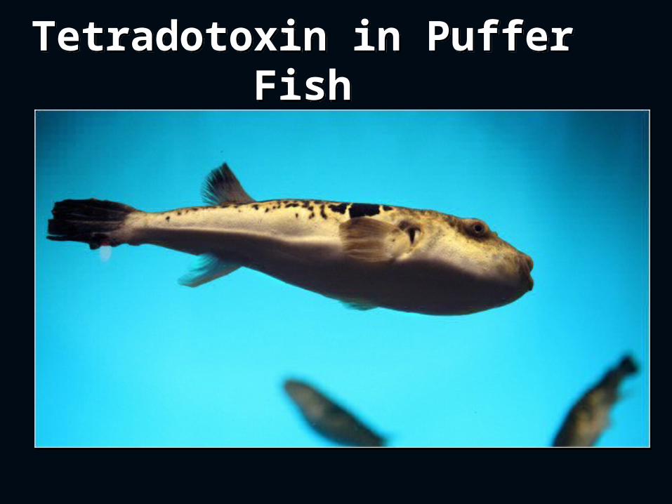
Tetradotoxin in Puffer Fish
Tetradotoxin in Puffer Fish
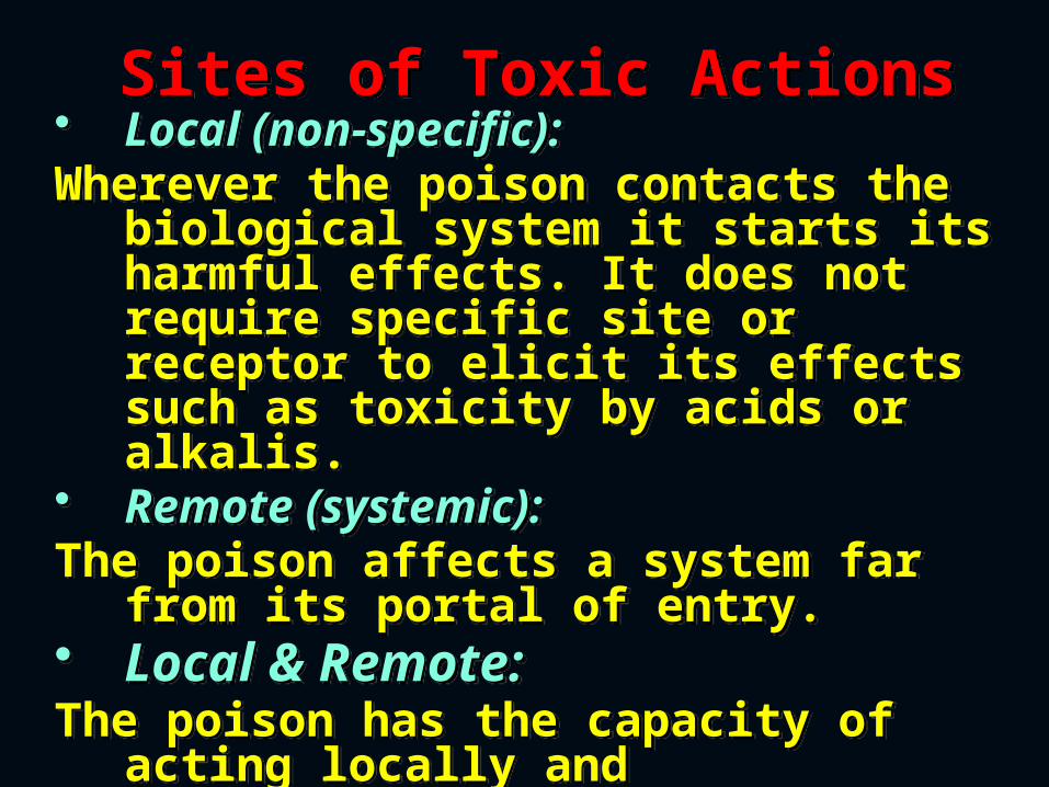
Sites of Toxic ActionsSites of Toxic Actions• Local (non-specific):Wherever the poison contacts the
biological system it starts its harmful effects. It does not require specific site or receptor to elicit its effects such as toxicity by acids or alkalis.
• Remote (systemic):The poison affects a system far
from its portal of entry.• Local & Remote:The poison has the capacity of
acting locally and systematically. Oxalic acid is an example of these
poisons.
• Local (non-specific):Wherever the poison contacts the
biological system it starts its harmful effects. It does not require specific site or receptor to elicit its effects such as toxicity by acids or alkalis.
• Remote (systemic):The poison affects a system far
from its portal of entry.• Local & Remote:The poison has the capacity of
acting locally and systematically. Oxalic acid is an example of these
poisons.
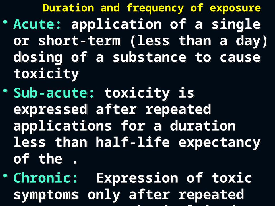
Duration and frequency of exposureDuration and frequency of exposure
• Acute: application of a single or short-term (less than a day) dosing of a substance to cause toxicity
• Sub-acute: toxicity is expressed after repeated applications for a duration less than half-life expectancy of the .
• Chronic: Expression of toxic symptoms only after repeated exposure to a chemical in doses regularly applied to the organism for a time greater than half of its life-expectancy
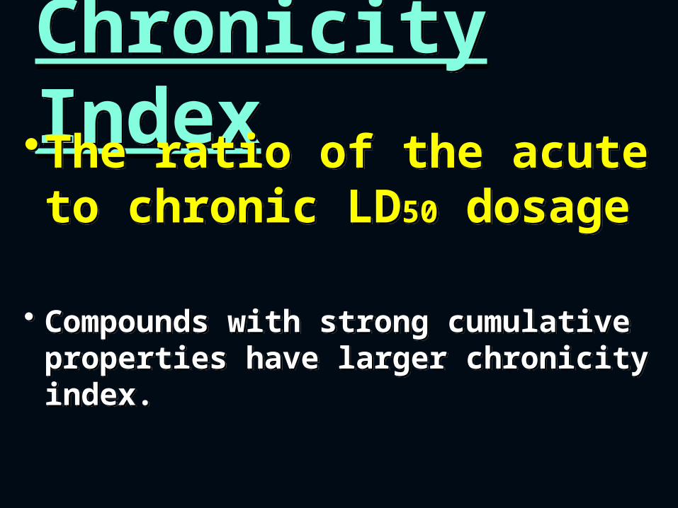
Chronicity IndexChronicity Index•The ratio of the acute to chronic LD50 dosage
• Compounds with strong cumulative properties have larger chronicity index.
•The ratio of the acute to chronic LD50 dosage
• Compounds with strong cumulative properties have larger chronicity index.
Types of Toxic Mechanisms
Types of Toxic Mechanisms
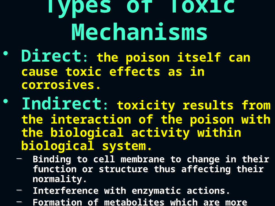
• Direct: the poison itself can cause toxic effects as in corrosives.
• Indirect: toxicity results from the interaction of the poison with the biological activity within biological system.
– Binding to cell membrane to change in their function or structure thus affecting their normality.
– Interference with enzymatic actions.– Formation of metabolites which are more toxic
than the parent poison.– Effects on DNA
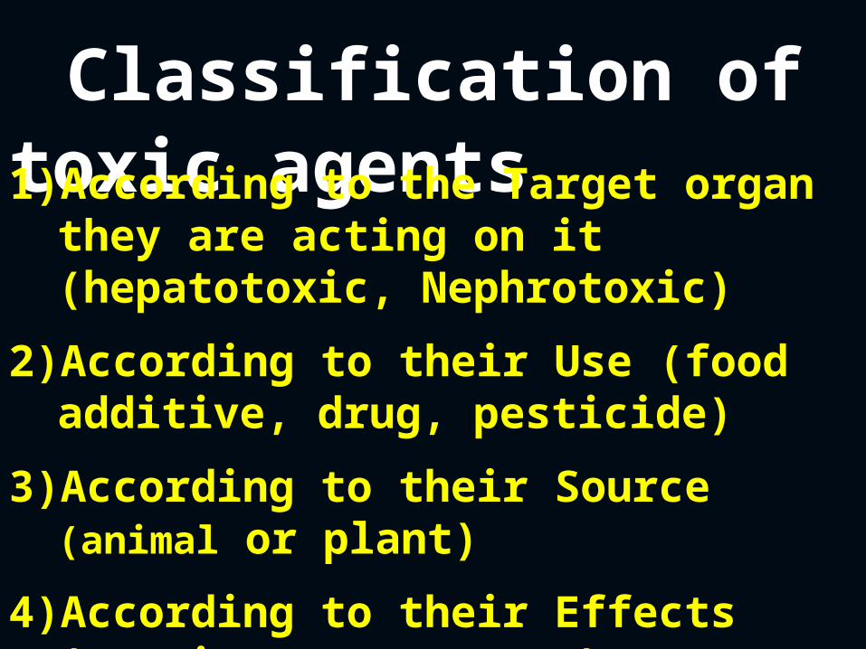
Classification of toxic agents1)According to the Target organ
they are acting on it (hepatotoxic, Nephrotoxic)
2)According to their Use (food additive, drug, pesticide)
3)According to their Source (animal or plant)
4)According to their Effects (carcinogen, mutagen)
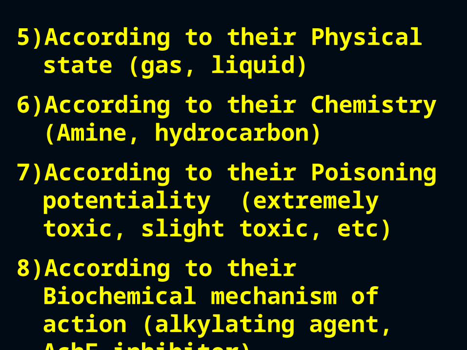
5)According to their Physical state (gas, liquid)
6)According to their Chemistry (Amine, hydrocarbon)
7)According to their Poisoning potentiality (extremely toxic, slight toxic, etc)
8)According to their Biochemical mechanism of action (alkylating agent, AchE inhibitor)
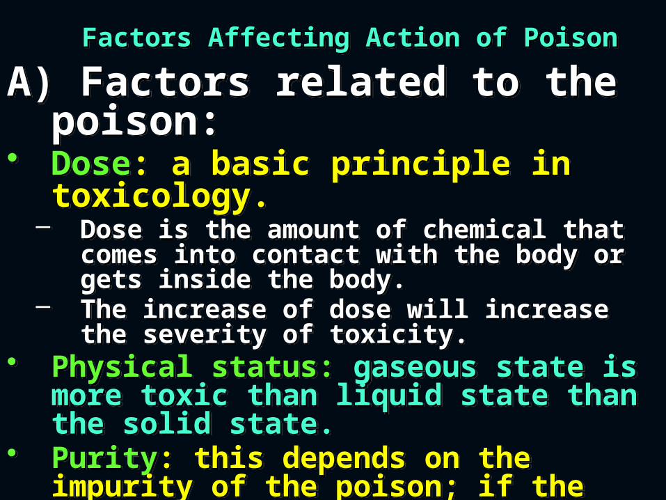
Factors Affecting Action of PoisonFactors Affecting Action of Poison
A) Factors related to the poison:
• Dose: a basic principle in toxicology.
– Dose is the amount of chemical that comes into contact with the body or gets inside the body.
– The increase of dose will increase the severity of toxicity.
• Physical status: gaseous state is more toxic than liquid state than the solid state.
• Purity: this depends on the impurity of the poison; if the impurities are more toxic than the poison, the toxicity will be more and vice versa.
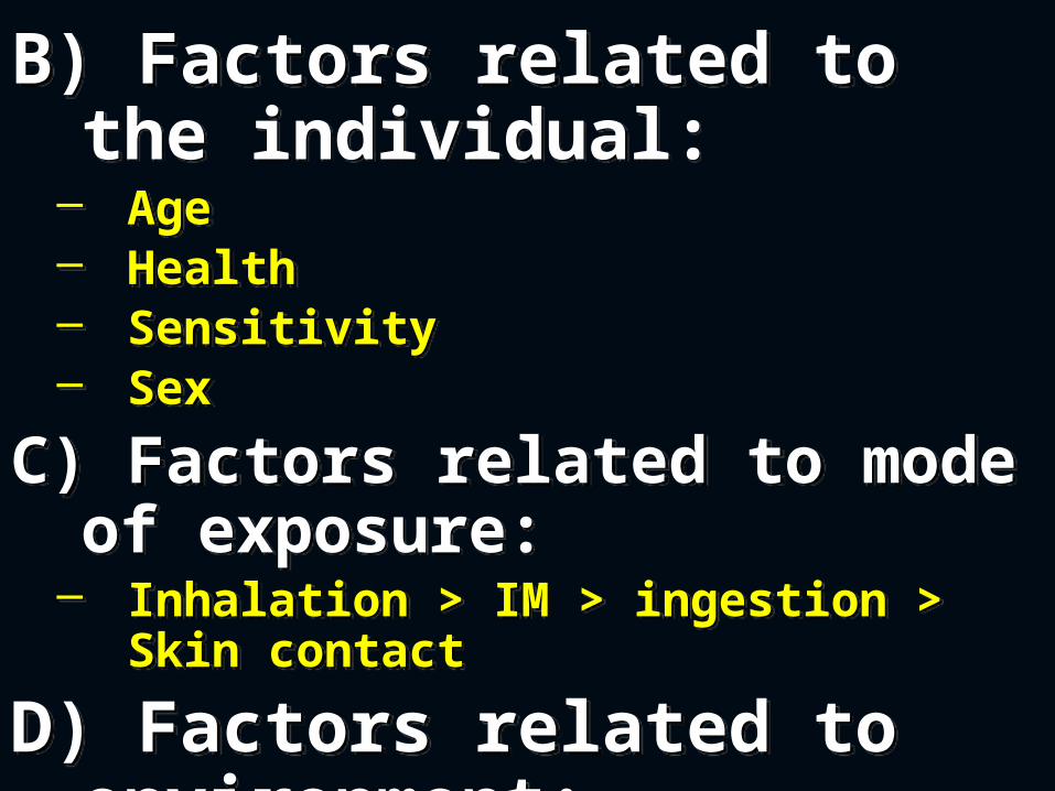
B) Factors related to the individual:
– Age– Health– Sensitivity– Sex
C) Factors related to mode of exposure:
– Inhalation > IM > ingestion > Skin contact
D) Factors related to environment:
– Temperature, pressure, humidity, radiation can cause alterations on poisons status.
B) Factors related to the individual:
– Age– Health– Sensitivity– Sex
C) Factors related to mode of exposure:
– Inhalation > IM > ingestion > Skin contact
D) Factors related to environment:
– Temperature, pressure, humidity, radiation can cause alterations on poisons status.
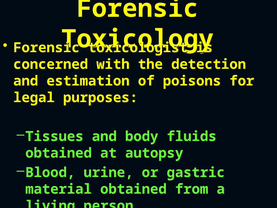
Forensic ToxicologyForensic
Toxicology• Forensic toxicologist is concerned with the detection and estimation of poisons for legal purposes:
–Tissues and body fluids obtained at autopsy
–Blood, urine, or gastric material obtained from a living person
• Forensic toxicologist is concerned with the detection and estimation of poisons for legal purposes:
–Tissues and body fluids obtained at autopsy
–Blood, urine, or gastric material obtained from a living person
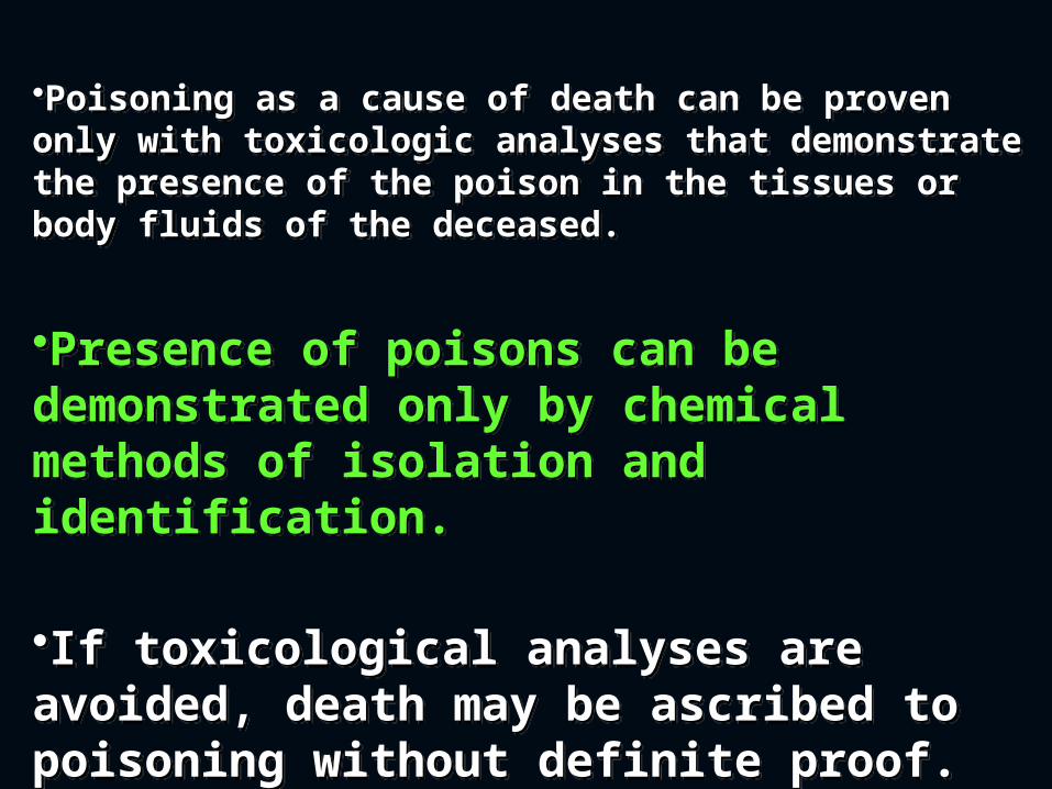
•Poisoning as a cause of death can be proven only with toxicologic analyses that demonstrate the presence of the poison in the tissues or body fluids of the deceased.
•Presence of poisons can be demonstrated only by chemical methods of isolation and identification.
•If toxicological analyses are avoided, death may be ascribed to poisoning without definite proof.
Analytical toxicology
deals with the detection, identification, and quantification of poisons.
Analytical toxicology
deals with the detection, identification, and quantification of poisons.
SAMPLES REQUIRED FOR TOXICOLOGICAL ANALYSIS FROM
AUTOPSIES
SAMPLES REQUIRED FOR TOXICOLOGICAL ANALYSIS FROM
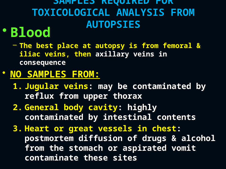
AUTOPSIES• Blood – The best place at autopsy is from femoral &
iliac veins, then axillary veins in consequence
• NO SAMPLES FROM:1. Jugular veins: may be contaminated
by reflux from upper thorax2. General body cavity: highly
contaminated by intestinal contents 3. Heart or great vessels in chest:
postmortem diffusion of drugs & alcohol from the stomach or aspirated vomit contaminate these sites
• Blood – The best place at autopsy is from femoral &
iliac veins, then axillary veins in consequence
• NO SAMPLES FROM:1. Jugular veins: may be contaminated
by reflux from upper thorax2. General body cavity: highly
contaminated by intestinal contents 3. Heart or great vessels in chest:
postmortem diffusion of drugs & alcohol from the stomach or aspirated vomit contaminate these sites
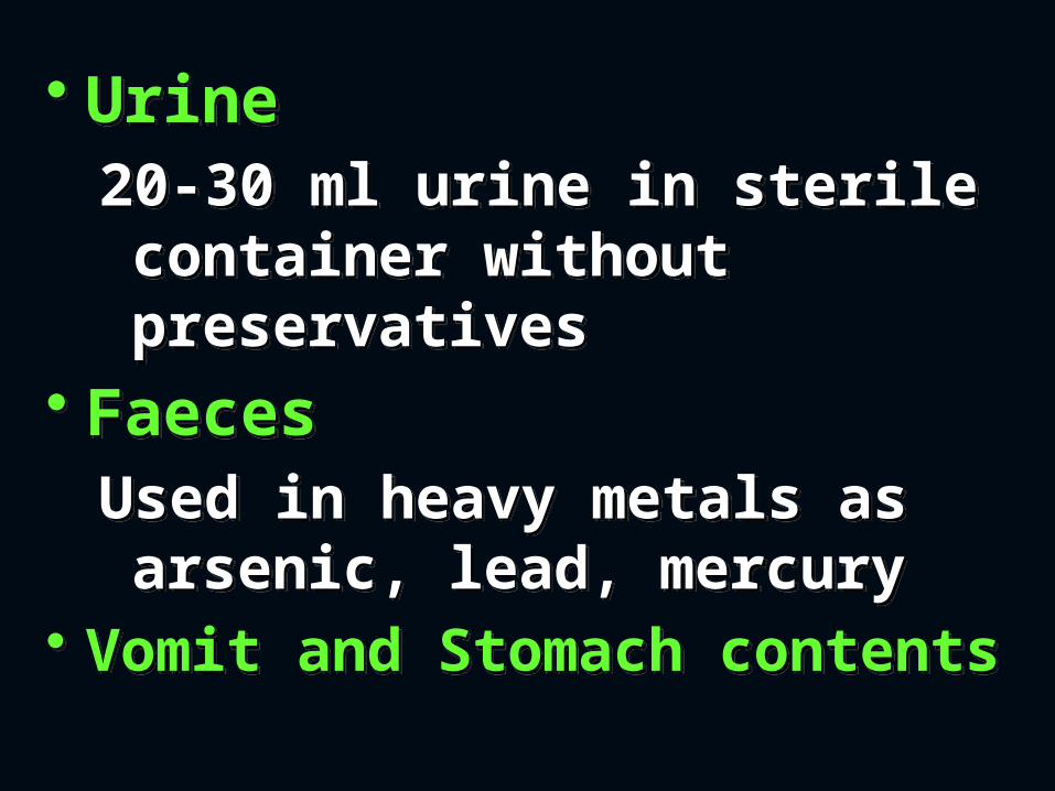
• Urine20-30 ml urine in sterile container without preservatives
• FaecesUsed in heavy metals as arsenic, lead, mercury
• Vomit and Stomach contents
• Urine20-30 ml urine in sterile container without preservatives
• FaecesUsed in heavy metals as arsenic, lead, mercury
• Vomit and Stomach contents
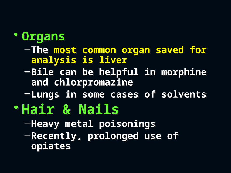
• Organs–The most common organ saved
for analysis is liver–Bile can be helpful in morphine
and chlorpromazine –Lungs in some cases of solvents
• Hair & Nails–Heavy metal poisonings–Recently, prolonged use of
opiates
• Organs–The most common organ saved
for analysis is liver–Bile can be helpful in morphine
and chlorpromazine –Lungs in some cases of solvents
• Hair & Nails–Heavy metal poisonings–Recently, prolonged use of
opiates
METHODS OF ANALYSISMETHODS OF ANALYSIS• QUALITATIVE METHODS
A) COLOR TESTThis is a rapid, easily performed,
qualitative, screening test, but not specific method
• Can be used as bed side rapid test
• QUALITATIVE METHODS
A) COLOR TESTThis is a rapid, easily performed,
qualitative, screening test, but not specific method
• Can be used as bed side rapid test
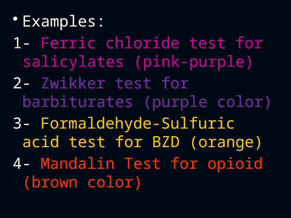
• Examples:1- Ferric chloride test for
salicylates (pink-purple)2- Zwikker test for barbiturates
(purple color)3- Formaldehyde-Sulfuric acid
test for BZD (orange)4- Mandalin Test for opioid
(brown color)
• Examples:1- Ferric chloride test for
salicylates (pink-purple)2- Zwikker test for barbiturates
(purple color)3- Formaldehyde-Sulfuric acid
test for BZD (orange)4- Mandalin Test for opioid
(brown color)
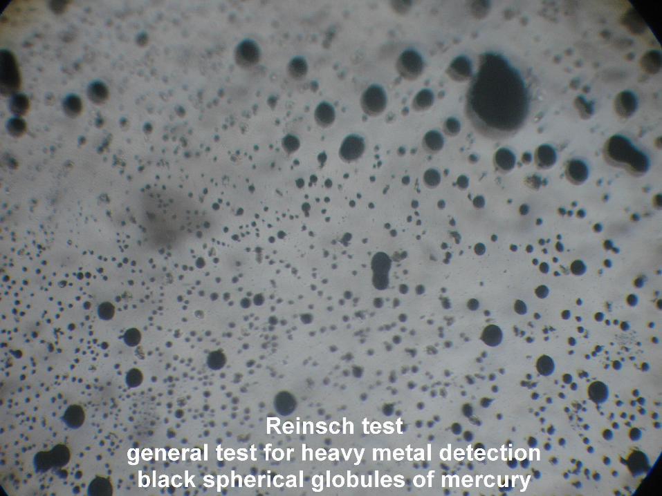
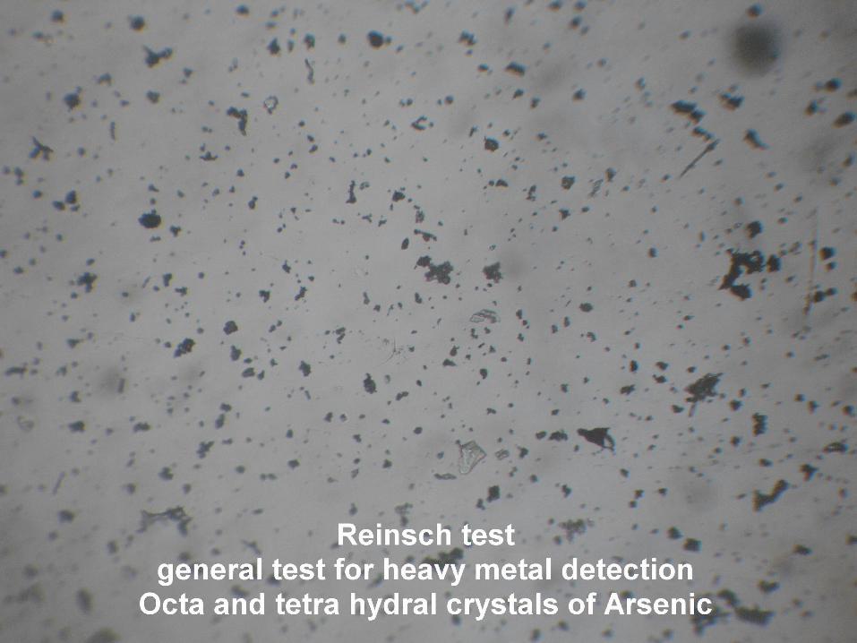
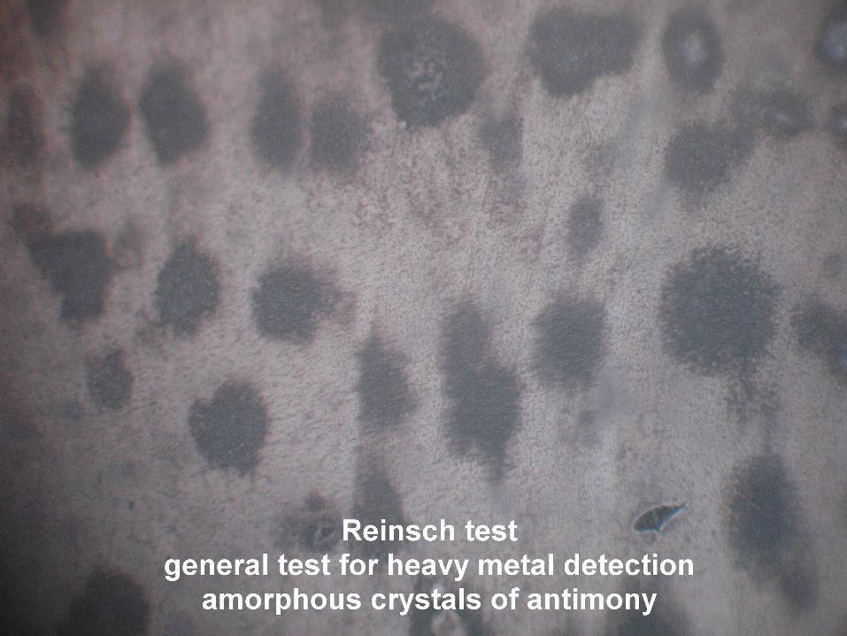
B) CHEMICAL TEST• Reinsch Test is an initial indicator
to detect the presence of one or more of the following Heavy Metals in a biological sample –Antimony–Arsenic–Bismuth–Selenium–Thallium–Mercury
B) CHEMICAL TEST• Reinsch Test is an initial indicator
to detect the presence of one or more of the following Heavy Metals in a biological sample –Antimony–Arsenic–Bismuth–Selenium–Thallium–Mercury
• QUANTITATIVE METHODSA) CHROMATOGRAPHY1- Thin-Layer Chromatography (TLC):
Mobile phase (a mixture of organic solvents such as chloroform, and methanol) is run across a Stationary phase (silica gel spread on a glass plate).
2- Gas Chromatography- Mass spectrometry:
Stationary phase is a liquid and the mobile phase (a carrier gas) is an inert gas such as helium or nitrogen.

Spotting Spotting Running Running
3- High-Performance Liquid Chromatography (HPLC):
In HPLC the stationary phase is a column packed with solid particles and the mobile phase is a liquid solvent.
B) IMMUNOASSAYS• Enzyme-multiplied immunoassay technique
(EMIT). frequently used to detect the presence of certain drugs in urine.
• Polarization immunoassay (FPIA)• Radioimmunoassay (RIA)
HPLCHPLC
ColumnColumnSystem System
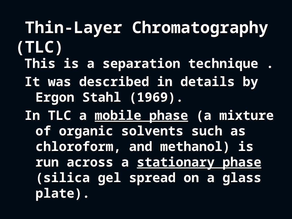
Thin-Layer Chromatography (TLC) Thin-Layer Chromatography (TLC)
This is a separation technique .It was described in details by
Ergon Stahl (1969). In TLC a mobile phase (a mixture
of organic solvents such as chloroform, and methanol) is run across a stationary phase (silica gel spread on a glass plate).
This is a separation technique .It was described in details by
Ergon Stahl (1969). In TLC a mobile phase (a mixture
of organic solvents such as chloroform, and methanol) is run across a stationary phase (silica gel spread on a glass plate).
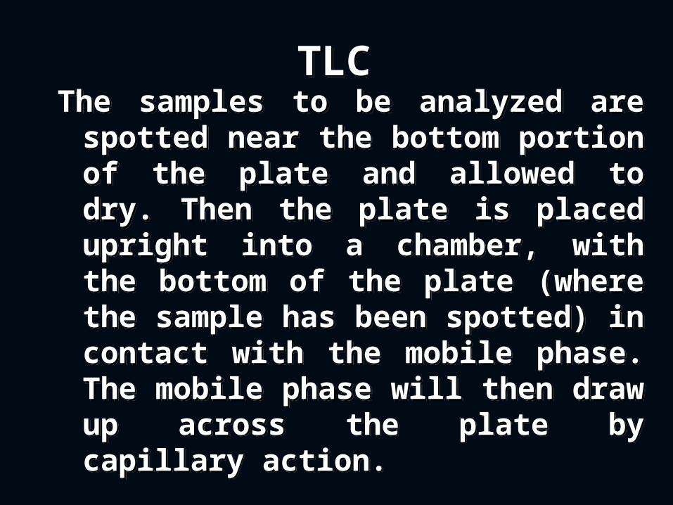
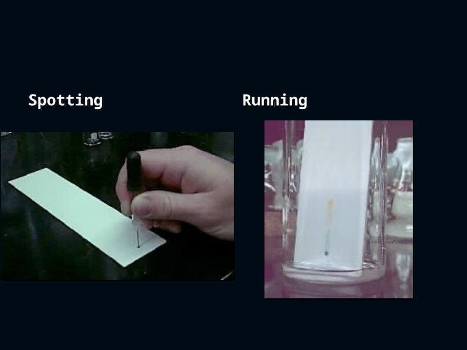
TLCTLCThe samples to be analyzed are
spotted near the bottom portion of the plate and allowed to dry. Then the plate is placed upright into a chamber, with the bottom of the plate (where the sample has been spotted) in contact with the mobile phase. The mobile phase will then draw up across the plate by capillary action.
The samples to be analyzed are spotted near the bottom portion of the plate and allowed to dry. Then the plate is placed upright into a chamber, with the bottom of the plate (where the sample has been spotted) in contact with the mobile phase. The mobile phase will then draw up across the plate by capillary action.
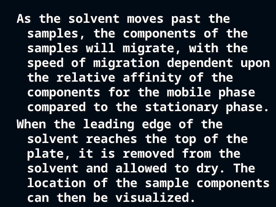
As the solvent moves past the samples, the components of the samples will migrate, with the speed of migration dependent upon the relative affinity of the components for the mobile phase compared to the stationary phase.
When the leading edge of the solvent reaches the top of the plate, it is removed from the solvent and allowed to dry. The location of the sample components can then be visualized.
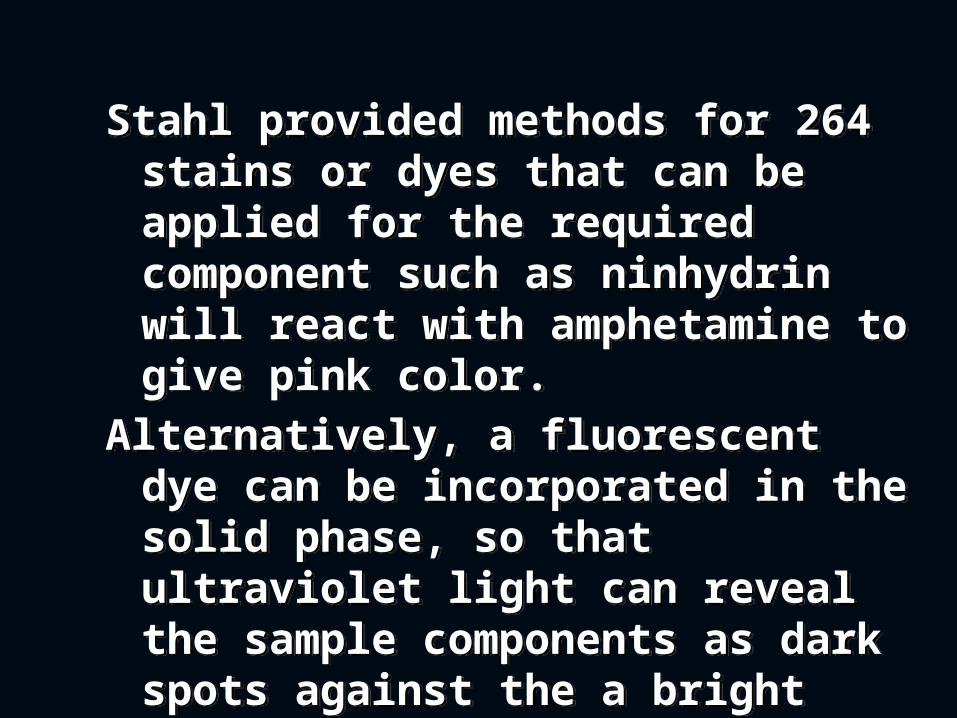
Stahl provided methods for 264 stains or dyes that can be applied for the required component such as ninhydrin will react with amphetamine to give pink color.
Alternatively, a fluorescent dye can be incorporated in the solid phase, so that ultraviolet light can reveal the sample components as dark spots against the a bright background.
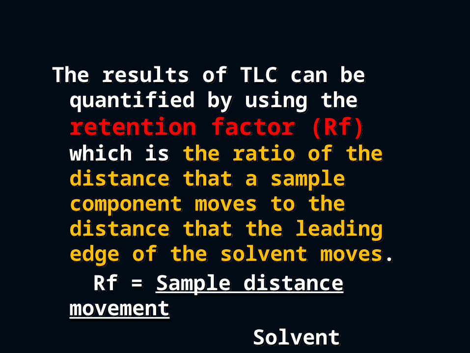
The results of TLC can be quantified by using the retention factor (Rf) which is the ratio of the distance that a sample component moves to the distance that the leading edge of the solvent moves.
Rf = Sample distance movement
Solvent distance movement
The results of TLC can be quantified by using the retention factor (Rf) which is the ratio of the distance that a sample component moves to the distance that the leading edge of the solvent moves.
Rf = Sample distance movement
Solvent distance movement
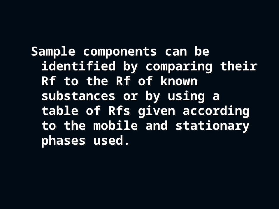
Sample components can be identified by comparing their Rf to the Rf of known substances or by using a table of Rfs given according to the mobile and stationary phases used.
Sample components can be identified by comparing their Rf to the Rf of known substances or by using a table of Rfs given according to the mobile and stationary phases used.
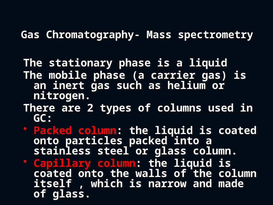
Gas Chromatography- Mass spectrometry Gas Chromatography- Mass spectrometry
The stationary phase is a liquid The mobile phase (a carrier gas) is an
inert gas such as helium or nitrogen. There are 2 types of columns used in
GC:• Packed column: the liquid is coated
onto particles packed into a stainless steel or glass column.
• Capillary column: the liquid is coated onto the walls of the column itself , which is narrow and made of glass.
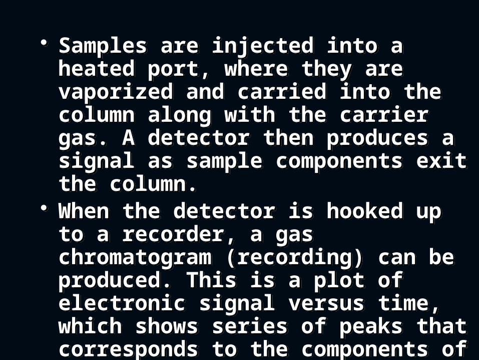
• Samples are injected into a heated port, where they are vaporized and carried into the column along with the carrier gas. A detector then produces a signal as sample components exit the column.
• When the detector is hooked up to a recorder, a gas chromatogram (recording) can be produced. This is a plot of electronic signal versus time, which shows series of peaks that corresponds to the components of the sample.
• The time it takes for a substance to pass thorough a column (retention time Rt) can be compared to standards to identify that substance. Also, since the area under each peak is proportional to the concentration of that substance, comparison with a standard of known concentration allows estimation of the concentrations.
• Flame photometric detectors increase the sensitivity over flame ionization.
• Electron capture detector uses radioactive source and it can detect picograms of DDT due to presence of 5 chlorine atoms in the molecule.
Mass Spectrometry (MS) Mass Spectrometry (MS) Although chromatography allows
identification of substances based on Rf and Rt with standards, definitive identification requires additional analysis. Mass spectrometry is one of these methods; it is used in combination with gas chromatography. As the sample components exit the GC column, they are routed into a vacuum chamber in the mass spectrometer, where they are hit with a beam of electrons.
Although chromatography allows identification of substances based on Rf and Rt with standards, definitive identification requires additional analysis. Mass spectrometry is one of these methods; it is used in combination with gas chromatography. As the sample components exit the GC column, they are routed into a vacuum chamber in the mass spectrometer, where they are hit with a beam of electrons.
This knocks electrons off the sample molecules, creating positive electrons and breaking them into fragments. These fragments are then passed through an electromagnetic field, which separates them by their mass/charge ratio. The resulting spectrum plotting the abundance and mass/ charge ration of each fragment is specific for a given substance.
This knocks electrons off the sample molecules, creating positive electrons and breaking them into fragments. These fragments are then passed through an electromagnetic field, which separates them by their mass/charge ratio. The resulting spectrum plotting the abundance and mass/ charge ration of each fragment is specific for a given substance.
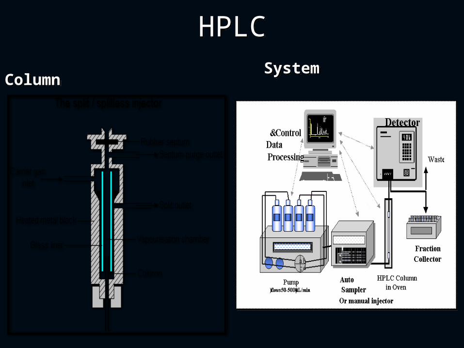
High-Performance Liquid Chromatography (HPLC) High-Performance Liquid Chromatography (HPLC)
In HPLC the stationary phase is a column packed with solid particles and the mobile phase is a liquid solvent.
As the mobile phase is pumped through the column, the sample is injected. A detector then identifies the components as they exit the column. Components are identified by their Rt, the length of time they take to pass through the column. And the results are compared with standards.
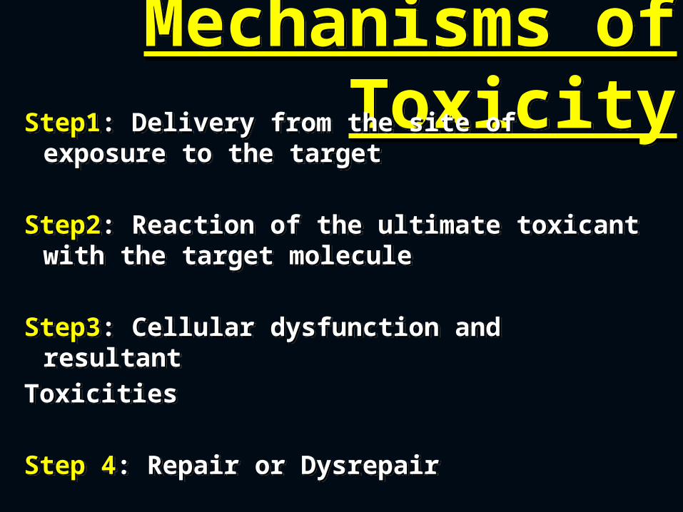
Mechanisms of Toxicity
Mechanisms of ToxicityStep1: Delivery from the site of exposure
to the target
Step2: Reaction of the ultimate toxicant with the target molecule
Step3: Cellular dysfunction and resultantToxicities
Step 4: Repair or Dysrepair
Step1: Delivery from the site of exposure to the target
Step2: Reaction of the ultimate toxicant with the target molecule
Step3: Cellular dysfunction and resultantToxicities
Step 4: Repair or Dysrepair
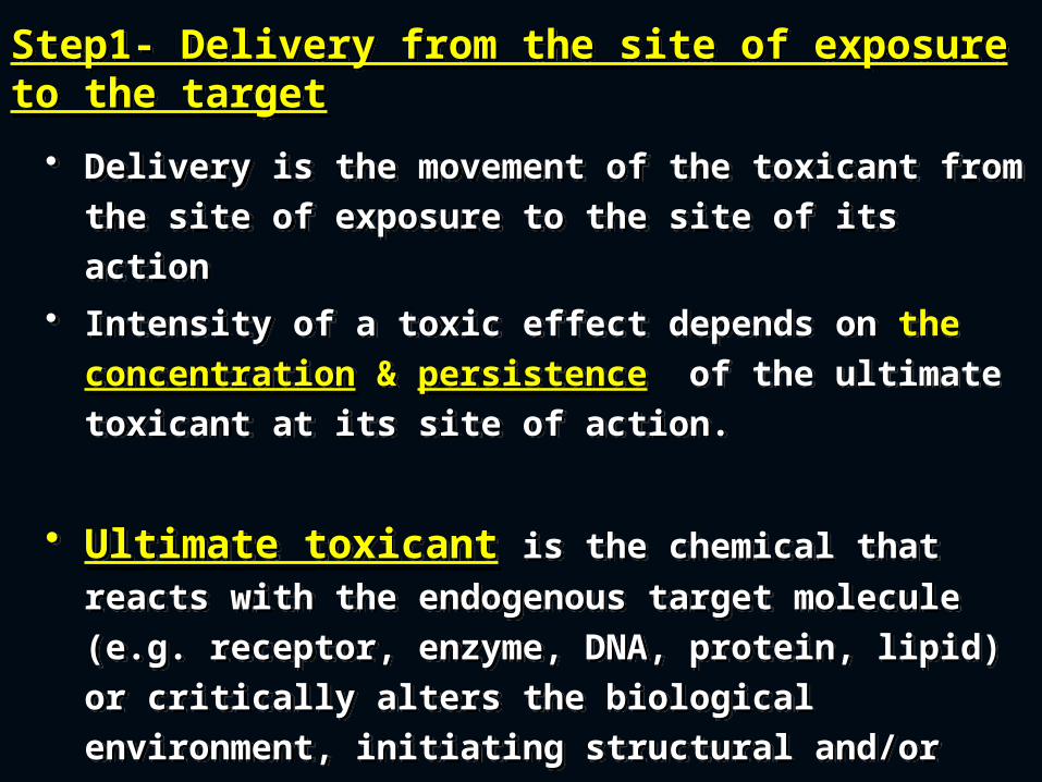
Step1- Delivery from the site of exposure to the targetStep1- Delivery from the site of exposure to the target
• Delivery is the movement of the toxicant from the site of exposure to the site of its action
• Intensity of a toxic effect depends on the concentration & persistence of the ultimate toxicant at its site of action.
• Ultimate toxicant is the chemical that reacts
with the endogenous target molecule (e.g. receptor, enzyme, DNA, protein, lipid) or critically alters the biological environment, initiating structural and/or functional alterations that result is toxicity.
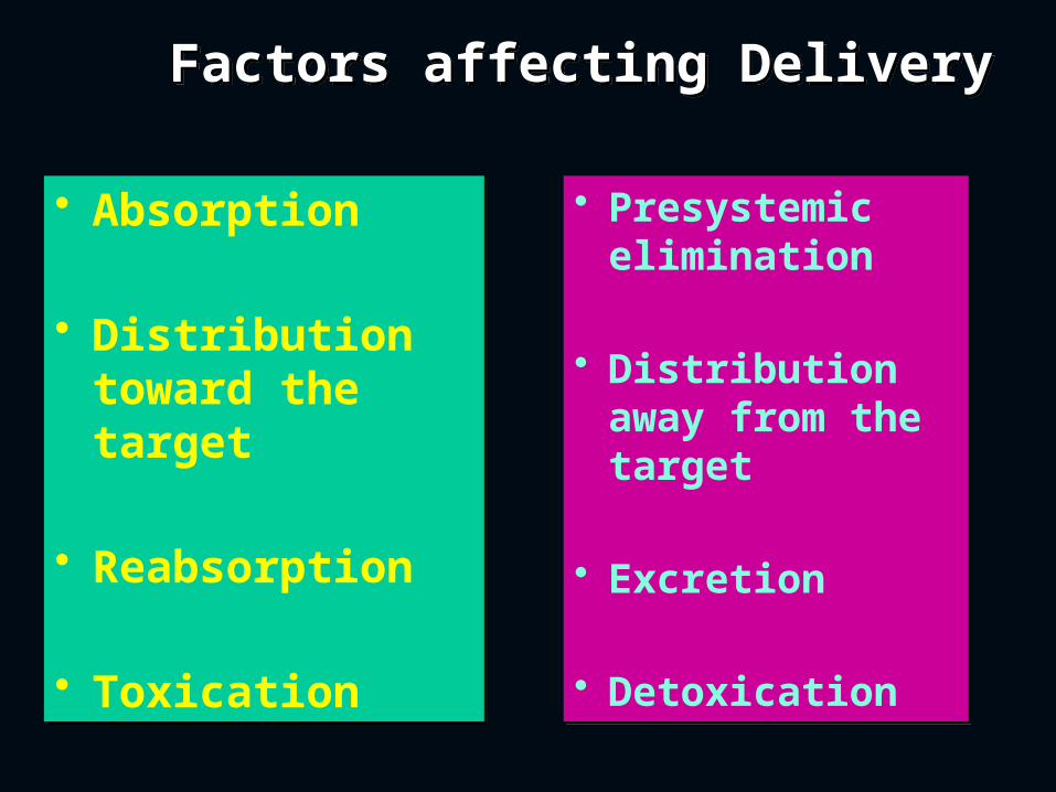
Factors affecting Delivery
Factors affecting Delivery
• Absorption
• Distribution toward the target
• Reabsorption
• Toxication
• Absorption
• Distribution toward the target
• Reabsorption
• Toxication
• Presystemic elimination
• Distribution away from the target
• Excretion
• Detoxication
• Presystemic elimination
• Distribution away from the target
• Excretion
• Detoxication
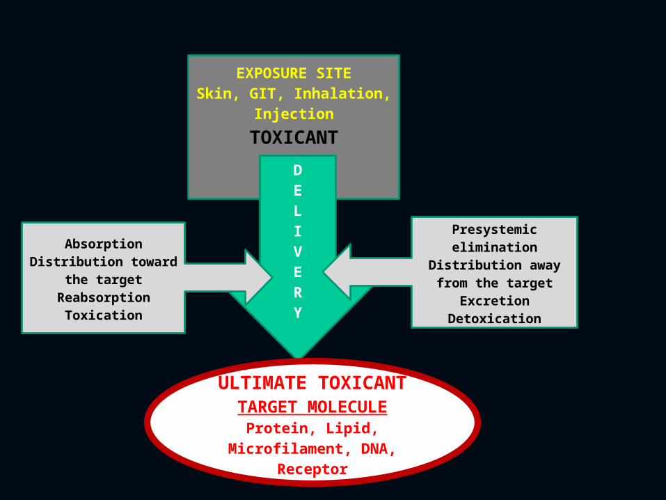
EXPOSURE SITESkin, GIT, Inhalation,
Injection
TOXICANT DELIVERY
ULTIMATE TOXICANTTARGET MOLECULE
Protein, Lipid, Microfilament, DNA,
Receptor
AbsorptionDistribution toward
the targetReabsorption
Toxication
Presystemic elimination
Distribution away from the target
ExcretionDetoxication
Absorption versus Presystemic Elimination
Absorption versus Presystemic Elimination
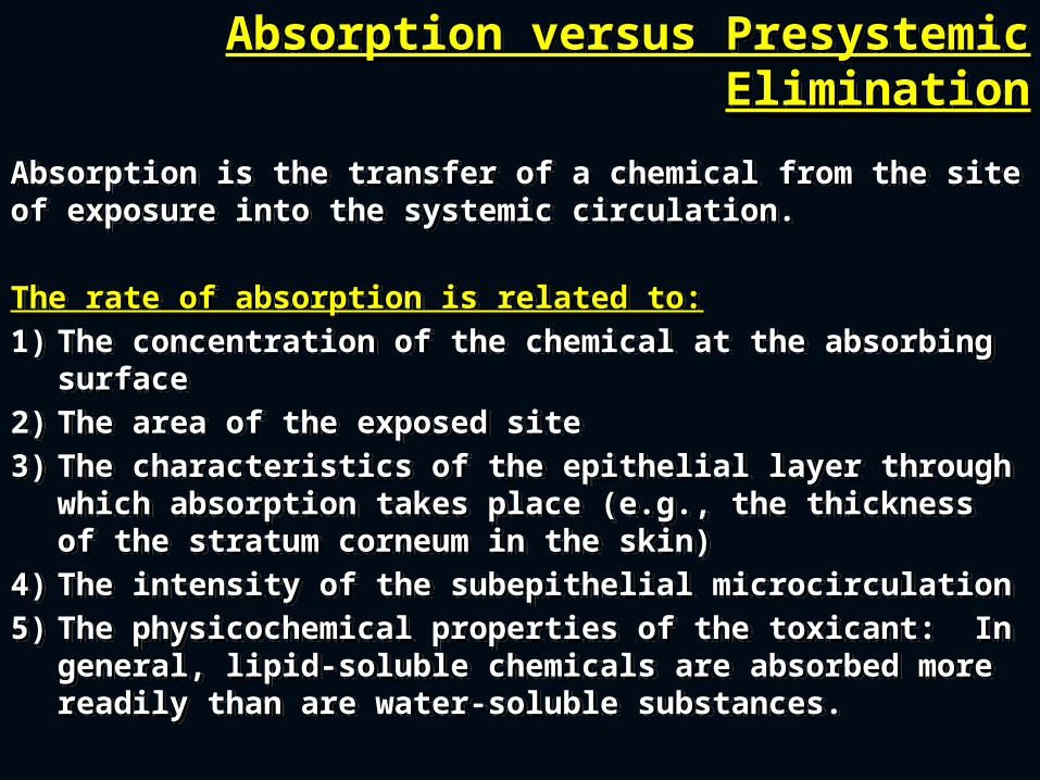
Absorption is the transfer of a chemical from the site of exposure into the systemic circulation.
The rate of absorption is related to:1) The concentration of the chemical at the absorbing
surface 2) The area of the exposed site3) The characteristics of the epithelial layer through
which absorption takes place (e.g., the thickness of the stratum corneum in the skin)
4) The intensity of the subepithelial microcirculation5) The physicochemical properties of the toxicant: In
general, lipid-soluble chemicals are absorbed more readily than are water-soluble substances.
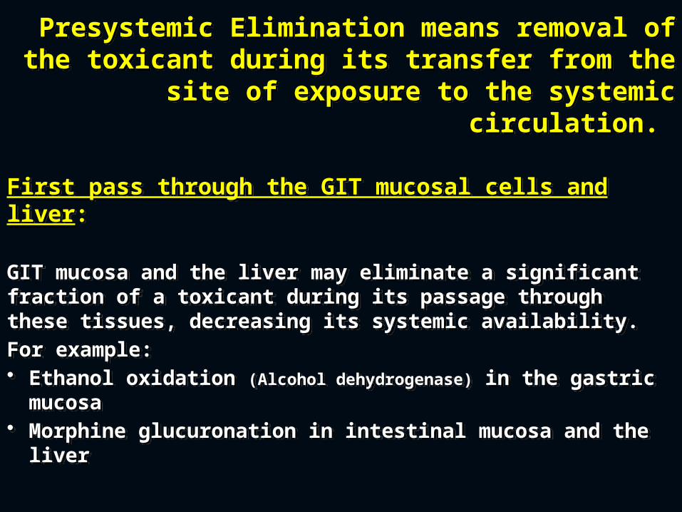
Presystemic Elimination means removal of the toxicant during its transfer from the
site of exposure to the systemic circulation.
Presystemic Elimination means removal of the toxicant during its transfer from the
site of exposure to the systemic circulation.
First pass through the GIT mucosal cells and liver:
GIT mucosa and the liver may eliminate a significant fraction of a toxicant during its passage through these tissues, decreasing its systemic availability. For example:• Ethanol oxidation (Alcohol dehydrogenase) in the gastric
mucosa• Morphine glucuronation in intestinal mucosa and the
liver
Distribution to and Away from the Target
Distribution to and Away from the Target
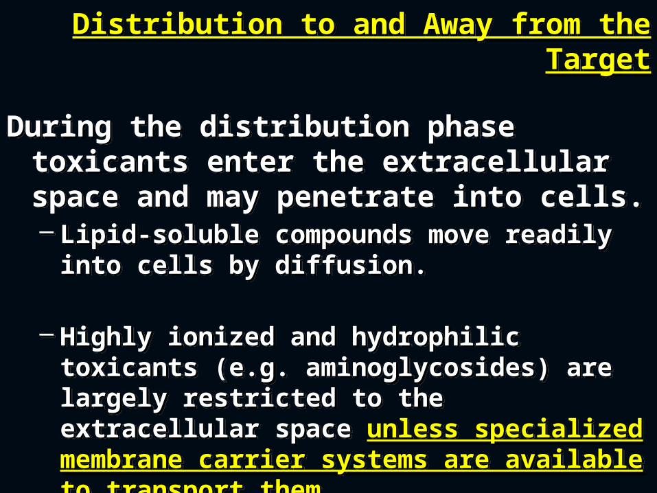
During the distribution phase toxicants enter the extracellular space and may penetrate into cells. – Lipid-soluble compounds move readily
into cells by diffusion.
– Highly ionized and hydrophilic toxicants (e.g. aminoglycosides) are largely restricted to the extracellular space unless specialized membrane carrier systems are available to transport them.
Mechanisms Opposing Distribution to a Target
Mechanisms Opposing Distribution to a Target
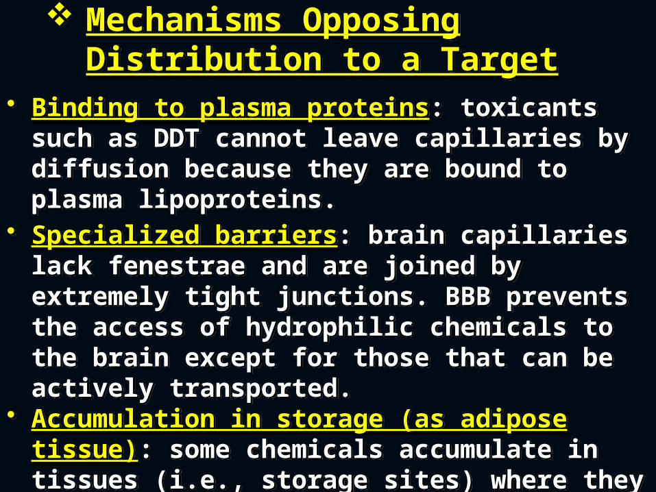
• Binding to plasma proteins: toxicants such as DDT cannot leave capillaries by diffusion because they are bound to plasma lipoproteins.
• Specialized barriers: brain capillaries lack fenestrae and are joined by extremely tight junctions. BBB prevents the access of hydrophilic chemicals to the brain except for those that can be actively transported.
• Accumulation in storage (as adipose tissue): some chemicals accumulate in tissues (i.e., storage sites) where they do not exert significant effects.
Excretion versus ReabsorptionExcretion versus Reabsorption
• Excretion is the removal of toxicants from the blood and their return to the external environment.
• Reabsorption is the reuptake of filtrated toxicants by renal tubules across their tubular cells into the peritubular capillaries.
• Reabsorption by diffusion is dependent on the lipid solubility of the chemical.
Excretion versus Reabsorption Excretion versus Reabsorption
• For organic acids and bases, diffusion is inversely related to the extent of ionization, because the nonionized molecule is more lipid-soluble.
• The ionization of weak organic acids such as salicylic acid and phenobarbital and bases such as amphetamine, procainamide, and quinidine is strongly pH-dependent in the physiologic range.
• Reabsorption is affected by the pH of the tubular fluid.
• Acidification favors excretion of weak organic bases
• Alkalinization favors elimination of weak organic acids.
Toxication versus DetoxicationToxication versus Detoxication
• Toxication (metabolic activation): biotransformation to harmful products.
Usually, toxication makes toxicants reactive toward endogenous molecules with susceptible functional groups.
Sometimes, toxication may have physicochemical properties to alter biological processes or structures. e.g, oxalic acid formed from E. glycol may cause acidosis.
Occasionally, chemicals acquire structural features and reactivity by biotransformation that allows for a more efficient interaction with specific receptors or enzymes.
Toxication (cont.) Increased reactivity may be due to
conversion into
Toxication (cont.) Increased reactivity may be due to
conversion into
• Electrophiles: molecules containing an electron-deficient atom with a partial or full positive charge that allows it to react by sharing electron pairs with electron-rich atoms in nucleophiles.
• Free radicals: molecules that contain one or more unpaired electrons in its outer orbital.
• Nucleophiles: uncommon mechanism for activating toxicants.– formation of cyanide from amygdalin.
DetoxicationDetoxication• Biotransformations that eliminate
the ultimate toxicant or prevent its formation
• Biotransformations that eliminate the ultimate toxicant or prevent its formation
Step2- Reaction of the ultimate toxicant with the target
molecule
Step2- Reaction of the ultimate toxicant with the target
molecule • This reaction leads to the injury to the target molecule itself, cell organelles, cells, tissues and organs, and even the whole organism.
• This reaction leads to the injury to the target molecule itself, cell organelles, cells, tissues and organs, and even the whole organism.
Step3- Cellular dysfunction and resultant toxicities Step3- Cellular dysfunction and resultant toxicities
• The reaction of toxicants with a target molecule may result in impaired cellular function.
• Normally, each cell has defined programs:
– Programs determine the destiny of cells—that is, whether they undergo division, differentiation (i.e., produce proteins for specialized functions) or apoptosis
– Programs control the activity of differentiated cells, determining whether they secrete more or less of a substance, whether they contract or relax, and whether they transport and metabolize nutrients at higher or lower rates.
Step 4- Repair or DysrepairStep 4- Repair or Dysrepair
• Many toxicants alter macromolecules, which, if not repaired, cause damage at higher levels in the organism.
• The organism trials to repair the
damaging effects from toxicants on molecular, cellular, and tissue levels
• Many toxicants alter macromolecules, which, if not repaired, cause damage at higher levels in the organism.
• The organism trials to repair the
damaging effects from toxicants on molecular, cellular, and tissue levels


![[Toxicology] toxicology introduction](https://static.fdocuments.in/doc/165x107/55c46616bb61ebb3478b4643/toxicology-toxicology-introduction.jpg)


