Introduction to Radiology. Oral and Maxillofacial Radiology I 343 DSD l Course Credits: 3 hours; 1...
27
Introduction to Introduction to Radiology Radiology
-
Upload
derek-hutchinson -
Category
Documents
-
view
224 -
download
1
Transcript of Introduction to Radiology. Oral and Maxillofacial Radiology I 343 DSD l Course Credits: 3 hours; 1...
Introduction to RadiologyIntroduction to Radiology
Oral and Maxillofacial Radiology I343 DSD
Course Credits: 3 hours; 1 hr Lecture per week, 2 hrs Practical per week
Prerequisite for: 443 DSD / Oral and Maxillofacial Radiology II
Course Description
Dental radiology is a branch of dentistry for diagnostic, treatment planning and following-up purposes. Complete examination of the oral cavity needs both clinical and radiographic investigations, therefore this course of dental radiology offers the way of examining the hidden parts of teeth and their supporting structures. This course is considered as an introductory course that includes lectures, demonstrations and practical applications on the previously given lectures. It is consisted of a series of weekly lectures and practical extending over two terms.
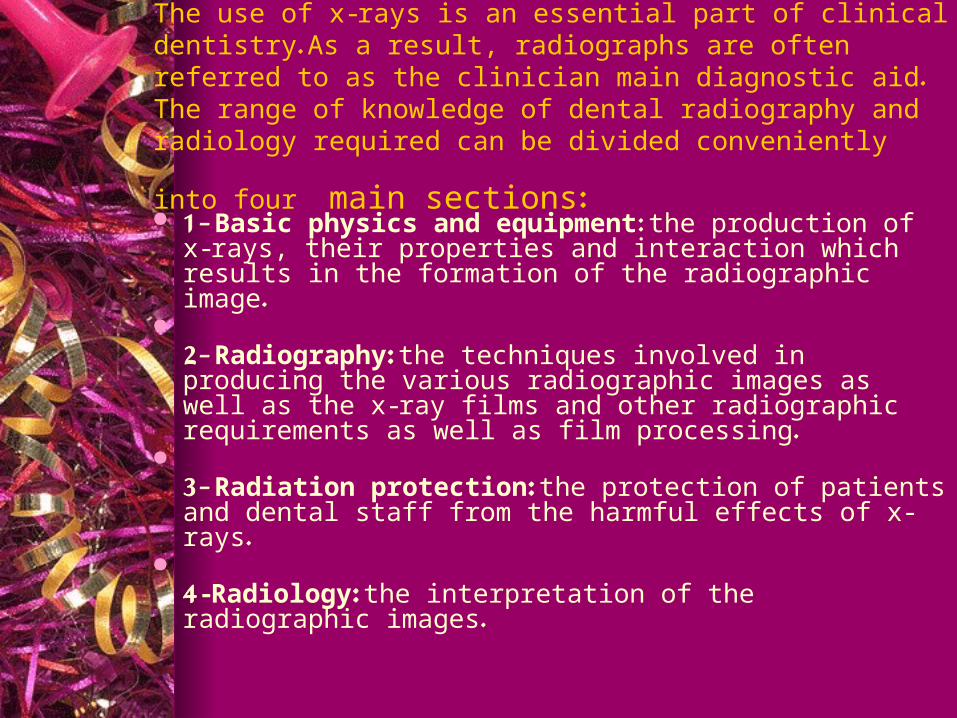
The use of x-rays is an essential part of clinical dentistry. As a result, radiographs are often referred to as the clinician main diagnostic aid. The range of knowledge of dental radiography and radiology required can be
divided conveniently into four main sections:
1- Basic physics and equipment: the production of x-rays, their properties and interaction which results in the formation of the radiographic image.
2- Radiography: the techniques involved in producing the various radiographic images as well as the x-ray films and other radiographic requirements as well as film processing.
3- Radiation protection: the protection of patients and dental staff from the harmful effects of x-rays.
4-Radiology: the interpretation of the radiographic images.
This course is primarily concerned with the first three topics namely physics, radiography and protection. Only part of film interpretation for simple lesions as caries, periodontal and periapical diseases will be covered. However, the more comprehensive cases and differential diagnosis will be covered in the third year course.

At the end of this course, the students will be able to:
Know how x-rays are produced, identify the component parts of the x-ray machine and its accessories and list and describe the possible interactions of x-rays with matter.
List and describe the different types of intra-oral and extra-oral x-ray films used in dentistry including their sizes, speeds as well as how to store them properly.
Identify the quality of x-ray image regarding the radiographic density, contrast, sharpness, magnification and distortion. Also, knows what is required to produce an ideal radiographic image.
List, discuss and practice the step-by-step procedures for both manual and automatic processing. Make complete intra-oral radiographic survey [CMS] on patients, process, mount then, detect and
retake the unsatisfactory radiographs. Identify the radiographic anatomical landmarks as seen in radiographs, distinguish between
normalities and normal variations. Also, to differentiate between those landmarks and some pathological lesions.
List and discuss the common causes of unsatisfactory radiographs, their causes and how to avoid them.
Discuss the harmful effects of radiation, both the short and long-term effects, whether they are somatic or genetic.
Protects his patients, the dental staff and people in the immediate environment from the harmful effects of radiation.
Detect, identify and describe the radiographic appearance of dental caries, periodontal and periapical pathosis. Also, to identify radiographically the existence of various dental anomalies and regressive changes that may affect teeth as well as various forms of trauma to the teeth.
Course Contents:
Introduction Lecture Radiation Physics (I, II) Radiation Biology Radiation Protection Dental X-ray Equipment and Films Dental X-ray Image Characteristics Processing of X-ray Film Intra-oral Radiographic Techniques (I, II, III) Normal Radiographic Anatomy Common Causes of Unsatisfactory Radiographs Interpretation of Dental Caries and the Assessment of Restorations Interpretation of Periapical Tissues and Pathosis Interpretation of Periodontal Tissues and Periodontal Disease
References
Oral Radiology Principles and Interpretation. 5th Edition. By White and Pharoh.
Dental Radiography, Principles and Techniques. 2nd Edition. By Joen Haring and Laura Jansen.
Essentials of Dental Radiography and Radiobiology. 3rd Edition. By Eric Whaites.

EvaluationGrades will be based on
Continuous Assessment (Quizzes, Homework, Assignment) 20 marks
Mid-Year Exam 20 marks Practical 20 marks Final Exam (Written) 40 marks
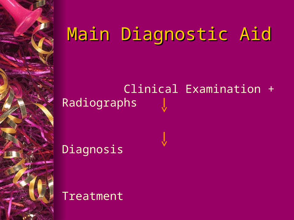
Main Diagnostic AidMain Diagnostic Aid
Clinical Examination + Radiographs
Diagnosis
Treatment
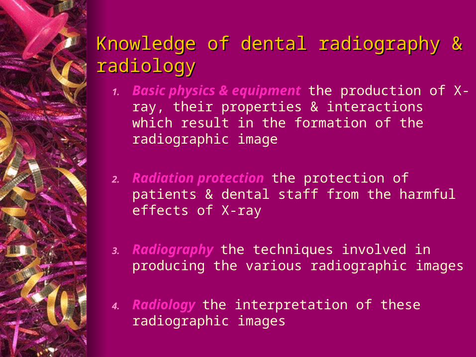
Knowledge of dental radiography & radiologyKnowledge of dental radiography & radiology
1. Basic physics & equipment the production of X-ray, their properties & interactions which result in the formation of the radiographic image
2. Radiation protection the protection of patients & dental staff from the harmful effects of X-ray
3. Radiography the techniques involved in producing the various radiographic images
4. Radiology the interpretation of these radiographic images
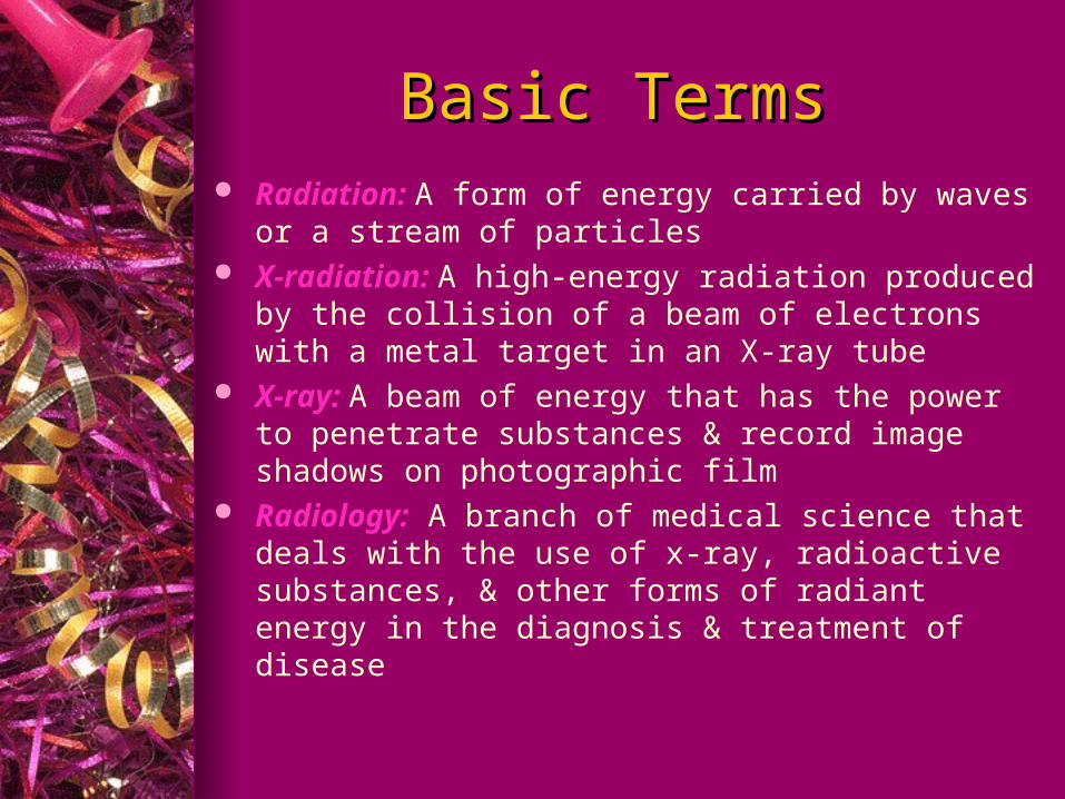
Basic TermsBasic Terms Radiation: A form of energy carried by waves or a stream of
particles X-radiation: A high-energy radiation produced by the collision
of a beam of electrons with a metal target in an X-ray tube X-ray: A beam of energy that has the power to penetrate
substances & record image shadows on photographic film Radiology: A branch of medical science that deals with the use
of x-ray, radioactive substances, & other forms of radiant energy in the diagnosis & treatment of disease
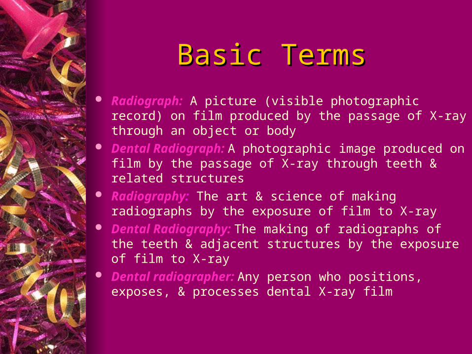
Basic TermsBasic Terms Radiograph: A picture (visible photographic record) on film
produced by the passage of X-ray through an object or body Dental Radiograph: A photographic image produced on film
by the passage of X-ray through teeth & related structures Radiography: The art & science of making radiographs by the
exposure of film to X-ray Dental Radiography: The making of radiographs of the teeth &
adjacent structures by the exposure of film to X-ray Dental radiographer: Any person who positions, exposes, &
processes dental X-ray film
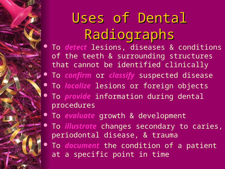
Uses of Dental RadiographsUses of Dental Radiographs To detect lesions, diseases & conditions of the teeth &
surrounding structures that cannot be identified clinically
To confirm or classify suspected disease To localize lesions or foreign objects To provide information during dental procedures To evaluate growth & development To illustrate changes secondary to caries, periodontal
disease, & trauma To document the condition of a patient at a specific
point in time
DiscoverDiscover of X-radiation of X-radiation
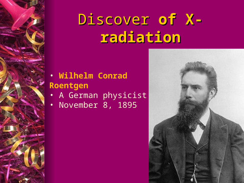
• Wilhelm Conrad Roentgen • A German physicist• November 8, 1895
DiscoverDiscover of X-radiation of X-radiation
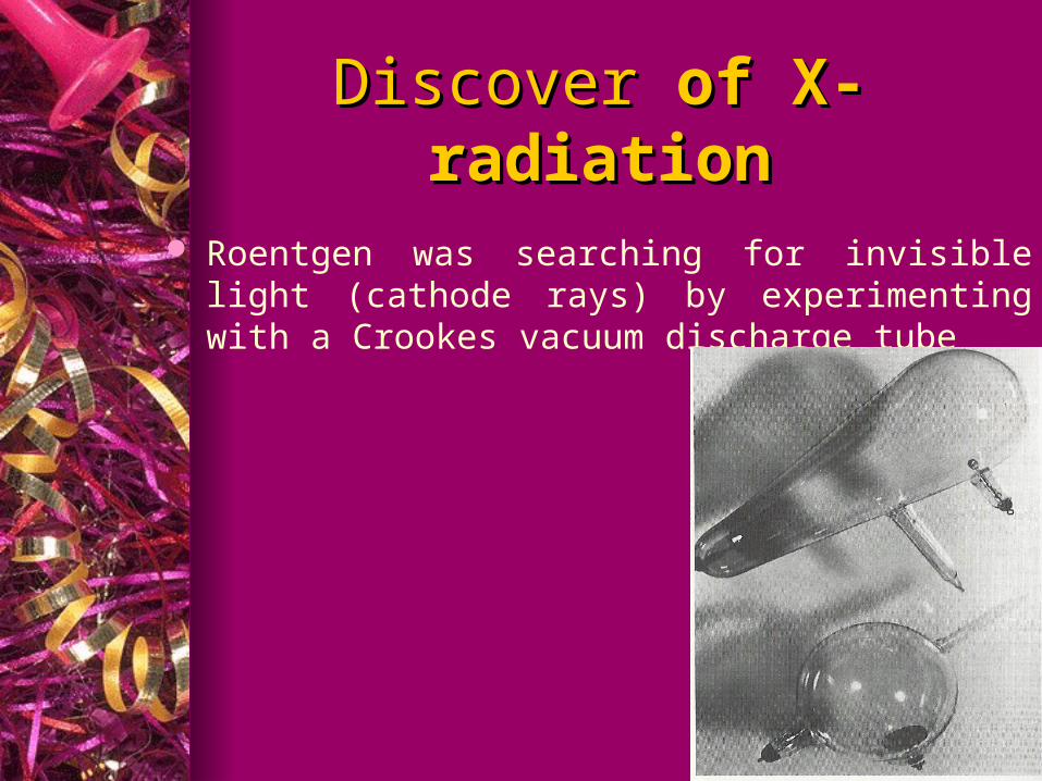
Roentgen was searching for invisible light (cathode rays) by experimenting with a Crookes vacuum discharge tube
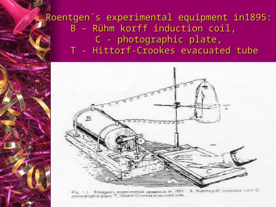
Roentgen´s experimental equipment in1895:Roentgen´s experimental equipment in1895:B – Rühm korff induction coil, B – Rühm korff induction coil,
C - photographic plate,C - photographic plate,T - Hittorf-Crookes evacuated tubeT - Hittorf-Crookes evacuated tube
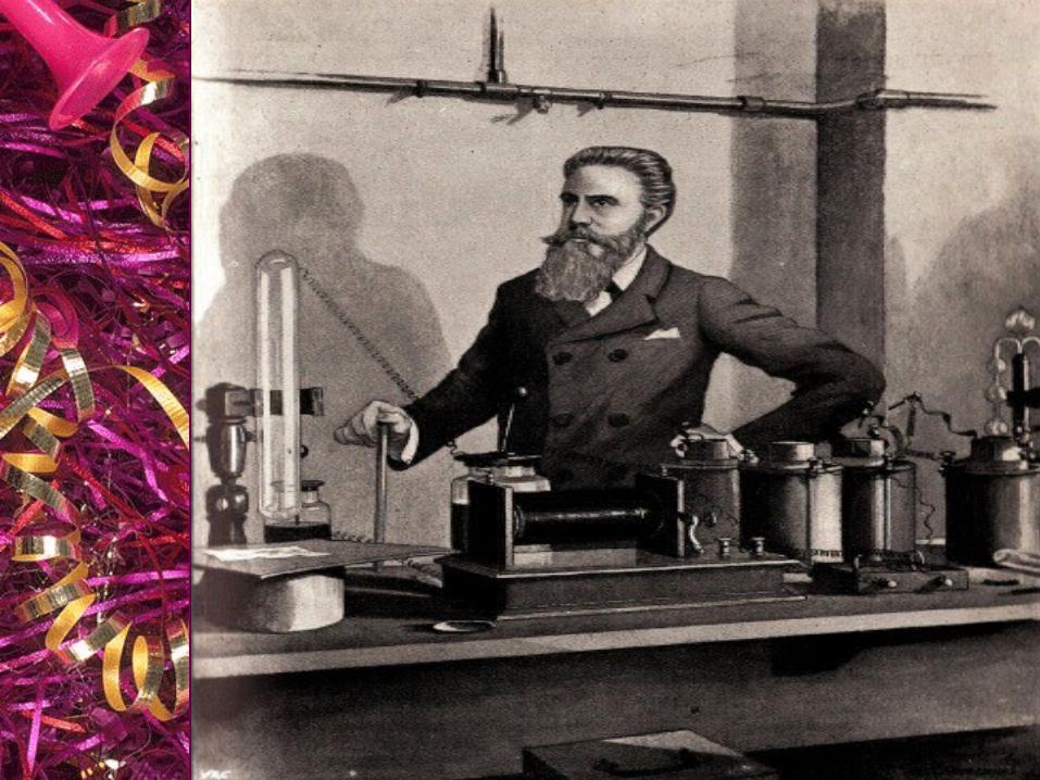
DiscoverDiscover of X-radiation of X-radiation
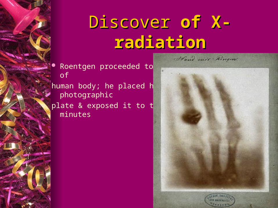
Roentgen proceeded to make the first radiograph of
human body; he placed his wife’s hand on a photographic
plate & exposed it to the unknown ray for 15 minutes

DiscoverDiscover of X-radiation of X-radiation
Roentgen named his discovery X-ray, the ‘’X’’ referring to the unknown nature & properties of such ray
Roentgen was awarded the first Nobel Prize ever awarded in Physics (1901)
After years of Roentgen discovery,
X-ray roentgen ray, radiology roentgenology, & radiographs roentgenographs
Pioneers in Dental X-radiationPioneers in Dental X-radiation In 1896 In 1896
German dentist, Otto WalkhoffOtto Walkhoff, made the first dental radiograph; he submitted himself to 25 minutes of x-ray exposure
W.J. MortonW.J. Morton, a New York physician, made the first dental radiograph in the United States using a skull
Pioneers in Dental X-radiationPioneers in Dental X-radiation In 1896 In 1896
C. Edmund KellsC. Edmund Kells, New Orleans dentist, exposed the first dental radiograph in the United States using a living person
During his many experiments, KellsKells exposed his hands to numerous x-rays every day for years. This overexposure to x-radiation caused the development of numerous cancers of his hand
Pioneers in Dental X-radiationPioneers in Dental X-radiation
William H. RollinsWilliam H. Rollins, a Boston dentist, developed the 1st dental x-ray unit. While experimenting with radiation, Dr. Rollins Dr. Rollins suffered a burn to his hand
This initiated an interest in radiation protection and later the publication of the 1st paper on the dangers associated with radiation
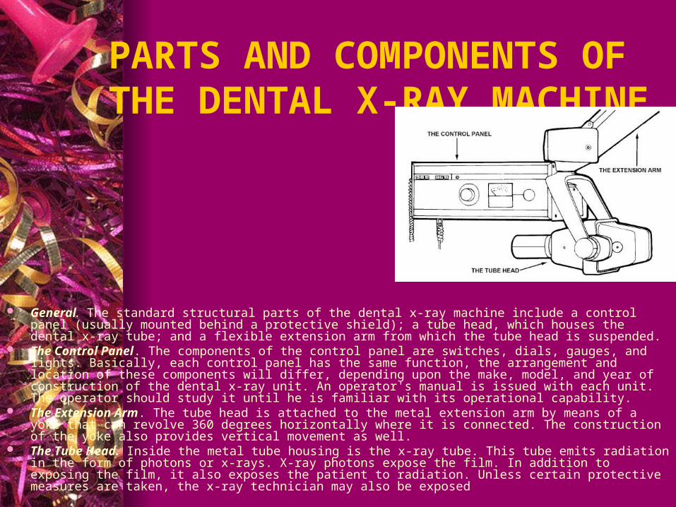
PARTS AND COMPONENTS OF THE DENTAL X-RAY MACHINE
General. The standard structural parts of the dental x-ray machine include a control panel (usually mounted behind a protective shield); a tube head, which houses the dental x-ray tube; and a flexible extension arm from which the tube head is suspended.
The Control Panel. The components of the control panel are switches, dials, gauges, and lights. Basically, each control panel has the same function, the arrangement and location of these components will differ, depending upon the make, model, and year of construction of the dental x-ray unit. An operator's manual is issued with each unit. The operator should study it until he is familiar with its operational capability.
The Extension Arm. The tube head is attached to the metal extension arm by means of a yoke that can revolve 360 degrees horizontally where it is connected. The construction of the yoke also provides vertical movement as well.
The Tube Head. Inside the metal tube housing is the x-ray tube. This tube emits radiation in the form of photons or x-rays. X-ray photons expose the film. In addition to exposing the film, it also exposes the patient to radiation. Unless certain protective measures are taken, the x-ray technician may also be exposed
THREE STEP PROCESS OF X-RAY PRODUCTION
The First Step. The first step in x-ray production is to turn on the machine. (If there is doubt on the part of the x-ray technician concerning the operation of the unit, reference should be made to the operator's manual.) When the unit is turned on, the filament of the cathode is heated by electrical current, causing it to emit electrons
THREE STEP PROCESS OF X-RAY PRODUCTION
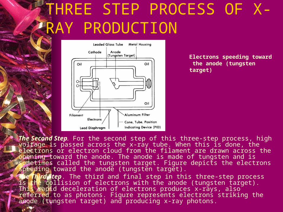
The Second Step. For the second step of this three-step process, high voltage is passed across the x-ray tube. When this is done, the electrons or electron cloud from the filament are drawn across the opening toward the anode. The anode is made of tungsten and is sometimes called the tungsten target. Figure depicts the electrons speeding toward the anode (tungsten target).
The Third Step. The third and final step in this three-step process is the collision of electrons with the anode (tungsten target). This rapid deceleration of electrons produces x-rays, also referred to as photons. Figure represents electrons striking the anode (tungsten target) and producing x-ray photons.
Electrons speeding toward the anode (tungsten target)
RADIATION PROTECTION
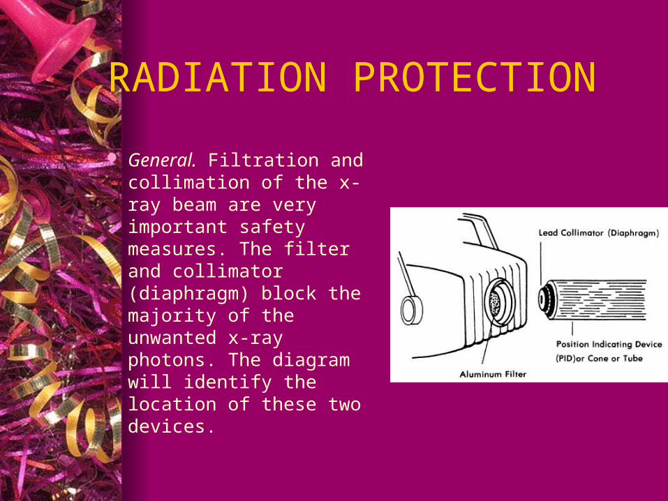
General. Filtration and collimation of the x-ray beam are very important safety measures. The filter and collimator (diaphragm) block the majority of the unwanted x-ray photons. The diagram will identify the location of these two devices.