Introduction to Head and Neck...
50
Introduction to Head and Neck Anatomy
Transcript of Introduction to Head and Neck...
Introduction to Head and Neck
Anatomy
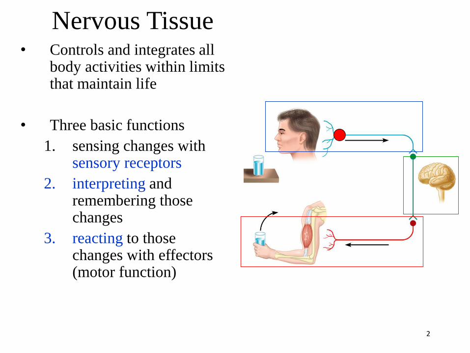
Nervous Tissue• Controls and integrates all
body activities within limits that maintain life
• Three basic functions
1. sensing changes with sensory receptors
2. interpreting and remembering those changes
3. reacting to those changes with effectors (motor function)
2

Cranial nerves12 pairs
Spinal nerves31 pairs

The PNS is divided into :
1- Somatic nervous system (SNS)
2- Autonomic nervous system (ANS)

Sensory (Afferent) vs. Motor (Efferent)
e.g., skin
e.g., muscle
Gray’s Anatomy 38 1999
sensory (afferent) nerve
motor (efferent) nerve
(pseudo-) unipolar neurons conducting impulses
from sensory organs to the CNS
multipolar neurons conducting impulses
from the CNS to effector organs (muscles & glands)

Organization
6
Integration MotorSensory
SNS
(Sensory)
ANS
(Sensory)
Brain
Spinal
cord
SNS
(Motor)
ANS
(Motor)
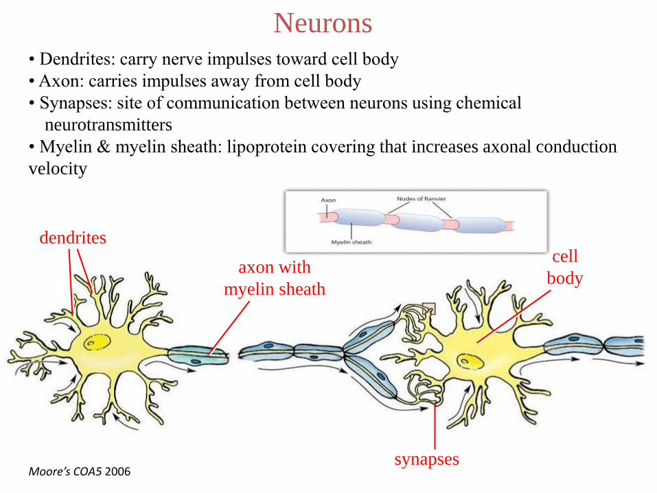
Neurons
cell
body
dendrites
axon with
myelin sheath
synapsesMoore’s COA5 2006
• Dendrites: carry nerve impulses toward cell body
• Axon: carries impulses away from cell body
• Synapses: site of communication between neurons using chemical
neurotransmitters
• Myelin & myelin sheath: lipoprotein covering that increases axonal conduction
velocity
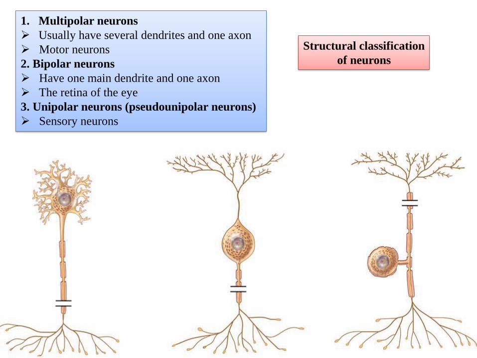
1. Multipolar neurons
➢ Usually have several dendrites and one axon
➢ Motor neurons
2. Bipolar neurons
➢ Have one main dendrite and one axon
➢ The retina of the eye
3. Unipolar neurons (pseudounipolar neurons)
➢ Sensory neurons
Structural classification
of neurons
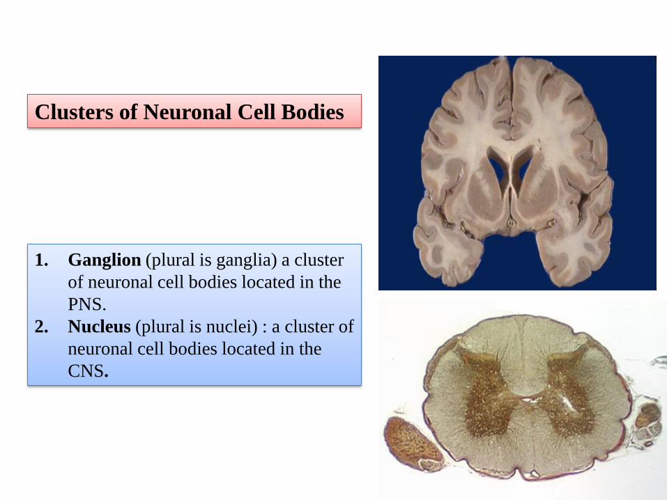
1. Ganglion (plural is ganglia) a cluster
of neuronal cell bodies located in the
PNS.
2. Nucleus (plural is nuclei) : a cluster of
neuronal cell bodies located in the
CNS.
Clusters of Neuronal Cell Bodies

• A nerve: is a bundle of axons
that is located in the PNS.
➢Cranial nerves connect the brain to
the periphery
➢ Spinal nerves connect the spinal
cord to the periphery
• A tract: is a bundle of axons
located in the CNS.
Bundles of Axons
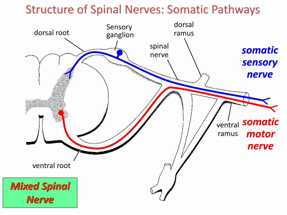
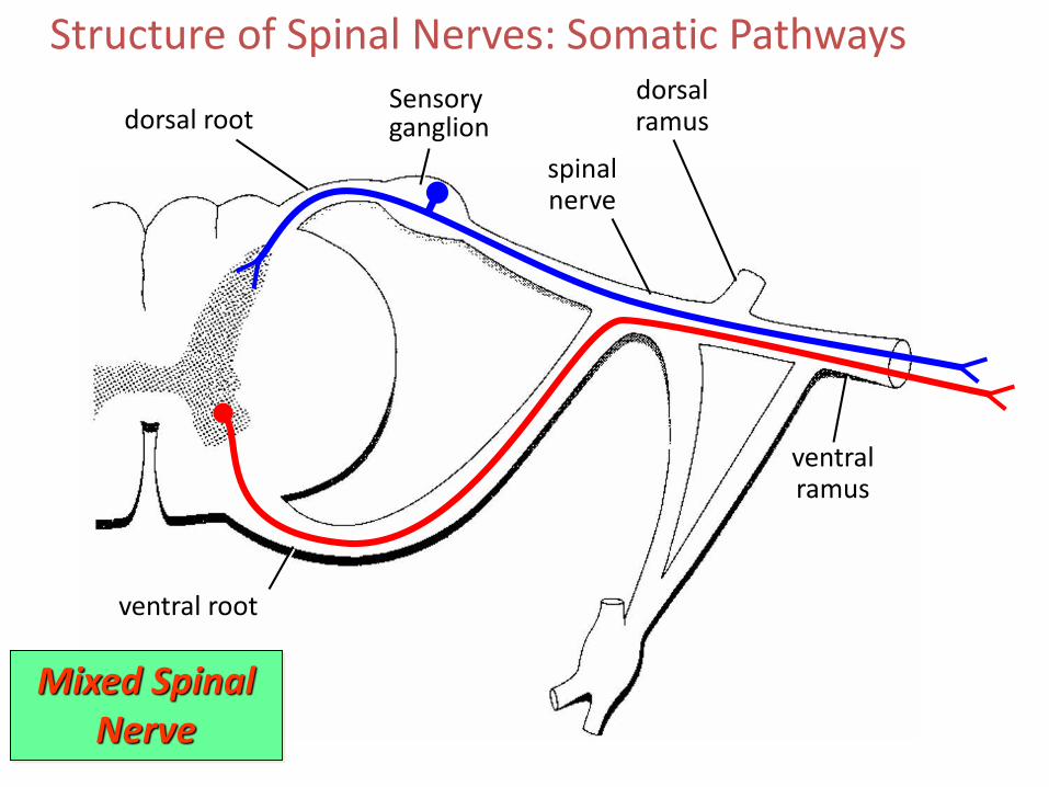
Structure of Spinal Nerves: Somatic Pathways
dorsal rootSensory ganglion
ventral root
spinalnerve
dorsalramus
ventralramus
somaticsensorynerve
somaticmotornerve
Mixed SpinalNerve
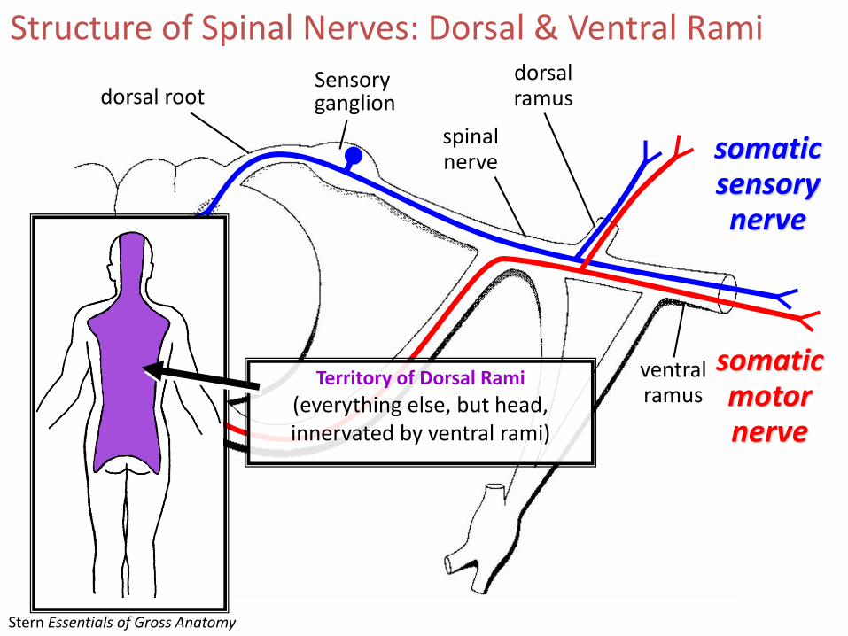
Structure of Spinal Nerves: Dorsal & Ventral Rami
spinalnerve
dorsalramus
somaticsensorynerve
somaticmotornerve
Territory of Dorsal Rami
(everything else, but head,innervated by ventral rami)
ventralramus
Stern Essentials of Gross Anatomy
dorsal rootSensory ganglion

13
SNS
PNS
ANS
Sensory
Motor
Sensory
Motor Parasympathetic
Sympathetic

• ANS is the subdivision of the peripheral nervous
system that regulates body activities that are generally
not under conscious control
• Visceral motor innervates non-skeletal (non-
somatic) muscles
• Composed of a special group of neurons serving:
– Cardiac muscle (the heart)
– Smooth muscle (walls of viscera and blood vessels)
– Glands
14
Autonomic nervous system
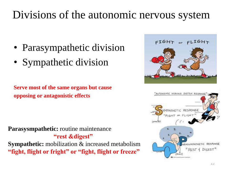
Divisions of the autonomic nervous system
• Parasympathetic division
• Sympathetic division
Serve most of the same organs but cause
opposing or antagonistic effects
15
Parasysmpathetic: routine maintenance
“rest &digest”
Sympathetic: mobilization & increased metabolism
“fight, flight or fright” or “fight, flight or freeze”

Basic anatomical difference between the motor pathways of the voluntary somatic nervous system (to skeletal muscles) and those of the autonomic nervous system
• Somatic division:– Cell bodies of motor neurons reside in CNS (brain or spinal
cord)
– Their axons (sheathed in spinal nerves) extend all the way to their skeletal muscles
• Autonomic system: chains of two motor neurons– 1st = preganglionic neuron (cell body in brain or cord)
– 2nd = postgangionic neuron (cell body in ganglion outside CNS)
16

• Axon of 1st (preganglionic) neuron leaves CNS to synapse with the 2nd
(postganglionic) neuron
• Axon of 2nd (postganglionic) neuron extends to the organ it serves
17
autonomic
somatic
Note: the autonomic ganglion is motor
this dorsal
root ganglion
is sensory

CNS ganglion
preganglionic
neuron
postganglionic
neuron
glands
smooth
muscle
cardiac
muscle
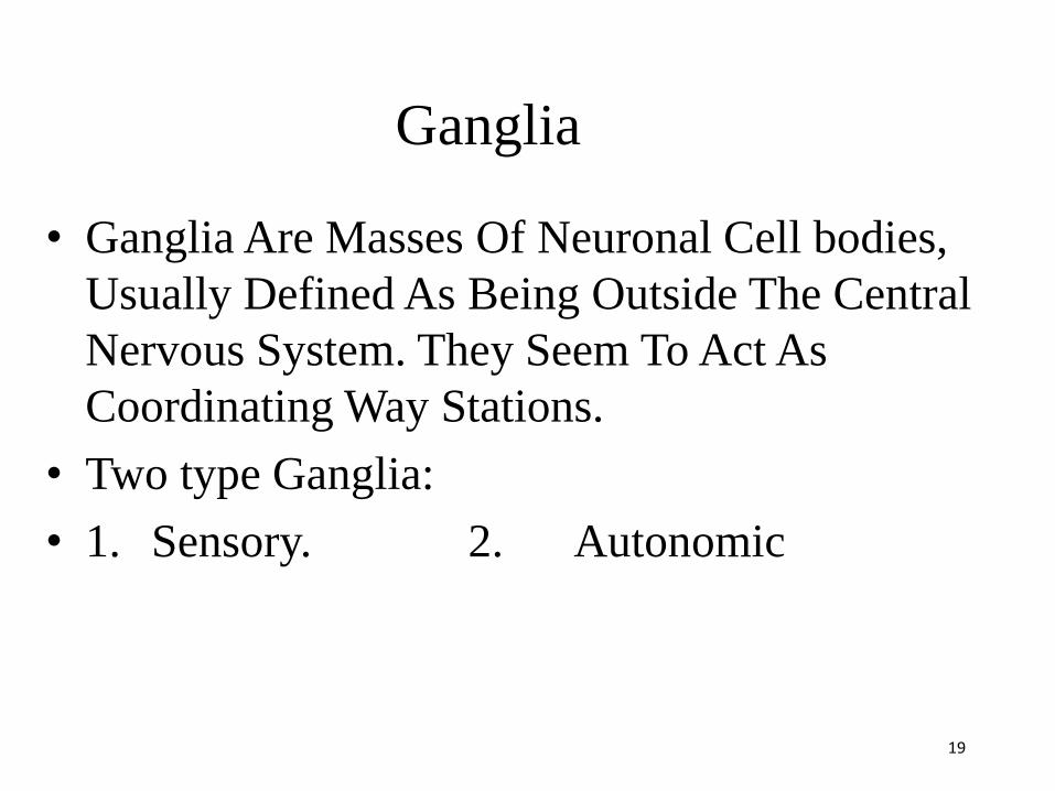
Ganglia
• Ganglia Are Masses Of Neuronal Cell bodies,
Usually Defined As Being Outside The Central
Nervous System. They Seem To Act As
Coordinating Way Stations.
• Two type Ganglia:
• 1. Sensory. 2. Autonomic
19

20
Sensory ganglion
Sensory ganglia do not receive synapses
21
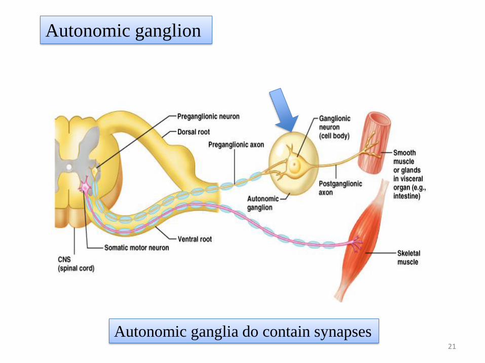
Autonomic ganglion
Autonomic ganglia do contain synapses
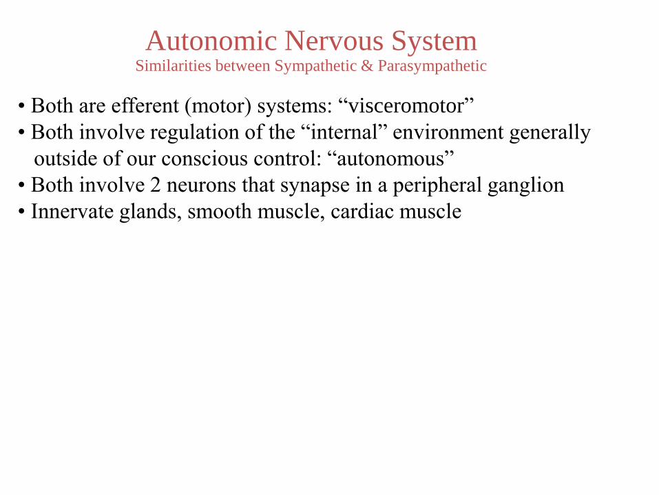
Autonomic Nervous SystemSimilarities between Sympathetic & Parasympathetic
• Both are efferent (motor) systems: “visceromotor”
• Both involve regulation of the “internal” environment generally
outside of our conscious control: “autonomous”
• Both involve 2 neurons that synapse in a peripheral ganglion
• Innervate glands, smooth muscle, cardiac muscle

Sympathetic
CNS ganglion
short preganglionic
neuron
ParasympatheticCNS ganglion
long preganglionic
neuron
Overview of the Autonomic Nervous SystemDifferences between Sympathetic & Parasympathetic
Relative Lengths of Neurons
long postganglionic
neuron
target
target
short postganglionic
neuron
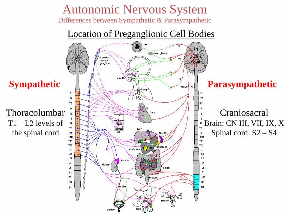
Autonomic Nervous SystemDifferences between Sympathetic & Parasympathetic
Location of Preganglionic Cell Bodies
ThoracolumbarT1 – L2 levels of
the spinal cord
Sympathetic
CraniosacralBrain: CN III, VII, IX, X
Spinal cord: S2 – S4
Parasympathetic
Structure of Spinal Nerves: Somatic Pathways
dorsal rootSensory ganglion
ventral root
spinalnerve
dorsalramus
ventralramus
Mixed SpinalNerve
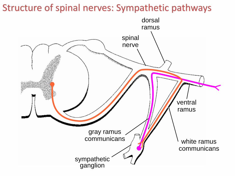
spinalnerve
dorsalramus
ventralramus
gray ramuscommunicans white ramus
communicans
sympatheticganglion
Structure of spinal nerves: Sympathetic pathways
27
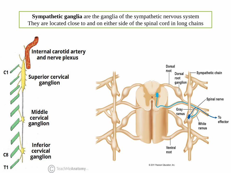
Sympathetic ganglia are the ganglia of the sympathetic nervous system
They are located close to and on either side of the spinal cord in long chains

Sympathetic ganglia are the ganglia of the sympathetic nervous system
They are located close to and on either side of the spinal cord in long chains
There are usually 22-23 pairs of
paravertebral sympathetic
ganglia:
3 in the cervical region
(cervical ganglia)
11 in the thoracic region
4 in the lumbar region
4-5 in the sacral region
1 unpaired coccygeal ganglion
Preganglionic nerves from the spinal cord synapse at one of the chain ganglia,
and the postganglionic fiber extends to an effector
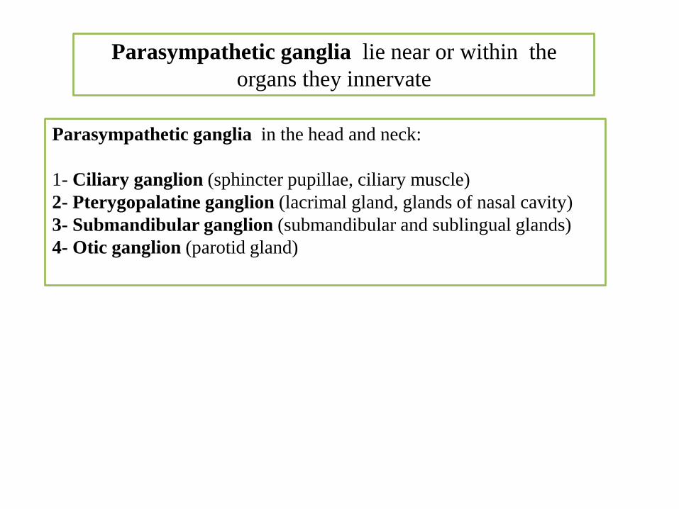
Parasympathetic ganglia lie near or within the
organs they innervate
Parasympathetic ganglia in the head and neck:
1- Ciliary ganglion (sphincter pupillae, ciliary muscle)
2- Pterygopalatine ganglion (lacrimal gland, glands of nasal cavity)
3- Submandibular ganglion (submandibular and sublingual glands)
4- Otic ganglion (parotid gland)
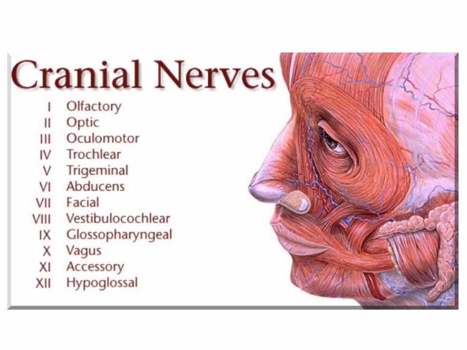

The numbering of the cranial nerves is based
on the order in which they emerge from the
brain, front to back
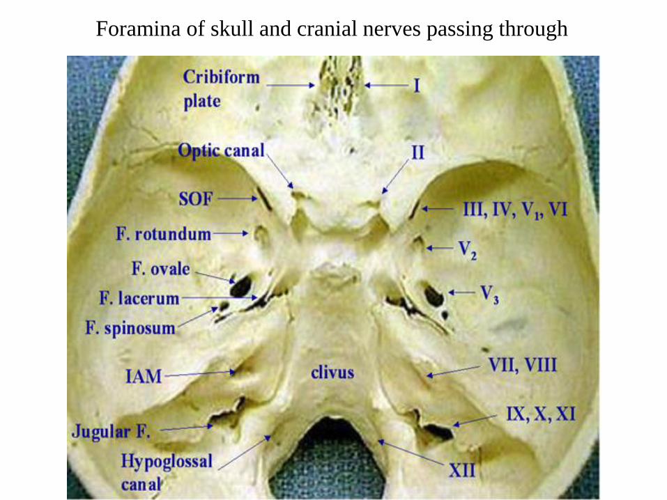
Foramina of skull and cranial nerves passing through
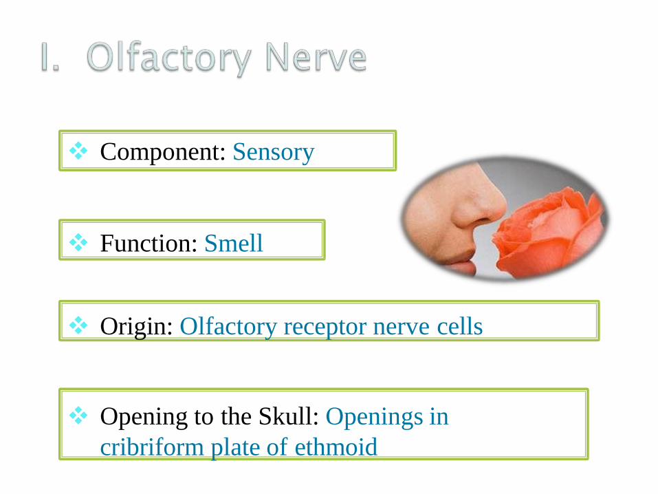
❖ Component: Sensory
❖ Function: Smell
❖ Origin: Olfactory receptor nerve cells
❖ Opening to the Skull: Openings in
cribriform plate of ethmoid
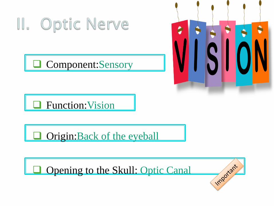
Component:Sensory
Function:Vision
Origin:Back of the eyeball
Opening to the Skull: Optic Canal

▪ Component: Motor
▪ Function:
▪ Raises upper eyelid
▪ Turns eyeball upward, downward and
medially, upward and laterally
▪ Constricts pupil
▪ Accommodates the eye
Opening to the Skull: Superior orbital fissure
contains parasympathetic
III- Oculomotor
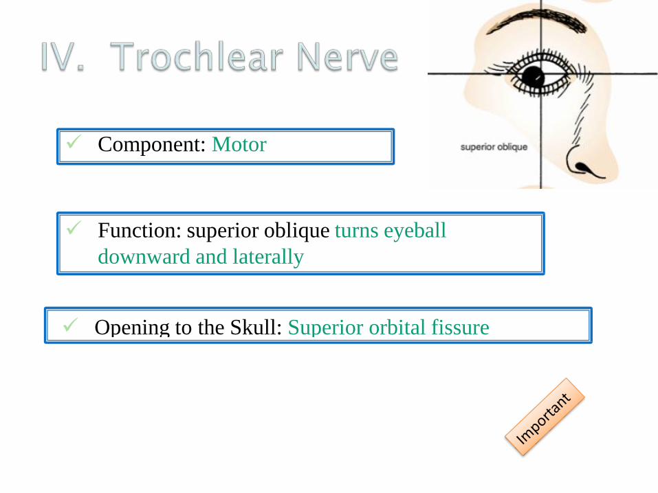
✓ Component: Motor
✓ Function: superior oblique turns eyeball
downward and laterally
✓ Opening to the Skull: Superior orbital fissure

V. Trigeminal Nerve
Mixed (sensory and motor)
Large sensory root
Small motor root
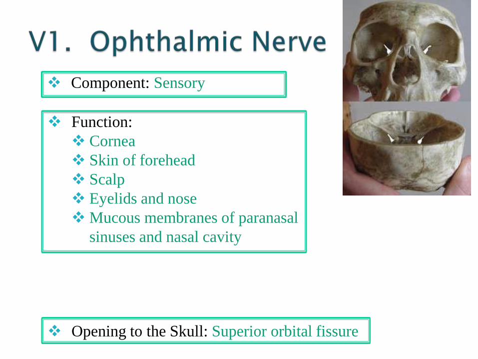
❖ Component: Sensory
❖ Function:
❖ Cornea
❖ Skin of forehead
❖ Scalp
❖ Eyelids and nose
❖Mucous membranes of paranasal
sinuses and nasal cavity
❖ Opening to the Skull: Superior orbital fissure

o Component: Sensory
o Function:
o Skin of the face over maxilla
o Upper lip
o Teeth of the upper jaw
o Mucous membrane of the nose, the
maxillary sinus and palate
Opening to the Skull: Foramen Rotundum
o Component: a. Motor
o Function:
o Muscles of mastication
o Mylohyoid
o Anterior belly of digastric
o Tensor veli palatine
o Tensor tympani
o Opening to the Skull: Foramen Ovale

• Component: b. Sensory
• Function:
• Skin of cheek
• Skin over mandible and side of head
• Teeth of lower jaw and TMJ
• Mucous membrane of mouth and anterior part of
tongue
Opening to the Skull: Foramen Ovale

✓ Component: Motor
✓ Function: Lateral rectus muscle turns eyeball laterally
Opening to the Skull: Superior orbital fissure

Component: Mixed
Function:
Motor• Muscles of the face and scalp
• Stapedius muscle
• Posterior belly of digastric
• Stylohyoid muscles
Function:
Sensory• Taste from ant. 2/3 of tongue
contains parasympathetic

Opening to the Skull: internal acoustic meatus,
facial canal, stylomastoid foramen
Function: parasympathetic
• Submandibular and sublingual salivary glands
• Lacrimal gland
• Glands of nose and palate
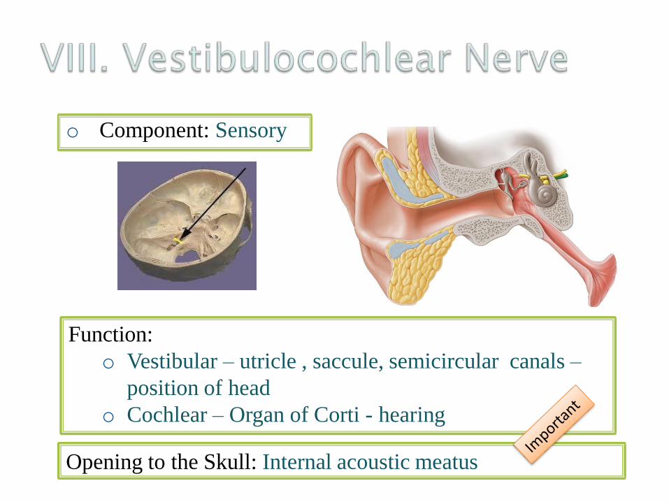
o Component: Sensory
Function:
o Vestibular – utricle , saccule, semicircular canals –
position of head
o Cochlear – Organ of Corti - hearing
Opening to the Skull: Internal acoustic meatus

✓ Component: Mixed
✓ Function:
Motor• Stylopharyngeus
muscle
✓ Function:
Sensory• General sensation and taste
from post. 1/3 of the tongue and
oropharynx
• Carotid sinus and carotid body
✓ Function:
parasympathetic
• Parotid gland
contains parasympathetic
✓ Opening to the Skull: Jugular foramen
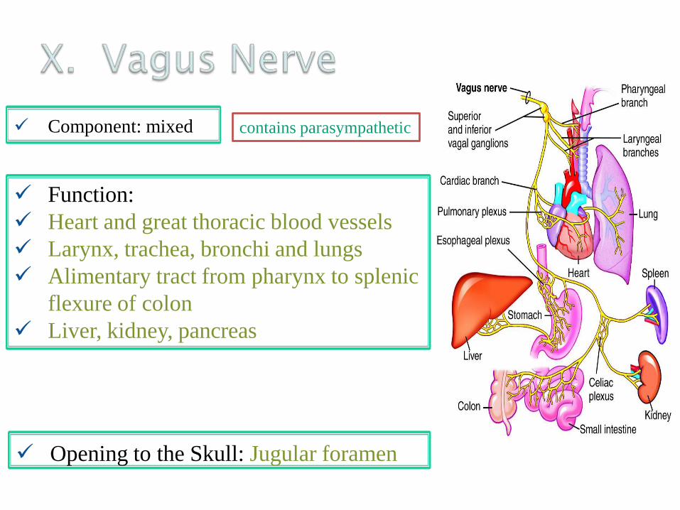
✓ Component: mixed
✓ Function:
✓ Heart and great thoracic blood vessels
✓ Larynx, trachea, bronchi and lungs
✓ Alimentary tract from pharynx to splenic
flexure of colon
✓ Liver, kidney, pancreas
✓ Opening to the Skull: Jugular foramen
contains parasympathetic
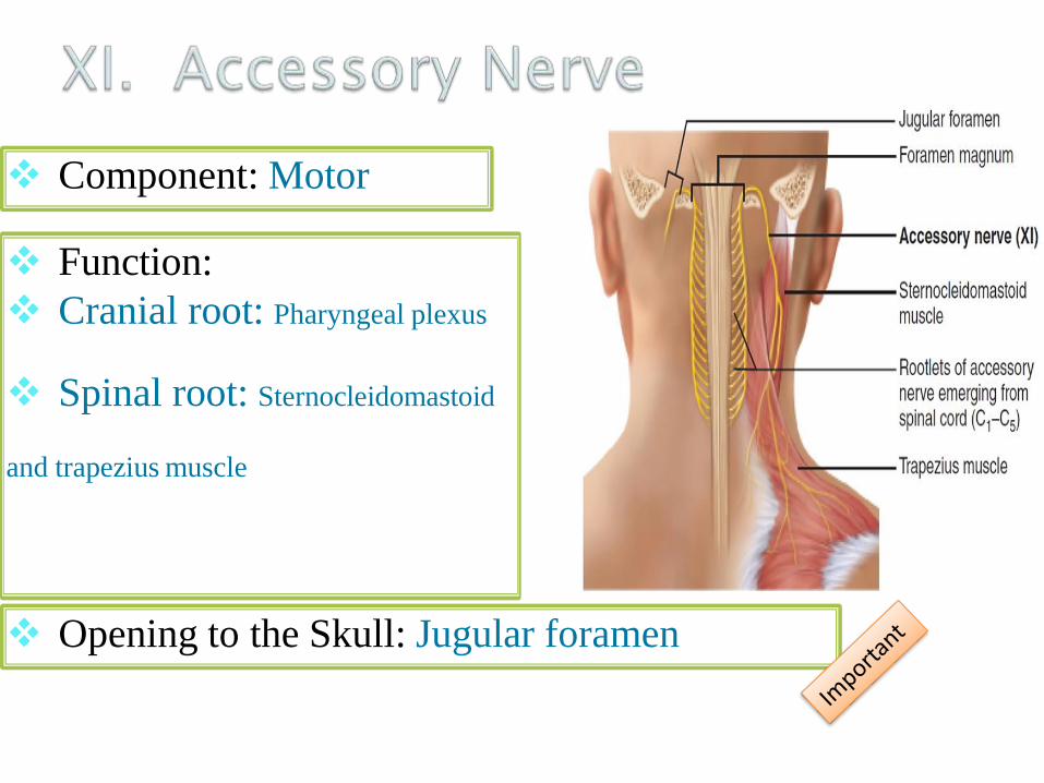
❖ Component: Motor
❖ Function:
❖ Cranial root: Pharyngeal plexus
❖ Spinal root: Sternocleidomastoid
and trapezius muscle
❖ Opening to the Skull: Jugular foramen

Motor to the muscles of the tongue

Pure sensory:
Olfactory
Optic
Vestibulocochlear
Pure motor:
Oculomotor
Trochlear
Abducent
Accessory
hypoglossal
Mixed (motor and sensory):
Trigeminal
Facial
Glossopharyngeal
Vagus
Contains parasympathetic
(secretomotor):
Oculomotor
Facial
Glossopharyngeal
Vagus