Introduction to Genes and Disease - Griffith University · Introduction to Genes and Disease . To...
15
1. Mitosis Introduction to Genes and Disease To appreciate the amazing potential of living organisms, and also to understand their limitations, it is essential to appreciate the body at a molecular level, and to understand the processes of cell division, inheritance and structure and function of the material that codes for life, DNA. Many of you will be pursuing careers in healthcare or biomedical research and you will encounter a range of pathologies at some stage in your career, for example cancer. Understanding cancer involves an understanding of many processes covered in this course – cell division and its control, gene expression, inheritance, DNA mutation etc. When we consider the impact of genes on the human condition, there are currently over 20,000 known human conditions/diseases that occur due to changes in our genomic sequence, and many more that occur due to alterations in how our genes are expressed. Understanding how to treat disease, maintain health and optimise performance requires an appreciation of how our genetic inheritance influences disease and how interventions affect function at the molecular level. Thus, an understanding of genetics and disease is essential for understanding the complexities of living organisms for a career in health. Module 1 focuses on the basic mechanisms of cell division, how it is controlled, and how cell division without control can lead to catastrophic results such as cancer. Module 2 introduces meiosis, the special type of cell division carried out by sexually reproducing organisms and how the processes of gamete formation and fertilization add to genetic variation in a population. Relevant case studies will be examined. Module 3 examines the principles of genetic inheritance, how genes, made up of DNA, provide information that influences certain characteristics and provides variation in organisms, and how characteristics are passed on from generation to generation. Module 4 focuses on chromosomes and their behaviour during cell division. It explores some human disorders resulting from gene and chromosomal mutation and how and why these anomalies may occur. Module 5 explores the structure and function of DNA, how DNA replicates prior to cell division, and how errors in replication and cell division can lead to mutation. A relevant case study is explored. Module 6 examines the fundamental relationship between genes and proteins, focusing on the process of protein synthesis and how the genetic language of DNA is decoded to produce a functional protein product. Relevant case studies are explored. Module 7 explores the intricate processes that cells use to precisely regulate the expression of their genes. Regulation of gene expression allows single celled organisms to rapidly respond to their environment while in multi-celled organisms, a
Transcript of Introduction to Genes and Disease - Griffith University · Introduction to Genes and Disease . To...

1. Mitosis
Introduction to Genes and Disease To appreciate the amazing potential of living organisms, and also to understand their limitations, it is essential to appreciate the body at a molecular level, and to understand the processes of cell division, inheritance and structure and function of the material that codes for life, DNA. Many of you will be pursuing careers in healthcare or biomedical research and you will encounter a range of pathologies at some stage in your career, for example cancer. Understanding cancer involves an understanding of many processes covered in this course – cell division and its control, gene expression, inheritance, DNA mutation etc. When we consider the impact of genes on the human condition, there are currently over 20,000 known human conditions/diseases that occur due to changes in our genomic sequence, and many more that occur due to alterations in how our genes are expressed. Understanding how to treat disease, maintain health and optimise performance requires an appreciation of how our genetic inheritance influences disease and how interventions affect function at the molecular level. Thus, an understanding of genetics and disease is essential for understanding the complexities of living organisms for a career in health. Module 1 focuses on the basic mechanisms of cell division, how it is controlled, and how cell division without control can lead to catastrophic results such as cancer. Module 2 introduces meiosis, the special type of cell division carried out by sexually reproducing organisms and how the processes of gamete formation and fertilization add to genetic variation in a population. Relevant case studies will be examined. Module 3 examines the principles of genetic inheritance, how genes, made up of DNA, provide information that influences certain characteristics and provides variation in organisms, and how characteristics are passed on from generation to generation. Module 4 focuses on chromosomes and their behaviour during cell division. It explores some human disorders resulting from gene and chromosomal mutation and how and why these anomalies may occur. Module 5 explores the structure and function of DNA, how DNA replicates prior to cell division, and how errors in replication and cell division can lead to mutation. A relevant case study is explored. Module 6 examines the fundamental relationship between genes and proteins, focusing on the process of protein synthesis and how the genetic language of DNA is decoded to produce a functional protein product. Relevant case studies are explored. Module 7 explores the intricate processes that cells use to precisely regulate the expression of their genes. Regulation of gene expression allows single celled organisms to rapidly respond to their environment while in multi-celled organisms, a

1. Mitosis
program of differential gene expression leads to the many different and specialized cell types in the organism. The role of aberrant gene expression and the types of genes involved in cancer is also explored. Module 8 focuses on embryonic development and how, through a pattern of differential gene expression, a single fertilized egg can develop into a multicelled organism. Teratogens, agents that can cause birth defects are explored. Module 9 introduces biotechnology, the practice of using plants, animals and micro-organisms such as bacteria, as well as biological processes to create products or provide benefit to mankind. Biotechnology is a rapidly growing field with applications in many areas of health and biomedical science. This Module not only introduces some of the techniques and applications of biotechnology, but also discusses the hazards and some of the ethical concerns associated with such technology. Module 10 introduces the theme of evolution, which ultimately accounts for the unity and diversity of life. Historic milestones and developments leading to Darwin’s proposal of evolution by natural selection are presented. This Module examines our current understanding of and evidence for evolution and demonstrates how evolutionary processes impact populations. The history of life on earth is discussed with a special focus on human evolution.
1. Mitosis
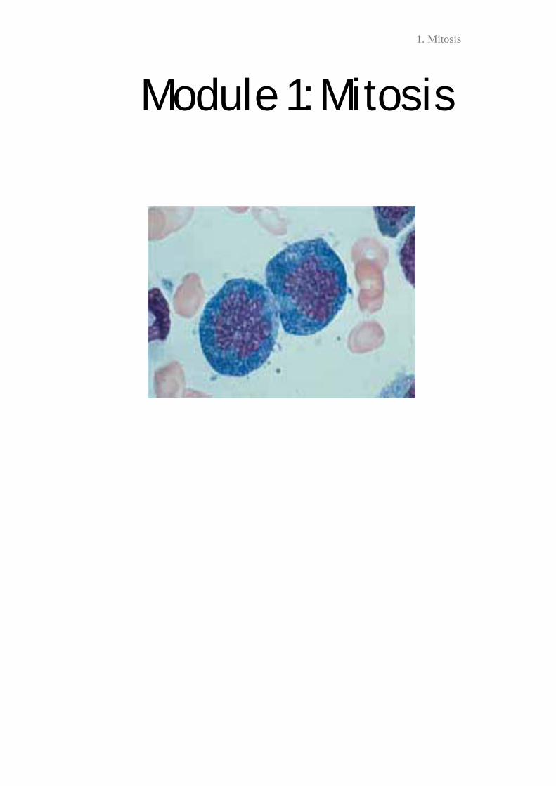
Module 1: Mitosis

1. Mitosis

1. Mitosis
Key Concepts 1. Cell division is used to reproduce single celled organisms and for growth and repair in multi-celled organisms 2. Most cell division results in genetically identical daughter cells 3. The cell cycle is the life of a cell from origin until it divides. The 2 main phases are interphase and the mitotic phase 4. The cell cycle is regulated by a molecular control system 5. Loss of cell cycle regulation can lead to cancer

2. Meiosis
7. Module 1 Case Study Adapted from Primary malignant melanoma of the esophagus: A case report. Joana Machado, Paula Ministro, Ricardo Araújo, Eugénia Cancela, António Castanheira, and Américo Silva. World J Gastroenterol. 2011 Nov 14; 17(42): 4734–4738. An 87-year-old Caucasian male was admitted to the emergency room with hematemesis. He had a history of intermittent dysphagia during the previous month. A CT scan was performed to characterise the nature of the lesion. Chest computed tomography demonstrates a solid mass in the middle third of the esophagus (arrow) causing obstruction and proximal dilatation. The distal third of the esophagus (arrowheads) is unremarkable. Endoscopic evaluation revealed a soft oesophageal lesion located 25-35 cm from the incisors, which appeared as a protrusion of the oesophagus wall, with active bleeding. A closer view of the lesion shows that tumour surface is darker than oesophageal mucosa due to its melanin content.
Biopsies were acquired. A: Hematoxylin and eosin staining discloses crowded rounded or elongated cells within distinct cytoplasm and hyperchromatic nuclei (magnification, x 100); B: Immunohistochemical study for HMB45 (anti-melanoma protein mAb). The positive reaction (brown cytoplasmic reaction) supports the diagnosis of malignant melanoma (magnification, x 100).
Tissue evaluation was compatible with a melanoma. After excluding other sites of primary neoplasm, the definitive diagnosis of Primary Malignant Melanoma of the Oesophagus (PMME) was made.
2. Meiosis
Primary malignant melanoma of the oesophagus is an extremely rare, neoplasm, with an incidence of 0.0036 cases per million people each year. Most cases occur in individuals >60years old. 1. Why is it more likely to occur in older individuals? Despite the significant advances in surgical techniques and chemoradiation therapy, the prognosis for patients with oesophageal cancer remains poor. The reported overall 5-year survival rates are at best 10-15%. 2. From what you understand about malignancy, provide a likely explanation for the low survival rate of this (and other) malignant cancers. An important aspect of diagnosis is microscopic visualisation of cells from a tissue biopsy 3. What characteristics of the cells are typical of cancer cells and what role do these particular characteristics play in cancer?
2. Meiosis
Module 1 Learning Objectives 1. Describe in detail the genomes of prokaryotes and eukaryotes. 2. Understand the terminology used in chromosome structure 3. Define telomeres, chromatids, centromeres, chromatin, heterochromatin and euchromatin 4. List the phases of the cell cycle and describe the sequence of events that occurs during each phase. 5. Explain the major events of cell division that enable the genome of one cell to be passed on to two daughter cells. . 6. Explain the phases of mitosis and describe the events characteristic of each phase. 7. Recognise the phases of mitosis from diagrams and micrographs. 8. Explain the process of binary fission in bacteria 9. Describe how the cell cycle is regulated. 10. Explain how loss of cell cycle control results in cancer 11. Distinguish between benign, malignant, and metastatic tumours. 12. Explain how cancer cells differ from normal cells. 13. Explain ER+ and HER2+ in relation to breast cancer 14. Explain why chemotherapeutic drugs that inhibit the cell cycle may be useful to treat cancer. 15. Explain why chemotherapeutic drugs can have severe side-effects. 16. Explain why cancer is considered a genetic disease 17. Relate your theoretical understanding of cell division and cell cycle control to real-life examples as presented in relevant case studies & critical thinking scenarios and propose answers to related questions.

2. Meiosis
MODULE 2: Meiosis

2. Meiosis
2. Meiosis
Key Concepts 1. Offspring inherit traits from parents due to genes on chromosomes 2. Sexually reproducing organisms use meiosis to produce gametes, which are combined from two different parents in fertilisation 3. Meiosis reduces the chromosome sets from diploid to haploid 4. Sexual reproduction produces genetic variation, important in evolution

10. Evolution III
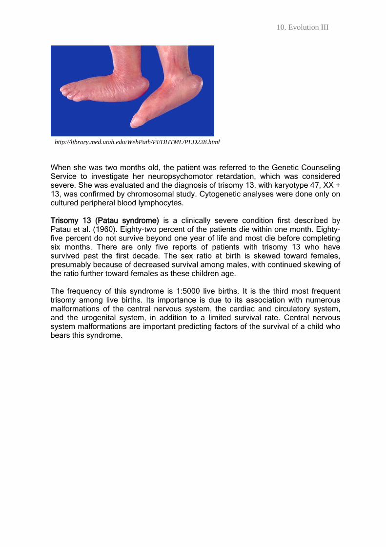
5. Module 2 Case Study (to be discussed in tutorial) Adapted from Patau syndrome with a long survival. A case report. A.C. Duarte, A.I.C. Menezes, E.S. Devens, J.M. Roth, G.L. Garcias and M.G. Martino-Roth. Genet. Mol. Res. 2004. 3 (2): 288-292. A child was born at term, from a vaginal delivery, and weighed 2,600 g. At birth, she was cyanotic (blue due to lack of oxygen in the blood), icteric (jaundice), spastic, and cried weakly. The initial clinical examination detected polydactyly (extra fingers) in the left hand, congenital clubfoot, convex foot soles and cardiac pathologies. There was no cleft lip or palate. Polydactyly was corrected on the child’s third day of life. Jaundice was treated and the left foot was immobilised. The infant was kept in an incubator for 19 days. Muscular hypertonia was observed during the first days of life; this changed to hypotonia at four months. The cardiac pathologies were initially treated with medication and the patient was under the care of a cardiologist until reparative surgery was done at 20 months.
http://www.orthopaedicsurgeon.com.sg/patients-education/childrens-orthopaedics/polydactyly/
http://orthoinfo.aaos.org/Module.cfm?Module=a00255
10. Evolution III
When she was two months old, the patient was referred to the Genetic Counseling Service to investigate her neuropsychomotor retardation, which was considered severe. She was evaluated and the diagnosis of trisomy 13, with karyotype 47, XX + 13, was confirmed by chromosomal study. Cytogenetic analyses were done only on cultured peripheral blood lymphocytes. Trisomy 13 (Patau syndrome) is a clinically severe condition first described by Patau et al. (1960). Eighty-two percent of the patients die within one month. Eighty-five percent do not survive beyond one year of life and most die before completing six months. There are only five reports of patients with trisomy 13 who have survived past the first decade. The sex ratio at birth is skewed toward females, presumably because of decreased survival among males, with continued skewing of the ratio further toward females as these children age. The frequency of this syndrome is 1:5000 live births. It is the third most frequent trisomy among live births. Its importance is due to its association with numerous malformations of the central nervous system, the cardiac and circulatory system, and the urogenital system, in addition to a limited survival rate. Central nervous system malformations are important predicting factors of the survival of a child who bears this syndrome.
http://library.med.utah.edu/WebPath/PEDHTML/PED228.html

10. Evolution III
1. Consider the human lifecycle and explain at what stage/s this chromosomal abnormality could occur.
2. Consider the cell cycle and explain at what stage/s this chromosomal abnormality could occur.
10. Evolution III
Module 2 Learning Objectives
1. Explain why humans resemble their parents but are not identical to them 2. Outline what is meant by the term karyotype and write and describe a human karyotype 3. Explain karyotyping and describe how chromosomes are recognised 4. Describe the concept of ploidy
5. Describe how the chromosome number changes throughout the human life cycle 6. Compare and contrast sister chromatids to homologous chromosomes 7. Explain the phases of meiosis and describe the events characteristic of each phase 8. Recognize the phases of meiosis from diagrams and micrographs 9. Define synapsis, crossing over, tetrad, chiasmata, Holliday junction and at which phase of meiosis these occur 10. Distinguish between mitosis and meiosis 11. Compare meiosis in female mammals to male mammals. 12. Explain how independent assortment, crossing over, and random fertilisation contribute to genetic variation in sexually reproducing organisms 13. Calculate the number of possible combinations of when chromosomes assort independently during meiosis (2n) 14. Explain the evolutionary significance of genetic variation 15. Explain the chromosomal basis of trisomy 13 (Patau syndrome) and describe how and when this chromosomal abnormality could occur.