Introduction to Fungi - KSU
24
Introduction to Fungi Classification, Morphology and Pathogenicity
Transcript of Introduction to Fungi - KSU

Introduction to Fungi
Classification, Morphology and Pathogenicity
• Mykes (Greek word) : Mushroom
• Differ from bacteria and other prokaryotes:
1. Cell walls containing chitin (rigidity & support), mannan &
other polysaccharides
2. Cytoplasmic membrane contains Ergosterols
3. Possess true nuclei with nuclear membrane & paired
chromosomes
4. Divide asexually, sexually or by both
5. Unicellular or multicellular
Diverse group of chemoheterotrophs
Over 100,000 fungal species identified
Only about 100 are human or animal pathogens
Most human fungal infections are nosocomial and/or occur in
immunocompromised individuals (opportunistic infections)
Saprophytic fungus
Parasitic fungus
Most are aerobes or facultative anaerobes

Simplest fungus >> Unicellular budding
yeast
Hyphae
Mycelium
Aerial Mycelium
Vegetative mycelium
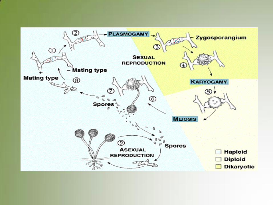
Reproduction in fungi
A. Sexual
B. Asexual reproduction
C. Asexual spores
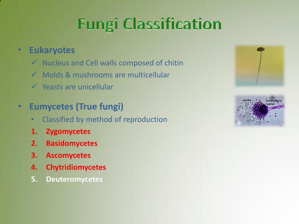
• Eukaryotes Nucleus and Cell walls composed of chitin
Molds & mushrooms are multicellular
Yeasts are unicellular
• Eumycetes (True fungi) • Classified by method of reproduction
1. Zygomycetes
2. Basidomycetes
3. Ascomycetes
4. Chytridiomycetes
5. Deuteromycetes
• Depending on Morphology
A. Yeasts
B. Yeast like fungi
C. Molds
D. Dimorphic fungi
E. Fleshy fungi

• Unicellular, Nucleated rounded fungi
• Reproduce by budding
• Colony on solid media are usually white to beige and appear much like bacterial colonies
• Some genera produce mucoid colonies
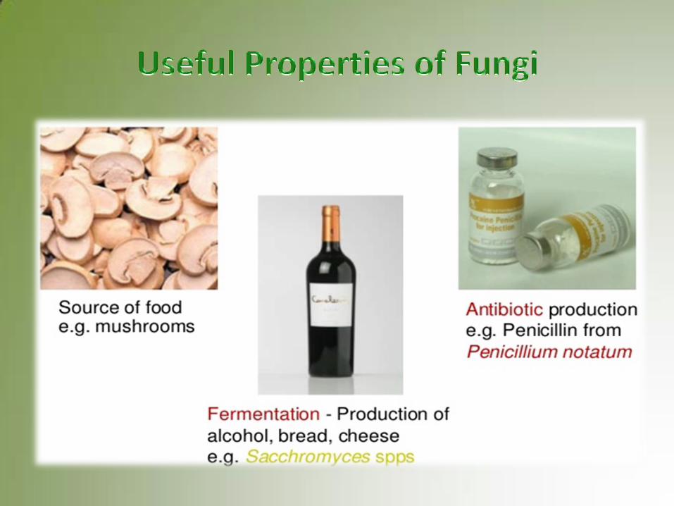
• Yeast are used in the preparation in the variety of foods
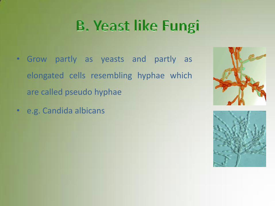
• Grow partly as yeasts and partly as
elongated cells resembling hyphae which
are called pseudo hyphae
• e.g. Candida albicans

• Multicellular, Filamentous with hyphae
• Produce conidia [spores]
• Colonies on solid agar are downy, fluffy, cottony
• Most mold colonies are pigmented which aid in identification hyphae spores
• Penicillium and Cephalosporium
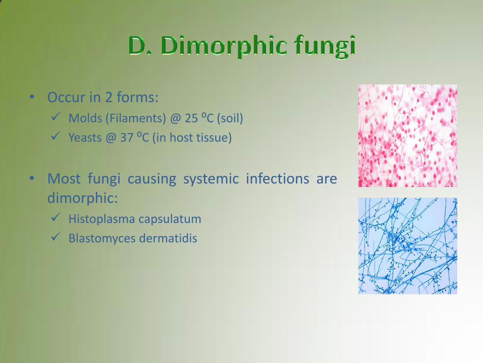
• Occur in 2 forms: Molds (Filaments) @ 25 ⁰C (soil)
Yeasts @ 37 ⁰C (in host tissue)
• Most fungi causing systemic infections are dimorphic: Histoplasma capsulatum
Blastomyces dermatidis
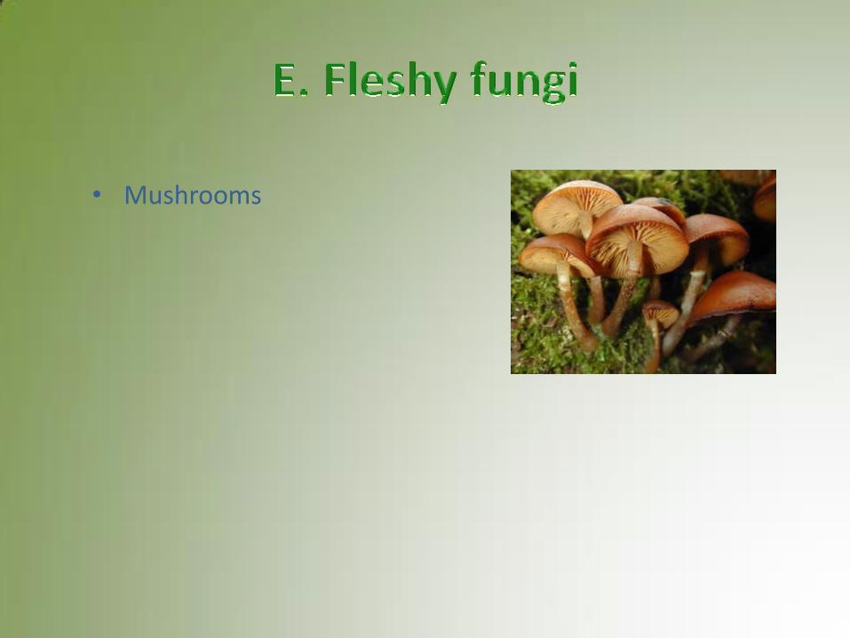
• Mushrooms
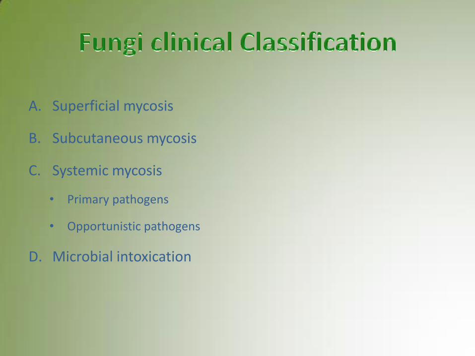
A. Superficial mycosis
B. Subcutaneous mycosis
C. Systemic mycosis
• Primary pathogens
• Opportunistic pathogens
D. Microbial intoxication
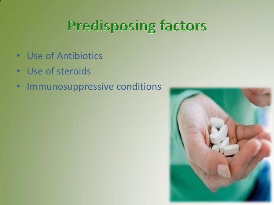
• Use of Antibiotics
• Use of steroids
• Immunosuppressive conditions
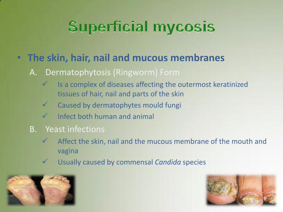
• The skin, hair, nail and mucous membranes
A. Dermatophytosis (Ringworm) Form Is a complex of diseases affecting the outermost keratinized
tissues of hair, nail and parts of the skin
Caused by dermatophytes mould fungi
Infect both human and animal
B. Yeast infections Affect the skin, nail and the mucous membrane of the mouth and
vagina
Usually caused by commensal Candida species
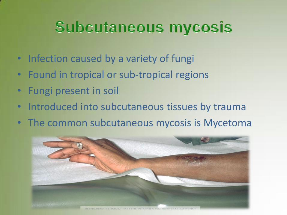
• Infection caused by a variety of fungi
• Found in tropical or sub-tropical regions
• Fungi present in soil
• Introduced into subcutaneous tissues by trauma
• The common subcutaneous mycosis is Mycetoma

– Deep seated fungal infection
– Occur through inhalation of air-borne spores
– Initially as a pulmonary infection
– Then may disseminated to other organ
– Caused by: • Primary pathogens
• Opportunistic pathogens

• 20 % KOH in water
• Gram stain
• India ink
• Calcoflour white (Electron Microscope)
• Giemsa staining
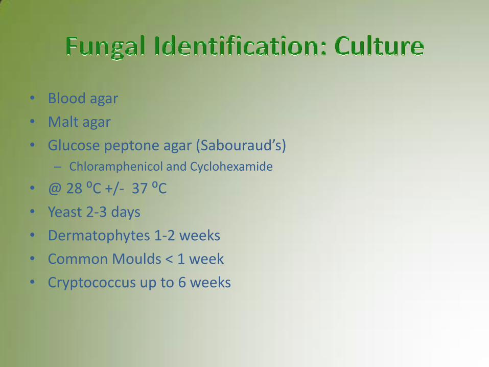
• Blood agar
• Malt agar
• Glucose peptone agar (Sabouraud’s) – Chloramphenicol and Cyclohexamide
• @ 28 ⁰C +/- 37 ⁰C
• Yeast 2-3 days
• Dermatophytes 1-2 weeks
• Common Moulds < 1 week
• Cryptococcus up to 6 weeks
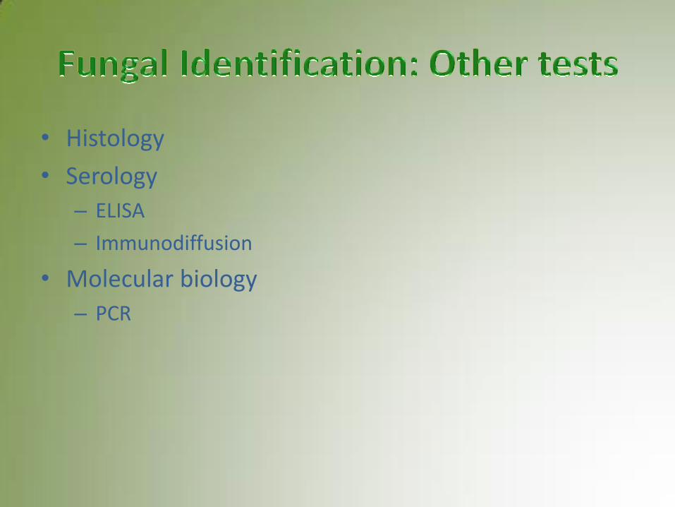
• Histology
• Serology
– ELISA
– Immunodiffusion
• Molecular biology
– PCR
• Most antifungal agent are for topical use
• Few administrated systemically