Introduction to Anatomy and Physiology (HK24CY005)
33
1 Introduction to Anatomy and Physiology (HK24CY005) Anatomy is about discovering how we are made, and where the different organs and parts of our body are located. Physiology is about understanding what the different organs do and how they inter- relate with other parts of the body. We are all made up of the same materials, yet each one of us is truly unique. This is one of the miracles of life – how there can be so many variants, even though we all (well, most of us) have two eyes, a nose a mouth and two ears. So what is it that makes our bodies how they are? Let’s start with the basic building blocks: Cells are the basis of all our bodies. They knit together to form all the different structures, tissues and organs that we need to function. The structure of a cell includes a nucleus, surrounded by protoplasm, encased by a cell membrane. Different cells have specialist functions, depending on the job they are required to do, and as a result, may also have different shapes, and may be referred to as specialised cells. Tissue is the collective term for a mass of cells specialised to perform a particular function. Most people think of tissue as soft, like skin or muscle, but tissue can be any part of the body. Different types of tissue are bone, cardiac, muscular, nervous, blood, connective or subcutaneous (tissue that lies under the skin). Different types of tissue are needed for the different jobs they have to do. Organs are complex structures that have a specific function. They are composed of more than one type of tissue. Better known organs include: the heart, lungs, eyes, kidneys and brain. Without organs, life would either be very difficult, or impossible. This is why in many cases organs come in pairs, so that if one fails, the other can take over the whole job. Systems encompass a group of organs and tissues, all functioning together to accomplish their task. So the circulatory system would include the heart (organ) and all the arteries, veins and blood (tissues). The respiratory system would include the lungs (organ), bronchial tubes and trachea (tissues). The nervous system would include the brain (organ) the spinal cord and all the nerves that branch off it (tissues).
Transcript of Introduction to Anatomy and Physiology (HK24CY005)

1
Introduction to Anatomy and Physiology (HK24CY005)
Anatomy is about discovering how we are made, and where the different organs and
parts of our body are located.
Physiology is about understanding what the different organs do and how they inter-
relate with other parts of the body.
We are all made up of the same materials, yet each one of us is truly unique. This is one
of the miracles of life – how there can be so many variants, even though we all (well,
most of us) have two eyes, a nose a mouth and two ears. So what is it that makes our
bodies how they are? Let’s start with the basic building blocks:
Cells are the basis of all our bodies. They knit together to form all the
different structures, tissues and organs that we need to function. The
structure of a cell includes a nucleus, surrounded by protoplasm,
encased by a cell membrane. Different cells have specialist functions,
depending on the job they are required to do, and as a result, may also
have different shapes, and may be referred to as specialised cells.
Tissue is the collective term for a mass of cells specialised to perform a
particular function. Most people think of tissue as soft, like skin or
muscle, but tissue can be any part of the body. Different types of tissue
are bone, cardiac, muscular, nervous, blood, connective or subcutaneous
(tissue that lies under the skin). Different types of tissue are needed for
the different jobs they have to do.
Organs are complex structures that have a specific function. They are
composed of more than one type of tissue. Better known organs
include: the heart, lungs, eyes, kidneys and brain. Without organs, life
would either be very difficult, or impossible. This is why in many cases
organs come in pairs, so that if one fails, the other can take over the
whole job.
Systems encompass a group of organs and tissues, all functioning
together to accomplish their task. So the circulatory system
would include the heart (organ) and all the arteries, veins and
blood (tissues). The respiratory system would include the lungs
(organ), bronchial tubes and trachea (tissues). The nervous
system would include the brain (organ) the spinal cord and all the
nerves that branch off it (tissues).

2
All the body’s systems are connected to each other via the Central Nervous System
(CNS). This enables each independent system to react to the needs of the body and
keep it in optimum condition.
Anatomical Terms of Reference
To make things easier when describing positions of the body, clinical terms of
reference have been devised that are used by all health professionals. All descriptions
of the body assume that the person is standing in the anatomical position. This is
standing erect, head, eyes and toes directed forward, the heels and toes together. The
upper limbs are hanging by the sides with the palms of the hands facing forward.
Median Plane: this is when the body, positioned in the anatomical position, has been
divided longitudinally down through the midline, separating the body into right and left
halves.
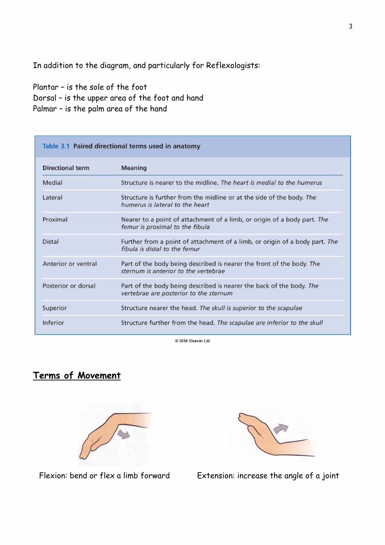
3
In addition to the diagram, and particularly for Reflexologists:
Plantar – is the sole of the foot
Dorsal – is the upper area of the foot and hand
Palmar – is the palm area of the hand
Terms of Movement
Flexion: bend or flex a limb forward Extension: increase the angle of a joint

4
Abduction: move away from the midline Adduction: move closer to the midline
Plantar flexion: flexing foot toward the
ground
Dorsiflexion: bending foot up
Supination: to turn the palm up Pronation: to turn the palm down
Inversion: turning towards the centre Eversion: turning outwards away from
centre
Rotation: to move a bone around its
longitudinal axis

5
Cavities of the Body
The body is divided into four main areas: the cranial, thoracic, abdominal and pelvic
cavities.
Cranial Cavity – this is bordered by the
skull and contains the brain.
Thoracic Cavity - this is the upper part of the
trunk. It is bordered by the sternum (anteriorly),
the ribs with associated intercostal muscles and
costal cartilages, the thoracic vertebrae
(posteriorly), the root of the neck (superiorly),
and the diaphragm (inferiorly).
The main organs located in the thoracic cavity are
the lungs and heart.
Abdominal Cavity – this cavity forms the main part of the trunk and is the largest body
cavity. It is bordered by the diaphragm (superiorly), the muscles of the anterior
abdominal wall (anteriorly), the lumbar vertebrae and muscles of the posterior
abdominal wall (posteriorly), the lower ribs and muscles of the abdominal wall (laterally),
and inferiorly it is continuous with the pelvic cavity. The main organs and associated
structures found in the abdominal cavity are: the stomach, small intestine and most of
the large intestine, the liver, gall bladder, bile ducts, pancreas, spleen, the kidneys, and
the adrenal glands.
The anterior abdominal cavity The posterior abdominal and pelvic cavities

6
Pelvic Cavity - this cavity is situated
below the abdominal cavity. It borders
with the pubic bones (anteriorly), the
abdominal cavity (superiorly), the
sacrum and coccyx (posteriorly), the
innominate bones (laterally) and the
muscles of the pelvic floor (inferiorly).
It contains the sigmoid colon, rectum,
anus, some loops of the small intestine,
the urinary bladder, and the lower
parts of the ureters and the urethra. The pelvic cavity also contains part of the
reproductive system, so varies in the male and female. The female pelvic cavity also
contains the uterus, uterine tubes, ovaries and vagina.
The male pelvic cavity also contains the prostate
gland, seminal vesicles, spermatic cords, deferent
ducts (vas deferens), ejaculatory ducts and the
urethra.
7
Homeostasis (3.1)
You will frequently hear therapists say that the goal of their treatment is to bring
about homeostasis. But what do they mean? In simplistic terms they want to bring
about a state of balance and harmony to the body. Homeostasis is a physiological
process by which the internal systems of the body (e.g. blood pressure, body
temperature, and acid-base balance) are maintained at equilibrium, despite variations in
the external conditions. In other words, the internal environment (i.e. what is going on
inside the body) needs to be kept within certain parameters for the body to remain
healthy. Lots of factors outside the body (the external environment) may influence
how the body functions, and the body may need to overcome these factors to work
efficiently.
Example:
The body wants to maintain a constant temperature of approximately 36.8°C.
On a very hot day, the body needs to cool itself, to prevent
the core temperature from rising. It can do this by sweating
and re-directing blood flow to just under the skin, to try and
lose heat from the blood.
Conversely on a very cold day, the blood flow is directed away from the
skin to the vital organs in order to conserve heat, and shivering may occur
which helps to generate heat. These mechanisms are homeostatic
processes to try to keep the internal environment stable when the
external environment varies.

8
ORGANISATION OF THE BODY
Cells
Every living organism is made from a collection of cells, each evolving to carry out
specific functions and becoming a specialised cell. When cells group together, they
form specialised tissues. The cell is the smallest living organism in the body and,
depending on its structure, will have a specific job to do. The proper term for the study
of cells is “cytology”.
Below is a generalised picture of a cell to show the different possible organelles that
could be present.
Organelles of a Cell
Each cell consists of an outer membrane and inside this is a gel-like fluid called
cytoplasm in which float many different organelles. The organelles are like ‘little
organs’, each having a specific job to do in order for the cell to function. The largest
of all the organelles is the nucleus, and the nucleus contains the cells genetic material,

9
the DNA. The nucleus is the control centre directing the functions of the cell. The
membrane of a cell is semi-permeable - it will allow certain substances to pass through,
so that it can obtain the substances it requires from its environment (and expel waste
products). However, the membrane is impermeable to some substances, in order to
protect the cell.
The amount of organelles in a cell will vary, depending upon the cell’s functions. All
body cells contain a nucleus, with the exception of red blood cells (erythrocytes).
Skeletal muscle cells may have several nuclei, and very active cells such as liver, muscle
and spermatozoa will contain large numbers of mitochondria (which are required for
energy production). Certain body cells may have projections on their plasma membrane,
called cilia to help them expel foreign particles, such as those found in the respiratory
tract. The cells of the body can adapt in shape, function and content, to better enable
them to perform the specific tasks which they are required to do.
Cell Reproduction
All cells grow and die, so for the body to remain healthy, cells need to renew
themselves.
All body cells, with the exception of the reproductive cells, have diploid (or two
complete sets) of chromosomes, called somatic cells. Human cells contain 46
chromosomes in each. However, the reproductive cells (spermatozoa and ova) contain
only 23 chromosomes in each.
The way a somatic cell reproduces is by splitting into two, thereby ending up with a
perfect copy of itself. This process is called mitosis and it takes about two hours from
start to finish.
A different process called meiosis creates the reproductive cells (spermatozoa and
ova). Rather than an exact copy of the original cell being produced by splitting, a more
complicated procedure occurs where the DNA strands swap some of their genetic
material, creating new combinations, so that all the spermatozoa and ova have a
different combination of DNA, which enables new individuals to be born following
sexual reproduction.
How Does a Cell Get its Energy?
A cell cannot eat, as we would define eating. The materials/substances a cell needs have
to pass through the cell membrane and, depending on the substance, this is done in
various ways. These processes are also used to enable the cell to get rid of waste
products.

10
Transportation of Substances Across Cell Membranes
It is in the nature of all gaseous or liquid substances to want to spread out and fill the
space they are in. If there is a high concentration of a substance in an area, but a low
concentration of that same substance in an adjoining area, it will move down its
concentration gradient (like going downhill) until there is an equal concentration of it in
both areas (this is called equilibrium). This ability of substances to move forms the
basis of how substances can transfer across the cell membranes.
The simplest way for a substance to cross the membrane is by diffusion. This is the
movement of a substance down its concentration gradient, provided the membrane is
permeable to the substance. The movement of water is known as osmosis, which is a
type of diffusion.
Sometimes a cell requires substances to pass through their membrane, against their
concentration gradient. This cannot occur passively, therefore it requires energy to
move the substances, and this type of movement is called active transport. One major
type of active transport used by the body is the sodium-potassium pump. Active
transport is particularly important in maintaining a charge on the cell membrane,
required for nerve impulse transmission.
Some particles are too large to pass across a cell membrane, so instead the cell has to
engulf the particle; this is called endocytosis, of which there are two types: pinocytosis
for small particles; or phagocytosis for larger particles (such as cell fragments, foreign
materials or microbes). Particles may be brought into the cell for digestion, or
destruction. The reverse of this, to expel particles from the cell is called exocytosis.
Body Fluids
The human body is largely made up of fluids, 60% of the average weight of an adult
being water. Children, babies and underweight adults have a higher proportion of
water, whereas obese people and the elderly have a lower proportion of water. About

11
22% of this water is extracellular (i.e. outside the cells) and 38% of it is intracellular
(i.e. inside the cells). Many functions of the body take place in a fluid environment. It
is important; therefore, that we take in sufficient water for our bodies to function
properly, and that the concentrations of body fluids remain at the correct levels for
these bodily functions to work properly.
Extracellular Fluid (ECF)
The main extracellular fluids of the body are: blood, plasma, lymph, cerebrospinal fluid
and interstitial/intercellular fluid (this is the tissue fluid that bathes all cells of the
body, with the exception of the outer layers of the skin). Other extracellular fluids
found in lesser amounts are: synovial fluid (joint fluid), pericardial fluid (around the
heart) and pleural fluid (around the lungs). The interstitial fluid delivers nutrients to
the individual cells of the body, and all other fluids have equally important rolls. The
composition of each of the different fluids vary, however, the exact composition of
each is essential to the job that fluid performs. The body, therefore, has many
homeostatic mechanisms in place to keep body fluids at the correct composition.
Intracellular Fluid (ICF)
The composition of the fluids inside the cells is quite different from the fluids outside
the cells. This difference is maintained largely by the sodium-potassium pump and this
is needed for the correct functioning of the cells (especially muscle and nerve cells).
The cell membrane is also selectively permeable, i.e. it only allows certain substances to
pass through it, which also helps maintain the differences. Inside the cells the
potassium, ATP (energy source), and protein is higher than outside, whereas the sodium
levels are higher outside the cells.
These three pictures show how body cells can be affected if the extracellular fluid
concentrations are not correct.
A – The fluid is at the right concentration so the cell can function
properly.
B – The fluid is too dilute and water goes into the cell. There is a risk
that the cell may burst.

12
C – The fluid is too concentrated and fluid leaves the cell. The
intercellular fluid would no longer contain sufficient water to enable cell
functions to occur and the cell would die.
pH of Body Fluids
pH is measured on a scale of 0 to 14. The pH of a fluid is 7 if the fluid is neutral, e.g.
water, below 7 if it is acidic, e.g. hydrochloric acid, or above 7 if it is an alkali (also
called a base), e.g. ammonia solution.
Chemical reactions occur at specific pH values, but if the pH is wrong, then these
reactions either don’t happen, or are less efficient. Our bodies function as a result of
lots of chemical reactions, which means that the pH of fluid must be within a specific
range for particular reactions to occur. Therefore, different body fluids are
maintained within certain ranges to enable reactions to occur within those tissues.
Below is a table containing the pH values of some of the more common body fluids. Note
that gastric juice found in the stomach is highly acidic (i.e. a low pH value). This is
important for activation of certain enzyme reactions in the stomach and it is also good
at killing invading micro-organisms (bacteria/viruses/fungi) which may have been
ingested in our food, helping to keep our bodies safe from infection.

13
Tissues
There are various types of tissue found in the body, each
having specialised functions. The proper term for the
study of tissues is histology.
Epithelial Tissue (Soft Tissue)
Epithelial tissue covers the body surfaces (internal and
external), lines hollow organs, cavities and ducts, and it
forms glands. Its functions therefore include protection,
absorption, filtration and secretion, depending upon its
location. Epithelial tissue may be either wet (such as that
which lines our mouths) or dry (waterproof and found on
the top layer of skin).
A variety of different epithelial cell shapes can be found:
squamous, cuboidal, columnar and transitional. Epithelium
varies depending on its function, and is sometimes only a
single cell layer thick (simple) e.g., found in the alveoli; or
may be several layers thick (stratified), e.g. in the skin.
Some epithelium may be specialised to change shape
(transitional), e.g. found in the bladder; or have projections
on to help move particles (ciliated), e.g. found in the
fallopian tubes.
Connective Tissue
Connective tissues are the supporting tissues of the body. They provide protection,
transport, binding, structural support or insulation. The cells in connective tissue are
surrounded by a matrix made up of protein fibres and a fluid, gel or solid ground
substance, which determines whether the tissue is a liquid, semi-solid or liquid. There
are many types of connective tissue:
Areolar (or loose) connective tissue
Adipose tissue
Lymphoid tissue (reticular tissue)
Yellow elastic tissue (elastic connective tissue)
White fibrous tissue (dense regular connective tissue)
Bone
Blood
Hyaline Cartilage
14
Yellow elastic cartilage (elastic fibrocartilage)
White fibrocartilage
Nervous Tissue (Soft Tissue)
Nerve tissue is arranged in bundles of fibres composed of nerve cells. These fibres run
throughout the body like an electrical wiring system, and send the brain information
about the state of the body and its environment. Further information is found in the
session on the ‘Nervous System’.
Muscular Tissue (Soft Tissue)
There are three types of muscular tissue:
Striated
Smooth
Cardiac
Further information is found in the session on the ‘Muscular System’.
Tissue Regeneration
Some body tissues get worn out and need to regenerate. This requires new cells to be
produced. The actual function of the tissue will determine whether or not they can
regenerate.
Some tissues have a high turnover of cells, so they need to replicate continuously in
order to replace the cells that are dying, e.g. skin and mucous membranes. Some cells
of the body although they can still replicate, they do so more slowly, e.g. the liver,
kidney and smooth muscles. Certain types of tissue cannot regenerate, e.g. nerve cells,
skeletal and cardiac muscles cells. In situations where these tissues are damaged, and
the normal tissue cells cannot be replicated, cellular replacement is usually by fibrous
tissue. This reduces the functionality of the particular tissue.
Types of Membrane
Membranes are thin sheets that line cavities, cover surfaces and provide protective
layers around organs. They can either be epithelial membranes or connective tissue
(synovial) membranes.
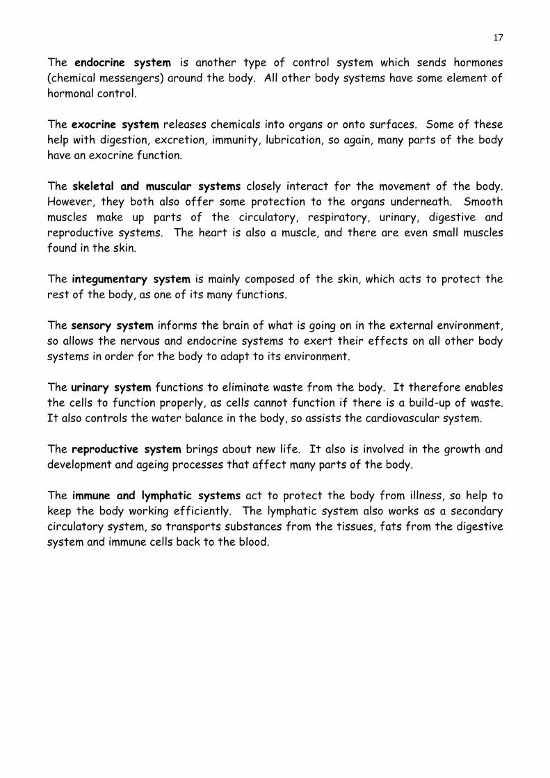
Mucous membrane (mucosa) – this is a moist
epithelial membrane. Goblet cells in the
epithelial layer secrete a fluid called mucous.
This fluid prevents the membrane from drying

15
out and also acts as lubrication, protecting against mechanical and chemical injury to
the underlying cells. In the respiratory tract, mucous helps trap foreign particles so
that they can be expelled. Mucous membranes line body cavities that open to the
exterior, so they are found in the digestive, respiratory, urinary and reproductive
systems.
Serous membrane (serosa) – this is also an
epithelial membrane. With serous membranes
there are two layers of areolar connective
tissue lined by a simple squamous epithelium.
Between the two layers a serous watery fluid is
secreted. The two layers are called the parietal
layer (lines a cavity) and the visceral layer
(surrounds the organs). The fluid between the
layers enables an organ to glide freely within a
cavity without being damaged by friction, so
these membranes are found around organs which need to change shape, such as around
the heart (the pericardium), the lungs (the pleura) or the abdominal cavity (the
peritoneum).
Synovial membrane – is a connective tissue membrane. It is
made up of areolar connective tissue, elastic fibres and fat.
Synovial fluid (a clear, sticky, oily fluid) is secreted into the
cavity that the membrane is lining and acts as lubrication for
movement or cushioning. Synovial membranes are found
lining the cavities of freely moveable joints and surrounding
the tendons, preventing damage from rubbing.
Glands
Glands are a group of epithelial cells secreting specialised substances. They are divided
into two specific types:
Endocrine glands – secrete their substances (hormones) directly into the blood or
lymph. Further information is found in the session on the ‘Endocrine system’.
Exocrine glands – discharge their secretions via a duct, or directly onto the epithelial
surface of a hollow organ. Secretions of exocrine glands include: mucus, digestive
juices, saliva and earwax.

16
Systems of the Body
The body is split into several systems according to the type of function they do. We
will be studying the following systems:
Digestive
Exocrine
Endocrine
Nervous
Skeletal
Muscular
Cardio-vascular and circulatory
Respiratory
Sensory
Integumentary
Immune
Lymphatic
Urinary
Reproductive
Although each system is studied separately, all the systems interact and are
interdependent on each other in order to allow the body to function. Below are some of
the ways the different systems interact.
The digestive system absorbs and processes food, and removes waste products. Every
cell of the body requires nutrients and energy, which are derived from food, and
removal of its waste, therefore the whole body is dependent (i.e. all other body
systems) on the digestive system functioning properly.
The respiratory system brings oxygen into the body, which all cells require to function,
and it removes carbon dioxide (waste gas) from the body, so again, every cell and
therefore body system is reliant on the respiratory system.
The cardiovascular/circulatory system is the body’s main transport system. It
transports nutrients, oxygen and waste products to all the cells, therefore enabling all
the cells to work efficiently. The digestive and respiratory systems could not get their
products to and from the cells without the circulatory system. It also transports white
blood cells and platelets, which are important for the body’s immunity. It also
transports chemicals and hormones, so aids the endocrine system.
The nervous system sends messages from the brain to control the body, so affects all
other systems.
17
The endocrine system is another type of control system which sends hormones
(chemical messengers) around the body. All other body systems have some element of
hormonal control.
The exocrine system releases chemicals into organs or onto surfaces. Some of these
help with digestion, excretion, immunity, lubrication, so again, many parts of the body
have an exocrine function.
The skeletal and muscular systems closely interact for the movement of the body.
However, they both also offer some protection to the organs underneath. Smooth
muscles make up parts of the circulatory, respiratory, urinary, digestive and
reproductive systems. The heart is also a muscle, and there are even small muscles
found in the skin.
The integumentary system is mainly composed of the skin, which acts to protect the
rest of the body, as one of its many functions.
The sensory system informs the brain of what is going on in the external environment,
so allows the nervous and endocrine systems to exert their effects on all other body
systems in order for the body to adapt to its environment.
The urinary system functions to eliminate waste from the body. It therefore enables
the cells to function properly, as cells cannot function if there is a build-up of waste.
It also controls the water balance in the body, so assists the cardiovascular system.
The reproductive system brings about new life. It also is involved in the growth and
development and ageing processes that affect many parts of the body.
The immune and lymphatic systems act to protect the body from illness, so help to
keep the body working efficiently. The lymphatic system also works as a secondary
circulatory system, so transports substances from the tissues, fats from the digestive
system and immune cells back to the blood.
18
HANDS AND FEET HK24CY005 1.1
The main structures found in the hands and feet are bones, muscles, tendons and
ligaments. There is also a nerve supply, blood supply, and lymphatic drainage. The outer
surface that is seen is covered in skin, which also has a covering of fine hairs, with the
exception of on the palms and soles. Therefore, as you can see there is an interaction
of many body systems in order for the hands and feet to function. For today, we shall
look at the bones, muscles, tendons and ligaments of the hands and feet, but more
detail of muscles and bones can be found in the lessons on those systems.
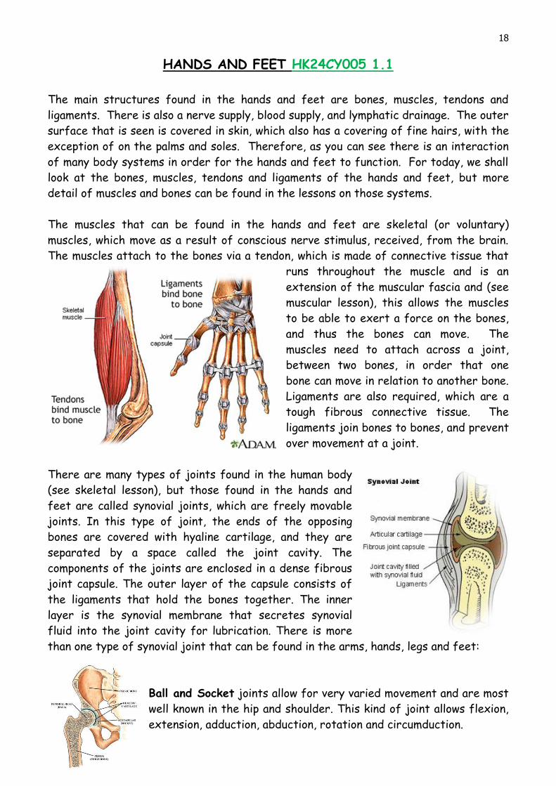
The muscles that can be found in the hands and feet are skeletal (or voluntary)
muscles, which move as a result of conscious nerve stimulus, received, from the brain.
The muscles attach to the bones via a tendon, which is made of connective tissue that
runs throughout the muscle and is an
extension of the muscular fascia and (see
muscular lesson), this allows the muscles
to be able to exert a force on the bones,
and thus the bones can move. The
muscles need to attach across a joint,
between two bones, in order that one
bone can move in relation to another bone.
Ligaments are also required, which are a
tough fibrous connective tissue. The
ligaments join bones to bones, and prevent
over movement at a joint.
There are many types of joints found in the human body
(see skeletal lesson), but those found in the hands and
feet are called synovial joints, which are freely movable
joints. In this type of joint, the ends of the opposing
bones are covered with hyaline cartilage, and they are
separated by a space called the joint cavity. The
components of the joints are enclosed in a dense fibrous
joint capsule. The outer layer of the capsule consists of
the ligaments that hold the bones together. The inner
layer is the synovial membrane that secretes synovial
fluid into the joint cavity for lubrication. There is more
than one type of synovial joint that can be found in the arms, hands, legs and feet:
Ball and Socket joints allow for very varied movement and are most
well known in the hip and shoulder. This kind of joint allows flexion,
extension, adduction, abduction, rotation and circumduction.

19
Ellipsoid joints are like a ‘cut down’ version of a ball and socket
joint. This kind of joint is found in the wrist, but the amount of
movement is not so great as a ball and socket joint.
Hinge Joints allow movement in only two directions (flexion
and extension). Think of a door hinge and you will get the
idea! A good example of a hinge joint is in the elbow or knee.
These are also found in the fingers and toes.
Saddle Joints allow all movement, except rotation. Saddle joints
are so called, because both surfaces look like a saddle (one
inverted and rotated 90º on top of the other). A good example of
a saddle joint would be found in the base of the thumb.
Gliding joints are so called because they allow bones
to glide at their joints and they have flat articulating surfaces –
where small amounts of flexibility are needed. Reflexologists and
podiatrists are most familiar with the metatarsal joints found in the
feet.
The Hands
According to Smith (2006), “Our hands do so much for us. They are capable of a wide
variety of functions: touching, grasping, feeling, holding, manipulating, caressing, and
more. They are a vitally important part of who we are and how we see ourselves.”
Our hands require a large portion of the
functioning of our brain, in relation to their size,
because they are so important to us.
On the right, from Life’s Journey in Words (2011)
is a “homunculus, a representation of how our brain

20
views our body. Tongue, face, fingers appear so huge because a bigger part of your
brain is devoted to their function and there are a huge number of sense receptors,
muscles, and nerves.”
Without the ability to hold objects and tools, the human race would not have developed
as it has done. Our hands can do both gentle actions and heavier labour. They also
contain many nerve endings so we can detect sensations. Without our hands, we would
not be able to perform a reflexology treatment. In addition, not only can the hands be
used to perform reflexology, but they also contain reflex points themselves, so
reflexology treatments can be done on the hands.
Structure of the Hand
Within the hands are many small bones, to allow for fine movements. Although there
are some muscles in the hands, much of the movement is created by muscles in the
arms, the tendons of which attach to the bones of the hands. This allows the hands to
be less bulky, and be able to grasp objects.
A mnemonic to help you remember the
bones:
Some Lovers Try Positions They Can’t
Handle
Or
Touching Toes Causes Happiness So
Let’s Touch Plenty
21
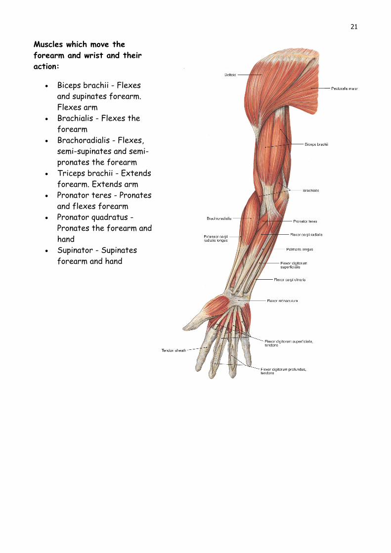
Muscles which move the
forearm and wrist and their
action:
Biceps brachii - Flexes
and supinates forearm.
Flexes arm
Brachialis - Flexes the
forearm
Brachoradialis - Flexes,
semi-supinates and semi-
pronates the forearm
Triceps brachii - Extends
forearm. Extends arm
Pronator teres - Pronates
and flexes forearm
Pronator quadratus -
Pronates the forearm and
hand
Supinator - Supinates
forearm and hand

22
In addition to the muscles shown in the above diagrams, it can be seen how the tendons
(shown as white) extend down into the hands and there is actually little muscle within
the hand. The flexor retinaculum is a band of ligaments around the wrist. The tendons
go beneath this band of ligament, so the ligaments hold the bones, muscles and tendons
in place during the movement of the wrist, hand and fingers.

23
The Feet
The feet’s main function is to support the body and allow us to walk and run. Within
reflexology, we regard the feet as a very important area for treatment, since the
whole body can be mapped onto the foot - so just by working on the feet, the whole
person can be affected.
Structure of the Feet
The bones of the feet do not go flat to the floor - they have arches. The arches allow
an element of springiness to aid in walking and running. Like the hands, the feet also
have many small bones to allow flexibility, yet they are generally less flexible than the
hands, so less able to manipulate objects. Also, similar to the hands, only some of the
movement comes from muscles in the foot, as muscles in the legs (attached by tendons)
control many of the bones.
24
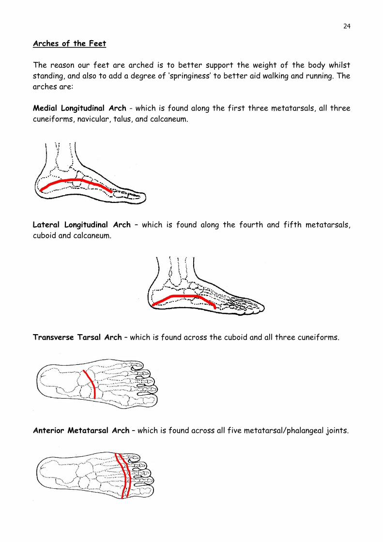
Arches of the Feet
The reason our feet are arched is to better support the weight of the body whilst
standing, and also to add a degree of ‘springiness’ to better aid walking and running. The
arches are:
Medial Longitudinal Arch - which is found along the first three metatarsals, all three
cuneiforms, navicular, talus, and calcaneum.
Lateral Longitudinal Arch – which is found along the fourth and fifth metatarsals,
cuboid and calcaneum.
Transverse Tarsal Arch – which is found across the cuboid and all three cuneiforms.
Anterior Metatarsal Arch – which is found across all five metatarsal/phalangeal joints.
25
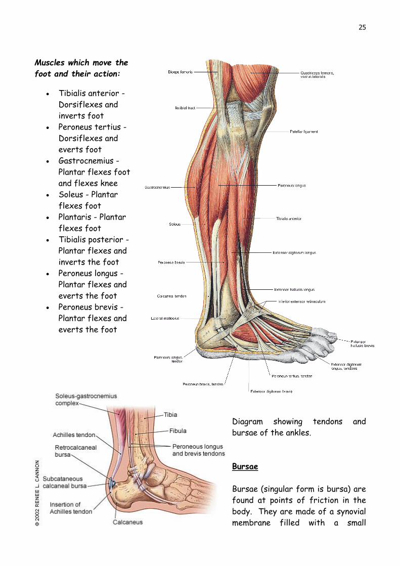
Muscles which move the
foot and their action:
Tibialis anterior -
Dorsiflexes and
inverts foot
Peroneus tertius -
Dorsiflexes and
everts foot
Gastrocnemius -
Plantar flexes foot
and flexes knee
Soleus - Plantar
flexes foot
Plantaris - Plantar
flexes foot
Tibialis posterior -
Plantar flexes and
inverts the foot
Peroneus longus -
Plantar flexes and
everts the foot
Peroneus brevis -
Plantar flexes and
everts the foot
Diagram showing tendons and
bursae of the ankles.
Bursae
Bursae (singular form is bursa) are
found at points of friction in the
body. They are made of a synovial
membrane filled with a small

26
amount of synovial fluid (so similar to a synovial joint). they can be found between bone
and another surface to such as tendons, ligaments or muscles. Bursae form a cushioning
to protect the bone and there are about 160 within an adult human body, some are
present at birth, but some develop later in life due to friction. In the above diagram,
the subcataneous calcaneal bursa and retrocalcaneal bursa can be seen which protect
the Achilles tendon where it attaches to the calcaneal bone. In addition to the bursae
shown above, bursae may form at any joints where there is too much friction, with a
common place in the foot for a bursa to be found being at the joint of the first
metatarsal with the distal phalanx of the large toe, and this bursa is commonly
referred to as a bunion.

27
Macro Skeleton on Micro bones of Foot HK24CY005 2.2
Spine Arm
Ribs & arm Scapula, arm & pelvis
Head & jaw

28
PRACTICAL REFLEXOLOGY – SESSION 2 (HK25CY008
The Reflexologist - Playing Detective!
As a Reflexologist, you begin to form an idea of a client’s state of health before they
even begin to answer questions on your Client Medical Form.
FIRST IMPRESSIONS
How do they walk towards you - are they limping, staggering, hesitant, confident,
twisted to one side?
The first glimpse of your new client may be when they are seated in the waiting room -
how do they rise?
Mentally note down everything you see and feel, intuitively, in that first meeting and
make notes later, so you have a comparison when they leave you, and on subsequent
visits.
FOOTWEAR
What sort of shoes are they wearing, if well worn, is one side worse than the other?
When they take their shoes off are they struggling? Do they fit properly? Look at the
dorsal aspect of the foot while they are standing, is it marked by the shoes, is it
veined, what colour is it?
HOW DO THE FEET LOOK?
Note:
The colour and pigmentation
The texture (dry, sweaty)
Any injuries (bruises, cuts)
Any foot conditions: ie bunions, verrucas, athletes foot, corns etc.
Any moles
Chilblains
Birthmarks
Cracks on heels or between toes
Dry flakes of skin
Oedema (swelling due to fluid)
Puffiness
Deformities
Amputations
Toenail conditions (fungal, thickened, discoloured)
Prominent veins/broken veins
Shape (broad/narrow/short or long toes – webbed toes)

29
Arches (raised, dropped/fallen)
When looking at the feet, there are conditions that may be commonly seen during a
treatment session. The following conditions can indicate further problems that the
client may be experiencing:
Ingrown toenails can also be a sign of headaches
Fallen longitudinal arch can signal back problems
Foot odour can be a sign of poor toxin elimination
A bunion at the end of a big toe can signal neck problems (stiff)
Calluses on the four toes can be a sign of sinus congestion.
When viewing the spine reflex on the foot, it can give clues as to the condition of the
client’s spine:
A high medial longitudinal arch can represent lordosis
A large bunion can represent kyphosis
Deep lines across a reflex can represent previous injury/problems
When next looking at a client’s or classmate’s feet, see if you can observe something
unusual. Then, by looking at the foot chart, try to work out what area of the body you
are looking at, and what it could possibly represent.
There are many more noteworthy observations you can make, just be vigilant to any
changes from before the treatment, during the treatment and after the treatment.
HOW DO THE FEET FEEL?
Temperature - compare both feet
Are they damp, clammy? This can be a sign of toxin build-up in the client.
Are they cold? This could be a sign of poor circulation.
Are they rigid or relaxed? This can be a sign of the client’s personality.
HOW DO THE FEET SMELL?
Cheesy, vinegary, other?
Note any increase or change of odour as you work, this could indicate the release of
toxins or a hormonal imbalance.

30
OTHER INTERPRETATIONS
Oriental Diagnosis, and various other schools of thought, put an interesting angle on
every visual aspect of the feet. They interpret every individual foot trait as an
emotional, hereditary or physical indicator as to that person's make up.
For example:
Dr Hirasawa from Japan reports a 90 per cent success rate using foot characteristics
as a diagnostic tool. The study of some 75,000 patients reveals that the toes are
particularly helpful in detecting illness and disease. He has found that a painful and
stiff big toe usually signifies liver trouble, in the second and third toe it signifies
stomach problems. Pain and stiffness in the fourth toe is related to spleen conditions
while the little toe suggests bladder problems. If one toe does not make contact with
the surface it can mean digestive or respiratory trouble. If all the small toes are
considerably shorter than the big toe there may be a problem with emotional
instability. Finally slippers which inhibit the use of heels can cause headaches!
There is more information on ‘reading feet’ which will be taught in session 5 - If this
aspect of foot observation fascinates you, then we also highly recommend a CPD course
with Jane Sheehan.
PRACTICAL TECHNIQUES HK25CY007/008
Breast / Intercostal reflexes
Working between the metatarsals is to work the
intercostal reflexes and the breast reflex. Finger walk
down, between the metatarsals, pushing against each
track of the metatarsal bones, two fingers at a time.
Start between the big and second toe, then work the next track and so on, until the full
width of the foot has been worked.
Next, snowplough down the entire length of the dorsal foot,
ensuring the fingers get right into the v-notch, which is the
groin/lymphatic reflex, then work across the v-notch with
both hands, simultaneously working under the medial and
lateral malleolus.
This ‘snowplough’ technique is excellent for lower lymphatic
drainage.

31
When the right foot has been completed, do the same with the left foot.
Shoulder Reflex
The shoulder reflex is under the little toe (zone 5), and often
becomes disturbed, as this is where most people carry tension.
Start by thumb walking across the pad, under the little toe.
When you get under the pad, you are working the scapula.
When you have covered the area, work now in an upward
movement, so the whole of the shoulder reflex has been
worked in two directions.
Now work the same area, but on the dorsal aspect, and
work using the fingers. Remember that the area needs to
be worked first across, then from top to bottom. When
working top to bottom, use the middle finger and use a
rocking/hooking technique.
To finish the shoulder area, sweep with the edge of the
index finger, from the dorsal aspect, right round to the
plantar aspect. Start at the top and repeat these sweeps
until the diaphragm line is reached.
Once all this is done on the right foot, repeat on the left.
32
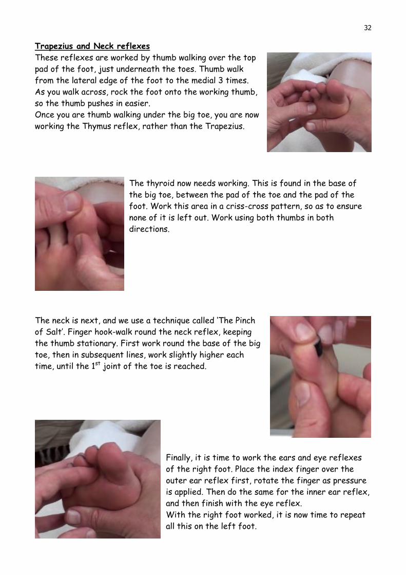
Trapezius and Neck reflexes
These reflexes are worked by thumb walking over the top
pad of the foot, just underneath the toes. Thumb walk
from the lateral edge of the foot to the medial 3 times.
As you walk across, rock the foot onto the working thumb,
so the thumb pushes in easier.
Once you are thumb walking under the big toe, you are now
working the Thymus reflex, rather than the Trapezius.
The thyroid now needs working. This is found in the base of
the big toe, between the pad of the toe and the pad of the
foot. Work this area in a criss-cross pattern, so as to ensure
none of it is left out. Work using both thumbs in both
directions.
The neck is next, and we use a technique called ‘The Pinch
of Salt’. Finger hook-walk round the neck reflex, keeping
the thumb stationary. First work round the base of the big
toe, then in subsequent lines, work slightly higher each
time, until the 1st joint of the toe is reached.
Finally, it is time to work the ears and eye reflexes
of the right foot. Place the index finger over the
outer ear reflex first, rotate the finger as pressure
is applied. Then do the same for the inner ear reflex,
and then finish with the eye reflex.
With the right foot worked, it is now time to repeat
all this on the left foot.

33
References
arthritis-health (2014) What is a Bursa? Available at: http://www.arthritis-
health.com/types/bursitis/what-bursa (Accessed: 3 September 2014).
Bupa (2013) Bunions. Available at: http://www.bupa.co.uk/individuals/health-
information/directory/b/bunions (Accessed: 3 September 2014).
Hull, H. (2009) Anatomy & Physiotherapy for beauty and complementary therapies.
Cambridge: The Write Idea.
Life’s Journey in Words (2011) Discovering Science @ Oman Children's Museum.
Available at: http://lifesjourneyinwords.blogspot.co.uk/2011/06/discovering-science-
oman-childrens.html (Accessed: 6 September 2013).
Mazzone, M. F. and McCue, T. Common Conditions of the Achilles Tendon. American
Family Physician. 2002 May 1;65(9):1805-1811. Available at:
http://www.aafp.org/afp/2002/0501/p1805.html (Accessed: 2 September 2014).
Medline Plus (2012) Tendon vs Ligament. Available at:
http://www.nlm.nih.gov/medlineplus/ency/imagepages/19089.htm (Accessed: 20
November 2013).
National Cancer Institute (no date) Seer Training Modules Available at:
http://training.seer.cancer.gov/anatomy/skeletal/articulations.html (Accessed: 20
November 2013).
Smith, D.G. ‘Grasping the Importance of Our Hands’. In Motion. 16.6 (2006).
Available at: http://www.amputee-coalition.org/inmotion/nov_dec_06/our_hands.html
(Accessed: 6 September 2013).
Tucker, L. (2005) An Introductory Guide to Anatomy and Physiology. Cambridge:
Holistic Therapy Books.
Waugh, A. and Grant, A. (2006) Ross and Wilson Anatomy and Physiology in Health and
Ilness. 10th edn. Evolve learning system. [Online]. Available at:
http://evolvels.elsevier.com/section/default.asp?id=0967_global_0001&mode
(Accessed: 16 August 2011).
Waugh, A. and Grant, A. (2010) Ross and Wilson Anatomy and Physiology in Health and
Illness. 11th Edn. London: Churchill Livingstone.