Introduction and general aspects 387 The red cell 389 ...
73
1 Introduction and general aspects 387 The red cell 389 Anaemia 392 Microcytic anaemia 393 Normocytic anaemia 397 Macrocytic anaemias 397 Megaloblastic anaemia 397 Macrocytosis without megaloblastic changes 401 Anaemia due to marrow failure (aplastic anaemia) 402 Haemolytic anaemias: an introduction 403 Inherited haemolytic anaemia 404 Red cell membrane defects 404 Haemoglobin abnormalities 406 The thalassaemias 407 Sickle syndromes 409 Metabolic disorders of the red cell 412 Acquired haemolytic anaemia 414 Autoimmune haemolytic anaemias 415 Drug-induced immune haemolytic anaemia 417 Alloimmune haemolytic anaemia 417 Non-immune haemolytic anaemia 418 Mechanical haemolytic anaemia 41 Myeloproliferative disorders 419 Polycythaemia 419 Myelofibrosis (myelosclerosis) 421 Myelodysplasia (MDS) 422 The spleen 422 Splenomegaly 423 Blood transfusion 423 Blood groups 424 Procedure for blood transfusion 424 Complications of blood transfusion 425 Blood, blood components and blood products 428 The white cell 430 Neutrophils 430 Eosinophils 431 Basophils 431 Monocytes 431 Lymphocytes 431 Haemostasis and thrombosis 431 Haemostasis 431 Vascular disorders 435 Platelet disorders 435 Inherited coagulation disorders 438 Acquired coagulation disorders 440 Thrombosis 442
Transcript of Introduction and general aspects 387 The red cell 389 ...
1
Introduction and general aspects 387
The red cell 389
Anaemia 392
Microcytic anaemia 393
Normocytic anaemia 397
Macrocytic anaemias 397
Megaloblastic anaemia 397
Macrocytosis without megaloblastic changes 401
Anaemia due to marrow failure
(aplastic anaemia) 402
Haemolytic anaemias: an introduction 403
Inherited haemolytic anaemia 404
Red cell membrane defects 404
Haemoglobin abnormalities 406
The thalassaemias 407
Sickle syndromes 409
Metabolic disorders of the red cell 412
Acquired haemolytic anaemia 414
Autoimmune haemolytic anaemias 415
Drug-induced immune haemolytic anaemia 417
Alloimmune haemolytic anaemia 417
Non-immune haemolytic anaemia 418
Mechanical haemolytic anaemia 41
Myeloproliferative disorders 419
Polycythaemia 419
Myelofibrosis (myelosclerosis) 421
Myelodysplasia (MDS) 422
The spleen 422
Splenomegaly 423
Blood transfusion 423
Blood groups 424
Procedure for blood transfusion 424
Complications of blood transfusion 425
Blood, blood components and blood
products 428
The white cell 430
Neutrophils 430
Eosinophils 431
Basophils 431
Monocytes 431
Lymphocytes 431
Haemostasis and thrombosis 431
Haemostasis 431
Vascular disorders 435
Platelet disorders 435
Inherited coagulation disorders 438
Acquired coagulation disorders 440
Thrombosis 442
2
Iron metabolism Absorption
upper small intestine
about 10% of dietary iron absorbed
Fe2+ (ferrous iron) much better absorbed than Fe3+ (ferric iron)
absorption is regulated according to body's need
increased by vitamin C, gastric acid
decreased by proton pump inhibitors, gastric achlorhydia, tetracycline, tannin (found in tea)
Distribution in body:
total body iron = 4g
haemoglobin = 70%
ferritin and haemosiderin = 25%
myoglobin = 4%
plasma iron = 0.1%
Transport
carried in plasma as Fe3+ bound to transferrin
Storage
stored as ferritin in tissues
Excretion
lost via intestinal tract following desquamination
Iron studies Serum iron
Total iron binding capacity (TIBC)
transferrin
raised in iron deficiency anaemia (IDA)
raised in pregnancy and by oestrogen
Transferrin saturation:
calculated by serum iron / TIBC
Ferritin:
raised in inflammatory disorders
low in IDA
Rarer tests:
transferrin receptors increased in IDA
Anaemia of chronic disease
normochromic/hypochromic, normocytic anaemia
reduced serum and TIBC
normal or raised ferritin
3
Folate metabolism Drugs which interfere with metabolism
trimethoprim methotrexate pyrimethamine
Drugs which can reduce absorption
phenytoin
Methaemoglobinaemia Methaemoglobinaemia describes haemoglobin which has been oxidised from Fe2+ (ferrous
iron) to Fe3+ (ferric iron).
This is normally regulated by NADH methaemoglobin reductase, which transfers electrons
from NADH to methaemoglobin resulting in the reduction of methaemoglobin to
haemoglobin.
There is tissue hypoxia as Fe3+ cannot bind oxygen, and hence the oxidation dissociation
curve is moved to the left
Congenital causes
1) haemoglobin chain variants: HbM, HbH
2) NADH methaemoglobin reductase deficiency
Acquired causes
1) drugs:
1. sulphonamides,
2. nitrates, sodium nitroprusside,
3. primaquine
4. dapsone
2) chemicals: aniline dyes
Features:
1) dyspnoea, anxiety, headache
2) 'chocolate' cyanosis
3) severe: acidosis, arrhythmias, seizures, coma
4) normal pO2 but decreased oxygen saturation
Management:
1) NADH - methaemoglobinaemia reductase deficiency: ascorbic acid
2) IV methylene blue if acquired
4
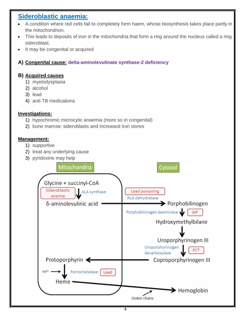
Sideroblastic anaemia: A condition where red cells fail to completely form haem, whose biosynthesis takes place partly in
the mitochondrion.
This leads to deposits of iron in the mitochondria that form a ring around the nucleus called a ring
sideroblast.
It may be congenital or acquired
A) Congenital cause: delta-aminolevulinate synthase-2 deficiency
B) Acquired causes
1) myelodysplasia
2) alcohol
3) lead
4) anti-TB medications
Investigations:
1) hypochromic microcytic anaemia (more so in congenital)
2) bone marrow: sideroblasts and increased iron stores
Management:
1) supportive
2) treat any underlying cause
3) pyridoxine may help

5
Porphyrias Overview
abnormality in enzymes responsible for the biosynthesis of haem results in overproduction of intermediate compounds (porphyrins) may be acute or non-acute
Acute intermittent porphyria: a rare autosomal dominant condition caused by a defect in porphobilinogen deaminase, an enzyme involved in the biosynthesis of
haem. This results in the toxic accumulation of delta aminolaevulinic acid and porphobilinogen. It characteristically presents with abdominal and neuropsychiatric symptoms in 20-40 year
olds. AIP is more common in females (5:1)
Features:
1) abdominal: abdominal pain, vomiting 2) neurological: motor neuropathy 3) psychiatric: e.g. depression 4) hypertension and tachycardia are common
Diagnosis:
1) classically urine turns deep red on standing 2) raised urinary porphobilinogen
(Elevated between attacks and to a greater extent during acute attacks) 3) assay of red cells for porphobilinogen deaminase 4) raised serum levels of delta aminolaevulinic acid and porphobilinogen
Drugs which may precipitate attack
1) barbiturates
2) benzodiazepines
3) halothane
4) alcohol
5) oral contraceptive pill
6) sulphonamides

6
Drugs considered safe to use
1) paracetamol
2) aspirin
3) codeine
4) morphine
5) chlorpromazine
6) beta-blockers
7) penicillin
8) metformin
Porphyria cutanea tarda (PCT):
most common hepatic porphyria defect in uroporphyrinogen decarboxylase may be caused by hepatocyte damage e.g. alcohol, oestrogens classically photosensitive rash with bullae, skin fragility on face and dorsal aspect of
hands urine: elevated uroporphyrinogen and pink fluorescence of urine under Wood's lamp manage with chloroquine
Variegate porphyria:
autosomal dominant defect in protoporphyrinogen oxidase photosensitive blistering rash abdominal and neurological symptoms more common in South Africans
7
Haemochromatosis: Haemochromatosis is an autosomal recessive disorder of iron absorption and metabolism
resulting in iron accumulation.
It is caused by inheritance of mutations in the HFE gene on both copies of chromosome 6*.
Epidemiology: 1 in 10 people of European descent carry a mutation genes affecting iron metabolism, mainly
HFE
prevalence in people of European descent = 1 in 200
Presenting features:
1) early symptoms include fatigue, erectile dysfunction and arthralgia (often of the hands) 2) 'bronze' skin pigmentation 3) diabetes mellitus 4) liver: stigmata of chronic liver disease, hepatomegaly, cirrhosis, hepatocellular
deposition) 5) cardiac failure (2nd to dilated cardiomyopathy) 6) hypogonadism (2nd to cirrhosis and pituitary dysfunction - hypogonadotrophic
hypogonadism) 7) arthritis (especially of the hands)
Questions have previously been asked regarding which features are reversible with treatment:
Reversible complications
Cardiomyopathy Skin pigmentation
Irreversible complications Liver cirrhosis** Diabetes mellitus Hypogonadotrophic hypogonadism Arthropathy
**whilst elevated liver function tests and hepatomegaly may be reversible, cirrhosis is not
Investigation:
The British Committee for Standards in Haematology (BCSH) published guidelines for the
investigation and management of haemochromatosis in 2000
There is continued debate about the best investigation to screen for haemochromatosis.
The 2000 BCSH guidelines suggest:
1) General population:
Transferrin saturation is considered the most useful marker.
Ferritin should also be measured but is not usually abnormal in the early stages of iron
accumulation
2) Testing family members: genetic testing for HFE mutation.
These guidelines may change as HFE gene analysis become less expensive
Diagnostic tests:
1) molecular genetic testing for the C282Y and H63D mutations
2) liver biopsy: Perl's stain

8
Typical iron study profile in patient with haemochromatosis
1) transferrin saturation:
> 55% in men or
> 50% in women
2) raised ferritin (e.g. > 500 ug/l) and iron
3) low TIBC
Monitoring adequacy of venesection:
BSCH recommend 'transferrin saturation should be kept below 50% and the serum ferritin
concentration below 50 ug/l'
Joint x-rays characteristically show chondrocalcinosis
*there are rare cases of families with classic features of genetic haemochromatosis but no mutation
in the HFE gene

9
Anaemia 392
Microcytic anaemia 393
Normocytic anaemia 397
Macrocytic anaemias 397
Megaloblastic anaemia 397
Macrocytosis without megaloblastic changes 401
Microcytic anaemia: Causes
1) iron-deficiency anaemia
2) thalassaemia: in beta-thalassaemia minor the microcytosis is often disproportionate to the
anaemia
3) congenital sideroblastic anaemia
4) lead poisoning
5) anaemia of chronic disease (more commonly a normocytic, normochromic picture)
A question sometimes seen in exams gives a history of a normal haemoglobin level associated with
a microcytosis. In patients not at risk of thalassaemia, this should raise the possibility of
polycythaemia rubra Vera which may cause an iron-deficiency secondary to bleeding.
Iron deficiency anaemia: Features:
1) koilonychia تقعر الأظفار
2) atrophic glossitis
3) post-cricoid webs
4) angular stomatitis
Blood film:
1) target cells
2) 'pencil' poikilocytes
3) if combined with B12/folate deficiency a dimorphic film occurs with mixed microcytic and macrocytic cells
Macrocytic anaemia: Macrocytic anaemia can be divided into causes associated with a megaloblastic bone marrow and
those with a normoblastic bone marrow
Megaloblastic causes Normoblastic causes
1) vitamin B12 deficiency
2) folate deficiency
1) alcohol
2) liver disease
3) hypothyroidism
4) pregnancy
5) reticulocytosis
6) myelodysplasia
7) drugs: cytotoxics
10
Pernicious anaemia investigation: Investigation:
anti gastric parietal cell antibodies in 90% (but low specificity)
anti intrinsic factor antibodies in 50% (specific for pernicious anaemia)
macrocytic anaemia
low WCC and platelets
LDH may be raised due to ineffective erythropoiesis
also low serum B12, hypersegmented polymorphs on film, megaloblasts in marrow
Schilling test
Schilling test
radiolabelled B12 given on two occasions
first on its own
second with oral IF
urine B12 levels measured

11
Anaemia due to marrow failure (aplastic anaemia)
Aplastic anaemia: Characterised by pancytopaenia and a hypoplastic bone marrow
Peak incidence of acquired = 30 years old
Features:
1) normochromic, normocytic anaemia
2) leukopenia, with lymphocytes relatively spared
3) thrombocytopenia
4) may be the presenting feature acute lymphoblastic or myeloid leukaemia
5) a minority of patients later develop paroxysmal nocturnal haemoglobinuria or myelodysplasia
Causes:
1) idiopathic
2) congenital: Fanconi anaemia, dyskeratosis congenita
3) drugs: cytotoxics, chloramphenicol, sulphonamides, phenytoin, gold
4) toxins: benzene
5) infections: parvovirus, hepatitis
6) radiation
Management:
1) Supportive:
blood products
prevention and treatment of infection
2) Anti-thymocyte globulin (ATG) and anti-lymphocyte globulin (ALG)
prepared in animals (e.g. rabbits or horses) by injecting human lymphocytes
is highly allergenic and may cause serum sickness (fever, rash, arthralgia), therefore steroid
cover usually given
3) immunosuppression using agents such as ciclosporin may also be given
4) Stem cell transplantation:
allogeneic transplants have a success rate of up to 80%
Fanconi anaemia Autosomal recessive
Features
aplastic anaemia
increased risk of AML
neurological & skeletal abnormalities
skin pigmentation
Drug-induced pancytopaenia Drug causes of pancytopaenia
1) cytotoxics
2) antibiotics: trimethoprim, chloramphenicol
3) anti-rheumatoid: gold, penicillamine
4) carbimazole: causes both agranulocytosis and pancytopaenia
5) anti-epileptics: carbamazepine
6) sulphonylureas: tolbutamide

12
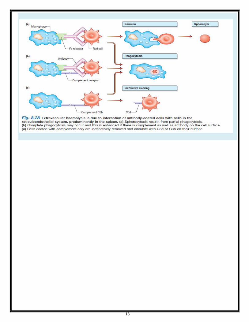
Haemolytic anaemias: by site In intravascular haemolysis:
Free haemoglobin is released which binds to haptoglobin.
As haptoglobin becomes saturated haemoglobin binds to albumin forming methaemalbumin
(detected by Schumm's test).
Free haemoglobin is excreted in the urine as haemoglobinuria, haemosiderinuria
Intravascular haemolysis: causes:
1) mismatched blood transfusion
2) G6PD deficiency*
3) red cell fragmentation: heart valves, TTP, DIC, HUS
4) paroxysmal nocturnal haemoglobinuria
5) cold autoimmune haemolytic anaemia
*strictly speaking there is an element of extravascular haemolysis in G6PD as well, although it is
usually classified as a intravascular cause
Extravascular haemolysis: causes
1) haemoglobinopathies: sickle cell, thalassaemia
2) hereditary spherocytosis
3) haemolytic disease of newborn
4) warm autoimmune haemolytic anaemia
13

14

15
Causes of haemolytic anaemia Inherited Acquired
Red cell membrane defect
1) Hereditary spherocytosis
2) Hereditary elliptocytosis
Haemoglobin abnormalities
1) Thalassaemia
2) Sickle cell disease
Metabolic defects
1) Glucose-6-phosphate dehydrogenase
deficiency
2) Pyruvate kinase deficiency
3) Pyrimidine kinase deficiency
Miscellaneous
1) Infections, e.g. malaria, mycoplasma
Clostridium welchii,
2) generalized sepsis
3) Drugs and chemicals causing damage to
the red cell membrane or oxidative
haemolysis
4) Hypersplenism
5) Burns
Immune
1) Autoimmune
Warm
Cold
2) Alloimmune
Haemolytic transfusion reactions
Haemolytic disease of the newborn
After allogeneic bone marrow or
organ transplantation
3) Drug-induced
Non-immune
1) Acquired membrane defects:
Paroxysmal nocturnal haemoglobinuria
2) Mechanical:
Microangiopathic haemolytic
anaemia
Valve prosthesis
March haemoglobinuria
3) Secondary to systemic disease
Renal and liver failure
16
Red cell membrane defect
1) Hereditary spherocytosis
2) Hereditary elliptocytosis
Hereditary spherocytosis Basics
most common hereditary haemolytic anaemia in people of northern European descent
autosomal dominant defect of red blood cell cytoskeleton
the normal biconcave disc shape is replaced by a sphere-shaped red blood cell
red blood cell survival reduced as destroyed by the spleen
Presentation:
1) failure to thrive
2) jaundice, gallstones
3) splenomegaly
4) aplastic crisis precipitated by parvovirus infection
5) degree of haemolysis variable
6) MCHC elevated
Diagnosis:
osmotic fragility test
Management:
1) folate replacement
2) splenectomy
Comparing G6PD deficiency to hereditary spherocytosis
G6PD deficiency Hereditary spherocytosis
Gender Male (X-linked recessive) Male + female (autosomal dominant)
Ethnicity African + Mediterranean descent Northern European descent
Typical history Neonatal jaundice
Infection/ drugs precipitate
haemolysis
Gallstones
Intravascular Heamolysis
Neonatal jaundice
Chronic symptoms although haemolytic
crises may be precipitated by infection
Gallstones
Splenomegaly is common
Extravascular Heamolysis
Blood film Heinz bodies Spherocytes (round, lack of central pallor)
Diagnostic test Measure enzyme activity of G6PD Osmotic fragility test
17
Haemoglobin Abnormalities
1) Thalassaemia
2) Sickle Cell Disease
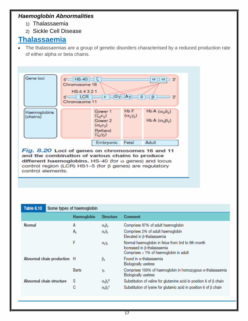
Thalassaemia The thalassaemias are a group of genetic disorders characterised by a reduced production rate
of either alpha or beta chains.
18
Alpha-thalassaemia: Alpha-thalassaemia is due to a deficiency of alpha chains in haemoglobin
Overview:
2 separate alpha-globulin genes are located on each chromosome 16
Clinical severity depends on the number of alpha chains present
If 1 or 2 alpha chains are absent
the blood picture would be hypochromic and microcytic, but
the Hb level would be typically normal
Loss of 3 alpha chains results in
Hypochromic microcytic anaemia with splenomegaly.
This is known as Hb H disease
If all 4 alpha chains absent (i.e. homozygote) then
death in utero (hydrops fetalis, Bart's hydrops)
Beta-thalassaemia trait: Beta-thalassaemia trait is an autosomal recessive condition Characterised by a mild hypochromic, microcytic anaemia. It is usually asymptomatic
Features: 1) mild hypochromic, microcytic anaemia - microcytosis is characteristically disproportionate to
the anaemia
2) HbA2 raised (> 3.5%) normally it is 2%

19
Haemoglobin abnormalities
1) Thalassaemia
2) Sickle cell disease
Sickle-cell crises: Sickle cell anaemia is characterised by periods of good health with intervening crises
Four main types of crises are recognised:
1) thrombotic, 'painful crises'
2) sequestration
3) aplastic
4) haemolytic
A) Thrombotic crises:
also known as painful crises or vaso-occlusive crises
precipitated by:
1) infection,
2) dehydration,
3) deoxygenation
infarcts occur in various organs including:
1) the bones e.g. avascular necrosis of hip,
2) hand-foot syndrome in children,
3) lungs, spleen and brain
B) Sequestration crises:
sickling within organs such as the spleen or lungs causes pooling of blood with worsening of
the anaemia
acute chest syndrome:
1) dyspnoea,
2) chest pain,
3) pulmonary infiltrates,
4) low pO2
5) the most common cause of death after childhood
C) Aplastic crises
caused by infection with parvovirus
sudden fall in haemoglobin
D) Haemolytic crises:
rare
fall in haemoglobin due an increased rate of haemolysis
Sickle cell anemia cause nephrogenic Diabetes Insipidus
20
G6PD deficiency Glucose-6-phosphate dehydrogenase (G6PD) deficiency is the commonest red blood cell
enzyme defect.
It is more common in people from the Mediterranean and Africa
Inherited in a X-linked recessive fashion.
Many drugs can precipitate a crisis as well as infections and broad (fava) beans
Pathophysiology:
↓ G6PD → ↓ glutathione → increased red cell susceptibility to oxidative stress
Features:
1) neonatal jaundice is often seen
2) intravascular haemolysis
3) gallstones are common
4) splenomegaly may be present
5) Heinz bodies on blood films
Diagnosis is made by using a G6PD enzyme assay
Some drugs causing haemolysis:
1) anti-malarials: primaquine
2) ciprofloxacin
3) sulph- group drugs: sulphonamides, sulphasalazine, sulfonylureas
Some drugs thought to be safe
1) penicillins
2) cephalosporins
3) macrolides
4) tetracyclines
5) trimethoprim
Comparing G6PD deficiency to hereditary spherocytosis
G6PD deficiency Hereditary spherocytosis
Gender Male (X-linked recessive) Male + female (autosomal dominant)
Ethnicity African + Mediterranean descent Northern European descent
Typical
history
• Neonatal jaundice
• Infection/drugs precipitate
haemolysis
• Gallstones
• Neonatal jaundice
• Chronic symptoms although haemolytic crises
may be precipitated by infection
• Gallstones
• Splenomegaly is common
Blood film Heinz bodies Spherocytes (round, lack of central pallor)
Diagnostic
test
Measure enzyme activity of
G6PD
Osmotic fragility test

21
Acquired haemolytic anaemia
Causes of immune destruction of red cells
Autoimmune haemolytic anaemias
Drug-induced immune haemolytic anaemia
Alloimmune haemolytic anaemia
Causes of non-immune destruction of red cells
Acquired membrane defects (e.g. paroxysmal nocturnal haemoglobinuria).
Mechanical factors (e.g. prosthetic heart valves, or microangiopathic haemolytic anaemia)
Secondary to systemic disease (e.g. renal and liver disease).
Autoimmune haemolytic anaemia Autoimmune haemolytic anaemia (AIHA) may be divided in to 'warm' and 'cold' types, according
to at what temperature the antibodies best cause haemolysis.
It is most commonly idiopathic but may be secondary to a lymphoproliferative disorder, infection
or drugs.
AIHA is characterised by a positive direct antiglobulin test (Coombs' test)
A) Warm AIHA:
In warm AIHA the antibody is usually IgG
causes haemolysis best at body temperature
Haemolysis tends to occur in extravascular sites, for example the spleen.
Management options include steroids, immunosuppression and splenectomy
Causes of warm AIHA
1) autoimmune disease: e.g. systemic lupus erythematosus*
2) neoplasia: e.g. lymphoma, CLL
3) drugs: e.g. methyldopa
*systemic lupus erythematosus can rarely be associated with a mixed-type autoimmune
haemolytic anaemia
B) Cold AIHA:
The antibody in cold AIHA is usually IgM
Causes haemolysis best at 4 deg C.
Haemolysis is mediated by complement and is more commonly intravascular.
Features may include symptoms of Raynaud's and acrocynaosis.
Patients respond less well to steroids
Causes of cold AIHA:
1) neoplasia: e.g. lymphoma
2) infections: e.g. mycoplasma, EBV

22
Drug-induced haemolytic anaemia: Drug-induced haemolytic anaemia can be classified according to three different mechanisms:
Type I:
antibody against drug-red cell membrane complex
penicillin
Type II:
deposition of complement via a drug-protein-antibody complex onto the red cell membrane
quinidine
rifampicin
Type III:
true autoimmune haemolytic anaemia - role of drug not known
methyldopa
L-dopa
mefanamic acid

23
Paroxysmal nocturnal haemoglobinuria: (PNH) An acquired disorder leading to haemolysis (mainly intravascular) of haematological cells.
It is thought to be caused by increased sensitivity of cell membranes to complement (see
below) due to a lack of glycoprotein glycosyl-phosphatidylinositol (GPI).
Patients are more prone to venous thrombosis
Pathophysiology:
GPI can be thought of as an anchor which attaches surface proteins to the cell membrane
complement-regulating surface proteins, e.g. decay-accelerating factor (DAF), are not properly
bound to the cell membrane due a lack of GPI
thrombosis is thought to be caused by a lack of CD59 on platelet membranes predisposing to
platelet aggregation
Features:
1) haemolytic anaemia
2) red blood cells, white blood cells, platelets or stem cells may be affected therefore
pancytopaenia may be present
3) haemoglobinuria: classically dark-coloured urine in the morning (although has been shown
to occur throughout the day)
4) thrombosis e.g. Budd-Chiari syndrome
5) aplastic anaemia may develop in some patients
Diagnosis:
flow cytometry of blood to detect low levels of CD59 and CD55 has now replaced Ham's test
as the gold standard investigation in PNH
Ham's test: acid-induced haemolysis (normal red cells would not)
Management:
1) blood product replacement
2) anticoagulation
3) Eculizumab:
a monoclonal antibody directed against terminal protein C5,
is currently being trialled and is showing promise in reducing intravascular haemolysis
4) stem cell transplantation

24
Myeloproliferative disorders 419
Polycythaemia 419
Myelofibrosis (myelosclerosis) 421
Myelodysplasia (MDS) 422
Polycythaemia: Polycythaemia may be relative, primary (polycythaemia rubra vera) or secondary
Relative causes:
1) dehydration
2) stress: Gaisbock syndrome
Primary:
polycythaemia rubra vera
Secondary causes:
1) COPD
2) altitude
3) obstructive sleep apnoea
4) excessive erythropoietin:
cerebellar haemangioma,
hypernephroma,
hepatoma,
uterine fibroids: uterine fibroids may cause menorrhagia which in turn leads to blood
loss - polycythaemia is rarely a clinical problem
To differentiate between true (primary or secondary) polycythaemia and relative polycythaemia
red cell mass studies are sometimes used.
In true polycythaemia the total red cell mass in males > 35 ml/kg and in women > 32 ml/kg
25
Polycythaemia Rubra Vera: (PRV) A myeloproliferative disorder caused by clonal proliferation of a marrow stem cell leading to an
increase in red cell volume.
Often accompanied by overproduction of neutrophils and platelets.
It has recently been established that a mutation in JAK2 is present in approximately 95% of
patients with PRV and this has resulted in significant changes to the diagnostic criteria.
The incidence of PRV peaks in the sixth decade.
Features:
1) hyperviscosity
2) pruritus, typically after a hot bath
3) splenomegaly
4) haemorrhage (secondary to abnormal platelet function)
5) plethoric appearance
6) hypertension in a third of patients
7) Other features that may be seen in PRV include a low ESR and a raised leukocyte alkaline
phosphotase
Following history and examination, the British Committee for Standards in Haematology (BCSH)
recommends the following tests are performed:
1) full blood count/film (raised haematocrit; neutrophils, basophils, platelets raised in half of
patients)
2) JAK2 mutation
3) serum ferritin
4) renal and liver function tests
If the JAK2 mutation is negative and there is no obvious secondary causes the BCSH suggest the
following tests:
1) red cell mass
2) arterial oxygen saturation
3) abdominal ultrasound
4) serum erythropoietin level
5) bone marrow aspirate and trephine
6) cytogenetic analysis
7) erythroid burst-forming unit (BFU-E) culture

26
The diagnostic criteria for PRV have recently been updated by the BCSH. This replaces the previous
PRV Study Group criteria.
JAK2-positive PRV - diagnosis requires both criteria to be present
Criteria Notes
A1 High haematocrit (>0.52 in men, >0.48 in women) OR
raised red cell mass (>25% above predicted)
A2 Mutation in JAK2
JAK2-negative PRV - diagnosis requires A1 + A2 + A3 + either another A or two B criteria
Criteria Notes
A1 haematocrit >0.60 in men, >0.56 in women OR
Raised red cell mass (>25% above predicted)
A2 Absence of mutation in JAK2
A3 No cause of secondary erythrocytosis
A4 Palpable splenomegaly
A5 Presence of an acquired genetic abnormality (excluding BCR-
ABL) in the haematopoietic cells
B1 Thrombocytosis (platelet count >450 * 109/l)
B2 Neutrophil leucocytosis
(neutrophil count > 10 * 109/l in non-smokers;
> 12.5*109/l in smokers)
B3 Radiological evidence of splenomegaly
B4 Endogenous erythroid colonies or
low serum erythropoietin
Polycythaemia rubra Vera management:
1) venesection - first line treatment
2) aspirin
3) hydroxyurea -slight increased risk of secondary leukaemia
4) phosphorus-32 therapy
Prognosis:
thrombotic events are a significant cause of morbidity and mortality
5-15% of patients progress to myelofibrosis
5-15% of patients progress to acute myeloid leukaemia (risk increased with chemotherapy
treatment)

27
Myeloproliferative disorders 419
Polycythaemia 419
Myelofibrosis (myelosclerosis) 421
Myelodysplasia (MDS) 422
Myelofibrosis: Overview:
a myeloproliferative disorder
thought to be caused by hyperplasia of abnormal megakaryocytes
the resultant release of platelet derived growth factor is thought to stimulate fibroblasts
haematopoiesis develops in the liver and spleen
Features:
1) e.g. elderly person with symptoms of anaemia e.g. fatigue (the most common presenting
symptom)
2) massive splenomegaly
3) hypermetabolic symptoms: weight loss, night sweats etc
Laboratory findings:
1) anaemia
2) high WBC and platelet count early in the disease
3) 'tear-drop' poikilocytes on blood film
4) unobtainable bone marrow biopsy - 'dry tap' therefore trephine biopsy needed
5) high urate and LDH (reflect increased cell turnover)

28
Thrombocytosis: Thrombocytosis is an abnormally high platelet count, usually > 400 * 10
9/l.
Causes of thrombocytosis
A) reactive: platelets are an acute phase reactant - platelet count can increase in response
to stress such as a severe infection or surgery
B) malignancy
C) essential thrombocytosis (see below), or as part of another myeloproliferative disorder
such as chronic myeloid leukaemia or polycythaemia rubra vera
D) hyposplenism
Essential Thrombocytosis Essential thrombocytosis is one of the myeloproliferative disorders which overlaps with
chronic myeloid leukaemia, polycythaemia rubra vera and myelofibrosis.
Megakaryocyte proliferation results in an overproduction of platelets.
Features:
1) platelet count > 600 * 109/l
2) both thrombosis (venous or arterial) and haemorrhage can be seen
3) a characteristic symptom is a burning sensation in the hands
4) a JAK2 mutation is found in around 50% of patients
Management
1) hydroxyurea (hydroxycarbamide) is widely used to reduce the platelet count
2) interferon-α is also used in younger patients
3) low-dose aspirin may be used to reduce the thrombotic risk
29
Hyposplenism: Causes:
1) splenectomy
2) sickle-cell
3) coeliac disease, dermatitis herpetiformis
4) Graves' disease
5) systemic lupus erythematosus
6) amyloid
Features:
1) Howell-Jolly bodies
2) siderocytes
Blood product transfusion complications 1) haemolytic: immediate or delayed
2) febrile reactions
3) transmission of viruses, bacteria, parasites
As platelet concentrates are generally stored at room temperature they provide a more
favourable environment for bacterial contamination than other blood products.
4) hyperkalaemia
5) iron overload
6) ARDS
7) clotting abnormalities
8) Massive blood tx may cause hypocalcemia
Immediate haemolytic reaction
E.g. ABO mismatch
massive intravascular haemolysis
Febrile reactions:
due to white blood cell HLA antibodies
often the result of sensitization by previous pregnancies or transfusions
Causes a degree of immunosuppression
e.g. patients with colorectal cancer who have blood transfusions have a worse outcome than
those who do not

30
The white cell
Eosinophilia Causes of eosinophilia may be divided into pulmonary, infective and other
Pulmonary causes:
1) asthma
2) allergic bronchopulmonary aspergillosis
3) Churg-Strauss syndrome
4) Loffler's syndrome
5) tropical pulmonary eosinophilia
6) eosinophilic pneumonia
7) hypereosinophilic syndrome
Infective causes:
1) schistosomiasis
2) nematodes: Toxocara, Ascaris, Strongyloides
3) cestodes: Echinococcus
Other causes
1) drugs: sulfasalazine, nitrofurantoin
2) psoriasis/eczema
3) eosinophilic leukaemia (very rare)
Neutropenic sepsis Neutropenic sepsis is a relatively common complication of cancer therapy, usually as a
consequence of chemotherapy.
It may be defined as a neutrophil count of < 0.5 * 109 in a patient who is having anticancer
treatment and has one of the following:
a temperature higher than 38C or
other signs or symptoms consistent with clinically significant sepsis
Prophylaxis:
if it is anticipated that patients are likely to have a neutrophil count of < 0.5 * 109 as a
consequence of their treatment they should be offered a fluoroquinolone
Management:
1) antibiotics must be started immediately, do not wait for the WBC
2) NICE recommend starting empirical antibiotic therapy with piperacillin with tazobactam
(Tazocin) immediately
3) many units add vancomycin if the patient has central venous access but NICE do not support
this approach
4) following this initial treatment patients are usually assessed by a specialist and risk-stratified
to see if they may be able to have outpatient treatment
5) if patients are still febrile and unwell after 48 hours an alternative antibiotic such as
meropenem is often prescribed +/- vancomycin
6) if patients are not responding after 4-6 days the Christie guidelines suggest ordering
investigations for fungal infections (e.g. HRCT), rather than just starting therapy antifungal
therapy blindly
7) there may be a role for G-CSF in selected patients
31
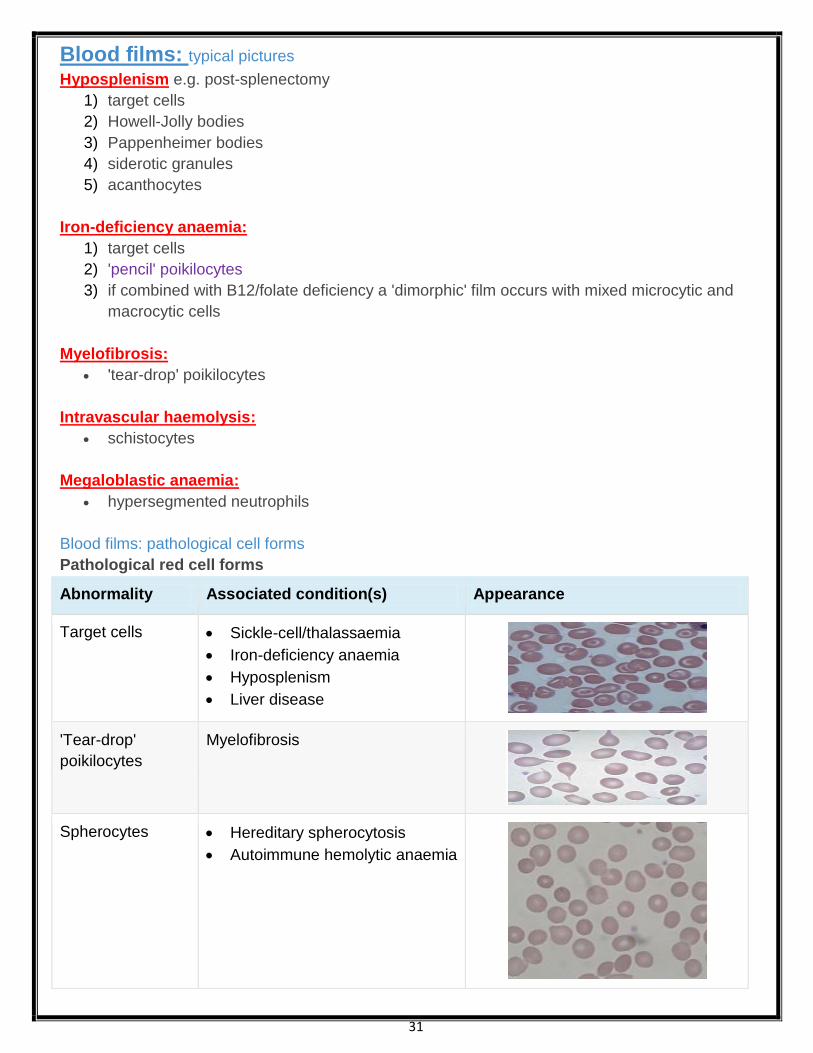
Blood films: typical pictures
Hyposplenism e.g. post-splenectomy
1) target cells
2) Howell-Jolly bodies
3) Pappenheimer bodies
4) siderotic granules
5) acanthocytes
Iron-deficiency anaemia:
1) target cells
2) 'pencil' poikilocytes
3) if combined with B12/folate deficiency a 'dimorphic' film occurs with mixed microcytic and
macrocytic cells
Myelofibrosis:
'tear-drop' poikilocytes
Intravascular haemolysis:
schistocytes
Megaloblastic anaemia:
hypersegmented neutrophils
Blood films: pathological cell forms
Pathological red cell forms
Abnormality Associated condition(s) Appearance
Target cells Sickle-cell/thalassaemia
Iron-deficiency anaemia
Hyposplenism
Liver disease
'Tear-drop'
poikilocytes
Myelofibrosis
Spherocytes Hereditary spherocytosis
Autoimmune hemolytic anaemia

32
Abnormality Associated condition(s) Appearance
Basophilic
stippling
Lead poisoning
Thalassaemia
Howell-Jolly
bodies
Hyposplenism
Heinz bodies G6PD deficiency
Alpha-thalassaemia
Schistocytes
('helmet cells')
Intravascular haemolysis
Mechanical heart valve
DIC
'Pencil'
poikilocytes
Iron defiency anaemia
Burr cells
(echinocytes)
Uraemia
Pyruvate kinase deficiency
Acanthocytes Abetalipoproteinemia
Other blood film abnormalities:
hypersegmented neutrophils: megaloblastic anaemia
33
Leucocyte alkaline phosphatase: Rose in:
pregnancy, oral contraceptive pill
infections
leukaemoid reactions
myelofibrosis
polycythaemia rubra vera
steroids, Cushing's syndrome
Low in:
chronic myeloid leukaemia
pernicious anaemia
paroxysmal nocturnal haemoglobinuria
infectious mononucleosis
Leukaemoid reaction: The leukaemoid reaction describes the presence of immature cells such as myeloblasts,
promyelocytes and nucleated red cells in the peripheral blood.
This may be due to:
1. infiltration of the bone marrow causing the immature cells to be 'pushed out' or
2. sudden demand for new cells
Causes:
1) severe infection
2) severe haemolysis
3) massive haemorrhage
4) metastatic cancer with bone marrow infiltration
A relatively common clinical problem is differentiating chronic myeloid leukaemia from a leukaemoid
reaction. The following differences may help:
Leukaemoid reaction Chronic myeloid leukaemia
high leucocyte alkaline phosphatase score
toxic granulation (Dohle bodies) in the white
cells
'left shift' of neutrophils i.e. three or less
segments of the nucleus
low leucocyte alkaline phosphatase score
34
Haematological Malignancies: Genetics:
Below is a brief summary of the common translocations associated with haematological malignancies
t(9;22) - Philadelphia chromosome:
present in > 95% of patients with CML
this results in part of the Abelson proto-oncogene being moved to the BCR gene on
chromosome 22
the resulting BCR-ABL gene codes for a fusion protein which has tyrosine kinase activity in
excess of normal
poor prognostic indicator in ALL
t(15;17):
seen in acute promyelocytic leukaemia (M3)
fusion of PML and RAR-alpha genes
t(8;14):
seen in Burkitt's lymphoma
MYC oncogene is translocated to an immunoglobulin gene
t(11;14)
Mantle cell lymphoma
deregulation of the cyclin D1 (BCL-1) gene
Haematological malignancies: Infections:
Viruses:
EBV: Hodgkin's and Burkitt's lymphoma, nasopharyngeal carcinoma
HTLV-1: Adult T-cell leukaemia/lymphoma
HIV-1: High-grade B-cell lymphoma
Bacteria:
Helicobacter pylori: gastric lymphoma (MALT)
Protozoa:
malaria: Burkitt's lymphoma
35
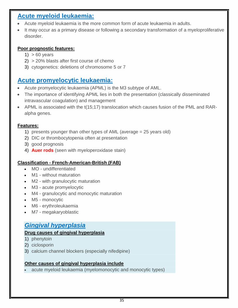
Acute myeloid leukaemia: Acute myeloid leukaemia is the more common form of acute leukaemia in adults.
It may occur as a primary disease or following a secondary transformation of a myeloproliferative
disorder.
Poor prognostic features:
1) > 60 years
2) > 20% blasts after first course of chemo
3) cytogenetics: deletions of chromosome 5 or 7
Acute promyelocytic leukaemia:
Acute promyelocytic leukaemia (APML) is the M3 subtype of AML.
The importance of identifying APML lies in both the presentation (classically disseminated
intravascular coagulation) and management
APML is associated with the t(15;17) translocation which causes fusion of the PML and RAR-
alpha genes.
Features:
1) presents younger than other types of AML (average = 25 years old)
2) DIC or thrombocytopenia often at presentation
3) good prognosis
4) Auer rods (seen with myeloperoxidase stain)
Classification - French-American-British (FAB)
MO - undifferentiated
M1 - without maturation
M2 - with granulocytic maturation
M3 - acute promyelocytic
M4 - granulocytic and monocytic maturation
M5 - monocytic
M6 - erythroleukaemia
M7 - megakaryoblastic
Gingival hyperplasia Drug causes of gingival hyperplasia
1) phenytoin
2) ciclosporin
3) calcium channel blockers (especially nifedipine)
Other causes of gingival hyperplasia include
acute myeloid leukaemia (myelomonocytic and monocytic types)
36
Chronic myeloid leukaemia: The Philadelphia chromosome is present in more than 95% of patients with chronic myeloid
leukaemia (CML).
It is due to a translocation between the long arm of chromosome 9 and 22 - t(9:22)(q34; q11).
This results in part of the ABL proto-oncogene from chromosome 9 being fused with the BCR
gene from chromosome 22.
The resulting BCR-ABL gene codes for a fusion protein which has tyrosine kinase activity in
excess of normal
Presentation (40-50 years):
1) middle-age
2) anaemia, weight loss, abdo discomfort
3) splenomegaly may be marked
4) spectrum of myeloid cells seen in peripheral blood
5) decreased leukocyte alkaline phosphatase
6) May undergo blast transformation (AML in 80%, ALL in 20%)
Management:
1) imatinib is now considered first-line treatment
2) hydroxyurea
3) interferon-alpha
4) allogenic bone marrow transplant
Imatinib: 1) inhibitor of the tyrosine kinase associated with the BCR-ABL defect
2) very high response rate in chronic phase CML
Hairy cell leukaemia: Hairy cell leukaemia is a rare malignant proliferation disorder of B cells.
It is more common in males (4:1)
Features:
1) pancytopenia
2) splenomegaly
3) skin vasculitis in 1/3 patients
4) 'dry tap' despite bone marrow hypercellularity
5) tartrate resistant acid phosphotase (TRAP) stain positive
Management:
1) chemotherapy is first-line: cladribine, pentostatin
2) immunotherapy is second-line: rituximab, interferon-alpha
Interferons (IFN)
These Are cytokines released by the body in response to viral infections and neoplasia.
They are classified according to cellular origin and the type of receptor they bind to:
1) IFN-alpha and IFN-beta bind to type 1 receptors whilst
2) IFN-gamma binds only to type 2 receptors.
IFN-alpha is produced by leucocytes and has an antiviral action.
It has been shown to be useful in the management of hepatitis B & C, Kaposi's sarcoma,
metastatic renal cell cancer and hairy cell leukaemia

37
Acute lymphoblastic leukaemia: Prognostic features
Good prognostic factors:
1) French-American-British (FAB) L1 type
2) common ALL
3) pre-B phenotype
4) low initial WBC
5) del(9p)
Poor prognostic factors:
1) FAB L3 type
2) T or B cell surface markers
3) Philadelphia translocation, t(9;22)
4) age < 2 years or > 10 years
5) male sex
6) CNS involvement
7) high initial WBC (e.g. > 100 * 109/l)
8) non-Caucasian
38
Chronic lymphocytic leukaemia: Chronic lymphocytic leukaemia (CLL) is caused by a monoclonal proliferation of well-
differentiated lymphocytes which are almost always B-cells (99%)
Features:
1) often none
2) constitutional: anorexia, weight loss
3) bleeding, infections
4) lymphadenopathy more marked than CML
Complications:
1) hypogammaglobulinaemia leading to recurrent infections
2) warm autoimmune haemolytic anaemia in 10-15% of patients
3) transformation to high-grade lymphoma (Richter's transformation)
Investigations:
1) blood film: smudge cells
2) immunophenotyping
Prognostic factors
Poor prognostic factors: (median survival 3-5 years)
1) male sex
2) age > 70 years
3) lymphocyte count > 50
4) prolymphocytes comprising more than 10% of blood lymphocytes
5) lymphocyte doubling time < 12 months
6) raised LDH
7) CD38 expression positive
Chromosomal changes:
A) Deletion of the long arm of chromosome 13 (del 13q) :
It is the most common abnormality, being seen in around 50% of patients.
It is associated with a good prognosis
B) deletions of part of the short arm of chromosome 17 (del 17p)
These are seen in around 5-10% of patients
associated with a poor prognosis
Indications for treatment
1) progressive marrow failure: the development or worsening of anaemia and/or thrombocytopenia
2) massive (>10 cm) or progressive lymphadenopathy
3) massive (>6 cm) or progressive splenomegaly
4) progressive lymphocytosis: > 50% increase over 2 months or lymphocyte doubling time < 6months
5) systemic symptoms:
weight loss > 10% in previous 6 months,
fever >38 C for > 2 weeks,
extreme fatigue, night sweats
6) autoimmune cytopaenias e.g. ITP
Management:
1) patients who have no indications for treatment are monitored with regular blood counts
2) fludarabine, cyclophosphamide and rituximab (FCR) has now emerged as the initial
treatment of choice for the majority of patients
39
Hodgkin's lymphoma Malignant proliferation of lymphocytes Characterised by the presence of the Reed-Sternberg cell.
It has a bimodal age distributions being most common in the third and seventh decades
Features:
1) lymphadenopathy (75%) - painless, non-tender, asymmetrical
2) systemic (25%): weight loss, pruritus, night sweats, fever (Pel-Ebstein)
3) alcohol pain in HL
4) normocytic anaemia, eosinophilia
5) LDH raised
Histological classification:
Type Frequency Prognosis Notes
Nodular sclerosing Most common
(around 70%)
Good prognosis More common in women.
Associated with lacunar cells
Mixed cellularity Around 20% Good prognosis Associated with a large number of
Reed-Sternberg cells
Lymphocyte
predominant
Around 5% Best prognosis
Lymphocyte depleted Rare Worst prognosis
'B' symptoms also imply a poor prognosis
1) weight loss > 10% in last 6 months
2) fever > 38 C
3) night sweats
Other factors associated with a poor prognosis identified in a 1998 NEJM paper included:
1) age > 45 years
2) male
3) stage IV disease
4) haemoglobin < 10.5 g/dl
5) white blood count > 15,000/�l
6) lymphocyte count < 600/�l or < 8%
7) albumin < 40 g/l
*Reed-Sternberg cells with nuclei surrounded by a clear space
Hodgkin's lymphoma: staging:
Ann-Arbor staging of Hodgkin's lymphoma
I: single lymph node
II: 2 or more lymph nodes/regions on same side of diaphragm
III: nodes on both sides of diaphragm
IV: spread beyond lymph nodes
Each stage may be subdivided into A or B
A = no systemic symptoms other than pruritus
B = weight loss > 10% in last 6 months, fever > 38c, night sweats (poor prognosis)
40
Burkitt's lymphoma Burkitt's lymphoma is a high-grade B-cell neoplasm.
There are two major forms:
1) Endemic (African) form: typically involves maxilla or mandible
2) Sporadic form:
Abdominal (e.g. ileo-caecal) tumours are the most common form.
More common in patients with HIV
Burkitt's lymphoma is associated with the c-myc gene translocation, usually t(8:14).
The Epstein-Barr virus (EBV) is strongly implicated in the development of the African form of
Burkitt's lymphoma and to a lesser extent the sporadic form.
Management:
1) Is with chemotherapy.
1) This tends to produce a rapid response which may cause 'tumour lysis syndrome'.
2) Rasburicase :
A recombinant version of urate oxidase,
An enzyme which catalyses the conversion of uric acid to allantoin is often given before the
chemotherapy to reduce the risk of 'tumour lysis syndrome'.
allantoin is 5-10 times more soluble than uric acid, so renal excretion is more effective
Complications of tumour lysis syndrome:
1) hyperuricaemia
2) hyperkalaemia
3) hyperphosphataemia
4) hypocalcaemia
5) acute renal failure
41
Paraproteinaemia: Causes of paraproteinaemia:
1) myeloma
2) monoclonal gammopathy of uncertain significance (MGUS)
3) benign monoclonal gammopathy
4) Waldenstrom's macroglobulinaemia
5) amyloidosis
6) CLL, lymphoma
7) heavy chain disease
8) POEMS
Benign monoclonal gammopathy:
1) non-lymphoid malignancy (e.g. colon, breast)
2) infections (CMV, hepatitis)
3) autoimmune disorders (RA, SLE)

42
Myeloma Overview:
neoplastic proliferation of bone marrow plasma cells
peak age = 60-70 years
equal sex ratio
Monoclonal products produced
IgG (50-60%)
IgA (20-30%)
Light chain disease (20%)
Clinical features:
1) bone disease:
bone pain,
osteoporosis + pathological fractures (typically vertebral),
osteolytic lesions
2) lethargy
3) infection
4) hypercalcaemia (see below)
5) renal failure
6) amyloidosis e.g. Macroglossia,
7) carpal tunnel syndrome;
8) neuropathy;
9) hyperviscosity
Diagnosis is based on: 2 of the following 3
1) monoclonal proteins in the serum and urine (Bence Jones proteins)
2) Increased plasma cells in the bone marrow (> 20%)
3) bone lesions on the skeletal survey
Hypercalcaemia in myeloma
1) primary factor: due primarily to increased osteoclastic bone resorption caused by local
cytokines (e.g. IL-1, tumour necrosis factor) released by the myeloma cells
2) much less common contributing factors:
1. impaired renal function,
2. increased renal tubular calcium reabsorption and
3. elevated PTH-rP levels
Myeloma prognosis:
1) B2-microglobulin is a useful marker of prognosis - raised levels imply poor prognosis.
2) Low levels of albumin are also associated with a poor prognosis
International prognostic index
Stage Criteria Median survival (months)
I B2 microglobulin < 3.5 mg/l
Albumin > 35 g/l
62
II Not I or III 45
III B2 microglobulin > 5.5 mg/l 29

43
MGUS: Monoclonal gammopathy of undetermined significance (MGUS) also known as benign
paraproteinaemia and monoclonal gammopathy.
It is a common condition that causes a paraproteinaemia and is often mistaken for myeloma.
Differentiating features are listed below.
Around 10% of patients eventually develop myeloma at 5 years, with 50% at 15 years
Features:
1) usually asymptomatic
2) no bone pain or increased risk of infections
3) around 10-30% of patients have a demyelinating neuropathy
Differentiating features from myeloma
1) normal immune function
2) normal beta-2 microglobulin levels
3) lower level of paraproteinaemia than myeloma (e.g. < 30g/l IgG, or < 20g/l IgA)
4) stable level of paraproteinaemia
5) no clinical features of myeloma (e.g. lytic lesions on x-rays or renal disease)
Waldenstrom's macroglobulinaemia An uncommon condition seen in older men.
It is a lymphoplasmacytoid malignancy characterised by the secretion of a monoclonal IgM
paraprotein
Features:
1) monoclonal IgM paraproteinaemia
2) systemic upset: weight loss, lethargy
3) hyperviscosity syndrome e.g. visual disturbance
4) hepatosplenomegaly
5) lymphadenopathy
6) cryoglobulinaemia e.g. Raynaud's
44
Amyloidosis Overview
amyloidosis is a term which describes the extracellular deposition of an insoluble fibrillar
protein termed amyloid
amyloid is derived from many different precursor proteins:
A) fibrillar component
B) non-fibrillary protein:
1) amyloid-P component, derived from the acute phase protein serum amyloid P
2) apolipoprotein E and
3) heparan sulphate proteoglycans
the accumulation of amyloid fibrils leads to tissue/organ dysfunction
Classification:
systemic or localized
further characterised by precursor protein (e.g. AL in myeloma - A for Amyloid, L for
immunoglobulin Light chain fragments)
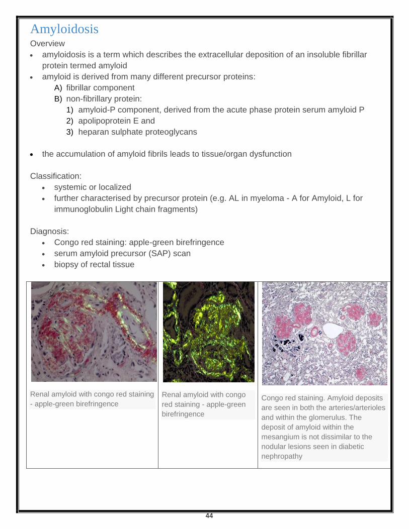
Diagnosis:
Congo red staining: apple-green birefringence
serum amyloid precursor (SAP) scan
biopsy of rectal tissue
Renal amyloid with congo red staining
- apple-green birefringence
Renal amyloid with congo
red staining - apple-green
birefringence
Congo red staining. Amyloid deposits
are seen in both the arteries/arterioles
and within the glomerulus. The
deposit of amyloid within the
mesangium is not dissimilar to the
nodular lesions seen in diabetic
nephropathy
45
Amyloidosis: types A) AL amyloid:
L for immunoglobulin Light chain fragment
due to myeloma, Waldenstrom's, MGUS
features include: cardiac and neurological involvement, macroglossia, periorbital
eccymoses
B) AA amyloid:
A for precursor serum amyloid A protein, an acute phase reactant
seen in chronic infection/inflammation
e.g. TB, bronchiectasis, rheumatoid arthritis
features: renal involvement most common feature
C) Beta-2 microglobulin amyloidosis:
precursor protein is beta-2 microglobulin, part of the major histocompatibility complex
associated with patients on renal dialysis
46
Haemostasis and thrombosis 431
Haemostasis 431 Vascular disorders 435
Platelet disorders 435
Inherited coagulation disorders 438
Acquired coagulation disorders 440
Thrombosis 442
Thrombocytopenia: Causes of severe thrombocytopenia
1) ITP
2) DIC
3) TTP
4) haematological malignancy
Causes of moderate thrombocytopenia:
1) heparin induced thrombocytopenia (HIT)
2) drug-induced (e.g. quinine, diuretics, sulphonamides, aspirin, thiazides)
3) alcohol
4) liver disease
5) hypersplenism
6) viral infection (EBV, HIV, hepatitis)
7) pregnancy
8) SLE/antiphospholipid syndrome
9) vitamin B12 deficiency
ITP
Idiopathic thrombocytopenic purpura (ITP) is an immune mediated reduction in the platelet count.
Antibodies are directed against the glycoprotein IIb/IIIa or Ib-V-IX complex.
ITP can be divided into acute and chronic forms:
Acute ITP:
more commonly seen in children
equal sex incidence
may follow an infection or vaccination
usually runs a self-limiting course over 1-2 weeks
Chronic ITP:
more common in young/middle-aged women
tends to run a relapsing-remitting course
Investigations:
1) antiplatelet autoantibodies (usually IgG)
2) Bone marrow aspiration shows megakaryocytes in the marrow. This should be carried out
prior to the commencement of steroids in order to rule out leukaemia
Management:
1) oral prednisolone (80% of patients respond)
2) splenectomy if platelets < 30 after 3 months of steroid therapy
3) IV immunoglobulins
4) immunosuppressive drugs e.g. cyclophosphamide
Evan's syndrome: ITP in association with autoimmune haemolytic anaemia (AIHA)

47
Thrombotic thrombocytopenic purpura: Pathogenesis of thrombotic thrombocytopenic purpura (TTP)
1) abnormally large and sticky multimers of von Willebrand's factor cause platelets to clump within
vessels
2) in TTP there is a deficiency of ADAMTS13 (a metalloprotease enzyme) which breakdowns large
multimers of von Willebrand's factor
3) overlaps with haemolytic uraemic syndrome (HUS)
Features:
1) rare, typically adult females
2) fever
3) fluctuating neuro signs (microemboli)
4) microangiopathic haemolytic anaemia
5) thrombocytopenia
6) renal failure
Causes:
1) post-infection e.g. urinary, gastrointestinal
2) pregnancy
3) drugs: ciclosporin, oral contraceptive pill, penicillin, clopidogrel, aciclovir
4) tumours
5) SLE
6) HIV
Management:
1) no antibiotics - may worsen outcome
2) plasma exchange is the treatment of choice
3) steroids, immunosuppressants
4) vincristine

48
Haemolytic uraemic syndrome Haemolytic uraemic syndrome is generally seen in young children and produces a triad of:
acute renal failure
microangiopathic haemolytic anaemia
thrombocytopenia
Causes
post-dysentery - classically E coli 0157:H7 ('verotoxigenic', 'enterohaemorrhagic')
tumours
pregnancy
ciclosporin, the Pill
systemic lupus erythematosus
HIV
Investigations
full blood count: anaemia, thrombocytopaenia, fragmented blood film
U&E: acute renal failure
stool culture
Management
treatment is supportive e.g. Fluids, blood transfusion and dialysis if required
there is no role for antibiotics, despite the preceding diarrhoeal illness in many patients
The indications for plasma exchange in HUS are complicated. As a general rule plasma
exchange is reserved for severe cases of HUS not associated with diarrhoea
Heparin-induced thrombocytopaenia (HIT)
immune mediated - antibodies form which cause the activation of platelets
usually does not develop until after 5-10 days of treatment
despite being associated with low platelets HIT is actually a prothrombotic condition
Features:
1) a greater than 50% reduction in platelets,
2) thrombosis and
3) skin allergy
Treatment options include
Alternative anticoagulants such as lepirudin and danaparoid
49
Haemophilia Haemophilia is X-linked recessive disorder of coagulation.
Up to 30% of patients have no family history of the condition.
A) Haemophilia A
1) It is due to a deficiency of factor VIII
2) accounts for 90% of cases of haemophilia
B) haemophilia B (Christmas disease) there is a lack of factor IX
Features:
1) haemoarthroses, haematomas
2) prolonged bleeding after surgery or trauma
Blood tests:
1) prolonged APTT
2) bleeding time, thrombin time, prothrombin time normal
3) Up to 10-15% of patients with haemophilia A develop antibodies to factor VIII treatment
A normal factor VIIIc activity points to a diagnosis of haemophilia B (lack of factor IX).
Von Willebrand's disease The most common inherited bleeding disorder.
The majority of cases are inherited in an autosomal dominant fashion
characteristically behaves like a platelet disorder i.e:
epistaxis and menorrhagia are common whilst
haemoarthroses and muscle haematomas are rare
Role of von Willebrand factor:
large glycoprotein which forms massive multimers up to 1,000,000 Da in size
promotes platelet adhesion to damaged endothelium
carrier molecule for factor VIII
Types:
Type 1: partial reduction in vWF (80% of patients)
Type 2: abnormal form of vWF
Type 3: total lack of vWF (most severe form) autosomal recessive
Investigation:
1) prolonged bleeding time
2) APTT may be prolonged
3) factor VIII levels may be moderately reduced
4) defective platelet aggregation with ristocetin
Management:
1) tranexamic acid for mild bleeding
2) desmopressin (DDAVP): raises levels of vWF by inducing release of vWF from Weibel-
Palade bodies in endothelial cells
3) factor VIII concentrate
A grossly elevated APTT may be caused by:
1) heparin therapy,
2) haemophilia or
3) Antiphospholipid syndrome.

50
Thrombophilia: Causes:
Inherited
A) activated protein C resistance (factor V Leiden)
B) antithrombin III deficiency
C) protein C deficiency
D) protein S deficiency
Acquired:
A) antiphospholipid syndrome
B) the Pill
A) Activated protein C resistance: (factor V Leiden)
The most common inherited thrombophilia. It is due to Factor V Leiden mutation.
Heterozygotes have a 5-fold risk of venous thrombosis whilst
Homozygotes have a 50-fold increased risk
B) Protein C deficiency An autosomal codominant condition which causes an increased risk of thrombosis
Features:
1) venous thromboembolism
2) skin necrosis following the commencement of warfarin:
When warfarin is first started biosynthesis of protein C is reduced.
This results in a temporary procoagulant state after initially starting warfarin,
Normally avoided by concurrent heparin administration.
Thrombosis may occur in venules leading to skin necrosis
C) Antithrombin III deficiency: autosomal dominant
An inherited cause of thrombophilia occurring in approximately 1:2,000 of the population.
Antithrombin III inhibits several clotting factors, primarily thrombin, factor X and factor IX. It
mediates the effects of heparin
Antithrombin III deficiency comprises a heterogeneous group of disorders:
1) some patients having a deficiency of normal antithrombin III whilst
2) others produce abnormal antithrombin III
Features:
1) recurrent venous thromboses
2) arterial thromboses do occur but is uncommon
Management:
1) thromboembolic events are treated with lifelong warfarinisation
2) heparinisation during pregnancy*
3) antithrombin III concentrates (often using during surgery or childbirth)
*as patients with antithrombin III deficiency have a degree of resistance to heparin anti-Xa levels should be
monitored carefully to ensure adequate anticoagulation

51
Antiphospholipid syndrome: pregnancy
an acquired disorder
Characterised by:
1) A predisposition to both venous and arterial thromboses,
2) recurrent fetal loss and
3) Thrombocytopenia.
It may occur as a primary disorder or secondary to other conditions, most commonly SLE.
In pregnancy the following complications may occur:
1) recurrent miscarriage
2) IUGR
3) pre-eclampsia
4) placental abruption
5) pre-term delivery
6) venous thromboembolism
Management:
1) low-dose aspirin should be commenced once the pregnancy is confirmed on urine testing
2) Low molecular weight heparin:
Started once a fetal heart is seen on ultrasound.
discontinued at 34 weeks gestation
These interventions increase the live birth rate seven-fold
Superficial Thrombophlebitis
52
Venous thromboembolism Risk factors: Common predisposing factors include:
1) malignancy,
2) pregnancy and
3) the period following an operation
The comprehensive list below is partly based on the 2010 SIGN venous thromboembolism
(VTE) guidelines:
General:
1) pregnancy (especially puerperium)
2) increased risk with advancing age
3) obesity
4) family history of VTE
5) immobility
6) hospitalisation
7) anaesthesia
8) central venous catheter: femoral >> subclavian
Underlying conditions:
1) malignancy
2) thrombophilia: e.g. Activated protein C resistance, protein C and S deficiency
3) heart failure
4) antiphospholipid syndrome
5) Behcet's
6) polycythaemia
7) nephrotic syndrome
8) sickle cell disease
9) paroxysmal nocturnal haemoglobinuria
10) hyperviscosity syndrome
11) homocystinuria
Medication
1) combined oral contraceptive pill: 3rd generation more than 2nd generation
2) hormone replacement therapy: the risk of VTE is higher in women taking oestrogen +
progestogen preparations compared to those taking oestrogen only preparations
3) raloxifene and tamoxifen
4) antipsychotics (especially olanzapine) have recently been shown to be a risk factor
It should be remembered however that around 40% of patients diagnosed with a PE have no
major risk factors.

53
Deep vein thrombosis: diagnosis and management
Diagnosis
NICE published guidelines in 2012 relating to the investigation and management of DVT.
If a patient is suspected of having a DVT a two-level DVT Wells score should be performed:
Two-level DVT Wells score
Clinical feature Points
Active cancer (treatment ongoing, within 6 months, or palliative) 1
Paralysis, paresis or recent plaster immobilisation of the lower extremities 1
Recently bedridden for 3 days or more or
major surgery within 12 weeks requiring general or regional anaesthesia
1
Localised tenderness along the distribution of the deep venous system 1
Entire leg swollen 1
Calf swelling at least 3 cm larger than asymptomatic side 1
Pitting oedema confined to the symptomatic leg 1
Collateral superficial veins (non-varicose) 1
Previously documented DVT 1
An alternative diagnosis is at least as likely as DVT -2
Clinical probability simplified score
DVT likely: 2 points or more
DVT unlikely: 1 point or less
A) If a DVT is 'likely' (2 points or more):
a proximal leg vein ultrasound scan should be carried out within 4 hours and, if the result is
negative, a D-dimer test
if a proximal leg vein ultrasound scan cannot be carried out within 4 hours a D-dimer test
should be performed and low-molecular weight heparin administered whilst waiting for the
proximal leg vein ultrasound scan (which should be performed within 24 hours)
B) If a DVT is 'unlikely' (1 point or less)
perform a D-dimer test and if it is positive arrange:
a proximal leg vein ultrasound scan within 4 hours
if a proximal leg vein ultrasound scan cannot be carried out within 4 hours low-molecular
weight heparin should be administered whilst waiting for the proximal leg vein ultrasound
scan (which should be performed within 24 hours)

54
Management:
Venous thromoboembolism - length of warfarin treatment
provoked (e.g. recent surgery): 3 months
unprovoked: 6 months
Cancer patients with VTE - 6 months of LMWH
Low molecular weight heparin (LMWH) or fondaparinux should be given initially after a DVT is
diagnosed.
a vitamin K antagonist (i.e. warfarin) should be given within 24 hours of the diagnosis
the LMWH or fondaparinux should be continued for at least 5 days or until the international
normalised ratio (INR) is 2.0 or above for at least 24 hours, whichever is longer, i.e. LMWH or
fondaparinux is given at the same time as warfarin until the INR is in the therapeutic range
warfarin:
should be continued for at least 3 months.
At 3 months, NICE advise that clinicians should 'assess the risks and benefits of
extending treatment'
NICE add 'consider extending warfarin beyond 3 months for patients
withunprovoked proximal DVT if their risk of VTE recurrence is high and there is no
additional risk of major bleeding'.
This essentially means that if there was no obvious cause or provoking factor (surgery,
trauma, significant immobility) it may imply the patient has a tendency to thrombosis and
should be given treatment longer than the norm of 3 months.
In practice most clinicians give 6 months of warfarin for patients with an unprovoked
DVT/PE
for patients with active cancer NICE recommend using LMWH for 6 months
Further investigations and thrombophilia screening
As both malignancy and thrombophilia are obvious risk factors for deep vein thrombosis NICE
make recommendations on how to investigate patients with unprovoked clots.
Investigations for cancer:
1) a physical examination (guided by the patient's full history) and
2) a chest X-ray and
3) Blood tests (full blood count, serum calcium and liver function tests) and urinalysis.
4) Consider further investigations for cancer with an abdomino-pelvic CT scan (and a
mammogram for women) in all patients aged over 40 years with a first unprovoked DVT or PE
Thrombophilia screening
not offered if patients will be on lifelong warfarin (i.e. won't alter management)
consider testing for antiphospholipid antibodies
consider testing for hereditary thrombophilia in patients who have had unprovoked DVT or PE
and who have a first-degree relative who has had DVT or PE

55
56
Pregnancy: DVT/PE Overview
pregnancy is a hypercoagulable state
majority occur in last trimester
Pathophysiology
1) increase in factors VII, VIII, X and fibrinogen
2) decrease in protein S
3) uterus presses on IVC causing venous stasis in legs
Management
1) warfarin contraindicated
2) S/C low-molecular weight heparin preferred to IV heparin (less bleeding and
thrombocytopenia)
Superior vena cava obstruction: Superior vena cava (SVC) obstruction is an oncological emergency caused by compression of
the SVC.
It is most commonly associated with lung cancer.
Features:
1) dyspnoea is the most common symptom
2) swelling of the face, neck and arms - conjunctival and periorbital oedema may be seen
3) headache
4) visual disturbance
5) pulseless jugular venous distension
Causes:
1) common malignancies: small cell lung cancer, lymphoma
2) other malignancies: metastatic seminoma, Kaposi's sarcoma, breast cancer
3) aortic aneurysm
4) mediastinal fibrosis
5) goitre
6) SVC thrombosis
Management:
1) general: dexamethasone, balloon venoplasty, stenting
2) small cell: chemotherapy + radiotherapy
3) non-small cell: radiotherapy

57
Heparin There are two main types of heparin - unfractionated, 'standard' heparin or low molecular weight
heparin (LMWH).
Heparins generally act by activating antithrombin III.
Unfractionated heparin forms a complex which inhibits thrombin, factors Xa, IXa, XIa and XIIa.
LMWH however only increases the action of antithrombin III on factor Xa
The table below shows the differences between standard heparin and LMWH:
Standard heparin Low molecular weight heparin
Administration Intravenous Subcutaneous
Duration of action Short Long
Mechanism of
action
Activates antithrombin III.
Forms a complex that inhibits
thrombin, factors Xa, IXa, Xia
and XIIa
Activates antithrombin III.
Forms a complex that inhibits
factor Xa
Side-effects Bleeding
HIT
Osteoporosis
Bleeding
Lower risk of HIT and
osteoporosis with LMWH
Monitoring Activated partial thromboplastin
time (APTT)
Anti-Factor Xa (although routine
monitoring is not required)
Notes
Useful in situations where there is
a high risk of bleeding as
anticoagulation can be terminated
rapidly
Now standard in the management of
venous thromboembolism treatment
and prophylaxis and acute coronary
syndromes
Both unfractionated and low-molecular weight heparin can cause hyperkalaemia.
This is thought to be caused by inhibition of aldosterone secretion.
Heparin overdose may be reversed by protamine sulphate, although this only partially reverses
the effect of LMWH.
58
Warfarin Warfarin is an oral anticoagulant which inhibits the reduction of vitamin K to its active
hydroquinone form, which in turn acts as a cofactor in the carboxylation of clotting factor II, VII, IX
and X (mnemonic = 1972) and protein C.
Indications:
1) venous thromboembolism: target INR = 2.5, if recurrent 3.5
2) atrial fibrillation, target INR = 2.5
3) Mechanical heart valves, target INR depends on the valve type and location. Mitral valves
generally require a higher INR than aortic valves.
Patients on warfarin are monitored using the INR (international normalised ration), the ratio of the
prothrombin time for the patient over the normal prothrombin time.
Warfarin has a long half-life and achieving a stable INR may take several days.
There a variety of loading regimes and computer software is now often used to alter the dose.
Factors that may potentiate warfarin:
1) liver disease
2) P450 enzyme inhibitors, e.g.: amiodarone, ciprofloxacin
3) cranberry juice
4) drugs which displace warfarin from plasma albumin, e.g. NSAIDs
5) inhibit platelet function: NSAIDs
Side-effects:
1) haemorrhage
2) teratogenic, although can be used in breast-feeding mothers
3) Skin necrosis: when warfarin is first started biosynthesis of protein C is reduced. This results
in a temporary procoagulant state after initially starting warfarin, normally avoided by
concurrent heparin administration. Thrombosis may occur in venules leading to skin necrosis
4) purple toes
59
Warfarin overdose The following is based on the BNF guidelines, which in turn take into account the British
Committee for Standards in Haematology (BCSH) guidelines.
A 2005 update of the BCSH guidelines emphasised the preference of prothrombin complex
concentrate over FFP in major bleeding.
Situation Management
Major bleeding 1) Stop warfarin
2) Give intravenous vitamin K 5mg
3) Prothrombin complex concentrate - if not available then FFP*
INR > 8.0
Minor bleeding
1) Stop warfarin
2) Give intravenous vitamin K 1-3mg
3) Repeat dose of vitamin K if INR still too high after 24 hours
4) Restart warfarin when INR < 5.0
INR > 8.0
No bleeding
1) Stop warfarin
2) Give vitamin K 1-5mg by mouth, using the intravenous preparation
orally
3) Repeat dose of vitamin K if INR still too high after 24 hours
4) Restart when INR < 5.0
INR 5.0-8.0
Minor bleeding
1) Stop warfarin
2) Give intravenous vitamin K 1-3mg
3) Restart when INR < 5.0
INR 5.0-8.0
No bleeding
1) Withhold 1 or 2 doses of warfarin
2) Reduce subsequent maintenance dose
*as FFP can take time to defrost prothrombin complex concentrate should be considered in
cases of intracranial haemorrhage

60
Hereditary haemorrhagic telangiectasia Also known as Osler-Weber-Rendu syndrome,
Hereditary haemorrhagic telangiectasia (HHT) is an autosomal dominant condition
characterised by (as the name suggests) multiple telangiectasia over the skin and mucous
membranes.
20% of cases occur spontaneously without prior family history.
There are 4 main diagnostic criteria:
1) epistaxis : spontaneous, recurrent nose bleeds
2) telangiectases: multiple at characteristic sites (lips, oral cavity, fingers, nose)
3) visceral lesions: for example
gastrointestinal telangiectasia (with or without bleeding),
pulmonary arteriovenous malformations (AVM),
hepatic AVM,
cerebral AVM,
spinal AVM
4) family history: a first-degree relative with HHT
If the patient has 2 then they are said to have a possible diagnosis of HHT.
If they meet 3 or more of the criteria they are said to have a definite diagnosis of HHT:
The chest x-ray shows multiple
pulmonary nodules representing
arteriovenous malformations, the largest
in the right mid-zone.
The CT scan shows multiple hepatic
arteriovenous malformations
61
حكاية شعب بلا وطن.. الروهينجا
من المحتلة أراكان في الأصليين المواطنين المسلمين على وتطلق القديم أراكان دولة اسم "روهانج" من مأخوذة "روهنجيا" كلمة
إلى وانتمائها البلاد هذه تاريخ أعماق في للغوص و .حقيقية مأساة تعيش مضطهدة مسلمة أقلية وهم ،(بورما)ميانمار دولة قبل
في استمرت آسيا، شرق بجنو في حرة مستقلة إسلامية دولة مضى ما في كانت أراكان أن القول فيمكننا الإسلامية الحضارة
.حاليا ميانمار – بورما لاتحاد ومقاطعة ولاية 41 من واحدة ذلك بعد فأصبحت م4871 عام بورما من تحتل أن قبل قرون عدة الوجود
مع الحدودي والشريط البنغال، خليج ساحل على لميانمار الغربي الجنوب في يقع أراكان إقليم فإن الجغرافيا حيث من أما
الجنوب في حاليا ميانمار وتقع .تقريبا ميانمار مساحة عشر أي مربع كيلومتر ألف خمسين حوالي الإقليم مساحة وتبلغ ديش،بنغلا
وبنغلاديش والهند البنغال خليج الجنوب ومن والهند، الصين الشمال من ويحدها آسيا، لقارة الشرقي
-:كالتالي فيأتي للسكان الديمغرافي التقسيم أما .رانجون فهي ميانمار ةعاصم أما له، عاصمة اكياب مدينة من الإقليم هذا يتخذ
الروهنجيا تضامن منظمة حسب نسمة مليون 45 بحدود %15 تبلغ المسلمين ونسبة (1544)08،999،75 ميانمار سكان عدد (4
نسبة فإن Book Fact CIA حسبفب عددهم تقليص في تبالغ المصادر بعض وهناك 4978 منذ اراكان أبناء حقوق عن تدافع التي
%.1 تبلغ المسلمين
الآخرون أما ميانمار، داخل مليونان منهم ويعيش 1 %95 فيها المسلمين ونسبة ملايين 0.0 حوالي أراكان منطقة سكان عدد (1
.ضدهم الدولة هذه تمارسه الذي الاضطهاد بسبب البلاد خارج إلى فهاجروا
يشبه ومعظمهم والباتان والمنغول والفرس والأتراك والمورو والعرب البنغالية مثل مختلفة أصول من ميانمار مسلمي أصول وتنحدر (8
.ولونا شكلا الهندية القارة أهل
: اللغة
البنغالية، و الأردية و الفارسية و العربية اللغة من تعبيرات و كلمات من مؤلفة هي و "الروهنجيان" أراكان مسلمي لهجة تسمى
.الآن لحد مكتوبة تليس وهي

62
الإسلامية الوفود تتابعت ثم إليها، المسلمين العرب التجار قدوم مع الميلادي السابع القرن في أراكان إلى الإسلام دخول ويرجع
من عاما 805 حكمها دام مملكة الروهنجيا شعب وكون الإسلام، اعتناق على الأهالي من كبير عدد فأقبل .المعمورة أنحاء من إليها
مسلما ملكا (17) بعده وحكم شاه، سليمان الملك بقيادة م4185 عام في إسلامية دولة أول شكلت فقد م،4871 عام إلى م4185
هذه في المسلمين وجود قدم على يدل ومما .التوحيد كلمة مثل إسلامية شعارات تتضمن نقدية عملات لهم وكان التوالي، على
عاما، 065 منذ بني الذي (خان سندي مسجد) ،(أراكان) عاصمة (أكياب) في (مقام بدر) كمسجد لتاريخيةا الآثار بعض أيضا الدولة
.الميلادي عشر الخامس القرن في بني الذي (خان ولي) ومسجد م،4107 عام بني الذي (موسى الديوان) ومسجد
في الإسلام انتشار من خوفا ميانمار إلى الإقليم بضم قام الذي م4871 عام (بوداباي) البوذي الملك قبل من أراكان واحتلت
طوال ذلك على (هندي أصل) الماغ البوذيين وتشجيع خيراتهم ونهب المسلمين اضطهاد في البورميون البوذيون واستمر المنطقة،
.احتلالهم فترة
ميانمار بريطانيا جعلت م4988 عام وفي .تعماريةالاس البريطانية الهند حكومة إلى وضم تها ميانمار، بريطانيا احتلت م4711 عام في
بحكومة وعرفت آنذاك، الإمبراطورية في مستعمراتها كباقي الاستعمارية البريطانية الهند حكومة عن مستقلة مستعمرة أراكان مع
.البريطانية ميانمار
باعتماد المسلمين نفوذ من للتخل ص هاحملت فبدأت تخشاهم، بريطانيا جعل مما بقوة الإنجليزي الاستعمار المسلمون واجه
مذبحة بالمسلمين أوقعوا حتى بالسلاح وأمد تهم المسلمين، ضد البوذيين تحريض على فعمدت (تسد فر ق) المعروفة سياساتها
!أراكان في مسلم ألف مائة بحوالي خلالها فتكوا 4911م عام
في رغبت إذا سنوات عشر بعد عنها الاستقلال العرقيات لكل تمنح أن شريطة رلميانما الاستقلال بريطانيا منحت م،4917 عام وفي
من سكانها رغبة بدون أراكان احتلال في واستمروا وعودهم، ونكثوا عهودهم، نقضوا حتى الاستقلال على حصلوا أن ما ولكن ذلك،
.المسلمين ضد الممارسات بأبشع وقاموا أيضا ، (الماغ) والبوذيين (الروهنجيا) المسلمين

63

64
الروهنجيا مسلمي عذابات
أراكان من المسلمين إخراج مخطط مازال حيث م،1545 نوفمبر في جرت التي الانتخابات بعد الروهنجيا، المسلمين أحوال تتغير لم
بالحقيقة، له علاقة لا إعلان هذا لكن طي،ديمقرا نظام إلى عسكري نظام من الدولة نظام تغيير ميانمار حكومة إعلان رغم موجودا ،
تخطط بورما إن» :الروهنجيا تضامن منظمة رئيس «يونس محمد.د» ويقول .انتخاباتهم ونتائج إعلانهم المتحدة والأمم العالم يقبل ولم
التهديد تحت إجراؤها مت التي بالانتخابات التغيير يحدث ولن الجبليين، للبوذيين مستوطنة وجعلها أراكان من المسلمين لإخراج
تجرى أن وإما مستقلة، إسلامية دولة أراكان تكون أن إما :طريقي ن بأحد إلا حقوقهم الروهنجيا المسلمون ينال ولن العسكري،
.«المتحدة الأمم رعاية تحت أراكان في انتخابات
هذا فبموجب م،4971 عام ميانمار في الجديد يةالجنس قانون تطبيق بعد جديد اتجاه إلى الروهنجيا المسلمين معاناة تحولت وقد
تم كما الحكومية، والهيئات الجيش في الوظائف وتقلد التجارة أعمال وممارسة العقارات تملك من حرمانهم تم المزعوم القانون
المنظمات سوتأسي البرلمانية، الانتخابات في التصويت حق مثل والأساسية الطبيعية الإنسانية الحقوق جميع من حرمانهم
التاريخ ذلك منذ أراكان في المسلمين ضد الجديدة الخطوات البورمية الاحتلال سلطات تتخذ ولم .السياسية النشاطات وممارسة
مغادرة على وإجبارهم الإسلامية، العقيدة ترك على لإرغامهم المسلمين؛ ضد القديمة الخطط تطبيق تواصل زالت ما بل فحسب،
.بلدهم
.المسلمين تواجه التي ضطهااااتوالإ الصعوبات
والممتلكات، الأرواح ضحيتها يذهب التي الشغب أعمال من تنتهي لا لسلسلة أراكان في وخصوصا ميانمار في المسلمون يتعرض و
.المسلمين لحماية أمنية إجراءات أية السلطات تتخذ ولم
ويقومون المسلمة القرى بأنحاء يطوفون البوذيين (الماغ) وسفاحو ئيالقضا التنفيذ وهيئات الميانماريون الجنود إن عيان شهود ويقول
.الممتلكات وسلب المنازل ودخول المسلم الشباب وضرب السن كبار بإذلال

65
النار وحطب والماعز والدواجن الأرز تقديم على تجبرهم العسكرية الحكومة أن هناك المسلمين على تمارس التي الفظائع من و
أثناء الجيش لدى القسري العمل على السكان إجبار يتم كما . القانونية التنفيذ وهيئات الجنود إلى العام طوال المج انب البناء ومواد
وذلك مقابل دون سخرة والسدود الطرق بناء في أو الحكومية الأعمال من ذلك وغير طرق شق أو عسكرية ثكنات بناء أو التنقلات
.الجيش يعتمدها التي الذاتي الاكتفاء سياسة ضمن
توجد فلا ،المسلمين لمناطق السكانية التركيبة في جذرية تغييرات بإحداث تقوم زالت ما الحكومة فإن السكاني الصعيد على أما
ما بإنشاء الحكومة قامت م4977 عام ومنذ .فيها السلطة سل متهم البوذيين للمستوطنين منازل فيها وأنشأت إلا منطقة أو قرية أي
.المناطق هذه في الاستيطان على البوذيين أسر تشجيع يتسن ى حتى أراكان، شمال في "النموذجية القرى"ـب يسمى
على الدولة موافقة يشترط الذي الزواج قانون فهناك بينهم، أو المسلمين من الزواج يخص ما على معينة شروط الحكومة وتفرض
45 إلى إذن بغير الزواج عقوبة وتصل لسنوات، الإذن يتأخر وقد الإذن، هذا لقاء شاوىالر تدفع ما وغالبا مقابل عال مبلغ وبدفع الزواج
.سجن سنوات
السوداء القائمة على أولاده يوضع ولدين من أكثر له يولد ومن فقط بمولودين إلا للعائلة السماح عدم هناك المسلمين عذابات ومن
أولادهم إخفاء إلى كثيرة أحيان في العائلة يضطر مما للعقوبة العائلة ويعرض حقوق لهم وليس بهم معترف غير أنهم تعني وهي
.عليهم خوفا وأقاربهم جداتهم باسم السوداء القائمة أولاد يسجل ما وكثيرا السكاني التعداد عند
جريمة ويعتبر باتا ، نعا م فيمنع المبيت أما مسبق، بإذن إلا أقارب أو أشقاء كانوا ولو بيوتهم في أحد باستضافة لهم يسمح لا كما
.وأسرته هو البلاد من طرده أو اعتقاله أو منزله بهدم عليها يعاقب ربما كبرى
إذا أما القرية، سجلات من قيده يطوى للخارج يذهب ومن والجامعات، الكليات في التعلم مواصلة من المسلمين أبناء كذلك ويحرم
للعلم الانحناء على الحكومية المدارس في المسلمين الطلاب إرغام ويتم السجون، ياهبغ في به ويرمى عودته، عند فيعتقل عاد
السفر جوازات حجز يتم كما .عليه الحصول يصعب والذي تصريح، دون آخر إلى مكان من بالانتقال للمسلمين يسمح ولا .البورمي
أية أو (رانجون) الدولة عاصمة إلى السفر ويعتبر رسمي، بإذن إلا للخارج بالسفر لهم يسمح ولا الحكومة لدى بالمسلمين الخاصة
.عليها يعاقب جريمة أخرى مدينة
دليل وخير البلاد، عن لإبعادهم وعائلتهم المسلمين تستهدف متكررة جماعي وتهجير طرد عمليات بمليشياتها الحكومة وتمارس
.بنغلاديش إلى مسلم 855.555 من أكثر طرد حيث العسكري نقلابالا عقب م4961 عام :التالية الأعوام في حصل ما ذلك على
الشيوخ من 15.555 قرابة منهم مات جدا ، قاسية أوضاع في مسلم، مليون نصف أي (055.555) من أكثر طرد م4987 عام وفي
مسلم، 405.555 من أكثر دطر تم م4977 عام وفي .المتحدة للأمم التابعة اللاجئين غوث وكالة إحصائية حسب والأطفال والنساء
مليون نصف أي (055.555) قرابة طرد تم م4994 عام وفي .الديموغرافي للتغيير محاولة في للبوذيين النموذجية القرى بناء بسبب
مع صوتوا لأنهم المسلمين من انتقاما ساحقة، بأغلبية المعارضة فيها فازت التي العامة الانتخابات نتائج إلغاء عقب وذلك مسلم،
.الديمقراطي الوطني الحزب لصالح البلاد أهل عامة

66
:يلي كما المواطنين يقسم وهو 2891 عام صدر الذي الجديد الجنسية قانون وبحسب
(.والكامينيون والصينيون والباهييون والشائيون الكارينون) :وهم الأولى الدرجة من مواطنون -أ
.الأولى الدرجة أجناس من خليط :وهم الثانية الدرجة من مواطنون -ب
حسب البريطاني الاحتلال أثناء لاجئين بورما دخلوا أجانب أنهم على صن فوا حيث المسلمون :وهم الثالثة الدرجة من مواطنون -ج
.شاءت متى ترحيلهم الحكومة بإمكان وصار الأعمال كل من وحرموا هوية بلا وصاروا المسلمين جنسيات فسحبت الحكومة مزاعم
-: أهما ومن "أراكان" إقليم في المسلمين السكان على والاقتصادية الدينية العقوبات من مجموعة كذلك الحكومة وتفرض
:الاقتصااية الناحية
.واضح سبب دون السمك صيد وقوارب المسلمين أراضي الميانمارية الحكومة تصادر (4
إبقاء بهدف زهيد بسعر يمث لهم من أو للعسكر إلا المحاصيل بيع ومنع ية،المال والغرامات شيء، كل على الباهظة الضرائب فرض (1
.الديار ترك على لإجبارهم أو فقراء، المسلمين
.الزراعية مشاريعهم لتطوير الحديثة الزراعية الآلات شراء من المسلمين منع (8
.بقمس إنذار ودون تعويض، دون من وآخر وقت بين المتداولة العملات إلغاء (1
.مواشيهم وقتل الزراعية المسلمين محاصيل إحراق (0
.أراكان في الصناعي القطاع ضمن بالعمل للمسلمين السماح عدم (6
:الدينية الناحية
.جدا صعب أمر وهذا الحكومية الجهات من إجازتها بعد إلا الإسلامية المطبوعات وإصدار الدينية الكتب بطباعة الحكومة تسمح لا (4
.عملهم أماكن في الإسلامي الزي لبس أو لحاهم بإطلاق للمسلمين السماح عدم (1
أو عامة مراحيض إلى وتحو لها أو غيرهم على وتوز عها المسلمين لدفن المخصصة والمقابر الأوقاف ممتلكات الحكومة تصادر (8
!!والمواشي للخنازير حظائر
.الاعتقال معسكرات في العمل على إرغامهم مويت والضرب للامتهان الدين رجال كبار يتعر ض (1
(.م4978) هـ 4158 رمضان بعد للصلاة الأذان منع وقد الصلاة، أذان لإطلاق الصوت مكبرات استخدام يمنع (0
.عليها إرادتها فرض بهدف والمدارس المساجد إدارة في مشروعة غير بطريقة الحكومة تتدخل (6
.سلوكهم عن وترضى الحكومة تعرفهم الذين الأفراد من قلة باستثناء الحج ةفريض أداء من المسلمون يمنع (8
67
.الأضاحي ذبح منع (7
ومصادرة عامة، ومتنز هات عسكرية وثكنات مستودعات إلى تحويلها أو سكن ودور وخم ارات مراقص إلى وتحويلها المساجد هدم (9
إقليم في المسلمين الطلاب اتحاد رئيس نائب قال وقد .البوذيين الماغ على عهاوتوزي الإسلامية بالأوقاف الخاصة والعقارات الأراضي
بتدمير م1554 عام خلال قامت ميانمار حكومة إن ":إينا ـ الإسلامية الأنباء وكالة"لـ حديث في الرحمن عتيق محمد إبراهي أراكان
هذا أن كما القديمة، المساجد وإصلاح ترميم أو الجديدة المساجد بناء بموجبه منعت أصدرته قانون بموجب وذلك مسجدا 81 نحو
.الأخيرة سنوات العشر خلال بني مسجد أي هدم على ينص القانون
قتيل ألف 87 الأقل على الإعصار هذا ضحايا عدد بلغ حيث 1557 عام بورما ضرب الذي نرجس إعصار بعد وخاصة التنصير حملات (45
.دينالمشر وملايين مفقود ألف 06و
الثقافة فرضوا فلقد قسرا ، البورمي البوذي المجتمع في المسلمين وتذويب الإسلامية الثقافة لطمس المستميتة المحاولات (44
.بوذية بأسماء والتسم ي المسلمات للبنات الحجاب لبس وعدم البوذيات من والزواج البوذية
الترميم من باتا منعا يمنع بقي وما تاريخية، ومدارس مساجد من الإسلامية الآثار بتدمير وذلك :الإسلامية والآثار الهوية طمس (41
على تهوي وبعضها وغيرها، للأيتام ودور ومكتبات ومدارس مساجد من بالدين علاقة لـه جديد شيء أي بناء أو البناء إعادة عن فضلا
.خر يجيها أو لشهاداتها والمصادقة الحكومي الاعتراف أو رالتطوي من تمنع الإسلامية والمدارس الزمن، مرور بسبب الناس رؤوس
المسلمة المرأة وضطع
المسلمين صفوف في السكان نسبة إرتفاع من تحد لكي الحكومة فإن "أركان " في المسلمة المرأة عذابات عن نتحدث حينما أما
وإعطاء السلطات، من وإذن طويلة إجراءات بعد إلا نكاحال عقود عاما ،ومنعت 85 والرجال عاما 10 لـ للفتيات الزواج سن برفع قامت
أو للمطلق أخرى مرة الزواج منع السبب، كان مهما باتا منعا التعدد منع كثيرة، حالات في المسلمات للنساء للحمل مانعة حقن
القضاء هو ذلك كل من والهدف .البلد من الطرد أو الباهظة والغرامات للسجن نفسه يعر ض ذلك يخالف ومن سنة، مرور بعد إلا الأرمل
.أعدادهم تقليل أو عليهم
الأمن قو ات إدارة إلى الحاكمة السلطات لقرار طب قا ذهابها من بد فلا الزوجة حملت إذا أنه أيضا منها تعسفية إجراءات فرضت و
بمبلغ الرسوم دفع من بد لا مر ة كل وفي حملها، تضع حت ى شهر كل مرور بعد بطنها كاشفة الملو نة صورتها لأخذ "ناساكا" الحدودية
.الولادة بعد المولود إحصائية ولتسهيل الجنين، سلامة من السلطة تد عي كما للتأك د وذلك كبير،
الفتيات إجبار إلى بالإضافة .مقابل دون الجيش معسكرات في العمل على وإجبارهن منازلهن من عنوة النساء أخذ يتم كما
القوات قيادة إلى الإجباري بالحضور المتزوجات غير المسلمات الفتيات هناك القوانين وتلزم .البوذيين من الزواج على مسلماتال
الكثير حرمات وتنتهك الحجاب خلع على المسلمات تجبر كما الحدود، حرس قوات أفراد إشراف تحت أشهر 6 لمدة والعمل المسلحة
.منهن
68
واللاجئين الاجرة
هاجر حيث 4994 و 4987 العام في أكبرها كان جماعية هجرات قامت فقد الروهنجيا مسلموا لها تعرض التي الكبيرة المعاناة بسبب
المتحدة، العربية والإمارات وتايلاند، وليبيا، وماليزيا، وباكستان، السعودية، :مثل أخرى دول و بنجلاديش إلى الروهنجيين معظم
من أو بينهم الفاصل النهر عبر بنجلاديش إلى يصلوا من والمحظوظون .دائم بشكل يتم الحدود عبر والهروب الهجرة زالت وما وغيرها،
إلى ثانية بقذفهم تقوم والتي تايلاند إلى قذفهم يتم فقد الخليج خلال من بالقوارب بالهرب يقومون من ولكن الحدودي السياج خلال
.المحيط في المحقق الموت ليواجهوا واربالق محركات فصل بعد ولكن المحيط
التايلاندية السلطات تعتبره ما حسب تايلاند وفي ،155.555 إلى 155.555 بنحو بنجلاديش في الأراكانيين اللاجئين عدد ويقدر
المنظمات نم مجموعة وهو بورما، - تايلاند حدود لاتحاد ووفقا ،شخص 15.555 حوالي عددهم فيبلغ شرعيين غير مهاجرون أنهم
تديرها مخيمات تسعة في بورمي لاجئ 411.555 نحو يعيش كيلومتر، 4،755 البالغة الحدود طول على العاملة الدولية الإنسانية
في أما .تايلاند في بورمي لاجئ 455.555 بتسجيل الآن حتى قامت فقد اللاجئين لشؤون المتحدة الأمم مفوضية أما الحكومة
ماليزيا في المسجلين وغير المسجلين ميانمار من المهاجرين عدد كوالالمبور ومقرها بورما عمال حقوق مايةح لجنة فتقدر ماليزيا
.ألفا 805 باكستان وفي مليون، نصف نحو عددهم فيقدر السعودية في أما (.ايرين) 055.555 بحوالي
بالدعم الأراكانيين إخوانهم بجانب بالوقوف ميةالإسلا المنظمات من كثير بادرت بنجلادش، في اللاجئين عدد ازداد وعندما
الحكومة منعت المتحدة، بالولايات م1554 سبتمبر من عشر الحادي أحداث بعد ولكن، .وغيرها والطبية الغذائية والمساعدات
.الحدود على الروهنجيا اللاجئين مخيمات في العمل من الإسلامية المنظمات البنغالية
لة مخيمات فهناك نوعان؛ بنجلادش في رماويينالبو اللاجئين مخيمات لشؤون العليا المفوضية ولدى بنجلاديش حكومة لدى مسج
مخيم في يقيمون منهم 44.555 فقط، المفوضية لدى مسجلا روهينجيا 17.555 يوجد حيث المتحدة، للأمم التابعة اللاجئين
.جنوبا أبعد مسافة على يقع الذي نايابار خيمم في غيرهم 48.555و بازار كوكس خارج للاجئين كوتوبالونج
وعدد باحتياجات مقارنة جدا ضئيلة مساعدات أنها رغم المتحدة، الأمم منظمات من والعلاج الغذائية المواد بعض يجدون وهؤلاء
لة غير الأخرى المخيمات أما الروهنجيا اللاجئين مخيمات في يعيشون ،نسمة ألف مائتي من أكثر بها ويوجد ،(رسمية غير) مسج
في إلا لهم حياة ولا دواء ولا غذاء ولا ماء فلا إنسانية، بكارثة تنذر مأساوية أوضاع في وبورما، بنجلاديش بين الحدود على اللاجئين
السلطات قبل من القسري والطرد التعسفي الاعتقال بسبب المخيم من للخروج المحاولات كل تقيدت وقد .بلاستيكية خيام
المزمن، والإسهال الجلدية والأمراض والملاريا الحمى مثل الفتاكة، والأمراض الأوبئة من شتى لأنواع اللاجئون ويتعرض الية،البنغ
غير مستشفى وجود لعدم المتعثرة الولادة بسبب النساء من عدد يموت كما الخطيرة، الأمراض بسبب الأطفال من كبير عدد ويموت
في روهنجي ألف 155 أن لها تقرير في ،(الإنسان حقوق أجل من أطباء) الأمريكية الإنسان حقوق منظمة حذرت فقد الرسمي،
لع زيادة يتم لم إذا المجاعة لخطر سيتعرضون روهينجا مسلمي وأن الإنسانية، الناحية من متدهورة حالة من يعانون بنجلاديش الس
.حاد تغذية سوء من بالفعل يعانون الأطفال من % 47 إن :المنظمة وتقول الغذائية،
69
اللاجئين ضد والتحريض الكراهية لبث المحلية الإعلام وسائل عبر دعائية بحملات بنجلادش في السلطات تقوم أخرى جهة ومن
.هنجياالرو جيرانهم عن السلطة إبلاغ في بدورهم يقوموا لم إذا بالاعتقال القرويين بتهديد أيضا وتقوم الروهنجيين
في التايلاندية البحرية القوات بهم دفعت الذين تايلاند في الروهنجيا لاجئي بعض معاناة عن تتحدث التي الأنباء من كثير وهنالك
.العالية البحرية الأمواج وسط محركات دون قوارب
بالروهنجيا المتعلقة المتحدة الأمم قرارات
فيها الروهنجيا قضية مناقشة وتم جنيف في المتحدة للأمم التابعة الإنسان قحقو لمنظمة 40 ال الدورة عقدت ( 1545/ 45) •
:توصياتها وكانت
ودعوة بورما، في الروهنجيا ضد البورمي النظام يرتكبها التي الانسانية ضد الجرائم في للتحقيق لجنة لإنشاء الدولي المجتمع حث
والرعاية والتعليم الإنسانية المساعدات وتقديم الروهنجي، المجتمع على المفروضة القيود جميع رفع إلى بورما في الدولة مجلس
جذري حل عن والبحث وماليزيا والسعودية بنغلاديش في الروهنجيا واللاجئين أراكان ولاية في الروهنجي للمجتمع الصحية
ضد الإنسان حقوق انتهاكات جميع في والتحقيق الحقائق لتقصي بعثة لإنشاء والدعوة الروهنجيا، اللاجئين معاناة لمشكلة
.وبورما أراكان في الروهنجيا
(Geneva in session Council Rights Human UN 15th in discuss issues Rohingya)
( 1545/ 0 ) المتحدة الأمم قرار •
القيود جميع بورماورفع في والمتساوية الكاملة الحقوق لهم كمواطنين بالروهنجيا للاعتراف البورمي النظام الأمريكي الكونجرس دعا
.والعرقي الديني الاضطهاد من النظام لحملة حد وضع إلى أيضا القرار ويدعو والتعليم والتنقل الزواج، على المفروضة
(Congress US Burma: of citizens equal” and “full as Rohingya Recognize)
الروهنجيا مسلمي مهالب
.العسكرية ميانمار حكومة على للضغط الإسلامية منظماتوال الدول دعوة (4
.المشروعة حقوقهم ينالوا حتى للصمود ومادي ا ومعنوي ا سياسي ا الكاملة والمساعدة الدعم تقديم (1
. العالمية والإسلامية الخيرية والهيئات المنظمات قبل من والإغاثي المادي الدعم وزيادة تكثيف (8
للتعريف القنوات تلك في معينة أوقات وتخصيص والغربية، العربية الإعلام وسائل كبريات مع والتواصل الإعلامية، الجهود تكثيف (1
لممارسة القضية، بهذه المعنيين الدوليين الحقوقيين مع حوارات وإجراء لغات، بعدة تبث فضائية قنوات وفتح الراهن، والوضع بالقضية
.أجمع العالم أمام ممارساتها وفضح ذية،البو السلطات على الضغط من مزيد
70
عاجل نداء وإطلاق وباكستان بنجلاديش في خاصة أراكان للاجئي والتعليمية الصحية والرعاية والدواء الغذاء الإغاثة المعونات توفير (0
حكومة على والضغط يش،بنغلاد في بها المعترف غير اللاجئين من الفئة وحماية الإنسانية الإغاثة أجل من المانحة الدول إلى
.للخطر المعرضين السكان من للاجئين الإنسانية المساعدات بإيصال الفوري للسماح بنغلاديش
والإقامة والعمل التعليم مثل يحتاجونها التي التسهيلات تقديم أراكان مسلمي من لاجئون بها يوجد التي الإسلامية الدول على (6
الوطنية الدراسة" مادة فيها ويدرس العالم، في تواجدهم أماكن في الروهنجيا للمسلمين ةالتعليمي المؤسسات بناء ينبغي كما
رابطتهم تستمر حتى لمتطلباتهم، ومراعاة ضروراتهم لتلبية أساسية كمواد القومية ولغتهم وثقافتهم تاريخهم يدرس كما "لأراكان
.وخارجها أراكان بداخل والمواطنين الوطن تجاه بالمسئولية عوروالش العاطفي والارتباط والروح الحماس فيهم وينشأ الوطن مع
.اراكان في التعليمية البرامج وتنمية الدعوية والأنشطة الدينية المعاهد على للحفاظ الضرورية المساعدة تقديم (8
عن ويبتعدون الحرية إلى القوم ذاه يقترب اللغة بهذه الكتب تنشر ما فبقدر الروهنجية، اللغة إلى وترجمتها الكتب تأليف وينبغي (7
.بينهم فيما سائدا التعليم ويكون المثقفة، الأخرى الأمم مثل بأنفسهم مشاكلهم لحل فيتأهلون .العبودية
8991465153823/https://www.facebook.com/j.documentary/videos/54
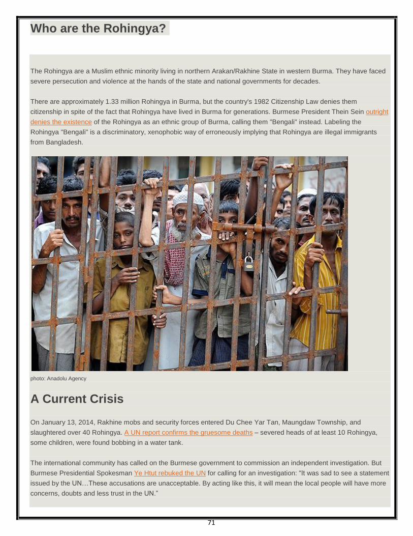
71
Who are the Rohingya?
The Rohingya are a Muslim ethnic minority living in northern Arakan/Rakhine State in western Burma. They have faced
severe persecution and violence at the hands of the state and national governments for decades.
There are approximately 1.33 million Rohingya in Burma, but the country's 1982 Citizenship Law denies them
citizenship in spite of the fact that Rohingya have lived in Burma for generations. Burmese President Thein Sein outright
denies the existence of the Rohingya as an ethnic group of Burma, calling them "Bengali" instead. Labeling the
Rohingya "Bengali" is a discriminatory, xenophobic way of erroneously implying that Rohingya are illegal immigrants
from Bangladesh.
photo: Anadolu Agency
A Current Crisis
On January 13, 2014, Rakhine mobs and security forces entered Du Chee Yar Tan, Maungdaw Township, and
slaughtered over 40 Rohingya. A UN report confirms the gruesome deaths – severed heads of at least 10 Rohingya,
some children, were found bobbing in a water tank.
The international community has called on the Burmese government to commission an independent investigation. But
Burmese Presidential Spokesman Ye Htut rebuked the UN for calling for an investigation: “It was sad to see a statement
issued by the UN…These accusations are unacceptable. By acting like this, it will mean the local people will have more
concerns, doubts and less trust in the UN.”

72
The government also vehemently refused the US’ request to allow international officials to sit on an investigation into the
incidents at Du Chee Yar Tan village. The government did eventually consent to commission two internal investigations,
but these severely biased investigations concluded that no massacre had taken place whatsoever, and deliberately
permitted perpetrators to go uncharged.
The government’s insistence that there were no Rohingya deaths in Du Chee Yar Tan in January 2014 is just one
recent example of the Government’s complicity in religious violence. Publicly rejecting UN counsel, the Government has
refused to hold perpetrators to account or provide legal redress to the many displaced, imprisoned, raped, and
murdered Muslims who have become victims of an avoidable, manmade humanitarian crisis.
Stranded at Sea
UNHCR estimates that 130,000 Rohingya have fled Bangladesh and western Arakan state since the outbreak of violence in
2012. As a result, the Burmese Navy and local security forces are profiting off the ethnic cleansing of the Rohingya in
western Burma by demanding payments from smugglers who devlier Rohingya to human traffickers, as well as Rohingya
desperately seeking passage.
Forced to venture by boat to trafficking camps on remote Thai islands, the Rohingya are faced with violence, lack of food and
water (often forced to drink their own urine), and those who have fallen victim to disease are thrown overboard if dead or close
to dying. In Thailand Rohingya are held in internment camps until they can either pay thousands to human traffickers to be
released or be sold as slaves to the highest bidder. Often Rohingya must resort to soliciting funds from their personal
networks, and if successful, they are pushed back out to sea. Those who are unable to pay become slaves: women and young
girls are forced into marriages and lifelong indentured servitude, and men sold to Thai fishermen.
Recently, mass graves, mostly Rohingya, have been uncovered in Thailand. Now, ASEAN countries (specifically Thailand,
Indonesia and Malaysia) are refusing to allow Rohingya refugees to seek assylum. They instruct their respective military to
take them back to sea, effectively abandoning the vulnerable Rohingya. It is suspected that thousands have died making the
deadly voyage to escape the ethnic cleansing they face in Burma.
Escalating Violence
Since June 2012, several hundred Rohingya have been killed because of their religion and ethnicity in widespread,
systematic attacks led by Rakhine Buddhists. Over 140,000 Rohingya have been displaced in inhumane internment
camps, and thousands have fled the country. Hundreds more, mostly men and boys, have been arrested on false
charges.
In June 2012, a Rakhine Buddhist woman was allegedly raped and murdered by three Rohingya Muslim men, and the
western Rakhine state subsequently broke out in violence. Refugees accumulated from both sides of the violence,
leaving thousands displaced and villages burned and abandoned. The Burmese military was called in to stop the
violence by means of further violence, firing tear gas and shooting at the mobs. The fury was temporarily subdued.
In October 2012, a more targeted surge occured against the Rohingya Muslim population. Rakhine Buddhist
communities formulated vigilante mobs, surrounding homes and razing them to flames, and Rohingya villagers fled.
Many Rohingya attempted to escape by boat to Bangladesh; some boats capsized leaving many missing, and some
reached the Bangladesh shore where they were told to immediately return to Burma.

73
Most alarming is the direct involvement of the local, state, and national government in the violence. Government officials
have enforced explicitly racist policies for decades, and have failed to intervene and even participated in violent
attacks against Rohingya. The government has been accused of implementing the crime against humanity of
persecution against the Rohingya, and Rohingya are increasingly considered to be targets of potential acts of genocide.
But Government officials and security forces have refused to implement impartial investigations into the violence. They
have instead subjected Rohingya and other Muslims to discriminatory restrictions and policies of apartheid and ethnic
cleansing. Rohingya internally displaced persons (IDPs) in Arakan State are now languishing in what UN officials have
called the most dismal and under-served IDP camps in the world.
Government persecution has enabled Rakhine Buddhist nationalists to implement a reign of terror against the Rohingya
with impunity. Hate speech directed against Rohingya Muslims appears regularly in state-run media, and some Buddhist
monks have instigated vitriolic rumors and discrimination against the minority group. Hate speech has helped fuel waves
of violence and systematic arson attacks in Rakhine State.
Of the hundreds of thousands of Rohingya that have fled to Bangladesh, Thailand, and Malaysia to escape persecution
and adversity, approximately 300,000 Rohingya live in squalid conditions in Bangladesh where they are denied access
to food supplies, medical aid, and education. In essence, the Rohingya are “Exiled to Nowhere.”
Take action with us and demand that the Rohingya Muslims be given citizenship, are treated with dignity as determined
by the Universal Declaration of Human Rights, and insist on an international investigation into systems of violence and
impunity encouraged by the Burmese government.