INTRODUCTION · 2013-11-19 · were evaluated for indices of anemia using SYSMEX KX 21N hematology...
34
0
Transcript of INTRODUCTION · 2013-11-19 · were evaluated for indices of anemia using SYSMEX KX 21N hematology...
0
1
INTRODUCTION
This compilation of abstracts will serve as a research
guide to support faculty and students in their search for
recorded literature in selected journals. Full texts of cited
articles are available in the LPU S.H.L. Learning
Resource Center. Online version if available, may be
browsed in the online databases of Academic OneFile
with a password.
Should you have comments on this compilation, please
call us at 723-07-06 local 113/114 or send message to

2
Clinical Laboratory Science Vol. 25(1), Winter 2012 A professional development model for medical laboratory scientists working in the immunohematology laboratory Garza, M. N., Pulido, L. A., Amerson, M., Ali, F. A., Greenhill, B. A., Griffin, G., & ... Hu, P. C.
6
Evaluation of iron status in anemia of chronic disease among patients with HIV infection Ogbe, P. J., Idoko, O. A., U, Digban, K. A., & Oguntayo, B. O.
8
Improving the inter-laboratory harmonization of the International Normalized Ratio (INR): utilizing the concept of transference to estimate and/or validate International Sensitivity Index (ISI) and mean normal prothrombin time (MNPT) values and/or to eliminate measurement bias Favaloro, E. J., McVicker, W., Hamdam, S., Huynh, M., Peris, P., O'Neal, M., & Hocker, N.
10
Novel test method (sickle confirm) to differentiate sickle cell anemia from sickle cell trait for potential use in developing countries Randolph, T. R., & Wheelhouse, J.
12

3
Proteasome inhibitors In cancer therapy: a novel approach to a ubiquitous problem... Focus: advances in clinical cancer research Landis-Piwowar, K. R.
14
Anti-hormones: mechanism and use of breast cancer... Focus: advances in clinical cancer research Dinda, S.
15
Cancer stem cells... Focus: advances in clinical cancer research Williams, J.
16
Vol. 2(2), Spring 2012
A professional development model for medical laboratory scientists working in the core Ali, F. A., Pulido, L. A., Garza, M. N., Amerson, M. H., Greenhill, B., Brown, K. N., & ... Hu, P. C.
17
A professional development model for medical laboratory scientists working in the microbiology laboratory Amerson, M. H., Pulido, L., Garza, M. N., Ali, F. A., Greenhill, B., Einspahr, C. L., & ... Hu, P. C.
18
Warfarin hypersensitivity due to gluten-sensitive enteropathy: a case study Kwolek, S., & Deming, P.
19
4
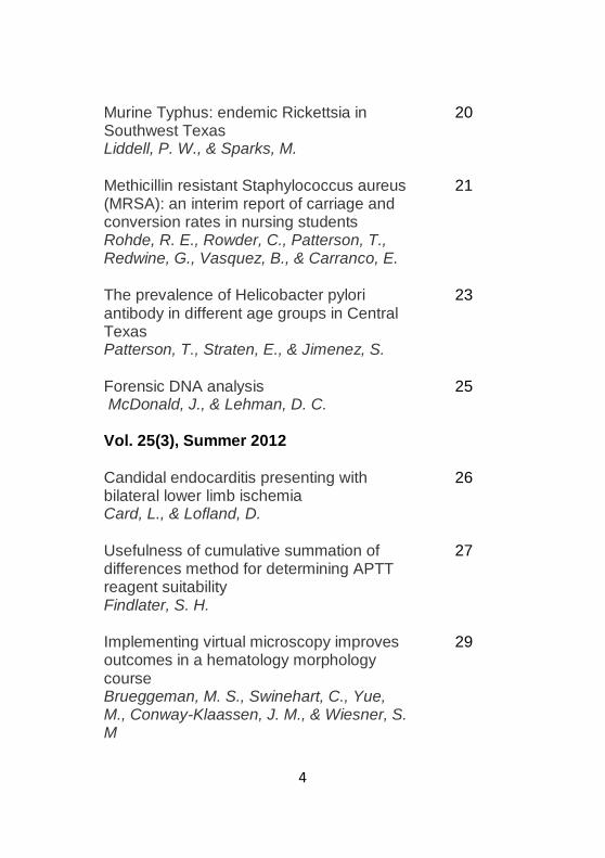
Murine Typhus: endemic Rickettsia in Southwest Texas Liddell, P. W., & Sparks, M.
20
Methicillin resistant Staphylococcus aureus (MRSA): an interim report of carriage and conversion rates in nursing students Rohde, R. E., Rowder, C., Patterson, T., Redwine, G., Vasquez, B., & Carranco, E.
21
The prevalence of Helicobacter pylori antibody in different age groups in Central Texas Patterson, T., Straten, E., & Jimenez, S.
23
Forensic DNA analysis McDonald, J., & Lehman, D. C.
25
Vol. 25(3), Summer 2012
Candidal endocarditis presenting with bilateral lower limb ischemia Card, L., & Lofland, D.
26
Usefulness of cumulative summation of differences method for determining APTT reagent suitability Findlater, S. H.
27
Implementing virtual microscopy improves outcomes in a hematology morphology course Brueggeman, M. S., Swinehart, C., Yue, M., Conway-Klaassen, J. M., & Wiesner, S. M
29
5
Snapshot prevalence and characterization of Staphylococcus species, including MRSA, in a student athletic facility: an undergraduate research project Garcia, S. A., Mckenzie, J. F., Patterson, T., & Rohde, R. E.
30
Lupus anticoagulant increases activated partial thromboplastin time (PTT) prolongation in Incubated 1:1 mix Monis, G., Ferrell, C., & Reyes, M.
32
6
Garza, M. N., Pulido, L. A., Amerson, M., Ali, F. A., Greenhill, B. A., Griffin, G., & ... Hu, P. C. (2012). A professional development model for medical laboratory scientists working in the immunohematology . Clinical Laboratory Science, 25(1), 2-6. Abstract: Transfusion medicine, a section of the Department of Laboratory Medicine at The University of Texas MD Anderson Cancer Center is committed to the education and advancement of its health care professionals. It is our belief that giving medical laboratory professionals a path for advancement leads to excellence and increases overall professionalism in the Immunohematology Laboratory. As a result of this strong commitment to excellence and professionalism, the Immunohematology laboratory has instituted a Professional Development Model (PDM) that aims to create Medical Laboratory Scientists (MLS) that are not only more knowledgeable, but are continually striving for excellence. In addition, these MLS are poised for advancement in their careers. The professional development model consists of four levels: Discovery, Application, Maturation, and Expert. The model was formulated to serve as a detailed path to the mastery of all process and methods in the Immunohematology Laboratory. Each level in the professional development model consists of tasks that optimize the laboratory workflow and allow for concurrent training. Completion of a level in the PDM is rewarded with fmanclal incentive and further advancement in the field. The PDM for Medical Laboratory Scientists in the Immunohematology Laboratory fosters personal development, rewards growth and competency, and sets high standards for all services and skills provided. This model is a vital component of the

7
Immunohematology Laboratory and aims to ensure the highest quality of care and standards in their testing. It is because of the success of this model and the robustness of its content that we hope other medical laboratories aim to reach the same level of excellence and professionalism, and adapt this model into their own environment. Subjects: 1. Blood Banks, 2. Medical Technologists
3. Professional Competence, 4. Professional Development
8
Ogbe, P. J., Idoko, O. A., U, Digban, K. A., & Oguntayo, B. O. (2012). Evaluation of iron status in anemia of chronic disease among patients with HIV infection. Clinical Laboratory Science, 25(1), 7-12. Abstract: OBJECTIVE: In HIV-infected populations from
developing countries, it is unclear what proportion of anemia is attributable to iron deficiency (ID). The objective of this study was to evaluate the iron status in anemia of chronic disease of patients with Human Immunodeficiency Virus (HIV) infection attending Federal Medical Centre, Makurdi. DESICN AND SETTINC: A total of 312 subjects comprising of 207 confirmed HIV positive patients and 105 apparently healthy subjects as control were evaluated for indices of anemia using SYSMEX KX 21N hematology analyzer machine (Kobe, Japan), CD4 count using CYFLOW SL machine (Artec, Cermany), and total iron binding capacity and serum iron using colorimetric method. RESULTS: While results showed that Serum Iron, Transferrin Saturation, PCV, RBC, MCV, MCH, MCHC and RDWCV are within normal reference range but statistically different (p<0.05) compared to the controls. Stratifying them on the basis of CD4 count showed that in AIDS patients the indicators are generally lower with Hb, PCV, MCH and RDWCV showing statistical significance (p<0.05) compared with patients with CD4 >200cells/mml Serum iron (50%) and transferring saturation (47.9%) contributed highest to anemia prevalence especially in males while Hb concentration (47.2%) is the major contributor to anemia in females. CONCLUSION: It was concluded therefore that albeit, on average, the parameters of iron status did not indicate iron deficiency or iron overload in the HIVstatus groups and AIDS patients, a large percentage
9
of patients did have anemia of chronic disease with HIVinfected women afflicted more often. The anemia is generally normocytic hypochromic in AIDS patierits. Subjects: 1. Acquired Immunodeficiency Syndrome -- Complications, 2. Anemia -- Diagnosis, 3. Chronic Disease, 4. HIV Infections -- Complications, 5. Iron -- Blood
10
Favaloro, E. J., McVicker, W., Hamdam, S., Huynh, M., Peris, P., O'Neal, M., & Hocker, N. (2012). Improving the inter-laboratory harmonization of the International Normalized Ratio (INR): utilizing the concept of transference to estimate and/or validate International Sensitivity Index (ISI) and mean normal prothrombin time (MNPT) values and/or to eliminate measurement bias. Clinical Laboratory Science, 25(1), 13-25. Abstract: BACKGROUND: The Prothrombin Time (PT) assay is clinically the most often requested coagulation test, as used primarily for monitoring of Vitamin K antagonist therapy where results are typically expressed as an International Normalized Ratio (INR). The INR reflects the patient's PT adjusted for the specific test reagent and instrument combination used by applying two correction factors, namely the International Sensitivity Index (ISI) and the Mean Normal Prothrombin Time (MNPT), according to the formula: INR = (patient PT/MNPT)'. When the manufacturer provides an ISI, laboratories are encumbered to check or locally validate the assigned value. Where a manufacturer does not provide an ISI, the laboratory needs to define its own (local ISI) value. The MNPT typically has to be locally defined, based on the population being tested. The main current CLSI recommendation for defining ISI values comprises use of commercial reference ('certified') plasma calibration sets, but FDA cleared material is limited, and different results may arise using different products. The MNPT can be defined using a WHO/CLSI recommended procedure requiring 20 normal individuals or with some calibration sets. Overall, there is limited data to validate the performance of these processes in
11
laboratory practice, and ongoing evidence from external quality assurance (proficiency testing) programs indicates continued failure in INR harmonization, suggesting that ISI and MNPT values used by laboratories (and presumably assessed using current recommended processes) continue to be inaccurate. OBJECTIVE: To assess some novel approaches to the laboratory estimation and/or validation of ISI and MNPT values for use in the INR calculation, and including the process of'transference', normally used to assess the comparability of analytical systems or to transfer reference intervals between comparable systems. RESULTS: We have successfully adapted these comparative procedures, including 'transference', to permit ongoing estimation and/or validation of ISI and MNPT values for use in INR calculations for a range of instrumentation, which has led to improved harmonization of INR values obtained in our pathology network. These processes do not require the use of any normal individual plasmas or calibrator sets and greatly simplifies the INR process. Evidence for validation of the processes used is provided by ongoing satisfactory performance in external quality assurance (proficiency testing). Subjects: 1. Calibration -- Methods, 2. International
Normalized Ratio, 3. Prothrombin Time, 4. Quality Control (Technology)

12
Randolph, T. R., & Wheelhouse, J. (2012). Novel test method (sickle confirm) to differentiate sickle cell anemia from sickle cell trait for potential use in developing countries. Clinical Laboratory Science, 25(1), 26-34. Abstract: The objective of this study was to develop a diagnostic testing method to detect HbS, distinguish sickle cell homozygotes from hétérozygotes, and overcome testing barriers encountered in laboratories in underdeveloped countries. Blood samples positive and negative for sickle cell were subjected to the standard hemoglobin solubility test followed by a variety of centrifugation and filtration procedures. Each procedure was evaluated for the ability to remove insoluble HbS from the sample. The hemoglobin types that remain (HbA, HbA2 and HbF) were measured spectrophotometrically or estimated visually allowing samples to be categorized into three genotypes (AA, AS and SS) as confirmed by hemoglobin electrophoresis. De-identified EDTA blood samples were obtained from Saint Louis University and Cardinal Glennon Children's hospitals and tested in the Department of Clinical Laboratory Science at Saint Louis University. The main outcome measures were turbidity of the solubility solution; color of the supernatant and the material on the surface of the solution following centrifugation; precipitate trapped on the filter paper; absorbance of the filtrate; and hemoglobin electrophoresis patterns. Centrifugation and filtration successfully separated HbS from HbA/A2/F allowing for the differentiation of seven sickle cell homozygotes from sixteen hétérozygotes with a sensitivity and specificity of 100%. This method has the potential to reliably distinguish homozygous from
13
heterozygous sickle cell patients and it is fast, inexpensive, and simple. These characteristics make Sickle Confirm a desirable method in developing countries like Haiti and Africa where sickle cell anemia is prevalent and modern diagnostic methods like electrophoresis, HPLC and nucleic acid testing are impractical. Subjects: 1. Anemia, Sickle Cell -- Diagnosis
2. Diagnosis, Differential, 3. Sickle Cell Trait
14
Landis-Piwowar, K. R. (2012). Proteasome inhibitors In cancer therapy: a novel approach to a ubiquitous problem... Focus: advances in clinical cancer research. Clinical Laboratory Science, 25(1), 38- 44. Abstract: LEARNING OBJECTIVES: 1. Describe the basic structure and function of the proteasome. 2. Identify the proteasomal subunit within the catalytic core that is crucial to cell survival. 3. List proteasome protein targets. 4. Evaluate the effect on cellular homeostasis following proteasome inhibition. 5. Detail the status of various proteasome inhibitors in pre-clinical and clinical trials. The cellular proteasome is an important molecular target in cancer therapy and drug resistance research. Proteasome inhibitors are effective agents against multiple myeloma and mantle cell lymphoma and display great potential as treatment for a variety of other malignancies. The proteasome is a large multicatalytic, proteinase complex located in the cytosol and the nucleus of eukaryotic cells. The ubiquitin proteasome system is responsible for the degradation of most intracellular proteins and therefore plays an essential regulatory role in critical cellular processes including cell cycle progression, proliferation, differentiation, angiogenesis, and apoptosis. Cancer cells are particularly sensitive to proteasome inhibitors, indicating the utility for inhibition of the ubiquitinproteasome pathway as an approach for cancer therapy. Subjects: 1. Lymphoma -- Drug Therapy, 2. Multiple Myeloma -- Drug Therapy, 3. Protease Inhibitors

15
Dinda, S. (2012). Anti-hormones: mechanism and use of breast cancer... Focus: advances in clinical cancer research. Clinical Laboratory Science, 25(1), 45-
49. Abstract: LEARNING OBJECTIVES: 1. Define the roles of estrogen and progesterone in breast and uterine cells. 2. DeBne hormone-dependent and independent tumors. 3. Describe the classiflcation and main functions of anti-hormone therapy. 4. Explain the mechanism of aromatase inhibitors, ERDs and SERMs. 5. Explain the differences between the mechanisms of tamoxifen and raloxifene. ABSTRACT: Breast cancer is the second-leading cause of cancer-related death in women. Recently, new drugs are being developed based on the molecular mechanisms of receptors, tumor suppressor genes, monoclonal antibodies, tumor markers and antihormone therapy. Anti-hormone therapy is used in the treatment of hormone-dependent breast tumors. Among the anti-hormone therapies, a substantial amount of research has been focused on the development of the ideal selective estrogen receptor modulator to treat metastatic breast tumors and to prevent breast cancer in high risk women. Subjects:
1. Antineoplastic Agents, Hormonal -- Therapeutic Use 2. Breast Neoplasms -- Drug Therapy, 3. Breast Neoplasms -- Physiopathology
16
Williams, J. (2012). Cancer stem cells... Focus: advances in clinical cancer research. Clinical Laboratory Science, 25(1), 50-57. Abstract: LEARNING OBJECTIVES: 1. Define the cancer stem cell model of tumorigenesis. 2. Describe the characteristics of cancer stem cells (CSCs). 3. Relate the biologic characteristics of CSCs to their impact on cancer therapeutics. 4. Identify novel targets for therapeutic approaches directed at CSCs. The cancer stem cell (CSC) hypothesis has had a major effect on the fields of cancer cell biology and clinical oncology. CSCs were originally described in hématologie malignancies, and subsequently in a variety of solid tumors. Their unique biological characteristics, including self-renewal capability, stem cell signaling pathways, relative quiescence and resistance to standard chemotherapy and radiotherapy are providing researchers and clinicians with new challenges. One important outcome of this new perspective on tumors is the recognition that effective treatment approaches will need to target both the rapidly proliferating bulk tumor cells, and the quiescent CSCs, which contain the ability to reestablish the malignancy when treatment is withdrawn. The clinical laboratory will undoubtedly see an infiux of new molecular and histopathological tests to augment initial diagnosis, treatment decisions, and prognostic monitoring of cancer patients related to identifying and quantifying these as CSCs.
Subjects: 1. Neoplasms -- Etiology, 2. Neoplasms -- Pathology, 3. Signal Transduction, 4. Stem Cells
17
Ali, F. A., Pulido, L. A., Garza, M. N., Amerson, M. H., Greenhill, B., Brown, K. N., & ... Hu, P. C. (2012). A professional development model for medical laboratory scientists working in the core laboratory. Clinical Laboratory Science, 25(2), 67- 73. Abstract:The Division of Pathology and Laboratory Medicine at The University of Texas MD Anderson Cancer Center has implemented a professional development model designed to further the education, expertise, and experiences of medical laboratory scientists in the core laboratory. The professional development model (PDM) has four competency levels: Discovery, Application, Maturation and Expert. All levels require the medical laboratory scientist to learn new skill sets, complete task and projects, and meet continuing education and certification requirements. Each level encourages personal development, recognizes increased competencies, and sets high standards for all services provided. Upon completion of a level within a given timeframe, the medical laboratory scientist receives a salary adjustment based on the competency level completed. Subjects: 1.Medical Technologists, 2. Professional Competence, 3. Professional Development
18
Amerson, M. H., Pulido, L., Garza, M. N., Ali, F. A., Greenhill, B., Einspahr, C. L., & ... Hu, P. C. (2012). A professional development model for medical laboratory scientists working in the microbiology laboratory. Clinical Laboratory Science, 25(2), 74-77. Abstract: The University of Texas M.D. Anderson Cancer Center, Division of Pathology arid Laboratory Medicine is committed to providing the best pathology and medicine through: state-of-the art techniques, progressive ground-breaking research, education and training for the clinical diagnosis and research of cancer and related diseases. After surveying the laboratory staff and other hospital professionals, the Department administrators and Human Resource generalists developed a professional development model for Microbiology to support laboratory skills, behavior, certification, and continual education within its staff. This model sets high standards for the laboratory professionals to allov* the labs to work at their fullest potential; it provides organization to training technologists based on complete laboratory needs instead of training technologists in individual areas in which more training is required if the laboratory needs them to work in other areas. This model is a working example for all microbiology based laboratories who want to set high standards and want their staff to be acknowledged for demonstrated excellence and professional development in the laboratory. The PDM model is designed to focus on the needs of the laboratory as well as the laboratory professionals. Subjects: 1. Medical Technologists, 2. Professional Competence, 3. Professional Development
19
Kwolek, S., & Deming, P. (2012). Warfarin hypersensitivity due to gluten-sensitive enteropathy: a case study. Clinical Laboratory Science, 25(2), 78-80. Abstract: A 53 year old female who was maintained on long-term warfarin therapy due to history of pulmonary embolism, repeatedly presents with an abnormally prolonged Prothrombin Time (PT) and Activated Partial Thromboplastin Time (APTT). After many asymptomatic episodes were corrected with Vitamin K therapy to temporarily reverse the effects of the warfarin, the cause of the apparent coagulopathy was further investigated. Factor Activity Assays of the common pathway factors II, IX, and X all revealed critically low values; below the threshold even a loading dose of warfarin is typically capable of eliciting. The patient tested strongly positive for Tissue Transglutaminase IgA, which is highly suggestive of a gluten-sensitive enteropathy. One effect of this condition is malabsorption due to flattened intestinal villi.' The patient was determined to have an acquired vitamin K deficiency secondary to gluten-sensitive enteropathy. Her condition was exacerbated by the long-term warfarin therapy, resulting in the prolonged PT and PTT. The patient was treated with vitamin K therapy, which reversed the deficiency and corrected her abnormal coagulation results. Subjects: 1. Celiac Disease -- Complications, 2. Drug Hypersensitivity -- Etiology, 3. Warfarin -- Adverse Effects

20
Liddell, P. W., & Sparks, M. (2012). Murine Typhus: endemic Rickettsia in Southwest Texas. Clinical Laboratory Science, 25(2), 81-87.
Abstract: Murine Typhus is a zoonosis caused by the organism Rickettsia typhi and is transmitted to humans by fieas. It is endemic in several areas of Texas, California and Hawaii where the vector is supported predominantly by rodents in addition to opossums, domestic and feral cats and domestic dogs. We present a typical case in an adult from Corpus Christi, located in one of the four endemic areas in Texas. Included is an overview of the organism's pathogenicity and our host responses, both infiuencing the milder clinical course seen with this species of Rickettsia. Subjects: 1. Ectoparasitic Infestations -- Epidemiology 2. Fleas, 3. Rickettsia, 4. Typhus
21
Rohde, R. E., Rowder, C., Patterson, T., Redwine, G., Vasquez, B., & Carranco, E. (2012). Methicillin resistant Staphylococcus aureus (MRSA): an interim report of carriage and conversion rates in nursing students. Clinical Laboratory Science, 25(2), 94-101. Abstract: OBJECTIVE: To evaluate and characterize MRSA and staphylococci carriage and conversion rates in nursing students across clinical semester rotations and to describe risk factors. DESIGN: A prospective, longitudinal cohort design (interim report) with three times of measurement. Data collected between August 2010 and May 2011 (ongoing longitudinal study to May 2012). Institutional Review Board approval (2010F5693). SETTING: Texas State University, San Marcos, TX. PARTICIPANTS: Eighty-seven nursing students. INTERVENTIONS: A positive MRSA swab represented an end-point for a participant. Intervention offered was bactroban (mupirocin) for nasal decolonization and an oral antibiotic, doxycycline; posttreatment collection to verify decolonization prior to next clinical rotation. MAIN OUTCOME MEASURES: Screening for Staphylococcus aureus and MRSA identification; confirmation and antibiotic susceptibility by Vitek 2. Self-administered questionnaires collected demographics and risk factors. Generalized estimating equations calculated population-averaged panel logistic regression models allowing for an AR(1) error by Stata version 12. RESULTS: MRSA colonization did not increase. 5. aureus prevalence (20-26%). Species prevalence other than S. aureus increased (9.2% to 80%). The following associations were found to be statistically significant: boil or skin infection odds with S. aureus (OR= 2.43, p< .05), working or volunteering in
22
healthcare facility odds with 5. other (OR= 2.72, p < .05) and gym and sports activities odds with S. other (OR= 4.98, p < .001). CONCLUSIONS: MRSA colonization did not increase. Knowledge and understanding of MRSA (risks) may play a role in compliance and barrier precautions. 5. aureus colonization remained stable (25- 30%). Species colonization other than S. aureus (e.g. S. epidermis, S. haemolyticus) increased to significant levels. ABBREVIATIONS: MRSA= Methicillin resistant Staphybcoccus aureus; CA-MRSA= Communityassociated methicillin resistant Staphylococcus aureus; HA-MRSA=Healthcare-associated methicillin resistant Staphylococcus aureus; CLS = Clinical Laboratory Science; OR = Odds Ration; CI = Confidence Interval; HCWs = Healthcare Workers; Healthcare associated infections = HAIs. INDEX TERMS: Methicillin resistant Staphylococcus aureus, MRSA, Community acquired infections. Nursing research. Nosocomial infections, HAIs. Subjects: 1. Methicillin-Resistant Staphylococcus Aureus
2. Staphylococcal Infections -- Microbiology, 3. Students, Nursing

23
Patterson, T., Straten, E., & Jimenez, S. (2012). The prevalence of Helicobacter pylori antibody in different age groups in Central Texas. Clinical Laboratory Science, 25(2), 102-106. Abstract: OBJECTIVE: The prevalence of exposure to
the bacteria Helicobacter pylori in Central Texas is unknown. It has been shown that elderly individuals have a higher rate of infection than younger individuals. Exposure is even higher in the elderly living in long term care facilities. Evidence of exposure can be demonstrated by the presence of antibody to H. pylori. Plasma samples collected from several age groups are tested for the antibody to determine the exposure rate for different age groups. DESIGN: An exemption was granted by the Texas State University Institutional Review Board (IRB) as the plasma samples had been previously collected for other types of laboratory assessments. Samples were tested with the Status H. pylori ® Immunoassay that identifies anú-H. pylori IgC antibody in plasma samples. SETTING: The research study took place in the Texas State University Clinical Laboratory Science Department. PATIENTS OR OTHER PARTICIPANTS: Blinded in-patient plasma samples were used that had been previously collected for other assays. MAIN SOURCE MEASURE(S): The percentage of positive antibody tests is determined by age group. RESULTS: The chi-squared X² results for each age group expressed a p-value of 0.000. The age group, 41- 60, had the highest rate of positive antibody tests at 24%. The second highest age group was the 61 and over age group at 17%. Third highest age group was the 21-40 age group at 16% positive tests. The lowest percentage testing positive was the 0-20 age group at 6% . CONCLUSION:
24
There was a higher prevalence of antibody in patients older than 40 years old than in younger patients. Curiously, the oldest age group (61years or older) did not have the highest rates of exposure. Exposure rates were much lower than rates seen in other areas. ABBREVIATION: Ethylenediaminetetraacetic acid = EDTA, Statistical Package for Social Sciences® = SPSS', Chi-Squared = X² Subjects: 1. Age Factors, 2. Antibodies, Bacterial --
Blood, 3. Helicobacter Infections -- Epidemiology -- Texas, 4. Helicobacter Pylori -- Immunology
25
McDonald, J., & Lehman, D. C. (2012). Forensic DNA analysis. Clinical Laboratory Science, 25(2), 109-
113. Abstract: LEARNING OBJECTIVES: 1. Discuss the important developments in the history of DNA profiling. 2. Compare and contrast restriction fragment length polymorphism and short tandem repeat analyses in the area of DNA profiling. 3. Describe tbe structure of sbort tandem repeats and their alíeles. 4. Identify the source of DNA in a blood sample. 5. Discuss tbe importance of the amelogenin gene in DNA profiling. 6. Describe the advantages and disadvantages of mitochondrial DNA analysis in DNA profiling. 7. Describe tbe type of DNA profiles used in the Combined DNA Index System. 8. Compare the discriminating power of DNA profiling and blood typing.
Subjects: 1. DNA Fingerprinting -- Methods, 2. Forensic
Medicine -- Methods, 3. Genetics
26
Card, L., & Lofland, D. (2012). Candidal endocarditis presenting with bilateral lower limb ischemia. Clinical Laboratory Science, 25(3), 130-
134. Abstract: The incidence of fungal endocarditis is
increasing. While the pathogenic mechanisms are not fully understood, infection is associated with underlying heart disease and is most often attributable to Candida species. Candidal endocarditis complications include heart damage, inflammation, and emboli with resulting ischemia and tissue death. Candidal endocarditis is difficult to diagnose as blood cultures are often negative. Treatment includes surgical intervention and antifungal therapy. This case study describes a 41-year-old female complaining of acute onset of pain with numbness and tingling in both lower extremities. Prior history was significant for mycotic valve aneurysm and replacement secondary to culture-negative endocarditis. Evidence of limb-threatening ischemia led to a bilateral thrombectomy. During the thrombectomy white debris, later identified as Candida albicans, was encountered. A transesophogeal echocardiogram revealed a pedunculated mass which was determined to be the source of infection. The patient was placed on micafungin and voriconazole and discharged with a diagnosis of C albicans fungal infection with descending aorta fungal mass. This case study illustrates an unusual presentation of candidal endocarditis with discussion of disease epidemiology, pathogenesis, diagnosis, and treatment. Subjects: 1. Aorta -- Pathology, 2. Candidiasis 3. Endocarditis -- Microbiology
27
Findlater, S. H. (2012). Usefulness of cumulative summation of differences method for determining APTT reagent suitability. Clinical Laboratory Science, 25(3), 142-148. Abstract: OBJECTIVE: The Cumulative Summation of
Differences (CUSUM) is a recommended method for determining the consistency of one lot of Activated Partial Thromboplastin Time (APTT) reagent to another. This study investigates the usefulness of the CUSUM as a primary method for determining reagent suitability for APTT testing. METHOD: Results for lot comparison, reference range and Ex-Vivo heparin sensitivity studies were obtained using the Beckman Coulter ACL TOPTM coagulation analyzer. APTT testing was performed using HemosILTM SynthASiL w/CaCl and Heparin Xa testing was performed using the HemosILTM Liquid Heparin Assay. Samples from normal patients and from patients taking heparin were tested. RESULTS: The CUSUM calculation showed a difference in APTT reagent lot means that is within the acceptable range for this method, suggesting that the reagents were comparable. Reference range and heparin sensitivity studies demonstrated a clinically significant difference between tbe two reagent lot numbers tested. CONCLUSION: The CUSUM method of evaluating reagent lot variation of APTT reagents should be used with caution as it may not completely reflect the performance of the reagent. Clinically significant différences between reagent sensitivity may not be detected. The results of reference range and heparin sensitivity studies should also be considered when determining the suitability of APTT reagents. In addition, due to research evidence that using the APTT test for monitoting patient anticoagulation
28
therapy is problematic, an evaluation of tbe benefits of using other study methods and multiple study methods is suggested as well as continued examination of the use of the APTT as the test of choice for UF heparin monitoring. Subjects: 1. Indicators and Reagents -- Analysis 2. Partial Thromboplastin Time, 3. Quality Control (Technology) -- Methods
29
Brueggeman, M. S., Swinehart, C., Yue, M., Conway- Klaassen, J. M., & Wiesner, S. M. (2012). Implementing virtual microscopy improves outcomes in a hematology morphology course. Clinical Laboratory Science, 25(3), 149- 155. Abstract: In this study, we evaluated the efficacy of virtual microscopy as the primary mode of laboratory instruction in undergraduate level clinical hematology teaching. Distance education (DE) has become a popular option for expanding education and optimizing expenses but continues to be controversial. The challenge of delivering an equitable curriculum to distant locations along with the need to preserve our slide collection directed our effort to digitize the slide sets used in our teaching laboratories. Students enrolled at two performance sites were randomly assigned to either traditional microscopy (TM) or virtual microscopy (VM) instruction. The VM group performed significantly better than the TM group. We anticipate that this approach will play a central role in the distributed delivery of hematology through distance education as new programs are initiated to address workforce shortage needs. Subjects: 1. Education, Medical Laboratory Technology
2. Education, Non-Traditional, 3. Hematology -- Education 4. Microscopy, Virtual
30
Garcia, S. A., Mckenzie, J. F., Patterson, T., & Rohde, R. E. (2012). Snapshot prevalence and characterization of Staphylococcus species, including MRSA, in a student athletic facility: an undergraduate research project. Clinical Laboratory Science, 25(3), 156-164. Abstract: OBJECTIVE: To evaluate and characterize the prevalence of MRSA, Staphylococcus aureus, and other Staphylococcus species found on exercise equipment on one day point of collection. DESIGN: A cross sectional, point prevalence design (pilot study) with a single time of measurement. Data collected in summer of 2011 (Undergraduate Research Project for CLS Program, CLS 4361 Clinical Research). Project received Institutional Review Board exemption. SETTING: In a higher education athletic facility in Texas. SAMPLES: One hundred twenty-five environmental samples were collected from various exercise equipment and areas within the facility. MAIN OUTCOME MEASURES: Samples were screened for Staphylococcus species using standard microbiological techniques. Confirmation of 5. aureus was conducted by DRYSPOT Staphytect PlusTM. MRSA isolates were confirmed with CHROMagarTM; VITEK' 2 antibiotic susceptibility testing and PFCE characterized all MRSA isolates. RESULTS: Prevalence of MRSA was 6%, S. aureus 38%, other Staphylococcus species 52% and no growth 4%. Prevalence of 5. aureus and MRSA was highest on free weights and mats, respectively. PFGE characterized all MRSA isolates as HA-MRSA (USAI00 strain). CONCLUSIONS: Although limitations exist for this study with a single time of measurement for data, the findings indicate potential exposure risks from Staphylococcus

31
species in college athletic facilities. Compliance (disinfection) and creative health education may reduce transmission of pathogens, environmental load, and incidence of colonization or infection in students. Subjects: 1. Equipment Contamination 2. Methicillin-Resistant Staphylococcus Aureus 3. Sports Equipment and Supplies, 4. Staphylococcus
32
Monis, G., Ferrell, C., & Reyes, M. (2012). Lupus anticoagulant increases activated partial thromboplastin time (PTT) prolongation in Incubated 1:1 mix. Clinical Laboratory Science, 25(3), 165-169. Abstract: Our study analyzes the effects of incubation time and strength of lupus anticoagulant (LAC) on clotting times and prolongation of activated partial thromboplastin time (PTT) 1:1 mix assays with incubation. The prolongation in seconds of PTT 1:1 mix after incubation in the confirmed presence or absence of LAC was correlated to strength of the LAC as well as length of incubation (1 vs. 2 hours). Our study suggests that when screening for possible Factor VIII (FVIII) inhibitors, a 2 hour incubation of a PTT 1:1 mix increases the frequency of false positives as compared to 1 hour incubation, and that most of these false positives are due to LACs. Prolongation of clotting times fot PTT 1:1 mixes in patients with LAC is influenced by both length of incubation time and strength of the LAC. Conclusions: When using PTT 1:1 mixes to screen for FVIII inhibitors, the effect of a possible LAC on the interpretation of the PTT prolongation should be considered. This effect is infiuenced by both incubation time and LAC strength. Subjects: 1. Autoantibodies, 2. Blood Coagulation Tests -- Methods, 3. Laboratory Test Interference 4. Partial Thromboplastin Time -- Methods
33
Compiled by: Ma. Elsa V. Guarino
Director Emma S. Alilio Librarian
Designed by: Diana Joy A. Sasuya Library Assistant
3rd Floor, SHL Bldg.
LPU Batangas Capitol Site, Kumintang Ibaba
Batangas City