Intro to Hemostasis,08!11!2008
94
-
Upload
eva-meltyza -
Category
Documents
-
view
14 -
download
0
Transcript of Intro to Hemostasis,08!11!2008


Is the complex process by which the body spontaneously stop bleeding and maintains blood in the fluid state within the vascular compartment.

The major role of the hemostatic system is to maintain a complete balance of the body’s tendency toward clotting and bleeding.

State of fluid equilibrium within the blood vessels
Vessels
Platelets
Fibrinolysis/Inhibitors
Coagulation Proteins

Bleeding Clotting
Hemostasis


Normal Hemostasis :RapidLocalized

***

Vascular injury
Exposed sub endothelium
Thrombin
Adhesion
Aggregation
Plug formation
Consolidation
Fibrin stabilization
1-2 sec
10-20 sec
1-3 min
3-5 min
5-10 min
ADP, TXA2
Platelet release
Fibrin formation
Retraction
***

Primary Hemostasis :◦ Platelet adhesion to exposed collagen within the
endothelium of the vessel wall. Secondary Hemostasis :
◦ Enzymatic activation of the coagulation proteins to produce fibrin from fibrinogen stabilizing fragile clot formed during primary hemostasis.

Primary Hemostasis (platelet plug)
Secondary Hemostasis (Hemostatic Plug)


Hemostasis is achieved by highly integrated and regulated interaction of Blood vessels Platelets Coagulation proteins Fibrinolysis

Platelets
Fibrinolysis/Inhibitors
Coagulation Proteins
Vessels

Vascular SystemVascular System

Elastic fibers on the outer layer of the artery provide resistance against pressure, such as the powerful heartbeat. The middle layer consists of elastic tissue and smooth muscles, which are important with regard to the regulation of the amount of blood dispatched. The flat epithelial tissue in the inner layer has a smooth, literally polished surface that permits the easy flow of blood by reducing friction.

The arteries divide into arterioles before leading into capillaries. They serve as control valves in the sending of blood to the capillaries. They therefore possess
very special features: Thanks to their strong muscular walls, they control the blood flowing into the capillaries and prevent them from being damaged.

The capillaries are so narrow that they refuse to permit many substances to pass through. Even tiny red blood cells can pass
through only in single file or by changing their shape.

a red blood cell approaches the tissue cell in need of oxygen and deposits oxygen in it. With the carbon dioxide it has taken from the cell, its charge is
now different and it now sets out through the veins toward the heart.

Unlike the arteries, the veins are not subjected to strong blood pressure, and therefore have different structures. Thanks to their muscular walls, the veins can
store large quantities of blood by enlarging and constricting. Thus blood stored in the veins is available for immediate use in emergencies.

As a requirement of the work the veins perform in our bodies, they possess a different structure. Valves in the veins can open and close as deoxygenated blood is carried back to the heart. Thanks to them, blood is prevented from
flowing backwards and always forwarded to the heart. How do the veins know that they need to possess such valves? This example once again shows us the superior knowledge and creation of our Lord, Who
created us so perfectly.


Synthesis and secretion of a vasodilator prostacyclin (PGI2).
Secretion of tissue Plasminogen Activator (t-PA).
Inactivation and clearance of thrombin. Activity of the cofactor thrombomodulin in
the thrombin dependent activation of protein C.
Degradation of proaggregating substances such as ADP and vasoactive amines.
***


Platelets
Fibrinolysis/Inhibitors
Coagulation Proteins
Vessels


Disk-shaped “cells”produced in the megakaryocytes ofthe bone marrow
Mature Platelet
Megakaryocyte
BoneMarrow

Quantity - 150,000 - 400,000/mm3
Life Span - 10 days33%pooling
67%in the
circulation
Megakaryocyte Spleen
Roughly 2-4 Roughly 2-4 μμm in diameter.m in diameter.

Dense BodyADP SerotoninATP Calcium
Microtubular System
Glycogen
Exterior Coat(Glycocalyx)
Mitochondria
SurfaceConnecting SystemCytosol
Factor XIII
Alpha GranulesPF4 FibrinogenFactor V FibronectinFactor VIII R:ag Thromboglobulin
(vWF)
Plasma MembranePF3
Dense Tubular System

Microtubules. Microfilaments. Submembranous filaments.

***

Providing a negatively charged phospholipid surface for factor X and prothrombin activation.
Release of substances that mediate vasoconstriction, platelet aggregation, coagulation (thrombin generation), and vascular repair.
Providing surface membrane glycoprotein such as GPIb and IIIa to attach to other platelet via fibrinogen.
***

Aggregation
Coagulation
Shape Change Release
FibrinFormation
A
B
C
3 seconds
10 seconds
5 minutes
ADPRelease
Adhesion

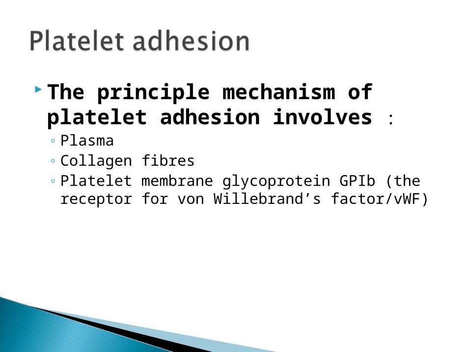
The principle mechanism of platelet adhesion involves :◦ Plasma◦ Collagen fibres◦ Platelet membrane glycoprotein GPIb (the
receptor for von Willebrand’s factor/vWF)

Platelet form pseudopods organelles (α-granules, dense bodies) reorganized to the center contraction granules spill their contents into the open canalicular system (OCS).

Mitogen Soted in and secreted from the α-granules
***

Activation of second passenger pathways with the platelet leads to intracellular :◦ Biochemical changes.◦ Culminating in platelet activation events such as :
Shape change Secretion Cytoskeletal reassembly
◦ Platelet aggregation

Platelet aggregation (platelet-to-platelet interaction) is an energy dependent process requires ATP (primarily derived from glycolysis)


Inactive Platelets

platelet activation

4 specific proteins secreted from the α- granules :◦ β - Thromboglobulin.◦ Platelet Factor 4 (Pf-4).◦ Thrombospondin.◦ PDGF.
***

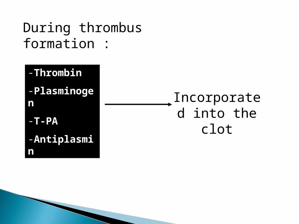
During thrombus formation :
-Thrombin
-Plasminogen
-T-PA
-Antiplasmin
Incorporated into the clot

PlateletsCoagulation Proteins
Fibrinolysis/Inhibitors
Vessels

Coagulation factors are designated by Roman numerals (I to XIII)
Activation of a particular factor is designated by a lower case “a” (X Xa)

Factor I FibrinogenFactor II ProthrombinFactor III Tissue ThromboplastinFactor IV Calcium IonsFactor V Labile Factor, ProaccelerinFactor VII Stable Factor, ProconvertinFactor VIII Antihemophilic FactorFactor IX Christmas FactorFactor X Stuart-Prower FactorFactor XI Plasma Thromboplastin AntecedentFactor XII Hageman FactorFactor XIII Fibrin Stabilizing Factor

Substrate Cofactors Enzymes
***

Fibrinogen Group Factors I, V, VIII and XIII
Prothrombin Group Factors II, VII, IX and X
Contact Group Factors XI, XII Prekallikrein (Fletcher Factor) High Molecular Weight Kininogen (Fitzgerald Factor)
***

Contact proteins Prothrombin proteins Fibrinogen or thrombin-sensitive proteins.
***

Extrinsic pathway Intrinsic pathway Common pathway


***

***


Decreased synthesis.Dysfunctional factor molecule(s).
Excessive destruction of factors.
Inactivation of factors.

Platelets
Fibrinolysis/Inhibitors
Coagulation Proteins
Vessels

Physiologic process of removing unwanted fibrin deposits.


Important to : Inflammation Vascular permeability Chemotaxis
Activated by coagulation & fibrinolytic system
***

Composed of approximately 22 serum proteins
Working together with antibodies and clotting factors,
Plays an important role as mediators of both immune and allergic reactions.
***

***

***

***
***

***
***

***

Screening assays in hemostasis Monitoring of anticoagulant therapy Disseminated Intravascular Coagulation Thrombophilia Inhibitor (Lupus Anticoagulant, Anti
Phospholipid Antibody)

1. Tourniquete Test (Rumple Leede)2. Bleeding Time3. Clot Retraction4. Platelet Count5. Clotting Time6. PT7. APTT8. TT9. Euglobulin Clot Lysis Time10. D=Dimer
-Vascular & Platelet-Platelet-Coagulation factors-Fibrinolysis

Platelet Rich Plasma (PRP)
Aggregating Reagent
AggregateClumping
Baseline Light Transmission
Increased LightTransmission
+


Typical Biphasic Pattern
100908070605040302010
0
PlateletRichPlasma
PlateletPoor
Plasma
Secondary Response(Release)
Primary Response
Injection Point
Lag
***

Cut 1 mm deep5 mm long
Constant Pressure
Expected Range 2 - 10 minutes
40 mm


Thromboplastinand Calcium
Patient’s Plasma
Factors
IIIVVIIX

Ca++
Patient’s Plasma
FactorsIIIV
VIIIIXXXIXII
Phospholipidand Activator

Highconcentrationof thrombin
1:10 dilutionpatient’splasma
Tim
e (
in s
e co
nd
s)
Fibrinogen in mg/dL
20 40 60 100 200 400 600
60
30
10
6
3
1:40 1:3
0 1:20
1:10
1:5

Low concentrationof thrombin
Undiluted patient’splasma
Screens foreffects of • Heparin • FDPs


Activators
Plasminogen Plasmin
Fibrinogen Fibrin
Degradation Products
R.E.S.


Antithrombin-IIIDecreased Levels
1. Congenital
2. Acquired – decreased synthesis
3. Acquired – increased utilization
4. Drug-induced

• Vitamin K-dependent plasma protein
• Inactivates Factors V and VIII
• Stimulates fibrinolysis

Protein CBiosynthesis: Liver, Vitamin K dependentMW: 56,000 daltonsPlasma Concentration: 3-5 mg/LIn Vivo Half-Life: 6-7 hours Pathology: Protein C deficiency, autosomal recessive (?)
* Requires Protein S for functional activity
Thrombo-modulin
FactorIIa
Protein C
ActivatedProtein C
Protein CInhibitor
Protein C
Inhibitor
ActivatedProtein
C*
Protein CActivation
Peptide
Activation
Procoagulant
Anticoagulant
Inhibition

I. Congenital Hereditary autosomal dominantII. Acquired
A. DICB. Liver diseaseC. During post-operative periodD. Anticoagulant therapy

• Superficial thrombophlebitis
• Venous thromboses in adolescents or young adults
• Arterial thromboses rarely observed
• Skin necrosis during onset of oral anticoagulant therapy

• Cofactor for Protein C
• Vitamin K-dependent protein
• Enhances binding of Protein C to phospholipid surfaces

PlasminogenActivity – Chromogenic Substrate
Antithrombin IIIAntigen – RID, ElisaActivity – Chromogenic Substrate
Protein CAntigen – EID, ElisaActivity – Chromogenic SubstrateFunctionality – Clot-based Assay


Thank you