AUDITION Functions: Adequate stimuli: Class # 12&13: Audition, p. 1.
Intro to Audition & Hearing
Lecture 16Chapter 9, part II
Jonathan PillowSensation & Perception (PSY 345 / NEU 325)
Spring 2015
1
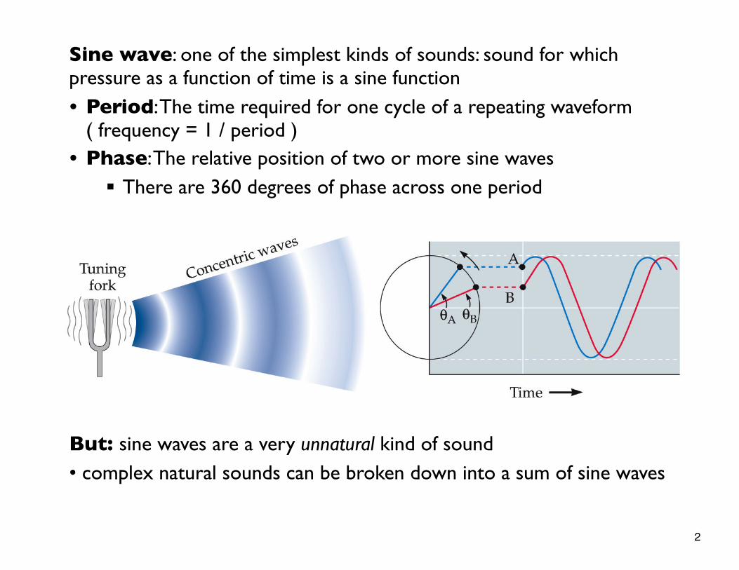
Sine wave: one of the simplest kinds of sounds: sound for which pressure as a function of time is a sine function
• Period: The time required for one cycle of a repeating waveform ( frequency = 1 / period )• Phase: The relative position of two or more sine waves
§ There are 360 degrees of phase across one period
But: sine waves are a very unnatural kind of sound• complex natural sounds can be broken down into a sum of sine waves
2
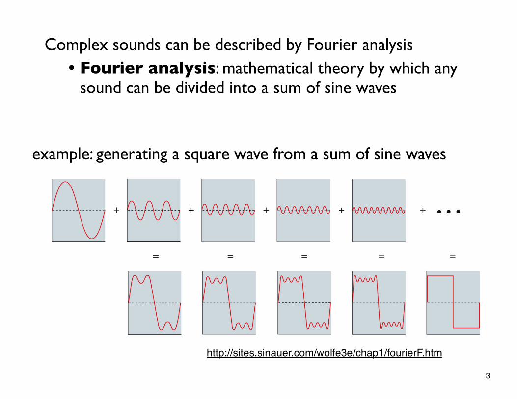
example: generating a square wave from a sum of sine waves
Complex sounds can be described by Fourier analysis• Fourier analysis: mathematical theory by which any
sound can be divided into a sum of sine waves
http://sites.sinauer.com/wolfe3e/chap1/fourierF.htm
3
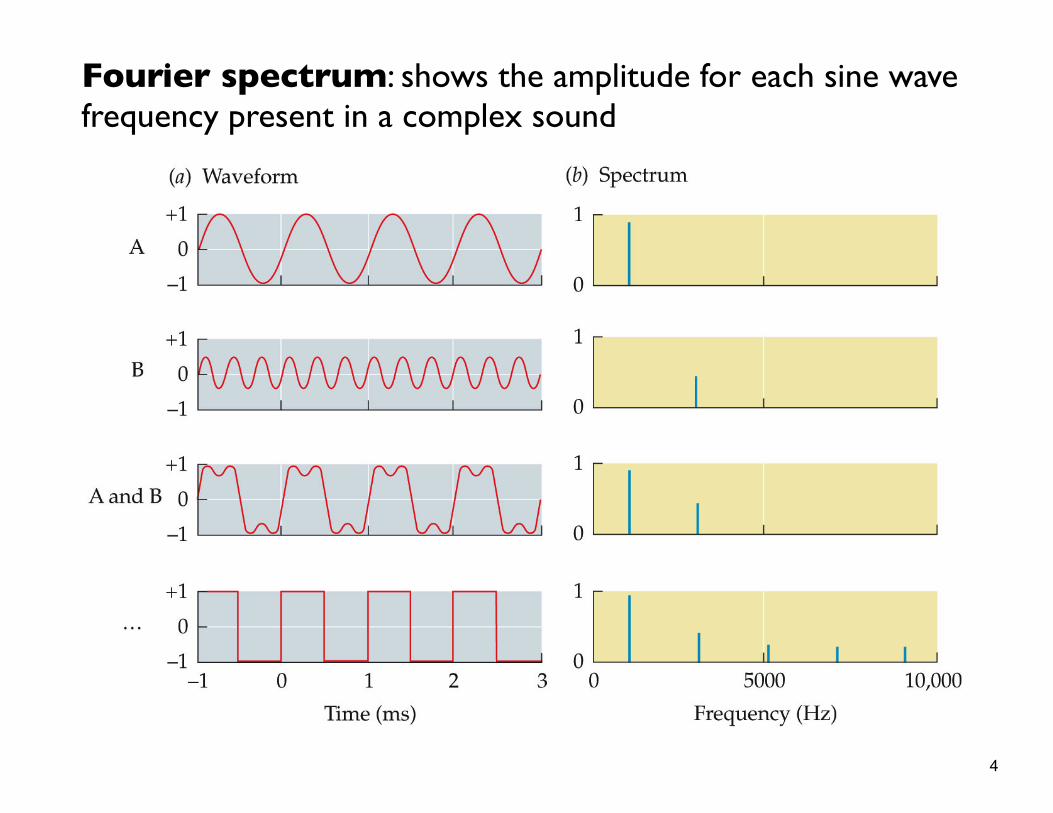
Fourier spectrum: shows the amplitude for each sine wave frequency present in a complex sound
4
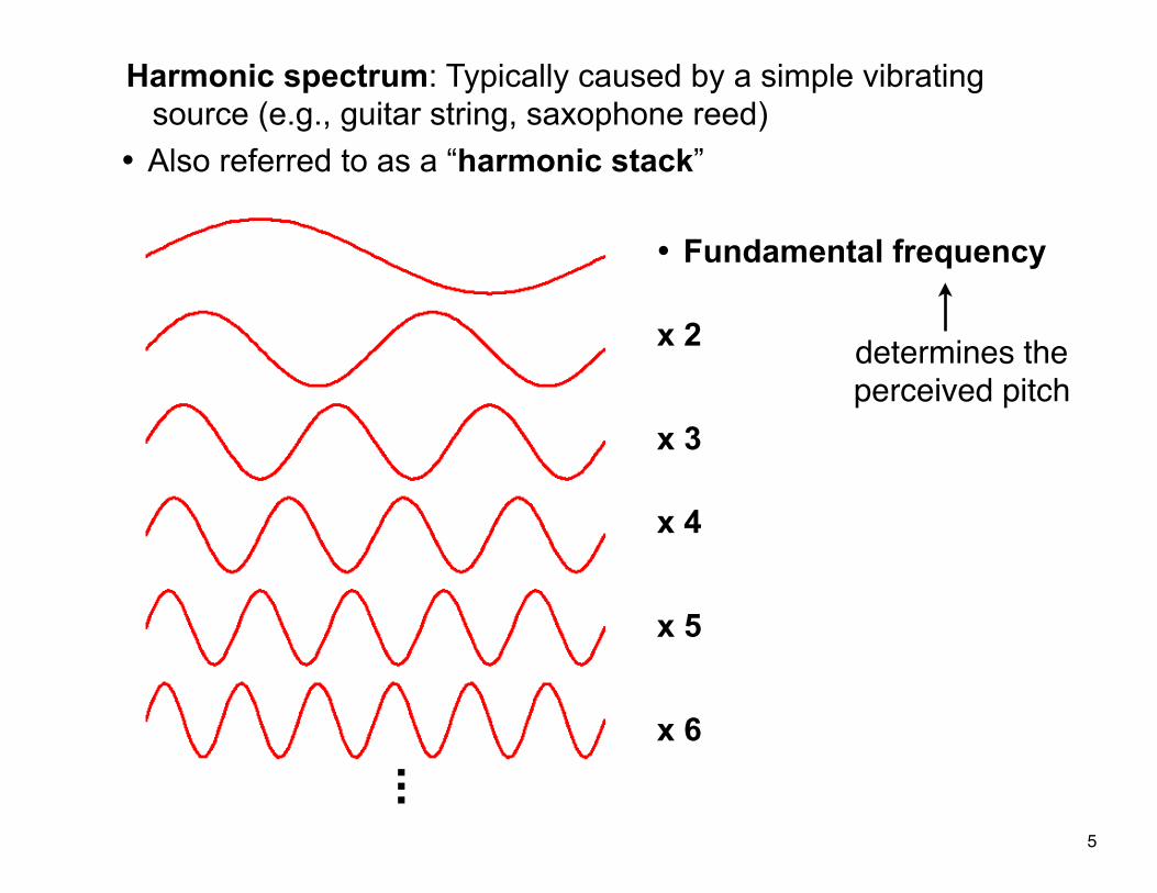
Harmonic spectrum: Typically caused by a simple vibrating source (e.g., guitar string, saxophone reed)
• Also referred to as a “harmonic stack”
• Fundamental frequency
x 2
x 3
x 4
x 5
x 6...
determines the perceived pitch
5
Timbre: psychological sensation by which a listener can judge that two sounds with the same loudness and pitch are dissimilar
§ timbre quality is conveyed by harmonics and other high frequencies
(more on this when we get to “music”)
6
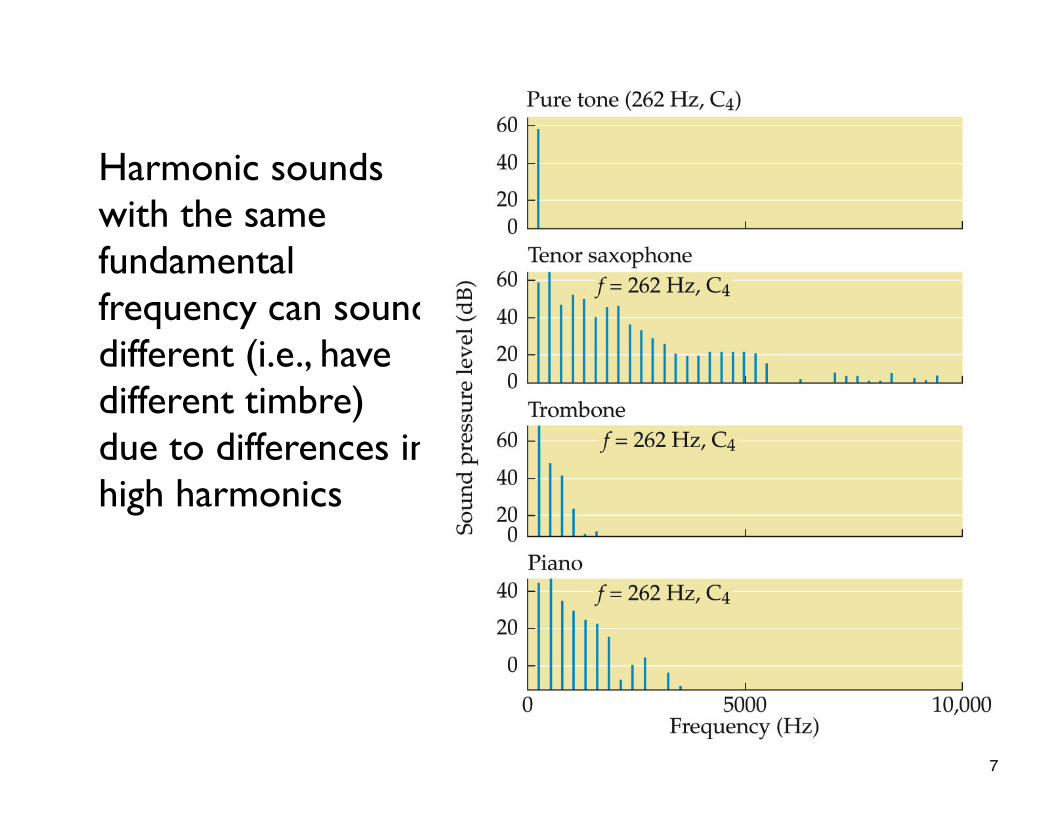
Harmonic sounds with the same fundamental frequency can sound different (i.e., have different timbre) due to differences in high harmonics
7
Part 2: The Auditory System
8
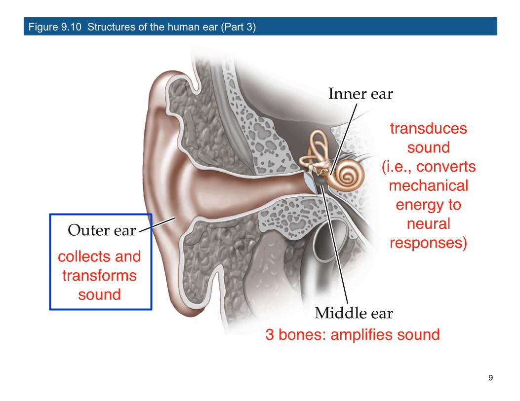
Figure 9.10 Structures of the human ear (Part 3)
collects and transforms
sound
3 bones: amplifies sound
transduces sound
(i.e., converts mechanical energy to
neural responses)
9
Basic Structure of the Mammalian Auditory System
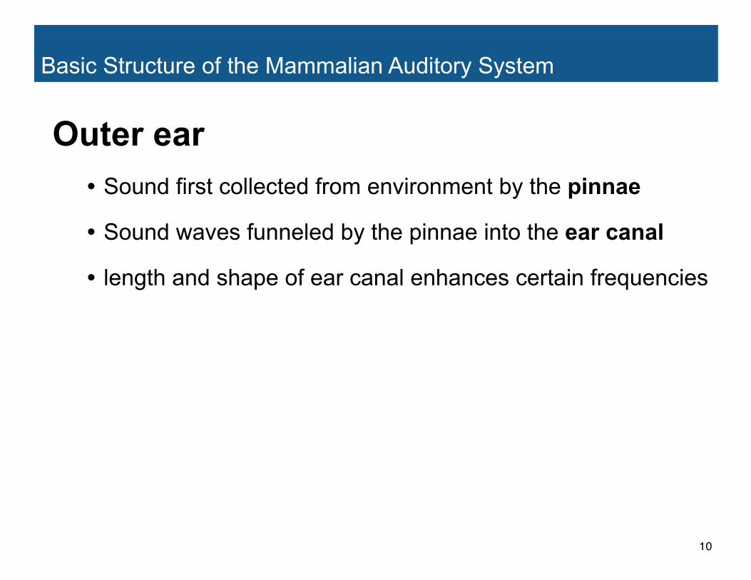
Outer ear• Sound first collected from environment by the pinnae
• Sound waves funneled by the pinnae into the ear canal
• length and shape of ear canal enhances certain frequencies
10
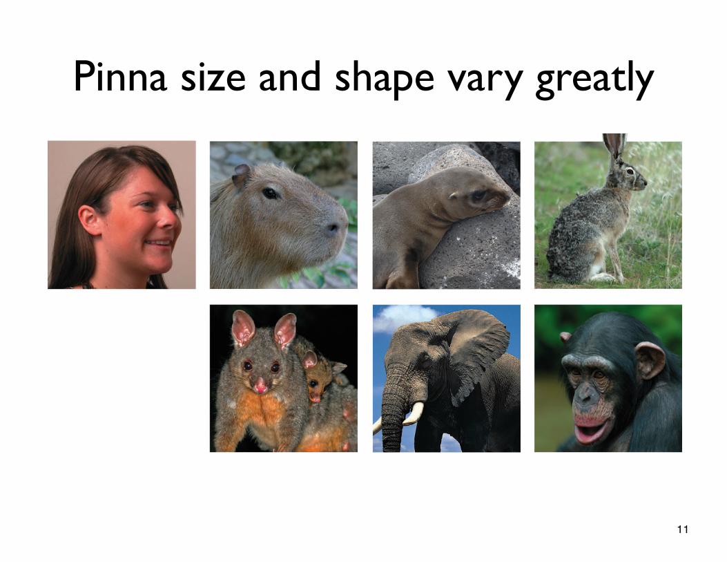
Pinna size and shape vary greatly
11
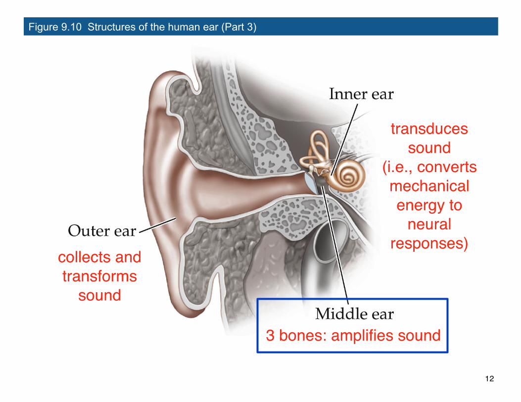
Figure 9.10 Structures of the human ear (Part 3)
collects and transforms
sound
3 bones: amplifies sound
transduces sound
(i.e., converts mechanical energy to
neural responses)
12
Basic Structure of the Mammalian Auditory System
Middle ear
13
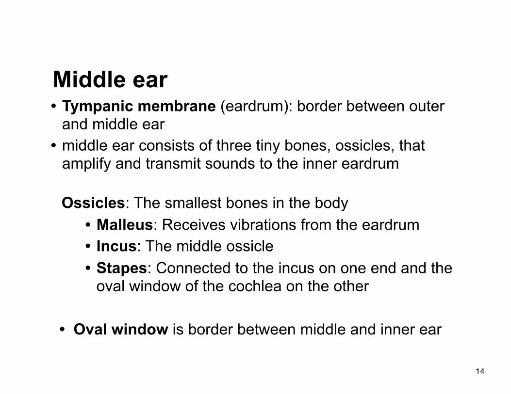
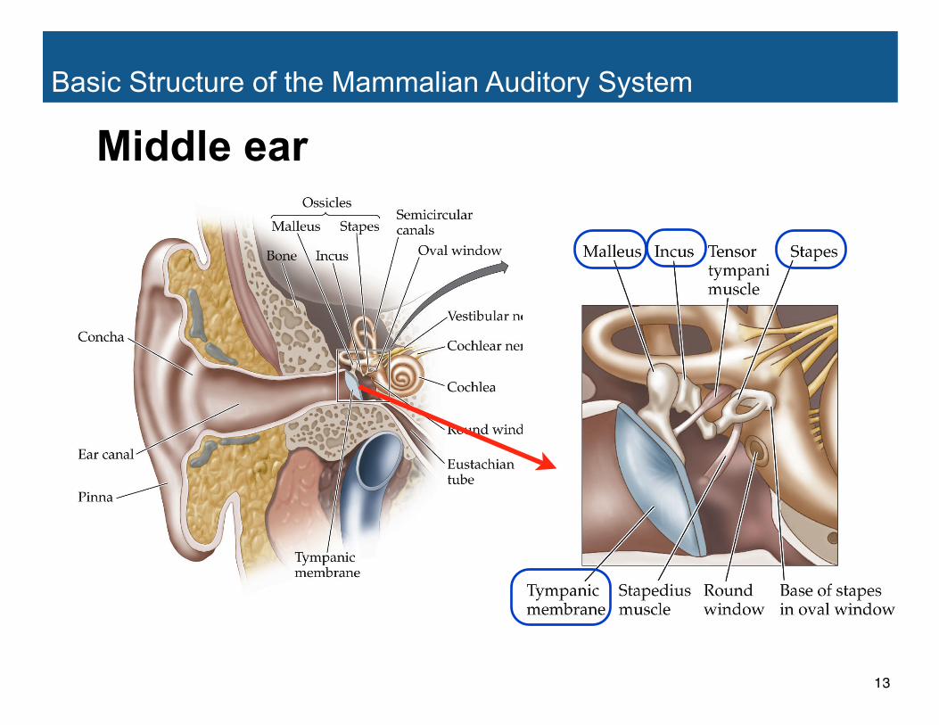
Middle ear• Tympanic membrane (eardrum): border between outer
and middle ear• middle ear consists of three tiny bones, ossicles, that
amplify and transmit sounds to the inner eardrum
Ossicles: The smallest bones in the body• Malleus: Receives vibrations from the eardrum• Incus: The middle ossicle• Stapes: Connected to the incus on one end and the
oval window of the cochlea on the other
• Oval window is border between middle and inner ear
14
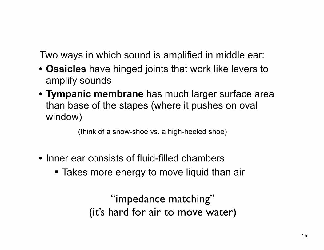
Two ways in which sound is amplified in middle ear:• Ossicles have hinged joints that work like levers to
amplify sounds• Tympanic membrane has much larger surface area
than base of the stapes (where it pushes on oval window)
(think of a snow-shoe vs. a high-heeled shoe)
• Inner ear consists of fluid-filled chambers§ Takes more energy to move liquid than air
“impedance matching”(it’s hard for air to move water)
15

16
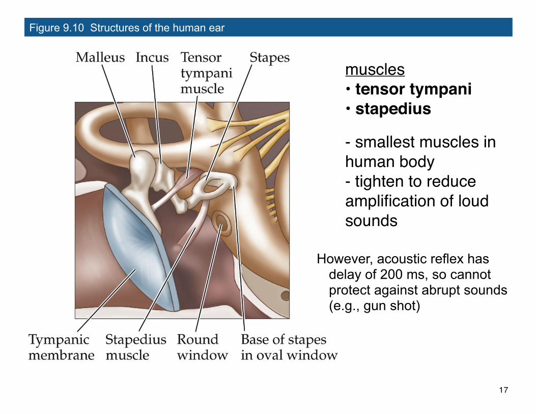
Figure 9.10 Structures of the human ear
muscles• tensor tympani• stapedius
- smallest muscles in human body- tighten to reduce amplification of loud sounds
However, acoustic reflex has delay of 200 ms, so cannot protect against abrupt sounds (e.g., gun shot)
17
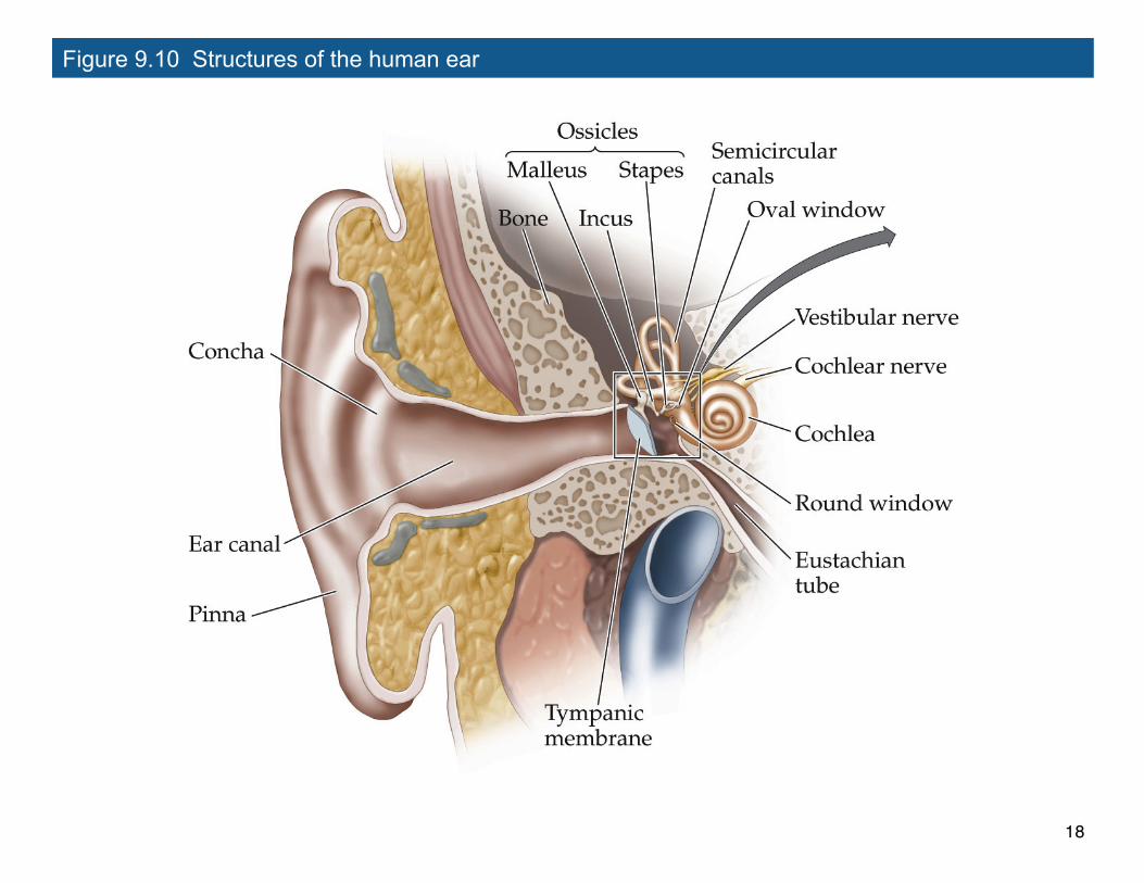
Figure 9.10 Structures of the human ear
18
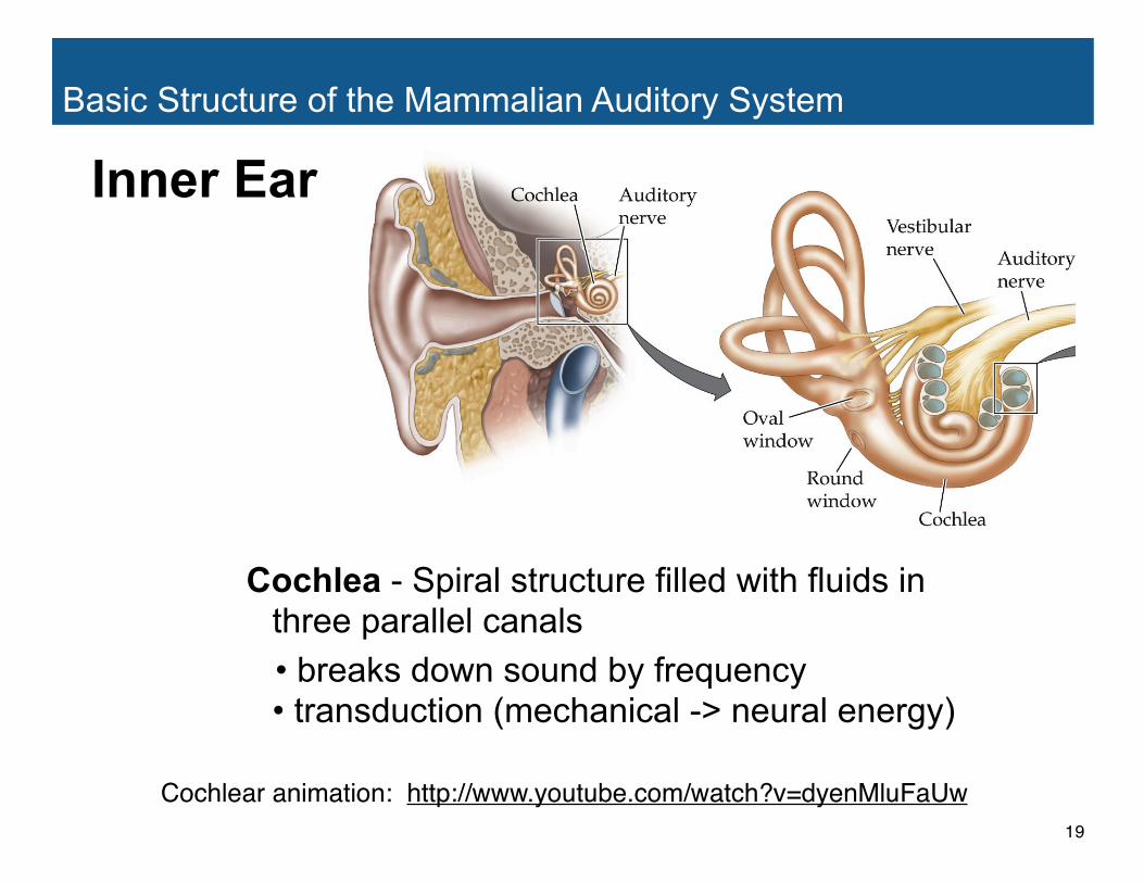
Basic Structure of the Mammalian Auditory System
Cochlea - Spiral structure filled with fluids in three parallel canals
• breaks down sound by frequency• transduction (mechanical -> neural energy)
Inner Ear
Cochlear animation: http://www.youtube.com/watch?v=dyenMluFaUw19
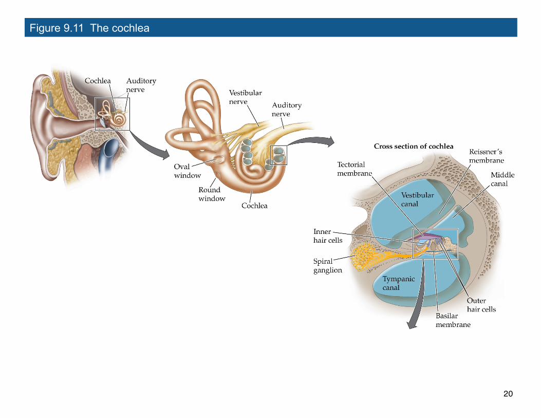
Figure 9.11 The cochlea
20
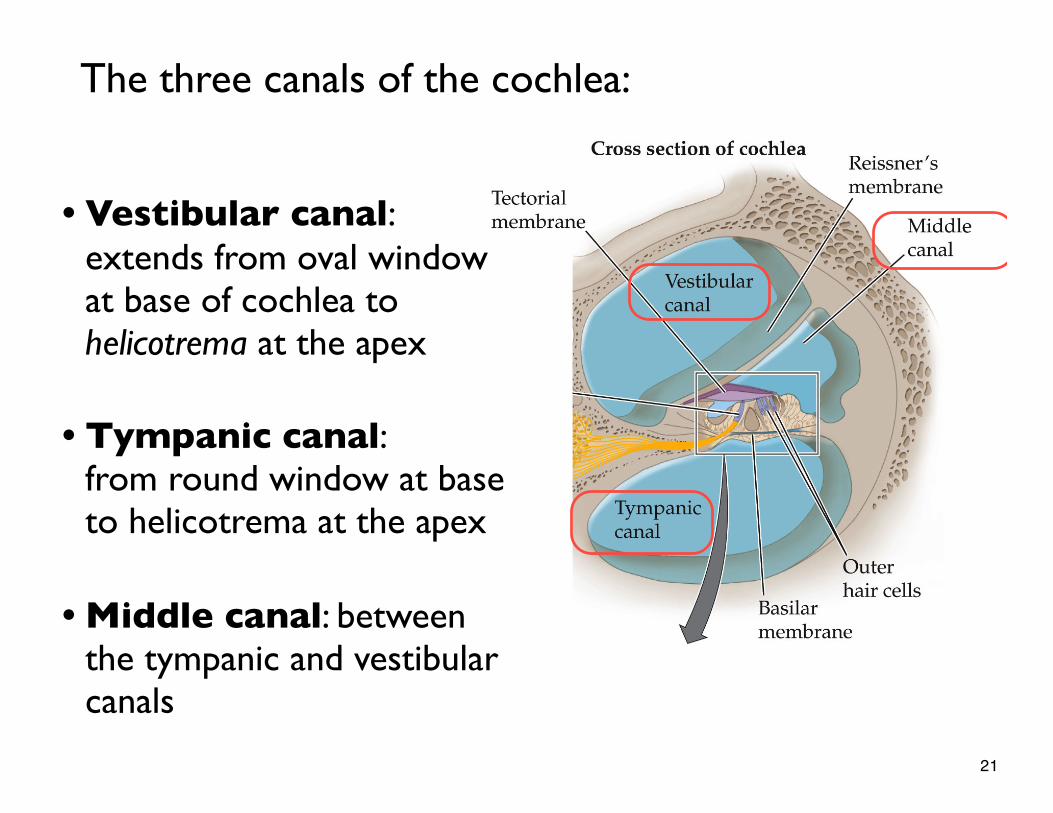
• Vestibular canal: extends from oval window at base of cochlea to helicotrema at the apex
• Tympanic canal: from round window at base to helicotrema at the apex
• Middle canal: between the tympanic and vestibular canals
The three canals of the cochlea:
21
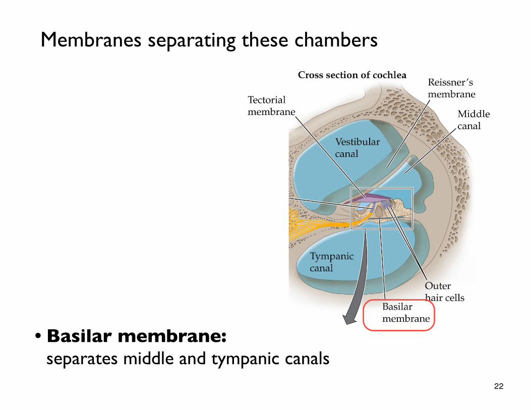
• Basilar membrane: separates middle and tympanic canals
Membranes separating these chambers
22
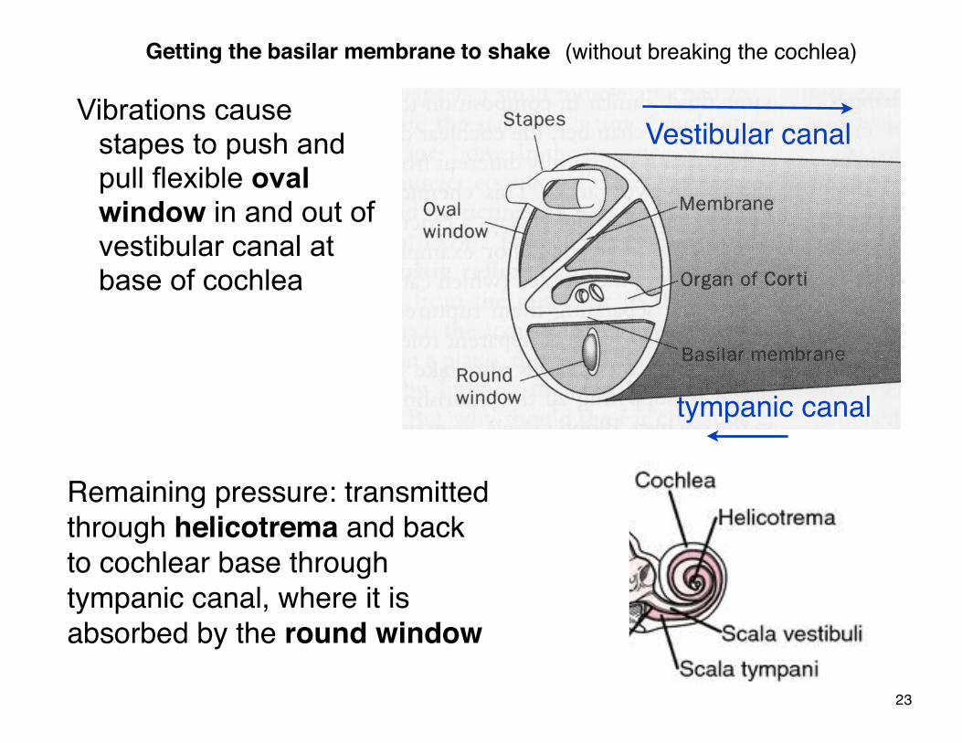
Getting the basilar membrane to shake (without breaking the cochlea)
Vibrations cause stapes to push and pull flexible oval window in and out of vestibular canal at base of cochlea
Remaining pressure: transmitted through helicotrema and back to cochlear base through tympanic canal, where it is absorbed by the round window
Vestibular canal
tympanic canal
23
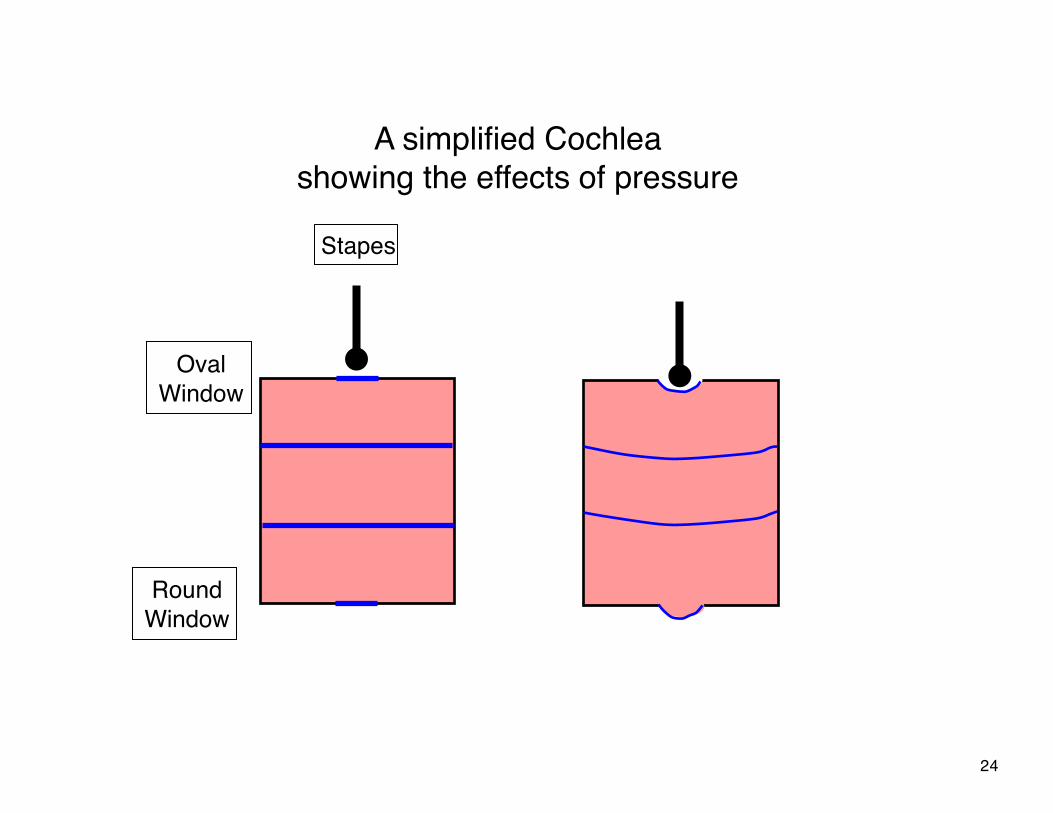
A simplified Cochlea showing the effects of pressure
OvalWindow
RoundWindow
Stapes
24
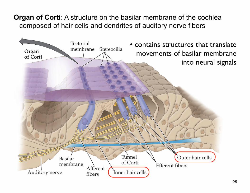
Figure 9.11 The cochlea (cont’d)
Organ of Corti: A structure on the basilar membrane of the cochlea composed of hair cells and dendrites of auditory nerve fibers
• contains structures that translate movements of basilar membrane
into neural signals
25
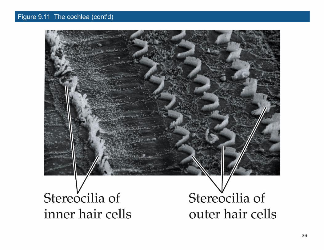
Figure 9.11 The cochlea (cont’d)
26
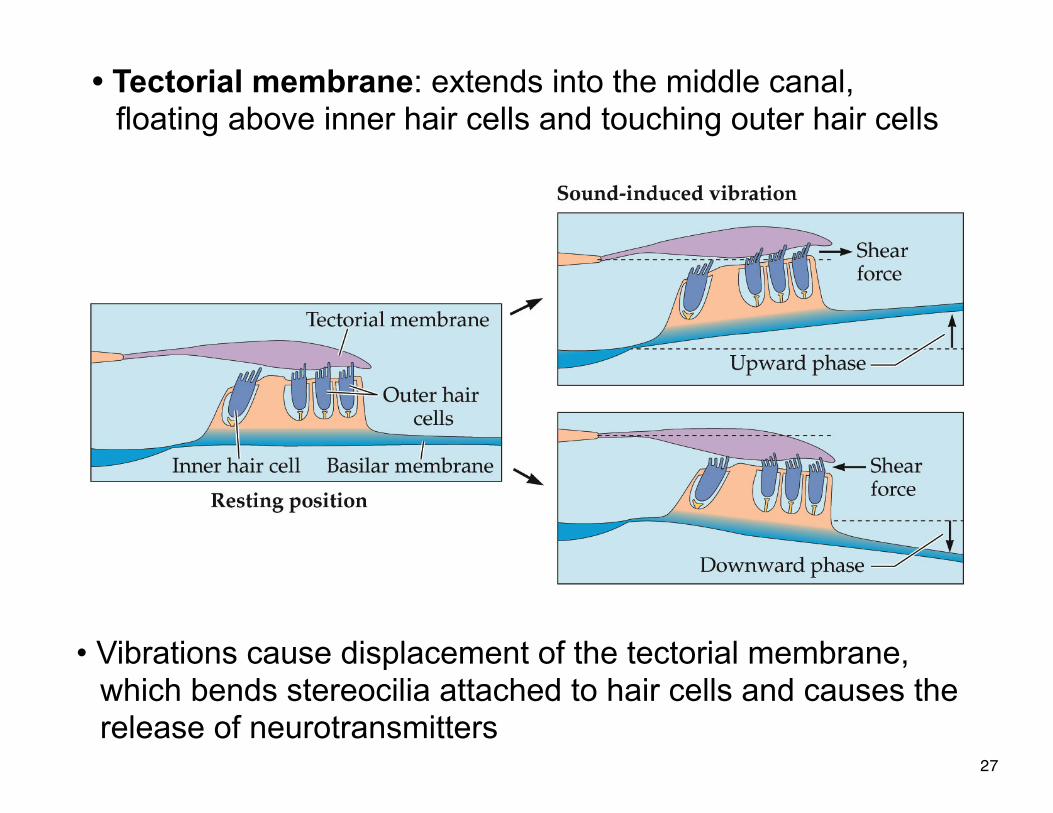
• Tectorial membrane: extends into the middle canal, floating above inner hair cells and touching outer hair cells
• Vibrations cause displacement of the tectorial membrane, which bends stereocilia attached to hair cells and causes the release of neurotransmitters
27
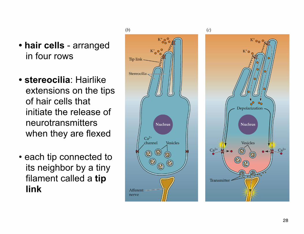
• hair cells - arranged in four rows
• stereocilia•
• hair cells - arranged in four rows
• stereocilia: Hairlike extensions on the tips of hair cells that initiate the release of neurotransmitters when they are flexed
• each tip connected to its neighbor by a tiny filament called a tip link
28
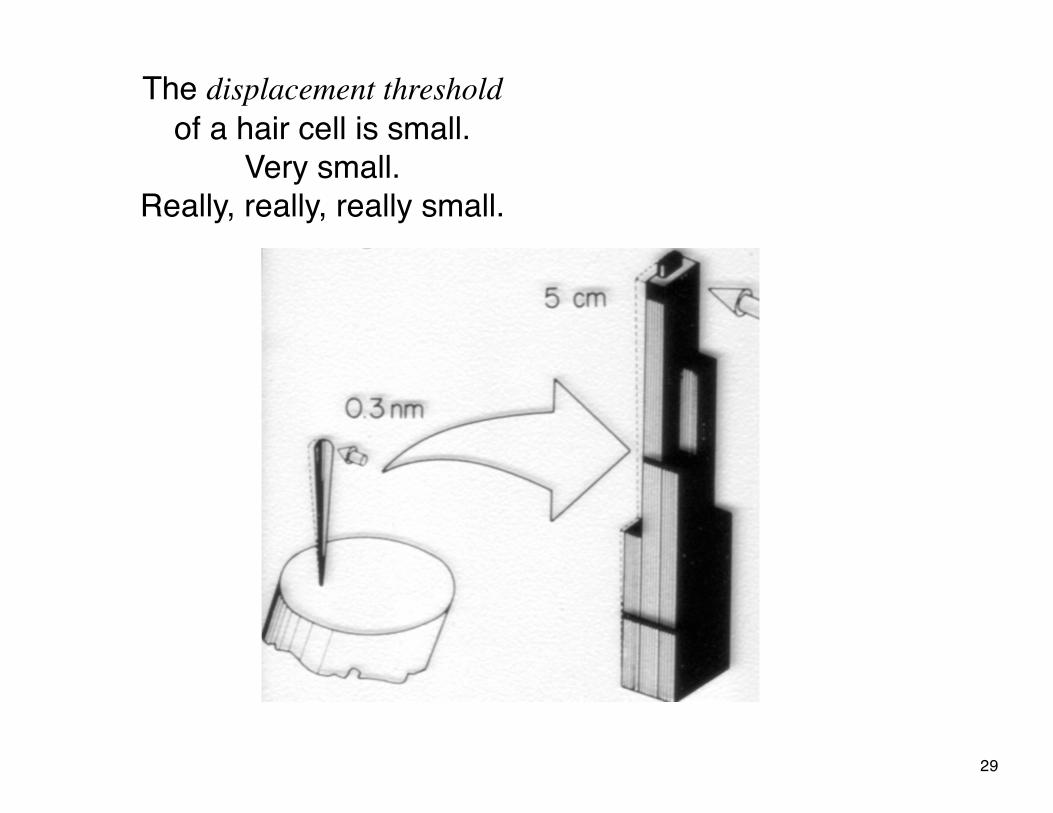
The displacement threshold of a hair cell is small.
Very small.Really, really, really small.
29
• Inner hair cells: Convey almost all information about sound waves to the brain (using afferent fibers)
• Outer hair cells: Convey information from the brain (using efferent fibers). § involved in an elaborate feedback system§ amplify sounds by increasing mechanical deflections
of the basilar membrane
30
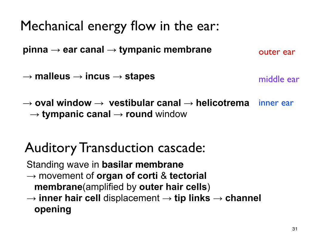
Standing wave in basilar membrane → movement of organ of corti & tectorial
membrane(amplified by outer hair cells) → inner hair cell displacement → tip links → channel
opening
pinna → ear canal → tympanic membrane
→ malleus → incus → stapes
→ oval window → vestibular canal → helicotrema → tympanic canal → round window
Mechanical energy flow in the ear:
outer ear
middle ear
inner ear
Auditory Transduction cascade:
31
additional topics this chapter
• auditory nerve• place code• temporal code• characteristic frequency• coding in the auditory system• psychoacoustics• hearing loss & hearing aides• cochlear implants
32
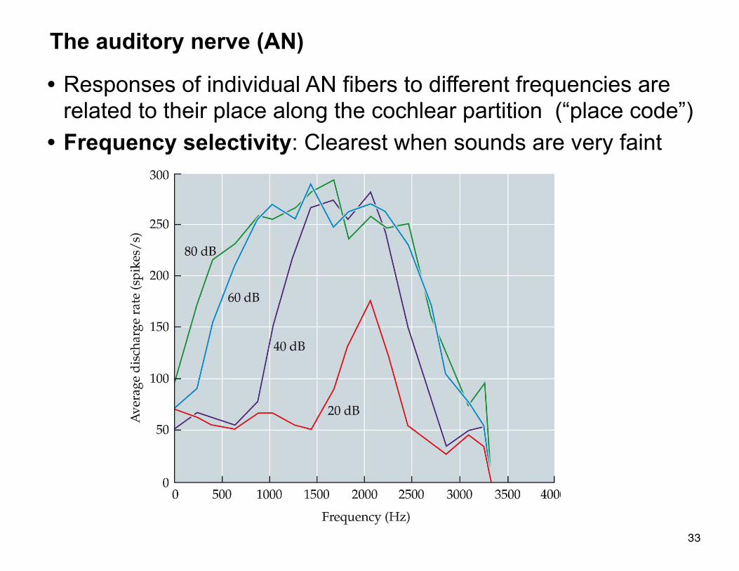
• Responses of individual AN fibers to different frequencies are related to their place along the cochlear partition (“place code”)
• Frequency selectivity: Clearest when sounds are very faint
The auditory nerve (AN)
33
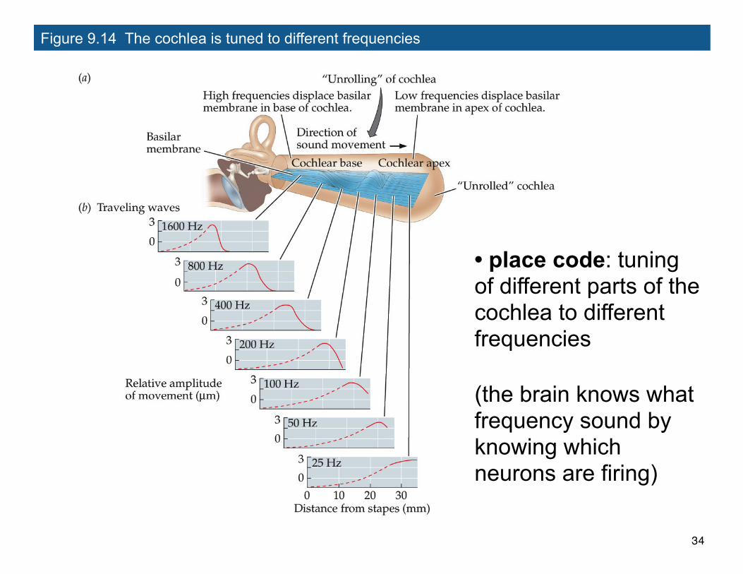
Figure 9.14 The cochlea is tuned to different frequencies
• place code: tuning of different parts of the cochlea to different frequencies
(the brain knows what frequency sound by knowing which neurons are firing)
34
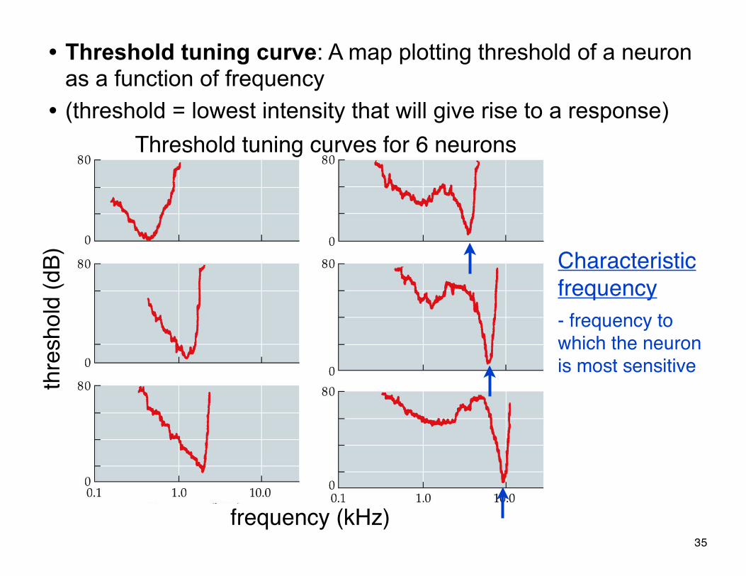
• Threshold tuning curve: A map plotting threshold of a neuron as a function of frequency
• (threshold = lowest intensity that will give rise to a response)
thre
shol
d (d
B)
frequency (kHz)
Characteristicfrequency- frequency to which the neuron is most sensitive
Threshold tuning curves for 6 neurons
35
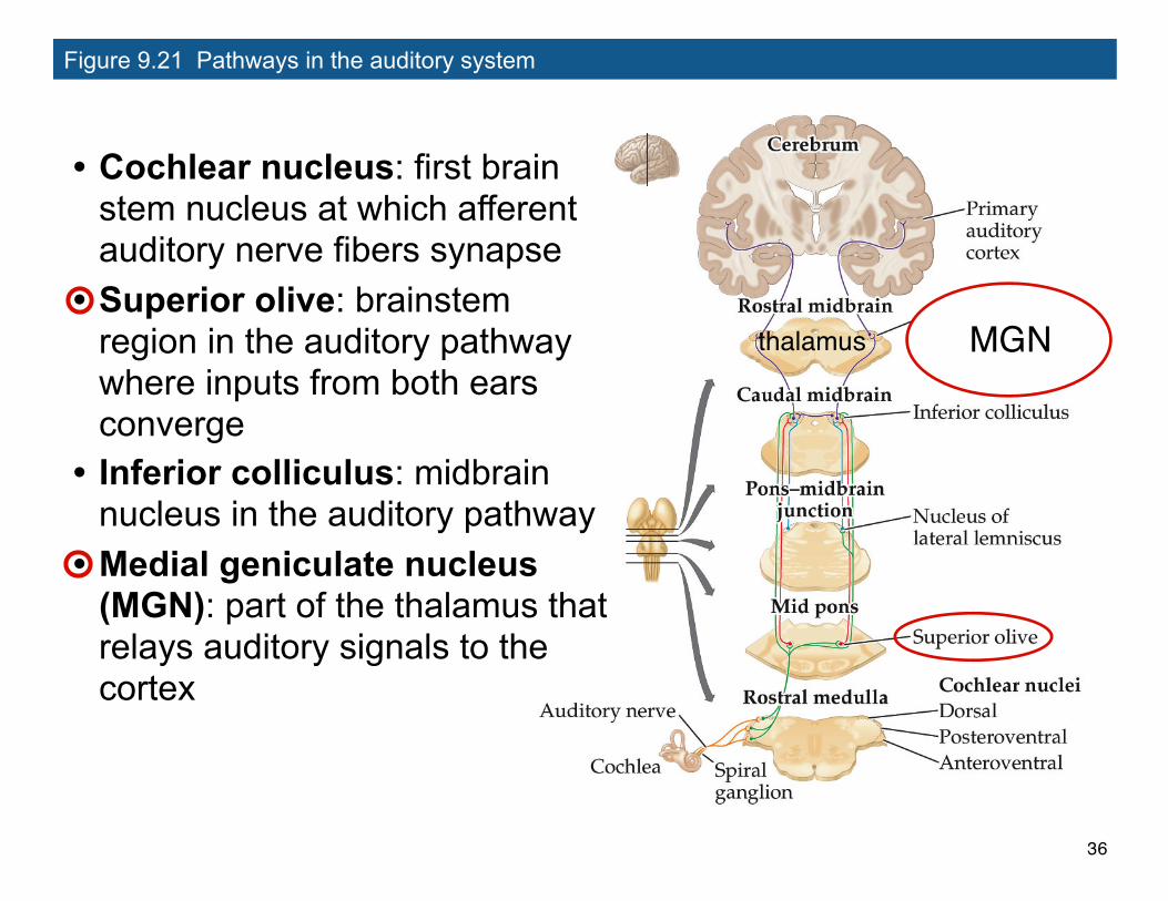
Figure 9.21 Pathways in the auditory system
• Cochlear nucleus: first brain stem nucleus at which afferent auditory nerve fibers synapse
• Superior olive: brainstem region in the auditory pathway where inputs from both ears converge
• Inferior colliculus: midbrain nucleus in the auditory pathway
• Medial geniculate nucleus (MGN): part of the thalamus that relays auditory signals to the cortex
MGNthalamus
36
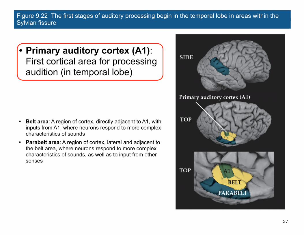
Figure 9.22 The first stages of auditory processing begin in the temporal lobe in areas within the Sylvian fissure
• Primary auditory cortex (A1): First cortical area for processing audition (in temporal lobe)
• Belt area: A region of cortex, directly adjacent to A1, with inputs from A1, where neurons respond to more complex characteristics of sounds
• Parabelt area: A region of cortex, lateral and adjacent to the belt area, where neurons respond to more complex characteristics of sounds, as well as to input from other senses
37
Basic Structure of the Mammalian Auditory System
Comparing overall structure of auditory and visual systems:
• Auditory system: Large proportion of processing is done before A1
• Visual system: Large proportion of processing occurs beyond V1
• Why? May be due to evolutionary reasons...
38
Basic Structure of the Mammalian Auditory System
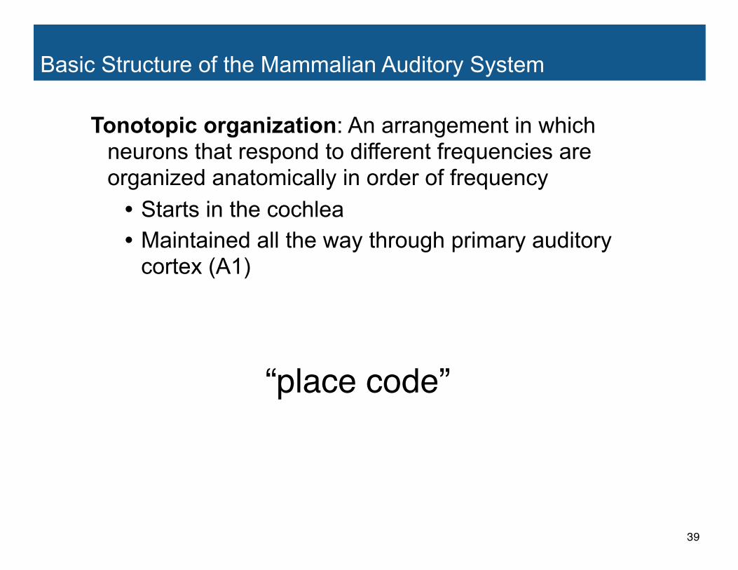
Tonotopic organization: An arrangement in which neurons that respond to different frequencies are organized anatomically in order of frequency
• Starts in the cochlea• Maintained all the way through primary auditory
cortex (A1)
“place code”
39
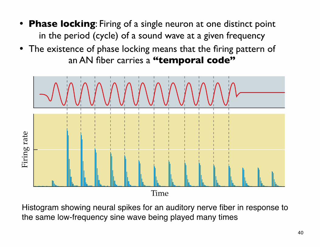
Histogram showing neural spikes for an auditory nerve fiber in response to the same low-frequency sine wave being played many times
• Phase locking: Firing of a single neuron at one distinct point in the period (cycle) of a sound wave at a given frequency
• The existence of phase locking means that the firing pattern of an AN fiber carries a “temporal code”
40
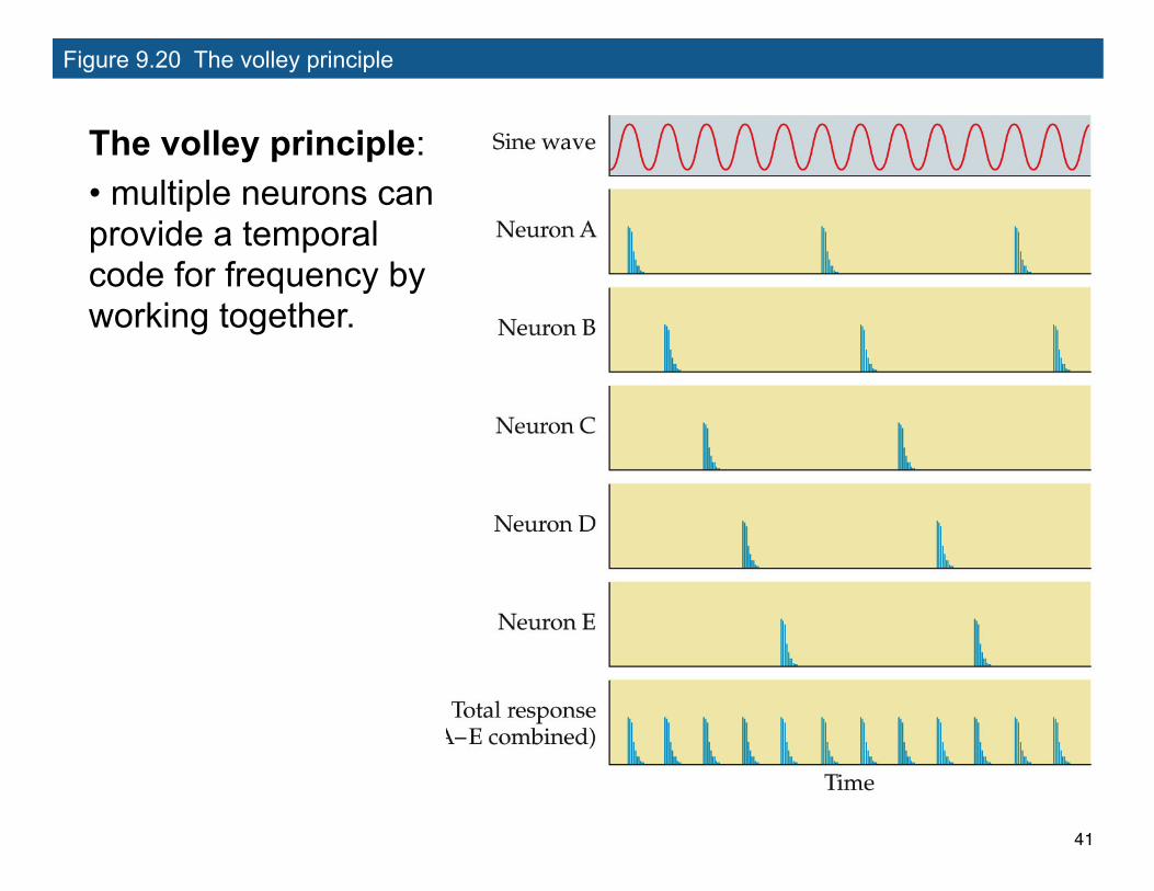
Figure 9.20 The volley principle
The volley principle:• multiple neurons can provide a temporal code for frequency by working together.
41
Psychoacoustics
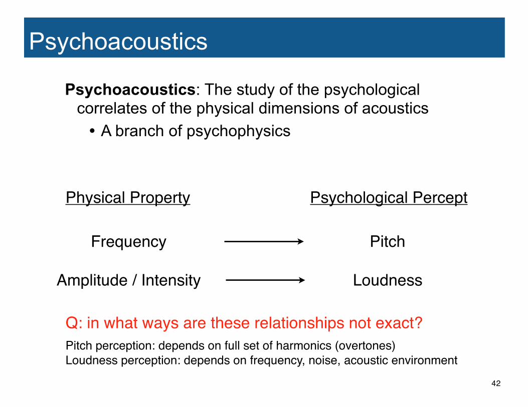
Psychoacoustics: The study of the psychological correlates of the physical dimensions of acoustics
• A branch of psychophysics
Physical Property
Frequency
Amplitude / Intensity
Psychological Percept
Pitch
Loudness
Q: in what ways are these relationships not exact?Pitch perception: depends on full set of harmonics (overtones)Loudness perception: depends on frequency, noise, acoustic environment
42
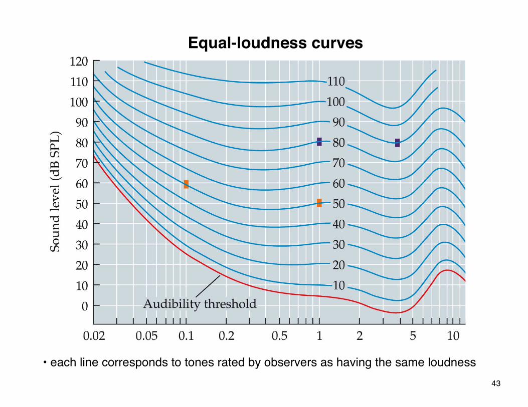
Equal-loudness curves
• each line corresponds to tones rated by observers as having the same loudness43