Intrinsically disordered proteins link alternative ...
45
ACCEPTED MANUSCRIPT Intrinsically disordered proteins link alternative splicing and post-translational modifications to complex cell signaling and regulation Jianhong Zhou 1 , Suwen Zhao 1,2 *, A. Keith Dunker 3 * 1.iHuman Institute, ShanghaiTech University. 2F Building 6, 99 Haike Road, Pudong New District, Shanghai, 201210, China. 2.School of Life science and Technology, ShanghaiTech University 3.Center for Computational Biology and Bioinformatics, Department of Biochemistry and Molecular Biology, Indiana University School of Medicine, 410 W 10th street, Suite 5000, Indianapolis, IN 46202, USA. Corresponding authors: Suwen Zhao, Email: [email protected] and A. Keith Dunker, Email: [email protected] Abstract: Intrinsically disordered proteins and regions (IDPs and IDRs) lack well-defined tertiary structures, yet carry out various important cellular functions, especially those associated with cell signaling and regulation. In eukaryotes, IDPs and IDRs contain the preferred loci for both alternative splicing (AS) and many post-translational modifications (PTMs). Furthermore, AS and/or PTMs at these loci generally alter the signaling outcomes associated with these IDPs or IDRs, where the functional cooperation of these three features is named the IDP-AS-PTM toolkit. However, the prevalence of such functional modulations remains unknown. Also, the signal- altering mechanisms by which AS, and PTMs modulate function and the extent to which AS and PTMs collaborate in their signaling modulations have not been well defined for particular protein examples. Here we focus on three important signaling and regulatory IDR-containing protein families in humans, namely G-protein coupled receptors (GPCRs), which are transmembrane ACCEPTED MANUSCRIPT ___________________________________________________________________ This is the author's manuscript of the article published in final edited form as: Zhou, J., Zhao, S., & Keith Dunker, A. (2018). Intrinsically disordered proteins link alternative splicing and post-translational modifications to complex cell signaling and regulation. Journal of Molecular Biology. https://doi.org/10.1016/j.jmb.2018.03.028
Transcript of Intrinsically disordered proteins link alternative ...
ACC
EPTE
D M
ANU
SCR
IPT
Intrinsically disordered proteins link alternative
splicing and post-translational modifications to
complex cell signaling and regulation
Jianhong Zhou1, Suwen Zhao1,2*, A. Keith Dunker3*
1. iHuman Institute, ShanghaiTech University. 2F Building 6, 99 Haike Road, Pudong
New District, Shanghai, 201210, China.
2. School of Life science and Technology, ShanghaiTech University
3. Center for Computational Biology and Bioinformatics, Department of Biochemistry
and Molecular Biology, Indiana University School of Medicine, 410 W 10th street, Suite
5000, Indianapolis, IN 46202, USA.
Corresponding authors: Suwen Zhao, Email: [email protected] and A. Keith
Dunker, Email: [email protected]
Abstract: Intrinsically disordered proteins and regions (IDPs and IDRs) lack well-defined tertiary
structures, yet carry out various important cellular functions, especially those associated with cell
signaling and regulation. In eukaryotes, IDPs and IDRs contain the preferred loci for both
alternative splicing (AS) and many post-translational modifications (PTMs). Furthermore, AS
and/or PTMs at these loci generally alter the signaling outcomes associated with these IDPs or
IDRs, where the functional cooperation of these three features is named the IDP-AS-PTM toolkit.
However, the prevalence of such functional modulations remains unknown. Also, the signal-
altering mechanisms by which AS, and PTMs modulate function and the extent to which AS and
PTMs collaborate in their signaling modulations have not been well defined for particular protein
examples. Here we focus on three important signaling and regulatory IDR-containing protein
families in humans, namely G-protein coupled receptors (GPCRs), which are transmembrane
ACCEPTED MANUSCRIPT
___________________________________________________________________
This is the author's manuscript of the article published in final edited form as:
Zhou, J., Zhao, S., & Keith Dunker, A. (2018). Intrinsically disordered proteins link alternative splicing and post-translational modifications to complex cell signaling and regulation. Journal of Molecular Biology. https://doi.org/10.1016/j.jmb.2018.03.028
ACC
EPTE
D M
ANU
SCR
IPT
2
proteins, the nuclear factors of activated T-cells (NFATs), which are transcription factors (TFs),
and the Src family kinases (SFKs), which are signaling enzymes. The goal here is to determine
how AS and PTMs individually alter the outcomes of the signaling carried out by the various IDRs
and to determine whether AS and PTMs work together to bring about differential cellular
responses. We also present data indicating that a wide range of other signaling IDPs or IDRs
undergo both AS- and PTM-based modifications, suggesting that they, too, likely take advantage
of signal outcome modulations that result from collaboration between these two events. Hence,
we propose that the widespread cooperation of IDPs, AS and/or PTMs provides a IDP-AS-PTM
toolkit and substantially contributes to the vast complexity of eukaryotic cell signaling systems.
Keywords: Intrinsic disorder, alternative splicing, post-translational modification, differential and
context-dependent signaling, signaling modulation, and regulation
Abbreviations: IDPs or IDRs, intrinsically disordered proteins or regions; AS, alternative splicing;
PTMs, post-translational modifications; GPCRs, G-protein coupled receptors; NFATs, nuclear
factor of activated T-cells; TFs, transcription factors; SFKs, Src family kinases; CD, circular
dichroism; NMR, nuclear magnetic resonance; ICL3, intercellular loop 3; CaN, calcineurin; GRK,
G-protein coupled receptor kinase; PKA/C, protein kinase A/C; CDK, cyclin-dependent kinase;
DBD, DNA-binding domain.
Highlights
We propose that IDPs work in concert with AS and PTMs to provide an IDP-AS-PTM toolkit
for complex context-dependent cell signaling and regulation.
Three intensely studied, highly divergent signaling protein families, namely G-protein
coupled receptors (GPCRs), nuclear factor of activated T-cells (NFAT) transcription factors (TFs)
ACCEPTED MANUSCRIPT
ACC
EPTE
D M
ANU
SCR
IPT
3
and Src family kinases (SFKs), all use this toolkit to increase context-dependent signaling
complexity.
PubMed text mining shows the widespread occurrence of IDPs, AS and PTMs in a large
number of proteins including those involved in developmental signaling pathways, which indicates
a common maybe even universal use of the IDP-AS-PTM toolkit in eukaryotic multicellular
signaling systems.
Introduction
Intrinsically disordered proteins and regions (IDPs and IDRs) have been found to be heavily
involved in cell signaling and regulation, especially in eukaryotes[1-9]. Due mainly to their amino
acid compositions[10-12], IDPs and IDRs lack stable tertiary structures under physiological
conditions and exist instead in highly dynamic, interconverting, flexible conformations[13]. Their
functions complement those of structured proteins and underlie cellular differentiation,
transcription, cell cycle regulation, DNA condensation, cell division and many other crucial
biological processes[3, 14].
Protein/nucleic acid/ligand binding sites are often located in IDPs or IDRs, and their flexibility
enable single, short IDRs to change their backbone and side-chain conformations and thereby
bind tightly with multiple, distinctly shaped binding partner surfaces[15, 16]. Thus IDPs and IDRs
can mediate interactions with a large number of partners and thus function as hubs or as partners
to structured hub proteins in signaling networks[17-19]. Furthermore, IDPs and IDRs contain
numerous sites for post-translational modifications (PTMs) that can reversibly regulate IDP- or
IDR-binding in a cellular context-dependent manner by adding binding sites for new partners
and/or by inhibiting binding by partners that recognize the unmodified IDR, thus further expanding
an IDR’s already formidable binding repertoire. In many cases the PTM-induced changes lead to
ACCEPTED MANUSCRIPT
ACC
EPTE
D M
ANU
SCR
IPT
4
initiation or inhibition of specific signaling pathways[20-23], or can be involved in diseases[24] or
protein translocation[25]. Another way of expanding the functionality of IDPs and IDRs is through
alternative splicing (AS) of pre-mRNA that codes for the same IDP or IDR, a post-transcriptional
process that generates two or sometimes many more protein forms from a single gene[26]. These
versatilities offered by the flexibility of IDPs or IDRs, with AS and PTMs adding further complexity,
enable signaling and regulatory proteins to efficiently accomplish dynamic functions in response
to changes in cellular environments.
Based on the observation that both AS and PTMs, especially multiple PTMs[27, 28], alter the
functions of many IDPs[23, 26], we previously proposed that IDPs work cooperatively with AS
and PTMs to provide a toolkit (namely IDP-AS-PTM), for signaling diversification[29, 30]. We also
showed that this toolkit is used by many of the proteins that carry out the functions underlying
multicellularity, functions such as cell-to-cell adhesion, intercellular communication, development
pathway specification, development pathway regulation over space and time, and tissue- or cell-
type-specific physiology[31]. An important feature of this proposal is that tissue- or cell-type-
specificity has been shown for both AS[32, 33] and PTMs[34, 35], suggesting that the new
biological function enabled by the IDP-AS-PTM toolkit provides context-dependent signaling. By
this we mean that specific tissues or specific cell-types are able to “rewire” or “remodel” protein
pathways and genetic networks depending on the local context, while still using the same sets of
genes. This rewiring results from the tissue- or cell-type-specific AS and/or the tissue- or cell-
type-specific PTMs. That is, both AS and PTMs have been shown to have the capability to alter
interactions between proteins or between proteins and nucleic acids, and both undergo tissue- or
cell-type specific alterations[16, 26, 32, 33, 36-38].
ACCEPTED MANUSCRIPT
ACC
EPTE
D M
ANU
SCR
IPT
5
So far, a significant number of important protein families have been predicted to contain
substantial amounts of intrinsic disorder. Some families have been analyzed in a large scale
(genome- or proteome-wide), including transcription factors[39, 40], nuclear hormone
receptors[41], membrane proteins[42, 43], histones[44], spliceosomal proteins[45, 46], ribosomal
proteins[47], DNA/RNA binding proteins[48], and many enzymes[49]. Other proteins have been
investigated on a smaller scale, but with detailed features (e.g. domain organizations, molecular
recognition features, binding interface properties), such as serine-arginine rich proteins[50],
scaffold proteins[51], autoinhibited proteins[52], cytoskeletal proteins[53]. In addition, many IDPs
are associated with human diseases, including cancer, neurondegeneration, cardiovascular
disease, amyloidosis, diabetes and many others[2, 54]. While more IDPs continue to be
characterized thus expanding the biological functions known to be associated with these
proteins[49, 55, 56], the frequency of occurrence of the IDP-AS-PTM toolkit mentioned above
remains unknown, and how IDPs or IDRs, AS and/or PTMs jointly regulate these proteins is not
well characterized for any particular IDP or IDR.
Here we examine whether the IDP-AS-PTM toolkit is used by three important signaling and
regulatory IDP families in humans, all of which contain members involved in development, namely
the G-protein coupled receptors (GPCRs, a membrane receptor protein family), the nuclear
factors of activated T-cells (NFATs, a transcription factor (TF) family), and the proto-oncogene
Src family kinases (SFKs, a regulatory enzyme family). First we map the sequence locations of
sites associated with AS (annotated in UniProt) and PTM (annotated in UniProt and
PhosphoSitePlus[57]), and then we determine whether their IDRs are enriched in these two
regulatory events as compared to their structured regions. Next we study selected examples for
each family to determine whether, and if so, how IDRs, AS and PTMs collaborate to regulate
signaling diversity for these particular protein examples.
ACCEPTED MANUSCRIPT
ACC
EPTE
D M
ANU
SCR
IPT
6
GPCRs are of particular interest because they are the largest known seven transmembrane
(7TM) protein family that have a particularly high amount of predicted disorder as compared to
other transmembrane proteins[43]. This a very large membrane receptor protein family with over
800 members in humans, with an enormous diversity of ligands from rhodopsin to peptides, and
with involvement in an extremely wide range of biological processes, including blood pressure
regulation, olfactory function, embryogenesis and nearly every other physiological process[58,
59].
TFs play a particularly central role in transcription regulation. Particular TF examples have
been known to contain IDRs since the 1980s[60], nearly two decades before bioinformatics
examinations showed the widespread existence of massive amounts of intrinsic disorder in the
eukaryotic TFs[39, 40]. Among the many well studied IDR-containing TFs, the NFAT family was
selected because it has widespread importance, including critical roles in T-cell function,
inflammation, angiogenesis, myocardial development, skeletal muscle development, cancer
metastasis, and many other biological processes; because its own regulation depends on two
very important signaling systems, namely regulation by calcium levels with calmodulin as the
calcium sensor and regulation by phosphorylation by various kinases and dephosphorylation by
calcineurin [61], and because its massive IDRs have been characterized by both circular
dichroism (CD) and nuclear magnetic resonance (NMR)[62].
SFKs are non-receptor tyrosine kinases, and they are key regulators in signal transduction.
The first-discovered SFK member, Src, is identified as the most highly connected hub in the
whole kinome. Members of this family are predicted to contain large IDRs at their N- or C-
terminus or between folded domains, and these predicted IDRs are significantly overlapped by
ACCEPTED MANUSCRIPT
ACC
EPTE
D M
ANU
SCR
IPT
7
regions of missing electron density from available SFK structures[63]. Also, SFKs help to regulate
a number of important processes such as differentiation, proliferation, migration, and survival[64].
For these three particular protein families, and many other IDPs mentioned above, numerous
publications show that their functional complexity is substantially enhanced by the combined use
of IDPs or IDRs, AS and/or PTMs. These observations support our hypothesis that the IDP-AS-
PTM toolkit is commonly used to provide a mechanism for sophisticated signaling processes and
indeed may be essential for the emergence of complex multicellular organisms as we suggested
previously[31].
Results
GPCRs and the IDP-AS-PTM toolkit
Co-occurrence of IDRs, AS and PTMs in GPCRs
The full-length GPCRs include the extracellular N-terminus, the intracellular C-terminus,
seven transmembrane helices (TM1-7), three intracellular loops (ICL1-3) and three extracellular
loops (ECL1-3) connecting the helices. While the overall TM topology is well conserved across
GPCR members, the N-terminus, ICL3 and the C-terminus exhibit high variability in terms of
length and amino acid composition[65], and are predicted to be the most disordered regions
(green histogram in Fig. 1A, left). This result is consistent with previous predictions[65-67] and the
experimental confirmation on a few GPCRs by CD and NMR[68-70]. In addition, these results
agree with the necessary truncation of these IDRs for most GPCRs during crystallization. Indeed,
for the >200 available crystal structures belonging to 44 GPCRs in Protein Data Bank (PDB), a
majority of their N- and C-termini and ICL3 regions are truncated to achieve crystallization
success.
ACCEPTED MANUSCRIPT

ACC
EPTE
D M
ANU
SCR
IPT
8
In accordance with the distribution of IDRs over GPCR secondary structures, the occurrence
of both AS and PTMs (purple line in Fig. 1A and stacked bar in Fig. 1B, respectively) more often
localize within the N- and C-termini and ICL3 regions as compared to other regions. Experimental
data provides evidence that GPCRs can be modified in multiple ways, and undergo tissue-
specific AS (Fig.1C), suggesting those two regulatory phenomena may mutually alter specific
receptor functions. Indeed, Among the 308 AS regions, 94 (~31%) of them contain known PTM
sites that may be altered by AS (Fig.2A). More importantly, as shown in the disorder distribution
in Fig. 2B, when predicted to be fully ordered (disorder percentage=0%), AS regions with PTMs
show lower percentage of than those without known PTMs (18% and 41% respectively); in
opposite, when predicted to be disordered, AS regions with PTMs generally show higher
percentage than those of without PTMs (Fig. 2B). This result suggests that AS regions with PTMs
are more likely to predicted to be disordered, indicating that IDRs provide the preferential
locations for AS to modulate PTMs. This enrichment of AS and multiple PTMs within IDRs of
GPCRs provide massive combinations of IDR, AS and/or PTMs that would differentially “encode”
receptor functional diversity, including differential downstream cellular signaling.
Regulation of GPCR functions by IDR-localized AS and/or PTMs
Given the preference of IDRs, AS and PTMs within N-terminus, ICL3 and C-terminus of
GPCRs, in the following we explore how these three most disordered regions work in concert with
AS and/or PTMs to enhance receptor functional diversity in a cell- or tissue- specific manner.
In general, the disordered regions of GPCRs are important for receptor ligand binding,
surface expression, trafficking and signaling, thus alteration of them by AS would substantially
affect GPCR activities. For instance, AS-isoforms differ in the N-terminus of many GPCRs show
decreased or abolished ligand-binding activity[71, 72]. In many cases, the functions of N-
ACCEPTED MANUSCRIPT
ACC
EPTE
D M
ANU
SCR
IPT
9
terminus-associated AS-variants remain to be elucidated, and some are speculated to act as a
dominant-negative mutant of the wild-type receptors[71]. Interestingly, some GPCRs have a very
long disordered ICL3 regions, within which the AS events often occur. For instance D2 dopamine
receptor (DRD2), with a long ICL3 having 148 residues, generates three isoforms, D2short,
D2long (canonical) and D2longer, with D2short and D2longer having deletion of 29 residues and
insertion of two residues in ICL3, respectively[73, 74]. As a result, D2short and D2long couple
with different α subunit of inhibitory G-proteins (D2short coupled preferentially with Gαi1, and
D2long couples selectively with Gαi2) and activate distinct signaling pathways accordingly[75].
The free fatty acid receptor 4 (FFAR4) generates an isoform with insertion of 16 residues within
ICL3, leading the receptor towards an arrestin-biased pathway[76]. Another extreme case is
CXCR3 isoform CXCR3Alt, which lacks the whole ICL3 region and fails to induce either Gαi
activation or β-arrestin recruitment[77]. As ICL3 is important for G-protein or arrestin interaction
and subsequent activation of intracellular events[78], other GPCRs with AS-isoforms differing
within this disordered region [e.g. D3 dopamine receptor (34 residues deletion),
Gastrin/cholecystokinin type B receptor (69 residues insertion), Histamine H3 receptor (80
residues deletion)] are likely to have different signaling pathways or especially biased signaling
pathways, thus illustrating the essential roles of IDR-localized AS in selective signal transduction.
A large number of phosphorylation sites are localized within the IDRs of GPCRs (Fig.1B),
and have been described to generate different combinational patterns linked to differential GPCR
signaling. Such patterns, which are termed phosphorylation codes, were initially discovered in
M3-muscarinic acetylcholine receptor[79] and β2AR[80], and are found in disordered C-tails/ICL3
for most GPCRs[81]. Besides phosphorylation, other PTMs, such as palmitoylation[82],
glycosylation[83], different patterns of ubiquitination (mono- or poly-ubiquitination)[84], also
participate in biased signaling, suggesting the likely presence of an expanded version to the
ACCEPTED MANUSCRIPT
ACC
EPTE
D M
ANU
SCR
IPT
10
phosphorylation code ––a PTM code that specifies GPCR activities using different types of PTMs.
The potential to regulate distinct signaling outcome by combinatorial PTMs is illustrated by
studies on CXCR4, which undergoes not only sulfation and glycosylation, but also
phosphorylation and ubiquitination (Fig.3). Thus, in the case of CXCR4, there is evidence that
site-specific PTMs can result in different signaling results, supporting the GPCR PTM code
hypothesis mentioned in reference[85].
AS further modulates PTM incidence in GPCRs
AS can alter multiple PTMs sites for many GPCRs (Fig1C and Fig.2), leading to another
layer of functional complexity by combining IDR-Co-occurring with AS and PTMs. This agrees
well with the functional influence of tissue-specific AS on IDPs in general[32, 33]. For example,
among the six GPCRs that undergo palmitoylation (Fig.1C), four of them (AVPR2[86], EDNRB
and EDNRA[87], OPRM1[88]) lose their palmitoylation sites induced by AS. AVPR2 isoform
without the palmitoylation sites (C341 and C342) and phosphorylation sites (S362, S363 and
S364) can adopt two different topologies[89] and mainly remain inside the cell and down-
regulates the surface expression of canonical AVPR2 by formation of heterodimers[90]. EDNRB
isoforms that lack the palmitoylation sites fail to activate G proteins[91]. Replacement of
phosphorylation sites within disordered C-terminus of TXA2R isoforms lead to distinct
combinations of kinase phosphorylation, and subsequent separated biological processes
(desensitization or internalization)[92, 93]. Although in many cases the AS-driven replacement or
addition of potential PTMs remain unknown (i.e. those three N-terminal AS-variants of CXCR4 in
Fig.3), the reported examples presented above show that the synergistic collaborations of AS and
PTMs leads to enhanced, context-dependent signaling complexity. That is, IDR-localized AS
creates alternative PTM patterns leading to different downstream outcomes. Overall, we conclude
ACCEPTED MANUSCRIPT
ACC
EPTE
D M
ANU
SCR
IPT
11
that diverse signaling carried out by GPCRs use the IDP-AS-PTM toolkit in multiple essential
ways.
TFs and the IDP-AS-PTM toolkit
Co-occurrence of IDRs, AS and PTMs in human TFs in general
Large portions of eukaryotic TFs have been predicted to contain IDRs, typically covering two-
thirds or more of the TF sequences, and these predictions are in good agreement with
experimental data[39, 40, 94]. Furthermore, recent work suggests that, for TF families associated
with development, there is a strong positive correlation between the amount of predicted disorder
and the complexity of the organism[95].
In human TFs, their intrinsic disorder is not restricted to DBDs but includes assigned Pfam
domains outside of the DBD and also not restricted to transactivation domains or protein-
interaction domains. Significantly, predicted IDRs include massive regions that have not yet been
assigned to a particular type of domain (green boxplot in Fig.4A, top axis).
In accordance with the distribution of disorder prediction, higher numbers of TFs have AS
events within the unassigned regions (URs) as compared to those in DBD and NonDBD (Fig.4A,
purple-border histogram, bottom axis). Likewise, significantly more PTM sites are located within
the more disordered URs than those in DBD and NonDBD (Fig.4B). Furthermore, the presence of
multiple types of PTMs in TFs is very common; among the 1345 TF members with PTM
annotations, 62% of them have more than one type of PTMs (Fig.4C). Besides, 93 of those 1345
TFs (~7%) are documented to have two or more types of PTMs targeting the same residues
(totally 202 sites), with lysine being the most frequent site for alternative PTMs (Fig.4D). In
addition, 882 (~41%) of all the AS regions (totally 2128 regions) contain known PTMs (Fig.5A),
and for the fully predicted to be ordered AS regions, those with PTMs are about two times less
ACCEPTED MANUSCRIPT
ACC
EPTE
D M
ANU
SCR
IPT
12
than those without PTMs (Fig.5B). That is, most of AS regions with PTMs (94%) are predicted to
be disordered (red histogram in Fig. 5B); in comparison, only ~70% of AS regions without known
PTMs show predicted disorder (blue histogram in Fig.5B). Thus, like GPCRs, human TFs
generally display co-occurrence of IDRs, AS, and PTMs that likely act synergistically to help
modulate the complicated aspects of transcriptional regulation.
Transcriptional regulation of NFATs by IDR-localized AS and PTMs
Extensive evidence indicates that AS and PTMs of TFs commonly alter DNA-binding
affinity/specificity or their interactions with cofactors in cell- or tissue-specific manner[28, 96-99].
Here we present data for one important subfamily––the nuclear factors of activated T-cells
(NFATs), to show how specific combinations of IDR-localized AS and PTMs affect the detailed
functions of NFATs. As key regulators in T-cell development and function, NFATs have five
members sharing a similar DNA-binding domain. Among them, four members (NFATc1-4) are
specifically regulated by calcineurin (CaN), a Ca2+/calmodulin-dependent serine phosphatase that
is involved in many important signaling pathways[100]. The distant member, NFAT5, is not
calcium-related due to the lack of regulatory domain that contains CaN-binding motifs, and it is
activated by osmotic stress instead. NFATs are of our particular interest because they have been
confirmed by CD and NMR to contain extremely long disordered domains besides the well-
defined DNA-binding domain[62]. These experimental data agree very well with the disorder
prediction in Fig.6A.
These IDRs of NFATs are the main locations where multiple splicing sites occur (Fig.6B).
NFATs have been suggested to undergo tissue-specific AS events that contribute to isoform-
specific transcriptional abilities[101]. The ten splice isoforms of NFATc1 are presented in Fig.6B.
ACCEPTED MANUSCRIPT
ACC
EPTE
D M
ANU
SCR
IPT
13
These variants differ in the length of their disordered N- and/or C-terminus, and all the other
NFAT genes are able to generate multiple AS isoforms in similar manner[101].
The NFATc proteins undergo multiple PTMs, including phosphorylation of the serine-rich
regions (SRR1&2) and Serine–Proline repeat motifs (SP1-3), and sumoylation, most of which
localize within the disordered region (Fig.6C). The large numbers of phosphorylation sites are
required for maintaining the NFATc molecules within the cytoplasm, whereas dephosphorylation
of these sites by CaN promotes nuclear import and initiation of target gene expression. These
regulatory serine sites are phosphorylated by different kinases, specifically by PKA, DYRK, CK1,
or GSK3 in a hierarchical pattern, creating a complex regulation that may allow for distinctive
activation profiles in different cell types[102]. The sumoylation of NFATs, which is cell-specific
and AS-isoform-specific, was recently shown to repress the transcriptional activity and regulate its
nuclear retention, providing a new regulatory mechanism for NFAT functions[103].
To give more structural details, the nuclear localization signals (NLS) contain clusters of
positively charged residues interspersed with serines (Fig.6C). Four of the five positively charged
residues are arginines. Having arginines rather than lysines is likely important here because
arginines form much stronger interactions with phosphates than do lysines. That is, each arginine
has two hydrogens that are well-placed to form two hydrogen bonds with two phosphate oxygens,
and it has even been observed that two arginines can bind to a single phosphate with the
concomitant formation of four hydrogen bonds[104]. Such hydrogen bonding between the
phosphates and arginines would inactivate the NLS, while dephosphorylation by calcineurin
would lead to activation and nuclear import. Interestingly, up to 13 different phosphates, most of
which are rather distant in the sequence from the NLS, are involved in this inactivation, and the
ACCEPTED MANUSCRIPT
ACC
EPTE
D M
ANU
SCR
IPT
14
collaboration among the phosphates leads to a sensitive, on-off switch-like behavior for the
nuclear import of NFAT1c[105, 106].
AS alter PTM sites of NFATs
AS-induced PTM changes are observed (Fig.6C and Fig.6D), and related functional
consequences have been reported in some cases. The final ~150 residues of C-terminal IDR
contain a second transactivation domain which contain two strong sumoylation sites (K702,
K914). Sumoylation leads NFATc1 to its subnuclear relocalization and enable NFATc1 to
suppress expression of its selective target genes (e.g.IL-2)[107]. The isoforms without these two
sumoylation sites (e.g. isoform A, isoform IA), but contains a weaker sumoylation site K349, does
not have the selectivity, suggesting the collaboration of AS and PTM bring out fine-tuning of
NFATc1 functions. The N-terminal-altered AS isoforms of NFATc1 lose the annotated R23
methylation, but the functions of methylated R23 remains unknown, so does the consequences of
losing this PTM. Also, the functions of the phosphorylation sites within the disordered C-terminus
remains unknown. It has been reported that the C-terminal-altered isoform of NFATc1, B-β, fails
to interact with TNF-α (a target gene of NFATc1) to which the common NFATc1 isoform strongly
binds[108]. In addition, two alternative N-terminal transactivation domain regions are generated
by AS in NFATc2, resulting isoforms having differential roles in the control of cell proliferation and
transformation[109]. It would be very interesting to test whether this different regulation by NFATc
is related to the AS-altered PTM mentioned above.
Taken together, the prevalence of the AS sites, as well as the large numbers of PTM sites
within the IDRs, suggest that the different NFAT molecules utilize the IDP-AS-PTM toolkit for
functional diversification.
ACCEPTED MANUSCRIPT
ACC
EPTE
D M
ANU
SCR
IPT
15
Src family kinases (SFKs) and the IDP-AS-PTM toolkit
SFKs act as important intermediaries regulating a variety of cellular activities, and increased
activity or overexpression of SFKs can lead to constant activation, which is considered as its
common mechanism of causing cancers and other diseases[110]. Src is the first identified proto-
oncogene[111], and its gene product is the first protein discovered to have tyrosine kinase
activity[112]. SFKs generally include nine members that are divided into two groups based on
sequence identity: Src, Fyn, Yes, Fgr, and Yrk, forming Group A, Lck, Hck, Blk and Lyn in Group
B.
Regulatory role of disordered N-terminus of SFKs
The N-terminal domains of SFKs, including SH4 and Unique, have been confirmed to be
disordered by NMR[113, 114], which is in good agreement with our prediction (Fig.7A). The other
important functional regions are the classic regulatory domains SH3 and SH2, the catalytic
domain SH1, SH2–SH1 linker and a regulatory C-terminal tail (Fig.7B), which are all involved in
kinase autoinhibitory functions[115]. Remarkably, recent studies have highlighted a more
complex regulatory mechanism mediated by the disordered SH4 and Unique domains[116-118].
Specifically, Unique is not only able to bind to SH3 and lipids, but also interacts with other
proteins, such as calmodulin, to regulate kinase activities[116]. The lipid binding of Unique may
change the ligands or substrates accessible to the kinase, thereby possibly inducing distinct
pathways. This new finding is further supported by a study demonstrating that disordered SH4
and Unique are directly involved in kinase regulation by using SH3 as a scaffold[117], and these
three domains form a fuzzy complex[118]. These recent investigations on SH4 and Unique have
significantly filled the gap between unrecognized functions of the N-terminus and the well-known
regulatory roles involving in SH2 and SH3.
ACCEPTED MANUSCRIPT
ACC
EPTE
D M
ANU
SCR
IPT
16
Multiple types of PTMs within the disordered N-terminus of SFKs dynamically control kinase
anchoring to the membrane. Specifically, all SFKs share the SH4 myristoylation sites (G2), a
PTM that is necessary but not sufficient for membrane anchoring. Multiple positively charged
residues at SH4, and the unique lipid binding regions of Unique domain, also markedly contribute
to lipid binding as well. More importantly, in the case of Src, the lipid interactions with SH4 and
Unique are disrupted to different extents by different combinations of phosphorylation. Those can
either be phosphorylation of SH4 S17 by PKA and/or phosphorylation of Unique T37 and/or S75
by cyclin-dependent kinase (CDK). The lipid binding is disturbed because the phosphorylated
residues are negatively charged, which electrostatically repels the negatively charged internal
lipids. In addition, except for Src and Blk, other SFK members all have a conserved SH4 cysteine
that can be palmitoylated after the N-myristoylation[119]. This dual acylation is critical for kinase
localization and trafficking, and also promotes the productive plasma membrane binding.
IDR-localized AS modify PTMs and binding interactions of SFKs
AS within the disordered N-terminus of SFKs can delete PTM and has strong effects on
SFKs cellular locations, protein partner binding and signaling pathways. In the case of Lck, the N-
terminus-associated-isoforms not only show tissue-specific distribution, but also are devoid of the
binding sites for plasma membrane and for other proteins[120] (Fig.7C and Fig.7D). Specifically,
alternative installation of exon 1 (which encodes the first 10 residues including G2 myristoylation
site and C3/C5 palmitoylation sites) and 1-prime (which encode 35 residues including a CD4/8
binding motif) altered these PTMs and bind sites (Fig.7D), thus may impact subcellular location
and binding interactions of Lck, respectively[120]. Type I Lck variants (without exon 1) are
speculated to show modified subcellular location and biological functions, the specific alteration
remains undefined though[114]. The other member, Hck, is reported to generate an AS-variant
without the plamitoylation site at the SH4 domain, which is required for significant plasma
ACCEPTED MANUSCRIPT
ACC
EPTE
D M
ANU
SCR
IPT
17
membrane association[121]. As a consequence, the variant of Hck is mainly bound to lysosome
membrane rather than plasma membrane, and triggers distinct biological responses as compared
to those of the more common AS isoform of Hck[122]. Similarly, altered N-terminus variants are
observed in Lyn, generating LynB with 21 missing residues required for protein-binding
missing[123]. The modified protein–protein interactions between the most common AS isoform of
Lyn and LynB affect their ability to associate with their antigen receptors in mast cell signaling
and response[124]. Taking the above studies together, AS and multiple PTMs within the IDRs of
SFKs provide the IDP-AS-PTM toolkit for multilevel regulation of kinase membrane anchoring,
protein–protein interactions.
Discussion
Following our initial characterization of the functions carried out by IDPs and IDRs[1], we
discovered that phosphorylation[20] and AS[26] individually show strong preferential mapping to
IDRs as compared to structured protein regions. Later we noticed that p53 has functionally
important IDRs that undergo both multiple PTMs as well as nearby AS. This co-localization of
PTMs and AS means that the two types of modification can in principle work together to produce
enormous signaling complexity[29]. Note that isoforms from a single gene of p53 arise not only by
means of AS but also by alternative start or stop sites or by alternative promoter usage[125], but
these various mechanisms all lead to the same result––the creation of different-length protein
isoforms from a single gene. Others independently discovered that the same combination of
features, e.g., co-localized IDRs, AS, and PTMs, are important for regulating the activities of the
protein B-cell lymphoma 2 (Bcl-2), a protein important for its complicated roles in controlling
apoptosis[126]. Further support for these concepts is provided by the observed correlations
between IDPs and the evolution of cell-type diversification, including the modulation of IDP
function by AS and PTMs[127].
ACCEPTED MANUSCRIPT
ACC
EPTE
D M
ANU
SCR
IPT
18
As we began to look for other proteins that cooperatively use IDPs, AS, and PTMs, we found
that these three features were very seldom considered together: one laboratory would focus on
PTMs (especially phosphorylation), another on AS, and very few would consider that AS and
PTMs, especially clusters of PTMs, preferentially occur in IDRs. For example, in our text-mining
experiment to determine whether various IDR-containing pathways and proteins might use IDP-
AS-PTM toolkit (Supplement Table S1), this tendency to focus on either PTMs or AS is very
evident in the data in Table S1, where the number of papers that consider both AS and PTMs
(the 5th column) is far fewer in every case as compared to the number that consider PTMs or AS
individually (the 3rd and 4th columns). Thus, it was necessary to choose a few important proteins
and carefully search the literature to determine whether or not there is evidence that IDRs, AS,
and PTMs collaborate to increase context-dependent signaling complexity. We also noticed that
very few of the researchers realized that the AS and PTMs they were studying were colocalized
in IDP regions.
As result of these investigations, herein we report clear evidence for co-localization of IDRs,
AS and PTMs for three important signaling proteins, and furthermore we report clear evidence
that these three features collaborate to enable highly complex, context-dependent signaling that
is important for cellular differentiation. Furthermore, we have searched in PubMed for various
IDR-containing pathways and proteins for their presence of PTMs or AS, and the results (Table
S1) indicate that the use of IDP-AS-PTM toolkit is likely very widespread. What is needed now is
further detailed study to determine if IDPs, PTMs and AS are indeed all co-localized in individual
proteins for the various pathways and in the various proteins, and, if so, how do these three
features collaborate to bring about the complex context-dependent signaling that underlies these
various important biological processes. In addition, we collected many IDPs that utilize PTM
codes (or other different names used by different researchers, Table S2), and point out that
ACCEPTED MANUSCRIPT
ACC
EPTE
D M
ANU
SCR
IPT
19
because multiple PTMs and AS both map to these IDPs (Table S2), it is likely that AS contributes
to and enhances the complexity of PTM codes. All of these observations support the importance
of the proposed IDP-AS-PTM toolkit.
Note that these three protein families exemplify a wide range of signaling proteins, namely a
membrane receptor family, a transcription factor family, and a kinase family. We did not know in
advance if these three mechanistically divergent protein families used IDPs, AS, and PTMs to
create an array of context-dependent signaling outcomes. The result that all three of these very
different signaling protein families utilize the IDP-AS-PTM toolkit indicates that this toolkit is very
widely used. Indeed, when we learned that the Nobel Prize was awarded for work on the
signaling system underlying circadian rhythms[128], we tested whether two of the key proteins
shown by the Nobel Laureates to underlie these rhythms, namely clock and period[129], also use
the IDP-AS-PTM toolkit. We found that these key proteins do contain large amounts of predicted
disorder (data not shown) which contain both multiple PTMs as well as co-localized AS (Table S1
and further work in progress).
Additional proteins related to development[31], proteins related to cancer[2], proteins related
to induction of pluripotent stem cells[7], and proteins that were studied specifically to understand
the roles of IDRs or flexible regions in their functions[4, 36-38, 130] all show evidence for their
likely use of the IDP-AS-PTM toolkit (Table S1). One of these proteins, p53, also shows up
multiple times in Table S2, suggesting that PTM codes should also be considered for all these
proteins. These data should encourage molecular biologists focusing on cancer or stem cells to
test for the use of the IDP-AS-PTM toolkit and also for AS-modulated PTM codes for both of
these processes.
ACCEPTED MANUSCRIPT
ACC
EPTE
D M
ANU
SCR
IPT
20
The inhibition of NFAT’s NLS depends on up to 13 well separated phosphorylation events,
and, furthermore, the collaboration among the multiple phosphorylation events leads to a
sensitive, switch-like on-off regulation of NFAT’s NLS that may also involve simultaneous
inhibition of the nuclear export signal (NES) upon dephosphorylation[105]. These data were
interpreted in terms of a “conformational switch” resulting from protein-structure based models of
allosterism[105, 106]. However, the multiple phosphorylation events are located in IDRs, not
structured regions, so a classical “conformational switch” seems very unlikely. A potential
alternative model is provided by the on-off regulation of Sic1-Cdc4 interaction, which also results
from multiple well separated phosphorylation events of Sic1. Unlike the NFAT “conformational
switch model” that depends on structure, the model explaining the on-off behavior of Sic1-Cdc4
interactions is based on a flexible IDP having multiple phosphate groups that bind to a single site,
so that, at higher levels of phosphorylation, rebinding by one of the other phosphates is the most
likely event upon dissociation of a currently bound phosphate. However, at low levels of
phosphorylation, escape is more likely than rebinding. The resulting kinetic model shows switch-
like on-off behavior as the phosphorylation levels are changed [131, 132]. Such a kinetic model
might also explain how multiple phosphorylations regulate NFAT’s NLS.
A key question concerns the origin of the IDP-AS-PTM toolkit, and especially the origin of its
ability to carry out context-dependent signaling. Tissue- or cell-type-specific AS has been solidly
connected to the rewiring of protein signaling pathways[32, 33]. Also, AS has been solidly
connected to alterations of gene regulatory networks[26], but to our knowledge it is yet to be
shown that tissue- or cell-type-specific AS is directly connected to gene regulation. As for PTMs,
such events have been shown to be both tissue- or cell-type-specific[34, 35] and to be capable of
rewiring both protein pathways[16] and genetic networks[36-38], but again experiments directly
connecting tissue- or cell-type-specific PTMs with rewiring protein pathway or genetic networks
ACCEPTED MANUSCRIPT
ACC
EPTE
D M
ANU
SCR
IPT
21
are currently lacking. Also, it is unclear how tissue- or cell-type-specific PTMs arise. Do these
arise from tissue- or cell-type specific expression of the proteins responsible for the PTMs or do
these arise from tissue- or cell-type specific AS that add or remove the site of modification?
Probably both mechanisms are involved. Thus, we encourage the development of experiments to
further test the IDP-AS-PTMhypothesis.
Since context-dependent signaling depends on the capacity of tissue- or cell-type-specific
PTMs or AS to rewire or remodel protein pathways or gene regulatory networks, it is tempting to
suggest that this toolkit originated with multicellularity and became more complex as organism
complexity increased. Indeed, the frequency of AS events has been shown to increase as the
organism complexity increases[133] and so far AS in many single cell eukaryotes appears to be
rather limited. However, there is another alternative, namely that the IDP-AS-PTM toolkit
originated in single cellular organisms. This conjecture rests on the observations that single cell
eukaryotic organisms have an abundance of IDRs[134], that they have AS events[135] even if
such events appear to be rare, at least in the single-cell eukaryotes studied so far, that they use
PTMs for signaling[135], and that they are sometimes observed to exist in quite different cellular
states[136-138]. Another alternative is that the single-cell organisms having different cellular
forms use a simpler toolkit consisting of just IDPs and PTMs; indeed, phosphorylation has already
been shown to be important for the development of different cell types for yeast[135], but it
remains to be determined for this example whether these important phosphorylation events occur
in structured or IDP domains. Thus, we encourage the investigation of whether the IDP-AS-PTM
toolkit or a slightly simpler IDP-PTM toolkit plays important roles in the formation of, or
maintenance of, the different cell types observed for some single-cell organisms. If the latter, did
the IDP-PTM toolkit provide a stepping stone to the IDP-AS-PTM toolkit? We look forward to a
wider recognition of the connections among IDPs, AS and PTMs and to experimental tests of
ACCEPTED MANUSCRIPT
ACC
EPTE
D M
ANU
SCR
IPT
22
whether these three features do or do not collaborate to form an important developmental toolkit
that contributed to the evolution of multicellular organisms.
Finally, here we propose an integration of the context-dependent signaling arising from IDP-
AS-PTM with more standard views of cellular differentiation. Explanations for cell-type
specialization and specification focus on gene regulatory networks[139], particularly with regard
to the use of differential gene expression to regulate cell-type specification[140-143]). Attempts to
explain the advantages of cell specialization are based mainly on multilevel selection theory,
soma-germ cell line requirements, and what it means to be an “individual”[144-148]. In our view
these widely discussed concepts likely account for the broad outlines of cellular differentiation,
but we speculate that gene regulatory networks alone are simply too coarse-grained for
successful multicellular life. According to this view, the signaling modulations provided by the
IDP-AS-PTM toolkit lead to the fine-tuning of the cell-cell signaling interactions provided by
differential gene expression, a fine-tuning brought about by modulating the signaling interactions
of these very same differentially expressed genes. This fine-tuning is proposed to enable
individual cells to more appropriately respond to the various signals received from their
surroundings, thereby promoting the integration of the different cell types into a more successful
multicellular organism.
Materials and Methods
Dataset construction
The sequences for human GPCRs, NFATs, and SFKs are collected from UniProt. Initially, a
full list of GPCR members from all species were obtained from the UniProtKB/Swiss-Prot
document named 7tmrlist.txt. Only human GPCRs were included by searching the single keyword
“Human” in the species column. A sequence identity cutoff of 100% was used to remove
ACCEPTED MANUSCRIPT
ACC
EPTE
D M
ANU
SCR
IPT
23
redundancy by using CD-Hit (with other parameters default), and a total of 822 GPCR sequences
with sequence identity range from 1.5% to 98.7% (average: 20%, standard deviation:10.9%) were
retrieved. The names of the five families of NFATs and nine families of SFKs are identified from
[100] and [115], respectively. One study reported herein is to carry out the alignment of disorder
predictions across the nine SFKs families using the human sequences for these comparisons.
However, in the human SFKs, the Yes kinase family member is a pseudogene, so the chicken
Yes sequence was used in place of the human pseudogene for these comparisons. The
sequence identity of the five NFAT sequences ranges from 26% to 47%, with an average of 36%
and standard deviation of 8%. The sequence identity of the nine SFK sequences ranges from
55% to 79%, with an average of 63% and standard deviation of 6.9%.
The initial set of human TFs sequences (including 1691 gene members belonging to ~70
families) are retrieved from AminalTFDB database[149], and a sequence identity cutoff of 100%
was used to remove redundancy. Among the retrieved 1568 sequences, 1392 of them can be
assigned to 99 specific DNA-binding domains (DBDs) by Pfam. There are also Pfam domains
outside the DBDs, which are called Non-DBDs. The segments that have not yet been assigned
with any Pfam domains are named unassigned regions (URs).
Disorder prediction
PONDR FIT was used for all of the disorder predictions reported herein. This is a meta-
predictor that uses a neural network to combine the normalized outputs of six different disorder
predictors, namely PONDR VLXT, PONDR VSL2, PONDR VL3, IUPred, FoldIndex, and TopIDP.
Overall, this meta-predictor outperforms all of the individual predictors for nearly every protein
and by an average of about 11%[134]. This predictor can be accessed at the following URL:
http://disorder.compbio.iupui.edu/ meta predictor.php#PONDR-FIT.
ACCEPTED MANUSCRIPT
ACC
EPTE
D M
ANU
SCR
IPT
24
Identification of AS events and PTM sites
AS events were retrieved from UniProt[150] by using the keywords “Event=Alternative
splicing” in the CC table of the downloaded flat format files. UniProt provides the information on
how each spliced isoform differs from the canonical sequence and, more importantly, often
includes the related literature suggesting the functional relevance or tissue distribution of AS-
isoforms. In addition, “alternative spliced variants + protein name” were used to search for
additional information on tissue-specific AS.
PTMs information was extracted from both UniProt and PhosphoSitePlus[57]. Annotations in
UniProt based on sequence similarity were not included in the current study. Phosphorylation,
acetylation, methylation and sulfation were obtained from “MOD_RES” in the features table (FT)
of UniProt flat format file. Glycosylation and palmitoylation were retrieved by using “CARBOHYD”
and “LIPID” keywords, respectively. Other PTMs sites, especially ubiquitination, were obtained in
PhosphSitePlus.
Acknowledgements This work was supported by the Science and Technology
Commission of Shanghai Municipality grant 16ZR1448500 and ShanghaiTech University.
References
[1] Dunker A.K., Brown C.J., Lawson J.D., Iakoucheva L.M., Obradovic Z. (2002). Intrinsic disorder and
protein function. Biochemistry. 41, 6573-6582.
[2] Iakoucheva L.M., Brown C.J., Lawson J.D., Obradovic Z., Dunker A.K. (2002). Intrinsic disorder in cell-
signaling and cancer-associated proteins. J Mol Biol. 323, 573-584.
[3] Ward J.J., Sodhi J.S., McGuffin L.J., Buxton B.F., Jones D.T. (2004). Prediction and functional analysis
of native disorder in proteins from the three kingdoms of life. J Mol Biol. 337, 635-645.
[4] Galea C.A., Wang Y., Sivakolundu S.G., Kriwacki R.W. (2008). Regulation of cell division by intrinsically
unstructured proteins: intrinsic flexibility, modularity, and signaling conduits. Biochemistry. 47, 7598-7609.
ACCEPTED MANUSCRIPT
ACC
EPTE
D M
ANU
SCR
IPT
25
[5] Babu M.M., van der Lee R., de Groot N.S., Gsponer J. (2011). Intrinsically disordered proteins:
regulation and disease. Curr Opin Struct Biol. 21, 432-440.
[6] Tantos A., Han K.H., Tompa P. (2012). Intrinsic disorder in cell signaling and gene transcription. Mol
Cell Endocrinol. 348, 457-465.
[7] Xue B., Oldfield C.J., Van Y.Y., Dunker A.K., Uversky V.N. (2012). Protein intrinsic disorder and induced
pluripotent stem cells. Mol Biosyst. 8, 134-150.
[8] Yruela I. (2015). Plant development regulation: Overview and perspectives. J Plant Physiol. 182, 62-78.
[9] Wright P.E., Dyson H.J. (2015). Intrinsically disordered proteins in cellular signalling and regulation. Nat
Rev Mol Cell Biol. 16, 18-29.
[10] Xie Q., Arnold G.E., Romero P., Obradovic Z., Garner E., Dunker A.K. (1998). The Sequence Attribute
Method for Determining Relationships Between Sequence and Protein Disorder. Genome Inform Ser
Workshop Genome Inform. 9, 193-200.
[11] Dunker A.K., Brown C.J., Obradovic Z. (2002). Identification and functions of usefully disordered
proteins. Adv Protein Chem. 62, 25-49.
[12] Uversky V.N., Gillespie J.R., Fink A.L. (2000). Why are "natively unfolded" proteins unstructured under
physiologic conditions? Proteins. 41, 415-427.
[13] Varadi M., Kosol S., Lebrun P., Valentini E., Blackledge M., Dunker A.K., et al. (2014). pE-DB: a
database of structural ensembles of intrinsically disordered and of unfolded proteins. Nucleic Acids Res.
42, D326-335.
[14] Xie H., Vucetic S., Iakoucheva L.M., Oldfield C.J., Dunker A.K., Uversky V.N., et al. (2007). Functional
anthology of intrinsic disorder. 1. Biological processes and functions of proteins with long disordered
regions. J Proteome Res. 6, 1882-1898.
[15] Oldfield C.J., Meng J., Yang J.Y., Yang M.Q., Uversky V.N., Dunker A.K. (2008). Flexible nets:
disorder and induced fit in the associations of p53 and 14-3-3 with their partners. BMC Genomics. 9 Suppl
1, S1.
[16] Hsu W.L., Oldfield C.J., Xue B., Meng J., Huang F., Romero P., et al. (2013). Exploring the binding
diversity of intrinsically disordered proteins involved in one-to-many binding. Protein Sci. 22, 258-273.
[17] Dunker A.K., Cortese M.S., Romero P., Iakoucheva L.M., Uversky V.N. (2005). Flexible nets. The roles
of intrinsic disorder in protein interaction networks. FEBS J. 272, 5129-5148.
[18] Haynes C., Oldfield C.J., Ji F., Klitgord N., Cusick M.E., Radivojac P., et al. (2006). Intrinsic disorder is
a common feature of hub proteins from four eukaryotic interactomes. PLoS Comput Biol. 2, e100.
[19] Kim P.M., Sboner A., Xia Y., Gerstein M. (2008). The role of disorder in interaction networks: a
structural analysis. Mol Syst Biol. 4, 179.
ACCEPTED MANUSCRIPT
ACC
EPTE
D M
ANU
SCR
IPT
26
[20] Iakoucheva L.M., Radivojac P., Brown C.J., O'Connor T.R., Sikes J.G., Obradovic Z., et al. (2004). The
importance of intrinsic disorder for protein phosphorylation. Nucleic Acids Res. 32, 1037-1049.
[21] Xie H., Vucetic S., Iakoucheva L.M., Oldfield C.J., Dunker A.K., Obradovic Z., et al. (2007). Functional
anthology of intrinsic disorder. 3. Ligands, post-translational modifications, and diseases associated with
intrinsically disordered proteins. J Proteome Res. 6, 1917-1932.
[22] Gao J., Xu D. (2012). Correlation between posttranslational modification and intrinsic disorder in
protein. Pac Symp Biocomput. 94-103.
[23] Pejaver V., Hsu W.L., Xin F., Dunker A.K., Uversky V.N., Radivojac P. (2014). The structural and
functional signatures of proteins that undergo multiple events of post-translational modification. Protein Sci.
23, 1077-1093.
[24] Huang Q., Chang J., Cheung M.K., Nong W., Li L., Lee M.T., et al. (2014). Human proteins with target
sites of multiple post-translational modification types are more prone to be involved in disease. J Proteome
Res. 13, 2735-2748.
[25] Ota M., Gonja H., Koike R., Fukuchi S. (2016). Multiple-Localization and Hub Proteins. PLoS One. 11,
e0156455.
[26] Romero P.R., Zaidi S., Fang Y.Y., Uversky V.N., Radivojac P., Oldfield C.J., et al. (2006). Alternative
splicing in concert with protein intrinsic disorder enables increased functional diversity in multicellular
organisms. Proc Natl Acad Sci U S A. 103, 8390-8395.
[27] Strahl B.D., Allis C.D. (2000). The language of covalent histone modifications. Nature. 403, 41-45.
[28] Benayoun B.A., Veitia R.A. (2009). A post-translational modification code for transcription factors:
sorting through a sea of signals. Trends Cell Biol. 19, 189-197.
[29] Dunker A.K., Silman I., Uversky V.N., Sussman J.L. (2008). Function and structure of inherently
disordered proteins. Curr Opin Struct Biol. 18, 756-764.
[30] Niklas K.J., Bondos S.E., Dunker A.K., Newman S.A. (2015). Rethinking gene regulatory networks in
light of alternative splicing, intrinsically disordered protein domains, and post-translational modifications.
Front Cell Dev Biol. 3, 8.
[31] Dunker A.K., Bondos S.E., Huang F., Oldfield C.J. (2015). Intrinsically disordered proteins and
multicellular organisms. Semin Cell Dev Biol. 37, 44-55.
[32] Buljan M., Chalancon G., Eustermann S., Wagner G.P., Fuxreiter M., Bateman A., et al. (2012).
Tissue-specific splicing of disordered segments that embed binding motifs rewires protein interaction
networks. Mol Cell. 46, 871-883.
[33] Ellis J.D., Barrios-Rodiles M., Colak R., Irimia M., Kim T., Calarco J.A., et al. (2012). Tissue-specific
alternative splicing remodels protein-protein interaction networks. Mol Cell. 46, 884-892.
[34] Abdel-Hafiz H.A., Horwitz K.B. (2014). Post-translational modifications of the progesterone receptors. J
Steroid Biochem Mol Biol. 140, 80-89.
ACCEPTED MANUSCRIPT
ACC
EPTE
D M
ANU
SCR
IPT
27
[35] Hussain M.M., Bucher N.L., Faris B., Franzblau C., Zannis V.I. (1988). Tissue-specific posttranslational
modification of rat apoE. Synthesis of sialated apoE forms by neonatal rat aortic smooth muscle cells. J
Lipid Res. 29, 915-923.
[36] Slupsky C.M., Gentile L.N., Donaldson L.W., Mackereth C.D., Seidel J.J., Graves B.J., et al. (1998).
Structure of the Ets-1 pointed domain and mitogen-activated protein kinase phosphorylation site. Proc Natl
Acad Sci U S A. 95, 12129-12134.
[37] Liu Y., Matthews K.S., Bondos S.E. (2008). Multiple intrinsically disordered sequences alter DNA
binding by the homeodomain of the Drosophila hox protein ultrabithorax. J Biol Chem. 283, 20874-20887.
[38] Nelson M.L., Kang H.S., Lee G.M., Blaszczak A.G., Lau D.K., McIntosh L.P., et al. (2010). Ras
signaling requires dynamic properties of Ets1 for phosphorylation-enhanced binding to coactivator CBP.
Proc Natl Acad Sci U S A. 107, 10026-10031.
[39] Liu J., Perumal N.B., Oldfield C.J., Su E.W., Uversky V.N., Dunker A.K. (2006). Intrinsic disorder in
transcription factors. Biochemistry. 45, 6873-6888.
[40] Minezaki Y., Homma K., Kinjo A.R., Nishikawa K. (2006). Human transcription factors contain a high
fraction of intrinsically disordered regions essential for transcriptional regulation. J Mol Biol. 359, 1137-
1149.
[41] Krasowski M.D., Reschly E.J., Ekins S. (2008). Intrinsic disorder in nuclear hormone receptors. J
Proteome Res. 7, 4359-4372.
[42] Xue B., Li L., Meroueh S.O., Uversky V.N., Dunker A.K. (2009). Analysis of structured and intrinsically
disordered regions of transmembrane proteins. Mol Biosyst. 5, 1688-1702.
[43] Minezaki Y., Homma K., Nishikawa K. (2007). Intrinsically disordered regions of human plasma
membrane proteins preferentially occur in the cytoplasmic segment. J Mol Biol. 368, 902-913.
[44] Peng Z., Mizianty M.J., Xue B., Kurgan L., Uversky V.N. (2012). More than just tails: intrinsic disorder
in histone proteins. Mol Biosyst. 8, 1886-1901.
[45] Korneta I., Bujnicki J.M. (2012). Intrinsic disorder in the human spliceosomal proteome. PLoS Comput
Biol. 8, e1002641.
[46] Coelho Ribeiro Mde L., Espinosa J., Islam S., Martinez O., Thanki J.J., Mazariegos S., et al. (2013).
Malleable ribonucleoprotein machine: protein intrinsic disorder in the Saccharomyces cerevisiae
spliceosome. PeerJ. 1, e2.
[47] Peng Z., Oldfield C.J., Xue B., Mizianty M.J., Dunker A.K., Kurgan L., et al. (2014). A creature with a
hundred waggly tails: intrinsically disordered proteins in the ribosome. Cell Mol Life Sci. 71, 1477-1504.
[48] Wang C., Uversky V.N., Kurgan L. (2016). Disordered nucleiome: Abundance of intrinsic disorder in
the DNA- and RNA-binding proteins in 1121 species from Eukaryota, Bacteria and Archaea. Proteomics.
16, 1486-1498.
ACCEPTED MANUSCRIPT
ACC
EPTE
D M
ANU
SCR
IPT
28
[49] DeForte S., Uversky V.N. (2017). Not an exception to the rule: the functional significance of intrinsically
disordered protein regions in enzymes. Mol Biosyst. 13, 463-469.
[50] Haynes C., Iakoucheva L.M. (2006). Serine/arginine-rich splicing factors belong to a class of
intrinsically disordered proteins. Nucleic Acids Res. 34, 305-312.
[51] Cortese M.S., Uversky V.N., Dunker A.K. (2008). Intrinsic disorder in scaffold proteins: getting more
from less. Prog Biophys Mol Biol. 98, 85-106.
[52] Trudeau T., Nassar R., Cumberworth A., Wong E.T., Woollard G., Gsponer J. (2013). Structure and
intrinsic disorder in protein autoinhibition. Structure. 21, 332-341.
[53] Guharoy M., Szabo B., Contreras Martos S., Kosol S., Tompa P. (2013). Intrinsic structural disorder in
cytoskeletal proteins. Cytoskeleton (Hoboken). 70, 550-571.
[54] Uversky V.N., Oldfield C.J., Midic U., Xie H., Xue B., Vucetic S., et al. (2009). Unfoldomics of human
diseases: linking protein intrinsic disorder with diseases. BMC Genomics. 10 Suppl 1, S7.
[55] van der Lee R., Buljan M., Lang B., Weatheritt R.J., Daughdrill G.W., Dunker A.K., et al. (2014).
Classification of intrinsically disordered regions and proteins. Chem Rev. 114, 6589-6631.
[56] Piovesan D., Tabaro F., Micetic I., Necci M., Quaglia F., Oldfield C.J., et al. (2017). DisProt 7.0: a
major update of the database of disordered proteins. Nucleic Acids Res. 45, D1123-D1124.
[57] Hornbeck P.V., Zhang B., Murray B., Kornhauser J.M., Latham V., Skrzypek E. (2015).
PhosphoSitePlus, 2014: mutations, PTMs and recalibrations. Nucleic Acids Res. 43, D512-520.
[58] Katritch V., Cherezov V., Stevens R.C. (2013). Structure-function of the G protein-coupled receptor
superfamily. Annu Rev Pharmacol Toxicol. 53, 531-556.
[59] Venkatakrishnan A.J., Deupi X., Lebon G., Tate C.G., Schertler G.F., Babu M.M. (2013). Molecular
signatures of G-protein-coupled receptors. Nature. 494, 185-194.
[60] Sigler P.B. (1988). Transcriptional activation. Acid blobs and negative noodles. Nature. 333, 210-212.
[61] Pan M.G., Xiong Y., Chen F. (2013). NFAT gene family in inflammation and cancer. Curr Mol Med. 13,
543-554.
[62] Park S., Uesugi M., Verdine G.L. (2000). A second calcineurin binding site on the NFAT regulatory
domain. Proc Natl Acad Sci U S A. 97, 7130-7135.
[63] Kathiriya J.J., Pathak R.R., Clayman E., Xue B., Uversky V.N., Dave V. (2014). Presence and utility of
intrinsically disordered regions in kinases. Mol Biosyst. 10, 2876-2888.
[64] Espada J., Martin-Perez J. (2017). An Update on Src Family of Nonreceptor Tyrosine Kinases Biology.
Int Rev Cell Mol Biol. 331, 83-122.
[65] Jaakola V.P., Prilusky J., Sussman J.L., Goldman A. (2005). G protein-coupled receptors show
unusual patterns of intrinsic unfolding. Protein Eng Des Sel. 18, 103-110.
ACCEPTED MANUSCRIPT
ACC
EPTE
D M
ANU
SCR
IPT
29
[66] Tovo-Rodrigues L., Roux A., Hutz M.H., Rohde L.A., Woods A.S. (2014). Functional characterization of
G-protein-coupled receptors: a bioinformatics approach. Neuroscience. 277, 764-779.
[67] Venkatakrishnan A.J., Flock T., Prado D.E., Oates M.E., Gough J., Madan Babu M. (2014). Structured
and disordered facets of the GPCR fold. Curr Opin Struct Biol. 27, 129-137.
[68] Veldkamp C.T., Seibert C., Peterson F.C., De la Cruz N.B., Haugner J.C., 3rd, Basnet H., et al. (2008).
Structural basis of CXCR4 sulfotyrosine recognition by the chemokine SDF-1/CXCL12. Sci Signal. 1, ra4.
[69] Chen A.S., Kim Y.M., Gayen S., Huang Q., Raida M., Kang C. (2011). NMR structural study of the
intracellular loop 3 of the serotonin 5-HT(1A) receptor and its interaction with calmodulin. Biochim Biophys
Acta. 1808, 2224-2232.
[70] Seebahn A., Dinkel H., Mohrluder J., Hartmann R., Vogel N., Becker C.M., et al. (2011). Structural
characterization of intracellular C-terminal domains of group III metabotropic glutamate receptors. FEBS
Lett. 585, 511-516.
[71] Markovic D., Grammatopoulos D.K. (2009). Focus on the splicing of secretin GPCRs transmembrane-
domain 7. Trends Biochem Sci. 34, 443-452.
[72] Oladosu F.A., Maixner W., Nackley A.G. (2015). Alternative Splicing of G Protein-Coupled Receptors:
Relevance to Pain Management. Mayo Clin Proc. 90, 1135-1151.
[73] Dal Toso R., Sommer B., Ewert M., Herb A., Pritchett D.B., Bach A., et al. (1989). The dopamine D2
receptor: two molecular forms generated by alternative splicing. EMBO J. 8, 4025-4034.
[74] Seeman P., Nam D., Ulpian C., Liu I.S., Tallerico T. (2000). New dopamine receptor, D2(Longer), with
unique TG splice site, in human brain. Brain Res Mol Brain Res. 76, 132-141.
[75] Guiramand J., Montmayeur J.P., Ceraline J., Bhatia M., Borrelli E. (1995). Alternative splicing of the
dopamine D2 receptor directs specificity of coupling to G-proteins. J Biol Chem. 270, 7354-7358.
[76] Watson S.J., Brown A.J., Holliday N.D. (2012). Differential signaling by splice variants of the human
free fatty acid receptor GPR120. Mol Pharmacol. 81, 631-642.
[77] Berchiche Y.A., Sakmar T.P. (2016). CXC Chemokine Receptor 3 Alternative Splice Variants
Selectively Activate Different Signaling Pathways. Mol Pharmacol. 90, 483-495.
[78] Rosenbaum D.M., Rasmussen S.G., Kobilka B.K. (2009). The structure and function of G-protein-
coupled receptors. Nature. 459, 356-363.
[79] Tobin A.B., Butcher A.J., Kong K.C. (2008). Location, location, location...site-specific GPCR
phosphorylation offers a mechanism for cell-type-specific signalling. Trends Pharmacol Sci. 29, 413-420.
[80] Butcher A.J., Prihandoko R., Kong K.C., McWilliams P., Edwards J.M., Bottrill A., et al. (2011).
Differential G-protein-coupled receptor phosphorylation provides evidence for a signaling bar code. J Biol
Chem. 286, 11506-11518.
ACCEPTED MANUSCRIPT
ACC
EPTE
D M
ANU
SCR
IPT
30
[81] Zhou X.E., He Y., de Waal P.W., Gao X., Kang Y., Van Eps N., et al. (2017). Identification of
Phosphorylation Codes for Arrestin Recruitment by G Protein-Coupled Receptors. Cell. 170, 457-469 e413.
[82] Zheng H., Loh H.H., Law P.Y. (2013). Posttranslation modification of G protein-coupled receptor in
relationship to biased agonism. Methods Enzymol. 522, 391-408.
[83] Marada S., Navarro G., Truong A., Stewart D.P., Arensdorf A.M., Nachtergaele S., et al. (2015).
Functional Divergence in the Role of N-Linked Glycosylation in Smoothened Signaling. PLoS Genet. 11,
e1005473.
[84] Skieterska K., Rondou P., Van Craenenbroeck K. (2017). Regulation of G Protein-Coupled Receptors
by Ubiquitination. Int J Mol Sci. 18.
[85] Prabakaran S., Lippens G., Steen H., Gunawardena J. (2012). Post-translational modification: nature's
escape from genetic imprisonment and the basis for dynamic information encoding. Wiley Interdiscip Rev
Syst Biol Med. 4, 565-583.
[86] Sadeghi H.M., Innamorati G., Dagarag M., Birnbaumer M. (1997). Palmitoylation of the V2 vasopressin
receptor. Mol Pharmacol. 52, 21-29.
[87] Stannard C., Lehenkari P., Godovac-Zimmermann J. (2003). Functional diversity of endothelin
pathways in human lung fibroblasts may be based on structural diversity of the endothelin receptors.
Biochemistry. 42, 13909-13918.
[88] Zheng H., Pearsall E.A., Hurst D.P., Zhang Y., Chu J., Zhou Y., et al. (2012). Palmitoylation and
membrane cholesterol stabilize mu-opioid receptor homodimerization and G protein coupling. BMC Cell
Biol. 13, 6.
[89] Gonzalez A., Borquez M., Trigo C.A., Brenet M., Sarmiento J.M., Figueroa C.D., et al. (2011). The
splice variant of the V2 vasopressin receptor adopts alternative topologies. Biochemistry. 50, 4981-4986.
[90] Sarmiento J.M., Anazco C.C., Campos D.M., Prado G.N., Navarro J., Gonzalez C.B. (2004). Novel
down-regulatory mechanism of the surface expression of the vasopressin V2 receptor by an alternative
splice receptor variant. J Biol Chem. 279, 47017-47023.
[91] Okamoto Y., Ninomiya H., Tanioka M., Sakamoto A., Miwa S., Masaki T. (1997). Palmitoylation of
human endothelinB. Its critical role in G protein coupling and a differential requirement for the cytoplasmic
tail by G protein subtypes. J Biol Chem. 272, 21589-21596.
[92] Reid H.M., Kinsella B.T. (2003). The alpha, but not the beta, isoform of the human thromboxane A2
receptor is a target for nitric oxide-mediated desensitization. Independent modulation of Tp alpha signaling
by nitric oxide and prostacyclin. J Biol Chem. 278, 51190-51202.
[93] Kelley-Hickie L.P., O'Keeffe M.B., Reid H.M., Kinsella B.T. (2007). Homologous desensitization of
signalling by the alpha (alpha) isoform of the human thromboxane A2 receptor: a specific role for nitric
oxide signalling. Biochim Biophys Acta. 1773, 970-989.
[94] Guo X., Bulyk M.L., Hartemink A.J. (2012). Intrinsic disorder within and flanking the DNA-binding
domains of human transcription factors. Pac Symp Biocomput. 104-115.
ACCEPTED MANUSCRIPT
ACC
EPTE
D M
ANU
SCR
IPT
31
[95] Yruela I., Oldfield C.J., Niklas K.J., Dunker A.K. (2017). Evidence for a Strong Correlation Between
Transcription Factor Protein Disorder and Organismic Complexity. Genome Biol Evol. 9, 1248-1265.
[96] Taneri B., Snyder B., Novoradovsky A., Gaasterland T. (2004). Alternative splicing of mouse
transcription factors affects their DNA-binding domain architecture and is tissue specific. Genome Biol. 5,
R75.
[97] Jackson Behan K., Fair J., Singh S., Bogwitz M., Perry T., Grubor V., et al. (2005). Alternative splicing
removes an Ets interaction domain from Lozenge during Drosophila eye development. Dev Genes Evol.
215, 423-435.
[98] Tootle T.L., Rebay I. (2005). Post-translational modifications influence transcription factor activity: a
view from the ETS superfamily. Bioessays. 27, 285-298.
[99] Meek D.W., Anderson C.W. (2009). Posttranslational modification of p53: cooperative integrators of
function. Cold Spring Harb Perspect Biol. 1, a000950.
[100] Hogan P.G., Chen L., Nardone J., Rao A. (2003). Transcriptional regulation by calcium, calcineurin,
and NFAT. Genes Dev. 17, 2205-2232.
[101] Vihma H., Pruunsild P., Timmusk T. (2008). Alternative splicing and expression of human and mouse
NFAT genes. Genomics. 92, 279-291.
[102] Sheridan C.M., Heist E.K., Beals C.R., Crabtree G.R., Gardner P. (2002). Protein kinase A negatively
modulates the nuclear accumulation of NF-ATc1 by priming for subsequent phosphorylation by glycogen
synthase kinase-3. J Biol Chem. 277, 48664-48676.
[103] Vihma H., Timmusk T. (2017). Sumoylation regulates the transcriptional activity of different human
NFAT isoforms in neurons. Neurosci Lett. 653, 302-307.
[104] Johnson L.N., O'Reilly M. (1996). Control by phosphorylation. Curr Opin Struct Biol. 6, 762-769.
[105] Okamura H., Aramburu J., Garcia-Rodriguez C., Viola J.P., Raghavan A., Tahiliani M., et al. (2000).
Concerted dephosphorylation of the transcription factor NFAT1 induces a conformational switch that
regulates transcriptional activity. Mol Cell. 6, 539-550.
[106] Salazar C., Hofer T. (2003). Allosteric regulation of the transcription factor NFAT1 by multiple
phosphorylation sites: a mathematical analysis. J Mol Biol. 327, 31-45.
[107] Nayak A., Glockner-Pagel J., Vaeth M., Schumann J.E., Buttmann M., Bopp T., et al. (2009).
Sumoylation of the transcription factor NFATc1 leads to its subnuclear relocalization and interleukin-2
repression by histone deacetylase. J Biol Chem. 284, 10935-10946.
[108] Park J., Takeuchi A., Sharma S. (1996). Characterization of a new isoform of the NFAT (nuclear
factor of activated T cells) gene family member NFATc. J Biol Chem. 271, 20914-20921.
[109] Lucena P.I., Faget D.V., Pachulec E., Robaina M.C., Klumb C.E., Robbs B.K., et al. (2016). NFAT2
Isoforms Differentially Regulate Gene Expression, Cell Death, and Transformation through Alternative N-
Terminal Domains. Mol Cell Biol. 36, 119-131.
ACCEPTED MANUSCRIPT
ACC
EPTE
D M
ANU
SCR
IPT
32
[110] Thomas S.M., Brugge J.S. (1997). Cellular functions regulated by Src family kinases. Annu Rev Cell
Dev Biol. 13, 513-609.
[111] Oppermann H., Levinson A.D., Varmus H.E., Levintow L., Bishop J.M. (1979). Uninfected vertebrate
cells contain a protein that is closely related to the product of the avian sarcoma virus transforming gene
(src). Proc Natl Acad Sci U S A. 76, 1804-1808.
[112] Hunter T., Sefton B.M. (1980). Transforming gene product of Rous sarcoma virus phosphorylates
tyrosine. Proc Natl Acad Sci U S A. 77, 1311-1315.
[113] Perez Y., Gairi M., Pons M., Bernado P. (2009). Structural characterization of the natively unfolded N-
terminal domain of human c-Src kinase: insights into the role of phosphorylation of the unique domain. J
Mol Biol. 391, 136-148.
[114] Kim P.W., Sun Z.Y., Blacklow S.C., Wagner G., Eck M.J. (2003). A zinc clasp structure tethers Lck to
T cell coreceptors CD4 and CD8. Science. 301, 1725-1728.
[115] Roskoski R., Jr. (2004). Src protein-tyrosine kinase structure and regulation. Biochem Biophys Res
Commun. 324, 1155-1164.
[116] Perez Y., Maffei M., Igea A., Amata I., Gairi M., Nebreda A.R., et al. (2013). Lipid binding by the
Unique and SH3 domains of c-Src suggests a new regulatory mechanism. Sci Rep. 3, 1295.
[117] Maffei M., Arbesu M., Le Roux A.L., Amata I., Roche S., Pons M. (2015). The SH3 Domain Acts as a
Scaffold for the N-Terminal Intrinsically Disordered Regions of c-Src. Structure. 23, 893-902.
[118] Arbesu M., Maffei M., Cordeiro T.N., Teixeira J.M., Perez Y., Bernado P., et al. (2017). The Unique
Domain Forms a Fuzzy Intramolecular Complex in Src Family Kinases. Structure. 25, 630-640 e634.
[119] Liang X., Lu Y., Wilkes M., Neubert T.A., Resh M.D. (2004). The N-terminal SH4 region of the Src
family kinase Fyn is modified by methylation and heterogeneous fatty acylation: role in membrane
targeting, cell adhesion, and spreading. J Biol Chem. 279, 8133-8139.
[120] Rouer E., Benarous R. (1992). Alternative splicing in the human lck gene leads to the deletion of exon
1' and results in a new type II lck transcript. Oncogene. 7, 2535-2538.
[121] Lock P., Ralph S., Stanley E., Boulet I., Ramsay R., Dunn A.R. (1991). Two isoforms of murine hck,
generated by utilization of alternative translational initiation codons, exhibit different patterns of subcellular
localization. Mol Cell Biol. 11, 4363-4370.
[122] Carreno S., Caron E., Cougoule C., Emorine L.J., Maridonneau-Parini I. (2002). p59Hck isoform
induces F-actin reorganization to form protrusions of the plasma membrane in a Cdc42- and Rac-
dependent manner. J Biol Chem. 277, 21007-21016.
[123] Yamanashi Y., Miyasaka M., Takeuchi M., Ilic D., Mizuguchi J., Yamamoto T. (1991). Differential
responses of p56lyn and p53lyn, products of alternatively spliced lyn mRNA, on stimulation of B-cell
antigen receptor. Cell Regul. 2, 979-987.
ACCEPTED MANUSCRIPT
ACC
EPTE
D M
ANU
SCR
IPT
33
[124] Alvarez-Errico D., Yamashita Y., Suzuki R., Odom S., Furumoto Y., Yamashita T., et al. (2010).
Functional analysis of Lyn kinase A and B isoforms reveals redundant and distinct roles in Fc epsilon RI-
dependent mast cell activation. J Immunol. 184, 5000-5008.
[125] Khoury M.P., Bourdon J.C. (2011). p53 Isoforms: An Intracellular Microprocessor? Genes Cancer. 2,
453-465.
[126] Rautureau G.J., Day C.L., Hinds M.G. (2010). Intrinsically disordered proteins in bcl-2 regulated
apoptosis. Int J Mol Sci. 11, 1808-1824.
[127] Niklas K.J., Dunker A.K., Yruela I. (2018). The evolutionary origins of cell type diversification and the
role of intrinsically disordered proteins. J Exp Bot. 69, 1437-1446.
[128] Burki T. (2017). Nobel Prize awarded for discoveries in circadian rhythm. Lancet. 390, e25.
[129] Price J.L., Dembinska M.E., Young M.W., Rosbash M. (1995). Suppression of PERIOD protein
abundance and circadian cycling by the Drosophila clock mutation timeless. EMBO J. 14, 4044-4049.
[130] Jie J., Lohr F., Barbar E. (2017). Dynein Binding of Competitive Regulators Dynactin and NudE
Involves Novel Interplay between Phosphorylation Site and Disordered Spliced Linkers. Structure. 25, 421-
433.
[131] Mittag T., Orlicky S., Choy W.Y., Tang X., Lin H., Sicheri F., et al. (2008). Dynamic equilibrium
engagement of a polyvalent ligand with a single-site receptor. Proc Natl Acad Sci U S A. 105, 17772-
17777.
[132] Klein P., Pawson T., Tyers M. (2003). Mathematical modeling suggests cooperative interactions
between a disordered polyvalent ligand and a single receptor site. Curr Biol. 13, 1669-1678.
[133] Chen L., Bush S.J., Tovar-Corona J.M., Castillo-Morales A., Urrutia A.O. (2014). Correcting for
differential transcript coverage reveals a strong relationship between alternative splicing and organism
complexity. Mol Biol Evol. 31, 1402-1413.
[134] Xue B., Dunker A.K., Uversky V.N. (2012). Orderly order in protein intrinsic disorder distribution:
disorder in 3500 proteomes from viruses and the three domains of life. J Biomol Struct Dyn. 30, 137-149.
[135] Hossain M.A., Rodriguez C.M., Johnson T.L. (2011). Key features of the two-intron Saccharomyces
cerevisiae gene SUS1 contribute to its alternative splicing. Nucleic Acids Res. 39, 8612-8627.
[136] Herskowitz I. (1989). A regulatory hierarchy for cell specialization in yeast. Nature. 342, 749-757.
[137] Niklas K.J. (2014). The evolutionary-developmental origins of multicellularity. Am J Bot. 101, 6-25.
[138] Duran-Nebreda S., Bonforti A., Montanez R., Valverde S., Sole R. (2016). Emergence of proto-
organisms from bistable stochastic differentiation and adhesion. J R Soc Interface. 13.
[139] Britten R.J., Davidson E.H. (1969). Gene regulation for higher cells: a theory. Science. 165, 349-357.
ACCEPTED MANUSCRIPT
ACC
EPTE
D M
ANU
SCR
IPT
34
[140] Carroll S.B. (2008). Evo-devo and an expanding evolutionary synthesis: a genetic theory of
morphological evolution. Cell. 134, 25-36.
[141] Erwin D.H., Davidson E.H. (2009). The evolution of hierarchical gene regulatory networks. Nat Rev
Genet. 10, 141-148.
[142] Levine M., Cattoglio C., Tjian R. (2014). Looping back to leap forward: transcription enters a new era.
Cell. 157, 13-25.
[143] Arenas-Mena C. (2017). The origins of developmental gene regulation. Evol Dev. 19, 96-107.
[144] Labarbera M. (1988). The Evolution of Individuality. Leo W. Buss. Princeton University Press,
Princeton, NJ, 1988. xvi, 203 pp., illus. $40; paper, $12.95. Science. 240, 1073-1074.
[145] John Damuth I.L.H. (1988). Alternative formulations of multilevel selection. Biology and Philosophy. 3,
407–430.
[146] Michod R.E. (1997). Evolution of the individual. Am Nat. 150 Suppl 1, S5-S21.
[147] Bonner J. (1998). The origins of multicellularity. Intergrative Biology: Issues, News, and Reviews. 1,
27–36.
[148] Folse H.J., 3rd, Roughgarden J. (2010). What is an individual organism? A multilevel selection
perspective. Q Rev Biol. 85, 447-472.
[149] Zhang H.M., Liu T., Liu C.J., Song S., Zhang X., Liu W., et al. (2015). AnimalTFDB 2.0: a resource for
expression, prediction and functional study of animal transcription factors. Nucleic Acids Res. 43, D76-81.
[150] UniProt C. (2015). UniProt: a hub for protein information. Nucleic Acids Res. 43, D204-212.
[151] Xue B., Dunbrack R.L., Williams R.W., Dunker A.K., Uversky V.N. (2010). PONDR-FIT: a meta-
predictor of intrinsically disordered amino acids. Biochim Biophys Acta. 1804, 996-1010.
[152] Seibert C., Veldkamp C.T., Peterson F.C., Chait B.T., Volkman B.F., Sakmar T.P. (2008). Sequential
tyrosine sulfation of CXCR4 by tyrosylprotein sulfotransferases. Biochemistry. 47, 11251-11262.
[153] Chabot D.J., Chen H., Dimitrov D.S., Broder C.C. (2000). N-linked glycosylation of CXCR4 masks
coreceptor function for CCR5-dependent human immunodeficiency virus type 1 isolates. J Virol. 74, 4404-
4413.
[154] Ahr B., Denizot M., Robert-Hebmann V., Brelot A., Biard-Piechaczyk M. (2005). Identification of the
cytoplasmic domains of CXCR4 involved in Jak2 and STAT3 phosphorylation. J Biol Chem. 280, 6692-
6700.
[155] Busillo J.M., Armando S., Sengupta R., Meucci O., Bouvier M., Benovic J.L. (2010). Site-specific
phosphorylation of CXCR4 is dynamically regulated by multiple kinases and results in differential
modulation of CXCR4 signaling. J Biol Chem. 285, 7805-7817.
ACCEPTED MANUSCRIPT

ACC
EPTE
D M
ANU
SCR
IPT
35
[156] Marchese A., Raiborg C., Santini F., Keen J.H., Stenmark H., Benovic J.L. (2003). The E3 ubiquitin
ligase AIP4 mediates ubiquitination and sorting of the G protein-coupled receptor CXCR4. Dev Cell. 5, 709-
722.
[157] Sheftic S.R., Page R., Peti W. (2016). Investigating the human Calcineurin Interaction Network using
the piLxVP SLiM. Sci Rep. 6, 38920.
ACCEPTED MANUSCRIPT
ACC
EPTE
D M
ANU
SCR
IPT
36
Figure Legends
Figure 1 Abundance of alternative splicing (AS) and post-translational modifications (PTMs)
within IDRs of human GPCRs. (A) Distribution of average percentage of predicted disordered
residues (score >=0.5 predicted by PONDR-FIT predictor [151], green histogram) and AS regions
(marked purple line) within each region. (B) Distribution of PTMs (stacked bar, multiple colors).
(C) AS and PTMs may cooperate to further enhance GPCR signaling complexity. Asterisks (*)
indicate GPCRs with reported tissue/region-specific AS.
Figure 2. AS-modulated PTMs are mostly localized within IDRs of GPCRs. (A) Fraction of all
GPCR AS regions with and without known PTMs. (B) Distribution of disorder prediction for AS
regions with and without known PTMs (red and blue histogram, respectively). X-axis is the range
of predicted disorder. Y-axis is the percentage of AS regions within each bin.
Figure 3. IDR-localized AS and PTMs regulate differential CXCR4 signaling. Predicted disorder
by PONDR-FIT is shown as heat map (lower left), where red and blue indicate predicted disorder
and order, respectively. A crystal structure of the structured regions (28–303 residues, PDB id:
3OE9) is represented in blue ribbon. AS generate three tissue-specific isoforms by replacing the
first five residues at the disordered N-terminus and strongly affect receptor functions. Multiple
PTM combinations lead to different signaling: sulfation of Y7, Y12, and Y21 regulates receptor-
ligand binding and dimerization[152], and glycosylation of N11 plays a role in masking the
coreceptor functional activity[153]; Phosphorylation of Y157 is required for activation of the Gi-
independent JAK2/STAT3 pathway[154]. C-terminal PTM combinations have been reported to
associate with three different biological processes. Specifically, GRK6 phosphorylation (S339),
and possibly GRK2 phosphorylation (two residues from S346-S348 and S351-S352), lead to
receptor-arrestin3 binding, G-protein uncoupling and subsequent receptor desensitization. In
contrast, GRK3 (at the same regions as GRK2, but probably different residues), and GRK6 (S330
and S339) phosphorylation result in arrestin2 recruitment and subsequent ERK1/2
activation[155]. Additionally, PKC and GRK6 phosphorylation (S324 or S325, S330 respectively)
initiate degradation modulated by ubiquitination of K327, K331, and K333[155, 156].
Figure 4 Both AS and PTMs of human transcription factors (TFs) are significantly abundant
within IDRs, and multiple PTMs are very common. (A) Distribution of percentage of predicted
disorder (Green boxplot) and the number of TFs with annotated AS regions (purple-border
ACCEPTED MANUSCRIPT
ACC
EPTE
D M
ANU
SCR
IPT
37
histogram) for three different sets of regions, including Pfam DNA-binding domains (DBD), Pfam
Non-DBD domains (NonDBD) and unassigned regions (URs). Note that each set of region
contains more outliers than it indicates in the graph, and the default value 1.5 was used to define
the outliers. The median value for each set is indicated as a thick line. Statistical significance
between sets was assessed by Wilcoxon rank sum test, and p values are indicated. (B)
Distribution of PTM sites. Different types of PTMs are indicated in distinct colors. (C) Percentage
of TFs with 1, 2, 3, 4, >=5 types of PTMs. (D) Multiple PTMs targeting the same residue.
Figure 5. AS-modulated PTMs are mostly localized within IDRs of TFs. (A) Fraction of all TF AS
regions with and without known PTMs. (B) Distribution of disorder prediction for AS regions with
and without known PTMs (red and blue histogram, respectively). X-axis is the range of predicted
disorder. Y-axis is the percentage of AS regions within each bin.
Figure 6 NFATs and the IDP-AS-PTM toolkit. (A) Disorder prediction of the five members of
NFATs (same heat map as Fig.2). (B) The splice variants of NFATc1. The missing regions are
indicated in dash line and replaced segments are presented in green boxes. (C) Multiple PTMs
from PhosphoSitePlus and available publications mainly localize within the IDRs of NFATc1.
PTMs are indicated by different colors. The important IDR-localized functional regions are: two
calcineurin-binding motifs (PxIxIT and LxVP, x indicates any residues), nuclear localization signal
(NLS), three serine-proline-rich repeat motifs (SP1–3) and two serine-rich regions (SRR1&2). The
structure of the second LxVP short motif is from the NFATc1–calcineurin binding complex (384-
390 residues, PDB id: 5SVE)[157].
Figure 7 IDR-localized AS regions modulate PTMs and binding interactions of SFKs. (A) Disorder
prediction of the nine members of SFKs by PONDR-FIT (same heap map as in Fig.2). (B)
Domain architecture of SFKs: SH4 (yellow), Unique (red), SH3 (blue), SH2 (green), SH1 (tan).
Linkers are colored in red line. (C) Gene structures and N-terminal alternative variants of Lck
gene. Exons are shown in colored boxes and introns are shown as lines. (D) Multiple functional
PTMs of the disordered N-terminal region of Lck.
ACCEPTED MANUSCRIPT
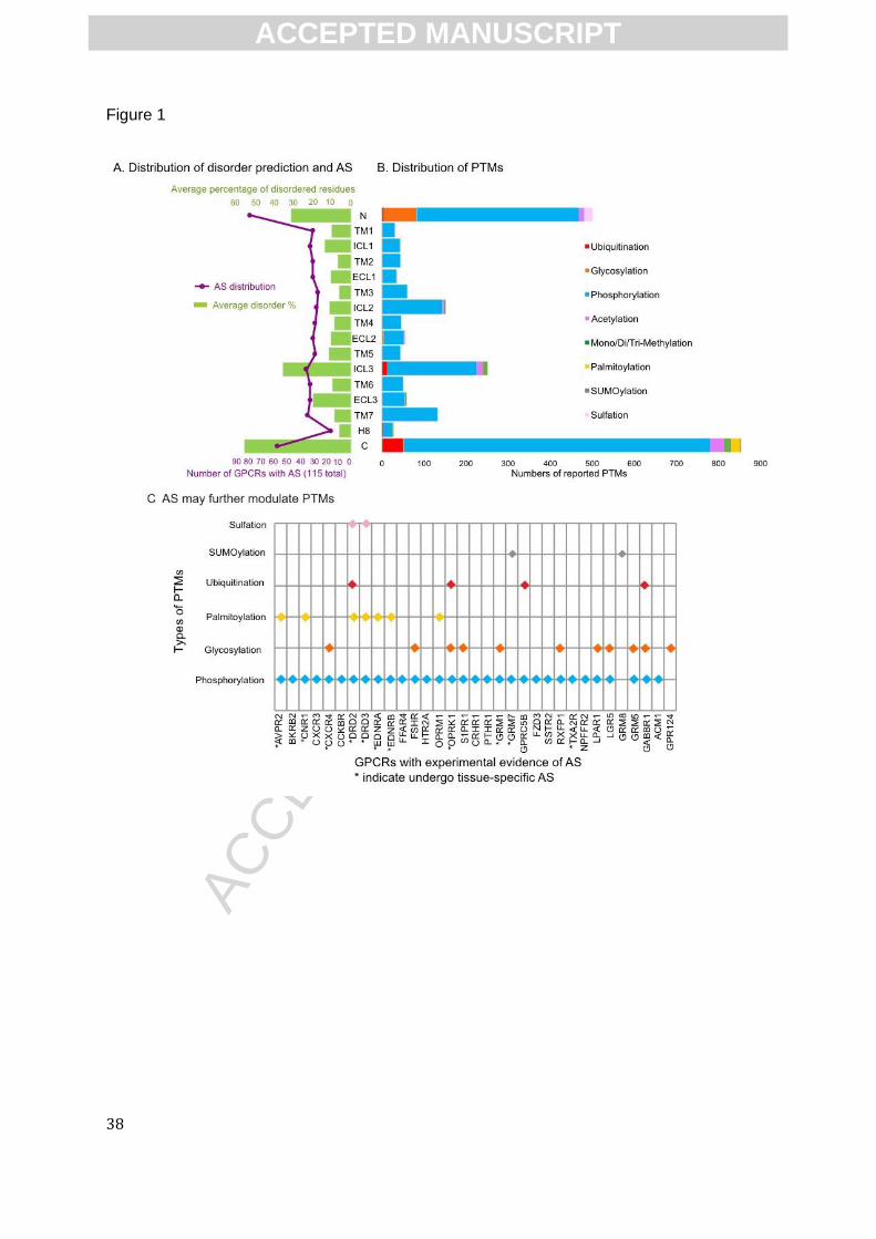
ACC
EPTE
D M
ANU
SCR
IPT
38
Figure 1
ACCEPTED MANUSCRIPT

ACC
EPTE
D M
ANU
SCR
IPT
39
Figure 2.
ACCEPTED MANUSCRIPT
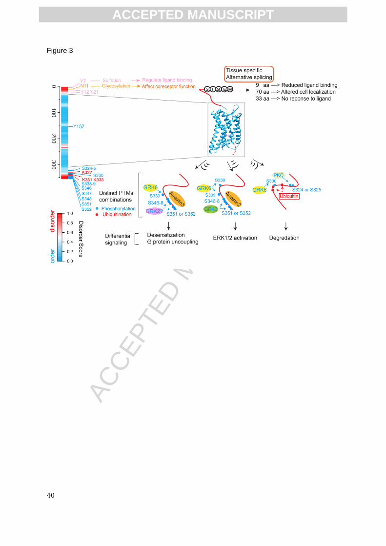
ACC
EPTE
D M
ANU
SCR
IPT
40
Figure 3
ACCEPTED MANUSCRIPT
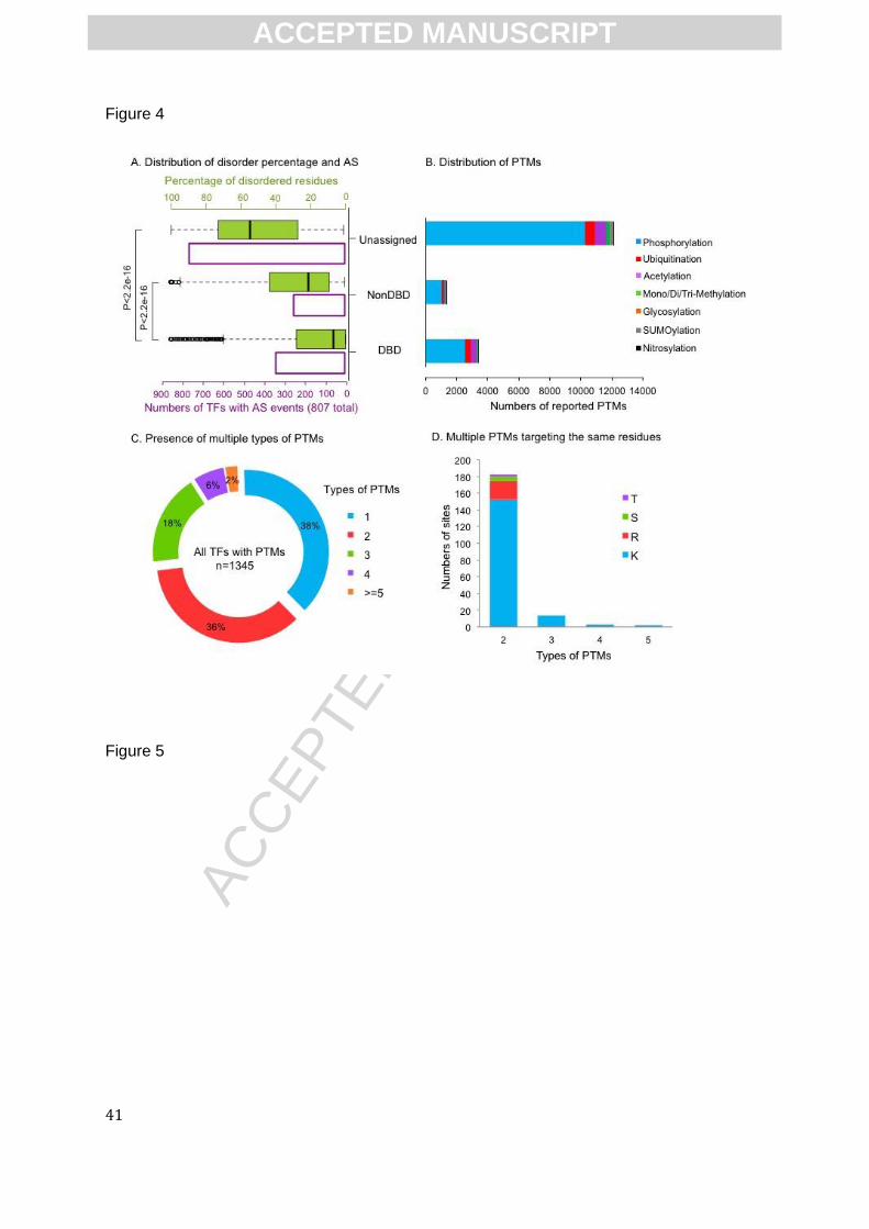
ACC
EPTE
D M
ANU
SCR
IPT
41
Figure 4
Figure 5
ACCEPTED MANUSCRIPT
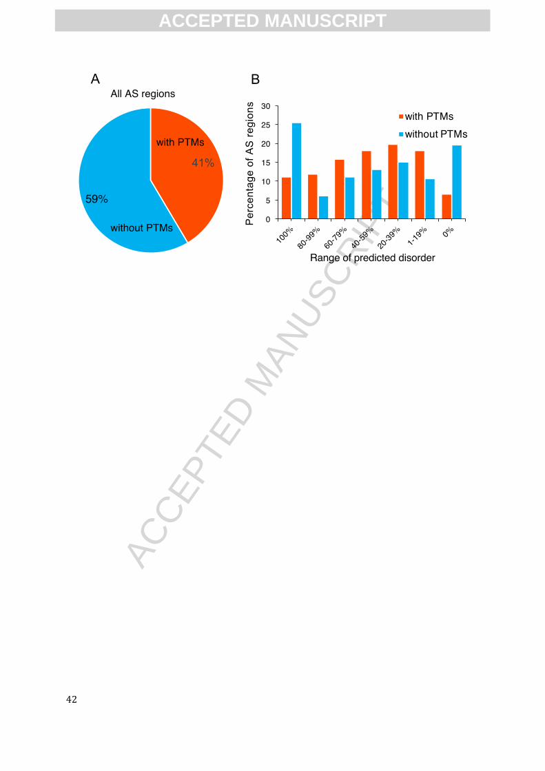
ACC
EPTE
D M
ANU
SCR
IPT
42
ACCEPTED MANUSCRIPT
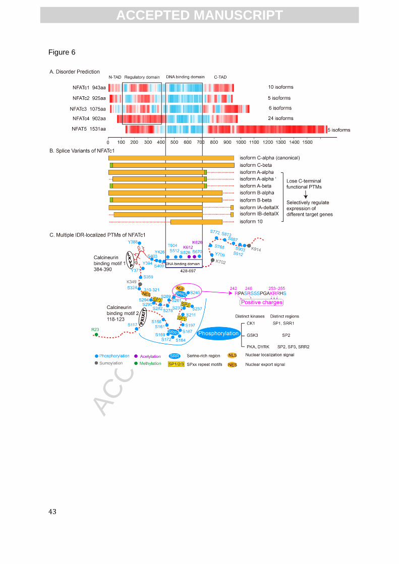
ACC
EPTE
D M
ANU
SCR
IPT
43
Figure 6
ACCEPTED MANUSCRIPT
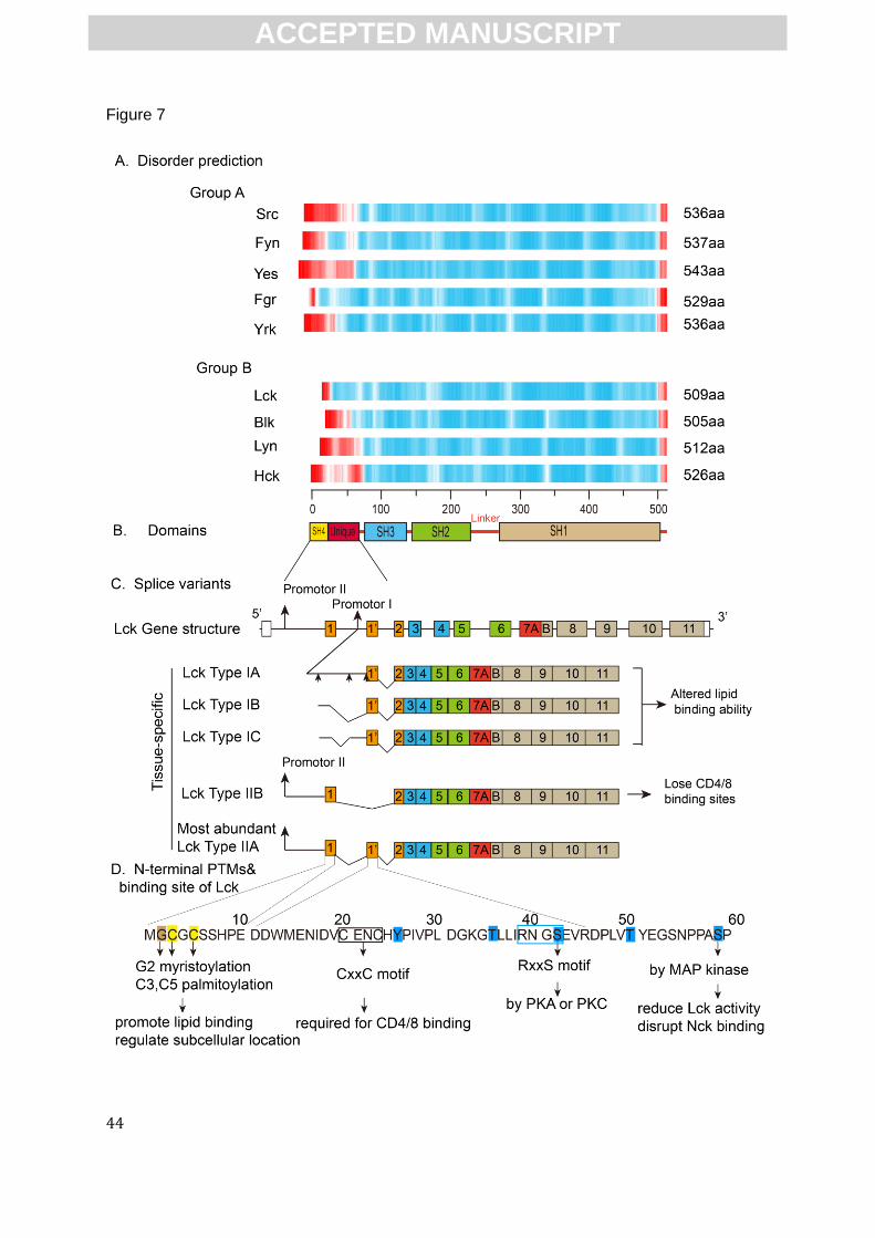
ACC
EPTE
D M
ANU
SCR
IPT
44
Figure 7
ACCEPTED MANUSCRIPT
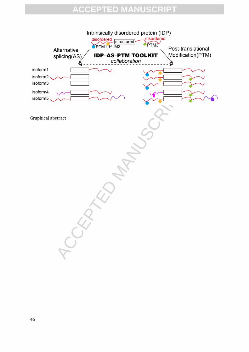
ACC
EPTE
D M
ANU
SCR
IPT
45
Graphical abstract
ACCEPTED MANUSCRIPT