Intrinsic and Extrinsic Factors influencing Properties ... · Intrinsic and Extrinsic Factors...
9
The Journal of Neuroscience, April 1990, 70(4): 1092-i 090 Intrinsic and Extrinsic Factors influencing Properties Patterns of ldentif ied Leech Neurons in Culture Growth Susan E. Acklin and J. G. Nicholls Department of Pharmacology, BiocenterMniversity of Basel, CH-4056 Basel, Switzerland An analysis has been made of intrinsic mechanisms influ- encing growth patterns of 2 identified leech interneurons in tissue culture. These small cells (known as DL and VL) dis- play unusual arborirations in the ganglion. The distinctive branching patterns resemble the letter “T” for VL and the letter “Y” for DL. DL and VL cells contain serotonin and can be identified in situ with Neutral red. 1. DL and VL cells were isolated and cultured on 2 ho- mogeneous substrates, concanavalin A (Con A) and leech extracellular matrix extract (ECM). Individual DL and VL cells in culture retained their ability to fire in spontaneous, rhyth- mical bursts. The synaptic connections formed in culture were specific and differed from those made by other sero- tonergic neurons. 2. On both Con A and ECM the neurons sprouted to re- produce their characteristic patterns. Every DL or VL cell that grew processes on ECM developed the appropriate Y- or T-shaped pattern. On Con A, most but not all of the cells showed specific Y or T patterns. The probability of sprouting was higher on Con A than on ECM. 3. Disruption of microtubular assemblies in freshly isolat- ed DL and VL cells by treatment with nocodazole did not affect the later outgrowth of specific patterns in culture. 4. These results provide evidence that adult DL and VL leech neurons retain intrinsic information for determining a variety of their properties in culture: In particular, they re- produce type-specific neuritic patterns in the absence of extrinsic guidance cues. Moreover, the intrinsic pattern de- termining mechanism is not lost after the destruction of mi- crotubular arrays within the cytoskeleton. A striking feature of the nervous system is the wide variety of distinctive shapes and branching patterns exhibited by neurons. The development of precise morphologies by individual cells constitutes a key mechanism for the establishment of neuronal circuits. Extrinsic factors that influence the direction, form, and extent of growing processesinclude soluble factors such as nerve growth factor and substrate-associated molecules such as lam- Received Aug. 2, 1989; revised Sept. 25, 1989; accepted Sept. 27, 1989. We wish to thank Drs. B. Stolz and H.-P. Hauri for help with the immunoflu- orescence, Dr. D. M. Grant for critical reading of the manuscript, as well as Ms. J. Wittker for typing the manuscript and Mr. P. Baettig for excellent photographic assistance. This work was supported by grants from the Swiss National Fonds, 3.525-1.83 and 3.556-0.86 (to J.G.N.), and a Fellowship from Boehringer Ingelheim, FRG (to S.E.A.). Correspondence should be addressed to Susan E. Acklin, Medical Sciences Building, University of Toronto, Toronto, Ontario M5S IAS, Canada. Copyright 0 1990 Society for Neuroscience 0270-6474/90/041082-09$02,00/O inin (Levi-Montalcini, 1975; Green and Shooter, 1980; Edgar et al., 1984; Letoumeau, 1985; Chiquet et al., 1988). A natural question concerns the extent to which intrinsic in- formation stored within a neuron can specify its individual out- growth pattern. Specific, identified axons in grasshopper and in zebra fish embryos grow in different patterns when presented with identical molecular cues (Raper et al., 1983; Eisen et al., 1986). The morphological features of a cell can also be repro- duced in culture. Thus, the daughter cells of mouse fibroblast 3T3 cells exhibit similarities in paths of migration, cell shape, and geometry of microfilament bundles (Albrecht-Buehler, 1977) and mitotic sister neuroblastoma cells display similar forms in culture (Solomon, 1979). Another striking example is provided by dispersed cultures of fetal rat hippocampal neurons; these cells develop morphological features similar to those of their counterparts in vivo (Banker and Cowan, 1977; Dotti et al., 1988). In the present experiments we made use of 2 identified leech neurons with unusual branching patterns to analyze how in- herent mechanisms can specify the shape of a cell. Many leech neurons can be identified by morphological, biochemical, and electrophysiological criteria; in culture, they retain their elec- trophysiological properties and the ability to form specific chem- ical or electrical synapses (Fuchs et al., 198 1). It has also been shown that when plated onto appropriate substrates such as Concanavalin A (Con A) or leech extracellular matrix extract (ECM) isolated neurons grow rapidly and extensively; each type of neuron shows a distinctive growth pattern (Chiquet and Ack- lin, 1986). The cells used in this study, known as dorsolateral (DL, 61) and ventrolateral (VL, 2 l), are small neurons that contain sero- tonin (5HT). In the ganglion they display unusual, easily rec- ognizable morphologies. Each cell has a single, large neurite that extends from the soma and bifurcates into 2 major branches pointing in opposite directions (Fig. 3). For DL the initial angle of bifurcation is 60”, which results in a Y-shaped morphology; for VL it is 180”, which results in a T-shaped morphology (Lent, 198 1; Nusbaum and Kristan, 1986). It will be shown that when DL and VL cells are cultured in absence of extrinsic guidance factors, they reproduce outgrowth patterns similar to the mor- phology established during development in vivo. As a first step in determining how intrinsic information is stored within the cell, microtubular assemblies were reversibly disrupted by ap- plication of nocodazole (De Brabander et al., 1976; Hoebeke et al., 1976; Solomon, 1980). After the neurons had recovered, we analyzed the new patterns of outgrowth. Another aim of our experiments was to assess whether the types of synaptic connections established by a neuron were in-
Transcript of Intrinsic and Extrinsic Factors influencing Properties ... · Intrinsic and Extrinsic Factors...

The Journal of Neuroscience, April 1990, 70(4): 1092-i 090
Intrinsic and Extrinsic Factors influencing Properties Patterns of ldentif ied Leech Neurons in Culture
Growth
Susan E. Acklin and J. G. Nicholls Department of Pharmacology, BiocenterMniversity of Basel, CH-4056 Basel, Switzerland
An analysis has been made of intrinsic mechanisms influ- encing growth patterns of 2 identified leech interneurons in tissue culture. These small cells (known as DL and VL) dis- play unusual arborirations in the ganglion. The distinctive branching patterns resemble the letter “T” for VL and the letter “Y” for DL. DL and VL cells contain serotonin and can be identified in situ with Neutral red.
1. DL and VL cells were isolated and cultured on 2 ho- mogeneous substrates, concanavalin A (Con A) and leech extracellular matrix extract (ECM). Individual DL and VL cells in culture retained their ability to fire in spontaneous, rhyth- mical bursts. The synaptic connections formed in culture were specific and differed from those made by other sero- tonergic neurons.
2. On both Con A and ECM the neurons sprouted to re- produce their characteristic patterns. Every DL or VL cell that grew processes on ECM developed the appropriate Y- or T-shaped pattern. On Con A, most but not all of the cells showed specific Y or T patterns. The probability of sprouting was higher on Con A than on ECM.
3. Disruption of microtubular assemblies in freshly isolat- ed DL and VL cells by treatment with nocodazole did not affect the later outgrowth of specific patterns in culture.
4. These results provide evidence that adult DL and VL leech neurons retain intrinsic information for determining a variety of their properties in culture: In particular, they re- produce type-specific neuritic patterns in the absence of extrinsic guidance cues. Moreover, the intrinsic pattern de- termining mechanism is not lost after the destruction of mi- crotubular arrays within the cytoskeleton.
A striking feature of the nervous system is the wide variety of distinctive shapes and branching patterns exhibited by neurons. The development of precise morphologies by individual cells constitutes a key mechanism for the establishment of neuronal circuits. Extrinsic factors that influence the direction, form, and extent of growing processes include soluble factors such as nerve growth factor and substrate-associated molecules such as lam-
Received Aug. 2, 1989; revised Sept. 25, 1989; accepted Sept. 27, 1989. We wish to thank Drs. B. Stolz and H.-P. Hauri for help with the immunoflu-
orescence, Dr. D. M. Grant for critical reading of the manuscript, as well as Ms. J. Wittker for typing the manuscript and Mr. P. Baettig for excellent photographic assistance.
This work was supported by grants from the Swiss National Fonds, 3.525-1.83 and 3.556-0.86 (to J.G.N.), and a Fellowship from Boehringer Ingelheim, FRG (to S.E.A.).
Correspondence should be addressed to Susan E. Acklin, Medical Sciences Building, University of Toronto, Toronto, Ontario M5S IAS, Canada. Copyright 0 1990 Society for Neuroscience 0270-6474/90/041082-09$02,00/O
inin (Levi-Montalcini, 1975; Green and Shooter, 1980; Edgar et al., 1984; Letoumeau, 1985; Chiquet et al., 1988).
A natural question concerns the extent to which intrinsic in- formation stored within a neuron can specify its individual out- growth pattern. Specific, identified axons in grasshopper and in zebra fish embryos grow in different patterns when presented with identical molecular cues (Raper et al., 1983; Eisen et al., 1986). The morphological features of a cell can also be repro- duced in culture. Thus, the daughter cells of mouse fibroblast 3T3 cells exhibit similarities in paths of migration, cell shape, and geometry of microfilament bundles (Albrecht-Buehler, 1977) and mitotic sister neuroblastoma cells display similar forms in culture (Solomon, 1979). Another striking example is provided by dispersed cultures of fetal rat hippocampal neurons; these cells develop morphological features similar to those of their counterparts in vivo (Banker and Cowan, 1977; Dotti et al., 1988).
In the present experiments we made use of 2 identified leech neurons with unusual branching patterns to analyze how in- herent mechanisms can specify the shape of a cell. Many leech neurons can be identified by morphological, biochemical, and electrophysiological criteria; in culture, they retain their elec- trophysiological properties and the ability to form specific chem- ical or electrical synapses (Fuchs et al., 198 1). It has also been shown that when plated onto appropriate substrates such as Concanavalin A (Con A) or leech extracellular matrix extract (ECM) isolated neurons grow rapidly and extensively; each type of neuron shows a distinctive growth pattern (Chiquet and Ack- lin, 1986).
The cells used in this study, known as dorsolateral (DL, 61) and ventrolateral (VL, 2 l), are small neurons that contain sero- tonin (5HT). In the ganglion they display unusual, easily rec- ognizable morphologies. Each cell has a single, large neurite that extends from the soma and bifurcates into 2 major branches pointing in opposite directions (Fig. 3). For DL the initial angle of bifurcation is 60”, which results in a Y-shaped morphology; for VL it is 180”, which results in a T-shaped morphology (Lent, 198 1; Nusbaum and Kristan, 1986). It will be shown that when DL and VL cells are cultured in absence of extrinsic guidance factors, they reproduce outgrowth patterns similar to the mor- phology established during development in vivo. As a first step in determining how intrinsic information is stored within the cell, microtubular assemblies were reversibly disrupted by ap- plication of nocodazole (De Brabander et al., 1976; Hoebeke et al., 1976; Solomon, 1980). After the neurons had recovered, we analyzed the new patterns of outgrowth.
Another aim of our experiments was to assess whether the types of synaptic connections established by a neuron were in-

The Journal of Neuroscience, April 1990, W(4) 1083
Figure 1. 5-HT-containing neurons in a leech ganglion stained with Neutral red (living, unfixed preparations). A, Ventral view: VL, Ret&s, M, cells. B, Dorsal view: DL cells. In A and B, anterior is to the left,
fluenced by its transmitter. Would all cells containing 5-HT make identical synapses on the same postsynaptic target? It will be shown that the synaptic connections made by DL and VL cells on P sensory cells in culture were different from those formed by Retzius cells (which also contain 5-HT). Moreover, DL and VL cells retained their membrane characteristics and the ability to produce rhythmical bursts of impulses in culture.
Materials and Methods Experiments were made on 2 small, 5-HT-containing interneurons (DL and VL) isolated from the CNS of the leech (Hirudo medicinalis).
Identification ofDL and VL within the ganglion and culturing method. Methods for isolating single neurons from the leech CNS have been described previously (Dietzel et al., 1986). Individual ganglia were dis- sected from the CNS of the leech and pinned in a Sylgard-coated dish in Leibovitz L- 15 medium (GIBCO) supplemented with 2 mM gluta- mine (GIBCO), 6 mg/ml glucose, 0.1 mg gentamicin sulfate (Garamycin; Schering, Kenilworth, NJ)/ml, and 2% fetal bovine serum (Kansas City Biological, Lenexa, KS). The connective tissue capsules surrounding the ganglia were opened with forceps, and the ganglia were incubated for 1 ir ai room temperature in collagenase/dispase (Boehringer Mannheim; 2 me/ml in L-15 medium). To identify 5-HT-containing cells and in oar&ular DL and VL, Neuiral red (Sigma; 1 mg/ml supplemented L- 15 medium plus collagen&e/dispase) was added 20 minutes before the end of the enzvme digestion (Stuart et al., 1974: Lent et al., 1979). Figure 1 shows Neutral red-stained 5-HT-containing neurons within a leech ganglion. After the enzyme and Neutral red treatment, the ganglia were washed extensively with supplemented L-15 medium. Single, stained DL and VL cells-and in certain experiments Retzius and medial (M) cells-were removed from the ganglion by suction into a fire-polished glass capillary. The cells were washed by transferring them through several drops of sterile, supplemented L- 15 medium. After 10 hr, cells were plated in culture Dishes (Falcon F-3034) coated with Con A (Sigma, 2 mg/ml) or with EDTA leech ECM extract (kindly provided by Ms. S. Grumbacher). The yield of Neutral red-stained DL and VL cells was low because of their small size and fragility. Cells could survive for up to 2 weeks in culture. Stained and nonstained Retzius cells did not show morphological or electrophysiological differences (Stuart et al., 1974; Lent and Frazer, 1977). - - -
Dye fills. Procedures for filling cells with Lucifer yellow and HRP
have been described elsewhere (Muller and McMahan, 1976; Stewart, 1978). To fill single DL and VL cells in culture with Lucifer yellow, the tips of microelectrodes (Haer Ultratip Omegadot 30-30-o. 30-40 MQ resistances) were filled with 3O!‘a Lucifer yellow and backfilled with 0.1 M lithium chloride (Sigma). Hvnernolarizina oulses (500 msec. 1 Hz. l-8 nA) were applied f& 1-i 0 r&n. After allo&g 5-l d min for drtisiod the live, unfixed preparations were photographed with Kodak Tri-X film. To pressure-inject HRP into identified VL and DL neurons within the ganglion, the tips of microelectrodes (thick-wall Haer Ultratip Ome- gadot 30-30-l) were filled with 20 mg HRP (Sigma, Type VI) and 2 mg Fast green (Sigma) dissolved in 1 ml potassium acetate (0.2 M). Electrode tips were bevelled to a resistance of 30-40 Ma. HRP was injected by applying pressure. After a diffusion time of 2-3 hr the preparations were fixed and then developed.
Selection of cells for measurement of neurite outgrowth in culture. Not all healthy DL and VL cells plated on Con A or ECM grew neurites (see also Results). Healthy cells were reliably recognized by their rounded and shiny appearance. These observations were confirmed by electro- physiological measurements. For assessment of growth we selected only the cells that had sprouted. Healthy cells without neurites were not considered. On Con A about 600/o of all plated, healthy cells sprouted. On ECM, however, only about 15% of all healthy cells grew out, and most of those that did, sprouted only after a delay of 6-8 d (see Results).
Assessment of outgrowth and pattern formation. The outgrowth pat- terns of DL and VL cells in the ganglion and in culture were photo- graphed, and the angle between the initial major branches was measured.
A number of variables had to be taken into account for the qualitative and quantitative assessment of T or Y patterns in culture: these included degree of attachment to the substrate and different lengths of axonal stump. After removal of DL and VL cells, an axonal stump of variable length (between a few microns and several cell diameters) remained attached to the soma. All neuronal arborization had been cut off at the end of the stump. When the axonal stump was short or hidden, the cell soma was carefully moved with a glass pipette to observe the sites from which growth arose. We did not measure minor processes growing out directly from the cell soma.
Nocodazole treatment. A stock solution of nocodazole (Sigma, 3 mg/ ml DMSO) was prepared. Freshly isolated Retzius, DL, and VL cells were incubated with nocodazole at a final concentration of 10 &ml supplemented L-15 medium for 20 min. The controls were incubated with the corresponding amount of DMSO alone. Cells were washed extensively in supplemented L- 15 medium.
1084 Acklin and Nicholls - Influences on Outgrowth Patterns
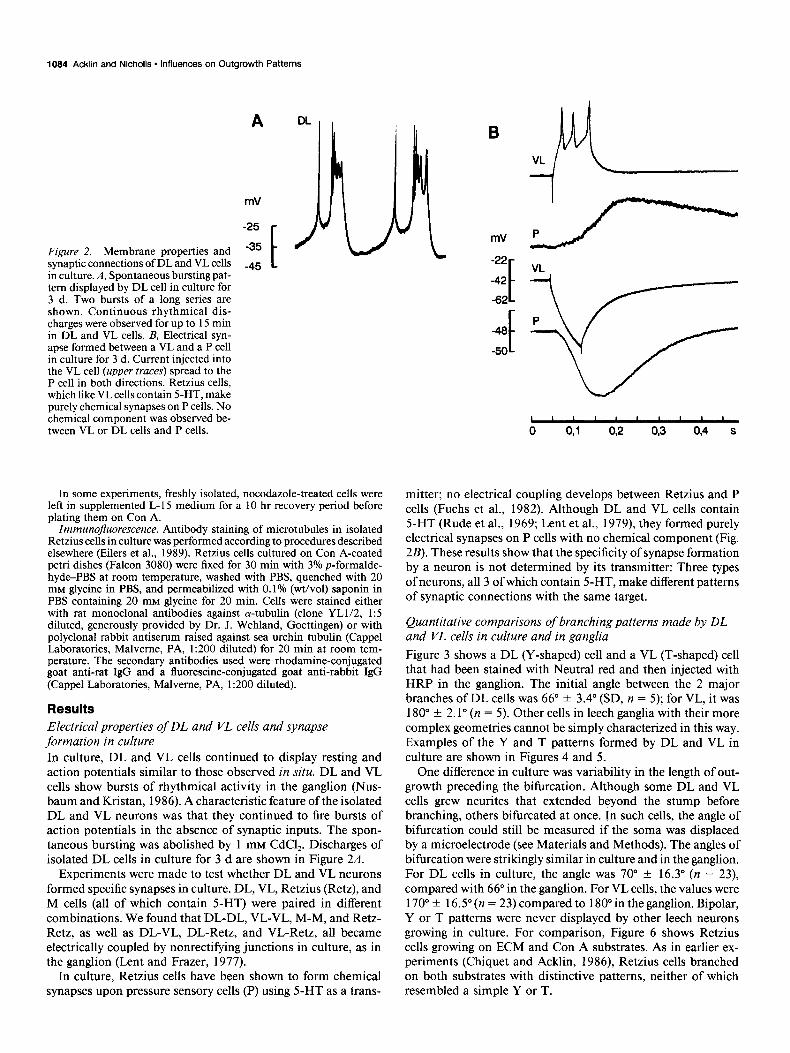
Figure 2. Membrane properties and synaptic connections of DL and VL cells in culture. A, Spontaneous bursting pat- tern displayed by DL cell in culture for 3 d. Two bursts of a long series are shown. Continuous rhythmical dis- charges were observed for up to 15 min in DL and VL cells. B, Electrical syn- apse formed between a VL and a P cell in culture for 3 d. Current injected into the VL cell (upper truces) spread to the P cell in both directions. Retzius cells, which like VL cells contain 5-HT, make purely chemical synapses on P cells. No chemical component was observed be- tween VL or DL cells and P cells.
A DL
mV
-25
-35
-45 t
/
B h 4 -K mV 1/-- LJ L -22
-42
-62
-46
-50 f
I ” ” ’ ’ “1 0 41 02 OB 0,4 s
In some experiments, freshly isolated, nocodazole-treated cells were left in supplemented L-15 medium for a 10 hr recovery period before plating them on Con A.
Immunojluorescence. Antibody staining of microtubules in isolated Retzius cells in culture was performed according to procedures described elsewhere (Eilers et al., 1989). Retzius cells cultured on Con A-coated petri dishes (Falcon 3080) were fixed for 30 min with 3% p-formalde- hyde-PBS at room temperature, washed with PBS, quenched with 20 mM glycine in PBS, and permeabilized with 0.1% (wt/vol) saponin in PBS containing 20 mM glycine for 20 min. Cells were stained either with rat monoclonal antibodies against a-tubulin (clone YLU2, 1:5 diluted, generously provided by Dr. J. Wehland, Goettingen) or with polyclonal rabbit antiserum raised against sea urchin tubulin (Cappel Laboratories, Malveme, PA, 1:200 diluted) for 20 min at room tem- perature. The secondary antibodies used were rhodamine-conjugated goat anti-rat IgG and a fluorescine-conjugated goat anti-rabbit IgG (Cappel Laboratories, Malveme, PA, 1:200 diluted).
Results Electrical properties of DL and VL cells and synapse formation in culture In culture, DL and VL cells continued to display resting and action potentials similar to those observed in situ. DL and VL cells show bursts of rhythmical activity in the ganglion (Nus- baum and Kristan, 1986). A characteristic feature of the isolated DL and VL neurons was that they continued to fire bursts of action potentials in the absence of synaptic inputs. The spon- taneous bursting was abolished by 1 mM CdCl,. Discharges of isolated DL cells in culture for 3 d are shown in Figure 2A.
Experiments were made to test whether DL and VL neurons formed specific synapses in culture. DL, VL, Retzius (Retz), and M cells (all of which contain 5-HT) were paired in different combinations. We found that DL-DL, VL-VL, M-M, and Retz- Retz, as well as DL-VL, DL-Retz, and VL-Retz, all became electrically coupled by nonrectifying junctions in culture, as in the ganglion (Lent and Frazer, 1977).
In culture, Retzius cells have been shown to form chemical synapses upon pressure sensory cells (P) using 5-HT as a trans-
mitter; no electrical coupling develops between Retzius and P cells (Fuchs et al., 1982). Although DL and VL cells contain 5-HT (Rude et al., 1969; Lent et al., 1979), they formed purely electrical synapses on P cells with no chemical component (Fig. 2B). These results show that the specificity of synapse formation by a neuron is not determined by its transmitter: Three types of neurons, all 3 of which contain 5-HT, make different patterns of synaptic connections with the same target.
Quantitative comparisons of branching patterns made by DL and VL cells in culture and in ganglia Figure 3 shows a DL (Y-shaped) cell and a VL (T-shaped) cell that had been stained with Neutral red and then injected with HRP in the ganglion. The initial angle between the 2 major branches of DL cells was 66” f 3.4” (SD, n = 5); for VL, it was 180” f 2.1” (n = 5). Other cells in leech ganglia with their more complex geometries cannot be simply characterized in this way. Examples of the Y and T patterns formed by DL and VL in culture are shown in Figures 4 and 5.
One difference in culture was variability in the length of out- growth preceding the bifurcation. Although some DL and VL cells grew neurites that extended beyond the stump before branching, others bifurcated at once. In such cells, the angle of bifurcation could still be measured if the soma was displaced by a microelectrode (see Materials and Methods). The angles of bifurcation were strikingly similar in culture and in the ganglion. For DL cells in culture, the angle was 70” f 16.3” (n = 23), compared with 66” in the ganglion. For VL cells, the values were 170” -t 16.5“ (n = 23) compared to 180” in the ganglion. Bipolar, Y or T patterns were never displayed by other leech neurons growing in culture. For comparison, Figure 6 shows Retzius cells growing on ECM and Con A substrates. As in earlier ex- periments (Chiquet and Acklin, 1986), Retzius cells branched on both substrates with distinctive patterns, neither of which resembled a simple Y or T.

The Journal of Neuroscience. April 1990, 1~34) 1085
Znfruence of substrates on branching patterns Branching patterns of DL and VL cells were more variable on Con A than on ECM. On Con A, 30% of the DL and VL cells grew out in ill-defined configurations; the remaining 70% formed the appropriate Y or T patterns. By contrast, although fewer cells sprouted on ECM (see below), 100% of those DL and VL cells that did sprout reproduced the appropriate T or Y patterns. A further characteristic difference was that DL and VL cells produced more branches on Con A than on ECM; in addition, the individual neurites were more curved on Con A (Figs. 4, 5). Comparable effects of substrate on the number and curvature
Figure 3. Characteristic branching patterns of (A) DL and (II) VL cells in a leech ganglion. These cells were iden- tified by staining with Neutral red and then pressure-injected with HRP.
of branches have been observed with other types of leech neu- rons (Chiquet and Acklin, 1986; Grumbacher-Reinert, 1989).
Con A and ECM also influenced the time course and success rate of neurite outgrowth by DL and VL cells. On Con A, ap- proximately 60% (n = 50) of all healthy, plated DL or VL cells showed neuronal outgrowth, whereas on ECM only 15% (n = 50) of all plated cells grew out (see also Materials and Methods). When neuronal outgrowth of DL and VL cells on ECM did occur it began late after a delay of 6 to 8 d (Fig. 7). These findings for DL and VL cells differ from other leech neurons, which sprout well on both substrates but more rapidly on ECM than on Con A (Chiquet and Acklin, 1986; Grumbacher-Reinert, 1989).

1066 Acklin and Nicholls l Influences on Outgrowth Patterns
Figure 4. Outgrowth patterns of DL and VL cells cultured for 10 d on ECM. Sprouting originated from the axonal stump. Sprouts (arrows) in B belong to a cocultured Retzius cell. Note the vari- ability in lengths of outgrowth from the stump before the bitbrcation into T or Y branches (C, D).
A
Figure 5. Sprouting patterns of DL (A) and VL (B) cells cultured for 3 d on Con A. The Y and T patterns are apparent. Note the thicker, more branched processes on Con A compared with ECM.

The Journal of Neuroscience, April 1990, fO(4) 1087
Figure 6. Outgrowth patterns of Retzius cells, cultured for 3 d on ECM (A) and Con A (B). The branching conforms to no simple T or Y configuration. The differences on Con A and ECM resemble those described earlier, with straighter, finer, less curved, less branched processes on ECM. Note the rapid growth on ECM.
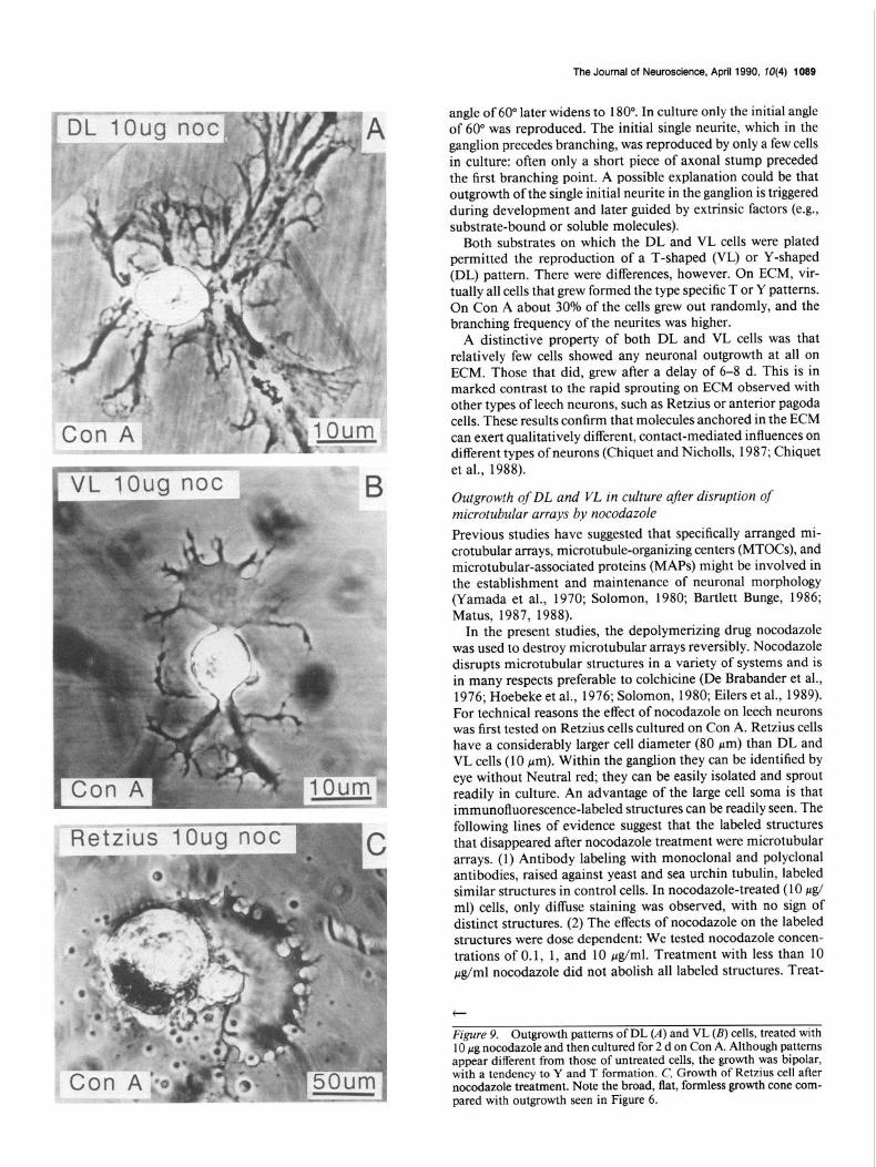
Outgrowth of DL and VL in culture after disruption of microtubular arrays by nocodazole The aim of the following experiments was to test whether DL and VL cells reproduced their original patterns in culture be- cause of specific preexisting microtubular arrays within the cy- toskeleton. A drug that disrupts microtubules (nocodazole) was applied to test whether Y- and T-shapes could still be repro- duced (De Brabander et al., 1976; Hoebeke et al., 1976; Solo- mon, 1980; Eilers et al., 1989). Immunofluorescence micro- graphs of the cytoskeleton were made in freshly isolated leech neurons labeled with anti-tubulin antibody. These pictures were compared with those obtained after treatment with nocodazole. Retzius cells rather than DL or VL were used because of their larger size and ease of dissection without Neutral red (see Dis- cussion). Incubation in 10 &ml nocodazole for 20 min de- stroyed all of the cytoskeletal microtubular arrays in Retzius cells (Fig. 8; see also Discussion). Moreover, nocodazole-treated Retzius cells grew out in broad veil-like structures (Fig. 9C). Isolated DL and VL cells were similarly treated with nocodazole, after which they were allowed to recover for 10 hr and plated on Con A. Figure 9A shows the sprouting pattern of a nocod- azole-treated DL cell after 1 d in culture. The outgrowth was highly symmetrical. The 2 broad, flat growth cones appeared similar in their divergence to the bipolar outgrowth of normal DL cells. The outgrowth of VL cells after nocodazole treatment also appeared bipolar, resembling the pattern of untreated VL cells (Fig. 9B). In contrast, other neurons, such as Retzius cells (Fig. 9C), treated and cultured under the same conditions showed no bipolar outgrowth patterns. Although the outgrowth patterns after nocodazole treatment showed recognizable differences for DL and VL cells, quantitative measurements of initial out-
growth angles were not possible because of the diffuse, ill-defined structures of the processes.
Discussion Adult leech neurons in culture retain their membrane properties and the ability to form specific synapses. Thus, Retzius cells continue to give characteristic impulses and can form chemical
mm 3-
Figure 7. Time course of neurite outgrowth of DL and VL neurons cultured on Con A (W,) and ECM (0). Each point represents the average neurite length per cell, measured from at least 5 neurons. Bars indicate SEMs. Note that growth did not begin on ECM for 6 d or more. By contrast, other cells (such as Retzius or AP cells) grew from the outset on both ECM and Con A, with growth rates that were significantly faster on ECM. Once DL and VL did grow on ECM, they grew rapidly (see Materials and Methods for description of how cells were selected for measurement).

1088 Acklin and Nicholls l Influences on Outgrowth Patterns
Figure 8. Effect of nocodazole on cy- toskeleton. A, Untreated control Ret- zius cell, labeled with a polyclonal rab- bit antiserum raised against sea urchin tubulin: B. Retzius cell. labeled with the same a&tubulin antibody as the con- trol cell, after treatment with 10 &ml nocodazole. C, Axonal stump of the same cell as shown in A. 0, Axonal stump of the same cell as shown in B. Both control and treated cells were fo- cused in the same plane by Nomarski optics prior to fluorescence photogra- phy. Note the absence of labeled struc- tures in the nocodazole-treated Retzius cell. Retzius cells rather than DL or VL were chosen because of their large size and the reliable identification without Neutral red.
synapses with one cell, the P cell, and electrical synapses with other cells (Fuchs et al., 1982). In culture, as in the ganglion, DL and VL cells became electrically coupled with other sero- tonergic neurons such as DL, VL, Retzius, and the M cell. Unlike Retzius cells, DL and VL formed electrical connections and did not form chemical synapses with the P cell (Fuchs et al., 198 1, 1982). This result shows that 2 cells with the same transmitter can form different connections in culture with specific targets. DL and VL cells were unusual in their ability to produce spon- taneous bursts of action potentials in culture.
An even more unusual property of DL and VL cells was the reproduction of the original arborization patterns in the absence of external cues. Thus, Retzius and other leech cells grow in highly distinctive patterns on Con A and ECM substrates, but the final picture is quite different from that produced in the ganglion by normal development (Grumbacher-Reinert, 1989). By contrast, adult DL and VL leech neurons appear to be pro- vided with intrinsic information that specifies their pattern of outgrowth, in the absence of extrinsic guidance factors.
A technical difficulty in our experiments was to measure quan- titatively the extent to which original patterns were reproduced. One clear result was that outgrowth was bipolar. Moreover, the angles between the first major branches were accurately repro- duced. Even in those cases where the neuronal outgrowth orig- inated directly at the axonal stump and was not preceded by a single neurite, the initial branches grew out at about the same angles as they did in the ganglion. To assess whether a T or Y had been formed when the axonal stump was hidden, we care- fully displaced the cell soma with a glass pipette. In a few cells on Con A, neurites also grew out directly from the cell soma. We ignored those processes and measured only outgrowth orig- inating at the axonal stump, since (1) the branches, originating from the cell soma, were thin and scattered, and (2) we never saw sprouting from the cell soma on ECM, which is an endog- enous substrate (Chiquet and Acklin, 1986; Chiquet et al., 1988).
The resemblance between branching patterns in the ganglion and in culture was only certain for the initial outgrowth of major branches. For DL cells in the ganglion, the initial branching
The Journal of Neuroscience, April 1990, W(4) 1089
angle of 60” later widens to 180”. In culture only the initial angle of 60” was reproduced. The initial single neurite, which in the ganglion precedes branching, was reproduced by only a few cells in culture: often only a short piece of axonal stump preceded the first branching point. A possible explanation could be that outgrowth of the single initial neurite in the ganglion is triggered during development and later guided by extrinsic factors (e.g., substrate-bound or soluble molecules).
Both substrates on which the DL and VL cells were plated permitted the reproduction of a T-shaped (VL) or Y-shaped (DL) pattern. There were differences, however. On ECM, vir- tually all cells that grew formed the type specific T or Y patterns. On Con A about 30% of the cells grew out randomly, and the branching frequency of the neurites was higher.
A distinctive property of both DL and VL cells was that relatively few cells showed any neuronal outgrowth at all on ECM. Those that did, grew after a delay of 6-8 d. This is in marked contrast to the rapid sprouting on ECM observed with other types of leech neurons, such as Retzius or anterior pagoda cells. These results confirm that molecules anchored in the ECM can exert qualitatively different, contact-mediated influences on different types of neurons (Chiquet and Nicholls, 1987; Chiquet et al., 1988).
Outgrowth of DL and VL in culture after disruption of microtubular arrays by nocodazole Previous studies have suggested that specifically arranged mi- crotubular arrays, microtubule-organizing centers (MTOCs), and microtubular-associated proteins (MAPS) might be involved in the establishment and maintenance of neuronal morphology (Yamada et al., 1970; Solomon, 1980; Bartlett Bunge, 1986; Matus, 1987, 1988).
In the present studies, the depolymerizing drug nocodazole was used to destroy microtubular arrays reversibly. Nocodazole disrupts microtubular structures in a variety of systems and is in many respects preferable to colchicine (De Brabander et al., 1976; Hoebeke et al., 1976; Solomon, 1980; Eilers et al., 1989). For technical reasons the effect of nocodazole on leech neurons was first tested on Retzius cells cultured on Con A. Retzius cells have a considerably larger cell diameter (80 Km) than DL and VL cells (10 pm). Within the ganglion they can be identified by eye without Neutral red; they can be easily isolated and sprout readily in culture. An advantage of the large cell soma is that immunofluorescence-labeled structures can be readily seen. The following lines of evidence suggest that the labeled structures that disappeared after nocodazole treatment were microtubular arrays. (1) Antibody labeling with monoclonal and polyclonal antibodies, raised against yeast and sea urchin tubulin, labeled similar structures in control cells. In nocodazole-treated (10 ~Lgl ml) cells, only diffuse staining was observed, with no sign of distinct structures. (2) The effects of nocodazole on the labeled structures were dose dependent: We tested nocodazole concen- trations of 0.1, 1, and 10 &ml. Treatment with less than 10 &ml nocodazole did not abolish all labeled structures. Treat-
t
Figure 9. Outgrowth patterns of DL (A) and VL (B) cells, treated with 10 hg nocodazole and then cultured for 2 d on Con A. Although nattems appear different from those of untreated cells, the growth was bipolar, with a tendency to Y and T formation. C, Growth of Retzius cell after nocodazole treatment. Note the broad, flat, formless growth cone com- pared with outgrowth seen in Figure 6.

1090 Acklin and Nicholls * Influences on Outgrowth Patterns
ment of control cells with the same amount of DMSO had no effect. (3) Nocodazole-treated Retzius cells (like DL and VL cells) grew out in broad, flat, veil-like structures. Distinct fine processes were absent. Nevertheless, after disruption of micro- tubular assemblies, DL and VL cells, unlike Retzius cells, still grew out in a bipolar manner. This could be the result of some microtubular structures remaining after nocodazole treatment. Another explanation might be that bipolar outgrowth is orga- nized by cytoskeletal components that are not affected by no- codazole, for example, MTOCs (Ng and Frankel, 1977; Schliwa et al., 1979; Spiegelman et al., 1979a, b). MTOCs could deter- mine cellular morphologies by organizing selective growth of certain neurites, while others remain stationary (Kirschner and Mitchison, 1986). It will be of interest to investigate further the role of cytoskeletal components in determining the morpholo- gies of DL and VL cells in culture and to analyze where in these cells intrinsic information for pattern formation is stored.
References Albrecht-Buehler, G. (1977) Daughter 3T3 cells. Are they mirror im-
aaes of each other? J. Cell Biol. 72: 595-603. Balker, G. A., and W. M. Cowan (1977) Rat hippocampal neurons
in dispersed cell culture. Brain Res. 126: 397425. - Bartlett Bunae. M. (1986) The axonal cvtoskeleton: Its role in aen-
erating and maintaining’cell form. TINS 9: 477-482. Chiquet, M., and S. E. Acklin (1986) Attachment to Con A or extra-
cellular matrix initiates rapid sprouting by cultured leech neurons. Proc. Natl. Acad. Sci. USA 83: 6 188-6 192.
Chiquet, M., and J. G. Nicholls (1987) Neurite outgrowth and synapse formation by identified leech neurones in culture. J. Exp. Biol. 132: 191-206.
Chiquet, M., L. Masuda-Nakagawa, and K. Beck (1988) Attachment to a laminin-like leech protein, but not to vertebrate laminin, initiates rapid sprouting by leech neurons. J. Cell Biol. 107: 1189-1198.
De Brabander, M. J., R. M. L. Van De Veire, F. E. M. Aerts, and P. A. J. Janssen (1976) The effects of methvll5-(2-thienvlcarbonvl)- lH-benzimidazbl-2-yllcarbamate, (R 17934; ‘NSC 238 i59), a new synthetic antitumoral drug interfering with microtubules, on mam- malian cells cultured in vitro. Cancer Res. 36: 905-916.
Dietzel, I. D., P. Drapeau, and J. G. Nicholls (1986) Voltage depen- dence of 5-hydroxytryptamine release at a synapse between identified leech neurons in culture. J. Physiol. (L0nd.j 3j2: 19 l-205.
Dotti, C. G., C. A. Sullivan. and G. A. Banker (1988) The establish- ment of polarity by hippdcampal neurons in culture. J. Neurosci. 8: 1454-1468.
Edgar, D., R. Timpl, and H. Thoenen (1984) The heparin-binding domain of laminin is responsible for its effects on neurite outgrowth and neuronal survival. Embo J. 3: 1463-1468.
Eilers, U., J. Klumperman, and H.-P. Hauri (1989) Nocodazole, a microtubule-active drug, interferes with apical protein delivery in cultured intestinal epithelial cells (Caco-2). J. Cell Biol. 108: 13122.
Eisen. J. S.. Z. M. Mvers. and M. Westerfield (1986) Pathwav selection by growth cones of identified motoneurones in live zebra fish embryos. Nature 320: 269-27 1.
Fuchs, P. A., J. G. Nicholls, and D. Ready (198 1) Membrane prop- erties and selective connexions of identified leech neurones in culture. J. Physiol. (Lond.) 316: 203-223.
Fuchs, P. A., L. P. Henderson, and J. G. Nicholls (1982) Chemical transmission between individual Retzius and sensory neurones of the leech in culture. J. Physiol. (Lond.) 323: 195-210. -
Green. L. A.. and E. M. Shooter (1980) The nerve arowth factor. Annu.
Grumbacher-Reinert, S. (1989) Local influence of substrate molecules in determining distinctive growth patterns of identified neurons in culture. Proc. Natl. Acad. Sci. USA 86: 7270-7274.
Hoebeke, J., G. Van Nijen, and M. De Brabander (1976) Interaction of oncodazole (R 17934), a new antitumoral drug, with rat brain tubulin. Biochem. Biophvs. Res. Commun. 69: 3 19-324.
Kirschner, M., and T. Mitchison (1986) Beyond self assembly: From microtubules to morphogenesis. Cell 45: 329-342.
Lent, C. M. (198 1) Morphology of neurons containing monoamines within leech segmental ganglia. J. Exp. Zool. 216: 3 1 l-3 16.
Lent, C. M., and B. M. Frazer (1977) Connectivity of the monoamine- containing neurones in central nervous svstem of leech. Nature 28: 844-847.-
Lent, C. M., J. Ono, K. T. Keyser, and H. J. Karten (1979) Identifi- cation of vital-stained neurons from leech ganglia. J. Neurochem. 32: 1559-1563.
Letoumeau, P. C. (1985) Axonal growth and guidance. In Molecular Bases of Neural Development, M. Edelman, ed., pp. 269-293, Wiley, New York.
Levi-Montalcini, R. (1975) NGF: an uncharted route. In The Neu- rosciences: Paths of Discovery, F. G. Worden, J. P. Swaze, and G. Adelman, eds., pp. 245-265, MIT Press, Cambridge, MA.
Matus, A. (1987) Putting together the neural cytoskeleton. TINS 10: 186-189.
Matus, A. (1988) Microtubule-associated proteins. Their potential role in determining neuronal morphology. Annu. Rev. Neurosci. I I: 29- 44.
Muller, K. J., and U. J. McMahan (1976) The shaves of sensory and motor neurones and the distribution of their synapses in ganglia of the leech. Proc. R. Sot. London IBiol.1 194: 481-499.
Ng, F., and J. Frankel (1977) 180” rdtation of ciliary rows and its morphogenetic implications. Proc. Natl. Acad. Sci. USA 74: 1115- 1119.
Nusbaum, M. P., and W. B. Kristan (1986) Swim initiation in the leech by serotonin containing intemeurones, cells 2 1 and 6 1. J. Exp. Biol. 122: 277-302.
Raper, J. A., M. Bastiani, and C. S. Goodman (1983) Pathfinders by neuronal growth cones in grasshopper embryos. I. Divergent choices made by the growth cones of sibling neurons. J. Neurosci. 3: 20-30.
Rude, S., R. E. Coggeshall, and L. S. Van Orden (1969) Chemical and ultrastructural identification of 5-hydroxytryptamine in an identified neuron. J. Cell Biol. 41: 832-854.
Schliwa, M., U. Euteneuer, W. Herzog, and K. Weber (1979) Evidence for rapid structural and functional changes of the melanophore mi- crotubule-organizing center upon pigment movements. J. Cell Biol. 83: 623-632.
Solomon, F. (1979) Detailed neurite morphologies of sister neuroblas- toma cells are related. Cell 16: 165-169.
Solomon, F. (1980) Neuroblastoma cells recapitulate their detailed neurite morphologies after reversible microtubule disassembly. Cell 21: 333-338.
Spiegelman, B. M., M. A. Lopata, and M. W. Kirschner (1979a) Mul- tiple sites for the initiation of microtubule assembly in mammalian cells. Cell 16: 239-252.
Spiegelman, B. M., M. A. Lopata, and M. W. Kirschner (1979b) Ag- gregation of microtubule initiation centers preceding neurite out- growth in mouse neuroblastoma cells. Cell 16: 253-263.
Stewart, W. W. (1978) Intracellular markina of neurons with a hiehlv fluorescent naphthalimide dye. Cell 14: 741-759.
Y 2
Stuart, A. E., A. J. Hudspeth, and Z. W. Hall (1974) Vital staining of specific monoamine-containing cells in the leech central nervous sys- tem. Cell Tissue Res. 153: 55-6 1.
Yamada, K. M., B. S. Spooner, and N. K. Wessells (1970) Axon growth: Roles of microfilaments and microtubules. Proc. Natl. Acad. Sci. USA 66: 1206-1212.
Rev. Neurosci. 4: 353-402. ~ ’