Intraoral Epithelioid Hemangioendothelioma a Case Report
7
e340 Med Oral Patol Oral Cir Bucal. 2010 Mar 1;15 (2):e340-6. Intraoral epithelioid hemangioendothelio ma Journal sect ion: Oral Medicine and Pathology doi:1 0.43 17 /medoral.15.e340 Publication Types: Case Report Intraoral epithelioid hemangioendo thelioma : A case report and review of the literature Manuel-Antonio Gordón-Núñez 1 , Leonardo-Miguel-Madeira Silva 2 , Maria-Fernanda-Fernandes Lopes 2 , Sebastião-Fernandes de Oliveira-Neto 3 , Alexandre-Pinto Maia 4 , Hébel-Cavalcanti Galvão 5 1 Postdoctoral Fellow IFARHU/SENA CYT 2005-201 0 Postdoctoral Progr am-Panamá / Post Gr aduation Program in Oral Patho- logy, Rio Grande do Norte Federal University-UFRN, Natal/RN, Brazil 2 Research Base in Oral Pathology, Dentistry Department, Rio Grande do Norte Federal University - UFRN, Natal/RN, Brazil 3 Surgery Discipline I, Dentistry Department, Rio Grande do Norte Federal University - UFRN, Natal/RN, Brazil 4 Postgraduation Program in Oral Pathology, Dentistry Department, Rio Grande do Norte Federal University - UFRN, Natal/ RN, Brazil 5 Oral Pathology Discipline, Dentistry Department, Rio Grande do Norte Federal University - UFRN, Natal/RN, Brazil Correspondence: Av.Senador Salgado Filho, 17 87 Lagoa Nova. Natal-RN, CEP: 5905 6-0 00 [email protected] Received: 03/02/2009 Accepted: 12/09/2009 Gordón-Núñez MA, Silva LM, Lopes MF, de Oliveira-Neto SF, Maia AP, Galvão HC. Intraoral epithelioid hemangioendothelioma: A case report and review of the literature. Med Oral Patol Oral Cir Bucal. 2010 Mar 1;15 (2):e340-6. http://www.medicinaoral.com/medoralfree01/v15i2/medoralv15i2p340.pdf Abstract The epithelioid hemangioendothelioma (EH) is an uncommon angiocentr ic neoplasm o f borderline or intermed iate malignant potential , between the hemangioma and conv entional angiosarcoma. It is character ized by the prolifera- tion of endothelial cells with epithelioid or histiocitóide morphology with vacuolated cytoplasm and occasional eosinophilic spindle cells. Shows potential for local recurrence as well as the ability to metastasize. Rarely affects the oral cavity, it have been described from 1975 until 2008 only 27 oral cases. Morphologically the EHs may be confused with other lesions, from a hemangioma to a squamous cell carcinoma, and thus immunohistochemical analysis is required. This paper reports the clinical and immunohistochemical characteristics of a case of EH in the gingiva of the tooth 35 of a 17 years-old-white-female. We present a review of the clinicopathological and im- munohistochemical characteristics of the intraoral epithelioid hemangioendothelioma cases previously reported. Key words: Epithelioid hemangi oendothelioma, int raoral, vascul ar neoplasm. Article Number: 2692 http://www .medicinaoral.com/ © Medicina Oral S. L. C.I.F . B 96689336 - pISSN 1698-4447 - eISSN: 1698-6946 eMail: [email protected] Indexed in: -SCI EXPANDED -JOURNAL CITATION REPORTS -Index Medicus / MEDLINE / PubMed -EMBASE, Excerpta Medica -SCOPUS -Indice Médico Español Introduction The term hemangioendothelioma was introduced by Borrmann who rst proposed the concept of vascular neoplasms with intermediate or low malignant potential (1). Three histological types of hemangioendothelioma are known: kaposiform, hobnail (or Dabska-retiform), and e pithelioid (2) . The epithelioid hemangioendothelioma (EH) type was rst described by Weiss and Enzinger in 1982 as a angiocentric neoplasm characterized by neoplastic proliferation of epithelioid or histiocy toid endothelial cells, showing eosinophilic vacuolated cytoplasm, and occasionally, fusiform cells. The cell proliferation can be ar ranged as s hort anastomosi ng cords , solid lobules or lining distinct primitive-appearing vascular chan- nels, with erythrocytes occasionally seen in the lumina.
-
Upload
atulsingh2001 -
Category
Documents
-
view
225 -
download
0
Transcript of Intraoral Epithelioid Hemangioendothelioma a Case Report

8/13/2019 Intraoral Epithelioid Hemangioendothelioma a Case Report
http://slidepdf.com/reader/full/intraoral-epithelioid-hemangioendothelioma-a-case-report 1/7
e340
Med Oral Patol Oral Cir Bucal. 2010 Mar 1;15 (2):e340-6. Intraoral epithelioid hemangioendothelioma
Journal sect ion: Oral Medicine and Pathology doi:10.4317/medoral.15.e340
Publication Types: Case Report
Intraoral epithelioid hemangioendothelioma: A case report
and review of the literature
Manuel-Antonio Gordón-Núñez 1, Leonardo-Miguel-Madeira Silva 2, Maria-Fernanda-Fernandes Lopes 2,
Sebastião-Fernandes de Oliveira-Neto 3, Alexandre-Pinto Maia 4, Hébel-Cavalcanti Galvão 5
1 Postdoctoral Fellow IFARHU/SENACYT 2005-2010 Postdoctoral Program-Panamá / Post Graduation Program in Oral Patho-
logy, Rio Grande do Norte Federal University-UFRN, Natal/RN, Brazil2 Research Base in Oral Pathology, Dentistry Department, Rio Grande do Norte Federal University - UFRN, Natal/RN, Brazil3 Surgery Discipline I, Dentistry Department, Rio Grande do Norte Federal University - UFRN, Natal/RN, Brazil4 Postgraduation Program in Oral Pathology, Dentistry Department, Rio Grande do Norte Federal University - UFRN, Natal/
RN, Brazil5 Oral Pathology Discipline, Dentistry Department, Rio Grande do Norte Federal University - UFRN, Natal/RN, Brazil
Correspondence:
Av.Senador Salgado Filho, 1787
Lagoa Nova. Natal-RN,
CEP: 59056-000
Received: 03/02/2009
Accepted: 12/09/2009
Gordón-Núñez MA, Silva LM, Lopes MF, de Oliveira-Neto SF, Maia AP,
Galvão HC. Intraoral epithelioid hemangioendothelioma: A case report
and review of the literature. Med Oral Patol Oral Cir Bucal. 2010 Mar
1;15 (2):e340-6.http://www.medicinaoral.com/medoralfree01/v15i2/medoralv15i2p340.pdf
AbstractThe epithelioid hemangioendothelioma (EH) is an uncommon angiocentric neoplasm of borderline or intermediate
malignant potential, between the hemangioma and conventional angiosarcoma. It is characterized by the prolifera-
tion of endothelial cells with epithelioid or histiocitóide morphology with vacuolated cytoplasm and occasional
eosinophilic spindle cells. Shows potential for local recurrence as well as the ability to metastasize. Rarely affects
the oral cavity, it have been described from 1975 until 2008 only 27 oral cases. Morphologically the EHs may be
confused with other lesions, from a hemangioma to a squamous cell carcinoma, and thus immunohistochemical
analysis is required. This paper reports the clinical and immunohistochemical characteristics of a case of EH in
the gingiva of the tooth 35 of a 17 years-old-white-female. We present a review of the clinicopathological and im-
munohistochemical characteristics of the intraoral epithelioid hemangioendothelioma cases previously reported.
Key words: Epithelioid hemangioendothelioma, intraoral, vascular neoplasm.
Article Number: 2692 http://www.medicinaoral.com/
© Medicina Oral S. L. C.I.F. B 96689336 - pISSN 1698-4447 - eISSN: 1698-6946
eMail: [email protected]
Indexed in:
-SCI EXPANDED
-JOURNAL CITATION REPORTS
-Index Medicus / MEDLINE / PubMed
-EMBASE, Excerpta Medica-SCOPUS
-Indice Médico Español
IntroductionThe term hemangioendothelioma was introduced by
Borrmann who rst proposed the concept of vascular
neoplasms with intermediate or low malignant potential
(1). Three histological types of hemangioendothelioma
are known: kaposiform, hobnail (or Dabska-retiform),
and epithelioid (2).
The epithelioid hemangioendothelioma (EH) type
was rst described by Weiss and Enzinger in 1982 as
a angiocentric neoplasm characterized by neoplastic
proliferation of epithelioid or histiocytoid endothelial
cells, showing eosinophilic vacuolated cytoplasm, and
occasionally, fusiform cells. The cell proliferation can
be arranged as short anastomosing cords, solid lobules
or lining distinct primitive-appearing vascular chan-
nels, with erythrocytes occasionally seen in the lumina.
8/13/2019 Intraoral Epithelioid Hemangioendothelioma a Case Report
http://slidepdf.com/reader/full/intraoral-epithelioid-hemangioendothelioma-a-case-report 2/7
e341
Med Oral Patol Oral Cir Bucal. 2010 Mar 1;15 (2):e340-6. Intraoral epithelioid hemangioendothelioma
Frequently, the tumor cells are arranged within a bro-
myxoid stroma (3).
This tumor primarily committed to the soft tissues of
the extremities (4), cases have been reported in lung,
liver, bones, skin and the head and neck region, includ-
ing the oral cavity (4-6). The EH is rare in the head and
neck region and even more rare in the oral cavity, with27 cases described from 1975 to 2008.
The EHs are usually treated with surgical removal of
primary tumors, research the possibility of metastatic
lesions and clinical follow-up of the patient due to risk
of recurrence (7).
It reported a case of intraoral epithelioid hemangioen-
dothelioma and a literature review is presented fo-
cused on clinical-pathological features and immuno-
histochemistry of the intraoral cases reported in the
MEDLINE data base.
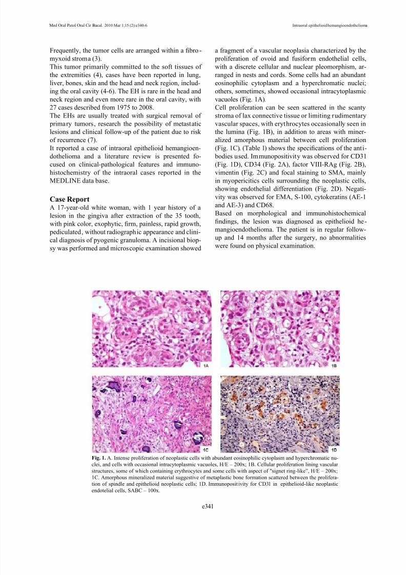
Case ReportA 17-year-old white woman, with 1 year history of a
lesion in the gingiva after extraction of the 35 tooth,
with pink color, exophytic, rm, painless, rapid growth,
pediculated, without radiographic appearance and clini-
cal diagnosis of pyogenic granuloma. A incisional biop-
sy was performed and microscopic examination showed
a fragment of a vascular neoplasia characterized by the
proliferation of ovoid and fusiform endothelial cells,
with a discrete cellular and nuclear pleomorphism, ar-
ranged in nests and cords. Some cells had an abundant
eosinophilic cytoplasm and a hyperchromatic nuclei;
others, sometimes, showed occasional intracytoplasmic
vacuoles (Fig. 1A).Cell proliferation can be seen scattered in the scanty
stroma of lax connective tissue or limiting rudimentary
vascular spaces, with erythrocytes occasionally seen in
the lumina (Fig. 1B), in addition to areas with miner-
alized amorphous material between cell proliferation
(Fig. 1C). (Table 1) shows the specications of the anti-
bodies used. Immunopositivity was observed for CD31
(Fig. 1D), CD34 (Fig. 2A), factor VIII-RAg (Fig. 2B),
vimentin (Fig. 2C) and focal staining to SMA, mainly
in myopericitics cells surrounding the neoplastic cells,
showing endothelial differentiation (Fig. 2D). Negati-
vity was observed for EMA, S-100, cytokeratins (AE-1
and AE-3) and CD68.
Based on morphological and immunohistochemical
ndings, the lesion was diagnosed as epithelioid he-
mangioendothelioma. The patient is in regular follow-
up and 14 months after the surgery, no abnormalities
were found on physical examination.
Fig. 1. A. Intense proliferation of neoplastic cells with abundant eosinophilic cytoplasm and hyperchromatic nu-
clei, and cells with occasional intracytoplasmic vacuoles, H/E – 200x; 1B. Cellular proliferation lining vascular
structures, some of which containing erythrocytes and some cells with aspect of "signet ring-like”, H/E – 200x;
1C. Amorphous mineralized material suggestive of metaplastic bone formation scattered between the prolifera-
tion of spindle and epithelioid neoplastic cells; 1D. Immunoposit ivity for CD31 in epithelioid-like neoplastic
endotelial cells, SABC – 100x.

8/13/2019 Intraoral Epithelioid Hemangioendothelioma a Case Report
http://slidepdf.com/reader/full/intraoral-epithelioid-hemangioendothelioma-a-case-report 3/7
e342
Med Oral Patol Oral Cir Bucal. 2010 Mar 1;15 (2):e340-6. Intraoral epithelioid hemangioendothelioma
ANTIBODY CLONE SOURCE DILUITION ANTIGENIC RETRIEVALINCUBATION
TIME
CD31 JC/70A Dako, CA, USA 1:40 Citrato pH 6.0, Pascal Overnight
CD34QBEnd-
10Dako, CA, USA 1.50 Tris EDTA pH 9, Pascal Overnight
Factor VIII F8/86 Dako, CA, USA 1:400 Citrato pH 6.0, Pascal 30 min
Vimentin V9 Dako, CA, USA 1:50 Citrato pH 6.0, Pascal 30 min
SMA asm-1 Novocastra, London, UK 1:40 Citrato pH 6.0, Pascal 60 min
EMA E29 Dako, CA, USA 1:40 Sin tratamiento 60 min
S-100 S-100 Dako, CA, USA 1:100 Citrato pH 6.0, Pascal 30 min
Cytokeratins
(AE-1/EA-3)
A E - 1 ,
EA-3Dako, CA, USA 1:500 Citrato pH 6.0, Pascal 120 min
CD68 KP1 Santa Cruz, CA, USA 1:50 Citrato pH 6.0, Pascal Overnight
Table 1. Specications of the antibodies used.
Fig. 2. A. Immunopositivity for CD34 in epiteliod-like neoplastic endotelial cells, SABC-100x; 2B. Cytoplasmic immunostaining for FVIII-
RAg, SABC-400x; 2C. Immunostaining for Vimentin, SABC-400x; 2D. Immunostaining for SMA, SABC-400x.

8/13/2019 Intraoral Epithelioid Hemangioendothelioma a Case Report
http://slidepdf.com/reader/full/intraoral-epithelioid-hemangioendothelioma-a-case-report 4/7
e343
Med Oral Patol Oral Cir Bucal. 2010 Mar 1;15 (2):e340-6. Intraoral epithelioid hemangioendothelioma
DiscussionThe analysis of cases of intraoral epithelioid heman-
gioma reported in the literature is a relatively difcult
task, once the lesion from years ago has received vari-
ous names, such as “hemangioendothelioma”, “malig-
nant hemangioendothelioma” and “histiocytoid heman-
gioma” (7-9), thus, the reference point selected for theliterature review presented here is the work published in
1975 by Wesley et al. (8), entitled “Primary malignant
hemangioendothelioma of the gingiva”, until the most
recent reported case by Mohtasham et al. (10).
(Table 2) shows the epidemiological data of the EHs re-
ported in the literature in journals indexed in PubMed,
including the case reported here. Most cases were clini-
cally diagnosed as benign entities including pyogenic
granuloma, broma, peripheral giant cell granuloma,
peripheral ossifying broma, inammatory brous hy-
perplasia and necrotizing ulcerative gingivitis (11). The
case reported here is consistent with the literature, once
the hypothesis of clinical diagnosis was pyogenic gran-
uloma. The fact that the majority of EHs are clinically
diagnosed as benign lesions has a great relevance, since
it is a neoplasm with malignant biological behavior, in-
termediate between the hemangioma and conventional
angiosarcoma, with reported cases displaying aggres-
sive behavior and multiples recurrences (10,12).
It was noted that only 2 cases (7.4%) reported pain as-
sociated with the lesion, while the majority had no in-
formation about the symptoms, which leads us to infer
that they were asymptomatic or that this was not just
informed by authors (Table 2). The case reported here
was asymptomatic.The analysis of radiographic features showed that
25.9% of the lesions were associated with resorption or
destruction of underlying bone (Table 2), highlighting
the need for careful radiographic examination of the le-
sions to investigate possible alterations of the bone (11).
It was not found any radiographic changes in the case
reported here.
The malignant potential of the EHs is not well estab-
lished, however, based on cases reported in the litera-
ture, these neoplasms exhibit a biological behavior in-
termediate between hemangioma and angiosarcoma, as
well as potential for recurrence and metastasis (3).Predictions regarding the biological behavior of EH in
the oral region are not entirely possible due to the rar-
ity of the lesion, the lack of agreement relative to ter-
minology and criteria for diagnosis of EH and because
of differences in the biologic behavior of these tumors
relative to both their anatomic sites and with regard to
age of occurrence (6).
Furthermore, EH doesn´t shows, histopathologically,
consistent criteria for predicting their biologic behavior,
however, it is reported that the presence of a increased
number of mitotic gures and cellular atypia, spindle
tumor cells, metaplastic bone formation, in addition to
areas of focal necrosis can be morphological signs of le-
sions with more aggressive phenotype and consequently
more reserved prognosis, with recurrences and regional
and/or distant metastasis (1,6,11). The case reported
here showed no relevant cellular atypia or mitotic activ-
ity, which leads us to suggest that probably could be alesion with more indolent behavior.
The review of the literature showed that the most preva-
lent intraoral site for EH was the tongue (7-25.9%), fol-
lowed by the mandibular gingiva (6-22.2%) and maxil-
lary gingiva (5–18.5%). The size of lesions ranged from
0.2 cm to 7.0 cm, with a mean of 1.7 cm. In the eight
reported cases with recurrence (29.6%) (Table 2), it was
noted that, in some of them, were multiple recurrences
and the recurrent lesions showed a more aggressive ap-
pearance of malignancy. These recurrences were more
frequent in the maxillary gingiva and buccal mucosa,
both with 25.9%, followed by the lower lip, tongue, jaw
and maxilla, with 12.9%, respectively. There are no re-
ports of recurrence in mandibular gingiva and this site
is the same of the case here reported.
The case reported here agrees with the literature (3)
regarding the histomorphological features. The lesion
exhibit cells with prominent cytoplasmic vacuoles,
which represent primitive-appearing vascular channels;
it is reported that these vacuoles are negative for mu-
cin and erythrocytes occasionally seen in the lumina.
The EHs can exhibit mineralized material, reported by
some authors as metaplastic bone formation, in addition
to numerous osteoclast-like giant cells. It reported that
the metaplastic bone formation with signicant cellularatypia, mitotic activity greater than one per 10 high-
power elds, an increased proportion of spindle tumor
cells and focal necrosis represent histopathological fac-
tors associated with a more aggressive behavior of the
lesion (1,13,14).
Due to the predominant morphological characteristics,
the EHs can be confused with other lesions including
hemangioma to squamous cell carcinoma, therefore,
emphasizes the importance of immunohistochemical
analysis to establish the denitive diagnosis. In this
context, the review of published cases veried that the
majority of intraoral EHs lesions were immunoreactivefor CD34, CD31, factor VIII-RAg and vimentin, these
markers characterize the epithelioid endothelial origin
of this entity (Table 3). It is reported that the immunore-
activity for factor VIII-RAg indicates the cytoplasmatic
luminal features and the epithelial cellular neoplastic
nature (11). Furthermore, we agree with the literature
that due to the morphological appearance of epithelioid
tumoral cells of the EHs an the fact that many lesions
are reactive for cytokeratin (AE-1/AE-3), requires a lot
of attention from the pathologist, to avoid the risk of
misdiagnosed as squamous cell carcinoma (13).

8/13/2019 Intraoral Epithelioid Hemangioendothelioma a Case Report
http://slidepdf.com/reader/full/intraoral-epithelioid-hemangioendothelioma-a-case-report 5/7
e344
Med Oral Patol Oral Cir Bucal. 2010 Mar 1;15 (2):e340-6. Intraoral epithelioid hemangioendothelioma
A variable immunohistochemical expression to SMA for
intraoral EHs has been observed previously (12,15,16)
(Table 3). In the case reported here the immunoreacti-
vity for this marker was focally, as evidenced at times
immunoreactivity in myopericitics cells surrounding
the neoplastic cells, showing the endothelial differen-
tiation of these. The nuclear transcription factor Fli-1,
a specic marker for Ewing’s sarcoma and primitive
neuroectodermal tumor, has been sensitive to vascular
lesions, including EH (15).
Considering the intermediate malignant potential of the
EH, wide local excision was the mode of choice in most
cases reported in the literature (6,11,15,17), it was observed
that conservative procedures such as curettage may favor
the recurrence of the lesions (18). Under this, after esta-
blished a diagnosis of EH the correct treatment should al-
AUTHOR (YEAR) AGE SEX LOCALIZATIONCLINICAL AND RADIOGRAPHIC
HISTORYFOLLOW-UP
1 Wesley et al (1975) 18 F Mandibular gingivaReddish erosive lesion, (34 to 36), bone
resorption2 years SFL
2 Ellis, Kratochvil (1986) 13 F Maxillary gingiva Swelling, pink, tooth mobility, 4 years 6 years SFL
3 Ellis, Kratochvil (1986) 4 F Mandibular gingiva Tooth mobility, bone resorption NI
4 Moran et al (1987) 25 F Palate Swelling, 1.0 cm, 1 year 21 months SFL
5 de Araujo et al (1987) 4 M Mandibular gingivaSwelling, ulcerat ion, tooth mobility, 9
months NI
6 Marrogi et al (1991) 45 M Maxillary gingiva Erythematous lesion, 1.5 cm 3,6 months Rec
7 Marrogi et al (1991) 36 F Tongue Painful nodules, 0.2 cm, 2 months 17 months SFL
8 Flaitz et al (1995) 7 F Mandibular gingivaReddish swelling, 1.5 cm, tooth mobility,
bone destruction52 months SFL
9 Hamakawa et al (1999) 76 FMandibular anterior re-
gion
Submucous swelling, soft, 4.5 cm, bone
destruction6 years SFL
10 Orsini et al (2001) 18 F Buccal mucosaAsymptomatic swelling, 1.5 cm, 7 mon-
ths
9 months Rec
11 Ramer et al (2001) 32 M Maxilla Swelling, 3.5 cm 6 months Rec
12Molina Palma et al
(2002)65 F Tongue Swelling ,0.5 cm, 2 months 21 months SFL
13 Machalka et al (2003) 65 M JawaSwelling at the anterior region of jaw,
tooth mobility4.8 years Rec
14 Anderson et al (2003) 18 F Lower lip Asymptomatic swelling, 6 months 4 months Rec
15 Chi et al (2005) 28 F Maxillary gingiva Purple swelling, 0.6 cm 8 months SFL
16 Chi et al (2005) 23 F Jaw 2.0 cm, bone destruction NI
17 Sun et al (2007) 12 M Maxillary gingivaUlcerated swelling, 3.0 cm, 3 months,
bone destruction, tooth mobility6 months SFL
18 Sun et al (2007) 53 M Buccal mucosa Swelling, 1.5, 6 months 9 months Rec
19 Sun et al (2007) 17 M Tongue Soft swelling, 0.5 cm, 2 months 18 months SFL
20 Sun et al (2007) 52 F Upper lip Purple swelling, 2.0 cm, 1 year 3 years SFL
21 Sun et al (2007) 21 M Tongue Reddish swelling, 0.5 cm, 2 months 2 years SFL
22 Sun et al (2007) 34 M Tongue Swelling, 1.0 cm, 4 months 6 years SFL
23 Sun et al (2007) 11 M Mandibular gingivaPainful swelling, 2.0 cm, 1 month, bone
destruction, tooth mobility8 years SFL
24 Sun et al (2007) 46 M Tongue Reddish swelling, 1.2 cm 4 months Rec
25 Sun et al (2007) 6 M Floor of mouth and tongue Reddish swelling, 7.0 cm, 6 months 2 years SL
26Mohtasham et al
(2008)9 M Maxillary gingiva
Ulcerated reddish swelling, asymptom-
atic, 1.0 cm, 6 months1 year, Rec
27Gordón-Núñez et al
(2008)
17 F Mandibular gingiva Swelling, pink, 2,0 cm,1 year 9 months, SFL
Table 2. Clinical and radiographic features of the intraoral EH cases reported in the literature.
M: male; F: female; NI: no information; SFL: survival free of lesion; SL: survival with lesion; Rec: recurrence.

8/13/2019 Intraoral Epithelioid Hemangioendothelioma a Case Report
http://slidepdf.com/reader/full/intraoral-epithelioid-hemangioendothelioma-a-case-report 6/7
e345
Med Oral Patol Oral Cir Bucal. 2010 Mar 1;15 (2):e340-6. Intraoral epithelioid hemangioendothelioma
ways be the total surgical removal of the lesion and follow-
up, to avoid the risk of permanence of neoplastic material
and consequently recurrence. Therapeutic modalities as
chemo and / or radiotherapy have been suggested for the
treatment of EH, but without satisfactory results.
Among the 27 cases of intraoral EHs reported in the
literature, including the case of this paper was observed
that 23 cases (85.2%) had clinical follow-up informa-
tion. Most of the patients (15-55.6%) survive free of
evidence of lesion. On the other hand, 8 lesions (29.6%)
recurred locally and fortunately was not registered a lo-
cal or distant metastasis, nor death associated with le-
sions (Table 2). These data reinforce the less aggressive
nature of the intraoral EHs when compared with those
who commit other regions of the body.
The differential diagnosis of EH with another enti-
ties that occurs at the oral cavity may be assisted by
certain clinical, histopathological, biochemical and/
or immunohistochemistry. In this context, relates that
epithelioid hemangioma, for example, is an lesion that
usually exhibits cells with voluminous and eosinophilic
cytoplasm, may be associated with lymphadenopathy
and eosinophilia with elevated IgE and unlike the HE,
epithelioid hemangiomas are located supercially, mul-
tilobular, not exhibit endothelial cells, cells with intra-
cytoplasmic vacuoles, atypia, mitosis and necrosis (11).
The association of epithelioid hemangioma with lym-
phadenopathy, peripheral eosinophilia and increased
IgE are factors that help in the differential diagnosis
(13).
AUTHOR
(year)CD34 CD31 CD68
Factor
VIII-RAgVIMENTIN SMA S-100 EMA
AE-1/
AE-3Fli-1 VEGF
1Ellis, Kratochvil
(1986)+
2Ellis, Kratochvil
(1986)
+
3Moran et al
(1987)+
4Flaitz et al
(1995)+
5Hamakawa et al
(1999)+ + + - - -
6Orsini et al
(2001)+
7Ramer et al
(2001)+ +
8Molina Palma et
al (2002)+ +
9Machalka et al
(2003)+ + - + + + - -
10 Chi et al (2005) - + - - - - - +
11 Chi et al (2005) + + - + - - - +
12 Sun et al (2007) + + + + - - +
13 Sun et al (2007) + + + + - - +
14 Sun et al (2007) + + + + - - +
15 Sun et al (2007) + + + + - - -
16 Sun et al (2007) + + + + - - -
17 Sun et al (2007) + + + + - - -
18 Sun et al (2007) + + + + - - -
19 Sun et al (2007) + + + + - - -
20 Sun et al (2007) + + - - -
21Mohtasham et al
(2008)+ +
22Gordón-Núñez
et al (2008)+ + - + + + - - -
Table 3. Immunohistochemical analisis of the intraoral EH cases reported in the literature.
(+) Immunopositivity, (-) Immunonegativity.

8/13/2019 Intraoral Epithelioid Hemangioendothelioma a Case Report
http://slidepdf.com/reader/full/intraoral-epithelioid-hemangioendothelioma-a-case-report 7/7


![Case 9298 Epithelioid hemangioendothelioma of the femur · 2017. 2. 4. · Anastomosis, Surgical [E04.035] Surgical union or shunt between ducts, tubes or vessels. It may be end-to-end,](https://static.fdocuments.in/doc/165x107/6116a7eb3ddb85207d316366/case-9298-epithelioid-hemangioendothelioma-of-the-femur-2017-2-4-anastomosis.jpg)