Intraoperative Patient Transfer within the O.R. Environment is not a Clinical Concern White Paper
2
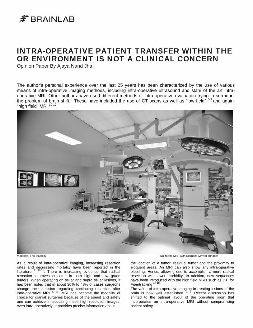
INTRA-OPERATIVE PATIENT TRANSFER WITHIN THE OR ENVIRONMENT IS NOT A CLINICAL CONCERN Opinion Paper By Ajaya Nand Jha The author’s personal experience over the last 25 years has been characterized by the use of various means of intra-operative imaging methods, including intra-operative ultrasound and state of the art intra- operative MRI. Other authors have used different methods of intra-operative evaluation trying to surmount the problem of brain shift. These have included the use of CT scans as well as “low field” 6-9 and again, “high field” MRI 10-12 . Medanta, The Medicity Two-room iMRI with Siemens Miyabi concept As a result of intra-operative imaging, increasing resection rates and decreasing mortality have been reported in the literature 1, 13-16 . There is increasing evidence that radical resection improves outcome in both high and low grade tumors. When operating on sellar and supra sellar lesions, it has been noted that in about 30% to 48% of cases surgeons change their decision regarding continuing resection after intra-operative MRI 5, 11 . MRI has become the modality of choice for cranial surgeries because of the speed and safety one can achieve in acquiring these high resolution images, even intra-operatively. It provides precise information about the location of a tumor, residual tumor and the proximity to eloquent areas. An MRI can also show any intra-operative bleeding. Hence, allowing one to accomplish a more radical resection with lower morbidity. In addition, new sequences have been introduced with the high field MRIs such as DTI for Fibertracking 17-18 . The value of intra-operative imaging in treating lesions of the brain is now well established 1, 2 . Recent discussion has shifted to the optimal layout of the operating room that incorporates an intra-operative MRI without compromising patient safety.
-
Upload
brainlab -
Category
Health & Medicine
-
view
156 -
download
1
Transcript of Intraoperative Patient Transfer within the O.R. Environment is not a Clinical Concern White Paper
INTRA-OPERATIVE PATIENT TRANSFER WITHIN THE OR ENVIRONMENT IS NOT A CLINICAL CONCERN Opinion Paper By Ajaya Nand Jha The author’s personal experience over the last 25 years has been characterized by the use of various means of intra-operative imaging methods, including intra-operative ultrasound and state of the art intra-operative MRI. Other authors have used different methods of intra-operative evaluation trying to surmount the problem of brain shift. These have included the use of CT scans as well as “low field” 6-9 and again, “high field” MRI 10-12.
Medanta, The Medicity Two-room iMRI with Siemens Miyabi concept As a result of intra-operative imaging, increasing resection rates and decreasing mortality have been reported in the literature 1, 13-16. There is increasing evidence that radical resection improves outcome in both high and low grade tumors. When operating on sellar and supra sellar lesions, it has been noted that in about 30% to 48% of cases surgeons change their decision regarding continuing resection after intra-operative MRI 5, 11. MRI has become the modality of choice for cranial surgeries because of the speed and safety one can achieve in acquiring these high resolution images, even intra-operatively. It provides precise information about
the location of a tumor, residual tumor and the proximity to eloquent areas. An MRI can also show any intra-operative bleeding. Hence, allowing one to accomplish a more radical resection with lower morbidity. In addition, new sequences have been introduced with the high field MRIs such as DTI for Fibertracking 17-18. The value of intra-operative imaging in treating lesions of the brain is now well established 1, 2. Recent discussion has shifted to the optimal layout of the operating room that incorporates an intra-operative MRI without compromising patient safety.
References [1] Senft C.et al., Lancet Oncol 12(11), 997, 2011 [2] Nimsky C. et al., Lancet Oncol 12(11), 982, 2011 [3] Black PM. et al., Neurosurg Clin N Am 16(1), 135, 2005 [4] Nimsky C. et al., Acta Neurochir Suppl 88, 21, 2003 [5] Vitaz TW. et al., Clin Neurol Neurosurg 113(9), 731, 2011 [6] Makary M. et al., J Magn Resin Imaging 34(5), 1022, 2011 [7] Seifert V. et al., Acta Neurochir Suppl 109, 35, 2011 [8] Senft C. et al., Clin Neurol Neurosurg 112(3), 237, 2010 [9] Pandin P. et al., Anesth Analg 105(3), 886, 2007
[10] Avula S. et al., Pediatr Radiol 2011 [11] Netuka D. et al., Acta Neurochir Suppl 109, 157, 2011 [12] Pamir MN. et al., Expert Rev Anticancer Ther 9(11), 1537, 2009 [13] Kuhnt D. et al., Neuro Oncol 13(12), 1339, 2011 [14] Kubben PL. et al., Lancet Oncol 12(11), 1062, 2011 [15] Gerganov VM. et al., Acta Neurochir (Wien) 153(3), 479, 2011 [16] Lenaburg HJ. et al., Technol Cancer Res Treat 8(2), 159, 2009 [17] Zhao Y. et al., J Clin Neurosci 19(2), 255, 2012 [18] Ulmer S. et al., Neuroimage 49(3), 2158, 2010
Europe | +49 89 99 1568 0 | [email protected] North America | +1 800 784 7700 | [email protected] South America | +55 11 3355 3370 | [email protected] IOR_FL_EN_INTRA-OPERATIVE PATIENT TRANSFER_OPINION PAPER_0812_Rev.1
Asia Pacific | +852 2417 1881 | [email protected] Japan | +81 3 3769 6900| [email protected]
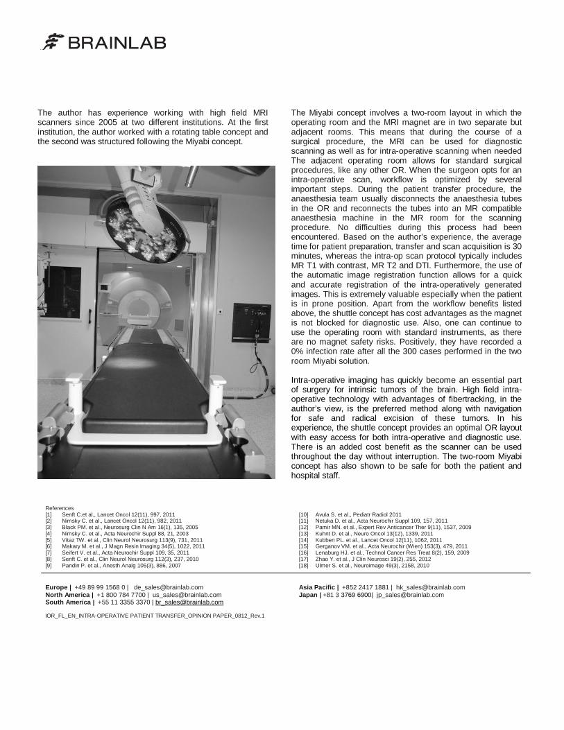
The author has experience working with high field MRI scanners since 2005 at two different institutions. At the first institution, the author worked with a rotating table concept and the second was structured following the Miyabi concept.
The Miyabi concept involves a two-room layout in which the operating room and the MRI magnet are in two separate but adjacent rooms. This means that during the course of a surgical procedure, the MRI can be used for diagnostic scanning as well as for intra-operative scanning when needed The adjacent operating room allows for standard surgical procedures, like any other OR. When the surgeon opts for an intra-operative scan, workflow is optimized by several important steps. During the patient transfer procedure, the anaesthesia team usually disconnects the anaesthesia tubes in the OR and reconnects the tubes into an MR compatible anaesthesia machine in the MR room for the scanning procedure. No difficulties during this process had been encountered. Based on the author’s experience, the average time for patient preparation, transfer and scan acquisition is 30 minutes, whereas the intra-op scan protocol typically includes MR T1 with contrast, MR T2 and DTI. Furthermore, the use of the automatic image registration function allows for a quick and accurate registration of the intra-operatively generated images. This is extremely valuable especially when the patient is in prone position. Apart from the workflow benefits listed above, the shuttle concept has cost advantages as the magnet is not blocked for diagnostic use. Also, one can continue to use the operating room with standard instruments, as there are no magnet safety risks. Positively, they have recorded a 0% infection rate after all the 300 cases performed in the two room Miyabi solution. Intra-operative imaging has quickly become an essential part of surgery for intrinsic tumors of the brain. High field intra-operative technology with advantages of fibertracking, in the author’s view, is the preferred method along with navigation for safe and radical excision of these tumors. In his experience, the shuttle concept provides an optimal OR layout with easy access for both intra-operative and diagnostic use. There is an added cost benefit as the scanner can be used throughout the day without interruption. The two-room Miyabi concept has also shown to be safe for both the patient and hospital staff.