Intraobserver and Interobserver Reliability of the Classification of Thoracic Adolescent Idiopathic...
18
Intraobserver and Interobserver Reliability of the Classification of Thoracic Adolescent Idiopathic Scoliosis*† by LAWRENCE G. LENKE, RANDAL R. BETZ, KEITH H. BRIDWELL, DAVID H. CLEMENTS, JÜRGEN HARMS, THOMAS G. LOWE, and HARRY L. SHUFFLEBARGER J Bone Joint Surg Am Volume 80(8):1097-1106 August 1, 1998 ©1998 by The Journal of Bone and Joint Surgery, Inc.
-
Upload
philomena-cook -
Category
Documents
-
view
221 -
download
0
Transcript of Intraobserver and Interobserver Reliability of the Classification of Thoracic Adolescent Idiopathic...

Intraobserver and Interobserver Reliability of the Classification of Thoracic Adolescent Idiopathic
Scoliosis*†
by LAWRENCE G. LENKE, RANDAL R. BETZ, KEITH H. BRIDWELL, DAVID H. CLEMENTS, JÜRGEN HARMS, THOMAS G. LOWE, and HARRY L.
SHUFFLEBARGER
J Bone Joint Surg AmVolume 80(8):1097-1106
August 1, 1998
©1998 by The Journal of Bone and Joint Surgery, Inc.

Figs. 1-A, 1-B, and 1-C: Radiographs of a fifteen-year and one-month-old girl.
LAWRENCE G. LENKE et al. J Bone Joint Surg Am 1998;80:1097-1106
©1998 by The Journal of Bone and Joint Surgery, Inc.

Fig. 1-B: Left-side-bending radiograph, made with the patient supine, showing correction of the lumbar curve to 50 degrees.
LAWRENCE G. LENKE et al. J Bone Joint Surg Am 1998;80:1097-1106
©1998 by The Journal of Bone and Joint Surgery, Inc.
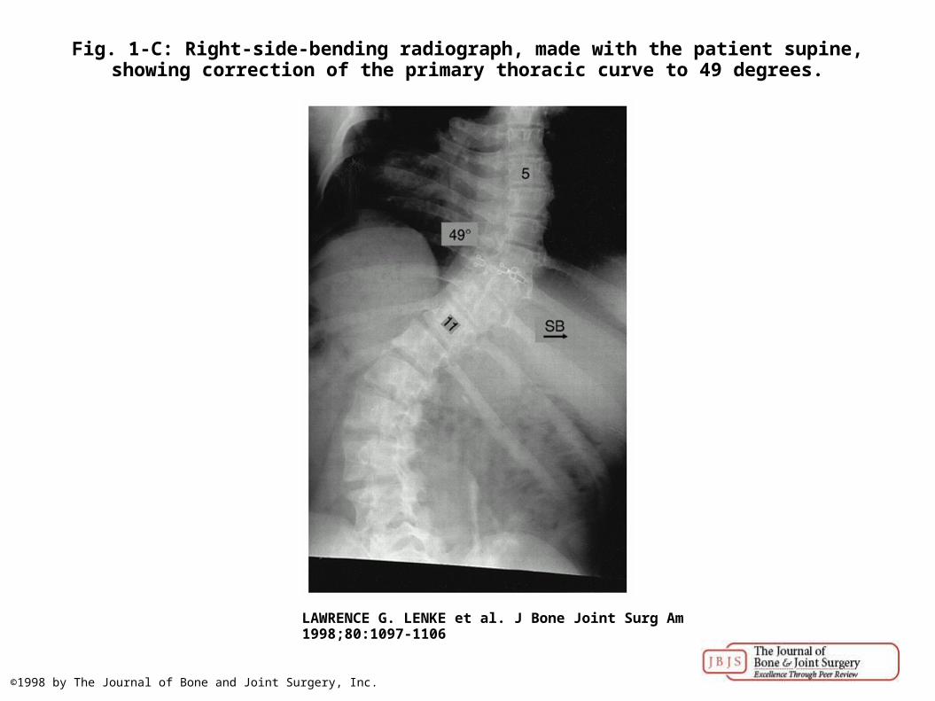
Fig. 1-C: Right-side-bending radiograph, made with the patient supine, showing correction of the primary thoracic curve to 49 degrees.
LAWRENCE G. LENKE et al. J Bone Joint Surg Am 1998;80:1097-1106
©1998 by The Journal of Bone and Joint Surgery, Inc.

Figs. 2-A, 2-B, and 2-C: Radiographs of a seventeen-year and nine-month-old girl.
LAWRENCE G. LENKE et al. J Bone Joint Surg Am 1998;80:1097-1106
©1998 by The Journal of Bone and Joint Surgery, Inc.

Fig. 2-B: Left-side-bending radiograph, made with the patient supine, showing correction of the cephalad thoracic curve to 10 degrees and correction of the lumbar curve to 13 degrees.
LAWRENCE G. LENKE et al. J Bone Joint Surg Am 1998;80:1097-1106
©1998 by The Journal of Bone and Joint Surgery, Inc.

Fig. 2-C: Right-side-bending radiograph, made with the patient supine, showing correction of the primary thoracic curve to 35 degrees.
LAWRENCE G. LENKE et al. J Bone Joint Surg Am 1998;80:1097-1106
©1998 by The Journal of Bone and Joint Surgery, Inc.

Fig. 3 Radiograph of a fourteen-year and five-month-old girl, showing a scoliotic curve of 30 degrees from the first through the fifth thoracic vertebra, 55 degrees from the fifth through the
twelfth thoracic vertebra, and 40 degrees from the twelfth thorac...
LAWRENCE G. LENKE et al. J Bone Joint Surg Am 1998;80:1097-1106
©1998 by The Journal of Bone and Joint Surgery, Inc.

Figs. 4-A through 4-D: Radiographs of a fourteen-year and three-month-old boy.
LAWRENCE G. LENKE et al. J Bone Joint Surg Am 1998;80:1097-1106
©1998 by The Journal of Bone and Joint Surgery, Inc.

Fig. 4-B: Left-side-bending radiograph, made with the patient supine, showing correction of the cephalad thoracic curve to 23 degrees and correction of the lumbar curve to 17 degrees.
LAWRENCE G. LENKE et al. J Bone Joint Surg Am 1998;80:1097-1106
©1998 by The Journal of Bone and Joint Surgery, Inc.

Fig. 4-C: Right-side-bending radiograph, made with the patient supine, showing correction of the primary thoracic curve to 29 degrees.
LAWRENCE G. LENKE et al. J Bone Joint Surg Am 1998;80:1097-1106
©1998 by The Journal of Bone and Joint Surgery, Inc.
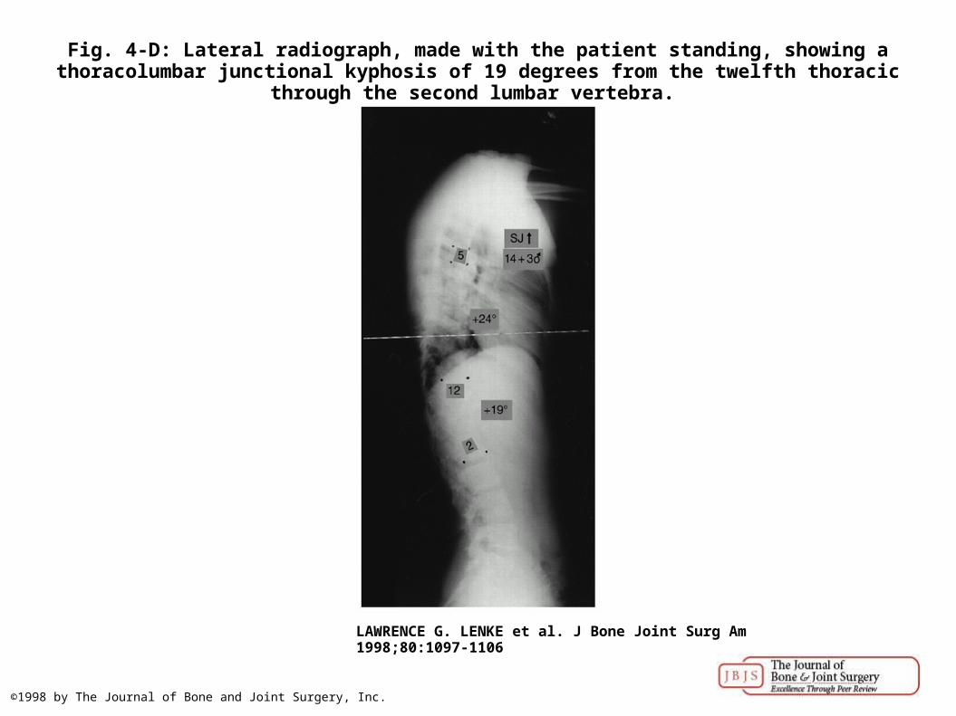
Fig. 4-D: Lateral radiograph, made with the patient standing, showing a thoracolumbar junctional kyphosis of 19 degrees from the twelfth thoracic through the second lumbar vertebra.
LAWRENCE G. LENKE et al. J Bone Joint Surg Am 1998;80:1097-1106
©1998 by The Journal of Bone and Joint Surgery, Inc.

Figs. 5-A, 5-B, and 5-C: Radiographs of a twelve-year and nine-month-old girl.
LAWRENCE G. LENKE et al. J Bone Joint Surg Am 1998;80:1097-1106
©1998 by The Journal of Bone and Joint Surgery, Inc.

Fig. 5-B: Left-side-bending radiograph, made with the patient supine, showing correction of the cephalad thoracic curve to 31 degrees and correction of the lumbar curve to 20 degrees.
LAWRENCE G. LENKE et al. J Bone Joint Surg Am 1998;80:1097-1106
©1998 by The Journal of Bone and Joint Surgery, Inc.

Fig. 5-C: Right-side-bending radiograph, made with the patient supine, showing correction of the primary thoracic curve to 61 degrees.
LAWRENCE G. LENKE et al. J Bone Joint Surg Am 1998;80:1097-1106
©1998 by The Journal of Bone and Joint Surgery, Inc.

Figs. 6-A, 6-B, and 6-C: Radiographs of a fifteen-year and nine-month-old girl.
LAWRENCE G. LENKE et al. J Bone Joint Surg Am 1998;80:1097-1106
©1998 by The Journal of Bone and Joint Surgery, Inc.

Fig. 6-B: Left-side-bending radiograph, made with the patient supine, showing correction of the cephalad thoracic curve to 41 degrees.
LAWRENCE G. LENKE et al. J Bone Joint Surg Am 1998;80:1097-1106
©1998 by The Journal of Bone and Joint Surgery, Inc.
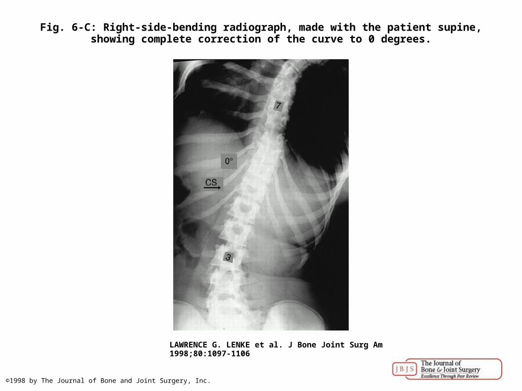
Fig. 6-C: Right-side-bending radiograph, made with the patient supine, showing complete correction of the curve to 0 degrees.
LAWRENCE G. LENKE et al. J Bone Joint Surg Am 1998;80:1097-1106
©1998 by The Journal of Bone and Joint Surgery, Inc.


![UseofFDG-PETinRadiationTreatmentPlanningfor ThoracicCancers · 2017. 11. 15. · spectively evaluated the role of FDG-PET in interobserver variability in 30 NSCLC patients [28]. Three](https://static.fdocuments.in/doc/165x107/60a84c1a0d1be65fbd244ca6/useoffdg-petinradiationtreatmentplanningfor-thoraciccancers-2017-11-15-spectively.jpg)