Intranasal insulin improves cognition and modulates...
9
Intranasal insulin improves cognition and modulates -amyloid in early AD M.A. Reger, PhD G.S. Watson, PhD P.S. Green, PhD C.W. Wilkinson, PhD L.D. Baker, PhD B. Cholerton, PhD M.A. Fishel, MD S.R. Plymate, MD J.C.S. Breitner, MD, MPH W. DeGroodt, MS P. Mehta, PhD S. Craft, PhD ABSTRACT Background: Reduced brain insulin signaling and low CSF-to-plasma insulin ratios have been ob- served in patients with Alzheimer disease (AD). Furthermore, intracerebroventricular or IV insulin administration improve memory, alter evoked potentials, and modulate neurotransmitters, possi- bly by augmenting low brain levels. After intranasal administration, insulin-like peptides follow extracellular pathways to the brain within 15 minutes. Objective: We tested the hypothesis that daily intranasal insulin treatment would facilitate cogni- tion in patients with early AD or its prodrome, amnestic mild cognitive impairment (MCI). The proportion of verbal information retained after a delay period was the planned primary outcome measure. Secondary outcome measures included attention, caregiver rating of functional status, and plasma levels of insulin, glucose, -amyloid, and cortisol. Methods: Twenty-five participants were randomly assigned to receive either placebo (n 12) or 20 IU BID intranasal insulin treatment (n 13) using an electronic atomizer, and 24 participants completed the study. Participants, caregivers, and all clinical evaluators were blinded to treat- ment assignment. Cognitive measures and blood were obtained at baseline and after 21 days of treatment. Results: Fasting plasma glucose and insulin were unchanged with treatment. The insulin-treated group retained more verbal information after a delay compared with the placebo-assigned group (p 0.0374). Insulin-treated subjects also showed improved attention (p 0.0108) and func- tional status (p 0.0410). Insulin treatment raised fasting plasma concentrations of the short form of the -amyloid peptide (A40; p 0.0471) without affecting the longer isoform (A42), resulting in an increased A40/42 ratio (p 0.0207). Conclusions: The results of this pilot study support further investigation of the benefits of intrana- sal insulin for patients with Alzheimer disease, and suggest that intranasal peptide administration may be a novel approach to the treatment of neurodegenerative disorders. Neurology ® 2008;70:1–1 GLOSSARY AD Alzheimer disease; APP amyloid precursor protein; BSA bovine serum albumin; CBG cortisol binding globulin; DSRS Dementia Severity Rating Scale; MCI mild cognitive impairment; PBS phosphate buffered saline; RIAs radioimmunoassays. Insulin, a highly conserved peptide whose function was once narrowly defined as the promotion of glucose turnover in skeletal muscle, has now emerged as a key neuromodu- lator in the CNS. Evidence has been established for its participation in neuronal mainte- nance, energy metabolism, neurogenesis, and neurotransmitter regulation. 1 Insulin also affects functional processes such as neuronal firing and long-term potentiation that un- e-Pub ahead of print at www.neurology.org. From the Departments of Psychiatry and Behavioral Science (M.A.R., G.S.W., C.W.W., L.D.B., B.C., J.C.S.B., S.C.), Medicine (P.S.G., S.R.P.), and Neurology (M.A.F.), University of Washington School of Medicine, Seattle; Geriatric Research, Education, and Clinical Center (G.S.W., C.W.W., L.D.B., B.C., M.A.F., S.R.P., J.C.S.B., S.C.), Research and Development (P.S.G.), Veterans Affairs Puget Sound Health Care System, Seattle; Kurve Technology, Inc. (W.D.), Bothell, WA; and Department of Immunology (P.M.), Institute for Basic Research in Developmental Disabilities, Staten Island, NY. Supported by the Department of Veterans Affairs, the Institute for the Study on Aging, and National Institute on Aging RO1 AG027415. Disclosure: W.D. is a founding member of Kurve Technology, the maker and patent holder of the electronic atomizer used in this study. W.D. holds equity interest in excess of $10,000 in Kurve Technology. The remaining authors have nothing to disclose. Address correspondence and reprint requests to Dr. Suzanne Craft, GRECC S-182, VAPSHCS, 1660 S. Columbian Way, Seattle, WA 98108 [email protected] Copyright © 2008 by AAN Enterprises, Inc. 1 Published Ahead of Print on October 17, 2007 as 10.1212/01.WNL.0000265401.62434.36
Transcript of Intranasal insulin improves cognition and modulates...
Intranasal insulin improves cognitionand modulates �-amyloid in early AD
M.A. Reger, PhDG.S. Watson, PhDP.S. Green, PhDC.W. Wilkinson, PhDL.D. Baker, PhDB. Cholerton, PhDM.A. Fishel, MDS.R. Plymate, MDJ.C.S. Breitner, MD,MPH
W. DeGroodt, MSP. Mehta, PhDS. Craft, PhD
ABSTRACT
Background: Reduced brain insulin signaling and low CSF-to-plasma insulin ratios have been ob-served in patients with Alzheimer disease (AD). Furthermore, intracerebroventricular or IV insulinadministration improve memory, alter evoked potentials, and modulate neurotransmitters, possi-bly by augmenting low brain levels. After intranasal administration, insulin-like peptides followextracellular pathways to the brain within 15 minutes.
Objective: We tested the hypothesis that daily intranasal insulin treatment would facilitate cogni-tion in patients with early AD or its prodrome, amnestic mild cognitive impairment (MCI). Theproportion of verbal information retained after a delay period was the planned primary outcomemeasure. Secondary outcome measures included attention, caregiver rating of functional status,and plasma levels of insulin, glucose, �-amyloid, and cortisol.
Methods: Twenty-five participants were randomly assigned to receive either placebo (n � 12) or20 IU BID intranasal insulin treatment (n � 13) using an electronic atomizer, and 24 participantscompleted the study. Participants, caregivers, and all clinical evaluators were blinded to treat-ment assignment. Cognitive measures and blood were obtained at baseline and after 21 days oftreatment.
Results: Fasting plasma glucose and insulin were unchanged with treatment. The insulin-treatedgroup retained more verbal information after a delay compared with the placebo-assigned group(p � 0.0374). Insulin-treated subjects also showed improved attention (p � 0.0108) and func-tional status (p � 0.0410). Insulin treatment raised fasting plasma concentrations of the shortform of the �-amyloid peptide (A�40; p � 0.0471) without affecting the longer isoform (A�42),resulting in an increased A�40/42 ratio (p � 0.0207).
Conclusions: The results of this pilot study support further investigation of the benefits of intrana-sal insulin for patients with Alzheimer disease, and suggest that intranasal peptide administrationmay be a novel approach to the treatment of neurodegenerative disorders.Neurology® 2008;70:1–1
GLOSSARYAD � Alzheimer disease; APP � amyloid precursor protein; BSA � bovine serum albumin; CBG � cortisol binding globulin;DSRS � Dementia Severity Rating Scale; MCI � mild cognitive impairment; PBS � phosphate buffered saline; RIAs �radioimmunoassays.
Insulin, a highly conserved peptide whose function was once narrowly defined as thepromotion of glucose turnover in skeletal muscle, has now emerged as a key neuromodu-lator in the CNS. Evidence has been established for its participation in neuronal mainte-nance, energy metabolism, neurogenesis, and neurotransmitter regulation.1 Insulin alsoaffects functional processes such as neuronal firing and long-term potentiation that un-
e-Pub ahead of print at www.neurology.org.
From the Departments of Psychiatry and Behavioral Science (M.A.R., G.S.W., C.W.W., L.D.B., B.C., J.C.S.B., S.C.), Medicine (P.S.G.,S.R.P.), and Neurology (M.A.F.), University of Washington School of Medicine, Seattle; Geriatric Research, Education, and Clinical Center(G.S.W., C.W.W., L.D.B., B.C., M.A.F., S.R.P., J.C.S.B., S.C.), Research and Development (P.S.G.), Veterans Affairs Puget Sound HealthCare System, Seattle; Kurve Technology, Inc. (W.D.), Bothell, WA; and Department of Immunology (P.M.), Institute for Basic Research inDevelopmental Disabilities, Staten Island, NY.
Supported by the Department of Veterans Affairs, the Institute for the Study on Aging, and National Institute on Aging RO1 AG027415.
Disclosure:W.D. is a founding member of Kurve Technology, the maker and patent holder of the electronic atomizer used in this study.W.D. holds equity interest in excess of $10,000 in Kurve Technology. The remaining authors have nothing to disclose.
Address correspondence andreprint requests to Dr. SuzanneCraft, GRECC S-182,VAPSHCS, 1660 S. ColumbianWay, Seattle, WA [email protected]
Copyright © 2008 by AAN Enterprises, Inc. 1
Published Ahead of Print on October 17, 2007 as 10.1212/01.WNL.0000265401.62434.36
derlie memory and other cognitive activi-ty.1 Given this multiplicity of actions, it isnot surprising that dysregulation of insulincan compromise brain function, and hasbeen observed in patients with neurode-generative disorders such as Alzheimer dis-ease (AD).2 Reduced levels of insulin andits signaling molecules have been noted inthe CSF and brain of patients with AD.3,4
Peripheral hyperinsulinemia and experi-mentally reduced insulin signaling also in-crease levels of amyloid (A�) and tauhyperphosphorylation, processes that con-tribute to the formation of senile plaquesand neurofibrillary tangles.5,6
The identification of reduced brain insulinsignaling in AD has important therapeuticimplications. Given insulin’s salutary effectson cognition and its modulation of ADpathophysiology, normalizing insulin avail-ability to the brain may have beneficial ef-fects. Raising insulin in the periphery is notlikely to be an effective long-term strategy toaccomplish this goal; chronic insulin eleva-tions may have undesirable metabolic conse-quences such as induction of hypoglycemiaand insulin resistance. Furthermore, patientswith AD often have concomitant insulin re-sistance, which causes a reduction in insulintransport capacity across the blood–brainbarrier that might prevent adequate levelsfrom reaching CNS targets.7 With intranasaladministration, insulin-like peptides bypassthe periphery and the blood–brain barrier,reaching the brain and CSF within minutesvia extracellular bulk flow transport alongolfactory and trigeminal perivascular chan-nels, as well as through more traditional ax-onal transport pathways.8,9 In the presentstudy, we used a randomized, placebo-controlled design to determine if 21 days ofintranasal insulin treatment would enhancecognition and daily function in patients withearly AD or amnestic mild cognitive impair-ment (MCI).
We also investigated the effects of intra-nasal insulin on plasma levels of the�-amyloid peptide (A�), based on previouswork showing that raising plasma insulinreduces plasma levels of the longer specie
(A�42) for normal adults, but not for pa-tients with AD.10 Although the role and sig-nificance of plasma A� is not well-understood, longitudinal studies havedocumented higher plasma A�42 levels atprodromal or early stages of AD.11 Onemechanism through which intranasal insu-lin may affect A� is through its effects onglucocorticoids, which regulate the amy-loid precursor protein (APP) and �-APPcleaving enzyme.12 A previous study re-ported that 8 weeks of intranasal insulintreatment reduced plasma cortisol levels inyoung adults.9 We therefore measuredplasma cortisol levels prior to and follow-ing the 21-day treatment period.
Results from our pilot clinical trial sug-gest that intranasal insulin treatment im-proves cognition and modulates plasmaA� and cortisol levels, thereby providing aclear rationale for further large-scale inves-tigation of this novel therapeutic approach.
METHODS Subjects. This study was approved by theHuman Subjects Review Committee of the University ofWashington, and conducted in the Clinical Research Centerof the VA Puget Sound. Written informed consent was ob-tained from all subjects and the legal representatives of pa-tients with AD. The trial was not registered withClinicalTrials.gov because it was a preliminary single-sitetrial of limited duration and enrollment. Eligibility screeningand enrollment occurred from August 2005 through May2006, and relevant data regarding study flow are provided infigure 1. Enrolled participants included 25 adults with AD(National Institute of Neurological and Communicative Dis-orders and Stroke–Alzheimer’s Disease and Related Disor-ders Association criteria; n � 11) or MCI with amnesticfeatures (n � 14), a disorder widely believed to represent aprodromal stage of AD in the vast majority of cases.13 Sam-ple size was determined based on previous studies of the ef-fects of insulin on memory.14 Diagnoses and study eligibilitywere determined by neurologist, psychiatrist, and neuropsy-chologist consensus following cognitive testing, medical his-tory, physical examination, electrocardiogram, and clinicallaboratory screening as previously described.15 All subjectswere free from psychiatric disorders, alcoholism, severe headtrauma, hypoxia, neurologic disorders other than AD, renalor hepatic disease, diabetes, chronic obstructive pulmonarydisease, congestive heart failure, or cardiac arrhythmias.Participants were randomly assigned using a random num-bers table to receive insulin (n � 13) or placebo (n � 12).Treatment assignments and preparation of insulin or salinevials was carried out by a research nurse who was not in-volved in data collection. All study personnel involved indata collection were blinded to treatment assignment. Treat-ment groups were comparable in terms of age, education,body mass index, or dementia severity as assessed by theMattis Dementia Rating Scale (table). One member of the
2 Neurology 70 January 1, 2008
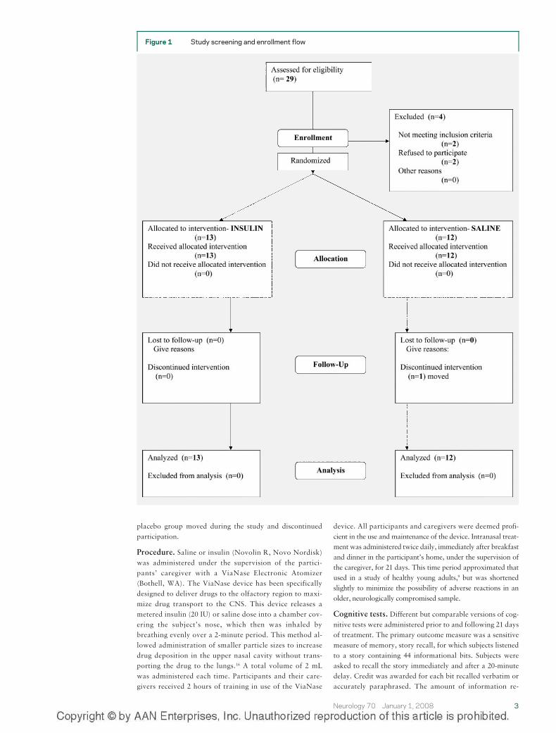
placebo group moved during the study and discontinuedparticipation.
Procedure. Saline or insulin (Novolin R, Novo Nordisk)was administered under the supervision of the partici-pants’ caregiver with a ViaNase Electronic Atomizer(Bothell, WA). The ViaNase device has been specificallydesigned to deliver drugs to the olfactory region to maxi-mize drug transport to the CNS. This device releases ametered insulin (20 IU) or saline dose into a chamber cov-ering the subject’s nose, which then was inhaled bybreathing evenly over a 2-minute period. This method al-lowed administration of smaller particle sizes to increasedrug deposition in the upper nasal cavity without trans-porting the drug to the lungs.16 A total volume of 2 mLwas administered each time. Participants and their care-givers received 2 hours of training in use of the ViaNase
device. All participants and caregivers were deemed profi-cient in the use and maintenance of the device. Intranasal treat-ment was administered twice daily, immediately after breakfastand dinner in the participant’s home, under the supervision ofthe caregiver, for 21 days. This time period approximated thatused in a study of healthy young adults,9 but was shortenedslightly to minimize the possibility of adverse reactions in anolder, neurologically compromised sample.
Cognitive tests. Different but comparable versions of cog-nitive tests were administered prior to and following 21 daysof treatment. The primary outcome measure was a sensitivemeasure of memory, story recall, for which subjects listenedto a story containing 44 informational bits. Subjects wereasked to recall the story immediately and after a 20-minutedelay. Credit was awarded for each bit recalled verbatim oraccurately paraphrased. The amount of information re-
Figure 1 Study screening and enrollment flow
Neurology 70 January 1, 2008 3
tained over the delay was calculated as immediate recall/delayed recall, and is a sensitive measure of early memoryimpairment in mild AD.17 Secondary measures included afrontal-executive test of selective attention and response in-hibition (Stroop Color-Word Interference Task). This testincluded two types of trial blocks.18 For both types of trials,color names were presented one at a time, via computerequipped with a voice key, with concordant or discordantfont colors (e.g., the word “red” presented in red or greenfont). At the beginning of each trial, an instruction screenwas presented to remind the participant to “NAME THEINK COLOR.” Practice trials were administered to ensurethe participant understood the task. Participants’ vocalizedresponses terminated each trial and initiated the next. Er-rors, response content, and voice onset time were recorded.The mean voice onset time for discordant trials was desig-nated as a secondary measure. Separate analyses were con-ducted with correct trials and with all trials, and producedidentical results. As an additional secondary measure, partic-ipants’ caregivers also completed the Dementia Severity Rat-ing Scale (DSRS) at baseline and at the end of the study, inwhich they rated change in the participant’s cognitive, social,and functional status over a specified period of time. TheDSRS includes items relating to memory, orientation, judg-ment, social interactions, home activities, personal care,speech/language, and recognition of others.19 Higher scoresindicate greater impairment.
Blood collection. Blood was collected at baseline and af-ter 21 days of treatment from fasting participants for analy-sis of biochemical secondary outcome measures: glucose,insulin, glucocorticoids, and plasma A�. Given that patientswith AD show greater differences in plasma A� followingnutrient and plasma insulin administration, we measured A�
90 minutes following a standard balanced meal. Sampleswere immediately placed on ice and spun at 2,200 rpm in acold centrifuge for 15 minutes, after which plasma, serum,lymphocytes, and red blood cells were aliquoted into sepa-rate storage tubes and flash frozen at �70°C.
�-Amyloid. To investigate the effects of daily intrana-
sal insulin administration on plasma A� levels, wemeasured A�40 and A�42 levels after overnight fast-ing and 90 minutes later, following a standard bal-anced meal, at baseline and day 21. Plasma A�40 and42 levels were quantified with a sandwich ELISA using6E10 (Signet Laboratories, Dedham, MA), a mousemonoclonal antibody to amino acids 3–8 of A�, forcapture and biotinylated rabbit polyclonal antibodiesspecific for the C-terminal of A�40 or 42 (Signet Lab-oratories) for detection. Nunc Maxisorp plates werecoated using 2 g of 6E10 per well in 0.05 M carbonatebuffer, pH 9.6, overnight at 4 °C. Plates were thenblocked with 1% bovine serum albumin (BSA) for 1hour shaking at room temperature. Synthetic A�40 or42 (Bachem, Torrance, CA) was diluted in 1 mg/mL in70% formic acid, then diluted in 0.5% BSA, 0.05%Tween-20, 1% Triton-X 100 in phosphate buffered sa-line (PBS), pH 7.4, to concentrations of 15.6 pg/mL to2,000 pg/mL for the standard curve. Tween-20 andTriton-X 100 were added to plasma samples to a finalconcentration of 0.05% and 1%. Addition of the deter-gents to the plasma increased the recovery of A� fromthe plasma. Complete protease inhibitor cocktail(Roche Applied Science, Indianapolis, IN) were addedto both standards and samples. Following blocking,samples and standards were added to the plate (100 �Lper well), covered, and incubated overnight shaking at4 °C. The plate was washed and 100 L of 1 g/mL bio-tinylated A�40 or 42 specific antibodies were addedand incubated for 1 hour shaking at room tempera-ture. Following another wash, bound antibody was de-tected using a streptavidin/horseradish peroxidaseconjugate and tetramethylbenzidine as a substrate.The limit of detection for this assay is 15 pg/mL and nocross-detection of A�40 or 42 with anti-A� 42 oranti-A� 40 is seen at concentrations as high as 3 ng/mL. All samples from the same subject, and equalnumbers of subjects from each treatment group, wereanalyzed on the same ELISA plate to reduce the contri-bution of plate to plate variability to any observed dif-ferences. Assignment of samples to plates wasarranged by a technician who was not associated withthe study and was not involved in sample analysis. Thecoefficient of variation for the plasma A� assay was12%. As a quality control measure, we included both astock pooled plasma standard on each plate and apooled plasma standard to which a known amount ofA� peptide had been added. The data from any platesfor which the values of the plasma standards are morethan 10% from the mean established by previous ex-periments were discarded and the samples reanalyzed.Additionally, plasma A� values were determined bythe average of two independent analyses performed ondifferent days to reduce the contribution of plate-to-plate variation to the overall values.
Hormone assays. Neuroendocrine markers were mea-sured with radioimmunoassays (RIAs) as describedpreviously.15 Cortisol binding globulin (CBG) levelswere measured in order to calculate free cortisol levels,given that approximately 95% of circulating cortisol isbound to CBG, and only the remaining 5% of free cor-tisol is biologically active.20 Due to limited availabilityof the supplies needed to make these measurements,
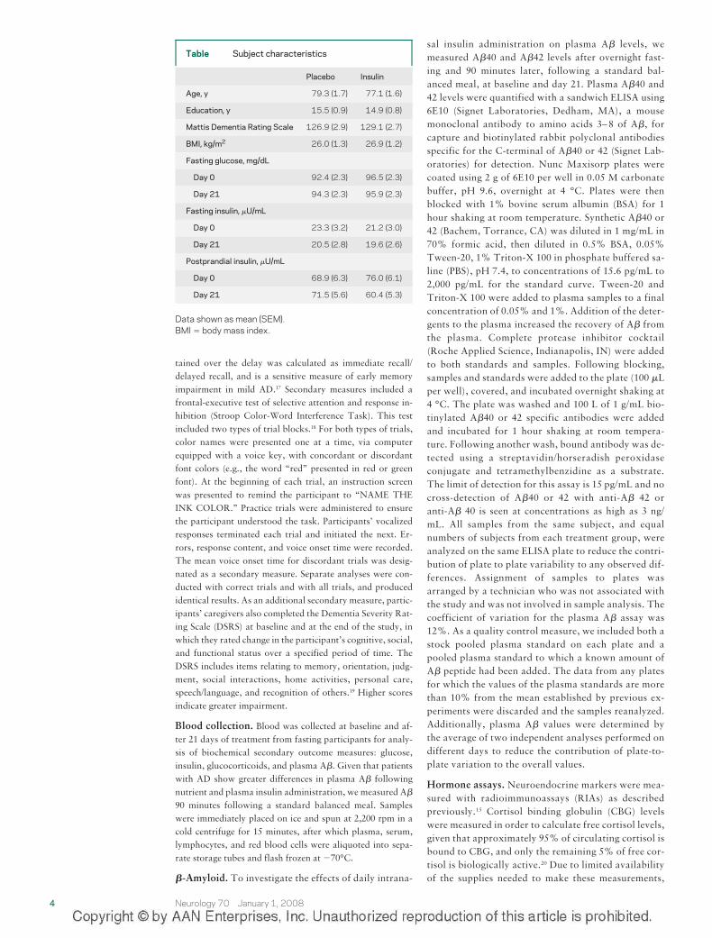
Table Subject characteristics
Placebo Insulin
Age, y 79.3 (1.7) 77.1 (1.6)
Education, y 15.5 (0.9) 14.9 (0.8)
Mattis Dementia Rating Scale 126.9 (2.9) 129.1 (2.7)
BMI, kg/m2 26.0 (1.3) 26.9 (1.2)
Fasting glucose, mg/dL
Day 0 92.4 (2.3) 96.5 (2.3)
Day 21 94.3 (2.3) 95.9 (2.3)
Fasting insulin, �U/mL
Day 0 23.3 (3.2) 21.2 (3.0)
Day 21 20.5 (2.8) 19.6 (2.6)
Postprandial insulin, �U/mL
Day 0 68.9 (6.3) 76.0 (6.1)
Day 21 71.5 (5.6) 60.4 (5.3)
Data shown as mean (SEM).BMI � body mass index.
4 Neurology 70 January 1, 2008
blood from a random sample of participants from eachof the two treatment groups was subjected to analysis(insulin n � 10, placebo n � 5).
Statistical analysis. Only one participant discontin-ued the trial and was lost to follow-up due to movingout of state; thus analyses were conducted with trialcompleters. The primary outcome measure (memorysavings score � delayed recall/immediate recall) andsecondary outcome measures (Stroop voice onsettimes and errors for concordant and discordant trials,and DSRS scores) were each subjected to a repeatedmeasures analysis of variance with treatment assign-ment as the between subjects factor, and phase oftreatment (baseline vs end) as the within subjects fac-tor. Covariates for all analyses included age, educa-tion, Mattis score, and an index of insulin resistance(difference in insulin values obtained at the 90-minutetimepoint following standard meal ingestion on day 1and day 21). Nonsignificant covariates were droppedfrom the model. Interactions with covariates were ex-amined in exploratory analyses with Pearson correla-tion. Effect sizes were estimated using Cohen’s f2, inwhich 0.02, 0.15, and 0.35 represent small, medium,and large effects.21 There were very few missing datapoints (�2% overall, with only one point missing forthe primary outcome measure). Missing data were re-placed using a conservative, last observation carriedforward approach; thus practically, because the studydesign involved two measurements (baseline and post-treatment), when post-treatment scores were missingthey were replaced with the subject’s baseline values.There was no instance in which post-treatment valuesexisted but baseline values were missing. If both base-line and post-treatment values were missing, as wasthe case for some biochemical assays due to difficultywith venous access, and for three participants whowere unable to comprehend/complete the computer-ized Stroop test, the participant was dropped fromanalysis.
RESULTS Intranasal insulin reduced postprandialplasma insulin levels. Baseline fasting plasma glu-cose and insulin values did not differ betweeninsulin-treated and placebo-assigned groups, andno change in either parameter was observed after21 days of treatment (table). However, the in-crease in plasma insulin 90 minutes following astandard meal was attenuated for insulin-treatedpatients compared with the placebo group [table;F(1,20) � 4.43, p � 0.0481]. These results areconsistent with other studies showing that manip-ulation of CNS insulin levels affects peripheral in-sulin sensitivity.22
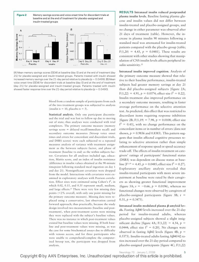
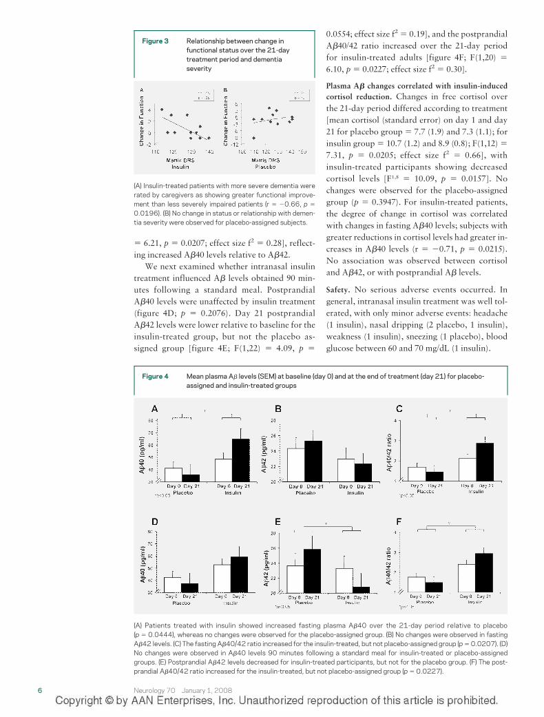
Intranasal insulin improved cognition. Analysis ofthe primary outcome measure showed that rela-tive to their baseline performance, insulin-treatedsubjects had greater memory savings at day 21than did placebo-assigned subjects [figure 2A;F(1,22) � 4.91, p � 0.0374; effect size f2 � 0.22].Insulin treatment also improved performance ona secondary outcome measure, resulting in fasteraverage performance on the selective attentiontest. As predicted, this effect that was restricted todiscordant items requiring response inhibition[figure 2B; F(1,19) � 7.98, p � 0.0108; effect sizef2 � 0.43], with no change performance on theconcordant items or in number of errors (data notshown, p � 0.9836 and 0.8383). This pattern sug-gests that insulin affected cognitive processes re-lating to selective attention rather than simpleenhancement of response speed or speed-accuracytrade-off. The effects of insulin treatment on care-givers’ ratings of participants’ functional status(DSRS) was dependent on disease status at base-line [F1,19 � 4.45, p � 0.0485; effect size f2 � 0.27].Exploratory ancillary analysis revealed thatinsulin-treated participants with more severe im-pairment at baseline were rated by their caregiv-ers as showing greater functional improvement(figure 3A; r � �0.66, p � 0.0196), whereas nofunctional changes were observed by caregivers ofplacebo-assigned participants (figure 3B; r �
0.31, p � 0.3475).
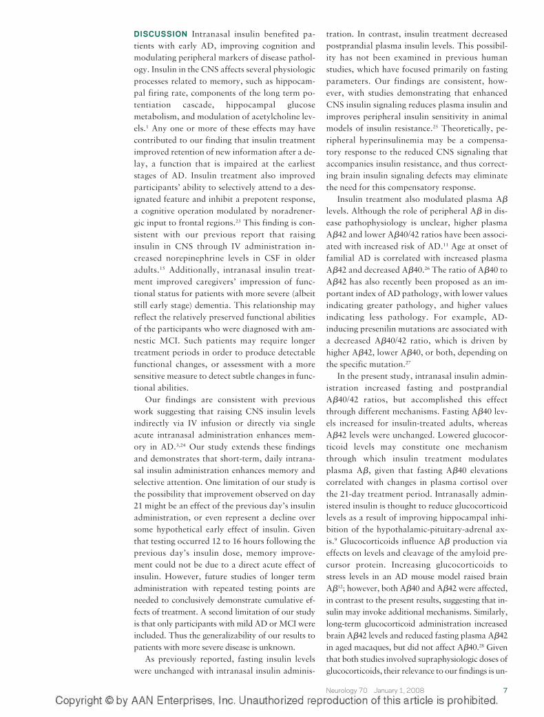
Intranasal insulin modulated plasma �-amyloid lev-els. Fasting A�40 levels increased over the 21-dayperiod for insulin-treated adults, whereasplacebo-assigned subjects showed a slight insig-nificant decline [figure 4A; F(1,22) � 4.54, p �
0.0444; effect size f2 � 0.20]. No changes wereobserved in fasting A�42 levels (figure 4B; p �
0.5373). Insulin-treated adults fasting A�40/42 ra-tios increased over the 21-day period compared toplacebo-assigned participants [figure 4C; F(1,22)
Figure 2 Memory savings scores and voice onset time for discordant trials atbaseline and at the end of treatment for placebo-assigned andinsulin-treated groups
(A) Mean memory savings scores (SEM) at baseline (day 0) and at the end of treatment (day21) for placebo-assigned and insulin-treated groups. Patients treated with insulin showedincreased memory savings over the 21-day period relative to placebo (p � 0.0499). (B) Meanvoice onset time (SEM) for discordant trials at baseline (day 0) and at the end of treatment(day 21) for placebo-assigned and insulin-treated groups. Patients treated with insulinshowed faster response time over the 21-day period relative to placebo (p � 0.0490).
Neurology 70 January 1, 2008 5
� 6.21, p � 0.0207; effect size f2 � 0.28], reflect-ing increased A�40 levels relative to A�42.
We next examined whether intranasal insulintreatment influenced A� levels obtained 90 min-utes following a standard meal. PostprandialA�40 levels were unaffected by insulin treatment(figure 4D; p � 0.2076). Day 21 postprandialA�42 levels were lower relative to baseline for theinsulin-treated group, but not the placebo as-signed group [figure 4E; F(1,22) � 4.09, p �
0.0554; effect size f2 � 0.19], and the postprandialA�40/42 ratio increased over the 21-day periodfor insulin-treated adults [figure 4F; F(1,20) �6.10, p � 0.0227; effect size f2 � 0.30].
Plasma A� changes correlated with insulin-inducedcortisol reduction. Changes in free cortisol overthe 21-day period differed according to treatment[mean cortisol (standard error) on day 1 and day21 for placebo group � 7.7 (1.9) and 7.3 (1.1); forinsulin group � 10.7 (1.2) and 8.9 (0.8); F(1,12) �7.31, p � 0.0205; effect size f2 � 0.66], withinsulin-treated participants showing decreasedcortisol levels [F1,8 � 10.09, p � 0.0157]. Nochanges were observed for the placebo-assignedgroup (p � 0.3947). For insulin-treated patients,the degree of change in cortisol was correlatedwith changes in fasting A�40 levels; subjects withgreater reductions in cortisol levels had greater in-creases in A�40 levels (r � �0.71, p � 0.0215).No association was observed between cortisoland A�42, or with postprandial A� levels.
Safety. No serious adverse events occurred. Ingeneral, intranasal insulin treatment was well tol-erated, with only minor adverse events: headache(1 insulin), nasal dripping (2 placebo, 1 insulin),weakness (1 insulin), sneezing (1 placebo), bloodglucose between 60 and 70 mg/dL (1 insulin).
Figure 4 Mean plasma A� levels (SEM) at baseline (day 0) and at the end of treatment (day 21) for placebo-assigned and insulin-treated groups
(A) Patients treated with insulin showed increased fasting plasma A�40 over the 21-day period relative to placebo(p � 0.0444), whereas no changes were observed for the placebo-assigned group. (B) No changes were observed in fastingA�42 levels. (C) The fasting A�40/42 ratio increased for the insulin-treated, but not placebo-assigned group (p � 0.0207). (D)No changes were observed in A�40 levels 90 minutes following a standard meal for insulin-treated or placebo-assignedgroups. (E) Postprandial A�42 levels decreased for insulin-treated participants, but not for the placebo group. (F) The post-prandial A�40/42 ratio increased for the insulin-treated, but not placebo-assigned group (p � 0.0227).
Figure 3 Relationship between change infunctional status over the 21-daytreatment period and dementiaseverity
(A) Insulin-treated patients with more severe dementia wererated by caregivers as showing greater functional improve-ment than less severely impaired patients (r � �0.66, p �
0.0196). (B) No change in status or relationship with demen-tia severity were observed for placebo-assigned subjects.
6 Neurology 70 January 1, 2008
DISCUSSION Intranasal insulin benefited pa-tients with early AD, improving cognition andmodulating peripheral markers of disease pathol-ogy. Insulin in the CNS affects several physiologicprocesses related to memory, such as hippocam-pal firing rate, components of the long term po-tentiation cascade, hippocampal glucosemetabolism, and modulation of acetylcholine lev-els.1 Any one or more of these effects may havecontributed to our finding that insulin treatmentimproved retention of new information after a de-lay, a function that is impaired at the earlieststages of AD. Insulin treatment also improvedparticipants’ ability to selectively attend to a des-ignated feature and inhibit a prepotent response,a cognitive operation modulated by noradrener-gic input to frontal regions.23 This finding is con-sistent with our previous report that raisinginsulin in CNS through IV administration in-creased norepinephrine levels in CSF in olderadults.15 Additionally, intranasal insulin treat-ment improved caregivers’ impression of func-tional status for patients with more severe (albeitstill early stage) dementia. This relationship mayreflect the relatively preserved functional abilitiesof the participants who were diagnosed with am-nestic MCI. Such patients may require longertreatment periods in order to produce detectablefunctional changes, or assessment with a moresensitive measure to detect subtle changes in func-tional abilities.
Our findings are consistent with previouswork suggesting that raising CNS insulin levelsindirectly via IV infusion or directly via singleacute intranasal administration enhances mem-ory in AD.3,24 Our study extends these findingsand demonstrates that short-term, daily intrana-sal insulin administration enhances memory andselective attention. One limitation of our study isthe possibility that improvement observed on day21 might be an effect of the previous day’s insulinadministration, or even represent a decline oversome hypothetical early effect of insulin. Giventhat testing occurred 12 to 16 hours following theprevious day’s insulin dose, memory improve-ment could not be due to a direct acute effect ofinsulin. However, future studies of longer termadministration with repeated testing points areneeded to conclusively demonstrate cumulative ef-fects of treatment. A second limitation of our studyis that only participants with mild AD orMCI wereincluded. Thus the generalizability of our results topatients with more severe disease is unknown.
As previously reported, fasting insulin levelswere unchanged with intranasal insulin adminis-
tration. In contrast, insulin treatment decreasedpostprandial plasma insulin levels. This possibil-ity has not been examined in previous humanstudies, which have focused primarily on fastingparameters. Our findings are consistent, how-ever, with studies demonstrating that enhancedCNS insulin signaling reduces plasma insulin andimproves peripheral insulin sensitivity in animalmodels of insulin resistance.25 Theoretically, pe-ripheral hyperinsulinemia may be a compensa-tory response to the reduced CNS signaling thataccompanies insulin resistance, and thus correct-ing brain insulin signaling defects may eliminatethe need for this compensatory response.
Insulin treatment also modulated plasma A�
levels. Although the role of peripheral A� in dis-ease pathophysiology is unclear, higher plasmaA�42 and lower A�40/42 ratios have been associ-ated with increased risk of AD.11 Age at onset offamilial AD is correlated with increased plasmaA�42 and decreased A�40.26 The ratio of A�40 toA�42 has also recently been proposed as an im-portant index of AD pathology, with lower valuesindicating greater pathology, and higher valuesindicating less pathology. For example, AD-inducing presenilin mutations are associated witha decreased A�40/42 ratio, which is driven byhigher A�42, lower A�40, or both, depending onthe specific mutation.27
In the present study, intranasal insulin admin-istration increased fasting and postprandialA�40/42 ratios, but accomplished this effectthrough different mechanisms. Fasting A�40 lev-els increased for insulin-treated adults, whereasA�42 levels were unchanged. Lowered glucocor-ticoid levels may constitute one mechanismthrough which insulin treatment modulatesplasma A�, given that fasting A�40 elevationscorrelated with changes in plasma cortisol overthe 21-day treatment period. Intranasally admin-istered insulin is thought to reduce glucocorticoidlevels as a result of improving hippocampal inhi-bition of the hypothalamic-pituitary-adrenal ax-is.9 Glucocorticoids influence A� production viaeffects on levels and cleavage of the amyloid pre-cursor protein. Increasing glucocorticoids tostress levels in an AD mouse model raised brainA�12; however, both A�40 and A�42 were affected,in contrast to the present results, suggesting that in-sulin may invoke additional mechanisms. Similarly,long-term glucocorticoid administration increasedbrain A�42 levels and reduced fasting plasma A�42in aged macaques, but did not affect A�40.28 Giventhat both studies involved supraphysiologic doses ofglucocorticoids, their relevance to our findings is un-
Neurology 70 January 1, 2008 7
clear; however, they serve to illustrate that modula-tion of glucocorticoids can affect A� levels inplasma and brain.
In contrast to changes observed for partici-pants while fasting, intranasal insulin administra-tion decreased A�42 levels measured 90 minutesfollowing a standard balanced meal, and therebyreduced the A�40/42 ratio. This effect may reflecta “normalization” of the action of postprandialelevations of peripheral insulin; in previous work,raising peripheral insulin to normal postprandiallevels reduced plasma A�42 levels for normaladults, but not for patients with AD.10 Althoughour results raise interesting questions about theinfluence of CNS insulin on peripheral A� andinsulin regulation, the significance of these obser-vations and their underlying mechanisms are un-clear at the present time.
Intranasal drug delivery is a potentiallypowerful therapeutic tool that allows largemolecules to bypass the blood–brain barrierand directly access the CNS. Our results pro-vide the first evidence of cognitive improve-ment following daily intranasal peptideadministration for patients with early AD, andmay serve as an impetus for a panoply of future ef-forts with insulin and other promising therapeuticagents. Our results also support brain insulin signal-ing as a promising target in the search for new ther-apeutic avenues in AD.
Received March 15, 2007. Accepted in final form June 27,2007.
REFERENCES1. Craft S, Watson GS. Insulin and neurodegenerative dis-
ease: shared and specific mechanisms. Lancet Neurol2004;3:169–178.
2. Haan MN. Therapy Insight: type 2 diabetes mellitusand the risk of late-onset Alzheimer’s disease. Nat ClinPract Neurol 2006;2:159–166.
3. Craft S, Peskind E, Schwartz MW, Schellenberg GD,RaskindM, Porte D Jr. Cerebrospinal fluid and plasmainsulin levels in Alzheimer’s disease: relationship to se-verity of dementia and apolipoprotein E genotype.Neurology 1998;50:164–168.
4. Hoyer S. Glucose metabolism and insulin receptor sig-nal transduction in Alzheimer disease. Eur J Pharmacol2004;490:115–125.
5. Zhao L, Teter B, Morihara T, et al. Insulin-degradingenzyme as a downstream target of insulin receptor sig-naling cascade: implications for Alzheimer’s disease in-tervention. J Neurosci 2004;24:11120–11126.
6. Freude S, Plum L, Schnitker J, et al. Peripheral hyperin-sulinemia promotes tau phosphorylation in vivo. Dia-betes 2005;54:3343–3348.
7. Kaiyala KJ, Prigeon RL, Kahn SE, Woods SC,Schwartz MW. Obesity induced by a high-fat diet is
associated with reduced brain insulin transport indogs. Diabetes 2000;49:1525–1533.
8. Thorne RG, Pronk GJ, Padmanabhan V, Frey WH II.Delivery of insulin-like growth factor-I to the rat brainand spinal cord along olfactory and trigeminal path-ways following intranasal administration. Neuro-science 2004;127:481–496.
9. Benedict C, Hallschmid M, Hatke A, et al. Intranasalinsulin improves memory in humans. Psychoneuroen-docrinology 2004;29:1326–1334.
10. Kulstad JJ, Green PS, Cook DG, et al. Differentialmodulation of plasma b-amyloid by insulin in patientswith Alzheimer disease. Neurology 2006;66:1506–1510.
11. Mayeux R, Honig LS, Tang MX, et al. Plasma Ab40and Ab42 and Alzheimer’s disease: relation to age,mortality, and risk. Neurology 2003;61:1185–1190.
12. Green KN, Billings LM, Roozendaal B, McGaugh JL,LaFerla FM. Glucocorticoids increase amyloid-betaand tau pathology in a mouse model of Alzheimer’sdisease. J Neurosci 2006;26:9047–9056.
13. Gauthier S, Reisberg B, ZaudigM, et al. Mild cognitiveimpairment. Lancet 2006;367:1262–1270.
14. Craft S, Asthana S, Cook DG, et al. Insulin dose-response effects on memory and plasma amyloid pre-cursor protein in Alzheimer’s disease: interactions withapolipoprotein E genotype. Psychoneuroendocrinology2003;28:809–822.
15. Fishel MA, Watson GS, Montine TJ, et al. Hyperinsu-linemia provokes synchronous increases in central in-flammation and b-amyloid in normal adults. ArchNeurol 2005;62:1539–1544.
16. Djupesland PG, Skretting A, Winderen M, Holand T.Bi-directional nasal delivery of aerosols can preventlung deposition. J Aerosol Med 2004;17:249–259.
17. Wicklund AH, Johnson N, Rademaker A, Weitner BB,Weintraub S. Word list versus story memory in Alzhei-mer disease and frontotemporal dementia. AlzheimerDis Assoc Disord 2006;20:86–92.
18. Spieler DH, Balota DA, Faust ME. Stroop performancein healthy younger and older adults and in individualswith dementia of the Alzheimer’s type. J Exp PsycholHum Percept Perform 1996;22:461–479.
19. Clark CM, Ewbank DC. Performance of the dementiaseverity rating scale: a caregiver questionnaire for rat-ing severity in Alzheimer disease. Alzheimer Dis AssocDisord 1996;10:31–39.
20. Coolens JL, Van Baelen H, Heyns W. Clinical use ofunbound plasma cortisol as calculated from total corti-sol and corticosteroid-binding globulin. J Steroid Bio-chem 1987;26:197–202.
21. Cohen J. Statistical power analysis for the behavioralsciences 2nd ed. Hillsdale, NJ: Lawrence Erlbaum As-sociates, 1988.
22. Morton GJ, Cummings DE, Baskin DG, Barsh GS,Schwartz MW. Central nervous system control of foodintake and body weight. Nature 2006;443:289–295.
23. Arnsten AF, Li BM. Neurobiology of executive func-tions: catecholamine influences on prefrontal corticalfunctions. Biol Psychiatry 2005;57:1377–1384.
24. Reger MA, Watson GS, Frey WH, 2nd, et al. Effects ofintranasal insulin on cognition in memory-impairedolder adults: modulation by APOE genotype. Neuro-biol Aging 2006;27:451–458.
8 Neurology 70 January 1, 2008

25. Plum L, Belgardt BF, Bruning JC. Central insulin ac-tion in energy and glucose homeostasis. J Clin Invest2006;116:1761–1766.
26. Kumar-Singh S, Theuns J, Van Broeck B, et al.Mean age-of-onset of familial Alzheimer disease caused by preseni-lin mutations correlates with both increased Ab42 anddecreased Ab40. HumMutat 2006;27:686–695.
27. BentahirM, Nyabi O, Verhamme J, et al. Presenilin clini-cal mutations can affect gamma-secretase activity by dif-ferent mechanisms. J Neurochem 2006;96:732–742.
28. Kulstad JJ, McMillan PJ, Leverenz JB, et al. Effects ofchronic glucocorticoid administration on insulin-degrading enzyme and amyloid-beta peptide in the agedmacaque. J Neuropathol Exp Neurol 2005;64:139–146.
Neurology 70 January 1, 2008 9