Elemental Spectroscopy ICP-OES. 2 Content: ICP-OES Fundamentals of ICP-OES Instrument Components.
Upload
murad-aamarCategory
view
85download
1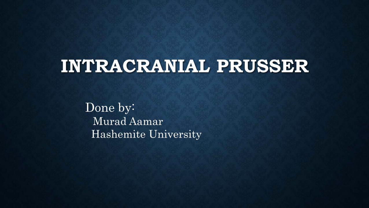
INTRACRANIAL PRUSSER
Done by: Murad Aamar
Hashemite University

Skull has three essential components:
- Brain tissue = 78%
- Blood = 12%
- Cerebrospinal fluid (CSF) = 10%
Any increase in any of these tissues causes increased ICP

Normal ICP = 4 -15 mmHg
Factors that influence ICP:
Arterial pressure
Venous pressure
Intraabdominal and intrathoracic pressure
Posture
Temperature
Blood gases (CO2 levels)
*The degree to which these factors ICP
depends on the ability of the brain to
accommodate to the changes
ICP and the Monro Kellie doctrine
Alexander Monro observed in 1783 that the cranium is a ‘rigid box’
containing a ‘nearly incompressible brain’. Therefore any expansion in
the contents, especially haematoma and brain swelling, may be
initially accommodated by exclusion of fluid components, venous
blood and cerebrospinal fluid (CSF). Further expansion is associated
with an exponential rise in intra-cranial pressure The result is
hypoperfusion and herniation.

Regulation and Maintenance
Normal intracranial pressureThe pressure exerted by the total volume from the brain tissue, blood, and CSF
If the volume in any one of the components increases within the cranial vault and the volume from another component is displaced, the total intracranial volume will not change


Normal compensatory adaptations
-Alteration of CSF absorption
-Displacement of CSF into spinal subarachnoid
space

Cerebral Blood Flow
Definition
The amount of blood in milliliters passing
through 100 g of brain tissue in 1 minute
About 50 ml/min per 100 g of brain tissue

Importance of ICP to BP and CPP
Brain needs constant supply O2 and Glucose
BP: heart delivers blood to brain at an average BP of 120/80 (Mean BP = 100); this mean arterial pressure (MAP) must be higher than ICP
CPP (Cerebral Perfusion Pressure): is the pressure needed to overcome ICP in order to deliver O2 & nutrients
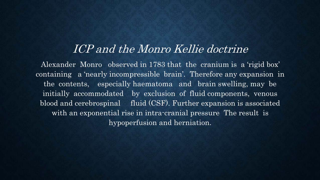
MAP is the DRIVING FORCE
ICP is the RESISTENCE
Cerebral Perfusion Prusser = MAP – ICP
= 100 mmHg – 15 mmHg
= 85 mmHg (Normal)
CPP < 50 mmHg→ cerebral ischemia
CPP < 30 mmHg → brain death

Regulatory Mechanisms of Cerebral Blood Flow
Autoregulation of cerebral blood flow
Metabolic Regulation of cerebral blood flow
Autoregulation
The automatic alteration in the diameter of the cerebral blood vessels to maintain a constant blood flow to the brain
Maintains CPP regardless of changes in BP

Problem: Autoregulation is limited
If BP and/or ICP rises: Autoregulation fails
When autoregulation fails, blood flow to brain increases or deceases → poor perfusion and cellular ischemia or death

Metabolic Regulation of cerebral blood flow
Factors affecting cerebral blood flowPCO2
PO2
Acidosis

Mechanisms of Increased ICP
Causes
Mass lesionCerebral edema
Head injury
Brain inflammation
Metabolic insult
Sustained increases in ICP result in brainstem
compression and herniation of the brain from one
compartment to another

CSF pathway:


GENERAL PRENCEPLES
A few basic principles concerning intracranial dynamics, CSF, CBF,
and ICP are essential to grasp at the outset and are sum- marized
here.

1-The first principle is obvious. The cranial cavity has a fixed
volume comprised of
(1) brain tissue (parenchyma),
(2) CSF, and
(3) blood vessels and intravascular blood.
According to the Monro-Kellie doctrine, the sum of these
components within the fixed volume of the cranial cavity
implies that an increase in one component must be
accompanied by an equal and opposite decrease in one or both
of the remaining components.

If this does not occur, the ICP will rise to levels close to the systemic
blood pressure, producing a reverberating blood flow pattern with no
net flow.
For each intracranial component, there is a family of pathologic
conditions of excess volume and a means to improve that excess
See the next slide:

-Brain tissue: brain edema due to inflammation , tumer, cyst,
abscess or hematoma.
-Vascular: elevated Pco2; hyperperfusion rate with loss of
autoregulation as in sever hepertention after a truma; venous
return obstructon
-CSF: impaired absorption with congenital, posthemorrhagic or
postinfectious hydrocephalus.

A consequence of this principle is that if there is an elevation in the
volume of any one compartment, there is a stage of compensation in
which the volume of one or more other compartments can be
reduced to avoid elevations in the ICP.

2-The second principle is not obvious, and may seem coun- terintuitive.
-Spinal fluid is produced at a constant rate (≈15 to20 mL/hr),
by an energy-dependent, physicochemical process, mainly by the choroid
plexus of the ventricles.
It is essential to understand that production is little affected by any
intracranial backpressure; thus, CSF production continues unabated, even to
lethal elevations of intracranial pressure.

Because production is almost always constant, it follows that
derangement of CSF dynamics almost always involves some aspect of
impeding CSF absorption through obstruction along the CSF pathways
inside the brain, subarachnoid spaces at the basal cisterns or
cerebralconvexity, or arachnoid granulations from which most absorp-
tion occurs.
The only exceptions to the almost constant CSF production are the
excess production associated with the rare choroid plexus papilloma
tumor and the occasional decreased CSF production seen with some
gram-negative bacte- rial meningitis with ventriculitis, usually in
neonates.

3-The third basic principle is that the CBF normally varies over a wide
range (30 to 100 mL/100 g brain tissue/min), depending on metabolic
demand from neuronal activity within a particular area of the brain.
The CBF may be considered in aggregate or of specific small regions,
pathologic or normal.
The blood flow to any brain area is generally abundant, exceeding
demand by a wide margin, so that O2 extraction ratios are often low.
The brain vasculature matches the blood flow to tissue metabolic
demand and the CBF generally maintains what is needed, despite wide
variations in systemic blood pressure, by a phenomenon known as
autoregulation. .


Factors such as an elevated or decreased arterial PCO2 shift the
curve as indicated. In the setting of traumatic brain injury, the
curve becomes more pronounced (i.e., smaller changes in blood
pressure or PCO2) and affects the CBF dramatically.

* If tissue demand exceeds autoregulation, or if CBF declines for
pathologic reasons, the first defense is that the O2 extraction will
increase (i.e., arteriovenous O2 difference, AVDO2) An important
implication is that if brain dysfunction is occurring clinically
because compensatory mechanisms (e.g., autoregulation changing
the vascular resistance, capacity to elevate mean systemic arterial
pressure, ability to increase O2 extraction) have been exceeded, the
tolerance for further decline in blood flow is low, and tissue damage
is seriously threatened. Therapy to increase blood pressure or
decrease ICP may be urgently needed.


A fourth principle derives from the other three and the fact that
injured tissue swells, making obvious the potential for a cascading
injury by a vicious cycle mechanism (next figure).
If the stage of compensation (see earlier), even with therapy, is
exceeded, and ICP is elevated high enough by some mechanism so that
cerebral perfusion pressure (CPP) declines, CBF can decline to levels
at which tissue injury occurs.
CPP = mean arterial pressure (MAP) − ICP


Brain edema swelling within the closed cranium will lead to further
increases in ICP with even further decreases in CPP in a stage of
decompensation. When the capacity for autoregulation is exceeded
or damaged so that it can no longer play a role, CBF is linked
directly to the CPP.

A fifth principle concerns focal mass effect and its progression in
regard to the complex anatomy of the cranial cavity. The cranial
cavity is not just a hollow spherical space but contains several
almost knifelike projections of folded dura, the falx and tentorium,
which divide the cavity into right and left supratentorial
compartment and an infratentorial compartment, the posterior
fossa. The sphenoid wing is a prominent, mostly bony ridge that
separates the anterior fossa containing the frontal lobe from the
middle fossa containing the temporal lobe.

it is unusual for focal mass effects not to be accompanied by an
overall increase in ICP. The point at which focal mass effect evolves
to include a rise in overall ICP depends largely on the compliance
within the cranial cavity. Young patients with so-called tight brains
can develop raised ICP, even with relatively small volumes of mass
that produce only effacement of the cortical gyri. On the other
hand, patients with advanced cerebral atrophy can, for example,
tolerate large frontal intracerebral hematomas or chronic subdural
hematomas with compression of the lateral ventricle and midline
shift while maintaining a tolerable ICP and a surprising degree of
intact neurologic function.

A sixth principle concerns the separateness of the phenomenology of
the following: (1) focal mass effect (as described earlier); (2) diffuse
raised ICP; and (3) ventriculomegaly (enlargement of the cerebral
ventricles).
Although these three processes often occur in combination, the
notion that they are separable comes from the observation that there
is a pure form of each

. The pure form of raised ICP—without focal mass lesion, trauma or
enlargement of the ventricular system—is a condition known as
idiopathic intracranial hypertension or pseudotumor cerebri.
The pure form of ventriculomegaly is a condition known as
adult chronic idiopathic or normal-pressure hydro- cephalus
(NPH).

The pure form of the focal mass lesion without increased ICP or
ventriculomegaly is common; it occurs with tumors too small to
raise ICP on their own that are not in a location to interfere with
CSF pathways. Instead, there focal mass lesions are typically
discovered incidentally or because of symptoms from a focal
neurologic deficit or seizure disorder.

CLINICAL POINT OF VIEW
Normal ICP varies over a wide range, with generally accepted
values between 0 and 20 mm Hg. Diffuse raised ICP, in the fully
evolved pure form, results in a clinical picture that may include
symptoms of headache, nausea and vomiting, double vision, and
obscuration of vision

The accompanying clinical signs may include papilledema and sixth
cranial nerve palsy with lateral rectus weakness and side by side
diplopia,
initially worse on far vision or gaze directed toward the side of the
palsy. The papilledema is a mostly chronic phenomenon and is not
seen acutely.

Papilledema (or papilloedema) is optic disc swelling that is caused by
increased intracranial pressure. The swelling is usually bilateral and can
occur over a period of hours to weeks. Unilateral presentation is
extremely rare

sixth cranial nerve palsy with lateral rectus weakness

. With raised ICP, there may also be obscurations of vision, in which
patients report that their vision temporarily fades or becomes gray,
visual loss, sometimes even to permanent blindness in combination
with headache. these obscurations are caused by the effect of
diffusely increased ICP on the sensitive optic nerves.

Pure ventriculomegaly—specifically, enlargement of the lateral
ventricles—is characterized by gait disturbance and incontinence
early in the clinical picture. As the process worsens, cognitive
disturbances may be added on. The early appearance of gait
disturbance and urinary incontinence is attributed to dysfunction
of the medial cerebral hemispheres in which the leg area of the
primary motor cortex and bladder control area reside

Parinaud’s syndrome results from dorsal midbrain com- pression, and its
features include a loss of upgaze known as
‘sun-setting’ . In infants, the fontanelle is tense and bulging, with an increase
in head circumference and bulging scalp veins.

The next table summarizes the relationships among ICP, mass
lesions, and ventriculomegaly.