Intra- and Interspecies Signaling between Streptococcus salivarius and Streptococcus pyogenes
8
JOURNAL OF BACTERIOLOGY, 0021-9193/01/$04.0010 DOI: 10.1128/JB.183.13.3931–3938.2001 July 2001, p. 3931–3938 Vol. 183, No. 13 Copyright © 2001, American Society for Microbiology. All Rights Reserved. Intra- and Interspecies Signaling between Streptococcus salivarius and Streptococcus pyogenes Mediated by SalA and SalA1 Lantibiotic Peptides M. UPTON, 1,2 ² J. R. TAGG, 2 P. WESCOMBE, 2 AND H. F. JENKINSON 1 * Department of Oral and Dental Science, University of Bristol Dental School, Bristol, BS1 2LY, United Kingdom, 1 and Department of Microbiology, University of Otago, Dunedin, New Zealand 2 Received 28 November 2000/Accepted 10 April 2001 Streptococcus salivarius 20P3 produces a 22-amino-acid residue lantibiotic, designated salivaricin A (SalA), that inhibits the growth of a range of streptococci, including all strains of Streptococcus pyogenes. Lantibiotic production is associated with the sal genetic locus comprising salA, the lantibiotic structural gene; salBCTX genes encoding peptide modification and export machinery proteins; and salYKR genes encoding a putative immunity protein and two-component sensor-regulator system. Insertional inactivation of salB in S. salivarius 20P3 resulted in abrogation of SalA peptide production, of immunity to SalA, and of salA transcription. Addition of exogenous SalA peptide to salB mutant cultures induced dose-dependent expression of salA mRNA (0.2 kb), demonstrating that SalA production was normally autoregulated. Inactivation of salR encoding the response regulator of the SalKR two-component system led to reduced production of, and immunity to, SalA. The sal genetic locus was also present in S. pyogenes SF370 (M type 1), but because of a deletion across the salBCT genes, the corresponding lantibiotic peptide, designated SalA1, was not produced. However, in S. pyogenes T11 (M type 4) the sal locus gene complement was apparently complete, and active SalA1 peptide was synthesized. Exogenously added SalA1 peptide from S. pyogenes T11 induced salA1 transcription in S. pyogenes SF370 and in an isogenic S. pyogenes T11 salB mutant and salA transcription in S. salivarius 20P3 salB. Thus, SalA and SalA1 are examples of streptococcal lantibiotics whose production is autoregulated. These peptides act as intra- and interspecies signaling molecules, modulating lantibiotic production and possibly influencing streptococcal population ecology in the oral cavity. Streptococcus salivarius is a primary colonizer of neonatal oral mucosal surfaces and a predominant component of the human adult oral microbiota and is not associated with disease in healthy individuals (39). Streptococcus pyogenes, on the other hand, persists in the pharynx in a carrier state in approximately 10% of the population, is a common cause of pharyngeal infections, especially in school-aged children, and is usually present in high numbers only during acute infection (1). All S. pyogenes strains tested have been found to be susceptible to growth inhibition by salivaricin A (SalA), a lantibiotic peptide produced by S. salivarius (34, 37), and it has been suggested that growth of S. pyogenes in vivo may be modulated by indig- enous SalA-producing S. salivarius. Lantibiotics are antimicrobial peptides that are produced by, and are active against, closely related gram-positive organisms. These peptides are ribosomally synthesized and then undergo posttranslational modifications, including amino acid dehydra- tion (38) and thioether bridge formation (20). Lantibiotics form two families; type A lantibiotics are linear, and type B lantibiotics have more globular conformations. The peptides are synthesized as prepropeptides consisting of an inhibitor propeptide and a leader region, the features of which have been utilized to group the type A lantibiotics into three sub- classes (36). In the members of one of these subclasses, sub- class AII, the amino acid (aa) residues Gly-Gly, Gly-Ser, or Gly-Ala immediately precede the site of leader cleavage. These Gly-Gly type cleavage sites are more commonly found in non- modified bacteriocins or pheromones, such as the ComC pep- tide responsible for competence induction in Streptococcus pneumoniae (15). The streptococcal pheromones are quorum- sensing molecules, analogous to the N-acyl homoserine lac- tones produced by gram-negative bacteria (35), that regulate microbial community responses. SalA (22 aa residues) is a subclass AII lantibiotic produced by S. salivarius 20P3 via processing of a 48-aa prepropeptide encoded by the salA gene (34). By utilizing salA as a DNA hybridization probe it was shown that all SalA peptide-produc- ing strains of S. salivarius contained salA sequences and that, somewhat surprisingly, 63 of 65 S. pyogenes strains of different M types contained a salA gene homolog designated salA1 (37). We hypothesized that S. pyogenes failed to produce SalA1 inhibitor and was sensitive to inhibition by SalA from S. sali- varius because the genetic locus for lantibiotic production and immunity was incomplete or transcriptionally inactive. In the present study, the structure of the sal genetic locus in S. sali- varius 20P3 was determined and the genes necessary for reg- ulation of lantibiotic production were identified. In S. pyogenes SF370 (M type 1), the corresponding sal genetic locus had a deletion spanning three genes encoding peptide modification and export proteins, and the salA1 gene was transcriptionally inactive, thus accounting for the lack of SalA1 production. However, the salA gene was actively transcribed in S. pyogenes * Corresponding author. Mailing address: Department of Oral and Dental Science, University of Bristol Dental School, Lower Maudlin Street, Bristol, BS1 2LY, United Kingdom. Phone: 44 117 928 4358. Fax: 44 117 928 4313. E-mail: [email protected]. ² Present address: Department of Medical Microbiology, Manches- ter Royal Infirmary, Manchester M13 9WL, United Kingdom. 3931 Downloaded from https://journals.asm.org/journal/jb on 10 December 2021 by 191.53.194.15.
Transcript of Intra- and Interspecies Signaling between Streptococcus salivarius and Streptococcus pyogenes
JOURNAL OF BACTERIOLOGY,0021-9193/01/$04.0010 DOI: 10.1128/JB.183.13.3931–3938.2001
July 2001, p. 3931–3938 Vol. 183, No. 13
Copyright © 2001, American Society for Microbiology. All Rights Reserved.
Intra- and Interspecies Signaling between Streptococcussalivarius and Streptococcus pyogenes Mediated by SalA and
SalA1 Lantibiotic PeptidesM. UPTON,1,2† J. R. TAGG,2 P. WESCOMBE,2 AND H. F. JENKINSON1*
Department of Oral and Dental Science, University of Bristol Dental School, Bristol, BS1 2LY, United Kingdom,1 andDepartment of Microbiology, University of Otago, Dunedin, New Zealand2
Received 28 November 2000/Accepted 10 April 2001
Streptococcus salivarius 20P3 produces a 22-amino-acid residue lantibiotic, designated salivaricin A (SalA),that inhibits the growth of a range of streptococci, including all strains of Streptococcus pyogenes. Lantibioticproduction is associated with the sal genetic locus comprising salA, the lantibiotic structural gene; salBCTXgenes encoding peptide modification and export machinery proteins; and salYKR genes encoding a putativeimmunity protein and two-component sensor-regulator system. Insertional inactivation of salB in S. salivarius20P3 resulted in abrogation of SalA peptide production, of immunity to SalA, and of salA transcription.Addition of exogenous SalA peptide to salB mutant cultures induced dose-dependent expression of salA mRNA(0.2 kb), demonstrating that SalA production was normally autoregulated. Inactivation of salR encoding theresponse regulator of the SalKR two-component system led to reduced production of, and immunity to, SalA.The sal genetic locus was also present in S. pyogenes SF370 (M type 1), but because of a deletion across thesalBCT genes, the corresponding lantibiotic peptide, designated SalA1, was not produced. However, in S.pyogenes T11 (M type 4) the sal locus gene complement was apparently complete, and active SalA1 peptide wassynthesized. Exogenously added SalA1 peptide from S. pyogenes T11 induced salA1 transcription in S. pyogenesSF370 and in an isogenic S. pyogenes T11 salB mutant and salA transcription in S. salivarius 20P3 salB. Thus,SalA and SalA1 are examples of streptococcal lantibiotics whose production is autoregulated. These peptidesact as intra- and interspecies signaling molecules, modulating lantibiotic production and possibly influencingstreptococcal population ecology in the oral cavity.
Streptococcus salivarius is a primary colonizer of neonataloral mucosal surfaces and a predominant component of thehuman adult oral microbiota and is not associated with diseasein healthy individuals (39). Streptococcus pyogenes, on the otherhand, persists in the pharynx in a carrier state in approximately10% of the population, is a common cause of pharyngealinfections, especially in school-aged children, and is usuallypresent in high numbers only during acute infection (1). All S.pyogenes strains tested have been found to be susceptible togrowth inhibition by salivaricin A (SalA), a lantibiotic peptideproduced by S. salivarius (34, 37), and it has been suggestedthat growth of S. pyogenes in vivo may be modulated by indig-enous SalA-producing S. salivarius.
Lantibiotics are antimicrobial peptides that are produced by,and are active against, closely related gram-positive organisms.These peptides are ribosomally synthesized and then undergoposttranslational modifications, including amino acid dehydra-tion (38) and thioether bridge formation (20). Lantibioticsform two families; type A lantibiotics are linear, and type Blantibiotics have more globular conformations. The peptidesare synthesized as prepropeptides consisting of an inhibitorpropeptide and a leader region, the features of which havebeen utilized to group the type A lantibiotics into three sub-
classes (36). In the members of one of these subclasses, sub-class AII, the amino acid (aa) residues Gly-Gly, Gly-Ser, orGly-Ala immediately precede the site of leader cleavage. TheseGly-Gly type cleavage sites are more commonly found in non-modified bacteriocins or pheromones, such as the ComC pep-tide responsible for competence induction in Streptococcuspneumoniae (15). The streptococcal pheromones are quorum-sensing molecules, analogous to the N-acyl homoserine lac-tones produced by gram-negative bacteria (35), that regulatemicrobial community responses.
SalA (22 aa residues) is a subclass AII lantibiotic producedby S. salivarius 20P3 via processing of a 48-aa prepropeptideencoded by the salA gene (34). By utilizing salA as a DNAhybridization probe it was shown that all SalA peptide-produc-ing strains of S. salivarius contained salA sequences and that,somewhat surprisingly, 63 of 65 S. pyogenes strains of differentM types contained a salA gene homolog designated salA1 (37).We hypothesized that S. pyogenes failed to produce SalA1inhibitor and was sensitive to inhibition by SalA from S. sali-varius because the genetic locus for lantibiotic production andimmunity was incomplete or transcriptionally inactive. In thepresent study, the structure of the sal genetic locus in S. sali-varius 20P3 was determined and the genes necessary for reg-ulation of lantibiotic production were identified. In S. pyogenesSF370 (M type 1), the corresponding sal genetic locus had adeletion spanning three genes encoding peptide modificationand export proteins, and the salA1 gene was transcriptionallyinactive, thus accounting for the lack of SalA1 production.However, the salA gene was actively transcribed in S. pyogenes
* Corresponding author. Mailing address: Department of Oral andDental Science, University of Bristol Dental School, Lower MaudlinStreet, Bristol, BS1 2LY, United Kingdom. Phone: 44 117 928 4358.Fax: 44 117 928 4313. E-mail: [email protected].
† Present address: Department of Medical Microbiology, Manches-ter Royal Infirmary, Manchester M13 9WL, United Kingdom.
3931
Dow
nloa
ded
from
http
s://j
ourn
als.
asm
.org
/jour
nal/j
b on
10
Dec
embe
r 20
21 b
y 19
1.53
.194
.15.

T11 (M type 4), which contained a complete sal genetic locus,and active SalA1 inhibitor peptide was produced. To the bestof our knowledge, this is the first report of an autoregulatorylantibiotic produced by streptococci, and the evidence pre-sented here suggests that SalA-like peptides form a family ofsignaling factors recognized by different species of strepto-cocci.
MATERIALS AND METHODS
Bacterial strains and media. S. salivarius 20P3 (wild type, SalA1) (34), S.pyogenes SF340 (M type 1, SalA2) (41), and S. pyogenes T11 (M type 4, SalA1
clinical isolate) were maintained on Columbia agar plates (Life TechnologiesLtd., Paisley, United Kingdom) supplemented with human blood (5%, vol/vol)and CaCO3 (0.1%, wt/vol) in a 5% CO2–air atmosphere at 37°C. Streptococcalstrains were routinely cultured in Todd-Hewitt broth (Difco Laboratories, De-troit, Mich.) or in M17 medium (Difco) supplemented with CaCO3 (10 mM) andsucrose (10 mM) (M17CS medium) for maximal SalA peptide production. Esch-erichia coli DH5a (9) and the highly electrocompetent derivative strain DH10B(14) were maintained on Luria-Bertani (LB) agar (Difco) or were grown in LBbroth with aeration at 37°C. The following antibiotic concentrations were used(where appropriate): ampicillin, 100 mg/ml (E. coli); and spectinomycin, 100mg/ml (E. coli), 60 mg/ml (S. pyogenes), or 600 mg/ml (S. salivarius).
PCR and sequence analysis. Primers for PCR amplification of salA and theregions downstream of salA in S. salivarius 20P3 were designed on the basis of thecorresponding sal locus sequence in S. pyogenes SF370 (University of OklahomaAdvanced Center for Genome Technology; http://www.genome.ou.edu/strep.html).The major primers utilized (and their corresponding target sites within the S.salivarius 20P3 sal locus [10,610 nucleotides; GenBank accession numberAY005472]) were SalAF (positions 604 to 629; 59GATATTTTGAACAATGCTATCGAAGA), DsintF (positions 856 to 876; 59CAACATCAGTTTTACGAATAC), ORFIR (positions 2340 to 2320; 59GTGAACTTCAATCTTTCATCG),SalYF (positions 6665 to 6684; 59GGTCAAGCTCTAGTTATCGC), SalYR(positions 8413 to 8393; 59GATTATGGATGTCACTAACCG), and SalRterm(positions 10610 to 10590; 59TCAGAAATCCATAAAATACCC). PCRs werecarried out with Taq polymerase (Roche Diagnostics Ltd., Lewes, England) for30 cycles consisting of denaturation at 95°C for 30 s, annealing at between 55 and64°C (as appropriate) for 30 s, and extension at 72°C for 1 min per kb of DNAto be amplified; this was followed by a final elongation step at 72°C for 5 min.PCR products were ligated into pGEM-T (Promega, Madison, Wis.) and se-quenced with a Perkin-Elmer ABI 377A sequencer. Primary sequence data werecollated with SeqEd sequencer software, and sequence alignment, translation,and general analyses were performed by using DNAMAN (Lynnon BioSoft,Vaudreuil, Canada). Deduced amino acid sequences for open reading frameswere compared with sequences in protein sequence databases by using theBLAST facilities on the National Center for Biotechnology Information server(www.ncbi.nlm.nih.gov) and the University of Oklahoma server.
DNA manipulation and transformation. Streptococcal genomic DNA wasisolated as described by Upton et al. (43). Briefly, confluent growth from one-halfof an agar plate culture was collected in TE buffer (10 mM Tris-HCl [pH 8.0]containing 10 mM EDTA), the cells were harvested by centrifugation (8,000 3g for 10 min) and suspended in 0.3 ml of TE buffer, and 0.3 ml of a lysis mixture(50 mM Tris-HCl [pH 8.0] containing 10 mM EDTA and 2% [vol/vol] TritonX-100) was added. Lysis was achieved by adding 30 U of mutanolysin (Sigma)and then 0.3 mg of pronase (Sigma) and incubating the preparation at 37°C for2 h. The suspension was extracted once with 0.6 ml of phenol and once with 0.6ml of phenol-chloroform (0.3 ml of phenol plus 0.3 ml of chloroform-isoamylalcohol [24:1]). Nucleic acids were precipitated from the aqueous phase with 2volumes of 100% ethanol at 270°C and collected by centrifugation (12,000 3 gfor 20 min), and the pellet was washed with 70% (vol/vol) ethanol, air dried, anddissolved in TE buffer. In a number of experiments the solutions were thenincubated with 50 mg of RNase A (Sigma)/ml at 37°C for 30 min and extractedwith phenol-chloroform, and the DNA was precipitated, washed, and dissolvedin TE buffer as described above.
Plasmid DNA was isolated from E. coli by using Qantum Prep (Bio-RadLaboratories, Hercules, Calif.). For insertional inactivation of streptococcalgenes, target fragments were generated by PCR amplification, cloned intopGEM-T, and recovered by restriction digestion with NcoI and NsiI endonucle-ases. Fragments were gel purified and ligated into vector pFW5 (29) digestedwith NcoI and NsiI. E. coli strains were transformed with plasmid DNA by usinga TransPorator (BTX, San Diego, Calif.) in 0.1-cm cuvettes (Bio-Rad Labora-tories, Richmond, Calif.). Following electroporation, bacteria were incubated for
1 h in 1 ml of 2 3 YT broth (16 g of Bacto Tryptone per liter, 10 g of Bacto YeastExtract per liter, 5 g of NaCl per liter; pH 7.0) at 37°C with shaking at 200 rpmbefore being spread onto LB agar plates containing the appropriate antibiotics(9). S. salivarius and S. pyogenes strains were prepared for electrotransformationby the method of Chen et al. (4); DL-threonine was included in the media at finalconcentrations of 0.6 and 0.4 M, respectively. Electroporation was carried out in0.1-cm cuvettes by using a Bio-Rad Gene Pulser II set at 1.8 kV, 25 mF, and 200V, and transformants (frequency, 50 to 100 transformants per mg of DNA) wereselected on agar containing spectinomycin.
Generation of mutants. To generate S. salivarius UB1309 salB::pMU1011 Spr,an internal 1.48-kb salB fragment (Fig. 1), PCR amplified from 20P3 DNA byusing primers DsintF and ORF1R, was cloned into pFW5 to produce pMU1001,and this plasmid was then transformed onto the S. salivarius 20P3 chromosome.S. pyogenes UB1308 salB::pMU1016 Spr was generated in the same way bytransforming pMU1016 (pFW5 carrying a 1.48-kb salB fragment from T11) ontothe S. pyogenes T11 chromosome. S. salivarius UB1310 salR::pMU1002 Spr wasgenerated by PCR amplifying a 1.16-kb fragment from the salKR gene region(Fig. 1) of 20P3 DNA with primers SalKF (positions 9249 to 9269; 59GTTGGATTGTACTCATGAAGG) and SalRR (positions 10411 to 10391; 59TCAACATAATCCTGAGATTCG), cloning this fragment into pFW5 to producepMU1002, and transforming this plasmid into wild-type strain 20P3 as describedabove. Integration of pFW5 plasmid constructs onto the streptococcal chromo-some was confirmed by PCR amplification with pFW5-specific primers pFW5F(59GATCAGGAGTTGAGAGTGGAC) and pFW5R (59TGGAGAAGATTCAGCCACTGC).
RNA analysis. To prepare total RNA from streptococci, cells were harvestedby centrifugation from M17CS medium, suspended in 0.2 ml of spheroplastingbuffer (20 mM Tris-HCl [pH 6.8] containing 10 mM MgCl2 and 26% [wt/vol]raffinose) containing 500 U of mutanolysin/ml and 0.1 mg of spectinomycin/ml,and incubated 37°C for 30 min. Spheroplasts were collected by centrifugation,and RNA was extracted by using a Qiagen RNAeasy kit and a Qiashreddercolumn (Qiagen Ltd., Crawley, England) as recommended by the manufacturer.RNA samples (10 mg per lane) were subjected to electrophoresis through 1%(wt/vol) agarose in 13 MOPS (morpholinepropanesulfonic acid) buffer (0.2 MMOPS containing 50 mM sodium acetate and 10 mM EDTA; pH 7.0) supple-mented with 1% (wt/vol) formaldehyde at 80 V for 3 h and were vacuum blottedonto a nylon membrane. For Northern analysis of sal transcripts, a PCR productgenerated with primers SalAF and SalAR (positions 901 to 883; 59AGAAGTATCTAGTATGTCG) was radioactively labeled with [32P]dATP by using Prime-a-Gene (Promega) as directed by the manufacturer. Hybridization reactionswere carried out at 65°C for 18 h as described elsewhere (5). For reversetranscription (RT)-PCR analysis, DNase-treated RNA that had been extractedfrom exponentially growing cells was utilized for cDNA synthesis by using theSalRterm oligonuclotide primer and avian myeloblastosis virus reverse transcrip-
FIG. 1. Genetic structures of the sal loci in S. salivarius 20P3 (A)and S. pyogenes SF370 (M type 1) (B). Open reading frames aredepicted by arrows which show the numbers of aa residues in thededuced polypeptides. The sites of insertional inactivation with plas-mid pFW5 constructs used to generate strains UB1309, UB1310, andUB1308 are shown. PCR-amplified segments of DNA used for hybrid-ization probes or cloning in pFW5 are indicated as follows: I, 0.3-kbsalA probe; II, 1.48-kb salB fragment; III, 1.16-kb salKR fragment.Symbols: }, putative promoter; F, inverted repeat.
3932 UPTON ET AL. J. BACTERIOL.
Dow
nloa
ded
from
http
s://j
ourn
als.
asm
.org
/jour
nal/j
b on
10
Dec
embe
r 20
21 b
y 19
1.53
.194
.15.

tase (First Strand cDNA synthesis kit; Roche Diagnostics). PCR amplificationswere then performed by utilizing the Expand Long Template PCR system(Roche Diagnostics) with primers SalAF and SalXR (positions 6510 to 6490;59CTCTCCTCTATCGGATAAAGC), primers SalY2S (positions 6571 to 6591;59CTATTTTCTAGCTACGATCGG) and SalRterm, or primers SalAF andSalRR.
SalA production and expression. SalA peptide was purified from S. salivarius20P3 M17CS broth culture supernatant as previously described (34). To test forSalA induction of transcription, streptococcal strains were grown for 18 h inM17CS medium with or without SalA peptide. Cells were harvested by centrif-ugation (16,000 3 g for 20 min at 4°C), and the cell-free supernatants werecollected in sterile tubes. For medium swap experiments the bacterial pelletswere suspended in the appropriate spent culture supernatant and incubated at37°C for 4 h before RNA was extracted as described above. Levels of inhibitorproduction and of immunity to inhibition were determined by using a deferredantagonism assay performed on Columbia blood agar plates supplemented with0.1% (wt/vol) CaCO3 as described elsewhere (42). Levels of lantibiotic produc-tion by producer strains and growth inhibition of test strains were scored as 0 (noinhibition zone), 1 (narrow inhibition zone that was 1 to 2 mm wide), 2 (mediumzone of inhibition that was 2 to 5 mm wide), 3 (larger zone of inhibition that was5 to 8 mm wide), or 4 (inhibition zone that was more than 8 mm wide).
Nucleotide sequence accession number. The nucleotide sequence data for theS. salivarius 20P3 sal locus have been deposited in the GenBank database underaccession number AY005472.
RESULTS
Structure of the sal locus in S. salivarius 20P3. To determinethe sequence and genetic structure of the S. salivarius 20P3 salgene locus, PCR primers were synthesized based on the nu-cleotide sequence of the approximately 7-kb region down-stream of the salA1 gene in S. pyogenes SF370 (Fig. 1). Theseprimers were utilized to PCR amplify segments that com-prised the complete sal genetic locus in S. salivarius 20P3. Thegenetic locus comprised eight open reading frames designatedsalABCTXYKR (Fig. 1A). These open reading frame designa-tions were assigned on the basis of database homologysearches and were in accordance with conventional lantibioticgene cluster nomenclature (7, 36).
The first gene (salA), encoding the 48-aa precursor lantibi-otic peptide, was preceded by a potential ribosome bindingsequence (GGGAG) and an AT-rich region containing puta-tive promoter sequences. Downstream of salA, the salB andsalC genes (Fig. 1A) are predicted to encode peptides withsignificant identities to the N-terminal and C-terminal se-quences, respectively, of lactococcin biosynthesis protein pro-duced by Lactococcus lactis (32) (Table 1). We identified salB
and salC as separate genes because there are multiple stopcodons present in all three reading frames in the putativeintergenic sequence. SalB contains a number of motifs that areconserved in LanM modification peptides (36), while SalC ispredicted to contain at least five membrane-spanning se-quences and is probably responsible for the formation ofthioether bridges in mature SalA peptide (12, 27). The de-duced aa sequences encoded by the next two genes, salT andsalX, exhibit significant identities with ATP binding cassette(ABC) transporters (16, 46) (Table 1). For lantibiotics andunmodified bacteriocins with a GG cleavage site, the leader isremoved by a cysteine protease activity in the N-terminal re-gion of the LanT polypeptide (16). Retention of the leaderpeptide might have an immunity function (45). Motifs charac-teristic of LanT protease regions (36) are present in the N-terminal region of the SalT polypeptide. Collectively, thesedata suggest that leader peptide cleavage and export of mod-ified SalA are carried out by a SalTX protein complex locatedwithin the cell membrane.
The salY gene, situated upstream of the salKR genes at thedistal end of the locus, encodes a 635-aa polypeptide for whichthere were no significant database matches. It is proposed thatthe products of the salKR genes form a two-component sensorkinase-response regulator system (40) based on significant se-quence identities between SalK and Bacillus subtilis ComP (47)and between SalR and the Bacillus brevis DegU transcriptionalregulator (24) (Table 1). Other features of the sal locus thathave relevance for transcriptional regulation include putativepromoter regions upstream of the salA and salY genes (Fig.1A) and inverted repeats with the potential to form stem-loopstructures immediately downstream of the salA and salX genes(Fig. 1A). An inverted repeat downstream of the inhibitorpeptide structural gene is present in several lantibiotic geneclusters (13, 18, 23, 33). The potential stem-loop structure thatis formed functions as a transcriptional attenuator that allowsonly partial readthrough transcription of the downstreamgenes.
Structure of the sal locus in S. pyogenes. S. pyogenes SF370(M type 1) contains the salA-like gene designated salA1 (37)but does not produce detectable levels of lantibiotic inhibitor(Table 2). While the deduced aa sequence of the mature SalA1peptide from strain SF370 has two conserved changes, residue
TABLE 1. Levels of homology of S. salivarius sal gene product sequences to peptide database sequences and to corresponding S. pyogenesSF370 sal gene product sequences
S. salivarius20P3 gene
Size ofprecursor
(aa)
Most similar sequence % Identity tocorresponding salgene product in
S. pyogenesSF370a
Molecule (accession no.) No. of aaresidues
Region of homology to salgene product (residues)
%Identity
salA 48 Salivaricin A, S. salivarius (P36500) 48 1–48 100 92salB 548 Lacticin 481 biosynthesis protein, L. lactis (P37609) 922 29–470 26 83salC 311 Lacticin 481 biosynthesis protein, L. lactis (P37609) 922 539–908 25salT 722 ComA transporter, S. pneumoniae (Q03727)b 717 11–697 24 92salX 245 ABC transporter, B. subtilis (P42423) 257 5–246 44 99salY 635 No significant database match 91salK 520 ComP sensor kinase, B. subtilis (Q99027) 769 564–754 21 85salR 201 DegU transcriptional regulator, B. brevis (P54662) 236 10–232 27 92
a salC is not present in SF370, while the salB and salT genes encode truncated proteins in SF370.b The N-terminal 100 aa of the salT product shows significant sequence identity with the cysteine protease activity regions of several bacteriocin transporters (16)
and includes the fully conserved Cys, His, and Asp residues involved in catalysis (36)
VOL. 183, 2001 LANTIBIOTIC SalA-MEDIATED SIGNALING IN STREPTOCOCCI 3933
Dow
nloa
ded
from
http
s://j
ourn
als.
asm
.org
/jour
nal/j
b on
10
Dec
embe
r 20
21 b
y 19
1.53
.194
.15.
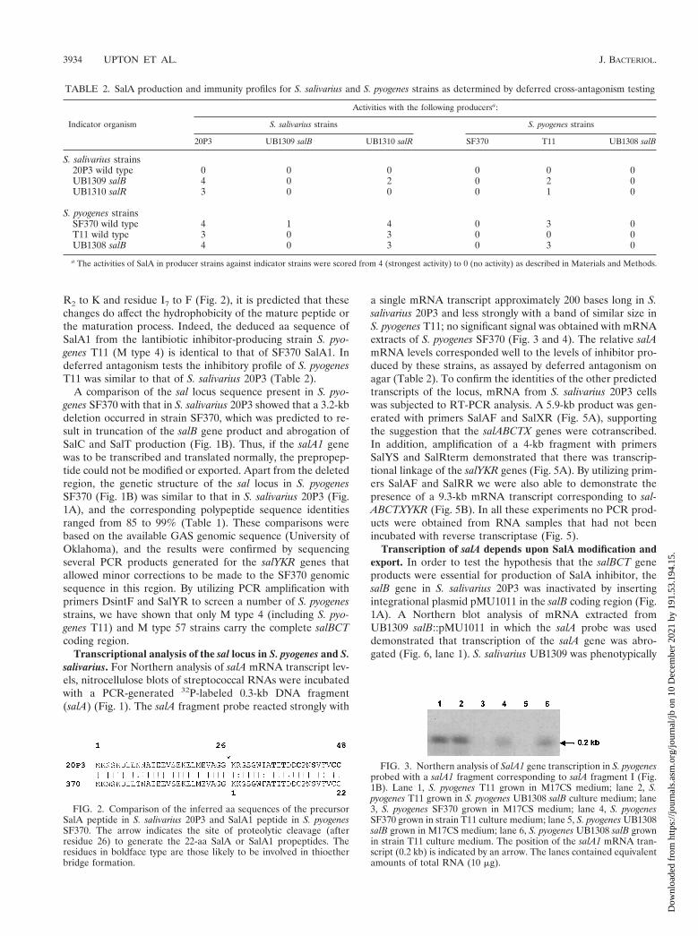
R2 to K and residue I7 to F (Fig. 2), it is predicted that thesechanges do affect the hydrophobicity of the mature peptide orthe maturation process. Indeed, the deduced aa sequence ofSalA1 from the lantibiotic inhibitor-producing strain S. pyo-genes T11 (M type 4) is identical to that of SF370 SalA1. Indeferred antagonism tests the inhibitory profile of S. pyogenesT11 was similar to that of S. salivarius 20P3 (Table 2).
A comparison of the sal locus sequence present in S. pyo-genes SF370 with that in S. salivarius 20P3 showed that a 3.2-kbdeletion occurred in strain SF370, which was predicted to re-sult in truncation of the salB gene product and abrogation ofSalC and SalT production (Fig. 1B). Thus, if the salA1 genewas to be transcribed and translated normally, the prepropep-tide could not be modified or exported. Apart from the deletedregion, the genetic structure of the sal locus in S. pyogenesSF370 (Fig. 1B) was similar to that in S. salivarius 20P3 (Fig.1A), and the corresponding polypeptide sequence identitiesranged from 85 to 99% (Table 1). These comparisons werebased on the available GAS genomic sequence (University ofOklahoma), and the results were confirmed by sequencingseveral PCR products generated for the salYKR genes thatallowed minor corrections to be made to the SF370 genomicsequence in this region. By utilizing PCR amplification withprimers DsintF and SalYR to screen a number of S. pyogenesstrains, we have shown that only M type 4 (including S. pyo-genes T11) and M type 57 strains carry the complete salBCTcoding region.
Transcriptional analysis of the sal locus in S. pyogenes and S.salivarius. For Northern analysis of salA mRNA transcript lev-els, nitrocellulose blots of streptococcal RNAs were incubatedwith a PCR-generated 32P-labeled 0.3-kb DNA fragment(salA) (Fig. 1). The salA fragment probe reacted strongly with
a single mRNA transcript approximately 200 bases long in S.salivarius 20P3 and less strongly with a band of similar size inS. pyogenes T11; no significant signal was obtained with mRNAextracts of S. pyogenes SF370 (Fig. 3 and 4). The relative salAmRNA levels corresponded well to the levels of inhibitor pro-duced by these strains, as assayed by deferred antagonism onagar (Table 2). To confirm the identities of the other predictedtranscripts of the locus, mRNA from S. salivarius 20P3 cellswas subjected to RT-PCR analysis. A 5.9-kb product was gen-erated with primers SalAF and SalXR (Fig. 5A), supportingthe suggestion that the salABCTX genes were cotranscribed.In addition, amplification of a 4-kb fragment with primersSalYS and SalRterm demonstrated that there was transcrip-tional linkage of the salYKR genes (Fig. 5A). By utilizing prim-ers SalAF and SalRR we were also able to demonstrate thepresence of a 9.3-kb mRNA transcript corresponding to sal-ABCTXYKR (Fig. 5B). In all these experiments no PCR prod-ucts were obtained from RNA samples that had not beenincubated with reverse transcriptase (Fig. 5).
Transcription of salA depends upon SalA modification andexport. In order to test the hypothesis that the salBCT geneproducts were essential for production of SalA inhibitor, thesalB gene in S. salivarius 20P3 was inactivated by insertingintegrational plasmid pMU1011 in the salB coding region (Fig.1A). A Northern blot analysis of mRNA extracted fromUB1309 salB::pMU1011 in which the salA probe was useddemonstrated that transcription of the salA gene was abro-gated (Fig. 6, lane 1). S. salivarius UB1309 was phenotypically
TABLE 2. SalA production and immunity profiles for S. salivarius and S. pyogenes strains as determined by deferred cross-antagonism testing
Indicator organism
Activities with the following producersa:
S. salivarius strains S. pyogenes strains
20P3 UB1309 salB UB1310 salR SF370 T11 UB1308 salB
S. salivarius strains20P3 wild type 0 0 0 0 0 0UB1309 salB 4 0 2 0 2 0UB1310 salR 3 0 0 0 1 0
S. pyogenes strainsSF370 wild type 4 1 4 0 3 0T11 wild type 3 0 3 0 0 0UB1308 salB 4 0 3 0 3 0
a The activities of SalA in producer strains against indicator strains were scored from 4 (strongest activity) to 0 (no activity) as described in Materials and Methods.
FIG. 2. Comparison of the inferred aa sequences of the precursorSalA peptide in S. salivarius 20P3 and SalA1 peptide in S. pyogenesSF370. The arrow indicates the site of proteolytic cleavage (afterresidue 26) to generate the 22-aa SalA or SalA1 propeptides. Theresidues in boldface type are those likely to be involved in thioetherbridge formation.
FIG. 3. Northern analysis of SalA1 gene transcription in S. pyogenesprobed with a salA1 fragment corresponding to salA fragment I (Fig.1B). Lane 1, S. pyogenes T11 grown in M17CS medium; lane 2, S.pyogenes T11 grown in S. pyogenes UB1308 salB culture medium; lane3, S. pyogenes SF370 grown in M17CS medium; lane 4, S. pyogenesSF370 grown in strain T11 culture medium; lane 5, S. pyogenes UB1308salB grown in M17CS medium; lane 6, S. pyogenes UB1308 salB grownin strain T11 culture medium. The position of the salA1 mRNA tran-script (0.2 kb) is indicated by an arrow. The lanes contained equivalentamounts of total RNA (10 mg).
3934 UPTON ET AL. J. BACTERIOL.
Dow
nloa
ded
from
http
s://j
ourn
als.
asm
.org
/jour
nal/j
b on
10
Dec
embe
r 20
21 b
y 19
1.53
.194
.15.
SalA2 as determined by an agar inhibition assay and did notsignificantly inhibit growth of S. pyogenes (Table 2). The anal-ogous salB gene inactivation experiment was then carried outwith S. pyogenes T11 by integrating plasmid pMU1016 in thesalB chromosomal gene (Fig. 1B). The resulting isogenic mu-tant, UB1308, did not produce a 0.2-kb salA1 transcript (Fig. 3,lane 5) and was also inhibitor production negative (SalA12) asdetermined by the agar inhibition assay (Table 2). These re-sults confirmed that the salBCT gene products were essentialfor production of SalA and SalA1 peptides and suggested thatSalA and SalA1 lantibiotics modulated salA and salA1 genetranscription.
Expression of SalA is autoregulated. To test the hypothesisthat salA expression depended upon the presence of activeSalA, S. salivarius 20P3 cells were removed from a late-expo-nential-phase culture in M17CS medium. The spent mediumwas inoculated with S. salivarius UB1309 salB cells; the controlconsisted of spent medium from a UB1309 culture. The cul-tures were incubated at 37°C for 4 h, and mRNAs were thenextracted, used for Northern analysis, and probed with the salAgene fragment as described above. Following incubation in theS. salivarius 20P3 spent culture medium, the UB1309 salB cellsproduced a 0.2-kb salA mRNA transcript (Fig. 6, lane 3) thatwas absent from the control cells (Fig. 6, lane 1).
In similar experiments, induction of salA1 transcription was
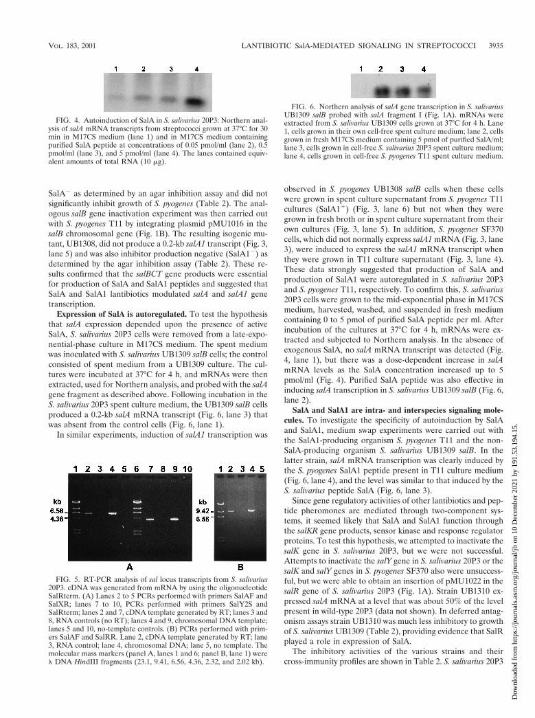
observed in S. pyogenes UB1308 salB cells when these cellswere grown in spent culture supernatant from S. pyogenes T11cultures (SalA11) (Fig. 3, lane 6) but not when they weregrown in fresh broth or in spent culture supernatant from theirown cultures (Fig. 3, lane 5). In addition, S. pyogenes SF370cells, which did not normally express salA1 mRNA (Fig. 3, lane3), were induced to express the salA1 mRNA transcript whenthey were grown in T11 culture supernatant (Fig. 3, lane 4).These data strongly suggested that production of SalA andproduction of SalA1 were autoregulated in S. salivarius 20P3and S. pyogenes T11, respectively. To confirm this, S. salivarius20P3 cells were grown to the mid-exponential phase in M17CSmedium, harvested, washed, and suspended in fresh mediumcontaining 0 to 5 pmol of purified SalA peptide per ml. Afterincubation of the cultures at 37°C for 4 h, mRNAs were ex-tracted and subjected to Northern analysis. In the absence ofexogenous SalA, no salA mRNA transcript was detected (Fig.4, lane 1), but there was a dose-dependent increase in salAmRNA levels as the SalA concentration increased up to 5pmol/ml (Fig. 4). Purified SalA peptide was also effective ininducing salA transcription in S. salivarius UB1309 salB (Fig. 6,lane 2).
SalA and SalA1 are intra- and interspecies signaling mole-cules. To investigate the specificity of autoinduction by SalAand SalA1, medium swap experiments were carried out withthe SalA1-producing organism S. pyogenes T11 and the non-SalA-producing organism S. salivarius UB1309 salB. In thelatter strain, salA mRNA transcription was clearly induced bythe S. pyogenes SalA1 peptide present in T11 culture medium(Fig. 6, lane 4), and the level was similar to that induced by theS. salivarius peptide SalA (Fig. 6, lane 3).
Since gene regulatory activities of other lantibiotics and pep-tide pheromones are mediated through two-component sys-tems, it seemed likely that SalA and SalA1 function throughthe salKR gene products, sensor kinase and response regulatorproteins. To test this hypothesis, we attempted to inactivate thesalK gene in S. salivarius 20P3, but we were not successful.Attempts to inactivate the salY gene in S. salivarius 20P3 or thesalK and salY genes in S. pyogenes SF370 also were unsuccess-ful, but we were able to obtain an insertion of pMU1022 in thesalR gene of S. salivarius 20P3 (Fig. 1A). Strain UB1310 ex-pressed salA mRNA at a level that was about 50% of the levelpresent in wild-type 20P3 (data not shown). In deferred antag-onism assays strain UB1310 was much less inhibitory to growthof S. salivarius UB1309 (Table 2), providing evidence that SalRplayed a role in expression of SalA.
The inhibitory activities of the various strains and theircross-immunity profiles are shown in Table 2. S. salivarius 20P3
FIG. 4. Autoinduction of SalA in S. salivarius 20P3: Northern anal-ysis of salA mRNA transcripts from streptococci grown at 37°C for 30min in M17CS medium (lane 1) and in M17CS medium containingpurified SalA peptide at concentrations of 0.05 pmol/ml (lane 2), 0.5pmol/ml (lane 3), and 5 pmol/ml (lane 4). The lanes contained equiv-alent amounts of total RNA (10 mg).
FIG. 5. RT-PCR analysis of sal locus transcripts from S. salivarius20P3. cDNA was generated from mRNA by using the oligonucleotideSalRterm. (A) Lanes 2 to 5 PCRs performed with primers SalAF andSalXR; lanes 7 to 10, PCRs performed with primers SalY2S andSalRterm; lanes 2 and 7, cDNA template generated by RT; lanes 3 and8, RNA controls (no RT); lanes 4 and 9, chromosomal DNA template;lanes 5 and 10, no-template controls. (B) PCRs performed with prim-ers SalAF and SalRR. Lane 2, cDNA template generated by RT; lane3, RNA control; lane 4, chromosomal DNA; lane 5, no template. Themolecular mass markers (panel A, lanes 1 and 6; panel B, lane 1) werel DNA HindIII fragments (23.1, 9.41, 6.56, 4.36, 2.32, and 2.02 kb).
FIG. 6. Northern analysis of salA gene transcription in S. salivariusUB1309 salB probed with salA fragment I (Fig. 1A). mRNAs wereextracted from S. salivarius UB1309 cells grown at 37°C for 4 h. Lane1, cells grown in their own cell-free spent culture medium; lane 2, cellsgrown in fresh M17CS medium containing 5 pmol of purified SalA/ml;lane 3, cells grown in cell-free S. salivarius 20P3 spent culture medium;lane 4, cells grown in cell-free S. pyogenes T11 spent culture medium.
VOL. 183, 2001 LANTIBIOTIC SalA-MEDIATED SIGNALING IN STREPTOCOCCI 3935
Dow
nloa
ded
from
http
s://j
ourn
als.
asm
.org
/jour
nal/j
b on
10
Dec
embe
r 20
21 b
y 19
1.53
.194
.15.
produced the highest SalA inhibitor activity of all the strains,and growth of wild-type 20P3 cells was not inhibited by any ofthe other strains. S. pyogenes T11 had an inhibitory and immu-nity profile similar to that of S. salivarius 20P3, but it wasweaker. By contrast, S. pyogenes SF370 and UB1309 and S.salivarius UB1308 did not produce inhibitory activities (Table2). These strains were also sensitive to growth inhibition bySalA and SalA1 peptides produced by S. salivarius 20P3 andUB1310 salR and S. pyogenes T11. S. salivarius UB1310 salR,which exhibited a moderate level of inhibitory activity againstS. pyogenes T11 and UB1308, was sensitive only to inhibition byS. salivarius 20P3 and S. pyogenes T11. Thus, streptococci thatproduce SalA or SalA1 peptides have increased immunity toboth lantibiotics compared with SalA-negative or SalA1-nega-tive streptococci.
DISCUSSION
It is now well established that gram-positive bacteria, espe-cially lactic acid bacteria, produce an array of extracellularpeptides with diverse functions. These molecules may act ashighly specific intercellular signals for controlling competencedevelopment, mating responses, and virulence factor produc-tion in bacterial communities or may act as bacteriocin-likeinhibitors of the growth of other neighboring species (10).Salivaricin A was one of the first bacteriocin-like inhibitors thatwere purified, sequenced, and characterized (34). This lantibi-otic is produced by S. salivarius, and homologous SalA1 pep-tides are produced by M type 4 strains of S. pyogenes (37). Inthis study we extended our understanding of the productionand function of this lantibiotic through comparative geneticanalysis of the sal genetic loci in S. salivarius and S. pyogenes.
The genetic organization of the sal locus in strain 20P3resembles that of other lantibiotic synthesis gene clusters (36).Transcriptional analyses suggested that there are at least twosal mRNA transcripts, a major (inducible) salA mRNA tran-script approximately 200 bases long and a salABCTXYKRreadthrough transcript. RT-PCR data are consistent with theproposal that the salBCTX genes are cotranscribed with salA,but the presence of an inverted repeat sequence downstreamof the salA structural gene would be predicted to form a hair-pin loop and attenuate transcriptional readthrough from salAinto salBCTX. This would allow production of higher levels ofSalA compared with the levels of SalBCTX, the modificationand export machinery, and is a control feature found in anumber of other lantibiotic synthesis gene operons (18, 23, 33).From the sequence analysis, we predicted that transcription ofsalYKR may also be initiated from a promoter immediatelyupstream of salY, but transcriptional start point mapping ex-periments would be necessary to confirm this. The significanceof the different transcripts to lantibiotic production and controlis not yet understood, but it is worth noting that among thetranscripts from the L. lactis nisin lantibiotic gene locus, atranscript corresponding to the entire locus has been reported(31).
Comparison of the polypeptide sequences encoded by thesal gene loci in S. salivarius and S. pyogenes with the sequencesin protein sequence databases allowed us to assign polypeptidefunctions. One unusual feature of the locus was the presence oftwo genes (salB and salC) encoding lantibiotic modification
enzymes, since subclass AII lantibiotics with the characteristicGly-Gly motif are usually modified by the product of a singlegene (lanM) (36). The only gene for which a possible functionof the product could not be inferred from a database matchwas salY. Since most lantibiotic gene clusters carry a self-immunity protein gene (36), it is possible that the 635-aa SalYprotein is associated with self-protection against SalA. How-ever, it is also possible that genetic determinants required forimmunity are located elsewhere on the chromosome (2, 11).
Inactivation of the salB gene in S. salivarius 20P3 or S.pyogenes T11 led to a lantibiotic-negative (SalA2 or SalA12)phenotype, confirming that the salB gene product plays anessential role in lantibiotic production. However, inactivationof salB also resulted in abrogation of salA and salA1 transcrip-tion. This transcription was restored by exogenous addition ofsubinhibitory levels of SalA or SalA1 peptides, demonstratingthat production of these lantibiotics is autoregulated, like pro-duction of nisin and subtilin (21, 23). Interestingly, salB mu-tants were also rendered sensitive to growth inhibition by SalAor SalA1 peptides, so lantibiotic production and immunity tothe lantibiotic are coregulated. These results explain clearlywhy S. pyogenes strains which contain a deletion across thesalBCT genes, such as SF370 (M type 1), are not producers ofSalA1 lantibiotic and are also very sensitive to growth inhibi-tion by SalA or SalA1 peptides.
A model for autoregulation of SalA production in S. saliva-rius is presented in Fig. 7; this model is based on experimentalevidence presented here and comparative data for other lan-tibiotic synthesis systems (7, 8, 20, 21, 24). Transcription of thesalABCTX genes is initiated at a promoter upstream of salA.The primary translation products include the 48-aa precursorpro-SalA and the SalB, SalC, SalT, and SalX protein modifi-cation and transport machinery. Modification of the precursorpeptide occurs with a membrane-associated SalB-SalC enzymecomplex, and the membrane-integral SalT-SalX polypeptidesthen mediate translocation of SalA across the cytoplasmicmembrane (Fig. 7). Cleavage of the precursor SalA leaderpeptide results from cysteine protease activity located in theN-terminal region of SalT, which generates extracellular ma-ture SalA peptide. The fate of the precursor SalA leader pep-tide is not known.
The gene clusters encoding production of nisin (12), strep-tococcin SA-FF22 (26), and subtilin (22) each contain twogenes for a two-component (sensor-regulator) system. It hasbeen shown that in the nisin and subtilin systems the matureinhibitor peptide is recognized by the sensor kinase (21, 23,30). We propose that mature SalA peptide is sensed by thesensor histidine kinase of a two-component system (putativelySalKR), which leads to activation of the response regulator(SalR) and induction of transcription from the salA promoter(Fig. 7). Although we were unable to generate a salK mutantand therefore were unable to formally test the notion that SalKis the cognate sensor for SalA, we generated a salR mutant thatcontained reduced salA mRNA transcript levels and exhibitedreduced SalA inhibitor production. This is consistent with theproposed role for SalR in autoregulation of SalA, but sincesalA mRNA transcription was not shut down in this mutant,other regulatory pathways must also be operative.
An interesting finding is that SalA production by one strep-tococcal species may be induced by sensing of the homologous
3936 UPTON ET AL. J. BACTERIOL.
Dow
nloa
ded
from
http
s://j
ourn
als.
asm
.org
/jour
nal/j
b on
10
Dec
embe
r 20
21 b
y 19
1.53
.194
.15.

peptide from another streptococcal species. Interspecies sig-naling between taxonomically diverse streptococcal species hasnot been demonstrated previously. Most peptide signalingmolecules are specific for their cognate sensor-response sys-tems in streptococci (6, 10), but the SalA peptide sensingsystem apparently does not discriminate between SalA andSalA1 (Fig. 2). Minor modifications to other inhibitor peptides,such as epilancin K7 (44), streptococcin SA-FF22 (19), andsubtilin (3), are sufficient to affect their biological activities, butthe two conservative changes in SalA1 do not significantlyaffect the inhibitor activity or profile. Interspecies communitysensing could regulate relative population levels of strepto-cocci. The ability of SalA1-producing S. pyogenes strains torespond to SalA from S. salivarius (and vice versa) could pro-vide a selective mechanism for cocolonization of the mucosalepithelium by pathogen and commensal cell populations. Forexample, SalA1 produced by rapidly multiplying S. pyogenescells might stimulate production of SalA by S. salivarius strains,leading to modulation of the number of S. pyogenes cells. Inthis way, the number of S. pyogenes cells may be self-limiting,thus promoting maintenance of the population in a carrierstate. An alternative scenario is that sensing of SalA by S.pyogenes may influence expression of other genes, includinggenes related to virulence. In this regard it has recently beendemonstrated that a bacteriocin-like peptide (BlpC) in S. pneu-moniae induces a set of 16 genes following activation of itscognate two-component sensor-regulator system (6). We arecurrently investigating the possibility that SalR, like CsrR (17)or Mga (25) proteins, may have a global regulatory function inS. pyogenes and modulate production of virulence factors, suchas streptolysin S (28).
In summary, here we describe the genetic locus encoding thepolypeptides necessary for autoregulated production of SalA
and SalA1 lantibiotics by S. salivarius and S. pyogenes. TheSalA and SalA1 peptides act as inhibitors of sensitive strepto-coccal strains which do not themselves produce these peptides,but they are also sensed by other SalA-producing or SalA1-producing strains and there is concomitant induction of lan-tibiotic synthesis. It could be envisaged, therefore, that SalAproduction and SalA1 production have a role in modulatingthe coexistence of streptococcal populations in vivo, where it isbecoming apparent that signaling peptides are likely to have amajor influence on oral microbial community structure.
ACKNOWLEDGMENTS
We thank Nancy Ragland for expert technical assistance, K. Rossand K. Dierksen for helpful discussions, R. Burne for advice on elec-troporation, A. Podbielski for kindly supplying plasmids, J. Ferretti forproviding strains, and N. Jakubovics for help with preparation of themanuscript. We gratefully acknowledge the BLAST search facilities atthe National Center for Biotechnology Information (National Libraryof Medicine, Washington, D.C.) and the University of OklahomaStreptococcal (GAS) Genome Sequencing Project funded by aUSPHS/NIH grant to B. A. Roe, S. P. Linn, L. Song, X. Yuan, S.Clifton, R. E. McLaughlin, M. McShan, and J. Ferretti.
This work was supported by grant UO0605 from the Marsden Fund,Royal Society of New Zealand.
REFERENCES
1. Bisno, A. L. 1991. Group A streptococcal infections and acute rheumaticfever. N. Engl. J. Med. 325:783–793.
2. Brurberg, M. B., I. F. Nes, and V. G. H. Eijsink. 1997. Pheromone-inducedproduction of antimicrobial peptides in Lactobacillus. Mol. Microbiol. 26:347–360.
3. Chan, W. C., B. W. Bycroft, M. L. Leyland, L. Y. Lian, and G. C. Roberts.1993. A novel post-translational modification of the peptide antibiotic sub-tilin: isolation and characterization of a natural variant from Bacillus subtilisATCC 6633. Biochem. J. 291:23–27.
4. Chen, Y. Y., C. A. Weaver, D. R. Mendelsohn, and R. A. Burne. 1998.Transcriptional regulation of the Streptococcus salivarius 57.I urease operon.J. Bacteriol. 180:5769–5775.
FIG. 7. Model for autoregulation of SalA production in S. salivarius and S. pyogenes. The product of the salA gene, preproSalA peptide, ismodified by the membrane-associated SalBC polypeptides and then exported with leader peptide cleavage by the membrane-integral SalTXpolypeptides. Extracellular lantibiotic peptide from S. salivarius (SalA) or from S. pyogenes (SalA1) is sensed at the cell surface, possibly by thetwo-component SalKR system, and transcription of the salA promoter is upregulated via a phosphorylated (P) regulatory protein (R). Only thebacteria that respond to extracellular SalA peptide by producing active SalA are immune to the inhibitory effects of the lantibiotic peptide, butthe mechanism of immunity is unknown.
VOL. 183, 2001 LANTIBIOTIC SalA-MEDIATED SIGNALING IN STREPTOCOCCI 3937
Dow
nloa
ded
from
http
s://j
ourn
als.
asm
.org
/jour
nal/j
b on
10
Dec
embe
r 20
21 b
y 19
1.53
.194
.15.

5. Church, G. M., and W. Gilbert. 1984. Genomic sequencing. Proc. Natl. Acad.Sci. USA 81:1991–1995.
6. de Saizieu, A., C. Gardes, N. Flint, C. Wagner, M. Kamber, T. J. Mitchell, W.Keck, K. E. Amrein, and R. Lange. 2000. Microarray-based indentification ofa novel Streptococcus pneumoniae regulon controlled by an autoinducedpeptide. J. Bacteriol. 182:4696–4703.
7. de Vos, W. M., G. Jung, and H.-G. Sahl. 1991. Appendix: definitions andnomenclature of lantibiotics, p. 457–464. In G. Jung and H.-G. Sahl (ed.),Nisin and novel lantibiotics. Escom Publishers, Leiden, The Netherlands.
8. de Vos, W. M., O. P. Kuipers, J. R. van der Meer, and R. J. Siezen. 1995.Maturation pathway of nisin and other lantibiotics: post-translationally mod-ified antimicrobial peptides exported by gram positive bacteria. Mol. Micro-biol. 17:427–437.
9. Dower, W. J., J. F. Miller, and C. W. Ragsdale. 1988. High efficiency trans-formation of E. coli by high voltage electroporation. Nucleic Acids Res.16:6127–6145.
10. Dunny, G. M., and B. A. B. Leonard. 1997. Cell-cell communication ingram-positive bacteria. Annu. Rev. Microbiol. 51:527–564.
11. Eijsink, V. G. H., M. Skeie, H. Middelhove, M. B. Brurberg, and I. F. Nes.1998. Comparative studies of class IIa bacteriocins of lactic acid bacteria.Appl. Environ. Microbiol. 64:3275–3281.
12. Engelke, G., Z. Gutowski-Eckel, M. Hammelmann, and K.-D. Entian. 1992.Biosynthesis of the lantibiotic nisin: genomic organization and membranelocalization of the NisB protein. Appl. Environ. Microbiol. 58:3730–3743.
13. Engelke, G., Z. Gutowski-Eckel, P. Kiesau, K. Siegers, M. Hammelmann,and K.-D. Entian. 1994. Regulation of nisin biosynthesis and immunity inLactococcus lactis 6F3. Appl. Environ. Microbiol. 60:814–825.
14. Hanahan, D., J. Jessee, and F. R. Bloom. 1991. Plasmid transformation ofEscherichia coli and other bacteria. Methods Enzymol. 204:63–113.
15. Havarstein, L. S., G. Coomaraswamy, and D. A. Morrison. 1995. An un-modified heptadecapeptide pheromone induces competence for genetictransformation in Streptococcus pneumoniae. Proc. Natl. Acad. Sci. USA92:11140–11144.
16. Havarstein, L. S., D. B. Diep, and I. F. Nes. 1995. A family of bacteriocinABC transporters carry out proteolytic processing of their substrates com-comitant with export. Mol. Microbiol. 16:229–240.
17. Heath, A., V. J. Di Rita, N. L. Barg, and N. C. Engleberg. 1999. A two-component regulatory system, CsrR-CsrS, represses expression of threeStreptococcus pyogenes virulence factors, hyaluronic acid capsule, streptolysinS, and pyogenic extoxin B. Infect. Immun. 67:5298–5305.
18. Hynes, W. L., J. J. Ferretti, and J. R. Tagg. 1993. Cloning of the geneencoding Streptococcin A-FF22, a novel lantibiotic produced by Streptococ-cus pyogenes, and determination of its nucleotide sequence. Appl. Environ.Microbiol. 59:1969–1971.
19. Jack, R. W., and J. R. Tagg. 1991. Isolation and partial structure of strep-tococcin SA-FF22, p. 171–179. In G. Jung and H.-G. Sahl (ed.), Nisin andnovel lantibiotics. Escom Publishers, Leiden, The Netherlands.
20. Jung, G. 1991. Lantibiotics: a survey, p. 1–34. In G. Jung and H.-G. Sahl(ed.), Nisin and novel lantibiotics. Escom Publishers, Leiden, The Nether-lands.
21. Kleerebezem, M., W. M. de Vos, and O. P. Kuipers. 1999. The lantibioticsnisin and subtilin act as extracellular regulators of their own biosynthesis, p.159–174. In G. M. Dunny and S. C. Winans (ed.), Cell-cell signaling inbacteria. American Society for Microbiology, Washington, D.C.
22. Klein, C., C. Kaletta, and K.-D. Entian. 1993. Biosynthesis of the lantibioticsubtilin is regulated by a histidine kinase/response regulator system. Appl.Environ. Microbiol. 59:296–303.
23. Kuipers, O. P., M. M. Beerthuyzen, P. G. G. A. de Reuyter, E. J. Luesink,and W. M. de Vos. 1995. Autoregulation of nisin biosynthesis in Lactococcuslactis by signal transduction. J. Biol. Chem. 270:27299–27304.
24. Louw, M. E., S. J. Reid, D. M. James, and T. G. Watson. 1994. Cloning andsequencing the degS-degU operon from an alkalophilic Bacillus brevis. Appl.Microbiol. Biotechnol. 42:78–84.
25. McIver, K., A. S. Thurman, and J. R. Scott. 1999. Regulation of mga tran-scription in the group A streptococcus: specific binding of Mga within its ownpromoter and evidence for a negative regulator. J. Bacteriol. 181:5373–5383.
26. McLaughlin, R. E., J. J. Ferretti, and W. L. Hynes. 1999. Nucleotide se-quence of the streptococcin A-FF22 lantibiotic regulon: model for the pro-duction of the lantibiotic SA-FF22 by strain of Streptococcus pyogenes. FEMSMicrobiol. Lett. 175:171–177.
27. Meyer, C., G. Bierbaum, C. Heidrich, M. Reis, J. Suling, M. I. Iglesias-Wind,
C. Kempter, E. Molitor, and H.-G. Sahl. 1995. Nucleotide sequence of thelantibiotic Pep5 biosynthetic gene cluster and functional analysis of PepPand PepC. Evidence for a role of PepC in thiether formation. Eur. J. Bio-chem. 232:478–489.
28. Nizet, V., B. Beall, D. J. Bast, V. Datta, L. Kilburn, D. E. Low, and J. C. S.de Azavedo. 2000. Genetic locus for streptolysin S production by group Astreptococcus. Infect. Immun. 68:4245–4254.
29. Podbielski, A., B. Spellberg, M. Woschnik, B. Pohl, and R. Luticken. 1996.Novel series of plasmid vectors for gene inactivation and expression analysisin group A streptococci (GAS). Gene 177:137–147.
30. Ra, S. R., M. Qiao, T. Immonen, I. Pujana, and P. E. J. Saris. 1996. Genesresponsible for nisin synthesis, regulation and immunity form a regulon oftwo operons and are induced by nisin in Lactococcus lactis N8. Microbiology142:1281–1288.
31. Ra, S. R., M. M. Beerthuyzen, W. M. de Vos, P. E. J. Saris, and O. P.Kuipers. 1999. Effects of gene disruptions in the nisin gene cluster of Lac-tococcus lactis on nisin production and producer immunity. Microbiology145:1227–1233.
32. Rince, A., A. Dufour, S. Le Pogam, D. Thuault, C. M. Bourgeois, and J. LePennec. 1994. Cloning, expression, and nucleotide sequence of genes in-volved in production of lactococcin DR, a bacteriocin from Lactococcuslactis subsp. lactis. Appl. Environ. Microbiol. 60:1652–1657.
33. Rince, A., A. Dufour, P. Uguen, J. Le Pennec, and D. Haras. 1997. Charac-terization of the lacticin 481 operon: the Lactococcus lactis genes lctF, lctE,and lctG encode a putative ABC transporter involved in bacteriocin immu-nity. Appl. Environ. Microbiol. 63:4252–4260.
34. Ross, K. F., C. W. Ronson, and J. R. Tagg. 1993. Isolation and character-ization of the lantibiotic salivaricin A and its structural gene salA fromStreptococcus salivarius 20P3. Appl. Environ. Microbiol. 59:2014–2021.
35. Salmond, G. P., B. W. Bycroft, G. S. Stewart, and P. Williams. 1995. Thebacterial enigma: cracking the code of cell-cell communication. Mol. Micro-biol. 16:615–624.
36. Siezen, R. J., O. P. Kuipers, and W. M. De Vos. 1996. Comparison oflantibiotic gene clusters and encoded proteins. Antonie Leeuwenhoek 69:171–184.
37. Simpson, W. J., N. L. Ragland, C. W. Ronson, and J. R. Tagg. 1995. Alantibiotic gene family widely distributed in Streptococcus salivarius andStreptococcus pyogenes. Dev. Biol. Stand. 85:639–643. Karger, Basel.
38. Skaugen, M., J. Nissen-Meyer, G. Jung, S. Stevanovic, K. Sletten, C. I. M.Abildgaard, and I. F. Nes. 1994. In vivo conversion of L-serine to D-alaninein a ribosomally synthesised polypeptide. J. Biol. Chem. 269:27183–27185.
39. Smith, D. J., J. M. Anderson, W. F. King, J. van Houte, and M. A. Taubman.1993. Oral streptococcal colonization of infants. Oral Microbiol. Immunol.8:1–4.
40. Stock, J. B., A. J. Ninfa, and A. M. Stock. 1989. Protein phosphorylation andregulation of adaptive responses in bacteria. Microbiol. Rev. 53:450–490.
41. Suvrov, A. N., and J. J. Ferretti. 1996. Physical and genetic map of an M type1 strain of Streptococcus pyogenes. J. Bacteriol. 178:5546–5549.
42. Tagg, J. R., and L. V. Bannister. 1979. “Fingerprinting” b haemolytic strep-tococci by their production of and sensitivity to bacteriocin-like inhibitors. J.Med. Microbiol. 12:379–411.
43. Upton, M., P. E. Carter, M. Morgan, G. F. S. Edwards, and T. H. Penning-ton. 1995. Clonal structure of invasive Streptococcus pyogenes strains inNorthern Scotland. Epidemiol. Infect. 115:231–241.
44. van de Kamp, M., L. M. Horstink, H. W. van den Hooven, R. N. Konings,C. W. Hilbers, A. Frey, H.-G. Sahl, J. W. Metzger, and F. J. van de Ven. 1995.Sequence analysis by NMR spectroscopy of the peptide lantibiotic epilancinK7 from Staphylococcus epidermidis. Eur. J. Biochem. 227:757–771.
45. van der Meer, J. R., J. Polman, M. M. Beerthuyzen, R. J. Siezen, O. P.Kuipers, and W. M. De Vos. 1993. Characterization of the Lactococcus lactisnisin A operon genes nisP, encoding a subtilisin-like serine protease involvedin precursor processing, and nisR, encoding a regulatory protein involved innisin biosynthesis. J. Bacteriol. 175:2578–2588.
46. Walker, J. E., M. Sarste, M. J. Runswick, and N. J. Gay. 1982. Distantlyrelated sequences in the a- and b-subunits of ATP synthase, myosin, kinaseand other ATP-requiring enzymes and a common nucleotide binding fold.EMBO J. 1:945–951.
47. Weinrauch, Y., R. Penchev, E. Dubnau, I. Smith, and D. Dubnau. 1990. ABacillus subtilis regulatory gene product for genetic competence and sporu-lation resembles sensor protein members of the bacterial two-componentsignal-transduction systems. Genes Dev. 4:860–872.
3938 UPTON ET AL. J. BACTERIOL.
Dow
nloa
ded
from
http
s://j
ourn
als.
asm
.org
/jour
nal/j
b on
10
Dec
embe
r 20
21 b
y 19
1.53
.194
.15.