Intestinal Lymphangiectasia: a Protein-Losing...
14
The Journal of Clinical Investigation Vol. 46, No. 10, 1967 Intestinal Lymphangiectasia: a Protein-Losing Enteropathy with Hypogammaglobulinemia, Lymphocytopenia and Impaired Homograft Rejection * W. STROBER,4 R. D. WOCHNER, P. P. CARBONE, AND T. A. WALDMANN (From the Metabolism Branch and the Medicine Branch, National Cancer Institute, National Institutes of Health, Bethesda, Maryland) Abstract. Intestinal lymphangiectasia is a disease characterized by dilated intestinal lymphatics, protein-losing enteropathy, hypoalbuminemia, and edema. The immunologic status of 18 patients with intestinal lymphangiec- tasia was studied. Concentrations of IgG, IgA, and IgM were measured by immune precipitation and metabolism of these three immunoglobulins was studied using purified radioiodinated proteins. The serum concentration and total body pool of each immunoglobin were greatly reduced. The fraction of the intravascular protein pool catabolized per day was increased to 34% for IgG, 59% for IgA, and 66% for IgM; these are in contrast with control values of 7%, 28%, and 17%, respectively. Synthetic rates of the immunoglobulins were normal or slightly increased. Primary circulating antibody response was tested in five patients with Vi and tularemia antigens. Titers elicited in patients with the Vi antigen were significantly lower than those seen in a control group, whereas no difference was seen between patient and control responses to the tularemia antigen. Lymphocytopenia was noted in patients with intestinal lymphangiectasia. The mean circulating lymphocyte count was 710 + 340/mm8 in contrast to 2500 + 600/ mm3 in controls. Cellular hypersensitivity was studied with skin tests and skin grafts. 91% of normal individuals reacted to at least one of the four skin test antigens: purified protein derivative, mumps, Trichophyton, and Candida albicans; in contrast, only 17% of patients with intestinal lymphangi- ectasia had a positive reaction. Each of three patients tested with dinitro- chlorobenzene had a negative reaction. Finally, all four patients who received skin homografts have retained these grafts for at least 12 months. The im- munological disorders in patients with intestinal lymphangiectasia appear to result from loss of immunoglobulins and lymphocytes into the gastrointestinal tract secondary to disorders of lymphatic channels. Lymphocyte depletion then leads to skin anergy and impaired homograft rejection. Introduction in the complex group of hypogammaglobulinemias, Immunologic deficiency may take a variety of in which case circulating antibody response to anti- forms. It may be manifest as a defect in the syn- genic challenge is greatly reduced (1). Alterna thesis of one or more serum immunoglobulins as tively, it may be seen as an abnormality in cellu- lar or delayed hypersensitivity as in Hodgkin's *Received for publication 17 March 1967 and in re- disease; in this instance, circulating antibody re- vised form 10 May 1967. Presented in part at the 58th Annual Meeting of the sponse is intact, but the ability to manifest de- American Society for Clinical, Investigation, Atlantic tAddress requests for reprints to Dr. Warren Strober, City, N. J., 1 May 1966; published in abstract form in Metabolism Branch, National Cancer Institute, National J. Clin. Invest. 1966, 45: 1077. Institutes of Health, Bethesda, Md. 20014. 1643
Transcript of Intestinal Lymphangiectasia: a Protein-Losing...
The Journal of Clinical InvestigationVol. 46, No. 10, 1967
Intestinal Lymphangiectasia: a Protein-Losing Enteropathywith Hypogammaglobulinemia, Lymphocytopenia and
Impaired Homograft Rejection *W. STROBER,4 R. D. WOCHNER,P. P. CARBONE,ANDT. A. WALDMANN
(From the Metabolism Branch and the Medicine Branch, National Cancer Institute, NationalInstitutes of Health, Bethesda, Maryland)
Abstract. Intestinal lymphangiectasia is a disease characterized by dilatedintestinal lymphatics, protein-losing enteropathy, hypoalbuminemia, andedema. The immunologic status of 18 patients with intestinal lymphangiec-tasia was studied. Concentrations of IgG, IgA, and IgM were measured byimmune precipitation and metabolism of these three immunoglobulins wasstudied using purified radioiodinated proteins. The serum concentration andtotal body pool of each immunoglobin were greatly reduced. The fraction ofthe intravascular protein pool catabolized per day was increased to 34% forIgG, 59% for IgA, and 66% for IgM; these are in contrast with control valuesof 7%, 28%, and 17%, respectively. Synthetic rates of the immunoglobulinswere normal or slightly increased.
Primary circulating antibody response was tested in five patients with Viand tularemia antigens. Titers elicited in patients with the Vi antigen weresignificantly lower than those seen in a control group, whereas no differencewas seen between patient and control responses to the tularemia antigen.
Lymphocytopenia was noted in patients with intestinal lymphangiectasia.The mean circulating lymphocyte count was 710 + 340/mm8 in contrast to2500 + 600/ mm3in controls. Cellular hypersensitivity was studied with skintests and skin grafts. 91% of normal individuals reacted to at least one of thefour skin test antigens: purified protein derivative, mumps, Trichophyton, andCandida albicans; in contrast, only 17% of patients with intestinal lymphangi-ectasia had a positive reaction. Each of three patients tested with dinitro-chlorobenzene had a negative reaction. Finally, all four patients who receivedskin homografts have retained these grafts for at least 12 months. The im-munological disorders in patients with intestinal lymphangiectasia appear toresult from loss of immunoglobulins and lymphocytes into the gastrointestinaltract secondary to disorders of lymphatic channels. Lymphocyte depletionthen leads to skin anergy and impaired homograft rejection.
Introduction in the complex group of hypogammaglobulinemias,
Immunologic deficiency may take a variety of in which case circulating antibody response to anti-forms. It may be manifest as a defect in the syn- genic challenge is greatly reduced (1). Alternathesis of one or more serum immunoglobulins as tively, it may be seen as an abnormality in cellu-
lar or delayed hypersensitivity as in Hodgkin's*Received for publication 17 March 1967 and in re- disease; in this instance, circulating antibody re-
vised form 10 May 1967.Presented in part at the 58th Annual Meeting of the sponse is intact, but the ability to manifest de-
American Society for Clinical, Investigation, Atlantic tAddress requests for reprints to Dr. Warren Strober,City, N. J., 1 May 1966; published in abstract form in Metabolism Branch, National Cancer Institute, NationalJ. Clin. Invest. 1966, 45: 1077. Institutes of Health, Bethesda, Md. 20014.
1643
STROBER, WOCHNER,CARBONE,AND WALDMANN
layed skin reactions to certain antigens and to re-ject homografts (allografts) is impaired (2, 3).
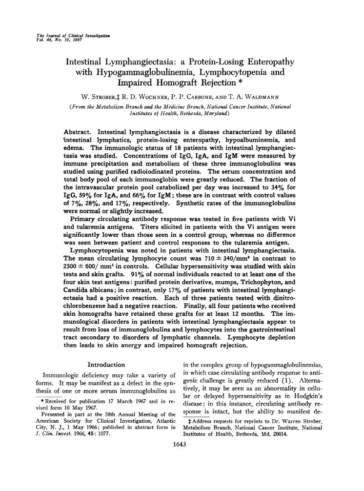
We now characterize an immune deficiencystate associated with the disease, intestinal lym-phangiectasia. Intestinal lymphangiectasia is achronic disorder of lymphatics associated withsevere protein-losing gastroenteropathy (4).Patients with this disorder have marked hypo-proteinemia and severe, often asymmetrical, edema.Gastrointestinal symptoms are usually mild. Mal-absorption of fat may occur, but is usually not se-vere. The hallmark morphological lesion of thisdisease is dilated lymphatic channels of the smallbowel (Fig. 1); peripheral lymphatic abnormali-ties may also be demonstrated by lymphangiog-raphy. These structural lymphatic abnormalities
FIG. 1. SMALL INTESTINAL BIOPSY FROM A PATIENT
WITH INTESTINAL LYMPHANGIECTASIA. Dilated lym-phatics are seen in the tips of villi and in the laminapropria.
occur as congenital malformations or as acquireddefects.
In this study, patients with intestinal lymphangi-ectasia were found to have low serum immuno-globulin levels secondary to increased fractionalcatabolic rates for these proteins. The increasedcatabolism was attributable to excessive loss ofserum proteins into the gastrointestinal lumen. Aconsiderable antibody response was observed afterantigenic challenge, but this response was slightlyless than that observed in normal individuals. Avery severe peripheral lymphocytopenia was regu-larly seen and this was associated with greatly de-creased skin reactivity to certain antigens. Moststriking was the fact that these patients acceptedskin grafts from unrelated donors.
MethodsPatients
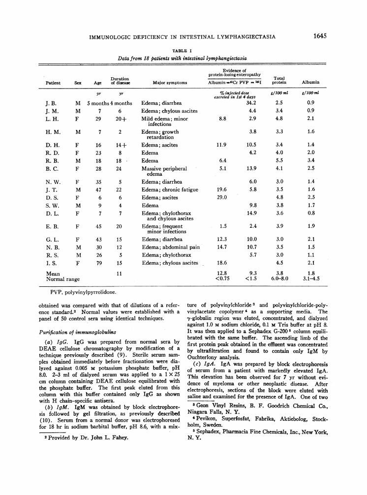
Data obtained from 18 patients with intestinal lymphan-giectasia are included in Table I. The subjects rangedin age from 4 months to 79 yr; the average duration ofdisease was 11 yr. Each patient was shown to have di-lated subepithelial lymphatic channels by peroral smallbowel biopsy or lapartomy. Edema and hypoproteinemiawere present in each case. The mean total serum pro-tein concentration was 3.8 g/100 ml (range: 2.5-5.5 g/100 ml) and the mean serum albumin concentration was1.8 g/100 ml (range: 0.8-3.4 g/100 ml). Each patienthad a markedly shortened albumin survival and a normalalbumin synthetic rate as demonstrated by iodinated al-bumin turnover studies. Each of the patients had eitherabnormally high 4 day polyvinylpyrrolidone-"I gastro-intestinal excretion (mean of 9.3% of injected dose vs.< 1.5% of injected dose in controls) or high 4 day albu-min-"Cr excretion (mean of 12.8%o of injected dose vs.< 0.75%o of injected dose in controls). These findingsindicated that the hypoproteinemia and shortened plasmaprotein survival were due to excessive gastroeintestinalprotein loss (5, 6).
The patients were clinically stable during the periods ofstudy; they were free of infection, were afebrile, andmaintained constant weight. Drug therapy was limitedto diuretics. Tests of liver and renal function werenormal and the patients did not have proteinuria. Therewere no concomitant illnesses except for pulmonaryemphysema in patient J. T., and diabetes mellitus inpatient I. S.
Seruin immunoglobulin quantitationcSerum immunoglobulin concentrations were determined
by radial immunodiffusion (7, 8). Samples were al-lowed to diffuse from wells made in agar impregnated withspecific antiserum.1 The diameter of the precipitin ring
1 Hyland Laboratories, Los Angeles, Calif.
1644
IMMUNOLOGICDEFICIENCY IN INTESTINAL LYMPHANGIECTASIA
TABLE I
Data from 18 patients with intestinal lymphangiectasia
Evidence ofprotein-losing enteropathy
Duration TotalPatient Sex Age of disease Major symptoms Albumin =56Cr PVP 131I protein Albumin
yr yr %injected dose g/100 ml g/100 mlexcreted in 1st 4 days
J. B. M 5 months 4 months Edema; diarrhea 34.2 2.5 0.9J. M. M 7 6 Edema; chylous ascites 4.4 3.4 0.9L. H. F 29 20+ Mild edema; minor 8.8 2.9 4.8 2.1
infectionsH. M. M 7 2 Edema; growth 3.8 3.3 1.6
retardationD. H. F 16 14+ Edema; ascites 11.9 10.5 3.4 1.4R. D. F 23 8 Edema 4.2 4.0 2.0R. B. M 18 18 - Edema 6.4 5.5 3.4B. C. F 28 24 Massive peripheral 5.1 13.9 4.1 2.5
edemaN. W. F 35 5 Edema; diarrhea 6.0 3.0 1.4J. T. M 47 22 Edema; chronic fatigue 19.6 5.8 3.5 1.6D. S. F 6 6 Edema; ascites 29.0 4.8 2.5
S. W. M 9 4 Edema 9.8 3.8 1.7D. L. F 7 7 Edema; chylothorax 14.9 3.6 0.8
and chylous ascitesE. B. F 45 20 Edema; frequent 1.5 2.4 3.9 1.9
minor infectionsG. L. F 43 15 Edema; diarrhea 12.3 10.0 3.0 2.1N. B. M 30 12 Edema; abdominal pain 14.7 10.7 3.5 1.5R. S. M 26 5 Edema; chylothorax 5.7 3.0 1.1I. S. F 79 15 Edema; chylous ascites 18.6 4.5 2.1
Mean 11 12.8 9.3 3.8 1.8Normal range <0.75 <1.5 6.0-8.0 3.1-4.5
PVP, polyvinylpyrrolidone.
obtained was compared with that of dilutions of a refer-ence standard.2 Normal values were established with apanel of 50 control sera using identical techniques.
Purification of immunoglobulins
(a) IgG. IgG was prepared from normal sera byDEAE cellulose chromatography by modification of atechnique previously described (9). Sterile serum sam-ples obtained immediately before fractionation were dia-lyzed against 0.005 M potassium phosphate buffer, pH8.0. 2-3 ml of dialyzed serum was applied to a 1 X 25cm column containing DEAEcellulose equilibrated withthe phosphate buffer. The first peak eluted from thiscolumn with this buffer contained only IgG as shownwith H chain-specific antisera.
(b) IgM. IgM was obtained by block electrophore-sis followed by gel filtration, as previously described(10). Serum from a normal donor was electrophoresedfor 18 hr in sodium 'barbital buffer, pH 8.6, with a mix-
2 Provided by Dr. John L. Fahey.
ture of polyvinylchloride3 and polyvinylchloride-poly-vinylacetate copolymers as a supporting media. The,y-globulin region was eluted, concentrated, and dialyzedagainst 1.0 M sodium chloride, 0.1 M Tris buffer at pH 8.It was then applied to a Sephadex G-200 5 column equili-brated with the same buffer. The ascending limb of thefirst protein peak obtained in the effluent was concentratedby ultrafiltration and found to contain only IgM byOuchterlony analysis.
(c) IgA. IgA was prepared by block electrophoresisof serum from a patient with markedly elevated IgA.This elevation has been observed for 7 yr without evi-dence of myeloma or other neoplastic disease. Afterelectrophoresis, sections of the block were eluted withsaline and examined for the presence of IgA. One of two
8 Geon Vinyl Resins, B. F. Goodrich Chemical Co.,Niagara Falls, N. Y.
'Pevikon, Superfosfat, Fabrika, Aktiebolog, Stock-holm, Sweden.
5 Sephadex, Pharmacia Fine Chemicals, Inc., NewYork,N. Y.
1645

STROBER, WOCHNER,CARBONE,AND WALDMANN
preparations was ultimately used for turnover study.Each of these preparations were analyzed by radioim-munoelectrophoresis and with radial immunodiffusionquantitation: preparation A contained 90% IgA and 10%transferrin; preparation B contained 85% IgA and 15%IgG.
Iodination of purified proteins.
Iodination of each of the above preparations was per-formed with either 1'1I or 'I by the iodine monochloridetechnique of McFarlane (11). All preparations werecalculated to have an average of less than one atom ofiodine per molecule of protein in the final product. Thefinal product contained less than 1% nonprecipitable ra-dioactivity. Normal human albumin was added to eachpreparation to prevent damage to the protein by ir-radiation.
Protocol for turnover studies
Each patient was admitted to the National Cancer In-stitute during the study period. 10 drops of Lugol's solu-tion were administered 3 times daily throughout thestudy period to prevent thyroidal uptake of the releasedisotope. Serum immunoglobulin concentrations were ob-tained at intervals throughout the study period to verifythat each patient was in a steady state. Turnover stud-ies of the protein preparations were done either simul-taneously or sequentially. From 10 to 50 utc of the iodi-nated proteins was administered intravenously from acalibrated syringe, and serum samples were obtained10 min after administration, and daily thereafter. Stooland urine specimens were collected in 24-hr lots. Se-rum and urine samples were counted with appropriatestandards to within ± 3% counting error in an automaticgamma-ray well-type scintillation counter with a thal-lium-activated sodium iodide crystal. When two iso-topes were studied simultaneously, they were differenti-ated with a pulse height analyzer.
Calculation of the metabolic data
The turnovers of the iodinated proteins were analyzedaccording to a modification of the methods of Berson,Yalow, Schreiber, and Post (12), and Pearson, Veall,and Vetter (13). The calculations are summarized asfollows: plasma volume (ml/kg) = radioactivity admin-istered/(radioactivity per milliliter plasma at 10 min Xbody weight); radioactivity retained in the body = radio-activity administered - cumulative excreted radioactivity.
Time plots of the plasma radioactivity and the radio-activity retained in the body were constructed on semi-logarithmic graph paper, and the survival half-times ofthe labeled proteins were determined graphically. Thefraction of the body protein remaining in the intravas-cular space = (plasma volume X plasma radioactivity permilliliter) /radioactivity retained in the body; this frac-tion was determined after equilibration of the labeledprotein among the body compartments. Total circu-lating protein = plasma volume X plasma concentrationof the protein. Total exchangeable pool of the protein =
total circulating protein/fraction of the protein in theintravascular space. Fraction of the circulating proteincatabolized per day = radioactivity excreted in each 24-hrperiod/mean circulating radioactivity during the sameperiod. This fraction was determined for each day, andthe mean value for days 3-15 was taken as the fractionalcatabolic rate. Absolute catabolic rates (turnover rates)= the total circulating protein X fraction of circulatingprotein catabolized per day.
Since the concentration of each of the plasma pro-teins studied remained constant throughout the periodof study, the patients were assumed to be in a steadystate as far as protein metabolism was concerned, andthe synthetic rates for the proteins were consideredto be equal to the turnover rates.
Lymphocyte levels
Blood lymphocyte levels were determined for 18 pa-tients with intestinal lymphangiectasia and 50 youngadult controls. Lymphocyte counts were computed fromthe products of the total venous white blood countsand differential counts. Each reported count representsthe mean of from 2 to 10 determinations.
Antibody stimulation studiesThe ability to produce antibodies in response to spe-
cific antigens was determined in five patients with in-testinal lymphangiectasia and 14 normal control sub-jects (mean age 25). The antigens used were (a) Viantigen (Vi), a polysaccharide isolated from 5396/38strain of E. coli (14, 15), and (b) Foshay tularemia vac-cine, the cell wall of Pasteurella tularensis (16). Theseantigens were chosen since prior exposure was unlikely,and measured antibody responses were therefore primaryin nature. 100 ,ug of Vi and 1 ml of Foshay vaccine weregiven intramuscularly to each patient. Antibody titerswere determined on specimens drawn at weekly intervalsfor 4 wk after injections. Titers were done concom-itantly on all samples using antigen-coated red cells in ahemagglutination titration method (17, 18). The high-est dilution giving a 2 + agglutination reaction was takenas the end point of the assay.
Skin tests
Purified protein derivative (PPD),ff Trichophyton,7Candida albicans, 7 and mumps8 antigens were obtainedfrom commercial sources and injected intradermally intothe forearm of both patients with intestinal lymphangiec-tasia and normal volunteer control subjects. Reactionswere considered positive if at least 1 cm of induration oc-curred at the test site at 48 hr.
Dinitrochlorobenzene (DNCB) skin test (19) sensiti-zation was performed by applying 2000 ,ug of DNCBinacetone on a midforearm site; 100 ,/g DNCBwas appliedconcomitantly to serve as a presensitizing dose control.The initial 2000 ltg dose caused a vesicant reaction in all
6 Parke, Davis & Co., Detroit, Mich.7 Hollister-Stier Laboratories, Inc., Yeadon, Pa.8 Eli Lilly & Co., Indianapolis, Ind.
1646
IMMUNOLOGICDEFICIENCY IN INTESTINAL LYMPHANGIECTASIA
subjects. 2 wk after sensitization, a 100 Ag challengedose was applied. A positive reaction was said to oc-
cur if at least 1 cm of induration was present 48 hr afterapplication and was significantly greater than the ini-tial 100 ug dose reaction.
Skin grafts
The skin grafts were obtained from nonrelated, healthydonors by taking an 8 mmpunch biopsy from the inter-scapular area. No information is available as to thewhite cell grouping of donors or recipients. The skinwas then placed on empty punch biopsy sites of the re-
cipient, usually on the upper medial arm region. Thegrafts were held in place with bandages and carefullyobserved over a prolonged period of time. Photographsof the grafts were taken at appropriate intervals.
Results
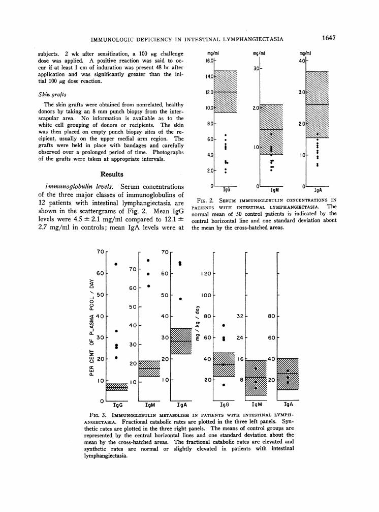
Immunoglobulin levels. Serum concentrationsof the three major classes of immunoglobulins of12 patients with intestinal lymphangiectasia are
shown in the scattergrams of Fig. 2. Mean IgGlevels were 4.5 + 2.1 mg/ml compared to 12.12.7 mg/ml in controls; mean IgA levels were at
70r
60
J- 500
0
<' 4Q0U)
-iAL
300
zW 20U.
w
a.
I 0
0
0
70
70r0
0
601
50 e
.
0
I
60
mg/ml mg/ml
3.0
2.0
1.0
rI
mg/ml4.0 -
3.0
2.0
I.0
°' IgG0 IM 0
IgA
FIG. 2. SERUMIMMUNOGLOBULINCONCENTRATIONSINPATIENTS WITH INTESTINAL LYMPHANGIECTASIA. Thenormal mean of 50 control patients is indicated by thecentral horizontal line and one standard deviation aboutthe mean by the cross-hatched areas.
120 F
100 F50~
40F40-
3030F
Eu
IgG IgM IgA
a
la
Ov
80 32
0
60F g
40
24
0
IgG IgM IgA
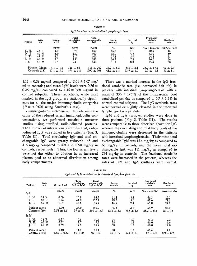
FIG. 3. IMMUNOGLOBULINMETABOLISM IN PATIENTS WITH INTESTINAL LYMPH-
ANGIECTASIA. Fractional catabolic rates are plotted in the three left panels. Syn-thetic rates are plotted in the three right panels. The means of control groups are
represented by the central horizontal lines and one standard deviation about themean by the cross-hatched areas. The fractional catabolic rates are elevated andsynthetic rates are normal or slightly elevated in patients with intestinallymphangiectasia.
1647
STROBER, WOCHNER,CARBONE,ANDWALDMANN
TABLE II
IgG Metabolism in intestinal lymphangiectasia
Total Total FractionalAge, Serum circulating exchangeable Intra- Survival catabolic Synthetic
Patient sex level IgG IgG vascular tj rate rate
mg/mI mg/kg mg/kg % days %IV pool/day mg/kg per dayL. H. 28 F 1.9 73 160 45.6 7.1 20.6 15N. B. 40 M 5.0 180 400 45.0 4.7 33.0 59J. T. 44 M 4.0 110 400 27.5 3.9 64.5 71E. B. 46 M 3.0 130 380 34.2 7.8 26.0 34I. S. 70 M 6.5 230 740 31.1 8.8 25.4 58
Patient Mean 4.1 i 1.7 145 i 62 416 ± 207 36.7 ± 8.2 6.5 =1 2.1 33.9 17.7 47 ± 22Controls (23) 12.1 i: 2.6 494 i: 116 1090 i 263 45.3 i- 6.1 22.9 :1 4.0 6.7 i 1.5 34 41 11
1.15 ± 0.32 mg/ml compared to 2.61 ± 1.07 mg/ There was a marked increase in the IgG frac-ml in controls; and mean IgM levels were 0.76 ± tional catabolic rate (i.e. decreased half-life) in0.26 mg/ml compared to 1.45 ± 0.68 mg/ml in patients with intestinal lymphangiectasia with acontrol subjects. These reductions, while most mean of 33.9 ± 17.7% of the intravascular poolmarked in the IgG group, are statistically signifi- catabolized per day as compared to 6.7 + 1.5% incant for all the major immunoglobulin categories normal control subjects. The IgG synthetic rates(P = < 0.001 using Student's t test). were normal or slightly elevated in the intestinal
Immunoglobulin metabolism. To determine the lymphangiectasia patients.cause of the reduced serum immunoglobulin con- IgM and IgA turnover studies were done incentrations, we performed metabolic turnover three patients (Fig. 3, Table III). The resultsstudies using purified radioiodinated proteins. were comparable to those described above for IgGThe turnover of intravenously administered, radio- wherein the circulating and total body pools of theiodinated IgG was studied in five patients (Fig. 3, immunoglobulins were decreased in the patientsTable II). Total circulating IgG and total ex- with intestinal lymphangiectasia. Their mean totalchangeable IgG were greatly reduced: 145 and exchangeable IgM was 15.4 mg/kg as compared to416 mg/kg compared to 494 and 1090 mg/kg in 66 mg/kg in controls, and the mean total ex-controls, respectively. Thus, the low serum levels changeable IgA was 111 mg/kg as compared towere not due either to dilution in an increased 234 mg/kg in controls. The fractional catabolicplasma pool or to abnormal distribution among rates were increased in the patients, whereas thebody compartments. rates of IgM and IgA synthesis were normal.
TABLE III
IgA and IgM metabolism in intestinal lymphangiectasia
Total Total FractionalAge, circulating exchangeable Intra- Survival catabolic Synthetic
Patient sex Serum level IgA or IgM IgA or IgM vascular I rate rate
mg/ml mg/kg mg/kg % days %IV pool/day mg/kg per dayIgA
L. H. 28 F 0.69 26.8 84.2 31.8 3.4 64.2 17.2I. S. 70 F 1.24 44.6 152.7 29.2 5.0 47.6 21.1J. T. 45 M 1.07 42.6 95.7 44.5 2.4 65.0 27.7Patient mean 1.00 38.0 110.9 35.2 3.6 58.9 22.0Controls (10) 2.53 :1: 1.1 97 52 234 i 135 42.1 d 8.6 6.7 2.3 28.3 1 6.3 25 i 15
IgML. H. 28 F 0.27 9.8 10.4 94 1.0 73.3 7.2N. B. 40 M 0.32 7.7 9.9 78 1.5 66.0 4.6J. T. 45 M 0.62 17.7 25.9 68 1.5 60.0 12.0Patient mean 0.40 11.7 15.4 80 1.3 66.4 7.9Controls (10) 1.45 ± 0.63 50 i 25 66 i 40 76 i 12 5.4 1 1.0 17 - 4.0 8.9 i 6.2
1648
IMMUNOLOGICDEFICIENCY IN INTESTINAL LYMPHANGIECTASIA
The mean fractional catabolic rates of IgM were66.4%o of the intravascular pool as compared to17%7o in controls. Fractional catabolic rates ofIgA averaged 58.97o in the patients as comparedto 28.3%o in controls. It is clear, therefore, thatthe reduced serum concentrations of IgG, IgA,and IgM in patients with intestinal lymphangiec-tasia were secondary to shortened survival of theseproteins.
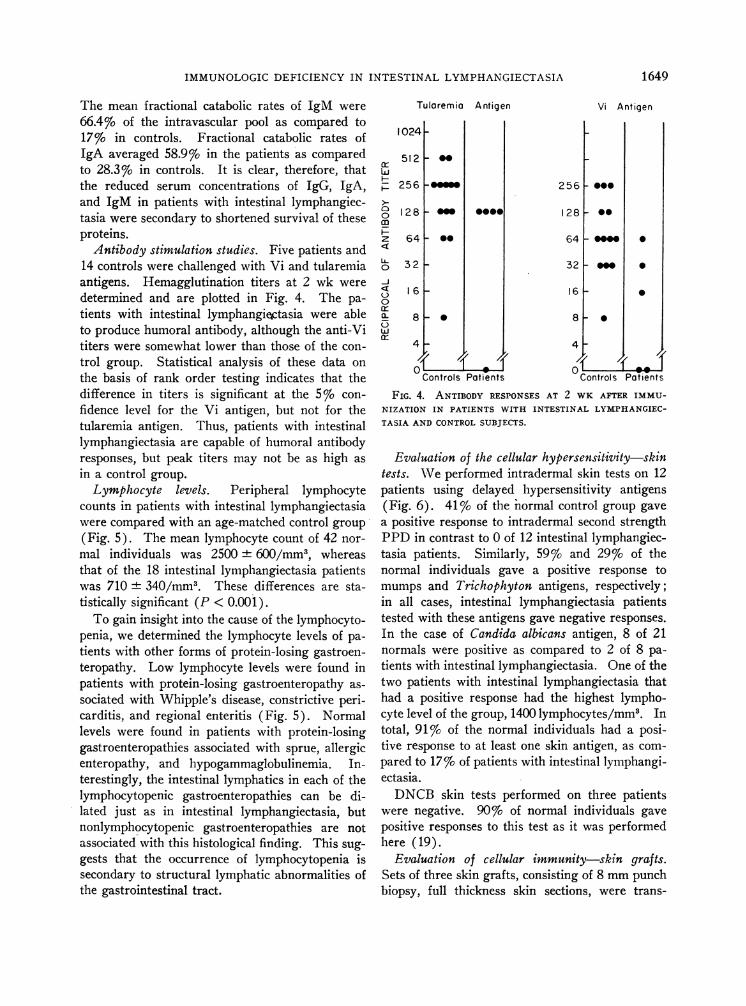
Antibody stimulation studies. Five patients and14 controls were challenged with Vi and tularemiaantigens. Hemagglutination titers at 2 wk weredetermined and are plotted in Fig. 4. The pa-tients with intestinal lymphangiectasia were ableto produce humoral antibody, although the anti-Vititers were somewhat lower than those of the con-trol group. Statistical analysis of these data onthe basis of rank order testing indicates that thedifference in titers is significant at the 5%o con-fidence level for the Vi antigen, but not for thetularemia antigen. Thus, patients with intestinallymphangiectasia are capable of humoral antibodyresponses, but peak titers may not be as high asin a control group.
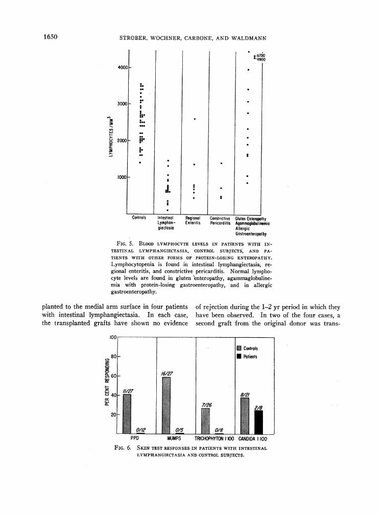
Lymphocyte levels. Peripheral lymphocytecounts in patients with intestinal lymphangiectasiawere compared with an age-matched control group(Fig. 5). The mean lymphocyte count of 42 nor-mal individuals was 2500 + 600/mm3, whereasthat of the 18 intestinal lymphangiectasia patientswas 710 + 340/mm3. These differences are sta-tistically significant (P < 0.001).
To gain insight into the cause of the lymphocyto-penia, we determined the lymphocyte levels of pa-tients with other forms of protein-losing gastroen-teropathy. Low lymphocyte levels were found inpatients with protein-losing gastroenteropathy as-sociated with Whipple's disease, constrictive peri-carditis, and regional enteritis (Fig. 5). Normallevels were found in patients with protein-losinggastroenteropathies associated with sprue, allergicenteropathy, and hypogammaglobulinemia. In-terestingly, the intestinal lymphatics in each of thelymphocytopenic gastroenteropathies can be di-lated just as in intestinal lymphangiectasia, butnonlymphocytopenic gastroenteropathies are notassociated with this histological finding. This sug-gests that the occurrence of lymphocytopenia issecondary to structural lymphatic abnormalities ofthe gastrointestinal tract.
Tularemia Antigen
10241-
512 .0w
00m
z
IL011
-lJ0ar-a.3w
256 P-.me128 F mm
641- 0.
32 -
16 k
0000
8 -*
4 -
Controls Patients
Vi Antigen
256
128
64
32
16
*0-
am
0
0
0
8 - ,
Controls Patients
FIG. 4. ANTIBODY RESPONSESAT 2 WK AFTER IMMU-NIZATION IN PATIENTS WITH INTESTINAL LYMPHANGIEC-TASIA AND CONTROLSUBJECTS.
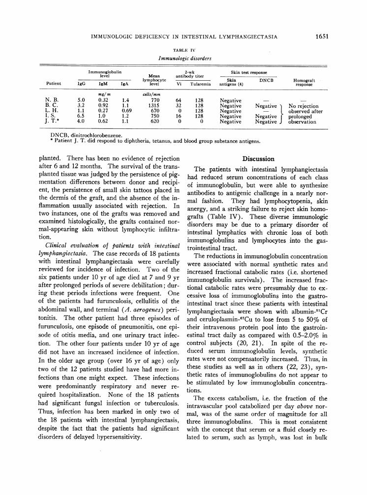
Evaluation of the cellular hypersensitivity-skintests. Weperformed intradermal skin tests on 12patients using delayed hypersensitivity antigens(Fig. 6). 41% of the normal control group gavea positive response to intradermal second strengthPPDin contrast to 0 of 12 intestinal lymphangiec-tasia patients. Similarly, 59% and 29%o of thenormal individuals gave a positive response tomumps and Trichophyton antigens, respectively;in all cases, intestinal lymphangiectasia patientstested with these antigens gave negative responses.In the case of Candida albicans antigen, 8 of 21normals were positive as compared to 2 of 8 pa-tients with intestinal lymphangiectasia. One of thetwo patients with intestinal lymphangiectasia thathad a positive response had the highest lympho-cyte level of the group, 1400 lymphocytes/mm3. Intotal, 91%o of the normal individuals had a posi-tive response to at least one skin antigen, as com-pared to 17%o of patients with intestinal lymphangi-ectasia.
DNCBskin tests performed on three patientswere negative. 90%o of normal individuals gavepositive responses to this test as it was performedhere (19).
Evaluation of cellular immunity-skin grafts.Sets of three skin grafts, consisting of 8 mmpunchbiopsy, full thickness skin sections, were trans-
1649
STROBER, WOCHNER,CARBONE,AND WALDMANN
4000
3000 _
2000 _
10001-
-
Jo
2
Controls Intestinal Regional Constrictive Gluten EnteropathyLymphan- Enteritis Pericard itis Agammoglobulinemiagiectasia Allergic
GastroenteropathyFIG. 5. BLOOD LYMPHOCYTELEVELS IN PATIENTS WITH IN-
TESTINAL LYMPHANGIECTASIA, CONTROL SUBJECTS, AND PA-TIENTS WITH OTHER FORMS OF PROTEIN-LOSING ENTEROPATHY.Lymphocytopenia is found in intestinal lymphangiectasia, re-gional enteritis, and constrictive pericarditis. Normal lympho-cyte levels are found in gluten 'enteropathy, agammaglobuline-mia with protein-losing gastroenteropathy, and in allergicgastroenteropathy.
planted to the medial arm surface in four patientswith intestinal lymphangiectasia. In each case,the transplanted grafts have shown no evidence
100
80
0C> 6016/2760-
MW 11/27"->40I
of rejection during the 1-2 yr period in which theyhave been observed. In two of the four cases, asecond graft from the original donor was trans-
1650
C-
U)
J0--
PPD MUMPS TRCHOPHYTON1:0 CANDIDA 1:100
FIG. 6. SKIN TEST RESPONSESIN PATIENTS WITH INTESTINALLYMPHANGIECTASIAANDCONTROLSUBJECTS.
U00
0
:11000
:*I,
0010
0
IMMUNOLOGICDEFICIENCY IN INTESTINAL LYMPHANGIECTASIA
TABLE IV
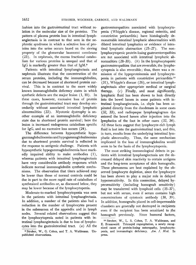
Immunologic disorders
Immunoglobulin 2-wk Skin test responselevel Mean antibody titer
lymphocyte Skin DNCB HomograftPatient IgG IgM IgA level Vi Tularemia antigens (4) response
mg/ m cells/mmN. B. 5.0 0.32 1.4 770 64 128 NegativeB. C. 3.2 0.92 1.1 1315 32 128 Negative Negative No rejectionL. H. 1.1 0.27 0.69 670 0 128 Negative - observed afterI. S. 6.5 1.0 1.2 750 16 128 Negative Negative F prolongedJ. T.* 4.0 0.62 1.1 620 0 0 Negative Negative J observation
DNCB, dinitrochlorobenzene.* Patient J. T. did respond to diphtheria, tetanus, and blood group substance antigens.
planted. There has been no evidence of rejectionafter 6 and 12 months. The survival of the trans-planted tissue was judged by the persistence of pig-mentation differences between donor and recipi-ent, the persistence of small skin tattoos placed inthe dermis of the graft, and the absence of the in-flammation usually associated with rejection. Intwo instances, one of the grafts was removed andexamined histologically, the grafts contained nor-mal-appearing skin without lymphocytic infiltra-tion.
Clinical evaluation of patients with intestinallymphangiectasia. The case records of 18 patientswith intestinal lymphangiectasia were carefullyreviewed for incidence of infection. Two of thesix patients under 10 yr of age died at 7 and 9 yrafter prolonged periods of severe debilitation; dur-ing these periods infections were frequent. Oneof the patients had furunculosis, cellulitis of theabdominal wall, and terminal (A. aerogenes) peri-tonitis. The other patient had three episodes offurunculosis, one episode of pneumonitis, one epi-sode of otitis media, and one urinary tract infec-tion. The other four patients under 10 yr of agedid not have an increased incidence of infection.In the older age group (over 16 yr of age) onlytwo of the 12 patients studied have had more in-fections than one might expect. These infectionswere predominantly respiratory and never re-quired hospitalization. None of the 18 patientshad significant fungal infection or tuberculosis.Thus, infection has been marked in only two ofthe 18 patients with intestinal lymphangiectasis,despite the fact that the patients had significantdisorders of delayed hypersensitivity.
DiscussionThe patients with intestinal lymphangiectasia
had reduced serum concentrations of each classof immunoglobulin, but were able to synthesizeantibodies to antigenic challenge in a nearly nor-mal fashion. They had lymphocytopenia, skinanergy, and a striking failure to reject skin homo-grafts (Table IV). These diverse immunologicdisorders may be due to a primary disorder ofintestinal lymphatics with chronic loss of bothimmunoglobulins and lymphocytes into the gas-trointestinal tract.
The reductions in immunoglobulin concentrationwere associated with normal synthetic rates andincreased fractional catabolic rates (i.e. shortenedimmunoglobulin survivals). The increased frac-tional catabolic rates were presumably due to ex-cessive loss of immunoglobulins into the gastro-intestinal tract since these patients with intestinallymphangiectasia were shown with albumin-51Crand ceruloplasmin-67Cu to lose from 5 to 50% oftheir intravenous protein pool into the gastroin-estinal tract daily as compared with 0.5-2.0%o incontrol subjects (20, 21). In spite of the re-duced serum immunoglobulin levels, syntheticrates were not compensatorily increased. Thus, inthese studies as well as in others (22, 23), syn-thetic rates of immunoglobulins do not appear tobe stimulated by low immunoglobulin concentra-tions.
The excess catabolism, i.e. the fraction of theintravascular pool catabolized per day above nor-mal, was of the same order of magnitude for allthree immunoglobulins. This is most consistentwith the concept that serum or a fluid closely re-lated to serum, such as lymph, was lost in bulk
1651
STROBER, WOCHNER,CARBONE,AND WALDMANN
fashion into the gastrointestinal tract without re-lation in the molecular size of the proteins. Thepattern of plasma protein loss in intestinal lymph-angiectasia is in contrast to that seen in the ne-phrotic syndrome in which a selective loss of pro-teins into the urine occurs based on the sievingproperty of the glomerular basement membrane(23). In nephrosis, the excess fractional catabo-lism for various proteins is unequal and that ofIgG is markedly greater than that of IgM.9
Patients with intestinal lymphangiectasia andnephrosis illustrate that the concentration of theserum proteins, including the immunoglobulins,can be decreased because of shortened protein sur-vival. This is in contrast to the more widelyknown immunoglobulin deficency states in whichsynthetic defects are the major lesion (1, 10). Inthese cases, however, excessive loss of proteinthrough the gastrointestinal tract may develop sec-ondarily without associated intestinal lymphaticabnormalities (22). Myotonic dystrophy is an-other example of an immunoglobulin deficiencystate due to shortened protein survival; here thelesion is increased endogenous catabolism specificfor IgG, and no excessive loss occurs (24).
The difference between hyposynthetic hypo-gammaglobulinemias and hypogammaglobulinemiasdue to shortened protein survival is reflected inthe response to antigenic challenge. Patients withhyposynthetic hypogammaglobulinemia have mark-edly impaired ability to make antibodies (1),whereas patients with intestinal lymphangiectasiahave very considerable antibody responses whichindicate normal immunoglobulin synthetic mecha-nisms. The observation that titers achieved maybe lower than those of normal controls could bedue in part to the more rapid rate of catabolism ofsynthesized antibodies or, as discussed below, theymay be lower because of the lymphocytopenia.
Moderate-to-marked lymphocytopenia was seenin the patients with intestinal lymphangiectasia.In addition, a number of the patients also had areduction in the number of lymphocytes presentin the submucosa of the appendix and in lymphnodes. Several related observations suggest thatthe lymphocytopenia noted in patients with in-testinal lymphangiectasia is due to loss of lympho-cytes into the gastrointestinal tract. (a) All the
9 Strober, W., G. Cohen, and T. A. Waldmann. Un-published observations.
gastroenteropathies associated with lymplhocyto-penia (Whipple's disease, regional enteritis, andconstrictive pericarditis) have histologically de-monstrable intestinal lymphatic abnormalities withdilated intestinal lymphatics or evidence of intes-tinal lymphatic obstruction (25-27). The non-lymphocytopenic protein-losing gastroenteropathiesare not associated with intestinal lymphatic ab-normalities (28-30). (b) In the lymphocytopenicgastroenteropathies that are reversible, the lympho-cytopenia is also reversible; thus, there was a re-mission of the hypoproteinemia and lymphocyto-penia in patients with constrictive pericarditis10(31), Whipple's disease (26), or intestinal lymph-angiectasia after appropriate medical or surgicaltherapy. (c) Finally, and most significantly,lymphatic fluid has been shown to leak directlyinto the bowel lumen in some patients with in-testinal lymphangiectasia, i.e. chyle has been as-pirated directly from the duodenum in some cases(32, 33), and radiopaque lymphangiogram dyeentered the bowel lumen after injection into thelymphatics of the foot in other cases (32, 34).All these facts suggest that lymphocyte-rich lymphfluid is lost into the gastrointestinal tract, and this,in turn, results from the underlying intestinal lym-phatic abnormality. Thus, the same mechanismimplicated in the loss of immunoglobulins wouldseem to be the basis of the lymphocytopenia.
The most striking immunological defects in pa-tients with intestinal lymphangiectasia are the de-creased delayed skin reactivity to certain antigensand the long-term acceptance of skin homografts.These phenomena are best explained by the ob-served lymphocyte depletion, since the lymphocytehas been shown to play a major role in delayedhypersensitivity. In this connection, delayed hy-persensitivity (including homograft sensitivity)may be transferred with lymphoid cells (35-37),but not with serum, even if serum contains highconcentrations of cytotoxic antibody (38, 39).In addition, homografts placed in cell-impermeablechambers are generally not destroyed in recipientseven if the recipient has been sensitized to thehomograft previously. Since humoral factors,
10 Strober, W., L. S. Cohen, T. A. Waldmann, andE. Braunwald. Tricuspid regurgitation, a newly recog-nized cause of protein-losing enteropathy, lymphocyto-penia, and immunologic deficiency. Am. J. Med. Inpress.
1652

IMMUNOLOGICDEFICIENCY IN INTESTINAL LYMPHANGIECTASIA
but not cells, may freely enter the chamber, theseexperiments show that direct cellular contact ofsensitized lymphoid tissue and foreign cells arenecessary for rejection (40, 41).
The importance of lymphocytes in delayed hy-persensitivity is also demonstrable in experi-mentally induced lymphocytopenia. Thus, anergyand lymphocytopenia are associated in certain ro-dents after neonatal thymectomy (42, 43), in ani--mals receiving antilymphocyte antisera (44), inpatients with Swiss-type agammaglobulinemia(45, 46), and in animals and man after artificialthoracic duct drainage (47, 48).
Of the various lymphocyte depletion states, pa-tients with intestinal lymphangiectasia appear tobe most analogous to animals with artificial thora-cic duct drainage. In both conditions, lympho-cytopenia, hypogammaglobulinemia, and severeimpairment of homograft immunity are evident(47-49). However, patients with intestinal lymph-angiectasia may have normal antibody responses,whereas animals and man with artificial thoracicduct drainage have profound depression in anti-body production to primary antigenic challenge(48, 49). The difference may be that lymphdrainage is more complete and the lymphocyto-penia more marked in the artificial drainage ex-periments than in the patients, or it may be re-lated to the different doses and types of stimu-lating antigens used in the studies. Thus, pa-tients with intestinal lymphangiectasia may re-tain enough lymphocytes to form antibodies, butnot enough to mount a graft rejection phenome-non. The ability to produce some antibodies tocertain antigens is retained in lymphocytopenicand anergic states other than intestinal lymphangi-ectasia, such as the thymectomized rodent (50),animals receiving antilymphocyte antisera (51),and patients with certain immunologic deficiencydiseases (52).
Although the lymphocytopenia seen in intesti-nal lymphangiectasia would appear to be an ade-quate explanation for the anergy observed, al-ternative or concomitant explanations may beconsidered. One such explanation is that the im-mune defect is secondary to an intrinsic abnor-mality of the lymphocyte itself. In this case, theimpaired cellular response seen in intestinal lymph-angiectasia would be more analogous to that ofHodgkin's disease, in which decreased delayed
skin responses, and defective in vitro lymphocytetransformation, as well as lymphocytopenia, havebeen observed (2, 3). Studies of in vitro lympho-cyte function of patients with intestinal lymphangi-ectasia are currently under investigation.
A second explanation for the observed anergyis that the loss of the essential humoral factors,rather than lymphocytes, is the underlying im-munologic defect in patients with intestinal lymph-angiectasia. This possibility is contrary to thedominant view that delayed hypersensitivity is notmediated or transferred by a circulating humoralfactor (38). Evidence to the contrary is pro-vided by Najarian and Feldman (53) who showedthat sensitized cells placed in cell-impermeable, in-traperitoneal Millipore chambers had augmentedability to reject homografts; however, similar ex-periments by Billingham and coworkers (54)could not corroborate these results. In sum, it isreasonable to conclude that while loss of humoralfactors may play a supportive or concomitant rolein the production of anergy in intestinal lymphangi-ectasia, the loss of lymphocytes is a sufficient con-dition for the immunologic defect observed. Itis of interest that patients with nonlymphocyto-penic gastroenteropathies provide a clinical situ-ation in which plasma proteins, but not lympho-cytes, are lost into the gastrointestinal tract; thisgroup of patients may be profitably studied for evi-dences of immune deficiency.
Clinical implications. Patients with protein-los-ing gastroenteropathy may be classified into lym-phocytopenic and nonlymphocytopenic categories.This fact may be useful in the differential diag-nosis of gastrointestinal protein loss and in deter-mining the therapy to be used. As to diagnosis,lymphocytopenia is usually not seen in patientswith gastrointestinal protein loss secondary to in-flammation or ulceration of the gastrointestinalmucosa, as in carcinoma of the bowel or ulcerativecolitis. In addition, it is not seen in patients withgastrointestinal allergy or with biochemical de-fects in the mucosal cells, as in allergic gastro-enteropathy or gluten-induced enteropathy. Lym-phocytopenia may be expected in patients withdisorders of small intestinal lymphatics: intestinallympangiectasia, cardiac gastroenteropathies, orWhipple's disease. The rate of gastrointestinalprotein loss is significantly reduced and lympho-cyte level returns toward normal in some of the
1653
STROBER, WOCHNER,CARBONE,ANDWALDMANN
patients in this latter group when appropriatetherapy is instituted. This is seen dramaticallyin constrictive pericarditis after pericardiectomy,as well as in tricuspid regurgitation after medicalrecompensation of cardiac failure.10 In addition,several patients with intestinal lymphangiectasiahave obtained elevations of serum protein levels(55, 56) as well as lymphocyte counts 11 when ona very low fat or middle chain triglyceride diet.
In view of the skin anergy seen in lymphocyto-penic patients, one cannot use delayed hyper-sensitivity skin tests to rule out tuberculosis orfungal disease. This is well illustrated in patientswith constrictive pericarditis, protein-losing en-teropathy, and lymphocytopenia, for which a nega-tive skin test may erroneously be used to eliminatethe possibility of tuberculosis. Wehave seen onesuch case in which surgical cure of the pericardiallesion led to a return to normal of the lymphocytecount and a conversion of a negative to positivetuberculin skin test.
Adults with intestinal lymphangiectasia haveprofound anergy, yet have few serious infections,in contrast to subjects with Swiss-type agamma-globulinemia (44, 45). In addition, patients withintestinal lympangiectasia have no increased in-cidence of fungal disease in contrast to subjectswith Hodgkin's lymphoma (1). That patientswith intestinal lymphangiectasia are generallyquite healthy, yet have profound anergy, sug-gests that the production of an internal fistula be-tween the thoracic duct and the gastrointestinal,biliary, or urinary tract might be a valuable ad-junct in the production of anergy required forsuccessful organ transplantation in man.
AcknowledgmentsWe wish to thank Dr. Donald Waldorf for aid in
performing several of the skin grafts and Miss DeloresHouston for technical assistance.
References
1. Good, R. A., W. D. Kelly, J. R6tstein, and R. L.Varco. 1962. Immunological deficiency diseases.Agammaglobulinemia, hypogammaglobulinemia,Hodgkin's disease, and sarcoidosis. Progr. Allergy.6: 187.
2. Kelly, W. D., D. L. Lamb, R. L. Varco, and R. A.Good. 1960. An investigation of Hodgkin's dis-
11 Waldmann, T. A. and W. Strober. Unpublishedobservations.
ease with respect to the problem of homotrans-plantation. Ann. N. Y. Acad. Sci. 87: 187.
3. Aisenberg, A. C., and S. Leskowitz. 1963. Anti-body formation in Hodgkin's disease. New Engl.J. Med. 268: 1269.
4. Waldmann, T. A. Protein-losing enteropathy. 1966.Gastroenterology. 50: 422.
5. Gordon, R. S., Jr. 1959. Exudative enteropathy:abnormal permeability of the gastrointestinal tractdemonstrable with labelled polyvinylpyrrolidone.Lancet. 1: 325.
6. Waldmann, T. A. 1961. Gastrointestinal proteinloss demonstrated by 'ICr-labelled albumin. Lan-cet. 2: 121.
7. Mancini, G., J. P. Vaerman, A. 0. Carbonara, andJ. F. Heremans. 1964. A single-radial-diffusionmethod for the immunological quantitation of pro-teins. In Protides of the Biological Fluids. Pro-ceedings of the 11th Colloquium. H. Peeters, edi-tor. Elsevier Publishing Co., Amsterdam. 370.
8. Fahey, J. L., and E. M. McKelvey. 1965. Quanti-tative determination of serum immunoglobulins inantibody-agar plates. J. Immunol. 94: 84.
9. Fahey, J. L., P. F. McCoy, and M. Goulian. 1958.Chromatography of serum proteins in normal andpathologic sera: the distribution of protein-boundcarbohydrate and cholesterol, siderophilin, thy-roxin-binding protein, B,2-binding protein, alka-line and acid phosphatases, radioiodinated albuminand myeloma proteins. J. Clin. Invest. 37: 272.
10. Barth, W. F., R. D. Wochner, T. A. Waldmann, andJ. L. Fahey. 1964. Metabolism of human gammamacroglobulins. J. Clin. Invest. 43, 1036.
11. McFarlane, A. S. 1958. Efficient trace-labelling ofproteins with iodine. Nature. 182: 53.
12. Berson, S. A., R. S. Yalow, S. S. Schreiber, and J.Post. 1953. Tracer experiments with I'-labeledhuman serum albumin: distribution and degrada-tion studies. J. Clin. Invest. 32: 746.
13. Pearson, J. D., N. Veall, and H. Vetter. 1958. Apractical method for plasma albumin turnoverstudies. Sonderbande Strahlentherapie. 38: 290.
14. Webster, M. E., M. Landy, and M. E. Freeman.1952. Studies on Vi antigen. II. Purification ofVi antigen from E. coli 5396/38. J. Immunol. 69:135.
15. Webster, M. E., J. F. Sagin, P. R. Anderson, S. S.Breese, M. E. Freeman, and M. Landy. 1954.Studies on Vi antigen. IV. Physico-chemical char-acterization of Vi antigens isolated from V formenterobacteriaceae. J. Immunol. 73: 16.
16. Kadull, P. J., H. R. Reames, L. L. Coriell, and L.Foshay. 1950. Studies on tularemia. V. Immu-nization of man. J. Immunol. 65: 425.
17. Landy, M., and E. Lamb. 1953. Estimation of Viantibody employing erythrocytes treated with puri-fied Vi antigen. Proc. Soc. Exptl. Biol. Med. 82:593.
18. Wright, G. G., and R. J. Feinberg. 1952. Hemag-glutination by tularemia antisera: further observa-
1654
IMMUNOLOGICDEFICIENCY IN INTESTINAL LYMPHANGIECTASIA
tions on agglutination of polysaccharide-treatederythrocytes and its inhibition by polysaccharide.J. Inimunol. 68: 65.
19. Kligman, A. M., and W. L. Epstein. 1959. Somefactors affecting contact sensitization in man. InMechanisms of Hypersensitivity. J. H. Shaffer,G. A. LoGrippo, and M. W. Chase, editors. Little,Brown and Company, Boston. 713.
20. Waldmann, T. A., and R. D. Wochner. 1964. Theuse of 'Cr-labelled albumin in the study of protein-losing enteropathy. In Protides of the BiologicalFluids. Proceedings of the 11th Colloquium. H.Peeters, editor. Elsevier Publishing Co., Amster-dam. II: 224.
21. Waldmann, T. A., A. G. Morell, R. D. Wochner, W.Strober, and I. Sternlieb. 1967. Measurement ofgastrointestinal protein loss using ceruloplasminlabeled with '7copper. J. Clin. Invest. 46: 10.
22. Waldmann, T. A., and P. J. Schwab. 1965. IgG (7sgamma globulin) metabolism in hypogammaglobu-linemia: studies in patients with defective gamma
globulin synthesis, gastrointestinal protein loss, or
both. J. Clin. Invest. 44: 1523.23. Joachim, G. R., J. S. Cameron, M. Schwartz, and E.
L. Becker. 1964. Selectivity of protein excre-
tion in patients with the nephrotic syndrome. J.Clin. Invest. 43: 2332.
24. Wochner, R. D., G. Drews, W. Strober, and T. A.Waldmann. 1966. Accelerated breakdown of im-munoglobulin G (IgG) in myotonic dystrophy: a
hereditary error of immunoglobulin catabolism.J. Clin. Invest. 45: 321.
25. Petersen, V. P., and J. Hastrup. 1963. Protein-losing enteropathy in constrictive pericarditis.Acta Med. Scand. 173: 401.
26. Laster, L., T. A. Waldmann, L. F. Fenster, andJ. W. Singleton. 1966. Albumin metabolism inpatients with Whipple's disease. J. Clin. Invest.45: 637.
27. Ammann, R. W. 1964. Pathogenesis and etiologyof regional enteritis. In Gastroenterology. H. L.Bockus, editor. W. B. Saunders Co., Philadelphia.II: 242.
28. Waldmann, T. A., R. D. Wochner, L. Laster, andR. S. Gordon, Jr. 1967. Allergic gastroenter-opathy. A cause of excessive gastrointestinal pro-
tein loss. NewEngl. J. Med. 276: 761.29. Huizenga, K. A., E. E. Wollaeger, P. A. Green, and
B. F. McKenzie. 1961. Serum globulin deficien-cies in non-tropical sprue, with report of twocases of acquired agammaglobulinemia. Am. J.Med. 31: 572.
30. Rubin, C. E., L. L. Brandborg, P. C. Phelps, andH. C. Taylor. 1960. Studies of coeliac disease.I. The apparent identical and specific nature ofthe duodenal and proximal jejunal lesion incoeliac disease and idiopathic sprue. Gastroenter-ology. 38: 28.
31. Davidson, J. D., T. A. Waldmann, D. S. Goodman,and R. S. Gordon, Jr. 1961. Protein-losing gas-
troenteropathy in congestive heart failure. Lancet.1: 899.
32. Vesin, P., A. Roberti, V. Bismuth, J. P. Desprez-Curely, G. Desbuquois, and R. Viguie. 1965.Protein and calcium-losing enteropathy with lym-phatic fistula into the small intestine. In Physiol-ogy and Pathophysiology of Plasma Protein Me-tabolism. Hans Koblet, editor. Hans Huber,Berne. 179.
33. Stoelinga, G. B. A., P. J. J. Van Munster, and J. P.Slooff. 1963. Chylous effusions into the intestinein a patient with protein-losing gastroenteropathy.Pediatrics. 31: 1011.
34. Mistilis, S. P., A. P. Skyring, and D. D. Stephen.1965. Intestinal lymphangiectasia: mechanism ofenteric loss of plasma-protein and fat. Lancet. 1:77.
35. Mitchison, N. A. 1955. Studies on the immunologi-cal response to foreign tumor transplants in themouse. I. The role of lymph node cells in con-ferring immunity by adoptive transfer. J. Exptl.Med. 102: 157.
36. Billingham, R. E., L. Brent, and P. B. Medawar.1954. Quantitative studies on tissue transplantationimmunity. II. The origin, strength, and durationof actively and adoptively acquired immunity.Proc. Roy. Soc. Ser. B. 143: 58.
37. Wesslen, T. 1952. Passive transfer of tuberculinhypersensitivity by viable lymphocytes from thethoracic duct. Acta Tuberc. Scand. 26: 38.
38. Billingham, R. E., and L. Brent. 1956. Furtherattempts to transfer transplantation immunity bymeans of serum. Brit. J. Exptl. Pathol. 37: 566.
39. Mitchison, N. A., and 0. L. Dube. 1955. Studieson the immunological response to foreign tumortransplants in the mouse. II. The relation betweenhemagglutinating antibody and graft resistancein the normal mouse and mice pretreated with tis-sue preparations. J. Exptl. Med. 102: 179.
40. Weaver, J. M., G. H. Algire, and R. T. Prehn.1955. The growth of cells in vivo in diffusionchambers. II. The role of cells in the destructionof homografts in mice. J. Natl. Cancer Inst. 15:1737.
41. Fujimoto, Y., T. Hasegawa, C. G. Watson, and J. R.Brooks. 1966. The role of lymphoid cells in homo-graft rejection: Circulating and cellular aspects.Transplant. Bull. 4: 668.
42. Miller, J. F. A. P., A. H. E. Marshall, and R. G.White. 1962. The immunological significance ofthe thymus. Advan. Immunol. 2: 111.
43. Good, R. A., A. P. Dalmasso, C. Martinez, 0. K.Archer, J. C. Pierce, and B. W. Papermaster.1962. The role of the thymus in the developmentof immunologic capacity in rabbits and mice. J.Exptl. Med. 116: 773.
44. Waksman, B. H., S. Arbouys, and B. G. Arnason.1961. The use of specific "lymphocyte" antisera
1655
STRO~tR, WOCHNER,CARBONE,AND WALDMANN
to inhibit hypersensitive reaction of the "delayed"type. J. Exptl. Med. 114: 997.
45. Hitzig, W. H., Z. Biro, H. Bosch, and H. J. Huser.1958. Agammaglobulinamie und alymphocytosemit schwund des lympatischen gewebes. Helv.Paediat. Acta 13: 551.
46. Rosen, F. S., D. Gitlin, and C. A. Janeway. 1962.Alymphocytosis, agammaglobulinemia, homografts,and delayed hypersensitivity: study of a case.Lancet. 2: 380.
47. McGregor, D. D., and J. L. Gowans. 1964. Survivalof homografts of skin in rats depleted of lympho-cytes by chronic drainage from the thoracic duct.Lancet. 1: 629.
48. Irvin, G. L., III, W. S. Tunner, and P. P. Carbone.1965. Effect of thoracic duct lymph drainage onthe immune response in man. Clin. Res. 13: 288.
49. McGregor, D. D., and J. L. Gowans. 1963. Theantibody responses of rats depleted of lymphocytesby chronic drainage from the thoracic duct. J.Exptl. Med. 117: 303.
50. Defendi, V., R. A. Roosa, and H. Koprowski. 1964.Effect of thymectomy at birth on response to tis-sue, cells and virus antigens. In The Thymus inImmunobiology. R. A. Good and A. E. Gabriel-sen, editors. Hoeber-Harper, New York. 504.
51. Monaco, A. P., M. L. Wood, and P. S. Russell.1965. Adult thymectomy: effect on recovery fromimmunologic depression in mice. Science. 149:432.
52. Nezelof, C., M.-L. Jammet, P. Lortholary, B. La-brune, and M. Lamy. 1964. L'hypoplasie heredi-taire du thymus: sa place et sa responsabilite dansune observation d'aplasie lymphocytaire, normo-plasmocytaire et normoglobulinemique du nourris-son. Arch. Franc. Pediat. 21: 897.
53. Najarian, J. S., and J. D. Feldman. 1962. Passivetransfer of transplantation immunity. I. Tritiatedlymphoid cells. II. Lymphoid cells in milliporechambers. J. Exptl. Med. 115: 1083.
54. Billingham, R. E., W. K. Silvers, and D. B. Wilson.1963. Further studies on adoptive transfer ofsensitivity to skin homografts. J. Exptl. Med.118: 397.
55. Jeffries, G. H., A. Chapman, and M. H. Sleisenger.1964. Low-fat diet in intestinal lymphangiectasia:its effect on albumin metabolism. New Engl. J.Med. 270: 761.
56. Holt, P. 1964. Dietary treatment of protein loss inintestinal lymphangiectasia. The effect of elimina-ting dietary long chain triglycerides on albuminmetabolism in this condition. Pediatrics. 34: 629.
1656