Intestinal Colonization of Enterococcus mundtii ST4SA and ...
152
Fluorescence and bioluminescence imaging of the intestinal colonization of Enterococcus mundtii ST4SA and Lactobacillus plantarum 423 in mice infected with Listeria monocytogenes EGde by Winschau Fayghan van Zyl Thesis presented in partial fulfilment of the requirements for the degree Master of Science at the University of Stellenbosch Supervisor: Prof. L.M.T. Dicks Co-supervisor: Dr. S.M. Deane Faculty of Science Department of Microbiology March 2015
Transcript of Intestinal Colonization of Enterococcus mundtii ST4SA and ...
Fluorescence and bioluminescence imaging of the
intestinal colonization of Enterococcus mundtii ST4SA and
Lactobacillus plantarum 423 in mice infected with Listeria
monocytogenes EGde
by
Winschau Fayghan van Zyl
Thesis presented in partial fulfilment of the requirements for the degree
Master of Science at the University of Stellenbosch
Supervisor: Prof. L.M.T. Dicks
Co-supervisor: Dr. S.M. Deane
Faculty of Science
Department of Microbiology
March 2015
i
Declaration
By submitting this thesis electronically, I declare that the entirety of the work contained
therein is my own, original work, that I am the sole author thereof (save to the extent
explicitly otherwise stated), that reproduction and publication thereof by Stellenbosch
University will not infringe any third party rights and that I have not previously in its entirety
or in part submitted it for obtaining any qualification.
March 2015
Winschau Fayghan van Zyl
Copyright © 2015 Stellenbosch UniversityAll rights reserved
Stellenbosch University https://scholar.sun.ac.za

ii
Summary
Lactic acid bacteria (LAB) are common inhabitants of the human gastro-intestinal tract (GIT).
Some LAB, especially lactobacilli, are well known for their application in fermented foods
and probiotic properties. These microorganisms exert many beneficial effects on human
health, such as digestion and assimilation of food and preventing pathogens colonising the
GIT. Furthermore, some selected probiotic strains are believed to perform a critical role in the
treatment of gastro-intestinal disorders, lactose intolerance and in the stimulation of the
immune system.
Despite the ever increasing consumer interest in probiotic LAB, the mechanisms by which
they exert their beneficial effects and the activities of probiotics in the GIT often remain
poorly understood. Understanding survival mechanisms of LAB in the GIT, especially the
interaction between LAB and pathogens, would be facilitated by the direct in vivo monitoring
of these processes.
Using the mCherry fluorescence gene, we successfully constructed Lactobacillus plantarum
423 and Enterococcus mundtii ST4SA reporter strains. With this study we showed that
fluorescence imaging can be used to detect Lb. plantarum 423 and Ent. mundtii ST4SA in the
GIT of mice. The two species colonized the cecum and colon for at least 24 h after one oral
administration. To our knowledge, this is the first report on fluorescence imaging of LAB
expressing mCherry in a mouse model.
Using a bioluminescence marker system, we evaluated the impact of Lb. plantarum 423 and
Ent. mundtii ST4SA on orally acquired Listeria monocytogenes infection and the ability of the
probiotics to compete with the pathogen in the GIT of mice. Challenging Lb. plantarum 423
and Ent. mundtii ST4SA that were already established in the GIT of mice with L.
monocytogenes EGDe had no effect on the survival and persistence of the probiotic strains.
Stellenbosch University https://scholar.sun.ac.za

iii
We demonstrated that the colonization of mice with Lb. plantarum 423 and Ent. mundtii
ST4SA, or a combination of the strains, protected the animals against colonization of the GIT
by L. monocytogenes EGDe. Enterococcus mundtii proved more effective than Lb. plantarum
423 in reducing the number of L. monocytogenes EGDe in the mouse model.
Stellenbosch University https://scholar.sun.ac.za

iv
Opsomming
Melksuurbakterieë (MSB) kom algemeen in die mens se spysverteringkanaal (SVK) voor.
Verskeie MSB, veral lactobacilli, is bekend vir hul gebruik in gefermenteerde voedsel en as
probiotika. Die bakterieë het baie eienskappe wat die mens se gesondheid kan bevoordeel,
insluitend vertering en assimilasie van voedsel en voorkoming van kolonisering van die SVK
deur patogeniese bakterieë. Sekere probiotiese rasse speel ook ʼn belangrike rol in die
behandeling van SVK versteurings, laktose intoleransie en die stimulering van die immuun
stelsel.
Alhoewel die belangstelling in probiotiese bakterieë toeneem, is daar min inligting bekend
oor die meganismes wat MSB gebruik om hulle voordelige eienskappe in die SVK uit te voer.
Die oorlewing van MSB in die SVK, veral die interaksies tussen MSB en patogene, kan met
behulp van ʼn in vivo moniteringsisteem bestudeer word.
Deur die mCherry fluorisensie geen in Lactobacillus plantarum 423 en Enterococcus mundtii
ST4SA te kloneer, het ons daarin geslaag om ʼn effektiewe verklikker sisteem te ontwikkel en
kon die voorkoms en migrasie van die twee spesies in die SVK van muise bestudeer word.
Lactobacillus plantarum 423 en Ent. mundtii ST4SA het veral die blindederm en kolon
gekoloniseer. Beide rasse het na ʼn enkele mondelingse toediening vir ten minste 24 h in die
SVK oorleef. Sover ons kennis strek, is hierdie die eerste verslag van fluoriserende MSB wat
met behulp van die mCherry geenproduk in die SVK bestudeer is.
Deur gebruik te maak van ʼn bioliggewende verklikker sisteem, het ons die vermoë van Lb.
plantarum 423 en Ent. mundtii ST4SA om met Listeria monocytogenes in die SVK te
kompeteer, bestudeer. Listeria monocytogenes het geen invloed gehad op die kolonisering van
Lb. plantarum 423 en Ent. mundtii ST4SA nie. Deur die muise vooraf met Lb. plantarum 423
en Ent. mundtii ST4SA te koloniseer (in kombinasie of met net een van die twee rasse), kon
Stellenbosch University https://scholar.sun.ac.za

v
ons daarin slaag om kolonisering van die SVK met L. monocytogenes te voorkom. In die muis
model wat in hierdie studie gebruik is, was Ent. mundtii ST4SA meer effektief as Lb.
plantarum 423 in die verlaging van Listeria selgetalle.
Stellenbosch University https://scholar.sun.ac.za

vi
This thesis is dedicated to my best friend and fiancé Lauren Jansen
Stellenbosch University https://scholar.sun.ac.za

vii
Biographical sketch
Winschau Fayghan van Zyl was born in Cape Town, South Africa on the 11th
of February,
1989. He matriculated at St. Andrews High School, South Africa, in 2006. In 2007 he
enrolled as B.Sc. student in Molecular Biology and Biotechnology at the University of
Stellenbosch and obtained the degree in 2011. In 2012 he obtained his B.Sc (Hons) in
Microbiology, also at the University of Stellenbosch. In January 2013 he enrolled as M.Sc.
student in Microbiology.
Stellenbosch University https://scholar.sun.ac.za

viii
Preface
All chapters have been written according to the instructions for the Journal of Applied and
Environmental Microbiology.
Stellenbosch University https://scholar.sun.ac.za

ix
Acknowledgements
I sincerely want to thank:
My family and friends for their constant motivation and support.
Prof. L.M.T. Dicks (Department of Microbiology, University of Stellenbosch) for granting me
this opportunity and all his support and guidance.
Dr. S.D. Deane for her valuable insight and assistance with some of the experiments.
All my co-workers in the lab and Department of Microbiology for their insight and moral
support.
The National Research Foundation (NRF) of South Africa for financial support.
Stellenbosch University https://scholar.sun.ac.za
x
Contents
Page
Chapter 1
Introduction 1
Chapter 2
Literature Review 7
1 Introduction 8
2 Reporter systems 8
2.1 In vivo fluorescence 10
2.1.1 Fluorescent proteins 10
2.1.1.1 Green fluorescent protein 10
2.1.1.2 Red fluorescent proteins 11
2.1.2 Alternatives to fluorescent proteins 12
2.1.2.1 Near-infrared molecular probes 12
2.1.3 Limitations and side-effects of fluorescent proteins in marker applications 13
2.2 In vivo bioluminescence imaging 14
2.2.1 Bacterial lux bioluminescence system 14
2.2.2 Firefly bioluminescence system 16
2.2.3 Limitations and side-effects of luciferase systems in marker applications 17
3 Probiotic lactic acid bacteria 18
3.1 Lactobacillus plantarum 423 18
Stellenbosch University https://scholar.sun.ac.za
xi
3.2 Enterococcus mundtii ST4SA 20
4 Expression of reporter genes in lactic acid bacteria 20
4.1 Electro-transformation of lactic acid bacteria 21
4.2 Integration and expression of reporter genes in lactic acid bacteria 22
4.2.1 Plasmid expression 22
4.2.2 Integration of reporter genes 22
4.3 Codon optimization of reporter genes 24
5 Effect of probiotic lactic acid bacteria on Listeria monocytogenes 24
References 27
Tables and Figures 42
Chapter 3
Construction of a Fluorescent Reporter System for Lactic Acid Bacteria, using the
mCherry Gene
Abstract 49
Introduction 50
Materials and Methods 52
Results 60
Discussion 63
References 68
Tables and Figures 73
Stellenbosch University https://scholar.sun.ac.za
xii
Chapter 4
Intestinal Colonization of Enterococcus mundtii ST4SA and Lactobacillus plantarum 423
in Mice, as Reported with Fluorescent Imaging
Abstract 91
Introduction 92
Materials and Methods 93
Results 96
Discussion 97
References 101
Figures 105
Chapter 5
Bioluminescence Imaging of Listeria monocytogenes in Mice Colonized with
Enterococcus mundtii ST4SA and Lactobacillus plantarum 423
Abstract 110
Introduction 111
Materials and Methods 112
Results 114
Discussion 116
References 120
Stellenbosch University https://scholar.sun.ac.za

xiii
Figures 124
Chapter 6
General Discussion and Conclusions 132
References 136
Stellenbosch University https://scholar.sun.ac.za

1
STELLENBOSCH UNIVERSITY
Chapter 1
Introduction
Stellenbosch University https://scholar.sun.ac.za
2
Introduction
Reporter genes are invaluable when studying gene expression, the labelling of bacteria for
molecular biology studies and biomedical tests, and in research on cellular processes and
microbial interactions. Reporter proteins used as molecular markers are essential to the better
understanding of all cellular processes and microbial functioning. Microorganisms are easily
identified in vivo and over a prolonged period, when the expression of reporter genes are
detected with sensitive imaging (1, 2), such as the Caliper in vivo imaging system (IVIS).
These proteins have to be expressed constitutively under physiological conditions (3).
Research using reporter systems is thus important in understanding the competition between
antimicrobial-producing probiotic bacteria and disease causing pathogens.
The common functional limitations and drawbacks of most reporter proteins are low photo
stability, photo bleaching and slow maturation of the expressed proteins (4). One of the new
variants of a reporter gene is the mCherry red fluorescent protein (rfp), derived by
mutagenesis from the more functionally limited tetrameric rfp from Discosoma, also known
as DsRed (5). The excitation and emission spectra of rfp’s occur in the region of the spectrum
where auto-fluorescence is minimal, rendering them the preferred choice as molecular
markers for in vivo experiments (6). In addition, the protein expressed by the mCherry gene is
stable and resistant to photo-bleaching (4).
One of the best examples in which reporter systems are used, is the study conducted by
Mortin et al. (2007) in the evaluation of daptomycin against infection by Staphylococcus
aureus (7). A luciferase-labelled strain of S. aureus was used in a murine model. The drug
was later approved by the Food and Drug Administration (FDA). The same technology was
used to study the spread of an infection in mice caused by Listeria monocytogenes (8).
Stellenbosch University https://scholar.sun.ac.za

3
Rat and mouse models are the predominant choice for in vivo evaluation of probiotic
properties and have been used to study the persistence and localization of lactic acid bacteria
(LAB) that show potential as probiotics (9, 10). Probiotic bacteria are evaluated by the ability
of the strains to colonise the intestinal tract or the ability to compete against pathogens for
adhesion sites (11, 12). Most studies performed on probiotic LAB are based on in vitro tests
and models simulating the human gastro-intestinal tract (GIT) (9, 11, 13). However, data
generated from these studies can only be used to predict the survival of LAB and serve only
as an indication as to where the strain may potentially colonize the GIT. Conventional in vivo
approaches are frequently limited by the need to sacrifice large numbers of animals to
establish the precise localization of these bacteria. Thus, a better understanding of the
persistence and colonization of LAB would be facilitated by direct in vivo monitoring of these
biological processes in animals.
Lactobacillus plantarum 423 and Enterococcus mundtii ST4SA are excellent probiotic strains
(14, 15). They are safe to use and survive conditions that simulate the GIT, as determined by a
computerised GIT model (9, 11, 14). Both strains inhibited the growth of L. monocytogenes
ScottA and adhered to Caco-2 cells (9, 15). The strains also reduced symptoms associated
with Salmonella enterica serovar Typhimurium infection in Wistar rats (11).
The present study evaluated fluorescent imaging (FI) in the real time monitoring of Lb.
plantarum 423 and Ent. mundtii ST4SA stably expressing the mCherry fluorescent gene. The
mCherry gene has been codon optimized to enhance expression in Lb. plantarum 423 and Ent.
mundtii ST4SA. Due to a growing interest in LAB in the biological sciences and industry,
there is a clear need for a more extensive range of genetic tools to facilitate their study. The
mCherry fluorescence marker was used to establish whether Lb. plantarum 423 and Ent.
mundtii ST4SA persists and colonises the GIT of mice. In addition, the viability and duration
of colonization was determined. The effect of Lb. plantarum 423 and Ent. mundtii ST4SA on
Stellenbosch University https://scholar.sun.ac.za

4
the colonization of L. monocytogenes in the GIT of mice were also investigated. Mice
colonized with labelled Ent. mundtii ST4SA and Lb. plantarum 423 were used to determine
whether these probiotic strains could inhibit intestinal infection by a bioluminescent L.
monocytogenes strain. Using bioluminescence, the progression of infection could be
monitored in real time without sacrificing the animals.
Stellenbosch University https://scholar.sun.ac.za
5
References
1. Contag, C.H. and M.H. Bachmann. 2002. Advances in in vivo bioluminescence
imaging of gene expression. Annu. Rev. Biomed. Eng. 4: 235-237.
2. Koo, J., Y. Kim, J. Kim, M. Yeom, I.C. Lee and H.G. Nam. 2007. A GUS/luciferase
fusion reporter for plant gene trapping and for assay of promoter activity with luciferin-
dependent control of the reporter protein stability. P. Cell Phys. 48 (8): 1121-1131.
3. Andrue, N., A. Zelmer and S. Wiles. 2011. Noninvasive biophotonic imaging for
studies of infectious disease. FEMS Microbiol. Rev. 35: 360-394.
4. Lagendijk, E.L., S. Validov, G.E.M. Lamers, S. de Weert and G.V. Bloemberg.
2010. Genetic tools for tagging Gram-negative bacteria with mCherry for visualization
in vitro and in natural habitats, biofilm and pathogenicity studies. FEMS Microbiol.
Letts. 305: 81-90.
5. Shaner, N.C., P.A. Steinbach and R.Y. Tsien. 2005. A guide to choosing fluorescent
proteins. Nat. Meth. 2 (12): 905.
6. Chapagain, P.P., C.K. Regmi and W. Castillo. 2011. Fluorescent protein barrel
fluctuations and oxygen diffusion pathways in mCherry. J. Chem. Phys. 135: 1-6.
7. Mortin, L.I., T. Li, D.G. Andrew, Van Praagh, S. Zhang, X. Zhang and J.D. Alder.
2007. Rapid bactericidal activity of Daptomycin against methicillin-resistant and
methicillin-susceptible Staphylococcus aureus Peritinitis in mice as measured with
bioluminescent bacteria. Antimicrob. Agents Chemother. (51) 5: 1787-1794.
8. Hardy, J., J.J. Margolis and C.H. Contag. 2006. Induced biliary excretion of Listeria
monocytogenes. Infect. Immun. 74: 1819-1827.
9. Botes, M., C.A. van Reenen and L.M.T. Dicks. 2008. Evaluation of Enterococcus
mundtii ST4SA and Lactobacillus plantarum 423 as probiotics using a gastro-intestinal
model with infant milk formulations as substrate. Int. J. Food Microbiol. 128: 362-370.
Stellenbosch University https://scholar.sun.ac.za
6
10. Duangjitchareon, Y., D. Kantachote, M. Ongsakul, N. Poosaran and C. Chaiyasut.
2009. Potential use of probiotic Lactobacillus plantarum SS2 isolated from fermented
plant beverage: safety assessment and persistence in the murine gastrointestinal tract.
World J. Microbiol. Biotechnol. 25: 315-321.
11. Dicks, L.M.T. and K. ten Doeschate. 2010. Enterococcus mundtii ST4SA and
Lactobacillus plantarum 423 alleviated symptoms of Salmonella Infection, as
determined in Wistar rats challenged with Salmonella enterica Serovar Typhimurium.
Curr. Microbiol. 61: 184-189.
12. Dicks, L.M.T. and M. Botes. 2010. Probiotic lactic acid bacteria in the gastro-intestinal
tract: health benefits, safety and mode of action. Benificial Microbes 1: 11-29.
13. Lin, W.H., C.F. Hwang, L.W. Chen and H.Y. Tsen. 2006. Viable counts,
characteristic evaluation for commercial lactic acid bacteria products. Food Microbiol.
23: 74-81.
14. Ramiah, K., K. ten Doeschate, R. Smith and L.M.T. Dicks. 2009. Safety Assessment
of Enterococcus mundtii ST4SA and Lactobacillus plantarum 423 determined in trials
with Wistar rats. Probiot. Antimicrob. Prot. 1: 15-23.
15. Botes, M., B. Loos, C.A. van Reenen and L.M.T. Dicks. 2008. Adhesion of the
probiotic strains Enterococcus mundtii ST4SA and Lactobacillus plantarum 423 to
Caco-2 cells under conditions simulating the intestinal tract, and in the presence of
antibiotics and inflammatory medicaments. Arch. Microbiol. 190: 573-584.
Stellenbosch University https://scholar.sun.ac.za

7
STELLENBOSCH UNIVERSITY
Chapter 2
Literature Review
Stellenbosch University https://scholar.sun.ac.za

8
1. Introduction
Bioluminescence (BLI) and fluorescence imaging (FI) are probably the most effective
methods to detect viable microorganisms in living tissue. Optical imaging is highly sensitive,
non-toxic and detects a signal from the excitation of fluorescent proteins, or through an
enzyme (luciferase)-catalysed oxidation reaction (1, 2). Moreover, optical imaging allows for
the non-invasive detection of microorganisms from within living tissue (3). Thus, optical
imaging technologies are at the forefront of shedding light on our understanding of numerous
host-microorganism interactions, including the beneficial health effects probiotic
microorganisms may have (4-6). In vivo application of BLI and FI has the added advantage of
reducing the number of animals required, and enables researchers to obtain more information
in less time compared to traditional pre-clinical animal models (7-8). The development of
recombinant bioluminescent/fluorescent microorganisms allows for the real-time monitoring
of their spatial and temporal persistence in living hosts.
This review discusses several advances in in vivo bioluminescence and fluorescence imaging
and the limitations encountered. The potential of applying optical imaging to lactic acid
bacteria (LAB) to improve our understanding of their survival in the gastro-intestinal tract
(GIT) is discussed. The idea of using BLI to monitor Listeria monocytogenes infection in
mice colonized with LAB is also reviewed.
2. Reporter Systems
Genes are selected as reporters when the characteristics of the expressed protein(s) allow easy
detection of the cells carrying the reporter in a complex microbial environment (9). A number
of reporter molecules with improved activity have been developed over the past two decades
(10-13). Light emitted by these molecules is detected using advanced photon detectors in
charge-coupled device (CCD) cameras mounted within light-tight specimen chambers (14).
Stellenbosch University https://scholar.sun.ac.za

9
Examples of useful imaging systems that are commercially available, including the various
features of each, are listed in Table 1. Over the past decade, advancements in detection
systems have significantly improved sensitivity and allowed for the detection of weak in vivo
light sources emitted by reporter proteins (15).
Before developing a bioluminescent or fluorescent microorganism, the suitability of the
reporter gene for a given experiment must be accessed, based on several factors. When a
bioluminescent phenotype is desired, the choice of the luciferase system generally depends on
the microorganism of interest. An inherent advantage of the bacterial luciferase system is the
ability to produce the substrate required for light production, thus it does not require the
substrate to be added exogenously. However, it is known that the lux genes are generally not
well-expressed in Gram-positive bacteria (6, 16). Some versions of the lux genes have been
developed in which the operon has been modified with the insertion of Gram-positive
ribosomal binding sites (4, 17). When a fluorescent phenotype is required, numerous factors
have to be taken into consideration including the excitation and emission wavelengths,
photostability and maturation speed (7).
A vast array of reporter systems is available (Tables 2 and 3). The most popular include
bioluminescence imaging with bacterial lux genes (lux), firefly and click beetle luciferase
(luc) and fluorescence imaging, i.e. green and red fluorescence proteins (34-36). Other well
established imaging systems include magnetic resonance imaging (MRI) and positron
emission tomography (1, 12). Bioluminescent and fluorescent reporters are the most popular
choices when tagging bacteria for in vitro and in vivo studies and will be focussed on in this
review.
Stellenbosch University https://scholar.sun.ac.za

10
2.1 In vivo Fluorescence
Just over 150 years has passed since John Herschel first described fluorescence in 1845. He
observed a blue glow shining from a solution of fluorescent quinine sulfate (37). Today,
fluorescent proteins (FPs) and probes are used universally in biological and molecular
research. Fluorescence is simply defined as the emission of light from a chemical substrate
upon light excitation. Green and red fluorescent proteins are discussed.
2.1.1 Fluorescent proteins
Fluorescent proteins are versatile, genetically encoded in vivo reporters, that are easily imaged
and they have become invaluable tools in biological and biotechnological sciences (18, 38).
These proteins are widely used to tag other proteins, eukaryotic cells and microorganisms (39,
40). Vectors expressing modified variants of red and green fluorescent proteins are used
extensively as reporters in bacteria and will be discussed in detail. A comparison of three of
the most common and widely used reporter systems that confer identifiable characteristics on
the organisms expressing them are listed in Table 4. For a fluorescent protein to be effectively
used as a reporter molecule for in vivo optical imaging, it must not lose FP emission when
constantly illuminated and has to reach peak intensity within a specific time period, which is
referred to as maturation time of the full chromophore (7).
2.1.1.1 Green fluorescent protein
Green fluorescent protein (GFP) is encoded by a gene that was originally isolated from the
bioluminescent jellyfish, Aequoria victoria (41). Osamu Shimomura, Roger Tsien and Martin
Chalfie were awarded the Nobel Prize in Chemistry for the remarkable impact GFP
technology has on research in life sciences. The first GFP gene was cloned and expressed in
other organisms, including E. coli, during the early 1990’s (42). Since then, numerous
Stellenbosch University https://scholar.sun.ac.za

11
derivatives of GFP with enhanced fluorescence and the potential to be expressed in a wide
variety of organisms have been constructed.
Cells expressing GFP, and derivatives thereof, emit light in the blue, green and yellow range
of the spectrum. The GFP protein emits light when excited by long-wavelength UV-light,
without the requirement for exogenous substrate or complex media (34). When the GFP gene
is cloned and transformed into the genome of a living organism, the resultant fluorescent
protein functions as a reporter molecule for detection and visualization of cells and even
whole organisms under UV-light.
Green fluorescent proteins have been expressed in numerous LAB species, such as Lb. sakei,
Lb. fructosus, Lb. delbrueckii subsp. lactis and Ent. faecalis (43-45). Most notably, Yu and
co-workers (2007), illustrated that GFP could be used to detect Lactobacillus spp. in the
gastro-intestinal tract (GIT) of chickens (43).
2.1.1.2 Red fluorescent proteins
A wide variety of FPs have been discovered and developed since the first application of GFP
as a marker of gene expression in the nematode Caenorhabditis elegans (46). This includes
the discovery of the DsRed FP from the coral Discosoma sp. and the development of a range
of far-red FPs termed the ‘mFruits’ (18). The use of the DsRed fluorescent protein in live-cell
imaging was hampered due to its slow maturation time (caused by its tetrameric form) and
low photo stability (47). The ‘mFruit’ FPs, developed in the laboratory of Roger Tsien are
based on molecular-directed evolution of the DsRed FP (18). The characteristics of FPs that
render them most suitable for optical imaging are listed in Table 2.
To improve maturation time, brightness and photo stability, optimized variants of RFP had
been developed, of which mCherry is one of the best known derivatives (18, 48).
Additionally, the mCherry gene was used successfully as a reporter in several in vitro studies
Stellenbosch University https://scholar.sun.ac.za

12
(49, 50). The mCherry protein has a monomeric structure which matures from a nascent
polypeptide into a folded fluorescent form within 15 min. In addition, the mCherry molecule
reaches 100% fluorescence in less than 2 h in E. coli, and matures four times faster than the
DsRed protein it was derived from (18).
The monomeric structure of the mCherry fluorescent protein makes it ideal for protein
fusions, as monomeric proteins tend to be the least disruptive to the function of the protein to
which they are fused. The mCherry protein is non-toxic, and it can be expressed at high levels
without adding any unwanted physiological stresses on host organisms (5). The mCherry
fluorophore is also highly resistant to photo bleaching, which means that the fluorophore will
not lose its ability to fluoresce during continuous illumination (51). The coding sequence of
the mCherry gene has also been codon-optimized to be expressed in mammalian cells.
2.1.2 Alternatives to fluorescent proteins
2.1.2.1 Near-infrared molecular probes
Genetic engineering of microorganisms is often time consuming and requires a considerable
level of expertise in molecular biology. Therefore, although genetically encoded markers have
proven to be very useful in the biological sciences, they are not always easy to apply. A
second strategy employs an injectable near-infrared molecular (NIR) probe consisting of a
fluorescent reporter group (52). The authors used a synthetic zinc(II) dipicolyamine (Zn-
DPA) coordination complex that binds to the anionic surfaces of bacterial cells. Carbocyanine
dye, as NIR probe, attaches to an affinity group with two Zn-DPA units on the bacterial cell
surface, which leads to the selective staining of the whole cell. The resulting bacterial imaging
probe has an excitation wavelength of 794 nm and emission wavelength of 810 nm. By using
in vitro fluorescence microscopy, the authors have shown that the probe can be used to stain
Stellenbosch University https://scholar.sun.ac.za
13
the periphery of Staphylococcus aureus cells. In addition, they also demonstrated that the
probe could be used in vivo to target S. aureus in a mouse leg infection model (52).
2.1.3 Limitations and side effects of fluorescent proteins in marker applications
In recent years, bacterial imaging has become an emerging technology with many
applications in biological science. Despite this, there are numerous limitations and challenges
that affect the application of molecular markers (2). Optical imaging methods such as radio-
imaging and MRI, are much more developed than the in vivo imaging of bacteria (52). A clear
drawback of optical imaging within live animals is the penetration of light through tissue.
Although higher wavelength bacterial imaging systems such as those using NIR probes can
penetrate readily through animal tissue, the imaging of bacterial cells in deeper tissues with
higher background fluorescence could be problematic (7). The deeper the tissue penetration,
the more bacterial cells would be required to produce a detectable fluorescent signal.
In many applications, the use of fluorescent proteins such as DsRed is hampered by a slow
maturation time, photobleaching, oligomerization, brightness of the chromophore and cell
toxicity (18). According to Wiedemann et al. (2009), a critical characteristic of FPs suitable
for in vivo applications is their brightness (2). Brightness is defined as the combination of the
capabilities of the chromophores to absorb excitation light and to re-emit photons.
Furthermore, the brightness of a chromophore is significantly influenced by factors such as
the total number of functional molecules expressed, the strength by which it is transcribed or
translated and how many of the proteins develop into mature chromophores.
One of the most adverse side effects FPs may have on living cells and tissues is phototoxicity.
Light of short wavelengths induces a phototoxic effect in cells (53, 54). Many of the negative
effects related to the exposure of living cells to UV and visible light can be avoided by the use
of microscopic methods that employ the use of infrared light for imaging of FPs (55).
Stellenbosch University https://scholar.sun.ac.za

14
2.2 In vivo bioluminescence imaging
Bioluminescence is defined as the release of energy by a chemical reaction in the form of
light emission. Bioluminescence is a naturally occurring process and is widespread in nature.
Biological entities such as bacteria, eukaryotic cells or genes can be labelled with a reporter
gene or gene complex that encodes light-producing enzymes known as luciferases (14, 32).
A diverse set of organisms can produce bioluminescent light, and it occurs most commonly in
bacteria, marine crustaceans, fungi, fish and insects (7). Luciferase enzymes produced by
these organisms generate visible light that arises from the oxidation of a substrate in the
presence of ATP as an energy source (11). The most commonly used bioluminescent systems
applied in in vivo optical imaging originate from bacteria (Photorhabdus luminescens), the
firefly (Photinus pyralis) and click beetle (Pyrophorus plagiopthalamus) and the marine
copepod Gaussia princeps (7).
2.2.1 Bacterial lux bioluminescence system
Luminous bacteria are the most widely distributed light-emitting organisms, with the majority
existing in seawater and the remainder living in terrestrial or freshwater environments. The
bacteria are classified into three major genera; Photobacterium, Vibrio and Photorhabdus.
Species existing in the marine environment are mainly grouped into the genera
Photobacterium and Vibrio, with the terrestrial species being classified in the genus
Photorhabdus (56). Species found in the seawater environment are generally free-living, but
the majority live in symbiosis with host organisms that include fish, squid and nematodes
(57).
For bacterial bioluminescence production, a molecular substrate reacts with oxygen to create
light. The luciferase enzymes act as a catalyst to speed up this reaction. The expression of
Stellenbosch University https://scholar.sun.ac.za
15
genes related to bioluminescence is controlled by the lux operon (58). The lux operon used in
most molecular studies (16, 32), was originally isolated from a Photorhabdus sp (Fig. 1).
Bacterial luciferase is encoded by the structural genes luxA and luxB. Luciferase is thus a
heterodimer composed of two different polypeptides known as alpha and beta, respectively.
Substrates for the bacterial luciferase are reduced flavin mononucleotide (FMNH2), oxygen
(O2) and long chain fatty aldehyde produced by the fatty acid reductase complex that is
encoded by the lux genes CDE (Fig. 1). Two regulatory genes, luxR and luxI, encoding the
regulatory protein and auto-inducer synthetase, respectively, are also required for light
emission.
One of the most significant advantages of the bacterial luciferase system is the ability of the
bacteria to synthesize the substrate (long chain aldehydes and FMNH2) required for light
production. When eukaryotic bioluminescence systems such as the click beetle luciferase are
expressed by bacteria, the substrate (luciferin) has to be added exogenously (6). The optimum
temperature of the P. luminescence lux operon lies within the same range as that of
mammalian tissues, making it one of the most suitable bacterial luciferase systems for in vivo
applications (59).
The bacterial luciferase system is predominantly used when bacterial infection models are
studied (16, 60). Example of this are the cloning of the P. luminescence lux operon into
infectious pathogens such as E. coli, Citrobacter rodentium and Salmonella spp. (26, 28, 36).
Pathogenicity of the labeled bacteria remained unaffected and they were visualized
throughout infection. Bacteria labelled with the lux genes can be detected in the tissue of
mice, revealing the precise location of the infection. Furthermore, the integration of the lux
genes into the chromosome of bacteria significantly increases the stability in terms of the light
emitted and is generally preferred over the use of plasmid expression.
Stellenbosch University https://scholar.sun.ac.za

16
2.2.2 Firefly bioluminescence system
Other than the bacterial luciferase, the most commonly used luciferases for in vivo
bioluminescence imaging are the luciferase (luc) genes from the North American firefly, P.
pyralis, and the click beetle, P. plagiopthalamus. Expression in bacteria requires codon-
optimization of the luc genes (6). The firefly luciferase enzyme is approximately 62 kDa in
size and is encoded by a single gene, luc (61). The luminescence reaction involves the
oxidation of the native D-luciferin (D-(-)-2-(6’-hydroxy-2’benzothiazolyl) thiazone-4-
carboxylic acid) substrate in the presence of Mg-ATP, resulting in the production of
oxyluciferin, CO2 and the emission of light (14, 62).
The P. pyralis luciferase enzyme produces light with an emission peak at 560 nm (Hastings,
1996). However, the generation of light by firefly luciferase is affected by temperature,
producing light at a peak of 610 nm at 37oC, rendering it suitable for in vivo applications (14).
Researchers have developed a number of luc genes with improved expression in bacterial and
eukaryotic cells, with shifted emission spectra by genetically modifying the wild type luc
genes (58).
The amount of light emitted in relation to energy consumed by the firefly luciferase represents
the most efficient bioluminescent reaction known and is therefore the most attractive choice
for expression by mammalian cells (7, 63). Researchers have expressed the firefly luciferase
successfully in the muscle tissue and nasal airways of mice (64, 65). Daniel and co-workers
(6) tagged lactic acid bacteria with the P. pyralis luciferase using a plasmid expression
system, revealing valuable information about the spatio-temporal persistence of the bacteria in
mice.
Stellenbosch University https://scholar.sun.ac.za

17
2.2.3 Limitations and side-effects of luciferase systems in marker applications
One of the most significant potential drawbacks of the bacterial and firefly luciferases is the
relation of bioluminescent light production to the metabolic activity of the microorganisms
expressing the luciferase enzymes (7). Luciferases rely heavily on the availability of microbial
metabolites in order for the light reactions to occur continuously. Bacterial luciferase relies on
the bio-availability of FMNH2, whereas firefly luciferase requires ATP. This is important to
note during in vivo infection studies, as a decrease or an absence of bioluminescence
production by marked pathogens could be due to the bacteria being in stationary growth phase
and not due to eradication of the infection (15, 17, 26). Supporting this notion, an absence of
bioluminescence production as a result of bacteria being in a phase of dormancy has been
reported for lux-expressing Leishmania amazonesis (66) and Mycobacterium smegmatis (5).
The absorption of light by mammalian tissues is one of the main factors influencing the
sensitivity and detection limits of optical imaging (Table 5). Haemoglobin serves as one of
the most significant causes of light absorption in animals (67). While haemoglobin absorbs
light in the visible spectrum (400-760 nm), longer wavelengths of light (above 600 nm) can
propagate through mammalian tissues more readily (68). Furthermore, melanin influences the
absorption of light in animals with dark fur. Nude animals or animals with white fur are thus
more suited for in vivo optical imaging studies.
Since luciferases are oxygenases, molecular oxygen is a basic requirement for all luciferase
enzymes to produce light. The lack of oxygen may thus limit the application of luciferases as
reporters in anaerobic environments such as the gastro-intestinal tract (GIT) (14). However,
oxygen enters the GIT via diffusion from oxygen-rich tissue surrounding the lumen. A low
level of oxygen was sufficient for production of detectable levels of bioluminescence by lux-
expressing Citrobacter rodentium cells that colonized the murine GIT as reported by Wiles
and co-workers (26).
Stellenbosch University https://scholar.sun.ac.za
18
3. Probiotic lactic acid bacteria
Probiotics are defined as microorganisms that provide the consumer with health-promoting
effects when ingested in certain numbers (69). Over 2000 years has passed since the ingestion
of live bacteria was recorded for the first time (70). However, in the early 20th
century Nobel
prize recipient Elie Metchnikoff was the first to suggest that colonizing the gut with beneficial
flora could exert beneficial effects on the host (71). Since then, a remarkable number of
microorganisms have been described as probiotics.
Some of the most notable potential beneficial effects of probiotics are the alleviation of
constipation, treatment and prevention of atopic dermatitis, cancer treatment, prevention of
cardiovascular incidents and the treatment of irritable bowel syndrome, also known as IBS
(70, 72, 73). However, for the maintenance of a healthy human gastro-intestinal microflora
and treatmeant of GIT disorders, lactic acid bacteria are the most important species used as
probiotics (74, 75). Moreover, species of the genera Lactobacillus and Bifidobacterium,
dominate a large percentage of the probiotic market (76). The evaluation of two relatively
new LAB strains as probiotics will be discussed. Lactobacillus plantarum 423 and Ent.
mundtii ST4SA are commercially available as probiotics and are distributed by Cipla Medpro
(Pty) Ltd., South Africa. Both strains have excellent adhesion properties and displace
Clostriduim sporogenes and Ent. faecalis in competitive exclusion experiments (77).
Moreover, both strains constantly produced antimicrobial peptides active against a number of
commonly occurring intestinal Gram-positive pathogens (78).
3.1 Lactobacillus plantarum 423
Lactobacillus plantarum is encountered in many habitats, including fermented milk, meat and
vegetables. The species is a naturally occurring inhabitant of the human GIT (79).
Lactobacillus plantarum 423 was originally isolated from sorghum beer (80). Probiotic
Stellenbosch University https://scholar.sun.ac.za
19
properties of strain 423 have been reported on in extensive in vitro and in vivo studies.
Lactobacillus plantarum 423 is also regarded as safe to use as a probiotic as was determined
in trials using Wistar rats (78). The bacterium was administered to animals via intragastric
gavage over a period of 14 days and the rats showed no physical or behavioural abnormalities
or changes in bodyweight, suggesting that Lb. plantarum 423 is not pathogenic. Using
fluorescent in situ hybridization (FISH) with strain specific probes, it was indicated that Lb.
plantarum 423 colonises the lower section of the small intestine. However, this method was
insufficient in providing information on the amount of viable cells in the different sections of
the GIT and feces (78).
Previous studies reported that Lb. plantarum 423 survived the harsh conditions of the GIT
(78, 81, 82). Furthermore, an in vitro model simulating the human GIT and using infant milk
as substrate was used to demonstrate the survival of the strain (82). The authors showed that
the survival of strain 423 was significantly improved when used in combination with Ent.
mundtii ST4SA. This suggests that the strains grew in symbiosis, thus degrading complex
sugars to fermentable substrates more rapidly when both strains are present.
A variety of potential pathogens such as L. monocytogenes, Bacillus cereus, Ent. faecalis and
Clostridium sporogenes are inhibited in vitro by Lb. plantarum 423 (78, 80, 83). Furthermore,
symptoms of Salmonella infection were alleviated by Lb. plantarum 423 as determined in
Wistar rats (84). When administered as a single culture Lb. plantarum 423 was the most
effective at reducing the symptoms associated with Salmonella infection. According to these
results, it can be speculated that Lb. plantarum 423 should be the probiotic of choice for
treatment of S. enterica serovar Typhimurium infections compared to Ent. mundtii ST4SA.
Stellenbosch University https://scholar.sun.ac.za

20
3.2 Enterococcus mundtii ST4SA
Enterococcus mundtii ST4SA, isolated from soybeans, is regarded as a safe probiotic as
shown by the absence of haematological and histological abnormalities in studies using the rat
model (85). Enterococci are common inhabitants of the human GIT and produce antimicrobial
peptides active against intestinal pathogens. However, it is known that in some cases
Enterococcus spp., such as Ent. faecalis have been associated with immunosuppression,
bacteraemia and urinary tract infections (86). Botes and co-workers (2008) reported on the
presence of three virulence factors in Ent. mundtii ST4SA that are transcriptionally silent
(81). Furthermore, the safety of the strain was confirmed in rat studies (78).
A previous study recorded high numbers of Ent. mundtii ST4SA in the ileum as determined in
in vitro conditions simulating the GIT (82). This suggested that the strain survives the more
anaerobic conditions of the colon. The ability of strain ST4SA to adhere to Caco-2 cells under
conditions simulating those of the intestinal tract was also reported (81). The strain also
relieved symptoms of Salmonella infection as determined in Wistar rats (84).
Enterococcus mundtii ST4SA and Lb. plantarum 423 produce antimicrobial peptides with
activity against a number of Gram-positive and Gram-negative bacteria (85). These
antimicrobial peptides, termed bacteriocins, may play a critical role in the competitive
exclusion of pathogens in the human GIT.
4. Expression of reporter genes in LAB
Lactic acid bacteria are widely distributed in nature, representing one of the most important
groups of microorganisms. They are very well known for being exploited in the commercial
industry either in the production of fermented foods, or for their medicinal properties (87, 88).
Hence, tools to genetically engineer LAB strains to improve their performance are constantly
being developed (89).
Stellenbosch University https://scholar.sun.ac.za

21
A critical part of improving and studying the interaction of LAB strains with hosts is the
introduction of foreign genes into bacterial cells. In fact, the idea of adding new and improved
properties to microorganisms has underpinned biotechnology for decades (90). An example of
this is the growing interest in using reporter genes to track labelled bacteria in a complex
environment such as the human GIT. Using controlled in vivo conditions or in vitro
simulations of the human GIT, the ability to track marked strains in real time can provide
valuable information such as the location of bacterial colonization (6, 44).
4.1 Electro-transformation of LAB
Lactic acid bacteria are Gram-positive, and have a very thick and rigid cell wall. This affects
the transformation of LAB, resulting in variable transformation efficiencies. Furthermore, the
heterogeneity of LAB, means that electroporation protocols have to be optimized for each
strain, including strains within the same species (91). To support this, Sieo et al. (2006)
showed that by using the same method, strains of Lb. crispatus electro-transformed with
plasmid DNA produced very different numbers of transformants per µg DNA (89, 92, 95).
Electroporation is generally regarded as the most efficient and reproducible method of
transferring foreign genetic material to LAB (93, 94). Almost three decades ago, Chassy and
Flickinger (1987) were one of the first to successfully use electroporation to transform LAB
with plasmid DNA (96). Since then, researchers have developed methods to transform a
number of LAB species (97, 98).
For some LAB species, failure to obtain transformants after electro-transformation may be
due to the presence of restriction modification (R-M) systems (93). In bacteria, R-M enzymes
are responsible for the cleavage of foreign genetic material that enters the cell by
transformation, conjugation or by infectious prophages (99). Almost all bacteria have R-M
systems that protect them from possibly harmful foreign DNA. Hence, the R-M status of any
Stellenbosch University https://scholar.sun.ac.za
22
LAB strain must be taken into consideration when attempting the transformation of the cells
with recombinant DNA.
4.2 Integration and expression of reporter genes in LAB
Lactic acid bacteria can be marked with reporter genes by using either replicative plasmids or
by integration of the gene into the host chromosome. The stable expression of genes by
replicative plasmids in LAB is achieved when the plasmid DNA can be maintained stably in
the host for many generations without antibiotic selection. The integration of genes into the
host chromosome, on the other hand, provides genetic stability and eliminates selection
requirements (100).
4.2.1 Plasmid expression
Maintenance of replicative plasmids inside LAB requires selective pressure, which is usually
achieved by the presence of antibiotic marker genes. Clearly, this limits the utility of plasmids
for in vivo applications (100). Yet, the use of multiple copy number plasmids may seem an
attractive choice for the constitutive expression of a reporter gene (101). As a result,
numerous studies have successfully used LAB marked with reporter plasmids for in vivo
applications, providing that the plasmids are maintained by the host (6, 87).
4.2.2 Integration of reporter genes
Replicative plasmids are inherently unstable, while the integration of a reporter gene into the
LAB host chromosome provides genetic stability. The integration of genes into the host
bacterial chromosome eliminates selection requirements for maintenance inside the bacterial
cell. Integration also provides the added advantage of stabilizing the exogenous DNA in the
host chromosome by irreversible incorporation (90). However, very few chromosomal sites
have been investigated that will allow high levels of reporter gene expression in LAB.
Stellenbosch University https://scholar.sun.ac.za
23
Furthermore, plasmid reporter gene expression levels are higher than those of some
chromosomally integrated genes (102).
Plasmids that are unable to replicate in a specific host LAB, such as plasmids harbouring
replication genes of Gram-negative bacteria (termed ‘suicide vectors’), but contain sequences
that share homology with the selected strain’s DNA, may integrate into the host chromosome
by homologous recombination (103). Consequently, the reporter gene will be incorporated at
the chromosomal location corresponding to that of the homologous fragment. This type of
integration has been described in numerous LAB species, including Lb. plantarum, Lc. lactis
and Streptococcus pneumonia (104, 105).
Target sequences for the genomic integration of reporter genes should be genes or regions that
are non-essential to the host. It is essential that the physiology of the host cell remains
unchanged. The region or gene that is disrupted has to have no impact on the phenotype
(106). Furthermore, recombinant strains must only differ from the wild type by the newly
acquired characteristics (fluorescence or bioluminescence). Complex experiments to prove
that the host cell physiology has not been negatively affected, can be avoided by disrupting a
non-functional genetic locus on the host chromosome. Prophage genes that are generally
considered to be transcriptionally silent in the lysogenic phase are a good example of genetic
loci that are often used for homologous recombination of foreign genes in LAB. Several LAB
species harbour phage genes on their chromosomes, and heterologous DNA can be inserted,
for example into the attB sites present in many prophages (100, 102). A more refined
integration strategy makes use of two recombination events for integration. Following the
integration of the entire plasmid, a second recombination event occurs to remove the plasmid
backbone, while the gene of interest is irreversibly left behind in the host chromosome (107).
The event in which recombination occurs followed by looping out of the vector is determined
by how the homologous sequences are arranged on the vector. Double recombination
Stellenbosch University https://scholar.sun.ac.za
24
integration has been used with many LAB species, e.g. Lb. casei, Lb. plantarum and Lb.
acidophilus (108).
4.3 Codon optimization
This is a technique employed by many researchers with the aim of maximizing the
translational efficiency of a gene in a particular host organism. To encode any particular
amino acid, different organisms have their preferred choice of nucleotide usage (109). In other
words, the codon usage of a plant will differ from that of a bacterium for the expression of the
same gene product. Therefore, the translation efficiency of a protein in a heterologous host
organism can be significantly increased by modifying the codon usage frequency.
To enhance the translational efficiency of a reporter gene in LAB, the gene can be chemically
synthesized with an optimized codon usage for expression in the specific LAB host. Garcia-
Cayuela et al. (2011) constructed fluorescent protein vectors for analysing the expression
strength of different promoters in LAB and E. coli (38). Here, the researchers used a synthetic
red fluorescent protein that had been codon-optimized for increased expression in Lb.
plantarum and Ent. faecalis.
Luciferase reporter genes from eukaryotes such as the firefly (P. pyralis) or click beetle (P.
plagiopthalamus) have a codon usage that is not suitable for expression in bacteria. Therefore,
the nucleotide sequences have to be altered to the preferred ones to maximize the luciferase
reporter protein expression in LAB. Firefly luciferase expression in M. tuberculosis increased
30-fold in signal intensity after codon-optimization (7).
5. Effect of probiotic LAB on Listeria monocytogenes
Listeria monocytogenes is an opportunistic food-borne pathogen, which can often cause a life-
threatening systemic disease, known as listeriosis, in individuals with a weakened immune
Stellenbosch University https://scholar.sun.ac.za

25
system (110). Newborns, elderly people and those with HIV or any other disease
compromising immunity are the most susceptible to listeriois (111). Listeria monocytogenes
has an innate ability to grow at low temperatures (15). Furthermore, L. monocytogenes is
widely distributed in nature, thus rendering many food products vulnerable to contamination
(112).
Listeriosis affects the central nervous system, and may lead to meningitis, brain abscess and
bacteraemia (113, 114) Furthermore, previous studies reported that infections of the liver and
placenta were due to L. monocytogenes (115, 116). Healthy individuals may develop
gastroenteritis when high cell numbers of L. monocytogenes are ingested. The replication and
consequent colonization of a bioluminescent strain of L. monocytogenes in bone marrow, the
intestinal tract and the gall bladder have been investigated (15, 110, 113). The murine
listeriosis model remains one of the best studied models of infection in which bacterial
pathogens are studied. However, the understanding of colonization and the processes of
infection by L. monocytogenes remain unclear, despite decades of research on host-pathogen
interactions.
Treatment of infections caused by Listeria, generally includes administration of antibiotics to
which the organism is highly susceptible, such as ampicillin, gentamicin, erythromycin and
vancomycin (117). However, with an increase in antibiotic resistant pathogens primarily
resulting from antibiotic over dosage, the need for alternative antimicrobial agents against
resistant pathogens is obvious. Several studies reported that antimicrobial peptides, classified
as bacteriocins and produced by some LAB, can be used to effectively treat multi-drug
resistant S. aureus infections in the respiratory tract and peritoneal cavity as determined in
Wistar rats (118, 119). These antimicrobial agents are ribosomally synthesized peptides which
have a bactericidal or bacteriostatic effect on other species (111, 120). Nisin V, a
bioengineered version of the nisin A bacteriocin that was first discovered in 1928, was found
Stellenbosch University https://scholar.sun.ac.za

26
to be an effective treatment for an infection by a lux-tagged L. monocytogenes strain in a
murine model (119).
Bacteriocins have been applied as bio-preservative in the food industry for decades,
particularly against contamination by L. monocytogenes. A bacteriocin produced by Lb. casei
CRL 705, known as Lactocin 705 was reported to have a bacteriostatic effect on the growth of
L. monocytogenes in ground beef (112).
Numerous studies have suggested that probiotic LAB, especially Lactobacillus spp., can
inhibit and control the growth of L. monocytogenes in vitro (121-123). A recent study
reported that an infection by L. monocytogenes in gnotobiotic mice could be modulated by
treatment with Lb. casei BL23 and Lb. paracasei CNCM I-3689 (124). Treatment of the mice
with the two Lactobacilli strains may have altered the regulation of several L. monocytogenes
genes involved in metabolism. Additionally, treatment of the mice with Lactobacillus caused
a decrease in L. monocytogenes dissemination in the gnotobiotic humanized mouse model
(125).
Stellenbosch University https://scholar.sun.ac.za
27
References
1. Dothager, R.S, K. Flentie, B. Moss, M. Pan, A. Kesarwala and D. Piwnica-Worms.
2009. Advances in bioluminescence imaging of live animals. Curr. Opin. Biotechnol.
20: 45-53.
2. Wiedenmann, J., F. Oswald and G.U. Nienhaus. 2009. Fluorescent proteins for live
cell imaging: Oppurtunities, limitations and challenges. Life 61 (11): 1029-1042.
3. Lehmann, S., E.G. Garayoa, A. Blanc, R. Keist, R. Schibli and m. Rudin. 2011.
Recoding intracellular molecular events from the outside:
Glycosylphosphadtidylinositol - anchored avidin as a reporter protein for in vivo
imaging. J. Nucl. Med. 52: 445-452.
4. Cronin, M., R.D. Sleator, C. Hill, G.F. Fitzgerald and D. van Sinderin. 2008.
Development of luciferase-based reporter system to monitor Bifidobacterium breve
UCC2003 persistence in mice. BMC Microbiol. 8: 161.
5. Parish, T., P. Carroll, L.J. Schreuder, J. Muwanguzi-Karugaba, S. Wiles, B.D.
Robertson et al. 2010. Sensitive detection of gene expression in Mycobacteria under
replicating and non-replicating conditions using optimized far-red reporters. PLoS ONE
5 (3): 9823.
6. Daniel, C., S. Poiret, V. Dennin, D. Boutillier and B. Pot. 2013. Bioluminescence
imaging study of spatial and temporal persistence of Lactobacillus plantarum and
Lactococcus lactis in living mice. Appl. Environ. Microbiol. 79 (4): 1086-1094.
7. Andrue, N., A. Zelmer and S. Wiles. 2011. Noninvasive biophotonic imaging for
studies of infectious disease. FEMS Microbiol. Rev. 35: 360-394.
8. Lin, W., C. hwang, L. Chen and H. Tsen. 2006. Viable counts, characteristic
evaluation for commercial lactic acid bacteria products. Food Microbiol. 23: 74-81.
Stellenbosch University https://scholar.sun.ac.za
28
9. Koo, J., Y. Kim, J. Kim, M. Yeom, I.C. Lee and H.G. Nam. 2007. A GUS/luciferase
fusion reporter for plant gene trapping and for assay of promoter activity with luciferin-
dependent control of the reporter protein stability. P. Cell Phys. 48 (8): 1121-1131.
10. Hooper, C.E., R.E. Ansorge, H.M. Browne and P. Tomkins. 1990. CCD imaging of
luciferase gene expression in single mammalian cells. J. Biolumin. Chemilumin. 5: 123-
130.
11. Wilson, T. and J.W. Hastings. 1998. Bioluminescence. Annu. Rev. Cell. Dev. Biol. 14:
197-230.
12. Maire, E. 2000. Development of an ultra-low-light-level luminescence image analysis
system for dynamic measurements of transcriptional activity in living and migrating
cells. Anal. Biochem. 280: 188-27.
13. White, M.R., M. Masuko, L. Amet, G. Elliot and M. Braddock. 1995. Real-time
analysis of the transcriptional regulation of HIV and hCMV promoters in single
mammalian cells. J. Cell. Sci. 108: 411-455.
14. Contag, C.H. and M.H. Bachmann. 2002. Advances in in vivo bioluminescence
imaging of gene expression. Annu. Rev. Biomed. Eng. 4: 235-237.
15. Hardy, J., J.J. Margolis and C.H. Contag. 2006. Induced biliary exretion of Listeria
monocytogenes. Infect. Immun. 74 (2): 1819-1827.
16. Francis, K.P., D. Joh, C. Bellinger-Kawahara, M.J. Hawkinson, T.F. Purchio and
P.R. Contag. 2000. Monitoring of bioluminescent Staphylococcus aureus infections in
living mice using using a novel luxABCDE construct. Infect. Immun. 68: 3594-3600.
17. Francis, K.P., J. Yu and C. Bellinger-Kawahara. 2001. Visualizing pneumococcal
infections in the lungs of live mice using bioluminescent Streptococcus pneumonia
transformed with a novel gram-positive lux transposon. Infect. Immun. 69: 3350-3358.
Stellenbosch University https://scholar.sun.ac.za
29
18. Shaner, N.C., R.E. Campbell, P.A. Steinbach, B.N. Giepmans, A.E. Palmer and
R.Y. Tsien. 2004. Red fluorescent proteins. Nat. Biotechnol. 22 (12): 1567.
19. Wang, L., W.C. Jackson, P.A. Steinbach and R.Y. Tsien. 2004. Evolution of new
nonantibody proteins via iterative somatic hypermutation. P. Natl. Acad. Sci. USA. 101:
16745-16749.
20. Merzlyak, E.M. J. Goedhart and D. Shcherbo. 2007. Bright monomeric red
fluorescent protein with an extended fluorescence lifetime. Nat. Methods 4: 555-557.
21. Shcherbo, D., E.M. Merzlyak and T.V. Chepurnykh. 2007. Bright far-red fluorescent
protein for whole-body imaging. Nat. Methods 4: 741-746.
22. Kredel, S., k. Nienhaus and F. Oswalk. 2008. Optimized far-red emitting variants of
fluorescent protein eqFP611. Chem. Biol. 15: 224-233.
23. Shkrob, M.A., Y.G. Yanushevich and D.M. Chudakov. 2009. Far-red fluorescent
proteins evolved from a blue chromoprotein from Actinia equine. Biochem. J. 392: 649-
654.
24. Shu, X., A. Royant, M.Z. Lin, T.A. Aguilera, V. Lev-Ram, P.A. Steinbach and R.Y.
Tsien. 2009. Mammalian expression of infrared fluorescent proteins engineered from a
bacterial phytochrome. Sci. 324: 804-807.
25. Crawford, M. A., Y. Zhu and C.S. Green. 2009. Antimicrobial effects of interferon-
inducible CXC chemokines against Bacillus anthracis spores and bacilli. Infect. Immun.
77: 1664–1678.
26. Wiles, S., K.M. Pickard, K. Peng, T.T. MacDonald and G. Frankel. 2006. In vivo
bioluminescence imaging of the murine pathogen Citrobacter rodentium. Infect. Immun.
74: 5391-5396.
Stellenbosch University https://scholar.sun.ac.za

30
27. Radhakrishnan, G.K., Q. Yu , J.S. Harms and G.A. Splitter. 2009. Brucella TIR
domain-containing protein mimics properties of the Toll-like receptor adaptor protein
TIRAP. J. Biol. Chem. 284: 9892–9898.
28. Foucault, M.L., L. Thomas, S. Goussard, B.R. Branchini and C. Grillot-Courvalin.
2010. In vivo bioluminescence imaging for the study of intestinal colonization by
Escherichia coli in mice. Appl. Environ. Microbiol. 76: 264-274.
29. Andreu, N. A. Zelmer and T. Fletcher. 2010. Optimisation of bioluminescent
reporters for use with mycobacteria. PLoS One 5: e10777.
30. Burkatovskaya, M., G.P. Tegos, E. Swietlik, T.N. Demidova, A.P. Castano and
M.R. Hamblin. 2006. Use of chitosan bandage to prevent fatal infections developing
from highly contaminated wounds in mice. Biomats. 27: 4157-4164.
31. Sedgley, C., B. Applegate, A. Nagel and D. Hall. 2004. Real-time imaging and
quantification of bioluminescent bacteria in root canals in vitro. J. Endodont. 30: 893–
898.
32. Contag, C.H., P.R. Contag, J.I. Mullins, S.D. Spilman, D.K. Stevenson DK and
D.A. Benaron. 1995. Photonic detection of bacterial pathogens in living hosts. Mol.
Microbiol. 18: 593–603.
33. Mortin, L.I., T. Li, D.G. Andrew, Van Praagh, S. Zhang, X. Zhang and J.D. Alder.
2007. Rapid bactericidal activity of Daptomycin against methicillin-resistant and
methicillin-susceptible Staphylococcus aureus Peritinitis in mice as measured with
bioluminescent bacteria. Antimicrob. Agents Chemother. 51 (5): 1787-1794.
34. Pinheiro, L.B, M.D. Gibbs, G. Vesey and J.J. Smith. 2007. Fluorescent reference
strains of bacteria by chromosomal integration of a modified green fluorescent protein
gene. Appl. Microbiol. Biotechnol. DOI 10.1007/s00253-007-1253-9.
Stellenbosch University https://scholar.sun.ac.za

31
35. Parish, T., P. Carroll, L.J. Schreuder, J. Muwanguzi-Karugaba, S. Wiles, B.D.
Robertson et al. 2010. Sensitive detection of gene expression in Mycobacteria under
replicating and non-replicating conditions using optimized far-red reporters. PLoS ONE
5 (3): 9823.
36. Rhee, K.J., H. Cheng, A. Harris, C. Morin, J.B. Kaper and G. Hecht. 2011.
Determination of spatial and temporal colonization of enterpathogenic E. coli and
enterohemorrhagic E. coli in mice. Gut Microbes 2: 34-41.
37. Herschel, J.F.W. 1845. On a case of superficial colour presented by a homogeneous
liquid internally colourless. Philos. T. Roy. Soc. B. 135: 143–145.
38. Garcia-Cayuela, T. and L.P. Gomez de Carinanos. 2012. Fluorescent protein vectors
for promoter analysis in lactic acid bacteria and Escherichia coli. Appl. Microbiol.
Biotechnol. 96: 171-181.
39. Frommer, W.B., M.W. Davidson, R.E. Campbell. 2009. Genetically encoded
biosensors based on engineered fluorescent proteins. Chem. Soc. Rev. 38: 2833-2841.
40. Chudakov, D.M., S. Lukyanov and K.A. Lukyanov. 2005. Fluorescent protein as a
toolkit for in vivo imaging. Trends Biotechnol. 26: 605-613.
41. Shimomura, O., F.H. Johnson and Y. Saiga. 1962. Extraction, purification and
properties of aequorin, a bioluminescent protein from the luminous hydromedusan,
Aequorea. J. Cell. Compar. Physl. 59: 223–239.
42. Prasher, D.C., V.K. Eckenrode, W.W. Ward, F.G. Prendergast and M.J. Cormier.
1992. Primary structure of the Aequorea victoria green-fluorescent protein. Gene 111
(2): 229-233.
43. Yu, Q., S. Dong, W. Zhu and Q. Yang. 2007. Use of green fluorescent protein to
monitor Lactobacillus in the gastro-intestinal tract of chicken. FEMS Microbiol. Letts.
275: 207-213.
Stellenbosch University https://scholar.sun.ac.za
32
44. Scott, K.P., D.K. Mercer, A.J. Richardson, C.M. Melville, L.A. Glover and H.J.
Flint. 2000. Chromosomal integration of the green fluorescent protein gene in lactic
acid bacteria and the survival of the marked strains in human gut simulations. FEMS
Microbiol. Letts. 182: 23-27.
45. Valdivia, R.H., A.E. Hromockyi, D. Monack, L. Ramakrishan and S. Falkow. 1996.
Applications for the green fuorescent protein (GFP) in the study of host-pathogen
interactiosn. Gene 173: 74-52.
46. Chalfie, M., Y. Tu, G. Euskirchen, W.W. Ward and D.C. Prasher. 1994. Green
fluorescent protein as a marker for gene expression. Science 263 (5148): 802-805.
47. Bevis, B.J. and B.S. Glick. 2002. Rapidly maturing variants of the Discosoma red
fluorescent protein (DsRed). Nat. Biotechnol. 20: 83-87.
48. Shaner, N.C., P.A. Steinbach and R.Y. Tsien. 2005. A guide to choosing fluorescent
proteins. Nat. Methods 2 (12): 905.
49. Chapagain, P.P., C.K. Regmi and W. Castillo. 2011. Fluorescent protein barrel
fluctuations and oxygen diffusion pathways in mCherry. J. Chem. Phys. 135: 235101,
1-6.
50. Doherty, G.P., K. Bailey and P.J. Lewis. 2010. Stage-specific fluorescence intensity
of GFP and mCherry during sporulation in Bacillus subtilis. BMC Research notes 3:
303.
51. Viegas, M.S., T.C. Martins, F. Seco and A. do Carmo. 2007. An improved and cost-
effective methodology for the reduction of autofluorescence in direct
immunofluorescence studies on formalin-fixed paraffin-embedded tissues. Eur J.
Histochem. 51 (1): 59-66.
Stellenbosch University https://scholar.sun.ac.za

33
52. Leevy, W.M., S.T. Gammon and H. Jiang. 2006. Optical imaging of bacterial
infection in living mice using a fluorescent nearinfrared molecular probe. J. Am. Chem.
Soc. 128: 16476–16477.
53. Morys, M. and D. Berger. 1993. Accurate measurements of biologically effective
ultraviolet radiation. Proc. SPIE. 2049: 152–161.
54. Setlow, R. B., E. Grist, K. Thompson and A.D. Woodhead. 1993. Wavelengths
effective in induction of malignant melanoma. Proc. Natl. Acad. Sci. USA 90: 6666–
6670.
55. Tsutsui, H., S. Karasawa, H. Shimizu, N. Nukina and A. Miyawaki. 2005. Semi-
rational engineering of a coral fluorescent protein into an efficient highlighter. EMBO.
Rep. 6: 233–238.
56. Lin, L.Y. and E.A. Meighen. 2009. Bacterial bioluminescence. Opin. Struct. Biol. 5:
798-809.
57. Meighen, E.A. 1992. Bioluminescence, bacterial. Encyclopedia of Microbiol. 1: 309-
319.
58. Hastings, J.W. 1996. Chemistries and colors of bioluminescent reactions: a review.
Gene 173: 5-11.
59. Szittner, R. and E. Meighen. 1990. Nucleotide sequence, expression, and properties of
luciferase coded by lux genes from a terrestrial bacterium. J. Biol. Chem. 265: 16581–
16587.
60. Brand, A.M. and R. Smith. 2011. Development of a murine model with optimal routes
for bacterial infection and treatment, as determined with bioluminescent imaging in
C57BL/6 Mice. Prob. Antimicrob. Prot. 3: 125-131.
Stellenbosch University https://scholar.sun.ac.za
34
61. De Wet, J.R., K.V. Wood, M. DeLuca, D.R. Helsinki and S. Subramani. 1987.
Firefly luciferase gene: structure and expression in mammalian cells. Mol. Cell. Biol. 7:
725-37.
62. McElroy, W.D. and M.A. DeLuca. 1983. Firefly and bacterial luminescence: basic
science and applications. J. Appl. Biochem. 5: 197-209.
63. Ando, Y., K. Niwa and N. Yamada. 2007. Firefly bioluminescence quantum yield and
colour change by pH-sensitive green emission. Nat. Photonics 2: 44–47.
64. Bloquel, C., C. Trollet, E. Pradines, J. Seguin, D. Scherman and M.F. Bureau.
2006. Optical imaging of luminescence for in vivo quantification of gene electrotransfer
in mouse muscle and knee. BMC Biotechnol. 6: 16.
65. Doyle, T.C., K.A. Nawotka, C.B. Kawahara, K.P. Francis and P.R. Contag. 2006.
Visualizing fungal infections in living mice using bioluminescent pathogenic Candida
albicans strains transformed with the firefly luciferase gene. Microb. Pathogenesis 40:
82–90.
66. Lang, T., S. Goyard, M. Lebastard and G. Milon. 2005. Bioluminescent Leishmania
expressing luciferase for rapid and high throughput screening of drugs acting on
amastigote harbouring macrophages and for quantitative real-time monitoring of
parasitism features in living mice. Cell Microbiol. 7: 383–392.
67. Taroni, P., A. Pifferi, A. Torricelli, D. Comelli and R. Cubeddu. 2003. In vivo
absorption and scattering spectroscopy of biological tissues. Photochem. Photobio. S. 2:
124-129.
68. Rice, B.W., M.D. Cable and M.B. Nelson. 2001. In vivo imaging of light-emitting
probes. J. biomed. Opt. 6: 432-440.
Stellenbosch University https://scholar.sun.ac.za
35
69. Bron. P.A., P. van baarden and M. Kleerebezem. 2012. Emerging molecular insights
into the interaction between probiotics and the host intestinal mucosa. Nat. Revs. 10:
66-78.
70. Harzallah, D and H. Belhadj. 2013. Lactic acid bacteria as probiotics: Characteristics,
selection criteria and role in immunomodulation of human GI mucosal barrier. In M.
Kongo (eds). Lactic acid bacteria – R & D for food, health and livestock purposes.
Intech. pp. 198-216.
71. Metchnikoff, E. 1908. The prolongation of life, New York, G.P. Putnam’s Sons, 376 p.
72. Boyle, R. J., F.J. Bath-Hextall, J. Leonardi-Bee, D. Murrell and M.L. Tang. 2009.
Probiotics for the treatment of eczema: a systematic review. Clin. Exp. Allergy 39:
1117–1127.
73. Rioux, K. P. and R.N. Fedorak. 2006. Probiotics in the treatment of inflammatory
bowel disease. J. Clin. Gastroenterol. 40: 260–263.
74. Kandler, O. and N. Weiss. 1986. Genus Lactobacillus Beijerinck 1901, 212 AL. In:
Sneath PHA, Mair NS, Sharpe ME, Holt JG (eds) Bergeys manual of systematic
bacteriology, vol 2. The Williams & Wilkens Co., Baltimore, pp 1209–1234.
75. Franz, C.M.A.P, M.E. Stiles, K.H. Schleifer and W.H. Holzapfel. 2003. Enterococci
in foods—a conundrum for food safety. Int. J. Food Microbiol. 88:105–122.
76. Marco, M.L., S. Pavan and M. Kleerebezem. 2006. Towards understanding
molecular modes of probiotic action. Curr. Opin. Biotechnol. 17: 204-210.
77. Ramiah, K., C. van Reenen and L.M.T. Dicks. 2008. Surface-bound proteins of
Lactobacillus plantarum 423 that contribute to adhesion of Caco-2 cells and their role in
competitive exclusion and displacement of Clostridium sporogenes and Enterococcus
faecalis. Res. Microbiol. 159: 470-475.
Stellenbosch University https://scholar.sun.ac.za

36
78. Ramiah, K., K. ten Doeschate, R. Smith and L.M.T. Dicks. 2009. Safety Assessment
of Enterococcus mundtii ST4SA and Lactobacillus plantarum 423 determined in trials
with Wistar rats. Probiot. Antimicrob. Prot. 1: 15-23.
79. Bron, P. A., C. Grangette, A. Mercenier, W.M. de Vos and M. Kleerebezem. 2004.
Identification of Lactobacillus plantarum genes that are induced in the gastro-intestinal
tract of mice. J. Bacteriol. 186: 5721–5729.
80. Van Reenen, C.A., L.M.T. Dicks and M.L. Chikindas. 1998. Isolation, purification
and partial characterization of plantaricin 423, a bacteriocin produced by Lactobacillus
plantarum. J. Appl. Microbiol. 84: 1131-1137.
81. Botes, M., B. Loos, C.A. van Reenen and L.M.T. Dicks. 2008. Adhesion of the
probiotic strains Enterococcus mundtii ST4SA and Lactobacillus plantarum 423 to
Caco-2 cells under conditions simulating the intestinal tract, and in the presence of
antibiotics and inflammatory medicaments. Arch. Microbiol. 190: 573-584.
82. Botes, M., C.A. van Reenen and L.M.T. Dicks. 2008. Evaluation of Enterococcus
mundtii ST4SA and Lactobacillus plantarum 423 as probiotics using a gastro-intestinal
model with infant milk formulations as substrate. Int. J. Food Microbiol. 128: 362-370.
83. Knoetze, H., S.D. Todorov, C.A. Van Reenen and L.M.T. Dicks. 2006.
Characteriziation of bacteriocin ST4SA, produced by Enterococcus mundtii ST4SA
isolated from soya beans. Department of Microbiology, University of Stellenbosch,
South Africa, S.A.
84. Dicks, L.M.T. and K. ten Doeschate. 2010. Enterococcus mundtii ST4SA and
Lactobacillus plantarum 423 alleviated symptoms of Salmonella Infection, as
determined in Wistar rats challenged with Salmonella enterica serovar Typhimurium.
Curr. Microbiol. 61: 184-189.
Stellenbosch University https://scholar.sun.ac.za

37
85. Dicks, L.M.T. and M. Botes. 2010. Probiotic lactic acid bacteria in the gastro-intestinal
tract: health benefits, safety and mode of action. Benificial Microbes 1: 11-29.
86. Willems, R.J., W.P. Hanage, D.E. Bessen and E.J Feil. 2011. Population biology of
gram-positive pathogens: high risk clones for dissemination of antibiotic resistance.
FEMS Microbiol. Rev. 35: 872-900.
87. Geoffroy, M., C. Guyard, B. Quatannens, S. Pavan, M. Lange and A. Mercenier.
2000. Use of green fluorescent protein to tag lactic acid bacterium strains under
development as live vaccine vectors. Appl. Environ. Microbiol. 66 (1): 383-391.
88. Marteau, P. and J.C. Rambaud. 1993. Potential of using lactic acid bacteria for
therapy and immunomodulation in man. FEMS. Microbiol. Rev. 12: 207-222.
89. Heravi, R.M., L.R. Nasiraii, M. Sankian, H. Kermanshahi and A.R. Varasteh.
2012. Optimization and comparison of two electrotransformation methods for
lactobacilli. Biotechnol. 11 (1): 50-54.
90. Heap, J.T., M. Ehsaan, C.M. Cooksley, Y. Ng, S.T. Cartman, K. Winzer and N.P
Minton. 2012. Integration of DNA into bacterial chromosomes from plasmids without a
counter selection marker. Nuc. Acids. Res. 1-10.
91. Aukrust, T.W., M.B. Bruberg and I.F. Nes. 1995. Transformation of Lactobacillus by
electroporation. In. Electroporation protocols for microorganisms, Nickoloff, J.A. (Ed).
Humana Press, New Jersey, USA, ISBN-13: 9780896033108, pp: 201-20.
92. Beasley, S.S., T.M. Takala, J. Reunanean, J. Apajalahti and P.E. Saris. 2004.
Characterization and electrotransformation of Lactobacillus crispatus isolated from
chicken crop and intestine. Poult. Sci. 83: 45-48.
93. Alegre, T., M.C. Rodriguez and J.M. Mesas. 2004. Transformation of Lactobacillus
plantarum by electroporation with in vitro modified plasmid DNA. FEMS Microbiol.
Letts. 241: 73-77.
Stellenbosch University https://scholar.sun.ac.za

38
94. Thompson, K. and M.A. Collins. 1996. Improvement in electroporation efficiency for
Lactobacillus plantarum by the inclusion of high concentrations of glycine in the
growth medium. J. Microbiol. Meth. 26: 73-79.
95. Sieo, C.C., N. Abdullah, W.S. Tan and Y.W. Ho. 2006. Optimization of
electrotransformation conditions to improve genetic engineering potential of
Lactobacillus spp. Isolated from gastro-intestinal tract of chickens. Biotechnol. 5: 244-
251.
96. Chassy, B.M. and J.L Flickinger. 1987. Transformation of Lactobacillus casei by
electroporation. FEMS Microbiol. Letts. 44: 173-177.
97. Dunny, G.M., L.N. Lee and D.J. LeBlanc. 1991. Improved electroporation and
cloning vector system for gram-positive bacteria. Appl. Environ. Microbiol. 57 (4):
1194-1201.
98. Solem, C., E. Defoor, P. Ruhdal and J. Martinussen. 2008. Plasmid pCS1966, a new
selection/counterselection tool for lactic acid bacterium strain construction based on the
oroP gene, encoding an orotate transporter from Lactococcus lactis. Appl. Environ.
Microbiol. 74 (15): 4472-4775.
99. Gallagher, L.A., M. McKevitt, E.R. Ramage and C. Manoil. 2008. Genetic
dissection of the Francisella novicida restriction barrier. J. Bacteriol. 190 (23): 7830-
7837.
100. Douglas, G.L. and T.R. Klaenhammer. 2011. Directed chromosomal integration and
expression of the reporter gene gusA3 in Lactobacillus acidophilus NCFM. Appl.
Environ. Microbiol. 77 (20): 7365-7371.
101. O’Sullivan, D.J. and T.R. Klaenhammer. 1993. High- and low- copy number
Lactococcus shuttle cloning vectors with features for clone screening. Gene 137: 227-
231.
Stellenbosch University https://scholar.sun.ac.za
39
102. Martin, M.C., J.C. Alonso, J.E. Suarez and M.A. Alvarez. 2000. Genetic food-grade
recombinant lactic acid bacterium strains by site-specific recombination. Appl. Environ.
Microbiol. 66 (6): 2555-2604.
103. Casey, J., C. Daly and G.F. Fitzgerald. 1991. Chromosomal integration of plasmid
DNA by homologous recombination in Enterococcus faecalis and Lactococcus lactis
subsp. lactis hosts harbouring Tn919. Appl. Environ. Microbiol. 57 (9): 2677-2682.
104. Scheirlinck, T., J. Mahillon, H. Joos, P. Dhaese and F. Michiels. 1989. Integration
and expression of α-amylase and endogluconase genes in the Lactobacillus plantarum
chromosome. Appl. Environ. Microbiol. 55: 2130-2137.
105. Leenhouts, K., J.J. Kok and G. Venema. 1989. Campbell-like integration of
heterologous plasmid DNA into the chromosome of Lactococcus lactis subsp. lactis.
Appl. Environ. Microbiol. 55: 394-400.
106. Rossi, F., A. Capoaglio and F. Dellaglio. 2008. Genetic modification of Lactobacillus
plantarum by heterologous gene integration in a not functional region of the
chromosome. Appl. Environ. Microbiol. 80: 79-86.
107. Gosalbes, M.J., C.D. Esteban, J.l. Galan and G. Perez-Martinez. 2000. Integrative
food-grade expression system based on the lactose regulon of Lactobacillus casei. Appl.
Environ. Microbiol. 66: 4822-4828.
108. Hols, P., T. Ferain, D. Garmyn, N. Bernard and J. Delcour. 1994. Use of
homologous expression-secretion signals and vector-free stable chromosomal
integration in engineering of Lactobacillus plantarum for alpha-amylase and levanase
expression. Appl. Environ. Microbiol. 60 (5): 1401-1413.
109. Gupta, S. and L. Joshi. 2003. Codon optimization. Computational Bioscience. Arizona
State University.
Stellenbosch University https://scholar.sun.ac.za

40
110. Hardy. J., K.P. Francis, M. DeBoer, P. Chu, K. Gibbs and C.H. Contag. 2004.
Extracellular replication of Listeria monocytogenes in the murin gall bladder. Sci. 303:
851-853.
111. Bendali, F., B. Gaillard-Martinie, M. Hebraud and D. Sadoun. 2008. Kinetic of
production and mode of action of the Lactobacillus paracasei subsp. paracasei anti-
listerial bacteriocin, an Algerian isolate. Food. Sci. Technol. 41: 1784-1792.
112. Vignolo, G., S. Fadda, M.N. de Kairuz, A.A.P. de Ruiz Holgado and G. Oliver.
1996. Control of Listeria monocytogenes in ground beef by Lactocin 705, a bacteriocin
produced by Lactobacillus casei CRL 705. Int. J. Food. Microbiol. 29: 397-402.
113. Hardy, J., P. Chu and C.H. Contag. 2009. Foci of Listeria monocytogenes persist in
the bone marrow. Disease Mod. Mech. 2: 39-46.
114. Frayne, J. and P. Gates. 1987. Listeria rhomboencephalitis. Clin. Exp. Neurol. 24:
175-179.
115. Marino, P., M. Maggioni, A. Preatoni, A. Cantoni and F. Invernizzi. 1996. Liver
abscesses due to Listeria monocytogenes. Liver 16: 67-69.
116. Mylonakis, E., E.L. Hohmann and S.B. Calderwood. 1998. Central nervous system
infection with Listeria monocytogenes. 33 years’ experience at a general hospital and
review of 776 episodes from the literature. Medicine (Baltimore) 77: 313-336.
117. Temple, M.E. and M.C. Nahata. 2000. Treatment of Listeriosis. Pharmac. 34 (5):
656-61.
118. De Kwaadsteniet, M., K. Doeschate and L.M.T Dicks. 2009. Nisin F in the treatment
of respiratory tract infections caused by Staphylococcus aureus. Lett Appl Microbiol.
48: 65-70.
Stellenbosch University https://scholar.sun.ac.za
41
119. Campion, A., P.G. Casey, D. Field, P.D. Cotter, C. Hill and R.P. Ross. 2013. In vivo
activity of nisin A and nisin V against Listeria monocytogenes in mice. BMC Microbiol.
13: 23.
120. Garneau, S., N.I. Martin and J.C. Vederas. 2002. Two-peptide bacteriocins produced
by lactic acid bacteria. Biochimie 84: 577-592.
121. Ennahar, S., D. Aoude-Werner, O. Assobhei and C. Hasselmann. 1998. Antilisterial
activity of enterocin 81, a bacteriocin produced by Enterococcus faecium WHE 81
isolated from cheese. J. Appl. Microbiol. 85: 521-526.
122. Matagaras, M., E.H. Drosinos and J. Metaxopoulos. 2003. Antgonistic activity of
lactic acid bacteria against Listeria monocytogenes in sliced cooked cured pork shoulder
stored under vacuum or modified atmosphere at 4 ± 2oC. Food Res. Technol. 20: 259-
265.
123. Lozo, J., M. Vukasinovic, I. Strahinic and L. Topisirovic. 2004. Characterization and
antimicrobial activity of bacteriocin 217 produced by natural isolate Lactobacillus
paracasei subsp. paracasei BGBUK2-16. J. Food Protection 67: 2727-2734.
124. Archambaud, C., m. Nahori, G. Soubigou, C. Becavin, L. Laval, P. Lechat, T.
Smokvina, P. Langella, M. Lecuit and P. Cossart. 2012. Impact of lactobacilli on
orally acquired listeriosis. PNAS 109 (41): 16684-16689.
125. Jacobsen, C.N., V.R. Nielsen, A.E. Hayford, P.L. Moller, K.F. Michaelsen, A.
Paerregaard, B. Sandstrom, M. Tvede and m. Jakobsen. 1999. Screening of
probiotic activities of forty-seven strains of Lactobacillus spp. By in vitro techniques
and evaluation of the colonization ability of five selected strains in humans. Appl.
Environ. Microbiol. 65 (11): 4949-4956.
Stellenbosch University https://scholar.sun.ac.za

42
TABLE 1. Commercially available Imaging Systems (7)
Manufacturer Instrument Features
Caliper Life Sciences
(http://www.caliperls.com)
IVIS (several models) Bioluminescence
Fluorescence
Digital X-ray (Lumina XR)
Image freely moving animals
(kinetic)
Berthold Technologies
(http://www.berthholdtech.com)
NightOwl (two camera options) Bioluminescence
Fluorescence
Biospace Lab
(http://www.biospacelab.com)
PhotonImager Bioluminescence
Fluorescence
Macrolens to convert to
bioluminescence microscope
Image freely moving animals
(In ActioR)
Cambridge Research and
Instrumentation (CRi)
(http://www.cri-inc.com)
Maestro Fluorescence
Spectral scanning
Carestream Health
(http://www.carestreamhealth.com)
Kodak In Vivo Imaging systems
(several models)
Bioluminescence
Fluorescence
Digital X-ray (FX/FX-Pro)
Li-Cor Biosciences
(http;//www.licor.com)
Pearl (1 mouse)
OdyseyR Imager+
Mousepod™
(3 mice)
Near infra-red fluorescence
VisEn
(http://www.visenmedical.com)
FMT 2500 Imaging system Near infra-red fluorescence
Two modes: Reflectance Imaging
and Quantitative Tomography
Multimodality adaptors for
CT/MR/PET
Stellenbosch University https://scholar.sun.ac.za
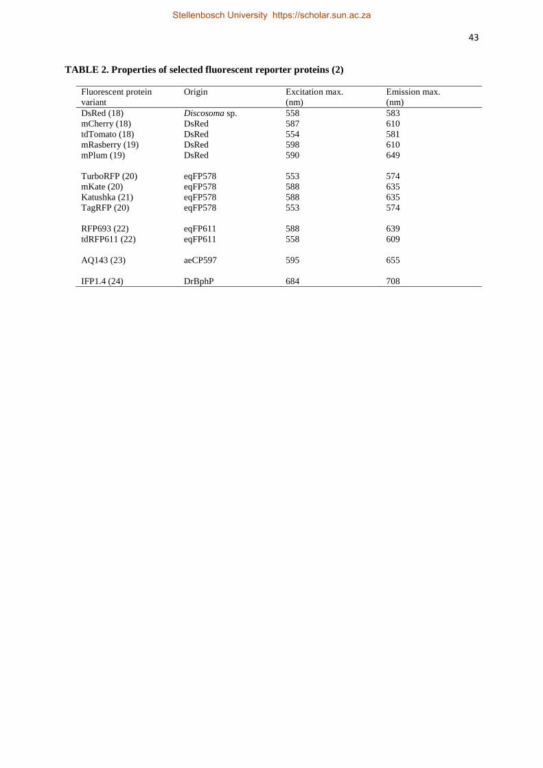
43
TABLE 2. Properties of selected fluorescent reporter proteins (2)
Fluorescent protein
variant
Origin Excitation max.
(nm)
Emission max.
(nm)
DsRed (18) Discosoma sp. 558 583
mCherry (18) DsRed 587 610
tdTomato (18) DsRed 554 581
mRasberry (19) DsRed 598 610
mPlum (19) DsRed
590 649
TurboRFP (20) eqFP578 553 574
mKate (20) eqFP578 588 635
Katushka (21) eqFP578 588 635
TagRFP (20) eqFP578
553 574
RFP693 (22) eqFP611 588 639
tdRFP611 (22) eqFP611
558 609
AQ143 (23) aeCP597
595 655
IFP1.4 (24) DrBphP 684 708
Stellenbosch University https://scholar.sun.ac.za

44
TABLE 3. Imaging systems utilizing bioluminescent bacteria (7)
Bacteria Reporter system
Bacillus anthracis (25) lux
Citrobacter rodentium (26) lux
Brucella melitensis (27) lux
Bifidobacterium breve (4) lux
Escherichia coli (28) lux, luc
Listeria monocytogenes (15) lux
Mycobacterium tuberculosis (29) lux, luc
Pseudomonas aeruginosa (30) lux
Pseudomonas fluorescence (31) lux
Salmonella enterica Typhimurium (32) lux
Staphylococcus aureus (16, 33) lux
Streptococcus pneumonia (17) lux
Stellenbosch University https://scholar.sun.ac.za

45
TABLE 4. Comparison of Lux, GFP and mCherry (18)
Characteristic Lux GFP mCherry
Color of light Bluish-white Green but variants are
yellow, blue and cyan
Red, shade of purple in
visible light
External requirements for light emission None; except must
view in dark room with
dark adapted eyes
Requires excitation by
UV light
Short wavelengths of
light – excitation
maximum of 587nm
Intrinsic requirements for light emission 5 additional genes plus
luxR and luxI
None None
Duration/stability of light emission Lasts 5-9 days, stops
after nutrient depletion
Last months as long as
plates do not dry out
Shade of purple in
visible light, resistant
to photo bleaching
O2 dependency for light emission Requires O
2 None; some O
2 needed
for chromophore
formation
Functions under
hypoxic conditions
Stellenbosch University https://scholar.sun.ac.za

46
TABLE 5. Factors that can affect the sensitivity of imaging systems (7)
Reporter gene (fluorescent/bioluminescent) expression level
Emission light wavelength (referring to reporter gene chosen)
Excitation wavelength (fluorescence only)
Sensitivity of detection system
Background fluorescence/bioluminescence
Animal fur colour or pigmentation
Signal location within the animal (type and depth of tissue)
Stellenbosch University https://scholar.sun.ac.za

47
FIG. 1. The arrangement of the luxCDABE open reading frames found in Photobacterium spp, Vibrio
spp. and Photorhabdus spp. (56). The luxI and luxR genes are adjacent to one another and luxI is the
first of the seven genes in the operon. LuxI and LuxR are required for activation of the luminescence
genes.
LuxI – Autoinducer
Synthetase
LuxR - Regulatory protein
Stellenbosch University https://scholar.sun.ac.za
48
STELLENBOSCH UNIVERSITY
Chapter 3
Construction of a Fluorescent Reporter System for Lactic
Acid Bacteria, using the mCherry Gene
Stellenbosch University https://scholar.sun.ac.za

49
Construction of a Fluorescent Reporter System for Lactic
Acid Bacteria, using the mCherry Gene
Abstract
Fluorescent reporter genes are invaluable when studying gene expressions and following the
migration of bacteria in the gastro-intestinal tract (GIT). The mCherry gene, encoding a red
fluorescent protein (rfp), was cloned into the pGKV223D LAB/E. coli expression vector and
two integration vectors were constructed. The integration vectors were constructed to allow
stable integration of mCherry into non-functional regions on the chromosomes of the
probiotics Lactobacillus plantarum 423 and Enterococcus mundtii ST4SA by homologous
recombination. Insertion of the mCherry gene did not alter the growth rate of strains 423 and
ST4SA and had no effect on bacteriocin production. Fluorescent cells were recorded using
the Caliper in vivo imaging system (IVIS). The mCherry gene may be used as a marker to tag
Lb. plantarum and Ent. mundtii in the GIT.
Stellenbosch University https://scholar.sun.ac.za
50
Introduction
Lactic acid bacteria (LAB) are used in many applications, such as probiotics, as starter
cultures in food fermentations, biological control agents and in pharmaceutical preparations.
They form part of the natural microbiota of the intestines, mouth, skin, urinary and genital
organs of both humans and animals (1). The use of some strains as probiotics exert beneficial
effects on human and animal health by improving the microbial balance of the gastro-
intestinal tract (GIT) and by producing antimicrobial peptides active against pathogens (2, 3).
Lactobacillus plantarum is a lactic acid bacterium that is commonly applied to industrial and
artisanal dairy, meat or plant fermentation and some strains are marketed as probiotics (4-6).
Lactic acid bacteria strains belonging to the genus Enterococcus, such as Enterococcus
faecium and Enterococcus faecalis, play a key role in environmental, food and clinical
microbiology (7, 8). Enterococci are also known to produce antimicrobial peptides active
against intestinal pathogens (9).
Due to the increasing consumer interest in probiotics as health supplements and natural
alternatives to antibiotics, techniques to study the survival and metabolic activities of LAB are
required. The in vivo monitoring of these processes is achieved by labelling LAB with
reporter genes. Fluorescence, the emission of light by fluorophores at different wavelengths,
is a versatile technique which may be applied in vitro and in vivo (10).
To develop a fluorescent microorganism, it is critical that the reporter gene is efficiently and
stably expressed (11). The Expression of reporter genes in LAB using plasmids requires
selective pressure for maintenance which complicates their use in in vivo applications.
Integration of a selected reporter gene into the genome of LAB for expression eliminates
selection requirements and provides genetic stability (12). Furthermore, the presence of the
Stellenbosch University https://scholar.sun.ac.za

51
fluorescent vector or gene in the recombinant organism should not alter the physiology of the
host or its ability to survive.
The green fluorescent protein (GFP) from the jellyfish Aequorea victoria is a versatile
reporter that is widely used to label bacteria, study gene functionality or tag proteins (13, 14).
In addition, GFP have been used to track labelled cells in tissues (15-17) and to provide
valuable information on the localization of bacteria in simulated GIT studies (18).
Since the discovery of GFP in 1962, numerous red-emitting variants have been developed
with optimized properties for in vivo applications (19). Of these, the mCherry red
fluorescence protein (rfp) is considered the most appropriate for in vivo studies. Fluorescent
reporters with emission wavelengths above 600 nm were required, as the absorption of light
by mammalian tissues is lower in this range of the light spectrum. Excitation and emission of
the mCherry rfp is at a wavelength where auto-fluorescence is minimal (20). Red fluorescent
proteins with excitation and emission wavelengths in the far-red region of the spectrum are
preferred for in vivo experiments.
The mCherry protein is highly photo-stable and was thus chosen as a reporter for in vivo
application in the current study. In addition, mCherry has an improved brightness and
maturation time compared to the Discosoma red (DsRed) protein it was derived from (21, 22).
The mCherry gene was used with great success in several bacteria, including LAB (23-25).
Furthermore, the mCherry protein is non-toxic and expression at high levels will not inflict
additional physiological stress on the host organism (26). The mCherry fluorophore is also
highly resistant to photo bleaching and the fluorophore will not lose its ability to fluoresce
during continuous illumination (27).
This paper describes the construction of mCherry reporter plasmids. The constructs were
specially designed for integration into the genomes of the probiotics, Lb. plantarum 423 and
Stellenbosch University https://scholar.sun.ac.za

52
Ent. mundtii ST4SA. We report on the introduction of the mCherry gene into the bacterial
chromosomes by homologous integration into selected target genes. Expression of
fluorescence in the two LAB was detected using the in vivo imaging system (IVIS). We also
show that introduction of the mCherry fluorescence gene into Lb. plantarum 423 and Ent.
mundtii ST4SA had no effect on their ability to produce antibacterial peptides (bacteriocins).
Materials and Methods
Bacterial strains, media and growth conditions
The bacterial strains and plasmids used in this study are listed in Table 1. All subcloning
experiments were carried out using Escherichia coli DH5α (28) as host. Escherichia coli
DH5α was grown in Brain Heart Infusion (BHI) or Luria-Bertani (LB) broth (Biolab
Diagnostics, Midrand, South Africa) at 37oC on an orbital shaker (200 rpm). All LAB were
grown in MRS broth (Biolab Diagnostics) without shaking, or on MRS agar at 30oC. When
necessary, antibiotics were added to growth media at the following final concentrations: 200
µg/ml erythromycin (Em) for E. coli DH5α, and 10 µg/ml Em for Lb. plantarum 423 and Ent.
mundtii ST4SA. Chloramphenicol (Cm) was incorporated at a final concentration of 5 µg/ml
for Ent. mundtii ST4SA, 10 µg/ml for Lb. plantarum 423 and 20 µg/ml for E. coli DH5α.
Ampicillin (Amp) was added to a final concentration of 100 µg/ml for E. coli DH5α.
General molecular techniques
Recombinant DNA methods and gel electrophoresis were carried out according to standard
protocols, as described by Sambrook and Russel (2001). DNA restriction and modification
enzymes were purchased from Roche Molecular Chemicals (Mannheim, Germany) or
Fermentas (Vilnuis, Lithuania) and were used according to the suppliers’ instructions.
Oligonucleotides (Table 2) were obtained from Inqaba Biotechnical Industries (Pretoria,
Stellenbosch University https://scholar.sun.ac.za
53
South Africa). PCR amplifications (Table 2) were performed on a Swift™
Minipro Thermal
Cycler (Esco Healthcare, Malaysia), using Takara Ex Taq High-Fidelity PCR DNA
polymerase (Clontech Laboratories, Inc., Randburg, South Africa). DNA sequencing was
performed by the Central DNA Sequencing Facility of the University of Stellenbosch. DNA
fragments were purified from agarose gels using the QIAquick PCR Purification Kit (Qiagen
Inc., Valencia, CA). Escherichia coli plasmid DNA was purified using the standard Qiagen®
Plasmid Mini and Midi Kits. Genomic DNA of LAB strains was isolated using the ZR
Fungal/Bacterial DNA Miniprep Kit (Zymoresearch Corporation, Irvine, CA, U.S.A.). The
presence of recombinant plasmid DNA in LAB transformants was confirmed by using the
plasmid isolation method of Qiagen®, but modified by using 10 mg/ml lysozyme for cell lysis
and STE buffer (6.7% saccharose; 50 mM Tris-Cl, pH 8.0; 1 mM EDTA) for wash steps.
Construction of pGKV223D expression and integration vectors containing the mCherry
fluorescence gene
Expression and integration plasmids constructed during this study are based on the
pGKV223D LAB/E. coli expression vector (University of Grönigen, Netherlands). The vector
contains an EmR gene for erythromycin resistance, the L23 LAB promoter, a multiple cloning
site (MCS) and the repA gene for replication in LAB/E.coli hosts, which was derived from
Lactococcus and Enterococcus spp. All primers are listed in Table 2. A schematic diagram
summarizing the construction of pGKCherry is shown in Figure 1. The mCherry cassette was
chemically synthesized by Invitrogen, Germany, with codon-optimization for enhanced
expression in Lb. plantarum and Ent. mundtii. The mCherry gene cassette was amplified from
plasmid pMK-RQCherry (Invitrogen, Germany) by using primers ForPldh and RevTT that
contain the restriction sites EcoRI and XbaI, respectively. The 1.5 kb amplicon, containing the
mCherry gene, flanked by the Pldhl promoter and a transcriptional terminator (TT), was
Stellenbosch University https://scholar.sun.ac.za

54
digested with EcoRI and XbaI and introduced into the MCS of pGKV223D after digestion of
the vector with the same restriction enzymes (Fig. 1). Fragments were ligated overnight at
15oC, before being transformed into competent E. coli DH5α for plasmid propagation.
Transformants with an Em-resistant phenotype and a color change from white to pink were
selected.
To construct the integration vectors PGKRm1CmRCherry and PGKEmuNCmRCherry, a
gene for chloramphenicol resistance (cat) was fused to the mCherry gene cassette as a second
selection marker (Fig. 2). Plasmid pKSCmR was digested with EcoRI and an 800 bp DNA
fragment containing the cat gene was purified from a 0.8% (w/v) agarose gel. Plasmid
pKSCherry carrying the codon-optimized mCherry gene cassette was linearized following
digestion with EcoRI, purified and then ligated with T4 DNA ligase (Roche Molecular
Chemicals, Mannheim, Germany) to the 800 bp cat gene fragment. Escherichia coli DH5α
electo-competent cells were then transformed for propagation of the resulting plasmid, named
pKSCmRCherry. Transformants were screened by selecting for a Cm resistant and a red
colony color phenotype. The integrity of the plasmid was verified by restriction digests and
PCR with primers ForCmR and RevTT.
The targets used for homologous recombination were the restriction modification type 1
(Rm1) and DNA-entry (EmuN) genes for Lb. plantarum 423 and Ent. Mundtii ST4SA,
respectively. Unique sites were chosen within these genes for the insertion of the CmRCherry
gene cassette. The integration vectors were constructed in such a way that, the CmRCherry
gene cassette (Fig. 2) was flanked with the target genes’ sequences including some of their
upstream and downstream regions, to facilitate homologous recombination. The Rm1 and
EmuN genes were amplified by PCR from genomic DNA extracted from Lb. plantarum 423
and Ent. mundtii ST4SA, respectively. Amplification of the target genes Rm1 and EmuN was
performed using primers ForRm1/RevRm1 and ForEmuN/RevEmuN, respectively. Both
Stellenbosch University https://scholar.sun.ac.za

55
amplicons were digested with EcoRI and XbaI and then ligated to the LAB/E. coli shuttle
vector pGKV223D that was cut with the same enzymes. The presence of target genes in the
resulting recombinant plasmids pGKRm1 and pGKEmuN isolated from Em resistant
transformants was confirmed by sequencing with primers ForRm1/RevRm1 and
ForEmuN/RevEmuN for both constructs, respectively.
For insertion of the CmRCherry cassette into the target gene sequences of plasmids pGKRm1
and pGKEmuN, pKSCmRCherry was digested with SalI and XbaI. The resulting 2.2 kb DNA
fragment containing the cat gene, Pldhl promoter, mCherry gene and the TT transcriptional
terminator, in that specific order (Fig. 2), was purified from a 0.8% agarose gel. The flushing
of both the SalI and XbaI restriction site ends of the 2.2 kb DNA fragment was accomplished
by filling in the 5’ sticky overhang ends with E. coli T4 DNA polymerase in the presence of
all four dNTP’s (31). Plasmids pGKRm1 and pGKEmuN were digested at unique restriction
sites in the target gene sequences namely, PvuI and EcoRV, respectively. After digestion of
the plasmids, the PvuI restriction site ends of the linearized pGKRm1 plasmid were flushed
by filling in the 5’ sticky ends as described before. Since, EcoRV is a blunt end restriction
enzyme, flushing of the ends was not necessary for compatibility with the blunt ended
CmRCherry gene cassette. The linearized blunt ended pGKRm1 and pGKEmuN fragments
were purified and ligated with T4 DNA ligase to the blunt ended CmRCherry gene cassette
from pKSCmRCherry. The ligation mixture was introduced into competent E. coli DH5α
cells through electroporation. Transformants with both an Em and Cm resistant phenotype
were selected. The presence of the CmRCherry gene cassette fused within upstream and
downstream flanking regions of the target genes Rm1 and EmuN in the resulting plasmids
designated pGKRm1CmRCherry and pGKEmuNCmRCherry was confirmed by DNA
sequencing. Sequence confirmation of pGKRm1CmRCherry and pGKEmuNCmRCherry was
carried out using primers ForCmR/RevRm1 and ForCmR/RevEmuN, respectively. Following
Stellenbosch University https://scholar.sun.ac.za
56
sequence confirmation of the integration vectors, the plasmids were extracted for subsequent
transformation of LAB. The Cm marker was associated with the mCherry gene to enable
selection of integrants that have lost the EmR-associated vector.
Targets for genomic integration
At the time of completion of this study, the whole genome sequences of Lb. plantarum 423
and Ent. mundtii ST4SA were not available. Therefore, published whole genome sequences of
closely related Lb. plantarum strains, WCFS1, JDM1 and ZJ316 were used as reference
strains in the search for non-essential target sequences for the genomic integration of the
CmRCherry gene cassette into the Lb. plantarum 423 chromosome. The presence of the five
selected targets genes was screened for by PCR using Lb. plantarum strains 423, ATCC8014
and ATCC14917 gDNA as templates. Three transcriptionally silent prophage and two
restriction modification (RM) genes were identified as potential non-essential target genes for
homologous recombination in the Lb. plantarum 423.
Similarly, two draft genome sequences of Ent. mundtii strains CRL1656 and ATCC882 were
used as reference in the screening of potential target gene sequences for homologous
recombination in strain ST4SA. Two RM genes and a DNA-entry nuclease gene (EmuN) were
identified as potential target sequences for homologous recombination in strain ST4SA.
Transformation of bacterial cells
Escherichia coli DH5α
Electrocompetent E. coli DH5α cells were prepared as previously described (32), except that
the LB growth medium contained 50% less NaCl in this study. Cells were electroporated with
5 µl plasmid DNA and transformants selected with Em, Amp or Cm.
Stellenbosch University https://scholar.sun.ac.za
57
Lactobacillus plantarum 423
Electroporation of Lb. plantarum was carried out as previously described (33), with minor
modifications. Overnight cultures of actively growing Lb. plantarum 423 cells were diluted
1/20 (5 ml) into filter-sterilized MRS growth medium (100 ml) supplemented with 8 %
glycine and 0.3 M sucrose as cell wall weakening agents. Cultures were incubated at 30oC for
approximately 4h until optical density at 600 nm of 0.4 was reached, representing the late
exponential growth phase. After incubation, cultures were harvested at 1500 x g for 5 min,
washed twice in ultra-pure water, kept on ice for 5 min in 50 mM EDTA, washed once more
in milliQ water, once in 0.3 M sucrose and resuspended in 2 ml of the latter solution. Aliquots
of 50 µl electro-competent bacteria were mixed with 1 µl plasmid DNA (500 ng) and
transferred to 2 mm cuvettes for electroporation. In addition, electro-competent Lb. plantarum
423 cells were used immediately for electroporation and not frozen. The electroporation
parameters used were 1.5 kV, 25 µF and 200Ω. Directly after electroporation, 1 ml pre-
warmed MRS media was added, the cells incubated at 30oC for 2h for phenotypic expression
before plating on MRS agar containing 10 µg/ml Em or Cm and subsequently incubated at
30oC for 48h.
Enterococcus mundtii ST4SA
A modified version of a Pediococcus sp. protocol for the electro-transformation of Ent.
mundtii ST4SA was used in this study (34). For the preparation of electro-competent Ent.
mundtii ST4SA cells, 10 ml of an overnight culture was inoculated into 100 ml MRS broth,
supplemented with 4.5 % glycine, 0.5 M sucrose and 40 mM threonine as cell wall weakening
agents. Incubation was at 30oC for 4h, without aeration, to an optical density at 600 nm of 0.5.
The cells were harvested (1500 x g, 10 min), resuspended in filter-sterilized buffer solution
(0.5 M sucrose and 10 % glycerol), and kept on ice for 1h before being washed three times
Stellenbosch University https://scholar.sun.ac.za

58
with the same solution. Cells were resuspended in electroporation buffer, containing 1 mM
K2HPO4/KH2PO4 (pH 7.0), 1 mM MgCl2 and 0.5 M sucrose. Subsequently, 100 µl aliquots
was mixed with 5 µl plasmid DNA (1 µg), transferred to 2 mm cuvettes and electroporated at
2.2 kV, 25 µF and 200Ω. Following electroporation, cells were promptly transferred into
MRS broth supplemented with 0.5 M sucrose, 20 mM MgCl2 and 2 mM CaCl2, before
incubation at 30oC for 2h and plating onto MRS agar containing 10 µg/ml Em.
Transformant selection
Escherichia coli DH5α and LAB transformants harbouring recombinant vectors encoding the
mCherry gene, were analyzed on agar plates for fluorescence emission using the IVIS® 100 In
Vivo Imaging System (Caliper Life Sciences, Hopkinton, MA, U.S.A.). Colonies with the
highest fluorescence emission were selected and transferred to 10 ml liquid broth containing
an appropriate antibiotic. Following the transformation of LAB strains with the integration
vectors pGKRm1CmRCherry and pGKEmuNCmRCherry, the electroporated culture was
directly transferred into 10 ml MRS broth containing Cm for the enrichment of successful
integrants. After a 48 hour incubation period, cultures were replica plated on MRS agar
supplemented with Em and Cm, respectively. Erythromycin-sensitive and Cm-resistant
colonies were then selected and scanned for fluorescence. Background auto fluorescence
emitted by the agar plate was used to normalize the fluorescence signal of colonies.
Fluorescence detection
Fluorescence assays of bacterial strains expressing the mCherry gene were carried out using
black non-autofluorescent 96-well microtiter trays. Four millilitres of 12 h-old cultures
expressing the mCherry gene were harvested (1500 x g for 3 min), the cells washed twice
with sterile saline solution (0.85% (w/v) NaCl) to remove trace elements of growth media and
Stellenbosch University https://scholar.sun.ac.za

59
then resuspended in 300 µl saline. Before fluorescent image capturing, the 300 µl cell
suspensions were transferred to wells in a microtiter tray. Bacterial strains with plasmid
constructs without the mCherry gene insert were used as controls.
Growth of Lb. plantatum 423 and Ent. mundtii ST4SA compared to the chromosomally
marked strains
Wild-type and recombinant Lb. plantarum 423 and Ent. mundtii ST4SA transformed with the
integration vectors were grown in MRS broth for 12 h. One millilitre of the 12-h old cultures
was used to inoculate 100 ml of pre-warmed MRS broth. The 100 ml cultures were then
aliquoted into nine 2 ml Eppendorf tubes (one Eppendorf tube for each time point) and
incubated at 30oC for 9 h. Every 1 h the optical density reading at 600nm of each of the
cultures in one Eppendorf tube was measured, serially diluted and plated on MRS agar.
Determination of antibacterial activities of fluorescing LAB strains
The ability of the recombinant LAB containing the mCherry gene to produce antimicrobial
peptides (bacteriocins) was tested using Listeria monocytogenes EGDe in a lawn assay, as
described by Van Reenen et al. (30). An actively growing overnight culture (12 h) of L.
monocytogenes EGDe was overlaid onto a BHI agar plate by inoculating 100 µl of the
bacterial suspension in 10 ml semi-solid BHI agar (0.8 %, w/v). The cell-free supernatants
(adjusted to pH 7.0 with NaOH) of 2 ml actively growing fluorescent LAB was collected by
centrifugation (8000 x g for 3 min). The supernatants were passed through a 0.22 µM pore
size filter, 10 µl was spotted onto BHI agar overlaid with L. monocytogenes EGDe, and the
plates incubated at 37oC for 24h. As positive controls, supernatants from LAB strains not
harbouring control plasmids vectors were used.
Stellenbosch University https://scholar.sun.ac.za
60
Quantification of fluorescent signals and CFU determination
The quantification of fluorescence signals relative to the amount of bacterial cells required for
a specific emission signal was determined. Overnight cultures (12 h) of Lb. plantarum
423::CmRCherry and Ent. mundtii ST4SA (pCmRCherry) were diluted as follows: 1:2, 1:4,
1:8, 1:16 and 1:32. Fluorescence of each dilution was measured using the IVIS imaging
system. For the determination of the colony forming units (CFU), each dilution was further
serially diluted in 0.85 % NaCl and plated out on MRS agar. For the estimation of CFU, 100
µl of each serial dilution was plated out, followed by incubation at 30o for 48h.
The amount of fluorescence emitted in p/cm2/s/sr for each dilution was plotted against the
corresponding CFUs of Lb. plantarum 423::CmRCherry and Ent. mundtii ST4SA
(pCmRCherry), with each point on the graphs representing the amount of photons emitted
correlating to the CFUs of each dilution.
Results
Cloning of pGKCherry and introduction into LAB
Escherichia coli colonies harbouring pGKCherry were easily detected on agar plates as a
result of their bright purple appearance (Fig. 3a). Bright fluorescence was detected on agar
plates using the IVIS (Fig. 3b). High fluorescence intensity was observed for cells harbouring
pGKCherry, compared to control cells harbouring the pGKV223D vector (Fig. 3c).
Lactobacillus plantarum 423 and Ent. mundtii ST4SA colonies transformed with pGKCherry
had a white appearance growing on agar plates, but showed varying intensities of
fluorescence. Colonies with the strongest intensity were selected for further analysis.
Fluorescent imaging of Lb. plantarum 423 (pGKCherry) and Ent. mundtii ST4SA
(pGKCherry) strains resuspended in microtitre trays revealed bright red fluorescence
Stellenbosch University https://scholar.sun.ac.za
61
compared to the control strains as shown in Figure 4a and Figure 4b, respectively.
Lactobacillus plantarum 423 pGKCherry was also imaged in microfuge tubes using the IVIS
with a pGKV223D transformant as control (Fig. 4c).
The presence of the 5 kb pGKCherry plasmid was confirmed in Lb. plantarum 423 strains by
isolation and visualization after electrophoresis. The presence of the pGKCherry vector in the
recombinant Lb. plantarum 423 is indicated in Fig. 5a. The probiotic Lb. plantarum 423
contain five natural plasmids as shown in Fig. 5b. The presence of the pGKCherry vector in
Ent. mundtii ST4SA was similarly confirmed, with Ent. mundtii ST4SA containing two large
natural plasmids of 50 kb and 100 kb with the bacteriocin ST4SA operon situated on the 50
kb plasmid as indicated in Fig. 6.
Screening of targets for genomic integration
Of the five target genes tested for, PCR results confirmed that the 1 kb type 1 RM
methyltransferase gene (Rm1) was the only gene present in Lb. plantarum 423 (Table 3).
Therefore, the Rm1 gene was used as target sequence for homologous recombination.
Lactobacillus plantarum 423 was significantly different compared to Lb. plantarum strains
that already have their genomes published, particularly with the regards to the genes we
screened for. One of the target genes tested, the Mrrp type IV RM gene, was present in six
sequenced Lb. plantarum strains and in three strains we have tested, but was not present in
Lb. plantarum 423.
In the case of Ent. mundtii ST4SA, PCR results confirmed that the DNA-entry nuclease gene
(EmuN), associated with the natural competence of the strain, was the only target sequence
present in the strain out of the three potential targets that was screened for. The EmuN gene
was used as target sequence for homologous recombination in Ent. mundtii ST4SA.
Stellenbosch University https://scholar.sun.ac.za

62
Introduction of integration vectors containing the mCherry gene into Lb. plantarum 423
and Ent. mundtii ST4SA
Escherichia coli DH5α cells transformed with the integration vectors, pGKRm1CmRCherry
and pGKEmuNCmRCherry, resulted in fluorescent colonies that were both Em and Cm
resistant. The vectors are able to replicate in Gram-negative and Gram-positive bacteria, and
contain a Cm marker that could be selected for in integrants that have lost the EmR-associated
vector. Lactobacillus plantarum 423 and Ent. mundtii ST4SA transformants could either
contain the autonomous plasmids, integrated plasmids with Cm and Em resistance, or an
integrated but recombined CmRCherry cassette in which the EmR-associated plasmid was
lost. Lactobacillus plantarum 423 transformants retained resistance to Cm, but not Em,
indicating that the CmRCherry gene cassette successfully integrated into the host
chromosome without the plasmid DNA encoding the EmR gene. However, Ent. mundtii
ST4SA transformants retained resistance to both Em and Cm, indicating that the EmR-
associated vector was not lost.
The presence of the cat and mCherry genes in Lb. plantarum 423 and Ent. mundtii ST4SA
transformants was confirmed by PCR using primers ForCmR/RevCmR and ForCherry/RevTT
(Fig. 7). No 1 kb Rm1 target gene fragments were amplified from potential Lb. plantarum 423
integrants with primers ForRm1 and RevRm1. However, amplification with primers ForCmR
and RevRm1 resulted in a 2.5 kb DNA fragment consisting of the CmRCherry gene cassette
and a 270 bp downstream Rm1 gene fragment (Fig. 8). These results suggested that the
Rm1gene was successfully disrupted by the insertion of the CmRCherrygene cassette.
Amplification of Ent. mundtii ST4SA transformant DNA with primers ForEmuN and
RevEMuN resulted in a 720 bp DNA-entry gene fragment (data not shown), indicating that
the EmuN gene was not knocked out. Lactobacillus plantarum 423 and Ent. mundtii ST4SA
Stellenbosch University https://scholar.sun.ac.za
63
transformed with the integration vectors were fluorescent when examined using the IVIS (Fig.
9).
Impact of the genomic disruption of target genes on LAB phenotypes
No significant difference in growth was observed between the wild-type and engineered
strains after a nine-hour incubation period (Fig. 10). The potential genomic interruption of the
target genes Rm1 and EmuN on the chromosomes of Lb. plantarum 423 and Ent. mundtii
ST4SA had no detectable impact on the bacterial growth.
Bacteriocins isolated from Lb. plantarum 423::CmRCherry and Ent. mundtii ST4SA
(pCmRCherry) resulted in the formation of clear inhibition zones of the sensitive L.
monocytogenes EGDe strain (Fig. 11).
Quantification of fluorescence signals and CFU determination
The excellent correlation between the fluorescence emitted and CFU counts for all the
dilutions made for Lb. plantarum 423 (R2=0.9889) and Ent. mundtii ST4SA (R
2=0.9788)
recombinant strains indicated that photon emission levels accurately reflect bacterial numbers
in total cultures (Fig. 12).
Discussion
The mCherry gene was chemically synthesized and codon optimized for optimal expression in
Lb. plantarum 423 and Ent. mundtii ST4SA. The expression and integration plasmids were
derived from the pGKV223D shuttle vector which is based on a Lactococcal spp. origin of
replication functional in E. coli and LAB.
Stellenbosch University https://scholar.sun.ac.za

64
Firstly, we developed the pGKCherry expression vector carrying the mCherry gene under
constitutive expression from the strong Lb. plantarum Pldhl promoter to evaluate the
expression of mCherry in LAB (36, 37). The expression of mCherry in E. coli DH5α resulted
in a change in colony colour on agar plates, indicating that the fluorescence protein was
functionally expressed from the Pldhl promoter. Following the electroporation of the
pGKCherry plasmid into Lb. plantarum 423 and Ent. mundtii ST4SA, fluorescence imaging
revealed strong fluorescence emission of the bacteria, suggesting that there was functional
expression of the mCherry protein in these Gram-positive hosts. Expression of the mCherry
protein in the LAB hosts did not affect colony color. Similarly, in another study the
expression of the mCherry gene in Lb. sakei was also reported not to affect colony color, with
subsequent observation of the bacteria by fluorescence microscopy revealing bright red
fluorescent cells (38). These results demonstrate the usefulness of the pGKCherry plasmid to
fluorescently tag LAB host strains for in vitro applications.
Our interest is in using the mCherry gene to track labeled strains of LAB in the GIT of mice,
thereby obtaining valuable information on the location and colonization by the bacteria.
Therefore, we developed a system to integrate the mCherry fluorescent marker gene onto the
chromosomes of Lb. plantarum 423 and Ent. mundtii ST4SA to circumvent the use of
autonomous plasmids. This ensures that the marker gene will be maintained inside the host
genomes without the need for antibiotic selection to ensure enhanced stability and continuous
expression (12, 39-41). Integration vectors pGKRm1CmRCherry and pGKEmuNCmRCherry
were constructed to enable the homologous recombination of the CmRCherry cassette
containing a gene for Cm resistance and the mCherry fluorescence gene into the Rm1 and
EmuN gene sequences of Lb. plantarum 423 and Ent. mundtii ST4SA, respectively. Previous
studies have described the integration of plasmids, unable to replicate in Gram-positive
bacteria, in LAB by using randomly selected heterologous chromosomal fragments as regions
Stellenbosch University https://scholar.sun.ac.za

65
of homology (39, 41, 42). However, the disruption or inactivation of random genes without
knowing their function may be detrimental to the relevant LAB strain.
We hypothesized that the disruption or inactivation of potentially inactive prophage and
restriction modification (RM) genes would not affect the host strains’ growth capacity or cell
physiology. Since, the homologous recombination of the engineered construct can only be
designed using known chromosomal DNA sequences, the screening for target sequences
proved to be challenging. As the whole genome sequences of Lb. plantarum 423 and Ent.
mundtii ST4SA had not yet been published, we had to turn to genome sequences of closely
related bacteria. Our investigation showed that both strains are significantly different to the
Lb. plantarum and Ent. mundtii strains that already had their genomes published. In support
of this notion, the Mrrp RM gene was present in all of the Lb. plantarum strains whose
genomic sequences were available and in the strains we tested for in our lab but was not in
strain 423.
The CmRCherry gene cassette was successfully integrated into the chromosome of Lb.
plantarum 423 by homologous recombination. Several other studies have reported on the
successful use of homologous recombination for the stable integration of genes in bacterial
chromosomes (18, 42, 43). Our results indicate that disruption or inactivation of the
chromosomally located Lb. plantarum 423 Rm1 gene is not lethal to the survival of the host.
In addition, it was essential that the genomic interruption of the Rm1 target gene by
homologous recombination on the chromosome of Lb. plantatum 423 have no impact on the
strain’s growth capacity. Thus, in favour of this, the growth capacity of the chromosomally
marked Lb. plantatum 423 remained unaffected following the disruption of the target gene
sequence.
Stellenbosch University https://scholar.sun.ac.za
66
Fluorescent Ent. mundtii ST4SA (pCmRCherry) transformants retained resistance to both Em
and Cm. This was an indication that the integration of the CmRCherry gene cassette without
the plasmid backbone did not occur. PCR amplification confirmed that the target EmuN gene
was not disrupted. Therefore, it can be speculated that disruption of the EmuN gene was not
achieved as its inactivation could potentially be harmful to the host cell. However, despite
this, Ent. mundtii ST4SA (pCmRCherry) transformants retained resistance to both Em and
Cm following culturing for five days in the absence of the antibiotics, indicating the stability
of the autonomous plasmid.
The recombinant fluorescent probiotic reporter strains, Lb. plantarum 423::CmRCherry and
Ent. mundtii ST4SA (pCmRCherry), were shown to still be producing their antimicrobial
peptides as evident from the formation of inhibition zones on overlaid MRS plates containing
the sensitive L. monocytogenes EGDe strain. This result also confirmed that the metabolic
load of carrying and expressing the fluorescence gene had no effect on the probiotic strains’
ability to produce their antimicrobial peptides. The operons coding for the bacteriocins
produced by Lb. plantarum 423 and Ent. mundtii ST4SA strains are situated on plasmids (34,
35). The replicons of the bacteriocin-containing plasmids and the plasmid vectors used to
introduce the mCherry marker are compatible, as was confirmed by plasmid isolations.
Furthermore, for both Ent. mundtii ST4SA (pCmRCherry) and Lb. plantarum
423::CmRCherry a positive or linear correlation between different fluorescence intensities
and CFUs exists. This suggests that in future experiments the amount of labeled bacteria
(CFU) present in a sample, correlating to a given fluorescent signal, can be estimated using
the linear graphs.
In this study, we describe the construction and application of a LAB/E. coli expression
plasmid as well as two integration vectors carrying the synthetic mCherry fluorescence gene.
Stellenbosch University https://scholar.sun.ac.za

67
It was shown that the mCherry gene can be used as a useful marker to fluorescently tag the
Lb. plantarum 423 and Ent. mundtii ST4SA probiotic LAB strains for in vivo application. The
introduction of the mCherry fluorescence gene into the LAB strains has no effect on the
bacteria’s ability to produce their antibacterial peptides. Furthermore, we have demonstrated
that target genes with known DNA sequences that are likely to be non-functional in the cells
can be used for the homologous integration of foreign genes in Lb. plantarum 423 and Ent.
mundtii ST4SA. The integration system described here may be tested and applied to other Lb.
plantarum and Ent. mundtii strains using the target genes we have identified.
Stellenbosch University https://scholar.sun.ac.za

68
References
1. Harzallah, D and H. Belhadj. 2013. Lactic acid bacteria as probiotics: Characteristics,
selection criteria and role in immunomodulation of human GI mucosal barrier. In M.
Kongo (eds). Lactic acid bacteria – R & D for food, health and livestock purposes.
Intech. pp. 198-216.
2. Foulquie Moreno, M.R., P. Sarantinopoulos, E. Tsakalidou and L. De Vuyst. 2006.
The role and application of enterococci in food and health. Int. J. Food Microbiol. 106:
1-24.
3. Bhardwaj, A., H. Gupta, S. Kapila, G. Kaur, S. Vij and R.V Malik. 2010. Safety
assessment and evaluation of probiotic potential of bacteriocinogenic Enterococcus
faecium KH 24 strain under in vitro and in vivo conditions. Int. J. Food Microbiol. 141:
156-164.
4. Marco, M.L., R.S. Bongers, W.M. de Vos and M. Kleerebezem. 2007. Spatial and
temporal expression of Lactobacillus plantarum genes in the gastro-intestinal tract of
mice. Appl. Environ. Microbiol. 73: 124-132.
5. Reid, G. 1999. The scientific basis for probiotic strains of Lactobacillus. Appl. Environ.
Microbiol. 65: 3763-3766.
6. Bron. P.A., P. van baarden and M. Kleerebezem. 2012. Emerging molecular insights
into the interaction between probiotics and the host intestinal mucosa. Nat. Revs. 10:
66-78.
7. Giraffa, G. 2002 Enterococci from foods. FEMS Microbiol. Revs. 26: 163-171.
8. Bhardwaj, A., R.K. Malik and P. Chauhan. 2008. Functional and safety aspects of
enterococci in dairy foods. Indian J. Microbiol. 48: 317-325.
Stellenbosch University https://scholar.sun.ac.za
69
9. Montalban-Lopez, M., M. Sanches-Hidalgo, E. Valdivia, M. Martinez-Bueno and
M. Maqueda. 2011. Are bacteriocins unexploited? Novel applications of old
antimicrobials. Curr. Pharm Biotechnol. 12: 1205-1220.
10. Wiedenmann, J., F. Oswald and G.U. Nienhaus. 2009. Fluorescent proteins for live
cell imaging: Oppurtunities, limitations and challenges. Life 61 (11): 1029-1042.
11. Andrue, N., A. Zelmer and S. Wiles. 2011. Noninvasive biophotonic imaging for
studies of infectious disease. FEMS Microbiol. Rev. 35: 360-394.
12. Douglas, G.L. and T.R. Klaenhammer. 2011. Directed chromosomal integration and
expression of the reporter gene gusA3 in Lactobacillus acidophilus NCFM. Appl.
Environ. Microbiol. 77 (20): 7365-7371.
13. Chudakov, D.M., S. Lukyanov and K.A. Lukyanov. 2005. Fluorescent protein as a
toolkit for in vivo imaging. Trends Biotechnol. 26: 605-613.
14. Frommer, W.B., M.W. Davidson, R.E. Campbell. 2009. Genetically encoded
biosensors based on engineered fluorescent proteins. Chem. Soc. Rev. 38: 2833-2841.
15. Tsien, R.Y. 1998. The green fluorescent protein. Annu. Rev. Biochem. 67: 509-544.
16. Griesbeck, O. 2004. Fluorescent proteins as sensors for cellular funcions. Curr. Opin.
Neurobiol. 14: 636-641.
17. Shaner, N.C., G.H. Patterson and M.W. Davidson. 2007. Advances in fluorescent
protein technology. J. Cell. Sci. 120: 4247-4260.
18. Scott, K.P., D.K. Mercer, A.J. Richardson, C.M. Melville, L.A. Glover and H.J.
Flint. 2000. Chromosomal integration of the green fluorescent protein gene in lactic
acid bacteria and the survival of the marked strains in human gut simulations. FEMS
Microbiol. Letts. 182: 23-27.
19. Shaner, N.C., R.E. Campbell, P.A. Steinbach, B.N. Giepmans, A.E. Palmer and
R.Y. Tsien. 2004. Red fluorescent proteins. Nat. Biotechnol. 22 (12): 1567.
Stellenbosch University https://scholar.sun.ac.za

70
20. Chapagain, P.P., C.K. Regmi and W. Castillo. 2011. Fluorescent protein barrel
fluctuations and oxygen diffusion pathways in mCherry. J. Chem. Phys. 135: 235101,
1-6.
21. Bevis, B.J. and B.S. Glick. 2002. Rapidly maturing variants of the Discosoma red
fluorescent protein (DsRed). Nat. Biotechnol. 20: 83-87.
22. Shaner, N.C., P.A. Steinbach and R.Y. Tsien. 2005. A guide to choosing fluorescent
proteins. Nat. Meth. 2 (12): 905.
23. Doherty, G.P., K. Bailey and P.J. Lewis. 2010. Stage-specific fluorescence intensity
of GFP and mCherry during sporulation in Bacillus subtilis. BMC Research notes 3:
303.
24. Chapagain, P.P., C.K. Regmi and W. Castillo. 2011. Fluorescent protein barrel
fluctuations and oxygen diffusion pathways in mCherry. J. Chem. Phys. 135: 1-6.
25. Garcia-Cayuela, T. and L.P. Gomez de Carinanos. 2012. Fluorescent protein vectors
for promoter analysis in lactic acid bacteria and Escherichia coli. Appl. Microbiol.
Biotechnol. 96: 171-181.
26. Parish, T., P. Carroll, L.J. Schreuder, J. Muwanguzi-Karugaba, S. Wiles, B.D.
Robertson et al. 2010. Sensitive detection of gene expression in Mycobacteria under
replicating and non-replicating conditions using optimized far-red reporters. PLoS ONE
5 (3): 9823.
27. Viegas, M.S., T.C. Martins, F. Seco and A. do Carmo. 2007. An improved and cost-
effective methodology for the reduction of autofluorescence in direct
immunofluorescence studies on formalin-fixed paraffin-embedded tissues. Eur J.
Histochem, 51 (1): 59-66.
28. Ausubel, F.M., R. Brent, R.E. Kingston, D.D. Moore, J.G. Seidman, J.A. Smith and
K. Struhl. 1994. Curr. Prot. Mol. Biol. John Wiley and Sons, Inc., New York, N.Y.
Stellenbosch University https://scholar.sun.ac.za

71
29. Sambrook, J. and D. Russel. 2001. Molecular Cloning: A Laboratory Manual. Cold
Spring Harbor Laboratory Press, Cold Spring Harbor, NY.
30. Van Reenen, C.A., L.M.T. Dicks and M.L. Chikindas. 1998. Isolation, purification
and partial characterization of plantaricin 423, a bacteriocin produced by Lactobacillus
plantarum. J. Appl. Microbiol. 84: 1131-1137.
31. Wilson, K. and J. Walker. 2008. Principles and techniques of biochemistry and
molecular biology. 6th ed. Cambridge University Press, New York, N.Y., USA.
32. Sharma, R.C. and R.T. Schimke. 1995. Preparation of electro-competent E. coli using
salt-free growth medium. Biotech. 20: 42-44.
33. Thompson, K. and M.A. Collins. 1996. Improvement in electroporation efficiency for
Lactobacillus plantarum by the inclusion of high concentrations of glycine in the
growth medium. J. Microbiol. Meth. 26: 73-79.
34. Van Reenen, C.A., W.H. Van Zyl and L.M.T. Dicks. 2006. Expression of the
immunity protein of plantaricin 423, produced by Lactobacillus plantarum 423, and
analysis of the plasmid encoding the bacteriocin. Appl. Environ. Microbiol. 72 (12):
7644.
35. Knoetze, H., S.D. Todorov, C.A. Van Reenen and L.M.T. Dicks. 2006.
Characteriziation of bacteriocin ST4SA, produced by Enterococcus mundtii ST4SA
isolated from soya beans. Department of Microbiology, University of Stellenbosch,
South Africa, S.A.
36. Geoffroy, M., C. Guyard, B. Quatannens, S. Pavan, M. Lange and A. Mercenier.
2000. Use of green fluorescent protein to tag lactic acid bacterium strains under
development as live vaccine vectors. Appl. Environ. Microbiol. 66 (1): 383-391.
Stellenbosch University https://scholar.sun.ac.za
72
37. Daniel, C., S. Poiret, V. Dennin, D. Boutillier and B. Pot. 2013. Bioluminescence
imaging study of spatial and temporal persistence of Lactobacillus plantarum and
Lactococcus lactis in living mice. Appl. Environ. Microbiol. 79 (4): 1086-1094.
38. Freiding, S., M.A. Ehrmann and R.F. Vogel. 2012. Comparison of different IlvE
aminotransferases in Lactobacillus sakei and investigation of their contribution to
aroma formation from branched chain amino acids. Food Microbiol. 29: 205-214.
39. Casey, J., C. Daly and G.F. Fitzgerald. 1991. Chromosomal integration of plasmid
DNA by homologous recombination in Enterococcus faecalis and Lactococcus lactis
subsp. lactis hosts harbouring Tn919. Appl. Environ. Microbiol. 57 (9): 2677-2682.
40. Cruz Martin, M., J.C. Alonso, J.E. Suarez and M.A. Alvarez. 2000. Generation of
food-grade recombinant lactic acid bacterium strains by site-specific recombination.
Appl. Environ. Microbiol. 66 (6): 2599-2604.
41. Heap, J.T., M. Ehsaan, C.M. Cooksley, Y. Ng, S.T. Cartman, K. Winzer and N.P
Minton. 2012. Integration of DNA into bacterial chromosomes from plasmids without a
counter selection marker. Nuc. Acids. Res. 1-10.
42. Sato, T., T. Fukui, H. Atomi and T. Imanaka. 2003. Targeted gene disruption by
homologous recombination in the hyperthermophillic archaeon Thermococcus
koadkaraensis KODI. J. Bacteriol. 185: 210-220.
43. Pinheiro, L.B, M.D. Gibbs, G. Vesey and J.J. Smith. 2007. Fluorescent reference
strains of bacteria by chromosomal integration of a modified green fluorescent protein
gene. Appl. Microbiol. Biotechnol. DOI 10.1007/s00253-007-1253-9.
Stellenbosch University https://scholar.sun.ac.za

73
TABLE 1. Description of bacterial strains and plasmids used in this study
Strain or plasmid Description Reference or source
Strains E. coli DH5α General purpose host strain for
subcloning
(28)
Lactobacillus plantarum
423 Probiotic (with adhesion genes)
which lines the gut; constantly
produces a powerful anti-
microbial peptide that kills
microbes; originally isolated from
sorghum beer
Cipla Medpro (Pty) Ltd
423 pGKCherry Contains pGKCherry plasmid;
EmR
This study
423::CmRCherry Contains CmR gene fused to
mCherry gene cassette stably
integrated to the Rm1 locus; CmR
This study
423 bac- Derivative of probiotic 423, cured
of plasmid harbouring plantaricin
423
(30)
ATCCR 8014
TM Quality control strain used for
molecular research on LAB;
origin not specified
ATCC
ATCCR 14917
TM Quality control strain; Originally
isolated from pickled cabbage.
ATCC
Enterococcus mundtii
ST4SA Probiotic (with adhesion genes)
which lines the gut and
constitutively produces
antimicrobial peptides; originally
isolated from soybeans
Cipla Medpro (Pty) Ltd
ST4SA pGKCherry Contains pGKCherry plasmid;
EmR
ST4SA pCmRCherry Contains pGKEmuNCmRCherry
plasmid; CmR, Em
R
Plasmids
pMK-RQmCherry Cloning vector carrying mCherry
gene cassette codon optimized for
Lb. plantarum and Ent. mundtii
fused to the Lb. plantarum Pldhl
promoter (lactacte
dehydrogenase) ; KanR
This study. Invitrogen, Germany
pGKV223D E. coli/Lactic acid bacteria shuttle
vector; EmR; LAB expression
vector with L23 promoter
University of Grönigen, The
Netherlands
pGKCherry pGKV223D plasmid carrying
mCherry gene cassette codon
optimized for Lb. plantarum and
Ent. mundtii fused to the Lb.
plantarum Pldhl promoter
(lactacte dehydrogenase); EmR
This study
pKSBluescript PCR cloning vector Promega, Madison, WI
pKSCherry pKSBluescript plasmid carrying
codon optimized mCherry gene
cassette; AmpR
This study
Stellenbosch University https://scholar.sun.ac.za

74
pKSCmR pKSBluescript plasmid carrying
Chloramphenicol resistance gene
(CmR); Amp
R
This study
pKSCmRCherry pKSBluescript plasmid carrying
CmR gene fused to mCherry gene
cassette; CmR , Amp
R
This study
pGKRm1 pGKV223D plasmid carrying Lb.
plantarum 423 Type 1 restriction
modification gene (Rm1); EmR
This study
pGKEmuN Contains pGKV223D plasmid
carrying Ent. mundtii ST4SA
DNA entry nuclease gene
(EmuN); EmR
This study
pGKRm1CmRCherry pGKV223D plasmid carrying Lb.
plantarum 423 Rm1 gene with
CmR and mCherry cassette insert;
CmR, Em
R
This study
pGKEmuNCmRCherry pGKV223D plasmid carrying
Ent. mundtii ST4SA EmuN gene
with CmR and mCherry cassette
insert; CmR, Em
R
This study
a Em
R, resistance to erythromycin
b Amp
R, resistance to ampicillin
c Kan
R, resistance to kanamycin
d Cm
R, resistance to chloramphenicol
Stellenbosch University https://scholar.sun.ac.za

75
TABLE 2. Oligonucleotides used in this study
Target Primer Sequence 5’ to 3’ Restriction
sites
Product
size
(bp)
Pldhl ForPldh GAATTCAATCTTCTCACCGTCTTG EcoR1 500
RevPldh GGATCCTCATCCTCTCGTAGTG
BamHI
TT ForTT AAGCTTCCTGCAGGCATG HindIII 274
RevTT GCTCTAGAACCGTTTCTACTCAATGAAC
XbaI
Lp2 ForLp2 GAATTCTGGTACTTCTTGCAATTCATCTG EcoR1 963
RevLp2 TCTAGATGCTAATTTCACGATCATCAATATACTTC
XbaI
Plys ForPlys GAATTCGTGATATATAATGATGGAATTTATTC EcoR1 900
RevPlys TCTAGAGCCTTAGCGTACTTATCCCAGGC
XbaI
Mrrp ForMrrp GAATTCATGAGTTATAAGCGTTGGAATG EcoR1 936
RevMrrp TCTAGATCAATCTTGTTCATAATAATATGC
XbaI
Lp1 ForLp1 GAATTCAAAGCATTGGCTATCATTACG EcoR1 2349
RevLp1 TCTAGATATCAAAGTCAACGTCATCTAATATTG
XbaI
Rm1 ForRm1 GAATTCTAGCGAATAATATTCTGGTCGAC EcoR1 1011
RevRm1 TCTAGACTGGCACAAGCAGAAACCGAG
XbaI
Emu ForEmu GAATTCAGGGATTGGCGGTTTTCG EcoR1 2157
RevEmu TCTAGATTTAACTTCCACTACTTCATAATGAG
XbaI
Emu1 ForEmu1 GAATTCATGCATTATTTTAGTATTGG EcoR1 743
RevEmu1 AAGCTTCAATTGATTTTTCAGTTC
HindIII
EmuN ForEmuN GAATTCTTAGCTTGCCCCGTTTGATAG EcoR1 720
RevEmuN TCTAGAACAAAAAAACAACCAAAACAATCATAG
XbaI
cat ForCmR GTTCGAAGGATCCATTCTAATGAAG AsuII 800
RevCmR
ATTCGAAGCTTAACGTCAATAAAGC AsuII
mCherry ForCherry ATGGCAATCATCAAAGAATTTATGCG - -
Stellenbosch University https://scholar.sun.ac.za
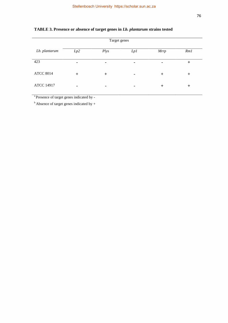
76
TABLE 3. Presence or absence of target genes in Lb. plantarum strains tested
Lb. plantarum
Target genes
Lp2 Plys Lp1 Mrrp Rm1
423 - - - - +
ATCC 8014 + + - + +
ATCC 14917 - - - + +
a Presence of target genes indicated by -
b Absence of target genes indicated by +
Stellenbosch University https://scholar.sun.ac.za

77
1 1485
mCherry gene Cassette
Pldhl mCherry TT
EcoRIBamHI HindIII
XbaI
pGKV223D(4 .8 k b )
EmR
repA
cat-86
L23
BamHIXbaISalI
PstIBspMIEcoRI
NdeI
BclIXbaI
NdeIHindIII
BbvIIEco57I
XbaI
BclI
1
pGKCherry(5 .0 k b )
EmR
repA
Pldhl mCherry
TT
BamHI
HindIII
EcoR1
NdeI
Bcll XbaI
FIG. 1. Schematic representing the construction of pGKCherry. Relevant restriction sites are shown.
For details, refer to “Materials and Methods”. Genes shown are mCherry and EmR that encode the
mCherry fluorescence gene and the protein for erythromycin resistance, respectively. Promoters: L23
originating from a Lactococcal spp.; Pldhl Lb. plantarum 423 lactate dehydrogenase promoter.
Stellenbosch University https://scholar.sun.ac.za
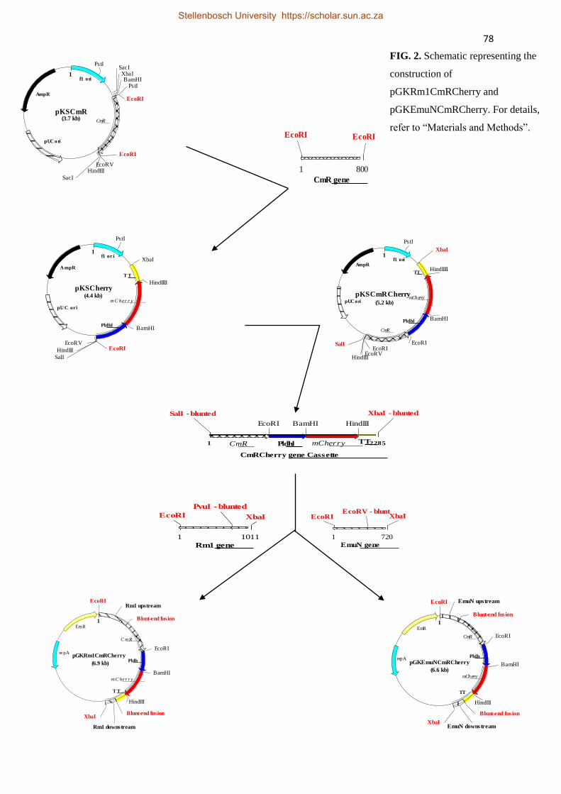
78
1
pKSCmR(3.7 kb)
f1 ori
pUC ori
AmpR
CmR
EcoRI
EcoRI
PstISacIXbaIBamHI
PstI
EcoRVHindIII
SacI
1 800
CmR gene
EcoRI EcoRI
1
pKSCherry(4.4 kb)
f1 or i
pU C ori
A mpR
mC her r y
T T
Pldhl
PstI
XbaI
HindIIII
BamHI
EcoRI
SalI
HindIII
EcoRV
1
pKSCmRCherry(5.2 kb)
f1 ori
pUC ori
AmpR
mCherry
TT
Pldhl
CmR
PstI
XbaI
HindIIII
BamHI
EcoRIEcoRI
SalI
HindIIIEcoRV
1 2285
CmRCherry gene Cassette
Pldhl mCherry TTCmR
EcoRI
SalI - blunted
BamHI HindIII
XbaI - blunted
1 1011
Rm1 gene
EcoRI XbaI
PvuI - blunted
1 720
EmuN gene
EcoRI XbaIEcoRV - blunt
1
pGKRm1CmRCherry
(6.9 kb)
EmR
re pA
Pldh
m C he r r y
T T
C m R
EcoRI
Blunt-end fus ion
Rm1 upstream
Blunt-end fus ion
Rm1 downstream
XbaI
EcoRI
BamHI
HindIII
1
pGKEmuNCmRCherry
(6.6 kb)
EmR
repA Pldh
mCherry
TT
CmR
EcoRI EmuN upstream
BamHI
HindIII
EmuN downstream
EcoRI
Blunt-end fus ion
XbaI
Blunt-end fus ion
FIG. 2. Schematic representing the
construction of
pGKRm1CmRCherry and
pGKEmuNCmRCherry. For details,
refer to “Materials and Methods”.
Stellenbosch University https://scholar.sun.ac.za
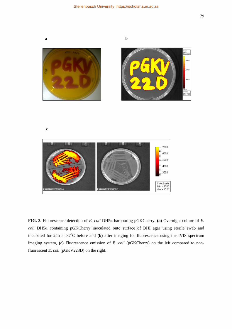
79
FIG. 3. Fluorescence detection of E. coli DH5α harbouring pGKCherry. (a) Overnight culture of E.
coli DH5α containing pGKCherry inoculated onto surface of BHI agar using sterile swab and
incubated for 24h at 37oC before and (b) after imaging for fluorescence using the IVIS spectrum
imaging system, (c) Fluorescence emission of E. coli (pGKCherry) on the left compared to non-
fluorescent E. coli (pGKV223D) on the right.
c
a b
Stellenbosch University https://scholar.sun.ac.za

80
FIG. 4. Fluorescence detection of Lb. plantarum 423 and Ent. mundtii ST4SA harbouring pGKCherry
(a) Left: control non-fluorescing Lb. plantarum 423 strain harbouring pGKV223D, right: fluorescing
Lb. plantarum 423 strain harbouring pGKCherry; (b) left: control non-fluorescing Ent.mundtii ST4SA
strain harbouring pGKV223D, right: fluorescing Ent. mundtii ST4SA strain harbouring pGKCherry;
(c) microfuge tube containing (left) non-fluorescing Lb. plantarum 423 strain harbouring pGKV223D,
and fluorescing (right) Lb. plantarum 423 strain harbouring pGKCherry. Regions of Interest (ROI)
represent the average amount of fluorescence emitted at 572 nm in p/cm2/s/sr.
c
a b
Stellenbosch University https://scholar.sun.ac.za
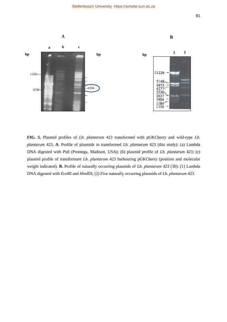
81
FIG. 5. Plasmid profiles of Lb. plantarum 423 transformed with pGKCherry and wild-type Lb.
plantarum 423. A. Profile of plasmids in transformed Lb. plantarum 423 (this study): (a) Lambda
DNA digested with PstI (Promega, Madison, USA); (b) plasmid profile of Lb. plantarum 423; (c)
plasmid profile of transformant Lb. plantarum 423 harbouring pGKCherry (position and molecular
weight indicated). B. Profile of naturally occurring plasmids of Lb. plantarum 423 (30): (1) Lambda
DNA digested with EcoRI and HindIII; (2) Five naturally occurring plasmids of Lb. plantarum 423.
a b c
1 2
A B
bp
11501---
4749--
--- --- ---
---4.934
---
---
bp bp
Stellenbosch University https://scholar.sun.ac.za

82
FIG. 6. Plasmid profiles of Ent. mundtii ST4SA transformed with pGKCherry and wild-type Ent.
mundtii ST4SA. A. Profile of plasmids in transformed Ent. mundtii ST4SA (this study). (a) Lambda
DNA digested with PstI (Promega, Madison, USA); (b) 100 kb and 50 kb plasmids of transformant
Ent. mundtii ST4SA with 4934 bp pGKCherry plasmid (indicated with dashed lines). B. Naturally
occurring plasmid profile of Ent. mundtii ST4SA (35). (1) Lambda DNA digested with EcoRI and
HindIII; (2 and 3) 50 kb and 100 kb natural plasmids of ST4SA.
a
---100000
---50000 11501---
4749--
---4934
A B
b
Stellenbosch University https://scholar.sun.ac.za
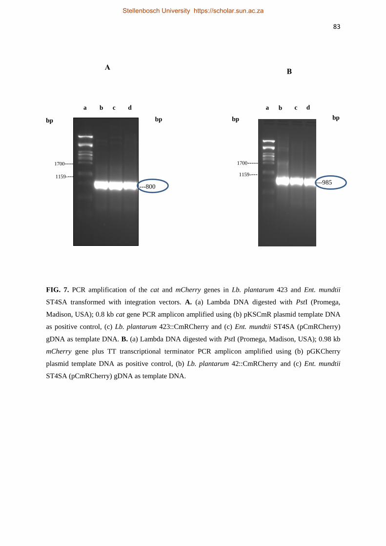
83
FIG. 7. PCR amplification of the cat and mCherry genes in Lb. plantarum 423 and Ent. mundtii
ST4SA transformed with integration vectors. A. (a) Lambda DNA digested with PstI (Promega,
Madison, USA); 0.8 kb cat gene PCR amplicon amplified using (b) pKSCmR plasmid template DNA
as positive control, (c) Lb. plantarum 423::CmRCherry and (c) Ent. mundtii ST4SA (pCmRCherry)
gDNA as template DNA. B. (a) Lambda DNA digested with PstI (Promega, Madison, USA); 0.98 kb
mCherry gene plus TT transcriptional terminator PCR amplicon amplified using (b) pGKCherry
plasmid template DNA as positive control, (b) Lb. plantarum 42::CmRCherry and (c) Ent. mundtii
ST4SA (pCmRCherry) gDNA as template DNA.
A
bp
a b c d
---800
bp
1700----
1159----
bp bp
a b c d
B
---985
1700-----
1159----
Stellenbosch University https://scholar.sun.ac.za

84
CmR+Pldhl+mCherry+TT+Rm1 downstream
Pldhl
mCherry
TT
CmR
Rm1 downstream region
1 bp 2555 bp
FIG. 8. A. Schematic diagram representing the 2.5 kb CmR+Pldhl+mCherry+TT+Rm1 amplicon. The
predicted fragment size and the binding regions of sequencing primers ForCmR and RevRm1 are
indicated. B. PCR amplification confirming the integration of the CmRCherry cassette in the
chromosome Lb. plantarum 423. (a) Lambda DNA digested with PstI (Promega, Madison, USA); 2.6
kb PCR DNA fragment containing the cat gene, Pldhl promoter, mCherry gene, TT transcriptional
terminator and the downstream Rm1 gene fragment amplified using (b) pGKRm1CmRCherry plasmid
template DNA as positive control and (b & c) gDNA of two transformants of Lb. plantarum
423::CmRCherry as template DNA.
bp bp a b c d
1700----
1159----
---2555
2883----
A.
B.
ForCmR Sequencing primer
RevRm1 Sequencing primer
Stellenbosch University https://scholar.sun.ac.za
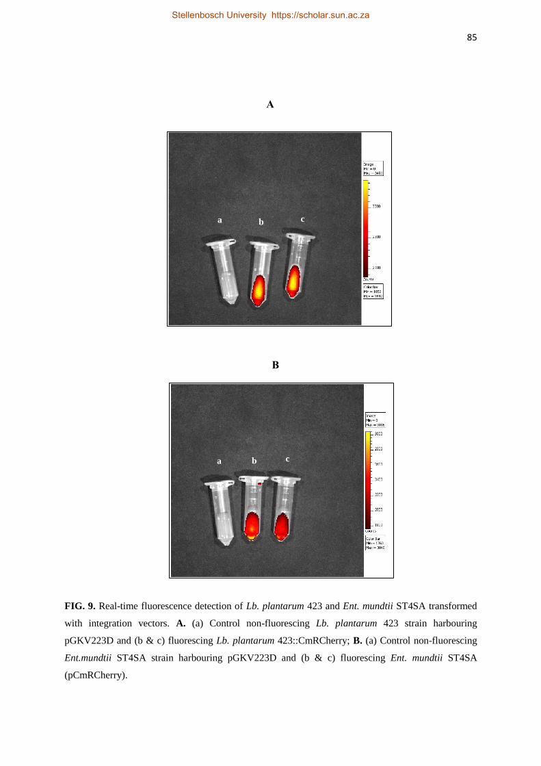
85
FIG. 9. Real-time fluorescence detection of Lb. plantarum 423 and Ent. mundtii ST4SA transformed
with integration vectors. A. (a) Control non-fluorescing Lb. plantarum 423 strain harbouring
pGKV223D and (b & c) fluorescing Lb. plantarum 423::CmRCherry; B. (a) Control non-fluorescing
Ent.mundtii ST4SA strain harbouring pGKV223D and (b & c) fluorescing Ent. mundtii ST4SA
(pCmRCherry).
B
a b c
a b c
A
Stellenbosch University https://scholar.sun.ac.za

86
FIG. 10. A.
0 2 4 6 8 100
2
4
6
8
10
12
CFU 423
CFU 423 CmRCherry
Time (h)
Lo
g C
FU
Stellenbosch University https://scholar.sun.ac.za

87
FIG. 10. B.
T im e ( h )
Lo
g C
FU
0 2 4 6 8 1 0
0
2
4
6
8
1 0
1 2
C F U S T 4 S A
C F U S T 4 S A C m R C h e r ry
FIG. 10. Growth comparison of wild-type probiotic strains (in blue) and probiotic strains harbouring
the Cherry gene (red): (A) Lb. plantarum::CmCherry and (B) Ent. mundtii ST4SA (pCmRCherry).
Values are averages of triplicate plate counts.
Stellenbosch University https://scholar.sun.ac.za
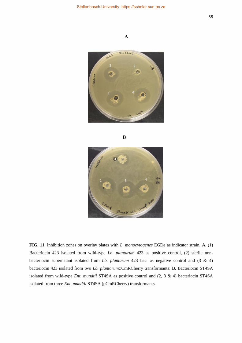
88
FIG. 11. Inhibition zones on overlay plates with L. monocytogenes EGDe as indicator strain. A. (1)
Bacteriocin 423 isolated from wild-type Lb. plantarum 423 as positive control, (2) sterile non-
bacteriocin supernatant isolated from Lb. plantarum 423 bac- as negative control and (3 & 4)
bacteriocin 423 isolated from two Lb. plantarum::CmRCherry transformants; B. Bacteriocin ST4SA
isolated from wild-type Ent. mundtii ST4SA as positive control and (2, 3 & 4) bacteriocin ST4SA
isolated from three Ent. mundtii ST4SA (pCmRCherry) transformants.
1 2
3 4
1
2 3 4
B
A
Stellenbosch University https://scholar.sun.ac.za

89
FIG. 12. A
FIG. 12. B
FIG. 12. Correlation between fluorescence signals and total CFUs of (A) Lb. plantarum
423::CmRCherry and (B) Ent. mundtii ST4SA (pCmRCherry). Positive correlations (R2=0.9889 and
R2=0.9788) between fluorescence signals and CFUs were determined for all dilutions for both strains.
Log10 averages of three repeats are plotted, with standard deviations. The logarithmic trendline (red
line) and the correlation coefficient of determination (R2) between fluorescence signals and CFUs are
shown.
1:2
1:4
1:8
1:16
1:32
R² = 0.9889
7.8
8
8.2
8.4
8.6
8.8
9
9.2
7 7.5 8 8.5 9
Ph
oto
ns
(lo
g)
p/c
m2/s
/sr
CFU (log)
Photons
Linear (Photons)
1:2
1:4
1:8
1:16
1:32
R² = 0.9788
7.6
7.8
8
8.2
8.4
8.6
8.8
9
7 7.5 8 8.5 9
Ph
oto
ns
(lo
g)
p/c
m2/s
/sr
CFU (log)
Photons
Linear (Photons)
Stellenbosch University https://scholar.sun.ac.za

90
STELLENBOSCH UNIVERSITY
Chapter 4
Intestinal Colonization of Enterococcus mundtii ST4SA
and Lactobacillus plantarum 423 in Mice, as Reported with
Fluorescent Imaging
Stellenbosch University https://scholar.sun.ac.za
91
Intestinal Colonization of Enterococcus mundtii ST4SA
and Lactobacillus plantarum 423 in Mice, as Reported with
Fluorescent Imaging
Abstract
Lactic acid bacteria (LAB) are frequently encountered as natural inhabitants of the gastro-
intestinal tract (GIT) of humans and animals. Some species received considerable attention
due to the beneficial effects they exert on health. In this study we report on the application of
the mCherry protein in the in vivo tagging of two probiotic strains, Enterococcus mundtii
ST4SA and Lactobacillus plantarum 423. Using fluorescence imaging, we determined that
both strains were predominantly colonized in the cecum and colon of mice. The location and
duration of colonization of the two strains in each section of the GIT were also revealed. We
report for the first time on the fluorescence imaging of LAB expressing mCherry in a mouse
model.
Stellenbosch University https://scholar.sun.ac.za
92
Introduction
Lactic acid bacteria (LAB) are common inhabitants of the human gastro-intestinal tract
(GIT). Some LAB, especially lactobacilli, are well known for their application in fermented
foods and probiotic properties (1, 2). These microorganisms exert many beneficial effects on
human health, such as the digestion and assimilation of food and the prevention of pathogens
from colonizing the GIT. (3-6). Furthermore, some selected probiotic strains are believed to
perform a critical role in the treatment of gastro-intestinal disorders, lactose intolerance and
in the stimulation of the immune system (7, 8).
Despite the ever increasing consumer interest in probiotic LAB due to their health-promoting
characteristics, the mechanisms by which they exert their beneficial effects and the activities
of the probiotics in the GIT often remain uncertain (9). The understanding of the survival
mechanisms of LAB in the GIT, especially the interaction between LAB and pathogens
would be facilitated by the direct in vivo monitoring of these processes.
During the past three decades, a wide range of molecular biology tools have been developed
to facilitate the study of LAB. The labelling of bacteria with marker genes with an easily
detectable phenotype has proven to be invaluable for the tracking of various LAB strains in
complex environments such as the mammalian GIT. Several reporter genes, such as the green
fluorescent protein (GFP) from the jellyfish Aequorea victoria (1, 10), the bacterial lux genes
from Photorabdus luminescence (11, 12), the click beetle luciferase system from Pyrophorus
plagiophthalamus (13, 14), chloramphenicol acetyl-transferase gene (15, 16), the Escherichia
coli β-glucoronidase gene (17) and the Vibrio fischeri luciferase gene (18) have been used to
label LAB for in vitro and in vivo studies. However, many of these systems require an
exogenous substrate to detect the expression of the reporter gene. Furthermore, oxygen
requirements needed for the expression of reporter genes is often a limiting factor for in vivo
applications (19). To circumvent these drawbacks, red fluorescent proteins, such as mCherry,
Stellenbosch University https://scholar.sun.ac.za
93
have been developed (20), and have been used successfully to label Mycobacterium
tuberculosis (21), E. coli, Lactococcus lactis and Enterococcus faecalis (23).
In this study we evaluate the potential of fluorescent imaging (FI) in real time monitoring of
Lb. plantarum 423 and Ent. mundtii ST4SA in vivo. Survival and colonization of Lb.
plantarum 423 and Ent. mundtii ST4SA in the GIT was studied using a simulated gastro-
intestinal model (32). However, simulated conditions of the GIT, are not always a true
indication of what happens in vivo. Here, we demonstrate that Lb. plantarum 423 and Ent.
mundtii ST4SA persists and colonises the GIT of mice. We also determined the location and
duration of colonization of the two strains in each section of the GIT.
Materials and methods
Ethical approval for in vivo experiments was obtained from the Ethics Committee of the
University of Stellenbosch (ethics reference number: SU-ACUM14-00015). Eight-week-old
female BALB/c mice were used in all experiments. The animals were kept under controlled
environmental conditions with water and feed provided ad libitum and changed daily.
Preparation of bacterial strains and administration to mice
Fluorescent strains of Lb. plantarum 423::CmRCherry and Ent. mundtii ST4SA
(pCmRCherry) that express the synthetic mCherry red fluorescence gene were constructed as
described previously (23). Both recombinant strains contain a gene for chloramphenicol
resistance. The strains were grown for 12 h at 30oC in 10 ml MRS broth (Biolab Diagnostics,
Midrand, South Africa), without aeration. One milliliter of each culture was inoculated into
fresh 10 ml MRS broth and incubated to an optical density at 600 nm of 2.5 for Lb.
plantarum 423::CmRCherry and 2.3 for Ent. mundtii ST4SA (pCmRCherry). The cells were
harvested (3 min at 8000 x g) and washed twice with sterile phosphate-buffered saline (PBS).
Stellenbosch University https://scholar.sun.ac.za
94
The cells of each of the strains were diluted to 2 x 10¹⁰
CFU/ml and resuspended in 200 µl
gavage buffer (0.2 M NaHCO3 buffer containing 1% glucose, pH 8) to a final concentration
of 4 x 10⁹ CFU. Viable bacterial cell suspensions were administered to mice by intragastric
gavage.
In vitro fluorescence detection
Fluorescence assays of bacterial strains expressing the mCherry gene were carried out using
black non-autofluorescent 96-well microtiter trays. Bacterial suspensions (4 ml of 12 h old
cultures) of strains with plasmids containing the mCherry gene were harvested (1500 x g for
3 min), the cell pellets were washed twice with sterile saline (0.85% (w/v) NaCl) to remove
traces of medium, and resuspended in 300 µl sterile saline. Before fluorescent image
capturing, the 300 µl cell suspensions were transferred to wells in a microtiter tray. Bacterial
strains with plasmid constructs without the mCherry gene insert were used as controls for
each fluorescing strain throughout.
Gastro-intestinal persistence of LAB
Two groups of nine mice each received one oral administration of fluorescent Lb. plantarum
423::CmRCherry and Ent. mundtii ST4SA (pCmRCherry), respectively. Control mice
received non-fluorescent Lb. plantarum 423 and Ent. mundtii ST4SA containing a vector
without the mCherry gene insert. Mice fecal material was collected at 30 min, 1, 2, 4, 6, 24,
48 and 72 h after administration of Lb. plantarum 423::CmRCherry and Ent. mundtii ST4SA
(pCmRCherry). For the enumeration of viable bacteria in feces, samples (100 mg) were
mechanically homogenized in 1 ml PBS and serially diluted in PBS and plated onto MRS
media containing chloramphenicol (Cm). Mice were sacrificed by cervical dislocation at 24,
48 and 72 h after the administration of bacteria. The GITs of the mice were dissected and the
small intestine (duodenum, jejunum and ileum) and large intestine (cecum and colon) were
Stellenbosch University https://scholar.sun.ac.za

95
studied for the presence of fluorescence signals. Background auto-fluorescence was removed
by overlaying images recorded of intestines with fluorescent bacteria with images of the
intestines of non-fluorescent bacteria. The small and large intestines were homogenized
separately in 3 ml PBS, serially diluted and plated onto MRS agar containing 5 and 10 µg/ml
Cm for Ent. mundtii ST4SA (pCmRCherry) and Lb. plantarum 423::CmRCherry,
respectively.
Evaluation of In vivo fluorescence
Fluorescence imaging (FI) was carried out using the IVIS® 100 In Vivo Imaging System
(Caliper Life Sciences, Hopkinton, MA, U.S.A.) which consists of a cooled charge-coupled-
device camera mounted on a light-tight specimen chamber. Photons emitted at 620 nm were
expressed as photons/cm2/second/steridan (p/cm
2/s/sr), using the Living Image
® software,
version 3.0 of Caliper Life Sciences. Regions of interest (ROI) were manually selected.
Signals produced from mice administered with fluorescent strains were compared to signals
produced from mice administered non-fluorescent strains. Before fluorescence imaging, mice
were anesthetized with 2% (v/v) isoflurane and placed into the IVIS camera chamber. The
anaesthetic (1.5% (v/v) isoflurane) was administered throughout examination through a nose
cone. Mice were killed at endpoints and the skin tissue of the abdomen was removed before
imaging in order to enhance fluorescence signal penetration. Reference images of mice
administered with control non-fluorescent strains were used for the removal of background
signals throughout the experiment.
Stellenbosch University https://scholar.sun.ac.za
96
Results
Monitoring of Lb. plantarum 423 and Ent. mundtii ST4SA in the GIT
At time zero (before administration of mice), no fluorescence signal was detected. During the
first 24 h post inoculation, no significant difference could be detected transcutaneously
between mice treated with fluorescent LAB and mice treated with control strains (data not
shown). However, when mice were imaged after sacrifice with the removal of the abdomen
skin tissue 24 h after administration of bacteria, a notable difference in fluorescence was
detected (Fig. 1).
The intestines were also imaged ex vivo, to determine the localization of the fluorescent
bacteria in the GIT (Fig. 2). At 24 h post inoculation of bacteria, both strains were observed
to have colonized predominantly in the cecum and colon. However, no fluorescence was
observed in the small intestines of mice administered Lb. plantarum 423::CmRCherry,
whereas fluorescent cells could be detected in the small intestines of mice administered Ent.
mundtii ST4SA (pCmRCherry). After 48 h, no fluorescent Lb. plantarum 423::CmRCherry
and Ent. mundtii ST4SA (pCmRCherry) could be detected in either the small or large
intestines.
Colonization of Lb. plantarum 423 and Ent. mundtii ST4SA in small and large intestine
After 24 h, mice administered with Lb. plantarum 423::CmRCherry harboured approximately
7.5 x 10² CFU in the small intestine (Fig. 3A) and more than 2 x 10
5 CFU in the large
intestine (Fig. 3B). Mice administered Ent. mundtii ST4SA (pCmRCherry) harboured
approximately 2 x 10⁴ CFU in the small intestinal compartment (Fig. 3A) and 2 x 10
5 in the
large intestinal compartment after 24 h (Fig. 3B). After 72 h, no Lb. plantarum
423::CmRCherry was detected in the intestines of mice. No viable Ent. mundtii ST4SA
(pCmRCherry) cells were detected in the small intestine 72 h after administration. However,
Stellenbosch University https://scholar.sun.ac.za
97
the number of Ent. mundtii ST4SA (pCmRCherry) cells in the large intestinal compartment
was still approximately 5 x 10³ CFU.
Enumeration of viable Lb. plantarum 423 and Ent. mundtii ST4SA in mouse feces
Fluorescent cells of Lb. plantarum 423::CmRCherry and Ent. mundtii ST4SA (pCmRCherry)
were observed in the feces collected after 24 h. High numbers of both strains were excreted in
the feces (Fig. 4). For Lb. plantarum 423::CmRCherry, the maximum level of viable cells
excreted after 4 h reached approximately 3 x 10⁷ CFU/100 mg feces. Enterococcus mundtii
ST4SA (pCmRCherry), on the other hand, reached a maximum level of 3 x 10⁸ CFU/100 mg
feces after 4 h and then slightly declined to 8 x 10⁷ CFU/100 mg feces after 24 h. After 72 h,
the number of Lb. plantarum 423::CmRCherry organisms declined to approximately 5 x 10³
CFU/100 mg feces, while the Ent. mundtii ST4SA (pCmRCherry) declined to 4 x 10⁴
CFU/100 mg feces.
Discussion
The emission of strong fluorescence by Lb. plantarum 423::CmRCherry and Ent. mundtii
ST4SA (pCmRCherry) harbouring the codon-optimized synthethic mCherry has been
reported (23). Similar results were recorded for the in vitro expression of the mCherry gene in
Lactococcus lactis, Enterococcus faecalis and Mycobacterium tuberculosis (21, 22).
In the current study, in vivo results demonstrated that transcutaneous signals in mice
administered fluorescent Lb. plantarum 423::CmRCherry and Ent. mundtii ST4SA
(pCmRCherry), could not be distinguished from the background signals of mice administered
non-fluorescent control strains. Factors that usually influence the application of a particular
fluorescence protein for in vivo experimentation include the propagation of fluorescent
signals through tissue and brightness of the fluorophore compared to the inherent background
Stellenbosch University https://scholar.sun.ac.za

98
auto-fluorescence of the sample or animal (21, 24, 25). However, the excitation and emission
spectrum of the mCherry fluorophore is outside the region where endogenous chromophores,
such as haemoglobin, absorb light within tissues (26).
In a similar study, abdominal auto-fluorescence effected the detection of intentional
fluorescence signals in mice (27). However, the effects of auto-fluorescence can usually be
avoided by the use of fluorophores with excitation and emission wavelengths in the far red
and near infrared region (600 nm > 780 nm) of the visible spectrum (28). Furthermore,
impairment of transcutaneous signals could also be explained by the inherent background
signals generated by the auto-fluorescence of the animal diet intake (29). This merits further
investigation. Since no other studies regarding the in vivo application of the mCherry gene
are reported in literature, this is the first study reporting the in vivo detection of LAB in mice
using mCherry.
By removal of the abdominal tissue and skin of euthanized animals at 24 h post-inoculation,
fluorescence was detected in mice colonized with Lb. plantarum 423::CmRCherry and Ent.
mundtii ST4SA (pCmRCherry). The fluorescent signal was weakened significantly by
passage through the animal tissue and skin for transcutaneous detection compared to the
signal obtained after tissue and skin removal.
The digestive tracts of mice colonized with fluorescent LAB were also imaged ex vivo. The
compensation for background auto-fluorescence, possibly due to chromophores in mice feed,
significantly increased detection of the signal. Both strains were predominantly detected in
the anaerobic cecum and colon. Similarly, previous studies found Lb. plantarum to
predominantly persist in the cecum and colon (5, 30). In another study, Lb. plantarum 423
was reported to adhere to mucus isolated from the ileum and cecum of piglets (31). The CFU
counts confirmed that the cecum and colon were the major sites of Lb. plantarum 423 and
Ent. mundtii ST4SA colonization (Fig. 3). However, a fluorescent signal and high numbers of
Stellenbosch University https://scholar.sun.ac.za
99
Ent. mundtii ST4SA cells were also found in the small intestine. This is in contrast to a
previous investigation which reported that Lb. plantarum 423 colonized the ileum, cecum and
colon, whereas Ent. mundtii ST4SA only colonized the cecum (8). The authors used
fluorescent in situ hybridisation (FISH) with strain specific oligonucleotide probes to detect
which surfaces in the GIT the strains adhered to. However, Lb. plantarum 423 was also
detected in the intestines of rats not treated with Lb. plantarum 423. This suggests that the
oligonucleotide probes that were used may have hybridized to DNA from other closely
related strains in the rat GIT and may have affected the results. The CFU counts revealed that
strain ST4SA colonises the gut more effectively as low levels of the bacteria were still
present 72 h post-feeding (Fig 3).
The colonization ability of Lb. plantarum 423 and Ent. mundtii ST4SA were assessed by
determining the number of cells excreted at different time points over 72 h. Both strains
survived conditions in the GIT as clearly indicated by the attainment of viable fluorescing
cells in the feces of mice (Fig. 4). Similar results were obtained in a previous study, with the
prediction of the survival of Lb. plantarum 423 and Ent. mundtii ST4SA in the GIT of infants
using a computerized gastro-intestinal model (32). High cell numbers were excreted for both
strains during the first 24 h after administration of the bacteria to mice (Fig. 4). Enterococcus
mundtii ST4SA was excreted in higher numbers during the first 24 h compared to Lb.
plantarum 423, suggesting that Lb. plantarum 423 colonized the GIT more effectively. After
72 h, the number of cells of both bacteria excreted declined to low levels. These transit
dynamics are similar to those reported for several LAB strains` including Lb. casei, Lb.
plantarum WCFS1 and Lc. lactis (5, 11, 33, 34). The decline in the number or Lb. plantarum
423 and Ent. mundtii ST4SA cells excreted over the 72 h period suggests that it is a non-
permanent colonization. However, Pavan and co-workers (2002), found that Lb. plantarum
WCFS1 was still excreted in the feces of mice for up to 7 days after one oral administration
(33).
Stellenbosch University https://scholar.sun.ac.za

100
In summary, we have shown that mCherry can be used to detect LAB ex vivo. Fluorescence
imaging proved useful in demonstrating that Lb. plantarum 423 and Ent. mundtii ST4SA
predominantly colonise the cecum and colon. Both strains persisted in the gut over 24 h.
However, no Lb. plantarum 423 could be found in the digestive tract after 72 h, while low
numbers of Ent. mundtii ST4SA were still present. The detection of fluorescence in whole
animals was greatly facilitated by the removal of the mice abdominal region skin tissue after
sacrifice. We could not detect any transcutaneous fluorescence. The propagation of mCherry
fluorescence through tissue was restricted. As far as we know, this is the first report on
fluorescence imaging of LAB expressing mCherry in a mouse model.
Stellenbosch University https://scholar.sun.ac.za

101
References
1. Geoffroy, M., C. Guyard, B. Quatannens, S. Pavan, M. Lange and A. Mercenier.
2000. Use of green fluorescent protein to tag lactic acid bacterium strains under
development as live vaccine vectors. Appl. Environ. Microbiol. 66 (1): 383-391.
2. Marteau, P. and J.C. Rambaud. 1993. Potential of using lactic acid bacteria for
therapy and immunomodulation in man. FEMS. Microbiol. Rev. 12: 207-222.
3. Xu. J. and J.I. Gordon. 2003. Honor thy symbionts. Proc. Natl. Acad. Sci. USA 100:
10452-10459.
4. Stanton, C., R.P. Ross, G.F. Fitzgerald and D. van Sindern. 2005. Feremented
functional foods based on probiotics and their biogenic metabolites. Curr. Opin.
Bioechnol. 16: 198-203.
5. Marco, M.L., R.S. Bongers, W.M. de Vos and M. Kleerebezem. 2007. Spatial and
temporal expression of Lactobacillus plantarum genes in the gastrointestinal tract of
mice. Appl. Environ. Microbiol. 73: 124-132.
6. Gerritsen, J., H. Smidt, G.T. Rijkers and G.M. de Vos. 2011. Intestinal microbiota
in human health and disease: the impact of probiotics. Genes Nutr. 6: 209-240.
7. Franz, C.M.A.P., M.E. Stiles, K.H. Schleifer and W.H. Holzapfel. 2003.
Enterococci in foods - a conundrum for food safety. Int. J. Food Microbiol. 88: 105-
122.
8. Ramiah, K., K. ten Doeschate, R. Smith and L.M.T. Dicks. 2009. Safety Assessment
of Enterococcus mundtii ST4SA and Lactobacillus plantarum 423 determined in trials
with Wistar rats. Probiot. Antimicrob. Prot. 1: 15-23.
9. Marco, M.L., S. Pavan and M. Kleerebezem. 2006. Towards understanding
molecular modes of probiotic action. Curr. Opin. Biotechnol. 17: 204-210.
Stellenbosch University https://scholar.sun.ac.za

102
10. Yu, Q., S. Dong, W. Zhu and Q. Yang. 2007. Use of green fluorescent protein to
monitor Lactobacillus in the gastro-intestinal tract of chicken. FEMS Microbiol. Letts.
275: 207-213.
11. Oozeer, R., N. Goupil-Feuillerat, C.A. Alpert, M. van de Guchte, J. Anba, J.
Mengaud and G. Corthier. 2002. Lactobacillus casei is able to survive and initiate
protein synthesis during its transit in the digestive tract of human flora-associated mice.
Appl. Environ. Microbiol. 68: 3570-3574.
12. Cronin, M., R.D. Sleator, C. Hill, G.F. Fitzgerald and D. van Sinderin. 2008.
Development of luciferase-based reporter system to monitor Bifidobacterium breve
UCC2003 persistence in mice. BMC Microbiol. 8: 161.
13. Foucault, M.L., L. Thomas, S. Goussard, B.R. Branchini and C. Grillot-Courvalin.
2010. In vivo bioluminescence imaging for the study of intestinal colonization by
Escherichia coli in mice. Appl. Environ. Microbiol. 76: 264-274.
14. Daniel, C., S. Poiret, V. Dennin, D. Boutillier and B. Pot. 2013. Bioluminescence
imaging study of spatial and temporal persistence of Lactobacillus plantarum and
Lactococcus lactis in living mice. Appl. Environ. Microbiol. 79 (4): 1086-1094.
15. Archen, M.G., B.E. Davidon and A.J. Hillier. 1986. Construction of plasmid vectors
for the detection of streptococcal promoters. Gene 45: 45-49.
16. De Vos, W.M. 1987. Gene cloning and expression in lactic streptococci. FEMS
Microbiol. Rev. 46: 281-295.
17. Platteeuw, C.G., G. Simons and W.M. de Vos. 1994. Use of Escherichia coli
βglucoronidase (gusA) gene as a reporter gene for analysing promoters in lactic acid
bacteria. Appl. Environ. Microbiol. 60: 587-593.
18. Eaton, T.J., C.A. Shearman and M.J. Gasson. 1993. The use of bacterial luciferase
genes as reporter genes in Lactococcus: regulation of the Lactococcus lactis subsp.
lactis lactose genes. J. Gen. Microbiol. 139: 1495-1501.
Stellenbosch University https://scholar.sun.ac.za
103
19. Contag, C.H. and M.H. Bachmann. 2002. Advances in in vivo bioluminescence
imaging of gene expression. Annu. Rev. Biomed. Eng. 4: 235-237.
20. Shaner, N.C., R.E. Campbell, P.A. Steinbach, B.N. Giepmans, A.E. Palmer and
R.Y. Tsien. 2004. Red fluorescent proteins. Nat. Biotechnol. 22 (12): 1567.
21. Parish, T., P. Carroll, L.J. Schreuder, J. Muwanguzi-Karugaba, S. Wiles, B.D.
Robertson et al. 2010. Sensitive detection of gene expression in Mycobacteria under
replicating and non-replicating conditions using optimized far-red reporters. PLoS ONE
5 (3): 9823.
22. Garcia-Cayuela, T. and L.P. Gomez de Carinanos. 2012. Fluorescent protein vectors
for promoter analysis in lactic acid bacteria and Escherichia coli. Appl. Microbiol.
Biotechnol. 96: 171-181.
23. Van Zyl, W.F., S.D. Deane and L.M.T. Dicks. 2014. Construction of a Fluorescent
Reporter System for Lactic Acid Bacteria, using the mCherry Gene. Department of
Microbiology, University of Stellenbosch, South Africa, S.A.
24. Taroni, P., A. Pifferi, A. Torricelli, D. Comelli and R. Cubeddu. 2003. In vivo
absorption and scattering spectroscopy of biological tissues. Photochem. Photobio. S.
2: 124-129.
25. Wiedenmann, J., F. Oswald and G.U. Nienhaus. 2009. Fluorescent proteins for live
cell imaging: Oppurtunities, limitations and challenges. Life 61 (11): 1029-1042.
26. Rice, B.W., M.D. Cable and M.B. Nelson. 2001. In vivo imaging of light-emitting
probes. J. biomed. Opt. 6: 432-440.
27. Inoue, Y., K. Izawa, S. Kiryu, A. Tojo and K. Ohtomo. 2008. Diet and abdominal
autofluorescence detected by in vivo fluorescence imaging of living mice. Mol. Imaging
7 (1): 21-27.
28. Volpi, D. and B. Vojnovic. 2012. Tissue autofluorescence: watch out what’s on the
menu. Gray Institute. Department of Oncology. University of Oxford.
Stellenbosch University https://scholar.sun.ac.za

104
29. Troy, T., D. Jekic-Mcmullen, L. Sambucett and B. Rice. 2004. Quantitative
comparison of the sensitivity of detection of fluorescent and bioluminescent reporters in
animal models. Mol. Imaging 3: 9-23.
30. Duangjitchareon, Y., D. Kantachote, M. Ongsakul, N. Poosaran and C. Chaiyasut.
2009. Potential use of probiotic Lactobacillus plantarum SS2 isolated from fermented
plant beverage: safety assessment and persistence in the murine gastro-intestinal tract.
World J. Microbiol. Biotechnol. 25: 315-321.
31. Mare, L., G.M. Wolfaardt and L.M.T. Dicks. 2006. Ahesion of Lactobacillus
plantarum 423 and Lactobacillus salivarus 241 to the intestinal tract of piglets, as
recorded with fluorescent in situ hybridisation (FISH) and production of plantaricin 423
by cells colonized to the ileum. J. Appl. Microbiol. 100: 838-845.
32. Botes, M., C.A. van Reenen and L.M.T. Dicks. 2008. Evaluation of Enterococcus
mundtii ST4SA and Lactobacillus plantarum 423 as probiotics using a gastro-intestinal
model with infant milk formulations as substrate. Int. J. Food Microbiol. 128: 362-370.
33. Pavan, S., P. Hols, J. Delcour, M.C. Geoffroy, C. Grangette, M. Kleerebezem and
A. Mercenier. 2000. Adaptation of the nisin-controlled expression system in
Lactobacillus plantarum: a tool to study in vivo biological effects. Appl. Environ.
Microbiol. 66: 4427-4432.
34. Grangette, C., H. Muller-Alouf, M. Geoffroy, D. Goudercourt, M. Turneer and A.
Mercenier. 2002. Protection against tetanus toxin after intragastric administration of
two recombinant lactic acid bacteria: impact of strain viability and in vivo persistence.
Vacinne 20: 3304-3309.
Stellenbosch University https://scholar.sun.ac.za

105
FIG. 1. Fluorescent imaging of Lb. plantarum 423 and Ent. mundtii ST4SA in vivo. Lactobacillus
plantarum 423::CmRCherry, Ent. mundtii ST4SA (pCmRCherry) and control strains, Lb. plantarum
423 (pGKV) and Ent. mundtii ST4SA (pGKV) (4 x 10⁹ CFU) were inoculated intragastrically into
sets of three mice, and the fluorescent signal was detected after removal of the skin tissue on the
ventral side after euthanisia. Shown is one representative image of one mouse, 24 h post-feeding. The
mouse abdominal regions are circled. (A) Mouse fed with control non-fluorescent strain 423 (pGKV)
and (B) mouse fed with fluorescent strain 423::CmRCherry. (C) Mouse fed with control non-
fluorescent strain ST4SA (pGKV) and (D) mouse fed with fluorescent strain ST4SA (pCmRCherry).
A. B.
C. D.
Stellenbosch University https://scholar.sun.ac.za

106
FIG. 2. Localization of fluorescent Lb. plantarum 423 and Ent. mundtii ST4SA in the digestive tracts
of mice. Groups of mice were gavaged once with Lb. plantarum 423::CmRCherry and Ent. mundtii
ST4SA (pCmRCherry) and the intestines resected 24 h post-feeding. Three mice were sacrificed, and
shown are representative images of the digestive tracts of two mice (1 and 2) at 24 h (H24), in mouse
fed with Lb. plantarum 423::CmRCherry (a) and mouse fed with Ent. mundtii ST4SA (pCmRCherry)
(b). The background auto-fluorescence was compensated for by using images of the digestive tracts of
mice fed with control strains, 423 (pGKV )and ST4SA (pGKV).
1
2
H24a H24b
Stellenbosch University https://scholar.sun.ac.za

107
24 48 72
100
101
102
103
104
105
423
ST4SA
Hours
Lo
g C
FU
/ml
Sm
all In
testi
nes
24 48 72
100
102
104
106
423
ST4SA
Hours
Lo
g C
FU
/ml
Larg
e in
testi
nes
FIG. 3. Persistence of Lb. plantarum 423 and Ent. mundtii ST4SA in the (A) small and (B) large
intestinal compartments of mice after one oral administration. Groups of mice were gavaged once
with Lb. plantarum 423::CmRCherry and Ent. mundtii ST4SA (pCmRCherry) and the intestines
resected 24, 48 and 72 h post-feeding. At the indicated times, log10, averages of three mice are plotted
with standard deviations.
A.
B.
Stellenbosch University https://scholar.sun.ac.za

108
0 6 12 18 24 30 36 42 48 54 60 66 72100
102
104
106
108
101 0
423
ST4SA
*
Hours
Lo
g C
FU
/100m
g f
eces
FIG. 4. Transit of Lb. plantarum 423 and Ent. mundtii ST4SA in the feces of mice. Sets of three mice
were fed once with 4 x 10⁹ CFU of Lb. plantarum 423::CmRCherry and Ent. mundtii ST4SA
(pCmRCherry). At every time point, log10, means of the CFU per 100 mg feces for each set of three
mice are plotted with standard deviations. At the times indicated, colonization was monitored by
enumeration of bacteria in feces plated on MRS containing chloramphenicol. Overall differences
between strains 423 and ST4SA, were assessed using the Man-Whitney nonparametric test and those
found to be significant (P<0.05) are shown with an asterisk at 4 h.
Stellenbosch University https://scholar.sun.ac.za

109
STELLENBOSCH UNIVERSITY
Chapter 5
Bioluminescence Imaging of Listeria monocytogenes in
Mice Colonized with Enterococcus mundtii ST4SA and
Lactobacillus plantarum 423
Stellenbosch University https://scholar.sun.ac.za
110
Bioluminescence Imaging of Listeria monocytogenes in
Mice Colonized with Enterococcus mundtii ST4SA and
Lactobacillus plantarum 423
Abstract
Listeria monocytogenes is an opportunistic food-borne pathogen, which can be life-
threatening to individuals with a weakened immune system. The aim of this study was to
evaluate whether Lactobacillus plantarum 423 and Enterococcus mundtii ST4SA could
prevent the colonization of L. monocytogenes in the gastro-intestinal tract of mice. Mice pre-
colonized with Lb. plantarum 423 and Ent. mundtii ST4SA were challenged with a
bioluminescent strain of L. monocytogenes. Using in vivo bioluminescence, we determined
that the colonization of mice with Lb. plantarum 423 and Ent. mundtii ST4SA, or a
combination of the strains, protected the animals from systemic L. monocytogenes EGDe
infection. Treatment of Listeria infections with probiotic lactic acid bacteria may offer an
alternative to conventional antibiotics.
Stellenbosch University https://scholar.sun.ac.za

111
Introduction
Listeria monocytogenes is the causative agent of a serious food-borne illness.
Immunocompromised individuals are the most susceptible to listeriosis and it results in a high
mortality rate (1, 2). Listeria monocytogenes is a ubiquitous gram positive organism that can
survive low pH, low water activity and high sodium chloride levels associated with meat
drying and fermentation (3, 4). Its ability to proliferate at low temperatures renders many
food products at risk to contamination.
Effective antibiotic treatment against infections caused by Listeria is essential. Although
several antibiotics have activity against the organism, the frequent use of antibiotics may
increase the chances of developing antibiotic resistant strains. Treatment with probiotic lactic
acid bacteria (LAB) may offer an alternative to conventional antibiotics. Several studies have
reported that LAB exert inhibitory activity against Listeria monocytogenes in vitro (5-9). The
anti-microbial effects of LAB are exerted either directly via competition of live microbial
cells with pathogens or indirectly via the production of organic acids, hydrogen peroxide or
bacteriocins (10).
Several bacteriocins, including nisin, lactocin 705, enterocin 81 and lacticin 3147 have been
described with in vitro activity against L. monocytogenes (4, 7, 11-13). They are small
ribosomally synthesized antimicrobial peptides that inhibit the growth of other species (14).
Bacteriocins have been applied as natural preservatives in the food industry for decades,
particularly against contamination by L. monocytogenes.
One of the prerequisites of a good probiotic is the ability of the strain to adhere to epithelial
cells or its ability to compete against pathogens for adhesion sites (13, 15). The effects of a
healthy intestinal microbiota composition on L. monocytogenes induced infection have
previously been assessed in vivo (16, 17). Treatment of infections caused by Listeria with live
probiotic LAB enhances the host’s resistance by stimulating the immune system (18-20).
Stellenbosch University https://scholar.sun.ac.za
112
In the current study, we evaluated the impact of Lb. plantarum 423 and Ent. mundtii ST4SA
on orally acquired L. monocytogenes infection and their ability to compete with the pathogen
in the gastro-intestinal tract (GIT) of mice. Mice were colonized with the probiotic LAB prior
to being challenged with a bioluminescent strain of L. monocytogenes. Changes in infection
were recorded by monitoring changes in bioluminescence and viable cell counts.
Materials and Methods
Approval for experiments on animals was obtained from the Ethics Committee of the
University of Stellenbosch (ethics reference number: SU-ACUM14-00015). Eight-week-old
female BALB/c mice were used in all experiments. The animals were kept under controlled
environmental conditions and fed a standard rodent diet.
Preparation of bacterial strains and growth conditions
The bioluminescent strain, L. monocytogenes EGDe, harbouring the pPL2 lux plasmid
containing the luxABCDE operon of Photorhabdus luminescence was purchased from Bio-
ware™
Microorganisms (Caliper Life Sciences, Hopkinton, MA, U.S.A.). Listeria
monocytogenes EGDe was cultured in Brain Heart Infusion (BHI) broth (Biolab Diagnostics,
Midrand, South Africa) or BHI agar at 37oC supplemented with 7.5 µg/ml chloramphenicol
(Cm). Enterococcus mundtii ST4SA and Lb. plantarum 423 harbouring a gene for Cm
resistance were grown in MRS broth (Biolab Diagnostics, Midrand, South Africa) or MRS
agar at 30oC containing 5 µg/ml Cm for strain ST4SA and 10 µg/ml Cm for strain 423 (21).
After 12 h, the cultures were used to inoculate fresh media and were grown to exponential
phase, harvested (8000g, 3 min) and washed twice with phosphate buffered saline. A dilution
series was prepared and the optical density of each culture recorded at 600 nm.
Stellenbosch University https://scholar.sun.ac.za
113
Dosage of mice
Sets of six mice received a daily dose of LAB for six consecutive days. Mice in group L
received 4 x 10⁹ CFU Lb. plantarum 423, mice in group E received 4 x 10
9 CFU Ent. mundtii
ST4SA, and mice in group EL received a 1:1 combination of Ent. mundtii ST4SA and Lb.
plantarum 423 in 200 µl gavage buffer (0.2 M NaHCO3buffer containing 1% glucose, pH 8).
Mice in group P received 200 µl sterile physiological water (control group). On day 6, the
mice were infected intragastrically with 9.6 x 10⁸ CFU of L. monocytogenes EGDe,
resuspended in 200 µl gavage buffer.
Mouse colonization
Fresh fecal samples were collected at 30 min, 1, 2, 4, 6 and 24 h after the administration of L.
monocytogenes EGDe. For the enumeration of viable bacteria, fecal samples were weighed
(100 mg), mechanically homogenized in PBS, serially diluted and plated on BHI and MRS
agar containing Cm at the concentrations described above. Mice were sacrificed at 4, 6 and
24 h by cervical dislocation and immediately thereafter the digestive tracts (stomach to
rectum) were resected and positioned in a Petri dish for ex vivo bioluminescence imaging.
The bioluminescence signal was enhanced by imaging longitudinally opened organs. To
determine the number of viable bacteria per organ homogenate, the small and large intestines
were homogenized in 3 ml PBS, respectively, diluted and plated on BHI and MRS agar
containing Cm as described above.
In vitro and in vivo bioluminescence imaging
Bioluminescent images were obtained using the IVIS® 100 In Vivo Imaging System (Caliper
Life Sciences, Hopkinton, MA, U.S.A.), which consists of a cooled charge-coupled-device
camera mounted on a light-tight specimen chamber. For the detection of bioluminescent L.
Stellenbosch University https://scholar.sun.ac.za

114
monocytogenes EGDe in fecal samples homogenized in PBS, 300 µl homogenate was added
to black 96-well microtiter trays and imaged by IVIS. Prior to live animal bioluminescence
imaging, isoflurane gas anesthetic was administered to mice at 2%, from which the mice
recovered within 2 min of removal of gas. During imaging, anesthesia was maintained by a
controlled air flow of 1.5% (v/v) isoflurane through a nose cone. Photon emission of bacteria
in microtitre trays, or anesthetized mice placed in the ventral position, was measured with
exposure times ranging from 1 to 5 min, depending on bioluminescence signal intensity.
Bioluminescence was quantified using the Living Image® software, version 3.0 (Caliper Life
Sciences, Massachusetts, U.S.A.) and measured as photons per second (p/s).
Bioluminescence signals originating from the abdomen of each individual mouse were
quantified using identical regions of interest (ROI) for all images and were determined
manually.
Results
Bioluminescence monitoring of colonization by Listeria monocytogenes in mouse
intestines
Before the administration of L. monocytogenes EGDe to mice, no bioluminescence signal
was detected (7.24 x 10⁴ p/s being the background signal). As early as 30 min after the
administration of L. monocytogenes EGDe, a strong bioluminescence signal was detected in
the abdominal region of all mice (Fig. 1A). After 24 h, a bioluminescent signal could be
observed from mice in the control group (group P). Mice in the control group, showed
symptoms of listeriosis, such as lethargy, hunched appearance and conjunctivitis of the eye
throughout the infection. Mice that had received Lb. plantarum 423, Ent. mundtii ST4SA and
a combination of the two strains showed no physical abnormalities. The bioluminescence
signal from mice that had received Lb. plantarum 423 (group L) could not be observed after 4
h post-administration of L. monocytogenes EGDe, whereas the signal from mice that had
Stellenbosch University https://scholar.sun.ac.za

115
received Ent. mundtii ST4SA (group E) or the combination of both strains (group EL) could
not be observed after 2 h. The highest signal measured in whole animals was recorded from
mice in the control group (mean value of 2.66 x 10⁶ p/s) compared to that from mice in group
L (mean value of 1.46 x 10⁶ p/s), group E (mean value of 1.26 x 10
6 p/s) and group EL (mean
value of 9.72 x 10⁵ p/s), respectively (Fig. 1B). After 6 h, the signal from mice in group L
decreased to the background level, whereas the signals from both groups E and EL declined
to approximately 1.30 x 10⁵ p/s. The bioluminescent signals of mice in the control group
declined to approximately 3 x 10⁵ p/s after 6 h, and remained at a plateau until 24 h.
Dissected intestines were also imaged ex vivo for L. monocytogenes EGDe bioluminescence
(Fig. 2). The highest bioluminescent signals were obtained when the small and large
intestines were longitudinally opened. By 6 h after infection, L. moncytogenes EGDe was
detected throughout the lower section of the small intestine (group P). After 24 h, a
bioluminescence signal was detected from the small intestine of mice in the control group
alone. After 6 h, no bioluminescent L. moncytogenes EGDe could be detected from the
intestines of mice that had received Lb. plantarum 423, Ent. mundtii ST4SA or the
combination of both strains, respectively. The CFU counts confirmed that the small and large
intestines were colonized by L. monocytogenes EGDe (Fig. 3). After 6 h, the number of L.
monocytogenes EGDe cells (mean value of 5 x 10² CFU) in the small intestines of mice that
had received Ent. mundtii ST4SA was significantly lower compared to that of group P (mean
value of 8.9 x 10⁶ CFU) as indicated in Fig. 3A. The number of L. monocytogenes EGDe
cells in the large intestines of mice that received Ent. mundtii ST4SA decreased to low levels
(mean value of 1 x 10³ CFU) after 24 h (Fig. 3B). While the number of L. monocytogenes
EGDe cells in the GIT of mice that had received Lb. plantarum 423 also decreased compared
to the control group. The amount of L. monocytogenes EGDe cells in the intestines of mice
that had received Ent. mundtii ST4SA was always lower. Viable cell counts of the individual
bacterial species in the intestines of mice in group EL could not be determined as L.
Stellenbosch University https://scholar.sun.ac.za
116
monocytogenes EGDe, Lb. plantarum 423 and Ent. mundtii ST4SA could not be
differentiated from each other based on colony morphology. Twenty four hours post-
administration of L. monocytogenes EGDe, the large intestine (cecum and colon) was the
major site of colonization of Lb. plantarum 423, whereas Ent. mundtii ST4SA was colonizing
both the small and large intestines (Fig. 4).
Enumeration of viable L. monocytogenes EGDe, Lb. plantarum 423 and Ent. mundtii
ST4SA in mouse feces
The number of viable L. monocytogenes EGDe increased with time in the feces of all the
mice, reaching a maximum level after 4 h in mice administered Lb. plantarum 423 (mean
value of 3.54 x 10⁸ CFU) and Ent. mundtii ST4SA (mean value of 2.61 x 10
8 CFU),
respectively (Fig. 5A). After 4 h, the number of L. monocytogenes EGDe excreted in the
feces of mice in groups E and L was significantly higher compared to that of group P (mean
value of 2 x 10² CFU). There was excellent correlation between this peak and the maximum
bioluminescence signal of 3.15 x 10⁵ p/s/100mg of feces and 1.03 x 10
6 p/s/100mg of feces
after 4 h for mice in groups E and L, respectively (Fig. 5B). By 6 h, a maximum of
approximately 1.78 x 10⁸ CFU L. monocytogenes EGDe with a bioluminescence signal of
5.87 x 10⁵ p/s/100mg of feces were detected in the feces of mice in group P and declined to
approximately 1.67 x 10⁵ CFU after 24 h. The number of Lb. plantarum 423 and Ent. mundtii
ST4SA, increased with time in the feces of mice in groups L and E, respectively, reaching
maximums of approximately 4.27 x 10⁷ and 1.53 x 10
8 CFU (Fig. 6).
Discussion
Bioluminescence has been used in a number of previous studies to follow in vivo infection
processes (12, 22-26). In the current study, Listeria monocytogenes EGDe bioluminescence
could be detected transcutaneously in whole animals after a single oral administration. No
Stellenbosch University https://scholar.sun.ac.za

117
physical symptoms of listeriosis were observed in any mice that received the probiotic LAB,
suggesting that the LAB protected the animals from systemic infection. The highest
bioluminescence signals were obtained with mice in the control group and could be observed
transcutaneously throughout the 24 h period of infection (Fig. 1). After 4 h, the
bioluminescent signal of mice that received Lb. plantarum 423, Ent. mundtii ST4SA and the
combination of the strains declined below transcutaneous detection levels. This suggests that
interactions between the probiotic LAB and L. monocytogenes EGDe in the mouse GIT
results in the inhibition of the pathogen as observed with live animal imaging. The ability of
Lb. plantarum 423 and Ent. mundtii ST4SA to inhibit the growth of L. monocytogenes EGDe
and L. monocytogenes ScottA in vitro has been shown previously (21, 27). Our results show
that the real-time bioluminescence imaging of live animals plays a key role in displaying the
difference in effects of L. monocytogenes EGDe infection on mice already colonized with Lb.
plantarum 423 and Ent. mundtii ST4SA compared to the control group.
Listeria monocytogenes EGDe lux bioluminescence emission is optimal in an aerobic
environment (28). Transcutaneous lux bioluminescence signal detection may not be a true
representation of the actual colonization due to limited oxygen availability in the intestinal
cavity. Longitudinally opened mouse digestive tracts were imaged ex vivo with the aim of
exposing L. monocytogenes EGDe to oxygen. A previous study has shown that a 10-fold-
higher bioluminescent signal was observed by imaging opened digestive tracts (24).
Bioluminescent L. monocytogenes EGDe was detected only in the small intestine of mice in
the control group after 24 h. However, CFU counts confirmed that L. monocytogenes EGDe
also colonized the large intestine but did not emit bioluminescence, suggesting that oxygen
may have been the limiting factor in the more anaerobic cecum and colon (Fig. 3B).
Microorganisms harbouring the bacterial lux genes can only produce bioluminescence when
the cells are metabolically active (28). Although CFU counts confirmed the presence of high
numbers of L. monocytogenes EGDe cells in the digestive tracts of mice that received Lb.
Stellenbosch University https://scholar.sun.ac.za

118
plantarum 423 and Ent. mundtii ST4SA, respectively, no bioluminescence was detected after
6 h. This suggests that the probiotic LAB may have had a bacteriostatic effect on L.
monocytogenes EGDe which resulted in the inability of the strain to emit bioluminescence.
Additionally, the lack of oxygen in the intestines having an effect on bioluminescence
emission can also be ruled out, since bioluminescence was detected from the mice in the
control group (no LAB administered). Bacteriocins produced by some LAB strains have
previously been shown to have a bacteriostatic effect on other species (14). Ramiah et al.
(2008) reported that Lb. plantarum 423 could compete with Clostridium sporogenes and Ent.
faecalis for adhesion to Caco-2 cells (29). In another study, treatment of an orally acquired L.
monocytogenes infection in a gnotobiotic humanized mouse model with live Lb. casei BL23
and Lb. paracasei CNCM limited L. monocytogenes systemic dissemination (30). Mice pre-
colonized with Ent. mundtii ST4SA showed the most rapid decline in viable L.
monocytogenes EGDe cell counts after infection. This suggests that Ent. mundtii ST4SA was
the most effective in controlling the growth of L. monocytogenes EGDe in the GIT compared
to Lb. plantarum 423. Using fluorescence imaging, Ent. mundtii ST4SA has been reported to
colonize in the ileum and cecum, whereas Lb. plantarum 423 only colonized the large
intestine (21). Challenging the two probiotic LAB strains colonized in mice with L.
monocytogenes EGDe did not affect the colonization ability of either probiotic strain.
Colonization was further assessed by monitoring the number of CFU per 100 mg feces at
different time points. Mice administered probiotic LAB excreted L. monocytogenes EGDe
more rapidly in the feces after 4 h compared to mice in the control group (Fig. 5A). This
indicates that the ability of L. monocytogenes EGDe to colonize the GIT in the presence of
either LAB strain was greatly reduced. The good correlation between the maximum CFU
counts and the maximum bioluminescence signals emitted indicates that photon emission
accurately reflectes bacterial numbers in the feces. Transit kinetics of the two LAB strains in
Stellenbosch University https://scholar.sun.ac.za

119
the feces of mice was not affected by challenging with L. monocytogenes EGDe and is
comparable to a previous study that assessed the colonization of the strains in vivo (21).
In conclusion, real-time bioluminescence monitoring proved useful in comparing the
inhibition of L. monocytogenes by Lb. plantarum 423, Ent. mundtii ST4SA and a
combination of the strains, respectively. Challenging Lb. plantarum 423 and Ent. mundtii
ST4SA colonized in the GIT of mice with L. monocytogenes EGDe had no effect on the
survival of the LAB strains. The results reported here suggest that the colonization of mice
with Lb. plantarum 423 and Ent. mundtii ST4SA or a combination of the strains protects the
animals from systemic L. monocytogenes EGDe infection. The probiotic LAB may have
inhibited L. monocytogenes EGDe, either directly by competing for adhesion sites in the GIT
or indirectly through the antimicrobial effect of bacteriocins produced. In addition, the
probiotic LAB may have stimulated the animals’ immune response against the pathogen.
Enterococcus mundtii proved more effective than Lb. plantarum 423 in reducing the number
of L. monocytogenes EGDe in vivo.
Stellenbosch University https://scholar.sun.ac.za
120
References
1. Mead, P.S., L. Slutsker, V. Dietz, L.F. McCaig, J.S. Bresee, C. Shapiro, P.M.
Griffin and R.V. Tauxe. 1999. Food-related illness and death in the United States.
Emerg. Infect. Dis. 5: 607-625.
2. Allerberger, F. and M. Wagner. 2010. Listeriosis: a resurgent foodborne infection.
Clin. Microbiol. Infect. 16: 16-23.
3. Johnson, J.L., M.P. Doyle, R.G. Cassens and J.L Schoeni. 1988. Fate of Listeria
monocytogenes in tissues of experimentally infected cattle and in hard salami. Appl.
Environ. Microbiol. 54: 497-501.
4. Vignolo, G., S. Fadda, M.N. de Kairuz, A.A.P. de Ruiz Holgado and G. Oliver.
1996. Control of Listeria monocytogenes in ground beef by Lactocin 705, a bacteriocin
produced by Lactobacillus casei CRL 705. Int. J. Food. Microbiol. 29: 397-402.
5. Harris, L.J., M.A. Daechsel, M.E. Stiles and T.R. Klaenhammer. 1989.
Antimicrobial activity of lactic acid bacteria against Listeria monocytogenes. J. Food.
Prot. 52: 384-387.
6. Ashenafi, M. 1991. Growth of Listeria monocytogenes in fermenting tempeh made of
various beans and its inhibition by Lactobacillus plantarum. Food Microbiol. 8: 303-
310.
7. Ennahar, S., D. Aoude-Werner, O. Assobhei and C. Hasselmann. 1998. Antilisterial
activity of enterocin 81, a bacteriocin produced by Enterococcus faecium WHE 81
isolated from cheese. J. Appl. Microbiol. 85: 521-526.
8. McAuliffe, O., C. Hill and R.P. Ross. 1999. Inhibition of Listeria monocytogenes in
cottage cheese manufactured with a lacticin 3147-producing starter culture. J. Appl.
Microbiol. 86: 251-256.
9. Matagaras, M., E.H. Drosinos and J. Metaxopoulos. 2003. Antgonistic activity of
lactic acid bacteria against Listeria monocytogenes in sliced cooked cured pork
Stellenbosch University https://scholar.sun.ac.za

121
shoulder stored under vacuum or modified atmosphere at 4 ± 2oC. Food Res. Technol.
20: 259-265.
10. Bendali, F., B. Gaillard-Martinie, M. Hebraud and D. Sadoun. 2008. Kinetic of
production and mode of action of the Lactobacillus paracasei subsp. paracasei anti-
listerial bacteriocin, an Algerian isolate. Food. Sci. Technol. 41: 1784-1792.
11. Lozo, J., M. Vukasinovic, I. Strahinic and L. Topisirovic. 2004. Characterization
and antimicrobial activity of bacteriocin 217 produced by natural isolate Lactobacillus
paracasei subsp. paracasei BGBUK2-16. J. Food Protection 67: 2727-2734.
12. Campion, A., P.G. Casey, D. Field, P.D. Cotter, C. Hill and R.P. Ross. 2013. In vivo
activity of nisin A and nisin V against Listeria monocytogenes in mice. BMC
Microbiol. 13: 23.
13. Botes, M., C.A. van Reenen and L.M.T. Dicks. 2008. Evaluation of Enterococcus
mundtii ST4SA and Lactobacillus plantarum 423 as probiotics using a gastro-intestinal
model with infant milk formulations as substrate. Int. J. Food Microbiol. 128: 362-370.
14. Garneau, S., N.I. Martin and J.C. Vederas. 2002. Two-peptide bacteriocins produced
by lactic acid bacteria. Biochimie 84: 577-592.
15. Dicks, L.M.T. and K. ten Doeschate. 2010. Enterococcus mundtii ST4SA and
Lactobacillus plantarum 423 alleviated symptoms of Salmonella Infection, as
determined in Wistar rats challenged with Salmonella enterica Serovar Typhimurium.
Curr. Microbiol. 61: 184-189.
16. De Waard, R., J. Garssen, J.G. Vos and E. Claasen. 2002. Modulation of delayed-
type hypersensitivity and acquired cellular resistance by orally administered viable
indigenous lactobacilli in Listeria monocytogenes infected Wistar rats. Lett. Appl.
Microbiol. 35: 256-260.
17. Sato, K. 1984. Enhancement of host resistance against Listeria infection by
Lactobacillus casei: role of macrophages. Infect. Immun. 44: 445-451.
Stellenbosch University https://scholar.sun.ac.za
122
18. Kim. Y.G. 2006. Probiotic Lactobacillus casei activates innate immunity via NF-
kappaB and p38 MAP kinase signalling pathways. Microbes. Infect. 8: 994-1005.
19. Bambirra, F.H. 2007. Protective effect of Lactobacillus sakei 2a against experimental
challenge with Listeria monocytogenes in gnotobiotic mice. Lett. Appl. Microbiol. 45:
663-667.
20. Dos Santos, L.M. 2011. Monoassociation with probiotic Lactobacillus delbrueckii
UFV-H2b20 stimulates the immune system and protects germfree mice against Listeria
monocytogenes infection. Med. Microbiol. Immunol. (Berl) 200: 29-38.
21. Van Zyl, W.F., S.D. Deane and L.M.T. Dicks. 2014. Construction of a Fluorescent
Reporter System for Lactic Acid Bacteria, using the mCherry Gene. Department of
Microbiology, University of Stellenbosch, South Africa, S.A.
22. Hardy. J., K.P. Francis, M. DeBoer, P. Chu, K. Gibbs and C.H. Contag. 2004.
Extracellular replication of Listeria monocytogenes in the murin gall bladder. Sci. 303:
851-853.
23. Wiles, S., K.M. Pickard, K. Peng, T.T. MacDonald and G. Frankel. 2006. In vivo
bioluminescence imaging of the murine pathogen Citrobacter rodentium. Infect.
Immun. 74: 5391-5396.
24. Foucault, M.L., L. Thomas, S. Goussard, B.R. Branchini and C. Grillot-Courvalin.
2010. In vivo bioluminescence imaging for the study of intestinal colonization by
Escherichia coli in mice. Appl. Environ. Microbiol. 76: 264-274.
25. Brand, A.M. and R. Smith. 2011. Development of a murine model with optimal
routes for bacterial infection and treatment, as determined with bioluminescent imaging
in C57BL/6 Mice. Prob. Antimicrob. Prot. 3: 125-131.
26. Rhee, K.J., H. Cheng, A. Harris, C. Morin, J.B. Kaper and G. Hecht. 2011.
Determination of spatial and temporal colonization of enterpathogenic E. coli and
enterohemorrhagic E. coli in mice. Gut Microbes 2: 34-41.
Stellenbosch University https://scholar.sun.ac.za

123
27. Botes, M., B. Loos, C.A. van Reenen and L.M.T. Dicks. 2008. Adhesion of the
probiotic strains Enterococcus mundtii ST4SA and Lactobacillus plantarum 423 to
Caco-2 cells under conditions simulating the intestinal tract, and in the presence of
antibiotics and inflammatory medicaments. Arch. Microbiol. 190: 573-584.
28. Hastings, J.W. 1996. Chemistries and colors of bioluminescent reactions: a review.
Gene 173: 5-11.
29. Ramiah, K., C. van Reenen and L.M.T. Dicks. 2008. Surface-bound proteins of
Lactobacillus plantarum 423 that contribute to adhesion of Caco-2 cells and their role
in competitive exclusion and displacement of Clostridium sporogenes and
Enterococcus faecalis. Res. Microbiol. 159: 470-475.
30. Archambaud, C., m. Nahori, G. Soubigou, C. Becavin, L. Laval, P. Lechat, T.
Smokvina, P. Langella, M. Lecuit and P. Cossart. 2012. Impact of lactobacilli on
orally acquired listeriosis. PNAS 109 (41): 16684-16689.
Stellenbosch University https://scholar.sun.ac.za

124
Group P
30 min 1 h 2 h 4 h 6 h 24 h
Group L
Group E
Figure 1. A.
Group EL
Stellenbosch University https://scholar.sun.ac.za

125
0 5 10 15 20 25104
105
106
107
Group P
Group L
Group E
*
Group EL
Hours
Lo
g1
0 (
ph
oto
ns/
sec)
in
wh
ole
an
ima
ls
FIG. 1. Monitoring of intestinal colonization by L. monocytogenes EGDe using bioluminescence
imaging in whole animals. Listeria monocytogenes EGDe (9.6 x 10⁸ CFU) was intragastrically
administered to groups of mice that had received sterile saline (Group P), 4 x 10⁹ CFU of Lb.
plantarum 423 (Group L), 4 x 10⁹ CFU of Ent. mundtii ST4SA (Group E) and a 1:1 combination of
the LAB strains (Group EL) for 6 days. (A) The bioluminescence signal was measured
transcutaneously in whole animals at different time points. The intensity of the photon emission is
represented as a pseudocolor image. One representative image of one mouse is shown. At every time
point, the bioluminescence signal in p/s in whole animals (B) is plotted for sets of three mice, with
standard deviations. Overall differences between the bioluminescence signals of the groups were
assessed using the Kruskal-Wallis nonparametric test and those found to be significant (P<0.05) are
indicated with an asterisk for comparison between Group P and Group L (24 h). The background level
of the bioluminescence signal is represented by a dashed line.
Figure 1. B.
Stellenbosch University https://scholar.sun.ac.za

126
FIG. 2. Bioluminescence monitoring of L. monocytogenes EGDe in the digestive tract of mice. Two
mice were sacrificed at each time point and one representative image of the digestive tract of one
mouse is shown at 4, 6 and 24 h after administration of L. monocytogenes EGDe (9.6 x 10⁸ CFU) on
day 6. Mice that had received sterile saline (Group P), 4 x 10⁹ CFU of Lb. plantarum 423 (Group L),
4 x 10⁹ CFU of Ent. mundtii ST4SA (Group E) and a 1:1 combination of the LAB strains (Group EL)
for 6 days.
Group P
4 h 6 h 24 h
Group L
Group E
Group EL
Stellenbosch University https://scholar.sun.ac.za

127
4 h
6 h
24 h
100
102
104
106
108
101 0
Placebo Group
Group L 423
Group E ST4SA
**
*
Hours
Lo
g C
FU
/ml
Lis
teri
a i
n s
mall
in
testi
ne
4 h
6 h
24 h
100
102
104
106
108
101 0
Placebo Group
Group L 423
Group E ST4SA
****
Hours
Lo
g C
FU
/ml
Lis
teri
a i
n l
arg
e i
nte
sti
ne
FIG. 3. Persistence of L. monocytogenes EGDe in the (A) small and (B) large intestinal compartments
of mice that had been administered sterile saline (Control Group), 4 x 10⁹ CFU of Lb. plantarum 423
(Group L 423) and 4 x 10⁹ CFU of Ent. mundtii ST4SA (Group E ST4SA) for 6 days. Two mice were
sacrificed at each time point and the digestive tracts resected at 4, 6 and 24 h after the administration
of L. monocytogenes EGDe (9.6 x 10⁸ CFU). Differences between the groups were assessed using the
Kruskal-Wallis nonparametric test and those found to be significant are indicated with one (P<0.05),
two (P<0.01) or four (P<0.0001) asterisks for comparison between the Control Group and Group E
ST4SA.
A.
B.
Stellenbosch University https://scholar.sun.ac.za

128
4 h
6 h
24 h
100
102
104
106
108423
ST4SA
*
Hours
Lo
g C
FU
/ml
stra
ins
42
3 a
nd
ST
4S
A i
n s
mal
l in
test
ine
4 h
6 h
24 h
100
102
104
106
108
101 0
**
ST4SA
423
Hours
Lo
g C
FU
/ml
stra
ins
42
3 a
nd
ST
4S
A i
n l
arg
e i
nte
stin
e
FIG. 4. Persistence of Lb. plantarum 423 and Ent. mundtii ST4SA in the (A) small and (B) large
intestinal compartments of mice that had been administered 4 x 10⁹ CFU of Lb. plantarum 423
(Group L) and 4 x 10⁹ CFU of Ent. mundtii ST4SA (Group E) for 6 days. Two mice were sacrificed at
each time point and the digestive tracts resected at 4, 6 and 24 h after the administration of L.
monocytogenes EGDe (9.6 x 10⁸ CFU). Differences between the groups were assessed using the Man-
Whitney nonparametric test and those found to be significant are indicated with one (P<0.05) or two
(P<0.01) asterisks.
A.
B.
Stellenbosch University https://scholar.sun.ac.za
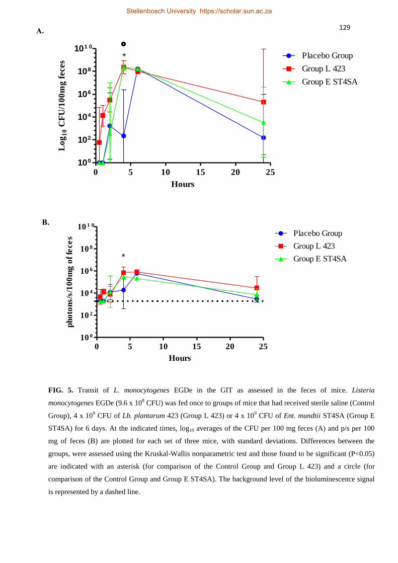
129
0 5 10 15 20 25100
102
104
106
108
101 0
Group E ST4SA
Placebo Group
Group L 423
*
Hours
Lo
g1
0 C
FU
/10
0m
g f
eces
0 5 10 15 20 2510 0
10 2
10 4
10 6
10 8
10 1 0
Group E ST4SA
Placebo Group
Group L 423
*
Hours
ph
oto
ns/s
/10
0m
g o
f fe
ce
s
FIG. 5. Transit of L. monocytogenes EGDe in the GIT as assessed in the feces of mice. Listeria
monocytogenes EGDe (9.6 x 10⁸ CFU) was fed once to groups of mice that had received sterile saline (Control
Group), 4 x 10⁹ CFU of Lb. plantarum 423 (Group L 423) or 4 x 10
9 CFU of Ent. mundtii ST4SA (Group E
ST4SA) for 6 days. At the indicated times, log10 averages of the CFU per 100 mg feces (A) and p/s per 100
mg of feces (B) are plotted for each set of three mice, with standard deviations. Differences between the
groups, were assessed using the Kruskal-Wallis nonparametric test and those found to be significant (P<0.05)
are indicated with an asterisk (for comparison of the Control Group and Group L 423) and a circle (for
comparison of the Control Group and Group E ST4SA). The background level of the bioluminescence signal
is represented by a dashed line.
A.
B.
Stellenbosch University https://scholar.sun.ac.za

130
0 5 10 15 20 2510 0
10 2
10 4
10 6
10 8
10 1 0
423
ST4SA
Hours
Lo
g1
0 C
FU
/10
0m
g o
f fe
ces
FIG. 6. Transit of Lb. plantarum 423 and Ent. mundtii ST4SA in the GIT as assessed in the feces of
mice challenged with L. monocytogenes EGde. Groups of mice were administered daily (4 x 10⁹
CFU) with Lb. plantarum 423 (Group L) and Ent. mundtii ST4SA (Group E) for 6 days before
administration of L. monocytogenes EGde (9.6 x 10⁸ CFU) on day 6. At selected time points, log10
averages of the CFU per 100 mg feces for each set of three mice are plotted with standard deviations.
Stellenbosch University https://scholar.sun.ac.za

131
STELLENBOSCH UNIVERSITY
Chapter 6
General Discussion and Conclusions
Stellenbosch University https://scholar.sun.ac.za

132
General Discussion and Conclusions
Genomic integration and expression of mCherry in LAB
The mCherry gene was successfully expressed in Lb. plantarum 423 and Ent. mundtii
ST4SA, resulting in the emission of bright fluorescence. The use of mCherry as a marker
gene to label LAB, including Lactobacillus sakei, Lactococcus lactis and Enterococcus
faecalis has been reported previously (1-3). Codon-optimization of the mCherry reporter gene
for expression in Lb. plantarum 423 and Ent. mundtii ST4SA significantly improved
fluorescence emission. A LAB/E. coli expression plasmid carrying the synthetic mCherry
gene was constructed and proved useful for the in vitro detection of fluorescent Lb.
plantarum 423 and Ent. mundtii ST4SA. However, our interest was in using the mCherry
gene to track labeled strains of LAB in the GIT of mice. Therefore, we developed a system to
integrate the mCherry fluorescent marker gene onto the chromosomes of Lb. plantarum 423
and Ent. mundtii ST4SA to circumvent the use of autonomous plasmids. This ensures that the
marker gene will be maintained inside the host genomes without the need for antibiotic
selection for enhanced stability and continuous expression (4-7).
The mCherry and a gene for chloramphenicol resistance (cat) were successfully integrated
into the chromosome of Lb. plantarum 423 by homologous recombination. This was an
indication that the disruption of the target gene for integration was not lethal to the survival of
the host. Disruption of the target gene for integration of the marker genes into the
chromosome of Ent. mundtii ST4SA proved unsuccessful. However, the integration plasmid
was stably maintained in Ent. mundtii ST4SA without recombination. Therefore, it appears
that disruption of the target gene was not achieved as its inactivation could potentially be
harmful to the host cell.
The introduction of the mCherry fluorescence gene into the LAB strains had no effect on the
bacteria’s ability to produce their antibacterial peptides or on their growth compared to the
Stellenbosch University https://scholar.sun.ac.za
133
wild-type strains. Furthermore, we have demonstrated that target genes with known DNA
sequences that are likely to be non-functional in the cells can be used for the homologous
integration of foreign genes in Lb. plantarum 423 and Ent. mundtii ST4SA. The integration
system we have described could potentially be applied to other Lb. plantarum and Ent.
mundtii strains using the target genes we have identified.
Intestinal colonization by Ent. mundtii ST4SA and Lb. plantarum 423 in mice
The aim of this work was to evaluate the potential of fluorescent imaging (FI) for the real
time monitoring of Lb. plantarum 423 and Ent. mundtii ST4SA in vivo, to reveal valuable
information on the strains’ ability to colonize the gastro-intestinal tract (GIT). The survival of
Lb. plantarum 423 and Ent. mundtii ST4SA in the GIT of infants using a computerized
gastro-intestinal model has been reported (Botes et al., 2008). However, simulated in vitro
conditions are not always the same as what happens in vivo.
We demonstrated that transcutaneous signals in mice administered fluorescent Lb. plantarum
423::CmRCherry and Ent. mundtii ST4SA (pCmRCherry), could not be distinguished from
the background signals of mice administered non-fluorescent control strains. This was
unexpected as the excitation and emission spectrum of the mCherry fluorophore is outside the
region where endogenous chromophores, such as haemoglobin, absorb light within tissues (8,
9). Factors that usually influence the application of a particular fluorescence protein for in
vivo experimentation include the penetration of fluorescent signals through tissue and
brightness of the fluorophore compared to the inherent background auto-fluorescence of the
sample or animal (9-11). Our findings merit further investigation.
Detection of the fluorescent signals emitted by the LAB strains was achieved by imaging of
the digestive tracts ex vivo. Therefore, there is no doubt that the signal was weakened by
passage through the mouse tissue and skin. Both strains were detected predominantly in the
Stellenbosch University https://scholar.sun.ac.za

134
anaerobic cecum and colon and persisted in the gut over 24 h. Similarly, previous studies
found Lb. plantarum to persist predominantly in the cecum and colon (12, 13). However, a
fluorescent signal and high numbers of Ent. mundtii ST4SA cells were also found in the small
intestine. The CFU counts from the intestines revealed that strain ST4SA colonises the gut
more effectively than strain 423 as low levels of the bacteria were still present 72 h post-
feeding. To our knowledge this is the first report on fluorescence imaging of LAB expressing
mCherry in a mouse model.
Alleviation of Listeria monocytogenes infection by Lb. plantarum 423 and Ent. mundtii
ST4SA
Effective treatment against Listeria infections is essential. Treatment of infections caused by
Listeria monocytogenes generally includes administration of antibiotics to which the
organism is highly susceptible (14). However, the frequent use of antibiotics may increase the
chances of developing antibiotic resistant strains. Therefore, the need for alternative
antimicrobial agents against resistant pathogens is obvious.
Treatment with probiotic lactic acid bacteria (LAB) may offer an alternative to conventional
antibiotics. Several studies have reported that LAB possess inhibitory activity against Listeria
monocytogenes in vitro (15-18). The anti-microbial effects of LAB are exerted either directly
via competition of live microbial cells with pathogens or indirectly via the production of
antimicrobial peptides, classified as bacteriocins (19).
The aim of this work was to evaluate the impact of Lb. plantarum 423 and Ent. mundtii
ST4SA on orally acquired L. monocytogenes infection and their ability to compete with the
pathogen in the GIT of mice. Mice pre-colonized with the probiotic LAB were challenged
with a bioluminescent strain of L. monocytogenes. Infection rates could be recorded by
monitoring bioluminescence emission which proved useful in comparing the inhibition of L.
Stellenbosch University https://scholar.sun.ac.za
135
monocytogenes by Lb. plantarum 423, Ent. mundtii ST4SA and a combination of the strains,
respectively.
Challenging Lb. plantarum 423 and Ent. mundtii ST4SA colonized in the GIT of mice with L.
monocytogenes EGDe had no effect on the survival of the strains. Our results demonstrated
that the colonization of mice with Lb. plantarum 423 and Ent. mundtii ST4SA or a
combination of the strains protected the animals from systemic L. monocytogenes EGDe
infection. The ability of Lb. plantarum 423 and Ent. mundtii ST4SA to inhibit the growth of
L. monocytogenes EGDe and L. monocytogenes ScottA in vitro has been shown previously
(20, 21). In another study it was reported that an infection by L. monocytogenes in
gnotobiotic mice could be modulated by treatment with live Lb. casei BL23 and Lb.
paracasei CNCM I-3689 (22).
In conclusion, the probiotic LAB tested in this study inhibited L. monocytogenes EGDe,
possibly directly, by competing for adhesion sites in the GIT, or indirectly through the
antimicrobial effect of the bacteriocins produced. Enterococcus mundtii ST4SA produces a
broad-spectrum antimicrobial peptide active against Gram-positive and Gram-negative
bacteria, whereas Lb. plantarum 423 produces a bacteriocin with activity against a number of
Gram-positive bacteria (23). Stimulation of the animals’ immune response by the probiotic
LAB may have played a key role in protection against pathogen colonization.
Stellenbosch University https://scholar.sun.ac.za
136
References
1. Doherty, G.P., K. Bailey and P.J. Lewis. 2010. Stage-specific fluorescence intensity
of GFP and mCherry during sporulation in Bacillus subtilis. BMC Research notes 3:
303.
2. Chapagain, P.P., C.K. Regmi and W. Castillo. 2011. Fluorescent protein barrel
fluctuations and oxygen diffusion pathways in mCherry. J. Chem. Phys. 135: 1-6.
3. Garcia-Cayuela, T. and L.P. Gomez de Carinanos. 2012. Fluorescent protein vectors
for promoter analysis in lactic acid bacteria and Escherichia coli. Appl. Microbiol.
Biotechnol. 96: 171-181.
4. Casey, J., C. Daly and G.F. Fitzgerald. 1991. Chromosomal integration of plasmid
DNA by homologous recombination in Enterococcus faecalis and Lactococcus lactis
subsp. lactis hosts harbouring Tn919. Appl. Environ. Microbiol. 57 (9): 2677-2682.
5. Martin, M.C., J.C. Alonso, J.E. Suarez and M.A. Alvarez. 2000. Genetic food-grade
recombinant lactic acid bacterium strains by site-specific recombination. Appl. Environ.
Microbiol. 66 (6): 255-2604.
6. Douglas, G.L. and T.R. Klaenhammer. 2011. Directed chromosomal integration and
expression of the reporter gene gusA3 in Lactobacillus acidophilus NCFM. Appl.
Environ. Microbiol. 77 (20): 7365-7371.
7. Heap, J.T., M. Ehsaan, C.M. Cooksley, Y. Ng, S.T. Cartman, K. Winzer and N.P
Minton. 2012. Integration of DNA into bacterial chromosomes from plasmids without
a counter selection marker. Nuc. Acids. Res. 1-10.
8. Rice, B.W., M.D. Cable and M.B. Nelson. 2001. In vivo imaging of light-emitting
probes. J. biomed. Opt. 6: 432-440.
9. Parish, T., P. Carroll, L.J. Schreuder, J. Muwanguzi-Karugaba, S. Wiles, B.D.
Robertson et al. 2010. Sensitive detection of gene expression in Mycobacteria under
Stellenbosch University https://scholar.sun.ac.za

137
replicating and non-replicating conditions using optimized far-red reporters. PLoS ONE
5 (3): 9823.
10. Taroni, P., A. Pifferi, A. Torricelli, D. Comelli and R. Cubeddu. 2003. In vivo
absorption and scattering spectroscopy of biological tissues. Photochem. Photobio. S.
2: 124-129.
11. Wiedenmann, J., F. Oswald and G.U. Nienhaus. 2009. Fluorescent proteins for live
cell imaging: Oppurtunities, limitations and challenges. Life 61 (11): 1029-1042.
12. Marco, M.L., R.S. Bongers, W.M. de Vos and M. Kleerebezem. 2007. Spatial and
temporal expression of Lactobacillus plantarum genes in the gastro-intestinal tract of
mice. Appl. Environ. Microbiol. 73: 124-132.
13. Duangjitchareon, Y., D. Kantachote, M. Ongsakul, N. Poosaran and C. Chaiyasut.
2009. Potential use of probiotic Lactobacillus plantarum SS2 isolated from fermented
plant beverage: safety assessment and persistence in the murine gastro-intestinal tract.
World J. Microbiol. Biotechnol. 25: 315-321.
14. Temple, M.E. and M.C. Nahata. 2000. Treatment of Listeriosis. Pharmac. 34 (5):
656-61.
15. Ennahar, S., D. Aoude-Werner, O. Assobhei and C. Hasselmann. 1998. Antilisterial
activity of enterocin 81, a bacteriocin produced by Enterococcus faecium WHE 81
isolated from cheese. J. Appl. Microbiol. 85: 521-526.
16. Matagaras, M., E.H. Drosinos and J. Metaxopoulos. 2003. Antgonistic activity of
lactic acid bacteria against Listeria monocytogenes in sliced cooked cured pork
shoulder stored under vacuum or modified atmosphere at 4 ± 2oC. Food Res. Technol.
20: 259-265.
17. Ashenafi, M. 1991. Growth of Listeria monocytogenes in fermenting tempeh made of
various beans and its inhibition by Lactobacillus plantarum. Food Microbiol. 8: 303-
310.
Stellenbosch University https://scholar.sun.ac.za
138
18. McAuliffe, O., C. Hill and R.P. Ross. 1999. Inhibition of Listeria monocytogenes in
cottage cheese manufactured with a lacticin 3147-producing starter culture. J. Appl.
Microbiol. 86: 251-256.
19. Bendali, F., B. Gaillard-Martinie, M. Hebraud and D. Sadoun. 2008. Kinetic of
production and mode of action of the Lactobacillus paracasei subsp. paracasei anti-
listerial bacteriocin, an Algerian isolate. Food. Sci. Technol. 41: 1784-1792.
20. Botes, M., B. Loos, C.A. van Reenen and L.M.T. Dicks. 2008. Adhesion of the
probiotic strains Enterococcus mundtii ST4SA and Lactobacillus plantarum 423 to
Caco-2 cells under conditions simulating the intestinal tract, and in the presence of
antibiotics and inflammatory medicaments. Arch. Microbiol. 190: 573-584.
21. Van Zyl, W.F., S.D. Deane and L.M.T. Dicks. 2014. Construction of a Fluorescent
Reporter System for Lactic Acid Bacteria, using the mCherry Gene. Department of
Microbiology, University of Stellenbosch, South Africa, S.A.
22. Archambaud, C., m. Nahori, G. Soubigou, C. Becavin, L. Laval, P. Lechat, T.
Smokvina, P. Langella, M. Lecuit and P. Cossart. 2012. Impact of lactobacilli on
orally acquired listeriosis. PNAS 109 (41): 16684-16689.
23. Botes, M., C.A. van Reenen and L.M.T. Dicks. 2008. Evaluation of Enterococcus
mundtii ST4SA and Lactobacillus plantarum 423 as probiotics using a gastro-intestinal
model with infant milk formulations as substrate. Int. J. Food Microbiol. 128: 362-370.
Stellenbosch University https://scholar.sun.ac.za