Internazionali - MLTJ
9
Allston Julius Stubbs Halis Atil Atilla Department of Orthopaedics, Wake Forest University Baptist Medical Center, Winston Salem, USA Corresponding author: Halis Atil Atilla Department of Orthopaedics, Wake Forest University Baptist Medical Center, Winston Salem, USA E-mail: [email protected] Summary Background: Despite the rapid advancement of imaging and arthroscopic techniques about the hip joint, missed diagnoses are still common. As a deep joint and compared to the shoulder and knee joints, localization of hip symptoms is diffi- cult. Hip pathology is not easily isolated and is of- ten related to intra and extra-articular abnormali- ties. In light of these diagnostic challenges, we recommend an algorithmic approach to effective- ly diagnoses and treat hip pain. Methods: In this review, hip pain is evaluated from diagnosis to treatment in a clear decision model. First we discuss emergency hip situations followed by the differentiation of intra and extra- articular causes of the hip pain. We differentiate the intra-articular hip as arthritic and non-arthritic and extra-articular pain as surrounding or remote tissue generated. Further, extra-articular hip pain is evaluated according to pain location. Finally we summarize the surgical treatment approach with an algorithmic diagram. Conclusion: Diagnosis of hip pathology is diffi- cult because the etiologies of pain may be vari- ous. An algorithmic approach to hip restoration from diagnosis to rehabilitation is crucial to suc- cessfully identify and manage hip pathologies. Level of evidence: V. KEY WORDS: hip pain, differential diagnosis, manage- ment, arthroscopy. Introduction Over the past 25 years, much ground has been cov- ered in the field of native hip preservation. In the last decade, there have been significant efforts directed to- ward preserving the hip joint. Some of the reasons be- hind the interest in hip preservation are the early onset of hip problems associated with the institutionalization of sport at the early ages of life and increasing activities of young people which encourage supraphysiologic range of motion. There has been improved recognition of the problem by medical professionals with high tech- nology imaging techniques and a better educated pa- tient population. Further, increased life expectancy en- courages us to preserve and restore the native hip as long as possible. While there is one outcome for the end-stage arthritic hip, there are many etiologies of hip degeneration or hip pain. Consequently, an algorithmic approach to hip restoration from diagnosis to rehabilita- tion is crucial to successfully identify and address prearthritic hip problems. Diagnostic algorithm To ensure the best outcome of hip restoration the ap- proach comprises four components: history, physical examination, radiology/laboratory and understanding patient expectations. Every type of pathology can be evaluated by this method called “HERE” (History, Ex- amination, Radiology- laboratory, Expectation of pa- tients). The first step of the diagnostic algorithm is to figure out the urgency of the pathology. Emergency hip pain The most important step of hip pain evaluation is the differentiation of emergency hip conditions from non- emergency conditions. Subluxation associated hip effusion: Space occupy- ing pathologic conditions in hip joint can force the head out from acetabulum and should be addressed urgently in order to prevent dislocation and possible avascular necrosis 1 . It is more common in children but major effu- sion of the hip is not uncommon in adults 2 . Tips (HERE) History: Very painful, with or without a trauma history. Pain in the groin or medial thigh Ex- amination: Decreased and painful of range of motion. Pain aggravated by lying on the side, Flexed, internal rotated position of the hip. Radiology: Increased joint space, with US effusion and MRI space occupying le- sion. Expectation: Immediately reducing the pain of the hip without sequelae. Stress Fractures: Femoral neck stress fractures are less common but require early diagnosis to prevent displacement and avascular necrosis of the femoral head. They present in two age groups: one is young, Muscles, Ligaments and Tendons Journal 2016;6 (3):300-308 300 The Hip Restoration Algorithm Review article @ CIC Edizioni Internazionali
Transcript of Internazionali - MLTJ

Allston Julius StubbsHalis Atil Atilla
Department of Orthopaedics, Wake Forest UniversityBaptist Medical Center, Winston Salem, USA
Corresponding author:Halis Atil AtillaDepartment of Orthopaedics, Wake Forest UniversityBaptist Medical Center, Winston Salem, USAE-mail: [email protected]
SummaryBackground: Despite the rapid advancement ofimaging and arthroscopic techniques about thehip joint, missed diagnoses are still common. Asa deep joint and compared to the shoulder andknee joints, localization of hip symptoms is diffi-cult. Hip pathology is not easily isolated and is of-ten related to intra and extra-articular abnormali-ties. In light of these diagnostic challenges, werecommend an algorithmic approach to effective-ly diagnoses and treat hip pain. Methods: In this review, hip pain is evaluatedfrom diagnosis to treatment in a clear decisionmodel. First we discuss emergency hip situationsfollowed by the differentiation of intra and extra-articular causes of the hip pain. We differentiatethe intra-articular hip as arthritic and non-arthriticand extra-articular pain as surrounding or remotetissue generated. Further, extra-articular hip painis evaluated according to pain location. Finally wesummarize the surgical treatment approach withan algorithmic diagram.Conclusion: Diagnosis of hip pathology is diffi-cult because the etiologies of pain may be vari-ous. An algorithmic approach to hip restorationfrom diagnosis to rehabilitation is crucial to suc-cessfully identify and manage hip pathologies.Level of evidence: V.
KEY WORDS: hip pain, differential diagnosis, manage-ment, arthroscopy.
IntroductionOver the past 25 years, much ground has been cov-ered in the field of native hip preservation. In the lastdecade, there have been significant efforts directed to-
ward preserving the hip joint. Some of the reasons be-hind the interest in hip preservation are the early onsetof hip problems associated with the institutionalizationof sport at the early ages of life and increasing activitiesof young people which encourage supraphysiologicrange of motion. There has been improved recognitionof the problem by medical professionals with high tech-nology imaging techniques and a better educated pa-tient population. Further, increased life expectancy en-courages us to preserve and restore the native hip aslong as possible. While there is one outcome for theend-stage arthritic hip, there are many etiologies of hipdegeneration or hip pain. Consequently, an algorithmicapproach to hip restoration from diagnosis to rehabilita-tion is crucial to successfully identify and addressprearthritic hip problems.
Diagnostic algorithmTo ensure the best outcome of hip restoration the ap-proach comprises four components: history, physicalexamination, radiology/laboratory and understandingpatient expectations. Every type of pathology can beevaluated by this method called “HERE” (History, Ex-amination, Radiology- laboratory, Expectation of pa-tients). The first step of the diagnostic algorithm is tofigure out the urgency of the pathology.
Emergency hip painThe most important step of hip pain evaluation is thedifferentiation of emergency hip conditions from non-emergency conditions.Subluxation associated hip effusion: Space occupy-ing pathologic conditions in hip joint can force the headout from acetabulum and should be addressed urgentlyin order to prevent dislocation and possible avascularnecrosis1. It is more common in children but major effu-sion of the hip is not uncommon in adults2.Tips (HERE) History: Very painful, with or without atrauma history. Pain in the groin or medial thigh Ex-amination: Decreased and painful of range of motion.Pain aggravated by lying on the side, Flexed, internalrotated position of the hip. Radiology: Increased jointspace, with US effusion and MRI space occupying le-sion. Expectation: Immediately reducing the pain ofthe hip without sequelae. Stress Fractures: Femoral neck stress fractures areless common but require early diagnosis to preventdisplacement and avascular necrosis of the femoralhead. They present in two age groups: one is young,
Muscles, Ligaments and Tendons Journal 2016;6 (3):300-308300
The Hip Restoration AlgorithmReview article
@ C
IC Ediz
ioni In
terna
ziona
li

active healthy individuals who has a history of over-loading by sports or military activities. The othergroup is the elderly females who suffer from osteope-nia or osteoporosis3. Tips (HERE) History: Young active, overloading sportactivities, military recruits. Elderly: Caucasian, female,osteoporosis. Examination: Nonspecific hip pain andpain with weight bearing. Radiology: Scintigraphy, MRI,Bone Mineral Densitometry. Expectation of Patient:Young; returning sports immediately, Elderly; avoidingfrom repeating surgeries and early mobilization. Hip Joint Infection: Septic arthritis is an importantand serious condition of the growing hip because ofits high potential to cause permanent sequelae4. Themost common joint infection in infants and in theyoung is the hip joint. In children differentiation ofseptic arthritis from transient synovitis is important astransient synovitis resolves spontaneously while theseptic arthritis needs operative treatment5. Althoughseptic arthritis of the hip is primarily recognized inchildren, it can be seen in adolescents and adults.Delayed or inadequate treatment of septic arthritis inadults can lead to irreversible joint destruction, dis-semination of the infection, and death with a rate of11%6. Gonococcal arthritis is the most common formof septic arthritis in United States. It is rarely associ-ated with joint destruction which is common inStaphylococcus aureus septic arthritis7. Tips (HERE) History: Children: Irritability, fever, non-weight bearing. Decline in general medical status. Ado-lescents and Adults: Sexual activity, rheumatoid arthri-tis or osteoarthritis, joint prosthesis, low socioeconomicstatus, intravenous drug abuse, alcoholism, diabetes,previous intra-articular corticosteroid injection, cuta-neous ulcers. Examination: Decreased and painful ofrange of motion. Avoidance of weight bearing. Flexed,internally rotated position of the hip. Fever. Radiology,Laboratory: US and MRI, Elevated ESR, WBC.Expectation: Elimination of the microorganisms fromthe joint without having a permanent sequelae or dis-semination. Slipped Capital Femoral Epiphysis (SCFE): Thereare stages as pre slip, acute, chronic, and acute on
chronic of the condition. The clinical presentation dif-fers from stage to stage. Early suspicion is crucial indiagnosis. Early diagnosis and fixation may preventarthritis8. Tips (HERE) History: In the pre-slip stage, weakness inthe leg, limping, or pain in the groin are dominant; inthe acute stage, antalgic gait and loss of range of mo-tion are dominant presentations. Endocrine disorder.Examination: Obesity, Black Male (higher incidence),Age: 9-16. Antalgic gait, Lack of internal rotation, ab-duction and flexion. Radiology, Laboratory: XR: Preslip:Non-specific, Acute: Slip at Frog leg views. MRI isworthwhile especially at early stages. Expectation: Pro-tecting from further slipping and early arthritis.Dislocation and Fracture: Diagnosis is not a realchallenge though presentation and history of trauma. Tips (HERE) History: Trauma Examination: Painfulmotion, deformity. Radiology/Laboratory: In somecases, the fracture line cannot be differentiated withstandard X-ray plain films Standard pelvis and inlet,outlet views and Judet views of acetabulum are re-quired. CT is helpful in most cases to identify fracturepatterns and loose bodies. Expectation: Reductionwith or without fixation.
Non emergency hip painAfter elimination of emergency causes of hip pain, wedetermine if the pain generator is intraarticular or ex-traarticular with the help of history and physical ex-amination. Though the hip is a deep and complexstructured joint there are many structures around thehip that can cause hip pain and disability. On the oth-er hand, there are many other extra articular remotepathologies that may causes hip pain, radiate, or af-fect the hip joint (Figure 1). Hip pathologies may mim-ic other organ pathologies or hip pain can radiate.I. Differentiation of intraarticular pathologies fromextraarticular: The most important clinical tools arehistory and physical examination. If there is hip painwith Byrd’s “C-sign”, loss of range of motion: asym-metry in hip motions, positive provocative tests, func-
Muscles, Ligaments and Tendons Journal 2016;6 (3):300-308 301
The Hip Restoration Algorithm
Figure 1. Hip Pain According to Urgent Care Need.
@ C
IC Ediz
ioni In
terna
ziona
li

tional hemipelvic muscle weakness, then we suspectintraarticular pathologies (Table I). These clinicalfindings are confirmed with radiographic findings. Ifthere are soft radiographic findings with pelvis, lum-bar, radiculopathic pain, hip range of motion is sym-metric, negative provocative tests and selective mus-cle weakness/dysfunction, then we should suspectextraarticular pathologies (Figure 2). II. Differentiation of Arthritic and Non-Arthritic HipPain: The physical examination is similar but ROMmight be much more affected in the arthritic hip thannon-arthritic. Differentiation can be done by radiologicfindings9. Non or mild arthritic hip pain can be ad-dressed by arthroscopic treatment; however, theseverity of hip arthritis may negatively affect the out-come of hip arthroscopy10. The severity of the arthri-tis in the hip can be classified with the Tönnis Radio-logic Classification as “Mild moderate and severe”11.The degree of osteoarthritis of the hip joint is as-
sessed by the Authors using the Tönnis radiologicalclassification and some additional signs of degenera-tion (Figure 3) including a central acetabular cotyloidosteophyte (the saber tooth sign) best visualized onthe AP view, remodeling of the superolateral acetabu-lum (the seagull sign), sclerosis of the inferior acetab-ulum (the hammock sign), remodeling of the inferiorfemoral neck, and osteophytic changes of the femoralhead fovea and posterosuperior femoral head-neckjunction. Joint space loss under 2 mm is a thresholdfor hip arthroscopy contraindication (Figure 4)12.Most of the cartilage loss can be seen at weightbearing area but non weight bearing but understress areas of the hip cartilage should be assessedwith joint space ratio test in the Faux-profile view al-so12 (Figure 4 b).
Non Arthritic or Mild Arthritic Hip PainWith the suspicion of intra-articular pathology and af-ter elimination of arthritis in hip joint, non or mildarthritic hip pain can be addressed. This type of paincan be classified by structural and most of them canbe treated by defined arthroscopic or open proce-dures. Most of them have similar findings preopera-tively (Figure 5).
Muscles, Ligaments and Tendons Journal 2016;6 (3):300-308302
A. J. Stubbs et al.
Table I. Findings Support Intra-articuar Pathology.
• Pain ‘C sign’• Asymmetric ROM• Positive provocative tests• Functional hemipelvic muscle weakness
Figure 2. Hip Pain According to Intra- or Extra-articular Causes.
Figure 3. Specific Radiographic Signs of Arthritis in the Hip.
@ C
IC Ediz
ioni In
terna
ziona
li
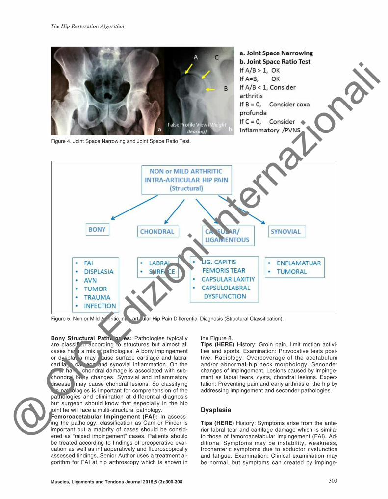
Bony Structural Pathologies: Pathologies typicallyare classified according to structures but almost allcases have a mix of pathologies. A bony impingementor dysplasia may cause surface cartilage and labralcartilage damage and synovial inflammation. On theother hand, chondral damage is associated with sub-chondral bony changes. Synovial and inflammatorydiseases may cause chondral lesions. So classifyingthe pathologies is important for comprehension of thepathologies and elimination at differential diagnosisbut surgeon should know that especially in the hipjoint he will face a multi-structural pathology. Femoroacetabular Impingement (FAI): In assess-ing the pathology, classification as Cam or Pincer isimportant but a majority of cases should be consid-ered as “mixed impingement” cases. Patients shouldbe treated according to findings of preoperative eval-uation as well as intraoperatively and fluoroscopicallyassessed findings. Senior Author uses a treatment al-gorithm for FAI at hip arthroscopy which is shown in
the Figure 8. Tips (HERE) History: Groin pain, limit motion activi-ties and sports. Examination: Provocative tests posi-tive. Radiology: Overcoverage of the acetabulumand/or abnormal hip neck morphology. Seconderchanges of impingement. Lesions caused by impinge-ment as labral tears, cysts, chondral lesions. Expec-tation: Preventing pain and early arthritis of the hip byaddressing impingement and seconder pathologies.
DysplasiaTips (HERE) History: Symptoms arise from the ante-rior labral tear and cartilage damage which is similarto those of femoroacetabular impingement (FAI). Ad-ditional Symptoms may be instability, weakness,trochanteric symptoms due to abductor dysfunctionand fatigue. Examination: Clinical examination maybe normal, but symptoms can created by impinge-
Muscles, Ligaments and Tendons Journal 2016;6 (3):300-308 303
The Hip Restoration Algorithm
Figure 5. Non or Mild Arthritic Intra-articular Hip Pain Differential Diagnosis (Structural Classification).
Figure 4. Joint Space Narrowing and Joint Space Ratio Test.
@ C
IC Ediz
ioni In
terna
ziona
li

ment test similar to FAI. Trendelenburg sign and cox-algic gait are additional examinational findings. Radi-ology and Laboratory: Radiology is the most commonassessment tool. The lateral center edge (LCE) angleis considered dysplastic below 20°; the Tönnis (sour-cil) angle is abnormal above 10º. MRI of cartilage(dGEMRIC) or hip arthroscopy can be a diagnostictool to identifying candidates for pelvic reorientation.Expectation: Long and painless serving of the nativehip. Injections, Arthroscopic treatment of intraarticularpathologies and bony reconstructions with periac-etabular osteotomy (PAO) and Femoral Osteotomiesor joint reconstruction can be done according to stageof the disease13.
Avascular Necrosis (AVN)Tips (HERE) History: Corticosteroid use, autoimmunedisorders, alcohol abuse, smoking, and coagulationdisorders, Previous hip surgery or trauma to hipshould be assessed as risk factors. In the earlystages it is asymptomatic. After it becomes sympto-matic deep groin pain is the most common symptom.Examination: Physical examination may be normalbut according to disease stage hip motion may bepainful and limited, especially internal rotation. Radi-ology and Laboratory: In the early stages radiographsmay be normal. At MRI necrotic-viable bone interfacecan be seen. Expectation: Prevention of future necro-sis and restoration of the sequelae14.
TumorsTips (HERE) History: Benign and malign tumors ofthe bone can be exactly realized by imaging studiesbut pain at night and at rest can be helpful at history.Examination: is usually non-specific. Radiology:Some of them can be easily seen and some of themmight be hidden on plain X-rays and seen at MRI, CTor Scintigraphic studies. The majority of tumors in thepelvis are malignant while in the proximal femur ma-jority of the tumors are benign15. Osteoid osteomaand bone cysts can be treated by arthroscopy assist-ed surgery16,17.Chondral Structure Pathologies: Almost all of thechondral, capsular, ligamentous and synovial patho -logies can be addressed arthroscopically so exact de-cision of treatment can be done after identifying thepathology by arthroscopy (Figure 8).
Labral PathologiesTips (HERE) History: Most patients have sharp, dollgroin pain, exaggerated by hip motion pain can beoccasionally described at buttock. Examination: Limp-ing, Trendelenburg sign, impingement sign and reliefwith intraarticular injection. Radiology: The most reli-able study for diagnosis of labral tears appears to besmall-field magnetic resonance arthrography (MRA).Expectation: Relieve of the pain with fixation of thelabrum and elimination of the cause of labral damage
Acetabular or Femoral Surface Chondral Patholo-gies: Articular cartilage defects have limited healingcapacity. Cartilage lesions of the hip are due to sev-eral causes like FAI, Dysplasia or inflammatory arthri-tis and cartilage lesion type and location differs ac-cording to cause. In CAM FAI abnormal femoralbump contacts with acetabular cartilage and causesdelamination of the cartilage. Pincer impingement is amore labral sacrificing, chondral sparing deformitythat typically involves repeated impingement of theanterior femoral head and neck junction on the ac-etabulum resulting in a lever arm on the posteroinferi-or acetabulum resulting in the so-called “contre-couplesion”. Inflammatory diseases cause central andglobal cartilage loss. In dysplasia, both the acetabularand femoral weight bearing areas have cartilage le-sions.Tips (HERE) History: Pure chondral injuries may notpresent with severe pain or symptoms because carti-lage tissue has no nociceptive receptors unless thereis a loose body or osteochondral lesion with subchon-dral bone involvement; however, synovial irritationmay cause pain. Examination: There is no specificexamination to assess cartilage damage but tests forthe cause of the chondral lesion can be done. Radiol-ogy: Plain radiographs are the most useful for reveal-ing the causes of chondral injury and severe chondrallesions. Mild to moderate chondral lesions can bebetter visualized by delayed gadolinium-enhancedMRI18 Expectation: Preventing arthritis by reconstruc-tion or regeneration of the articular cartilage by thetechniques of chondroplasty, abrasion arthroplasty,osteochondral drilling, osteoarticular autograft or allo-graft, hemicap resurfacing, autologous chondrocyteimplantation (ACI), or microfracture19.
Capsular/Ligamentous PathologiesLigamentum Teres (LT) Tears: The role of the liga-mentum teres is more important when the other stabi-lizers of the hip have some deficiency20. Tips (HERE) History: Completely non-specific andsame with intra-articular symptoms. But painful click-ing, locking or giving way can be assessed. Examina-tion: Log-roll test, resisted straight-leg raise and liga-mentum teres tests can be assessed21. Radiology:MR and CT arthrogram are helpful rather than MRI orCT alone. Adequate assessment of the LT requireddirect arthroscopic visualization and careful probing.Expectation: Relieving symptoms. Recommendedtreatment for LT tears has been simply debridementwith either mechanical shavers or radiofrequency withconcomitant anterior capsulorraphy22.Capsular Laxity: Microinstability of the hip joint dueto repetitive axial rotation and rotation of the capsuleis a common but underdiagnosed condition. Tips (HERE) History of overuse and supra physiolog-ic activities of the hip joint. Examination: Painful pas-sive hip extension and external rotation. Radiology:At MRA, Anterior capsule thinning and widening injoint recess23. Expectation: Preventing micro instabili-ty by addressing intraoperative pathologies and plica-tion of the capsule24.
Muscles, Ligaments and Tendons Journal 2016;6 (3):300-308304
A. J. Stubbs et al.
@ C
IC Ediz
ioni In
terna
ziona
li

Synovial PathologiesInflammatory Diseases: There are many inflamma-tory diseases that can affect hip joint.Tips (HERE) History: Positive family history. Previousdiagnosis for other joints. Joint stiffness. Examina-tion: Similar with other arthritis causes in the hip butthe other joints and remote findings of inflammatoryarthritis can be helpful as rheumatoid hand, Heber-den nodules, psoriatic plates, etc. Radiology and Lab-oratory: There is non-specific radiographic changesamong hip joint but central joint space loss at falseprofile images can be assessed for inflammatory dis-eases (Figure 4). Specific immunologic and biochem-istry studies can be done for revealing the underlyinginflammatory pathology. Expectation: Controlling theinflammatory process in the hip joint with systemicmedication. Addressing the intra-articular pathologiescaused by inflammatory process.Tumoral Lesions of Synovia: Synovial tumor basedlesions are rare but these lesions should be kept inmind in differential diagnosis. Tips (HERE) History and Examination can varies andmostly nonspecific. Radiology: Some of the aggres-sive lesions can be visualized by plain X rays but MRIis the valuable tool for synovial pathologies. Synovial-based tumor-like lesions can be revealed by MR sig-nal changes, such as the hemosiderin within Pig-mented villonodular synovitis (PVNS) or the cartilagewithin synovial chondromatosis25. Expectation: Elimi-nation of the tumor process via surgical and oncolog-ic treatments.Loose Bodies: Loose bodies can originate from ei-ther traumatic injury, degenerative changes, or otherdisease processes such as Perthes, spondyloepiphy-
seal dysplasia, or osteonecrosis19.Tips (HERE) History: Patients describe mechanicalsymptoms including catching, locking, clicking, or giv-ing way. Examination: Asymmetry in the hip joint mo-tion. Antalgic gait.Radiology can reveal some of the loose bodies butthe exact diagnosis can be done by MRI. Expecta-tion: The symptomatic loose bodies within the hip cancause the destruction of hyaline cartilage with resul-tant degenerative arthritis. Before destruction of thecartilage the removal of the loose bodies is the goaland the expectation. Arthroscopy is the emerginggold standard for removal of a loose body from a hipjoint.III. Differentiation of Extraarticular Pathologies:Hilton’s Law, simply stated, The nerve supplying themuscles surrounding a joint also innervates the jointsupports the theory that the surrounding tissue of ajoint may cause or mimic intra-articular pathologies orvice versa26. Extra-articular hip pain can be assessedby structural surrounding and distant tissues as wellas approach with the pain side as anterior, lateral orposterior pain (Figure 6).
Extra-Articular Causes of Hip PainLateral hip pain Greater Trochanteric Pain Syndrome (GTPS): In-cludes four conditions: greater trochanteric bursitis,gluteus medius - minimus tears, and external coxasaltans27.Tips (HERE) History: Lateral hip pain with or withoutsnapping and Trendelenburg sign can be observed.Examination: Provocative tests at physical examina-
Muscles, Ligaments and Tendons Journal 2016;6 (3):300-308 305
The Hip Restoration Algorithm
Figure 6. Evaluation of extraarticular hip pain according to pain side.* Can be treated endoscopically.
@ C
IC Ediz
ioni In
terna
ziona
li

tion are important for proper diagnosis. Radiologyand Laboratory: After classical radiographs, dynamicultrasound and MRI are useful to reveal pathology28.Expectation: Revealing of symptoms by treatment:Conservative with anti- inflammatory drugs, physicaltherapy, home exercises, and steroid injections. Withresistant cases, endoscopic debridement of the bursi-tis, release of the ilio-tibial band, and repair of thetears can be performed29.Anterior Hip Pain: It is important to differentiate thehip pain from other organ pains. Core Muscle Injury:Sports Hernia/Pubalgia/Gilmore’s Groin, classical in-guinal hernias and gynecologic or urologic problemscan be radiate as anterior hip pain. Hip surgeonshould assess these systems with the history and al-so examine these and refer to the related physicianwhen necessary. The mostly encountered anterior hippain cause is related with iliopsoas tendinitis or inter-nal snapping syndrome.Tips (HERE) History: Audible click and/or pain is oc-curred while the iliopsoas tendon moving over the an-terior hip structures. Examination: Anterior hip orgroin pain, weakness with resisted hip flexion in ab-duction, or symptomatic clicking or snapping with apositive iliopsoas test. Radiology: Conventional Xrays, bony prominence, Dynamic ultrasound and MRIcan show structural changes inside the muscle. Ex-pectation: Relieving of the symptoms. If the patientdoes not benefit from conservative management suchas drugs, physical therapy and injections, then an en-doscopic release of the iliopsoas may be consideredat insertion site or at pelvic rim30.
Posterior Hip PainDeep Gluteal Syndrome: There is a wide spectrumof sciatic nerve entrapment causes which is referredto as deep gluteal syndrome.Tips (HERE) History: Unable to sit more than half
hour, paresthesias and/or radicular pain in the affect-ed limb. Examination: Posterior hip tenderness withpalpation and exaggeration of the symptoms with piri-formis stretching tests. Pain which generated fromspine, sacroiliac or hip joint should be excluded. Ra-diology: MRI may reveal intensity changes at piri-formis muscle or space occupying lesion around thesciatic nerve. Expectation: Relieving the symptomsconservatively as drugs, injections and physical ther-apy or in resistant cases endoscopic sciatic nerve re-lease can be performed31.
Ischiofemoral (IF) ImpingementTips (HERE) History: Pain at the back of hip joint,pain can be radiate to anterior, groin or distal. me-chanical symptoms, neurologic symptoms along sci-atic nerve Examination: ischiofemoral impingementTests (Johnson Test, Dynamic IF Impingement Test)Radiology: Valgus neck, cystic or sclerotic changes atlesser trochanter or ischial tuberosity ischiofemoralspace calcification. Ischiofemoral space narrowingwith Flamingo view. MRI; ischiofemoral space nar-rowing, edema signal pattern within IF space, degen-erative signal (edema, tearing, fatty change) withinquadratus femoris muscle. Tendinopathy (edema andpartial tears), changes at hamstring origin32,33 (Figure7). Expectation: Reliving the symptoms by activitymodification, gait training, hip bracing, injection of IFspace, endoscopic surgical decompression.
Treatment AlgorithmDiagnosis and treatment of the hip pain is interwoven.Diagnosis can be change according to response ofthe treatment. Conservative treatment consist of ac-
Muscles, Ligaments and Tendons Journal 2016;6 (3):300-308306
A. J. Stubbs et al.
Figure 7. Radiologic Evaluation of the Ischiofemoral Impingement.
@ C
IC Ediz
ioni In
terna
ziona
li

tivity modification, resting, drugs, physical therapy,exercises. Invasive treatments consist of injection ofanesthetics, steroids, and biologics to the pathologygenerators. Relief of the symptoms sometimes treatthe condition and sometimes indicate the real pathol-ogy for surgical treatment (Figure 8). Surgical treat-ment of the hip pain should be followed by dedicatedrehabilitation program.The study was conducted according to internationalthe ethical standards34.
References1. Lachiewicz PF, Salvati EA, Hely D, Ghelman B. Pathological
dislocation of the hip in neurofibromatosis. A case report. JBone Joint Surg Am. 1983;65:414-415.
2. Bierma-Zeinstra SM, Bohnen AM, Verhaar JA, Prins A, Ginai-Karamat AZ, Laméris JS. Sonography for hip joint effusion inadults with hip pain. Ann Rheum Dis. 2000;59(3):178-82.
3. Egol KA, Koval KJ, Kummer F, Frankel VH. Stress fractures ofthe femoral neck. Clin Orthop Relat Res. 1998;(348):72-8. Re-view.
4. Ibn Yacoub Y, Amine B, Hajjaj-Hassouni N. Bilateral septicarthritis of the hip with osteitis and psoas abscess in a 17-year-old adolescent. J Pediatr Orthop B. 2011;20(4):238-41.
5. Kocher MS, Zurakowski D, Kasser JR. Differentiating betweenseptic arthritis and transient synovitis of the hip in children: anevidence-based clinical prediction algorithm. J Bone JointSurg Am. 1999;81(12):1662-70.
6. Mathews CJ, Weston VC, Jones A, Field M, Coakley G. Bac-terial septic arthritis in adults. Lancet. 2010 Mar 6;375(9717):846-55. doi: 10.1016/S0140-6736(09)61595-6. Review.
7. Dalla Vestra M, Rettore C, Sartore P, Velo E, Sasset L, ChiesaG, et al. Acute septic arthritis: remember gonorrhea. Rheuma-tol Int. 2008 Nov. 29(1):81-5.
8. Loder RT, Aronsson DD, Weinstein SL, Breur GJ, Ganz R, Le-unig M. Slipped capital femoral epiphysis. Instr Course Lect.2008;57:473-98. Review.
9. Bardakos NV, Villar RN. Predictors of progression of os-teoarthritis in femoroacetabular impingement: a radiologicalstudy with a minimum of ten years follow-up. J Bone Joint SurgBr. 2009;91(2):162-169.
10. Mofidi A, Shields JS, Stubbs AJ. Central acetabular osteo-phyte (saber tooth sign), one of the earliest signs of os-teoarthritis of the hip joint. Eur J Orthop Surg Traumatol.2011;21(2):71-74.
11. Tönnis D, Heinecke A. Acetabular and femoral anteversion:relationship with osteoarthritis of the hip. J Bone Joint SurgAm. 1999;81(12):1747-1770.
12. Howse E, Stubbs A J. Imaging in Hip Preservation Surgery P17 AANA Advanced Arthroscopic Surgical Techniques. TheHip / [edited by] J. W. Thomas Byrd, Asheesh Bedi, Allston J.
Muscles, Ligaments and Tendons Journal 2016;6 (3):300-308 307
The Hip Restoration Algorithm
Figure 8. Surgical Treatment Algorithm for Hip Pain.
@ C
IC Ediz
ioni In
terna
ziona
li

Stubbs. SLACK Incorporated, [2016]13. Gala L, Clohisy JC, Beaulé PE. Hip Dysplasia in the Young
Adult. J Bone Joint Surg Am. 2016;6;98(1):63-73. doi:10.2106/JBJS.O.00109. Review.
14. Van Thiel G, Mather RCThe Treatment of Osteonecrosis in theHip. OperTechSportsMed. 23:222-230.
15. Bloem JL, Reidsma II. Bone and soft tissue tumors of hip andpelvis. Eur J Radiol. 2012 Dec;81(12):3793-801. doi:10.1016/j.ejrad.2011.03.101. Epub. 2011;27. Review.
16. Tamam C, Howse EA, Tamam M, Barnes RH, Kelsey TJ, Per-ry B, Stubbs AJ. Arthroscopic Excision of Acetabular OsteoidOsteoma: Computer Tomography-Guided Approach.Arthrosc Tech. 2015;9;4(2):e101-5.
17. Jamali AA, Fritz AT, Reddy D, Meehan JP. Minimally invasivebone grafting of cysts of the femoral head and acetabulum infemoroacetabular impingement: arthroscopic technique andcase presentation. Arthroscopy. 2010;26(2):279-85.
18. Bittersohl B, Hosalkar HS, Haamberg T, et al. Reproducibilityof dGEMRIC in assessment of hip joint cartilage: a prospectivestudy. J Magn Reson Imaging. 2009;30:224-228.
19. Yen YM, Kocher MS. Chondral lesions of the hip: microfrac-ture and chondroplasty. Sports Med Arthrosc. 2010;18(2):83-9.
20. Martin RL, Palmer I, Martin HD. Ligamentum teres: a function-al description and potential clinical relevance. Knee SurgSports Traumatol Arthrosc. 2012;20:1209-14.
21. O’Donnell J, Economopoulos K, Singh P, et al. The ligamen-tum teres test: a novel and effective test in diagnosing tears ofthe ligamentum teres. Am J Sports Med. 2014;42:138-43.
22. Bardakos NV, Villar RN. The ligamentum teres of the adult hip.J Bone Joint Surg Br. 2009;91:8-15.
23. Magerkurth O, Jacobson JA, Morag Y, Caoili E, Fessell D,Sekiya JK. Capsular laxity of the hip: findings at magnetic res-onance arthrography. Arthroscopy. 2013;29(10):1615-22.
24. Ranawat AS, Sekiya JK. Arthroscopic capsular plication andthermal capsulorrhaphy. In: Sekiya JK, Safran M,Ranawat AS,Leunig M, eds. Techniques in hip arthroscopy and joint preser-vation surgery. Ed 1. Philadelphia: Saunders/ Elsevier.2010;131-138.
25. Bancroft LW, Peterson JJ, Kransdorf MJ. MR imaging of tu-mors and tumor-like lesions of the hip. Magn Reson ImagingClin N Am. 2005;13(4):757-74.
26. Moore KL, Dalley AF , Agur AMR. Clinically Oriented Anatomy,(2010) 6th Ed, p.633
27. Strauss EJ, Nho SJ, Kelly BT: Greater trochanteric pain syn-drome. Sports Med Arthrosc. 2010;18(2):113-119.
28. Yen YM, Lewis CL, Kim YJ. Understanding and Treating theSnapping Hip. Sports Med Arthrosc. 2015;23(4):194-9.
29. Redmond JM, Chen AW, Domb BG. Greater TrochantericPain Syndrome. J Am Acad Orthop Surg. 2016;24(4):231-40.
30. Brandenburg JB, Kapron AL, Wylie JD, Wilkinson BG, MaakTG, Gonzalez CD, Aoki SK. The Functional and StructuralOutcomes of Arthroscopic Iliopsoas Release. Am J SportsMed. 2016 Feb 12.
31. Martin HD, Reddy M, Gómez-Hoyos J. Deep gluteal syn-drome. J Hip Preserv Surg. 2015;2(2):99-107.
32. Torriani M, Souto SC, Thomas BJ, Ouellette H, Bredella MA.Ischiofemoral impingement syndrome: an entity with hip painand abnormalities of the quadratus femoris muscle. AJR Am JRoentgenol. 2009;193(1):186-90.
33. Tosun O, Algin O, Yalcin N, Cay N, Ocakoglu G, Karaogla -noglu M. Ischiofemoral impingement: evaluation with new MRIparameters and assessment of their reliability. Skeletal Radiol.2012;41(5):575-87.
34. Padulo J, Oliva F, Frizziero A, Maffulli N. Muscles, Ligamentsand Tendons Journal. Basic principles and recommendationsin clinical and field Science Research: 2016 Update. MLTJ.2016;6(1):1-5.
Muscles, Ligaments and Tendons Journal 2016;6 (3):300-308308
A. J. Stubbs et al.
@ C
IC Ediz
ioni In
terna
ziona
li