International Journal of Research in Pure and Applied ... · PDF file22 International Journal...
10
22 International Journal of Research in Pure and Applied Microbiology 2011; 1(2): 22-31 Original Article Isolation and Characterization of Microorganisms Responsible for Different Types of Food Spoilages Ashok Kumar 1* , Varun Bhushan 1 , Shikha Verma 1 , Gaurav Srivastav 2 and Sushil Kumar 3 1 Department of Biotechnology, Himachal Institute of Life Sciences Rampurghat Road, Paonta Sahib -173025 Himachal Pradesh INDIA 2 Research Scholar Biotechnology, Uttarakhand Technical University, Suddhowala, Dehradun-248002 Uttarakhand INDIA 3 Department of Microbiology, Himachal Institute of Life Sciences Rampurghat Road, Paonta Sahib -173025 Himachal Pradesh INDIA *Corresponding author: [email protected] Cont: +919450501471 Received 13 September 2011; accepted 20 September 2011 Abstract Bacteria are the main and an important cause of food spoilage. They thrive where food and water are present and the temperature is suitable. To resist harm, some bacteria can form spores tough reproductive cells that are able to survive under adverse conditions, that can resist damage by heat as in cooking, by cold as in freezing and by chemicals such as disinfectants. Bacteria need four hours to adapt to the new environment. 30 spoiled food samples were collected form Paonta sahib and seven bacterial isolates viz. Bacillus, Klebsiella, Pseudomonas, E.coli, Lactobacillus, Staphylococcus, and Micrococcus were isolated and characterized on the basis of morphology and biochemical reactions. It was found that the Bacillus, Klebsiella and Pseudomonas were the dominating species in the spoilage of every categories of food material. In fruits, vegetables and meat Klebsiella was the most potent spoiling bacteria. In meat, milk and in fatty products Pseudomonas was found in most of the spoilages. Although Klebsiella, Pseudomonas and Bacillus were dominant in spoilages E.coli, Staphylococcus and Micrococcus was also recovered from considerable categories of the spoiled sample. The bacteria especially gram negative, was key responsible for food spoilage. © 2011 Universal Research Publications. All rights reserved Key words: Food spoilage, Bacteria, Gram negative, Klebsiella, milk, Lactobacillus. Introduction There is potential for a wide range of vegetables and fruits products to become contaminated with microorganisms. The range of microorganisms associated with outbreaks linked to fresh produce encompasses bacteria, viruses and parasites. The products of most concern are sprouted seeds and unpasteurized juices. Most of the reported outbreaks have been associated with bacterial contamination, particularly members of the Enterobacteriaceae. Of these, Salmonella and Escherichia coli in sprouted seeds and fruit juices are of particular concern. Outbreaks linked to protozoa e.g. Cryptosporidium, Cyclospora, Giardia etc have been associated more with fruits than with vegetables. Protozoa and viruses are most often associated with contaminated water or food handlers. Fruits and vegetables normally carry a non-pathogenic epiphytic microflora (Ray, 2004). There is rapid progress in the field of chemical detection technology; little of this technology appears to have found application in estimation of the remaining shelf life of foods and early detection of spoilage. Predictive microbiology aims to summaries the probable behavior of specific spoilage organisms and the progression of spoilage processes in foods. The quantitative knowledge generated in the field of predictive microbiology provides a sound basis for the rational development of devices with which to monitor loss of product shelf life during storage, distribution and retail sale. To predict remaining shelf life accurately it is necessary, however, to consider the microbial ecology of the food system (McMeekin et al., 1996). A Bacillus strain was isolated from spoiled apple juice. This strain was acidophilic with a growth range between pH 2.5 and 5.5. Lipid analysis demonstrated the occurrence of omega Available online at http://www.urpjournals.com International Journal of Research in Pure and Applied Microbiology Universal Research Publications. All rights reserved
Transcript of International Journal of Research in Pure and Applied ... · PDF file22 International Journal...

22 International Journal of Research in Pure and Applied Microbiology 2011; 1(2): 22-31
Original Article
Isolation and Characterization of Microorganisms Responsible for Different Types of
Food Spoilages
Ashok Kumar1*
, Varun Bhushan1, Shikha Verma
1, Gaurav Srivastav
2 and Sushil Kumar
3
1 Department of Biotechnology, Himachal Institute of Life Sciences Rampurghat Road, Paonta Sahib -173025 Himachal Pradesh INDIA 2Research Scholar Biotechnology, Uttarakhand Technical University, Suddhowala, Dehradun-248002 Uttarakhand INDIA
3Department of Microbiology, Himachal Institute of Life Sciences Rampurghat Road, Paonta Sahib -173025 Himachal Pradesh INDIA
*Corresponding author: [email protected] Cont: +919450501471
Received 13 September 2011; accepted 20 September 2011 Abstract
Bacteria are the main and an important cause of food spoilage. They thrive where food and water are present and the
temperature is suitable. To resist harm, some bacteria can form spores tough reproductive cells that are able to survive under
adverse conditions, that can resist damage by heat as in cooking, by cold as in freezing and by chemicals such as disinfectants.
Bacteria need four hours to adapt to the new environment. 30 spoiled food samples were collected form Paonta sahib and seven
bacterial isolates viz. Bacillus, Klebsiella, Pseudomonas, E.coli, Lactobacillus, Staphylococcus, and Micrococcus were isolated and characterized on the basis of morphology and biochemical reactions. It was found that the Bacillus, Klebsiella and
Pseudomonas were the dominating species in the spoilage of every categories of food material. In fruits, vegetables and meat
Klebsiella was the most potent spoiling bacteria. In meat, milk and in fatty products Pseudomonas was found in most of the
spoilages. Although Klebsiella, Pseudomonas and Bacillus were dominant in spoilages E.coli, Staphylococcus and
Micrococcus was also recovered from considerable categories of the spoiled sample. The bacteria especially gram negative,
was key responsible for food spoilage.
© 2011 Universal Research Publications. All rights reserved
Key words: Food spoilage, Bacteria, Gram negative, Klebsiella, milk, Lactobacillus.
Introduction
There is potential for a wide range of vegetables and fruits
products to become contaminated with microorganisms. The
range of microorganisms associated with outbreaks linked to
fresh produce encompasses bacteria, viruses and parasites.
The products of most concern are sprouted seeds and
unpasteurized juices. Most of the reported outbreaks have
been associated with bacterial contamination, particularly
members of the Enterobacteriaceae. Of these, Salmonella
and Escherichia coli in sprouted seeds and fruit juices are of
particular concern. Outbreaks linked to protozoa e.g.
Cryptosporidium, Cyclospora, Giardia etc have been
associated more with fruits than with vegetables. Protozoa and viruses are most often associated with contaminated
water or food handlers. Fruits and vegetables normally carry
a non-pathogenic epiphytic microflora (Ray, 2004).
There is rapid progress in the field of chemical detection
technology; little of this technology appears to have found application in estimation of the remaining shelf life of foods
and early detection of spoilage. Predictive microbiology aims
to summaries the probable behavior of specific spoilage
organisms and the progression of spoilage processes in foods.
The quantitative knowledge generated in the field of
predictive microbiology provides a sound basis for the
rational development of devices with which to monitor loss
of product shelf life during storage, distribution and retail
sale. To predict remaining shelf life accurately it is necessary,
however, to consider the microbial ecology of the food
system (McMeekin et al., 1996).
A Bacillus strain was isolated from spoiled apple juice. This
strain was acidophilic with a growth range between pH 2.5
and 5.5. Lipid analysis demonstrated the occurrence of omega
Available online at http://www.urpjournals.com
International Journal of Research in Pure and Applied Microbiology
Universal Research Publications. All rights reserved

23 International Journal of Research in Pure and Applied Microbiology 2011; 1(2): 22-31
cyclohexane fatty acids and hopanoids. As these cell
constituents have among bacilli been found only in Bacillus
acidocaldarius strains, isolated microorganism seems to be
related to this species. The organism could be a threat to fruit
juices during storage at elevated temperatures, greater than or
equal to 26˚C because its spores were able to survive pasteurization conditions (Cerny et al., 1984). Species most
commonly implicated in fruit and fruit product disintegration
are Byssochlamy sfulva, Byssochlamys nivea, Neosartorya
fischeri, Talaromyces flavus and Eupenicillium brefeldianum.
They can survive heat treatments used for fruit processing
and can grow and spoil the products during storage at room
temperature, which results in great economic losses. Besides
spoilage, the heat-resistant molds produce a number of toxic
secondary metabolites, such as byssotoxin A; byssochlamic
acid; the carcinogen, patulin, the tremorgenic substances,
fumitremorgin A and C, and verruculogen; fischerin, which
caused fatal peritonitis in mice; and eupenifeldin, a compound possessing cytotoxicity as well as in vivo
antitumor activity (Tournas., 1994).
Strain A. acidoterrestris was identified as the causative agent
in spoilage of commercially pasteurized apple juice.
Alicyclobacillus sp. is soil borne bacteria, and do not strictly
require thermophilic and acidic environments.
Alicyclobacillus sp. possesses several distinct characteristics;
the major one is their ability to survive commercial
pasteurization processes and produce off-flavours in fruit
juices. Guaiacol and halophenols were identified as the offensive smelling agent in many Alicyclobacillus sp. related
spoilage. The Alicyclobacillus sp was identified as potential
spoilage bacteria in fruit juices (Chang et al., 2004).
Spoilage of milk products by Pseudomonas fragi is
characterized by the production of a strawberry like odour.
Ethyl esters of butyric, hexanoic, and 3-methylbutanoic acid
were shown to be the major contributors to the odour on the
basis of their relative concentration in the headspace
(Cormier et al., 1991). Proteolysis during storage of ultra high
temperature skim and whole milks processed by either direct or indirect systems has been studied. All the proteolysis
indices determined increased activities of both native milk
proteinase and proteinases of bacterial origin were observed
in skim ultra high temperature milks. The different behavior
of ultra high temperature skim and whole milks on storage
would have to be taken into account in establishing the
process conditions (Lopez et al., 1993). Proteolysis of milk
proteins is attributed to both native proteases and the
proteases produced by psychrotrophic bacteria during storage
of fresh raw milk. These proteases cause beneficial or
detrimental changes, depending on the specific milk product.
Plasmin, the major native protease in milk, is important for cheese ripening. A microbial protease from a psychrotrophic
microorganism can indirectly increase plasmin levels in the
casein curd. This relationship between the plasmin system
and microbial proteases in milk provides a means to control
levels of plasmin to benefit the quality of dairy products
(Nielsen, 2002).
The reason for the reported difference in spoilage behaviour
of skim and whole pasteurized milks was investigated. The rates of growth of psychrotrophic bacteria were not
significantly different in the two milks and the bacterial
types, all Pseudomonads, present at spoilage were also
similar. However, when representative spoilage organisms
were cultured into freshly pasteurized skim and whole milks,
skim milks exhibited predominantly bitter flavours while
whole milk showed mostly sour flavours. The different
spoilage behaviors can be largely explained by greater
proteolysis in skim milk than in whole milk, caused by higher
production of protease and greater susceptibility of the
protein to protease attack (Deeth et al., 2002).
Material and Methods:
1. Sample collection The samples were collected from the local market of Paonta
Sahib (Fig. 1). The sample collection was performed
according to standard method given by APHA 1998. The
sample names were coded for simplicity on the basis of their
types. The codes with their name are listed in table 1.
Fig. 1 Location Paonta Sahib in HP
2. Bacteriological Analysis: Bacteriological analysis was done by selective media method
by Presscott, 2002 and Sherman, 2005.
3. Morphological Characterization
The isolated microbes were characterized on the basis of
simple staining and gram staining (Holt et al., 1994;
Sherman, 2005).
4. Biochemical Characterization
The isolates were characterized by biochemical tests viz.
IMViC reactions i.e. indole test, Methyl Red test, Voges
Proskauer test and Citrate utilization test, Nitrate Reduction

24 International Journal of Research in Pure and Applied Microbiology 2011; 1(2): 22-31
Table 1: Sample Type and their codes
S No Sample Type Sample Code
1 Apple S1
2 Mango S2
3 Orange S3
4 Sugar cane S4
5 Pomegranate S5
6 Papaya S6
7 Litchi S7
8 Lady finger S8
9 Pumpkin S9
10 Guard S10
11 Bingil S11
12 Capsicum S12
13 Karella S13
14 Cucumber S14
15 Tomato S15
16 Egg S16
17 Fish S17
18 Pork S18
19 Hegot S19
10 Chicken S20
21 Milk S21
22 Cheese S22
23 Curd S23
24 Ice-cream S24
25 Ghee S25
26 Butter S26
27 Milk cream S27
28 Kneeled wheat S28
29 Bread S29
30 Honey S30
test, Lactose, Sucrose, Dextrose fermentation Reaction test by
standard method given by Sherman, 2005 and Holt et al.,
1994.
Results and Discussion
Bacteria can cause fruits and vegetables to get mushy or
slimy, or meat to develop a bad odor. There are different
spoilage bacteria which grow well at room temperature. The
large number of microorganisms and their waste products
cause the objectionable changes in odor, taste and texture. In
the present study different samples of spoiled food were
collected from the market of Paonta sahib.
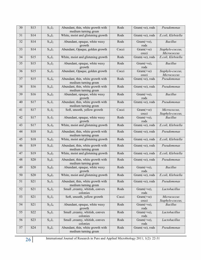
Morphological Characterization
The seventy three isolates were recovered from the thirty samples of the various categories of food. The recovered
isolate were characterized on the basis of colour, shape,
texture, margin, arrangement and staining characteristics
(Table 2).
Seventy three isolates were characterized on the basis of
colony morphology and the staining characteristics. It was
observed that thirty-nine isolates were gram (-ve) rods, twenty-five isolates was gram (+ve) rods and nine isolates
was gram (+ve) cocci (Fig 2).
Biochemical Characterization The seventy three isolates were characterized on the basis of
biochemical tests (Table 3). The tests performed to
characterize the isolates were Indole, MR, VP, citrate
utilization, nitrate reduction, fructose, sucrose and dextrose
fermentation (Sherman, 2009).
In the present study, the thirty spoiled samples of various
categories of food such as fruits, vegetables, meat, milk and milk products, ghee, butter, milk cream, kneeled wheat, bread
and honey were collected from the local market of Paonta
sahib. The seventy three isolates were characterized on the
basis of biochemical identification (Sherman, 2009). The
result obtained from the data shows that the bacteria found in
spoiled samples was Bacillus, Klebsiella, Pseudomonas,
E.coli, Lactobacillus, Staphylococcus, Micrococcus and there
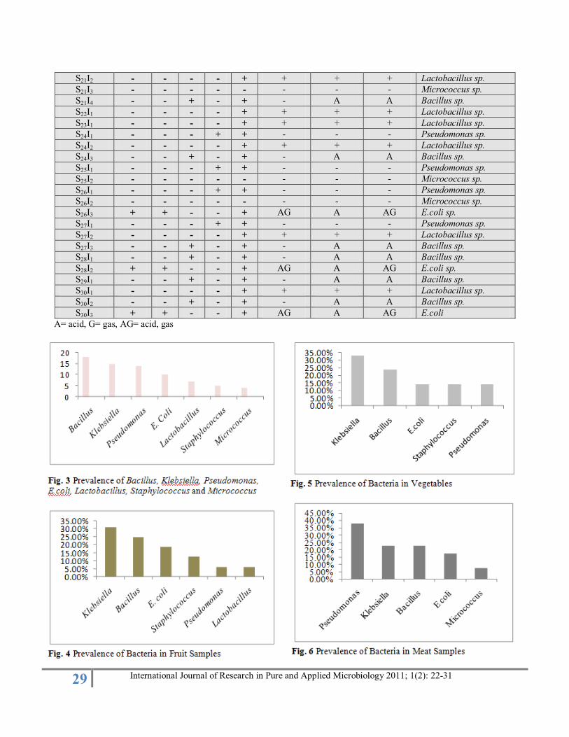
prevalence was 18%, 15%, 14%, 10%, 7%, 5%, 4%
respectively (Fig. 3). Klebsiella pneumonia is a potent
enteroinvasive food borne pathogen and causes serious illness
(Sabota et al., 1998).
In order to understand the prevalence of bacteria for different
categories of food individual study of different categories was
carried out. Sixteen isolates was recovered from the seven
samples of fruits. The bacteria found to spoil the fruits were
identified as Klebsiella, Bacillus, E.coli, Staphylococcus,
Pseudomonas, Lactobacillus and there prevalence was
31.25%, 25%, 18.75%, 12.5%, 6.25% and 6.25% respectively
(Fig. 4). A Bacillus strain was isolated from spoiled apple
juice. This strain was acidophilic with a growth range
between pH 2.5 and 5.5. This organism could be a threat to fruit juices during storage at temperatures greater than or
equal to 26°C because its spores were able to survive
pasteurization condition (Cerny et al., 1984). Lactobacillus
fermentum and Lactobacillus plantarum are the heat resistant
lactic acid bacteria obtained from spoiled acidified fruits
(Shearer et al., 2002).
Among the vegetable eight samples was collected and
twenty-one isolates were recovered from them. The bacteria
found to spoil the vegetables were identified as Klebsiella,
Bacillus, E.coli, Staphylococcus, Pseudomonas and there
prevalence in vegetable sample was found to be 33.33%, 23.80%, 14.28%, 14.28%, 14.28% respectively (Fig. 5).
Pseudomona and Klebsiella are responsible for spoilage even
in frozen vegetables (Manani et al., 2006).

25 International Journal of Research in Pure and Applied Microbiology 2011; 1(2): 22-31
Table 2: Morphological Characterization of Recovered Isolates
S.
No.
Sample
code
Isolate
code
Colony Morphology Simple
staining
Gram Staining Suspects
1 S1 S 1I1 Abundant, opaque, white waxy
growth
Rods Gram(+ve),
rods
Bacillus
2 S1 S1I2 Slimy, White, Somewhat
translucent, raised growth
Rods Gram(-ve), rods Klebsiella, E.coli,
3 S2 S2I1 Slimy, White, Somewhat
translucent, raised growth
Rods Gram(-ve), rods Klebsiella, E.coli,
4 S2 S2I2 White, moist and glistening growth Rods Gram(-ve), rods E.coli, Klebsiella
5 S3 S3I1 Small ,creamy, whitish, convex
colonies
Rods Gram(+ve),
rods
Lactobacillus
6 S3 S3I2 Slimy, White, Somewhat translucent, raised growth
Rods Gram(-ve), rods Klebsiella, E.coli,
7 S4 S4I1 Abundant, opaque, white waxy
growth
Rods Gram(+ve),
rods
Bacillus
8 S4 S4I2 Abundant, Opaque, golden growth Cocci Gram(+ve)
cocci
Staphylo-coccus,
Micrococcus
9 S5 S5I1 White, moist and glistening growth Rods Gram(-ve), rods E.coli, Klebsiella
10 S5 S5I2 Slimy, White, Somewhat
translucent, raised growth
Rods Gram(-ve), rods E.coli, Klebsiella
11 S6 S6I1 Abundant, opaque, white waxy
growth
Rods Gram(+ve),
rods
Bacillus
12 S6 S6I2 Abundant, Opaque, golden growth Cocci Gram(+ve)
cocci
Staphylo-coccus,
Micrococcus
13 S6 S6I3 Abundant, thin, white growth with
medium turning green
Rods Gram(-ve), rods Pseudomonas
14 S7 S7I1 White, moist and glistening growth Rods Gram(-ve), rods E.coli, Klebsiella
15 S7 S7I2 Slimy, White, Somewhat
translucent, raised growth
Rods Gram(-ve), rods E.coli, Klebsiella
16 S7 S7I3 Abundant, opaque, white waxy
growth
Rods Gram(+ve),
rods
Bacillus
17 S8 S8I1 Slimy, White, Somewhat translucent, raised growth
Rods Gram(-ve), rods E.coli, Klebsiella
18 S8 S8I2 White, moist and glistening growth Rods Gram(-ve), rods E.coli, Klebsiella
19 S9 S9I1 Slimy, White, Somewhat
translucent, raised growth
Rods Gram(-ve), rods E.coli, Klebsiella
20 S9 S9I2 Abundant, opaque, white waxy
growth
Rods Gram(+ve),
rods
Bacillus
21 S10 S10I1 White, moist and glistening growth Rods Gram(-ve), rods E.coli, Klebsiella
22 S10 S10I2 Abundant, thin, white growth with
medium turning green
Rods Gram(-ve), rods Pseudomonas
23 S11 S11I1 Slimy, White, Somewhat
translucent, raised growth
Rods Gram(-ve), rods E.coli, Klebsiella
24 S11 S11I2 Abundant, Opaque, golden growth Cocci Gram(+ve)
cocci
Staphylo-coccus,
Micrococcus
25 S12 S12I1 White, moist and glistening growth Rods Gram(-ve), rods E.coli, Klebsiella
26 S12 S12I2 Abundant, opaque, white waxy
growth
Rods Gram(+ve),
rods
Bacillus
27 S12 S12I3 White, moist and glistening growth Rods Gram(-ve), rods E.coli, Klebsiella
28 S13 S13I1 White, moist and glistening growth Rods Gram(-ve), rods E.coli, Klebsiella
29 S13 S13I2 Abundant, opaque, white waxy
growth
Rods Gram(+ve),
rods
Bacillus
26 International Journal of Research in Pure and Applied Microbiology 2011; 1(2): 22-31
30 S13 S13I3 Abundant, thin, white growth with
medium turning green
Rods Gram(-ve), rods Pseudomonas
31 S14 S14I1 White, moist and glistening growth Rods Gram(-ve), rods E.coli, Klebsiella
32 S14 S14I2 Abundant, opaque, white waxy
growth
Rods Gram(+ve),
rods
Bacillus
33 S14 S14I3 Abundant, Opaque, golden growth Cocci Gram(+ve)
cocci
Staphylo-coccus,
Micrococcus
34 S15 S15I1 White, moist and glistening growth Rods Gram(-ve), rods E.coli, Klebsiella
35 S15 S15I2 Abundant, opaque, white waxy
growth
Rods Gram(+ve),
rods
Bacillus
36 S15 S15I3 Abundant, Opaque, golden growth Cocci Gram(+ve)
cocci
Staphylo-coccus,
Micrococcus
37 S15 S15I4 Abundant, thin, white growth with
medium turning green
Rods Gram(-ve), rods Pseudomonas
38 S16 S16I1 Abundant, thin, white growth with
medium turning green
Rods Gram(-ve), rods Pseudomonas
39 S16 S16I2 Abundant, opaque, white waxy
growth
Rods Gram(+ve),
rods
Bacillus
40 S17 S17I1 Abundant, thin, white growth with
medium turning green
Rods Gram(-ve), rods Pseudomonas
41 S17 S17I2 Soft, smooth, yellow growth Cocci Gram(+ve)
cocci
Micrococcus,
Staphylo-coccus,
42 S17 S17I3 Abundant, opaque, white waxy growth
Rods Gram(+ve), rods
Bacillus
43 S17 S17I4 White, moist and glistening growth Rods Gram(-ve), rods E.coli, Klebsiella
44 S18 S18I1 Abundant, thin, white growth with
medium turning green
Rods Gram(-ve), rods Pseudomonas
45 S18 S18I2 White, moist and glistening growth Rods Gram(-ve), rods E.coli, Klebsiella
46 S19 S19I1 Abundant, thin, white growth with
medium turning green
Rods Gram(-ve), rods Pseudomonas
47 S19 S19I2 White, moist and glistening growth Rods Gram(-ve), rods E.coli, Klebsiella
48 S20 S20I1 Abundant, thin, white growth with
medium turning green
Rods Gram(-ve), rods Pseudomonas
49 S20 S20I2 Abundant, opaque, white waxy
growth
Rods Gram(+ve),
rods
Bacillus
50 S20 S20I3 White, moist and glistening growth Rods Gram(-ve), rods E.coli, Klebsiella
51 S21 S21I1 Abundant, thin, white growth with
medium turning green
Rods Gram(-ve), rods Pseudomonas
52 S21 S21I2 Small ,creamy, whitish, convex
colonies
Rods Gram(+ve),
rods
Lactobacillus
53 S21 S21I3 Soft, smooth, yellow growth Cocci Gram(+ve)
cocci
Micrococcus
Staphylo-coccus,
54 S21 S21I4 Abundant, opaque, white waxy
growth
Rods Gram(+ve),
rods
Bacillus
55 S22 S22I1 Small ,creamy, whitish, convex colonies
Rods Gram(+ve), rods
Lactobacillus
56 S23 S23I1 Small ,creamy, whitish, convex
colonies
Rods Gram(+ve),
rods
Lactobacillus
57 S24 S24I1 Abundant, thin, white growth with
medium turning green
Rods Gram(-ve), rods Pseudomonas

27 International Journal of Research in Pure and Applied Microbiology 2011; 1(2): 22-31
58 S24 S24I2 Small ,creamy, whitish, convex
colonies
Rods Gram(+ve),
rods
Lactobacillus
59 S24 S24I3 Abundant, opaque, white waxy
growth
Rods Gram(+ve),
rods
Bacillus
60 S25 S25I1 Abundant, thin, white growth with
medium turning green
Rods Gram(-ve), rods Pseudomonas
61 S25 S25I2 Soft, smooth, yellow growth cocci Gram(+ve)
cocci
Micrococcus,
Staphylo-coccus
62 S26 S26I1 Abundant, thin, white growth with
medium turning green
Rods Gram(-ve), rods Pseudomonas
63 S26 S26I2 Soft, smooth, yellow growth cocci Gram(+ve)
cocci
Micrococcus,
Staphylococcus
64 S26 S26I3 White, moist and glistening growth Rods Gram(-ve), rods E.coli ,Klebsiella
65 S27 S27I1 Abundant, thin, white growth with
medium turning green
Rods Gram(-ve), rods Pseudomonas
66 S27 S27I2 Small ,creamy, whitish, convex
colonies
Rods Gram(+ve),
rods
Lactobacillus
67 S27 S27I3 Abundant, opaque, white waxy growth
Rods Gram(+ve), rods
Bacillus
68 S28 S28I1 Abundant, opaque, white waxy
growth
Rods Gram(+ve),
rods
Bacillus
69 S28 S28I2 White, moist and glistening growth Rods Gram(-ve), rods E.coli, Klebsiella
70 S29 S29I1 Abundant, opaque, white waxy growth
Rods Gram(+ve), rods
Bacillus
71 S30 S30I1 Small ,creamy, whitish, convex
colonies
Rods Gram(+ve),
rods
Lactobacillus
72 S30 S30I2 Abundant, opaque, white waxy
growth
Rods Gram(+ve),
rods
Bacillus
73 S30 S30I3 White, moist and glistening growth Rods Gram(-ve), rods E.coli, Klebsiella
Among the meat products thirteen isolates were recovered
from the five samples. The bacteria found to spoil the
vegetables were identified as Pseudomonas, Klebsiella,
Bacillus, E. coli, Micrococcus and there prevalence in
vegetable sample was found to be 38.46%, 23.07%, 23.07%,
7.69%, 7.69% respectively (Fig. 6). A study of the bacteria
associated with spoilage of fresh meat was carried out. The
flora causing spoilage of meat includes Bacillus subtilis,
Escherichia coli, Klebsiella pneumoniae, Micrococcus
varians, Pseudomonas aeruginosa and Staphylococcus
aureus. Pseudomonas aeruginosa was the most dominant of
the isolated species. It was able to utilize glucose as its
primary carbon source and grew faster than the other meat
spoilage organisms (Olajuyigbe et al., 2006).
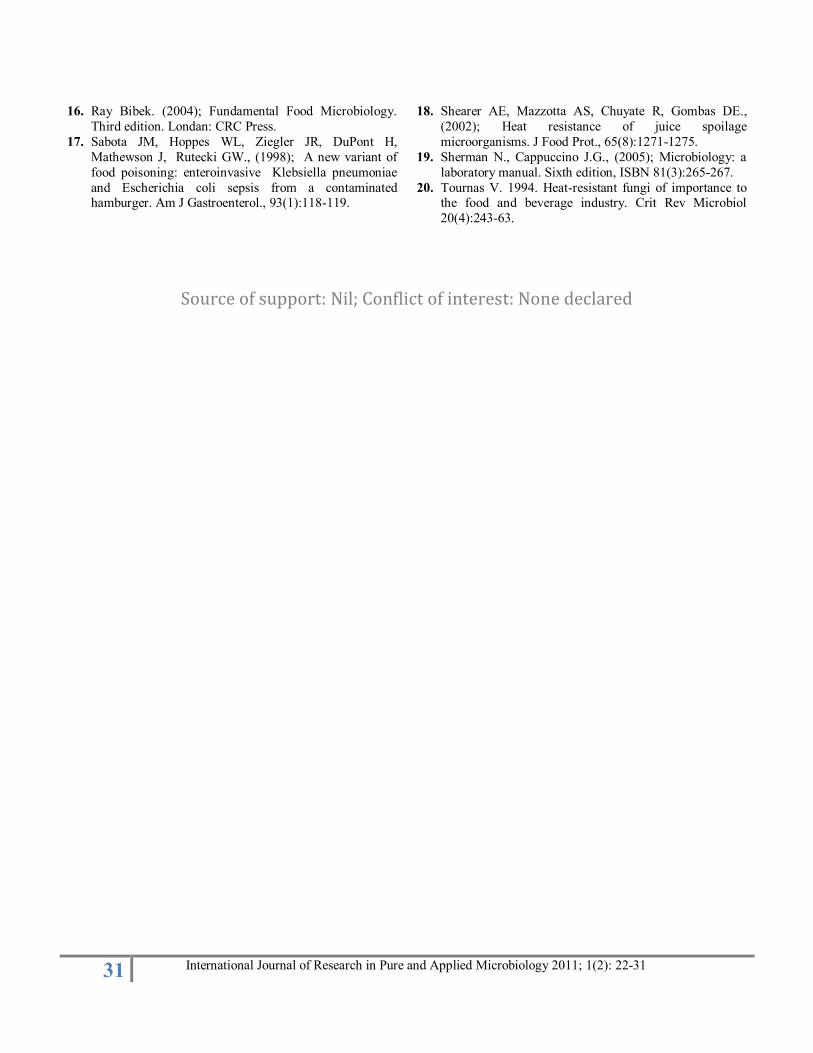
Among the milk and milk products nine isolate were recovered from the four samples. The bacteria found to spoil
the milk were identified as Lactobacillus, Pseudomonas,
Bacillus, Micrococcus and there prevalence in milk samples
was found to be 44.44%, 22.22%, 22.22%, 11.11%
respectively (Fig. 7). Mesophilic Lactobacillus sp. is the
dominant organisms in mature Cheddar cheese (Jordan et al.,
2002). Species of the genus Lactobacillus are widespread in
nature and can be found on plants or material of plant origin,
in manure and man-made habitats such as sewage, in
fermenting or spoiling food and also in association with the
intestinal tracts and mucous membranes of man and many
28 International Journal of Research in Pure and Applied Microbiology 2011; 1(2): 22-31
Table 3: Biochemical Characterization of the Recovered Isolate
Isolate code Indole MR VP CU NR Lactose Sucrose Dextrose Identified bacteria
S 1I1 - - - - + - A A Bacillus sp.
S1I2 - + - + + AG AG AG Klebsiella sp.
S2I1 - - - + + AG AG AG Klebsiella sp.
S2I2 + + - - + AG A AG E.coli
S3I1 - - - - + + + + Lactobacillus sp.
S3I2 - + - + + AG AG AG Klebsiella sp.
S4I1 - - - - + - A A Bacillus sp.
S4I2 - + + - + A A A Staphylococcus sp.
S5I1 + + - - + AG A AG E.coli sp.
S5I2 - - + + + AG AG AG Klebsiella sp.
S6I1 - - + - + - A A Bacillus sp.
S6I2 - + + - + A A A Staphylococcus sp.
S6I3 - - - + + - - - Pseudomonas sp.
S7I1 + + - - + AG A AG E.coli sp.
S7I2 - + + + + AG AG AG Klebsiella sp.
S7I3 - - - - + - A A Bacillus sp.
S8I1 - - + + + AG AG AG Klebsiella sp.
S8I2 + + - - + AG A AG E.coli sp.
S9I1 - + + + + AG AG AG Klebsiella sp.
S9I2 - - + - + - A A Bacillus sp.
S10I1 + + - - + AG A AG E.coli
S10I2 - - - + + - - - Pseudomonas sp.
S11I1 - + - + + AG AG AG Klebsiella sp.
S11I2 - + - - + A A A Staphylococcus sp.
S12I1 + + - - + AG A- AG E.coli sp.
S12I2 - - + - + - A A Bacillus sp.
S12I3 - + + + + AG AG AG Klebsiella sp.
S13I1 - + + + + AG AG AG Klebsiella sp.
S13I2 - - + - + - A A Bacillus sp.
S13I3 - - + + - - - Pseudomonas sp.
S14I1 - + + + + AG AG AG Klebsiella sp.
S14I2 - - + - + - A A Bacillus sp.
S14I3 - + - - + A A A Staphylococcus sp.
S15I1 - + - + + AG AG AG Klebsiella sp.
S15I2 - - - - + - A A Bacillus sp.
S15I3 - + + - + A A A Staphylococcus sp.
S15I4 - - - + + - - - Pseudomonas sp.
S16I1 - - - + + - - - Pseudomonas sp.
S16I2 - - + - + - A A Bacillus sp.
S17I1 - - - + + - - - Pseudomonas sp.
S17I2 - - - - - - - - Micrococcus sp.
S17I3 - - + - + - A A Bacillus sp.
S17I4 + + - - + AG A- AG E.coli sp.
S18I1 - - - + + - - - Pseudomonas sp.
S18I2 - + + + + AG AG AG Klebsiella sp.
S19I1 - - - + + - - - Pseudomonas sp.
S19I2 - + + + + AG AG AG Klebsiella sp.
S20I1 - - - + + - - - Pseudomonas sp.
S20I2 - - + - + - A A Bacillus sp.
S20I3 - + + + + AG AG AG Klebsiella sp.
S21I1 - - - + + - - - Pseudomonas sp.
29 International Journal of Research in Pure and Applied Microbiology 2011; 1(2): 22-31
S21I2 - - - - + + + + Lactobacillus sp.
S21I3 - - - - - - - - Micrococcus sp.
S21I4 - - + - + - A A Bacillus sp.
S22I1 - - - - + + + + Lactobacillus sp.
S23I1 - - - - + + + + Lactobacillus sp.
S24I1 - - - + + - - - Pseudomonas sp.
S24I2 - - - - + + + + Lactobacillus sp.
S24I3 - - + - + - A A Bacillus sp.
S25I1 - - - + + - - - Pseudomonas sp.
S25I2 - - - - - - - - Micrococcus sp.
S26I1 - - - + + - - - Pseudomonas sp.
S26I2 - - - - - - - - Micrococcus sp.
S26I3 + + - - + AG A AG E.coli sp.
S27I1 - - - + + - - - Pseudomonas sp.
S27I2 - - - - + + + + Lactobacillus sp.
S27I3 - - + - + - A A Bacillus sp.
S28I1 - - + - + - A A Bacillus sp.
S28I2 + + - - + AG A AG E.coli sp.
S29I1 - - + - + - A A Bacillus sp.
S30I1 - - - - + + + + Lactobacillus sp.
S30I2 - - + - + - A A Bacillus sp.
S30I3 + + - - + AG A AG E.coli
A= acid, G= gas, AG= acid, gas
30 International Journal of Research in Pure and Applied Microbiology 2011; 1(2): 22-31
animals (Chenoll et al., 2005). Lactobacilli from breast milk
could contribute to an anti-infective protection in neonates
and would be excellent candidates for the development of
infant probiotic products (Olivares et al., 2006). Degradation
of milk components through various enzymatic activities
associated with the contamination of dairy products by
Pseudomonas sp can reduce the shelf life of processed milk
(Cormier et al., 1991; Belgin et al., 2006).
On the basis of this observation it was estimated that the
bacteria causing spoilage in food product were Klebsiella,
E.coli, Bacillus, Lactobacillus, Pseudomonas, Micrococcus
and Staphylococcus.
Conclusion
From the present study it was concluded that seven
bacterial isolates viz. Bacillus, Klebsiella, Pseudomonas,
E.coli, Lactobacillus, Staphylococcus, Micrococcus was
isolated from the spoiled food samples. It was found that the
Bacillus, Klebsiella and Pseudomonas were the dominating species in the spoilage of every categories of food material.
In fruits, vegetables and meat Klebsiella was the most potent
spoiling bacteria. In meat, milk and in fatty products
Pseudomonas was found in most of the spoilages. Although
Klebsiella, Pseudomonas and Bacillus were dominant in
spoilages E.coli, Staphylococcus and Micrococcus was also
recovered from considerable categories of the spoiled sample.
Lactobacillus was recovered from the acidified spoilages as
well as from the milk spoilages. The strains of Lactobacillus
in fresh milk act as probiotic but in spoilages it is responsible
for the acid production and the sour taste and ranicity.
Acknowledgements:
Authors are thankful to Dr. Gaurav Gupta, Director Himachal
Institute of Life Sciences, Paonta Sahib (HP) INDIA for
providing the lab facility. The authors are also grateful to
Mrs. K.P. Rathoure for her technical support.
References
1. APHA (1998). Standard Methods for the Examination of
Water and Wastewater, 18th Ed. American Public Health
Association, Washington, DC: 45-60.
2. Belgin D, Kathryn JB., (2006); Genetic Diversity and Spoilage Potentials among Pseudomonas spp. Isolated
from Fluid Milk Products and Dairy Processing Plants, J
Applied Microbiology., 101:72–79
3. Cerny G, Hennlich W, Poralla K., (1984); Spoilage of
fruit juice by bacilli: isolation and characterization of the
spoiling microorganisms. Z Lebensm Unters Forsch.,
179(3):224-227.
4. Chang SS, Kang D H., (2004); Spoilage of fruit juice by
bacilli: isolation and characterization of the spoiling
microorganisms, Crit Rev Microbiol., 30(2):55-74.
5. Chenoll E, Carmen Macián M, Aznar R.,(2005);
Lactobacillus rennini sp. isolated from rennin and associated with cheese spoilage, Int J of systematic and
evolutionary microbiology., 130(2):138-139.
6. Cormier, F., Raymond, Y., Champagne, CP., Morin, A.,
(1991); Analysis of Odor active volatiles from
Pseudomonas fragi grown in milk. Journal of
Agricultural Food Chemistry, 39:159-161.
7. Deeth HC, Khusniati T, Datta N, Wallace RB., (2002);
Spoilage patterns of skim and whole milks, J Dairy Res.,
69(2):227-41.
8. Holt, J.G, Krieg, N.R, Senath, P.H.A Staley, J.T and
Williams, S.T., (1994); Bergey’s Manual of Determinative Bacteriology 9th Ed. Baltimore Md
Williams and Wilkins.
9. Jordan KN, Cogan TM., (2002); Heat resistance of
Lactobacillus spp. isolated from Cheddar cheese., Int J
Food Microbiol., 46(3):89-92.
10. López Fandiño R, Olano A, Corzo N, Ramos M., (1993);
Proteolysis during storage of UHT milk: differences
between whole and skim milk. J Dairy Res., Aug;
60(3):339-47.
11. McMeekin TA, Ross T., (1996); Shelf life prediction:
status and future possibilities. Int J Food Microbiol., 33(1):65-83.
12. Nielsen SS., (2002); Plasmin system and microbial
proteases in milk: characteristics, roles, and relationship.
J Agric Food Chem., 50(22):6628-34.
13. Olajuyigbe OO, Oluremi BB, Umaru DG., (2006);
Bacterial spoilage of fresh meat in some selected Lagos
markets. lfe Journal of Science., 8 (2): 193-198.
14. Olivares M, Díaz Ropero MP, Martín R, Rodríguez JM,
Xaus J., (2006); Antimicrobial potential of four
Lactobacillus strains isolated from breast milk. J Agric
Food Chem., 38(21):3128-3134.
15. Prescott LM, Harley JP, Klein DA., (2002); Microbiology, 5th Edi, McGraw-Hill, New York. Pp
1014.
31 International Journal of Research in Pure and Applied Microbiology 2011; 1(2): 22-31
16. Ray Bibek. (2004); Fundamental Food Microbiology.
Third edition. Londan: CRC Press.
17. Sabota JM, Hoppes WL, Ziegler JR, DuPont H,
Mathewson J, Rutecki GW., (1998); A new variant of
food poisoning: enteroinvasive Klebsiella pneumoniae
and Escherichia coli sepsis from a contaminated hamburger. Am J Gastroenterol., 93(1):118-119.
18. Shearer AE, Mazzotta AS, Chuyate R, Gombas DE.,
(2002); Heat resistance of juice spoilage
microorganisms. J Food Prot., 65(8):1271-1275.
19. Sherman N., Cappuccino J.G., (2005); Microbiology: a
laboratory manual. Sixth edition, ISBN 81(3):265-267.
20. Tournas V. 1994. Heat-resistant fungi of importance to the food and beverage industry. Crit Rev Microbiol
20(4):243-63.
Source of support: Nil; Conflict of interest: None declared