International Journal for Parasitology - molevol.hhu.de · Deep sequencing of Trichomonas vaginalis...
13
Deep sequencing of Trichomonas vaginalis during the early infection of vaginal epithelial cells and amoeboid transition q Sven B. Gould a,⇑ , Christian Woehle a , Gary Kusdian a , Giddy Landan a , Jan Tachezy b , Verena Zimorski a , William F. Martin a a Institute for Molecular Evolution, Heinrich-Heine-University, 40225 Düsseldorf, Germany b Laboratory of Molecular and Biochemical Parasitology, Charles University Prague, 12844 Prague, Czech Republic article info Article history: Received 4 December 2012 Received in revised form 8 April 2013 Accepted 9 April 2013 Available online 18 May 2013 Keywords: Trichomonas Infection Cytoskeleton Oxygen stress Gene families abstract The human pathogen Trichomonas vaginalis has the largest protozoan genome known, potentially encod- ing approximately 60,000 proteins. To what degree these genes are expressed is not well known and only a few key transcription factors and promoter domains have been identified. To shed light on the expres- sion capacity of the parasite and transcriptional regulation during phase transitions, we deep sequenced the transcriptomes of the protozoan during two environmental stimuli of the early infection process: exposure to oxygen and contact with vaginal epithelial cells. Eleven 3 0 fragment libraries from different time points after exposure to oxygen only and in combination with human tissue were sequenced, gen- erating more than 150 million reads which mapped onto 33,157 protein coding genes in total and a core set of more than 20,000 genes represented within all libraries. The data uncover gene family expression regulation in this parasite and give evidence for a concentrated response to the individual stimuli. Oxy- gen stress primarily reveals the parasite’s strategies to deal with oxygen radicals. The exposure of oxy- gen-adapted parasites to human epithelial cells primarily induces cytoskeletal rearrangement and proliferation, reflecting the rapid morphological transition from spindle shaped flagellates to tissue-feed- ing and actively dividing amoeboids. Ó 2013 Australian Society for Parasitology Inc. Published by Elsevier Ltd. All rights reserved. 1. Introduction The extracellular parasite Trichomonas vaginalis infects the uro- genital tract of approximately 3% of the world population annually and is thus the most widespread non-viral, sexually transmitted human parasite known (Schwebke et al., 2011). Although the vast majority of T. vaginalis infections proceed without apparent symp- toms, infection with the parasite decreases fertility, elevates the risk of prostate and cervical cancer and increases the risk of acquir- ing HIV (Petrin et al., 1998; Ryan et al., 2011). The more severe infections with manifest symptoms, Trichomoniasis, result in uro- genital tract swelling and inflammatory discharge and are com- monly treated with nitroimidazole derivates, although resistant strains are on the rise (Kulda, 1999; Upcroft and Upcroft, 2001; Benchimol, 2008; Pal and Bandyopadhyay, 2012). A crucial step of the infection process involves a dramatic mor- phological shift of the parasite. The free-swimming ovoid cells, which resemble the familiar image of a flagellated protozoan, transform into an amoeboid form (Fig. 1) upon contact with the urogenital tract. This process commences more or less immedi- ately upon contact with host tissue and the transformation of a cell takes only minutes to complete (Lal et al., 2006). The parasite’s morphogenesis entails at least two distinct but simultaneous pro- cesses: (i) the dramatic shape transition and (ii) the adherence to host cells. Recent studies on Trichomonas surface proteins that mediate host cell adherence and interactions with host extracellular matrix have identified three classes of proteins (recently reviewed by Hirt et al. (2011) and Ryan et al. (2011)). First there is the large BspA family (Bacteroides forsythus surface protein A), members of which have been localised to the parasite’s surface, but whose exact func- tion remains elusive (de Miguel et al., 2010; Noël et al., 2010). The second class comprises components of the thick glycocalyx, which is thought to also directly interact with the host extracellular ma- trix. Human Galectin-1 was identified as an interaction partner for a specific single lipophosphoglycan of the pathogen (Okumura et al., 2008). A third, and most controversial, class of suggested adhesion proteins is those that have been suggested to have dual functions, as they include enzymes of the glycolytic pathway such as glyceraldehyde-3-phosphate dehydrogenase (GAPDH) and 0020-7519/$36.00 Ó 2013 Australian Society for Parasitology Inc. Published by Elsevier Ltd. All rights reserved. http://dx.doi.org/10.1016/j.ijpara.2013.04.002 q Note: Nucleotide sequence data reported in this paper are available through the NCBI Single Read Archive Accession No. SRA059159. ⇑ Corresponding author. Address: Heinrich-Heine-University Düsseldorf, Univer- sitätsstr. 1, 40225 Düsseldorf, Germany. Tel.: +49 2118113983; fax: +49 2118113554. E-mail address: [email protected] (S.B. Gould). International Journal for Parasitology 43 (2013) 707–719 Contents lists available at SciVerse ScienceDirect International Journal for Parasitology journal homepage: www.elsevier.com/locate/ijpara
Transcript of International Journal for Parasitology - molevol.hhu.de · Deep sequencing of Trichomonas vaginalis...

Deep sequencing of Trichomonas vaginalis during the early infection ofvaginal epithelial cells and amoeboid transitionq
Sven B. Gould a,!, Christian Woehle a, Gary Kusdian a, Giddy Landan a, Jan Tachezy b, Verena Zimorski a,William F. Martin a
a Institute for Molecular Evolution, Heinrich-Heine-University, 40225 Düsseldorf, Germanyb Laboratory of Molecular and Biochemical Parasitology, Charles University Prague, 12844 Prague, Czech Republic
a r t i c l e i n f o
Article history:Received 4 December 2012Received in revised form 8 April 2013Accepted 9 April 2013Available online 18 May 2013
Keywords:TrichomonasInfectionCytoskeletonOxygen stressGene families
a b s t r a c t
The human pathogen Trichomonas vaginalis has the largest protozoan genome known, potentially encod-ing approximately 60,000 proteins. To what degree these genes are expressed is not well known and onlya few key transcription factors and promoter domains have been identified. To shed light on the expres-sion capacity of the parasite and transcriptional regulation during phase transitions, we deep sequencedthe transcriptomes of the protozoan during two environmental stimuli of the early infection process:exposure to oxygen and contact with vaginal epithelial cells. Eleven 30 fragment libraries from differenttime points after exposure to oxygen only and in combination with human tissue were sequenced, gen-erating more than 150 million reads which mapped onto 33,157 protein coding genes in total and a coreset of more than 20,000 genes represented within all libraries. The data uncover gene family expressionregulation in this parasite and give evidence for a concentrated response to the individual stimuli. Oxy-gen stress primarily reveals the parasite’s strategies to deal with oxygen radicals. The exposure of oxy-gen-adapted parasites to human epithelial cells primarily induces cytoskeletal rearrangement andproliferation, reflecting the rapid morphological transition from spindle shaped flagellates to tissue-feed-ing and actively dividing amoeboids.
! 2013 Australian Society for Parasitology Inc. Published by Elsevier Ltd. All rights reserved.
1. Introduction
The extracellular parasite Trichomonas vaginalis infects the uro-genital tract of approximately 3% of the world population annuallyand is thus the most widespread non-viral, sexually transmittedhuman parasite known (Schwebke et al., 2011). Although the vastmajority of T. vaginalis infections proceed without apparent symp-toms, infection with the parasite decreases fertility, elevates therisk of prostate and cervical cancer and increases the risk of acquir-ing HIV (Petrin et al., 1998; Ryan et al., 2011). The more severeinfections with manifest symptoms, Trichomoniasis, result in uro-genital tract swelling and inflammatory discharge and are com-monly treated with nitroimidazole derivates, although resistantstrains are on the rise (Kulda, 1999; Upcroft and Upcroft, 2001;Benchimol, 2008; Pal and Bandyopadhyay, 2012).
A crucial step of the infection process involves a dramatic mor-phological shift of the parasite. The free-swimming ovoid cells,
which resemble the familiar image of a flagellated protozoan,transform into an amoeboid form (Fig. 1) upon contact with theurogenital tract. This process commences more or less immedi-ately upon contact with host tissue and the transformation of a celltakes only minutes to complete (Lal et al., 2006). The parasite’smorphogenesis entails at least two distinct but simultaneous pro-cesses: (i) the dramatic shape transition and (ii) the adherence tohost cells.
Recent studies on Trichomonas surface proteins that mediatehost cell adherence and interactions with host extracellular matrixhave identified three classes of proteins (recently reviewed by Hirtet al. (2011) and Ryan et al. (2011)). First there is the large BspAfamily (Bacteroides forsythus surface protein A), members of whichhave been localised to the parasite’s surface, but whose exact func-tion remains elusive (de Miguel et al., 2010; Noël et al., 2010). Thesecond class comprises components of the thick glycocalyx, whichis thought to also directly interact with the host extracellular ma-trix. Human Galectin-1 was identified as an interaction partner fora specific single lipophosphoglycan of the pathogen (Okumuraet al., 2008). A third, and most controversial, class of suggestedadhesion proteins is those that have been suggested to have dualfunctions, as they include enzymes of the glycolytic pathway suchas glyceraldehyde-3-phosphate dehydrogenase (GAPDH) and
0020-7519/$36.00 ! 2013 Australian Society for Parasitology Inc. Published by Elsevier Ltd. All rights reserved.http://dx.doi.org/10.1016/j.ijpara.2013.04.002
q Note: Nucleotide sequence data reported in this paper are available through theNCBI Single Read Archive Accession No. SRA059159.! Corresponding author. Address: Heinrich-Heine-University Düsseldorf, Univer-
sitätsstr. 1, 40225 Düsseldorf, Germany. Tel.: +49 2118113983; fax: +492118113554.
E-mail address: [email protected] (S.B. Gould).
International Journal for Parasitology 43 (2013) 707–719
Contents lists available at SciVerse ScienceDirect
International Journal for Parasitology
journal homepage: www.elsevier .com/locate / i jpara

enzymes of pyruvate metabolism such as malic enzyme (ME),pyruvate:ferredoxin oxidoreductase (PFO) and subunits of succi-nyl-CoA synthetase (SCS) (Garcia and Alderete, 2007; Meza-Cer-vantez et al., 2011). These are common proteins with clearfunctions in energy metabolism (Müller et al., 2012). Their involve-ment in adhesion has been challenged (Addis et al., 1998; Bruge-rolle et al., 2000; Hirt et al., 2007; Ryan et al., 2011) andevidence for their secretion or their direct interactions with the hu-man extracellular matrix is yet to be presented. The machinery be-hind trichomonad morphogenesis is not well characterised butmicroscopic observations and the localisation of actin and actin-associated proteins have demonstrated these cytoskeletal ele-ments to play a crucial role (Gold and Ofek, 1992; Brugerolleet al., 1996; Bricheux et al., 1998, 2000; Kusdian et al., in press).
Trichomonas is considered a typical anaerobe. It has no oxygenrequirements for growth but sometimes is designated as a micro-aerophile because it grows slightly better at very low oxygen ten-sions of !0.25 lM, corresponding to about 1/1000th of ambientlevels at 25 "C, or 250 lM (Paget and Lloyd, 1990), while oxygentension above 60 lM has a detrimental effect (Ellis et al., 1994).The mitochondria of Trichomonas are designated as hydrogeno-somes because they produce hydrogen as an end product of ananaerobic, fermentative energy metabolism (Müller, 1988; Mülleret al., 2012). Core enzymes of the hydrogenosomal metabolism,such as PFO and Fe–Fe hydrogenases, are highly oxygen-sensitive(Williams et al., 1990; Hrdy and Müller, 1995; Page-Sharp et al.,1996). However, the parasite typically experiences oxygen stressin its natural environment, for example during the transmissionfrom one host to the other or with fluctuating vaginal oxygen lev-els during the menstruation cycle (Wagner and Levin, 1978; Elliset al., 1992; Hill et al., 2005) and hence must possess mechanismsto avoid inactivation of oxygen-sensitive enzymes and to removereactive oxygen species (ROS). Cytosolic NADH and NADPH oxi-dases are among the parasite’s most important oxygen scavengingenzymes (Linstead and Bradley, 1988). Glutathione, a widespreadantioxidant among eukaryotes, is absent in T. vaginalis, with cys-teine possibly acting in its place (Ellis et al., 1994). Individual pro-teins shown to be up-regulated during oxygen stress includesuperoxide dismutase (SOD) (Ellis et al., 1994; Rasoloson et al.,
2001) and peroxiredoxins (Coombs et al., 2004), ubiquitous en-zymes that are thought to be central to defenses against ROS (Gre-tes et al., 2012). Thioredoxin reductases are also present inT. vaginalis as a component of the hydrogenosomal thioredoxin-linked antioxidant system (Mentel et al., 2008). More recently,flavodiiron class A protein was shown to function as the major oxy-gen reductase responsible for respiration of hydrogenosomes(Smutná et al., 2009). This enzyme reduces oxygen to water in afour-electron reaction, while production of hydrogen peroxidewas not detected. Thus, the parasite seems to utilise a sophisti-cated system to buffer oxygen stress, which includes a cytosolicand a hydrogenosomal antioxidant system in combination (Coo-mbs et al., 2004; Mentel et al., 2008).
With approximately 160 Mbp, the genome of this protist is thelargest of any protozoan genome currently available (Carlton et al.,2007). The original genome annotation identified approximately60,000 potential protein encoding genes, which was reduced toapproximately 46,000 through subtracting genes that appear tobe only fragments of others (Smith and Johnson, 2011). A large por-tion of this redundancy of annotated genes is due to an unknownamount of duplications the genome has experienced. Approxi-mately 65% of the genome sequence consists of repetitive elementsand there are currently still >25,000 scaffolds that cannot beassembled. To what degree the parasite expresses this large arsenalof encoded genes is not known; expression evidence exists only for10,470 genes at TrichDB (http://trichdb.org/trichdb/). Transcrip-tional regulation in T. vaginalis and in protists in general is not wellcharacterised. Three core promoter elements have been identifiedin T. vaginalis, the initiator element (Inr) and the M3 and M5 ele-ments (Schumacher et al., 2003; Smith et al., 2011), and one whichappears to be the reverse sequence of the M3 motif and linked tothe 50 untranslated region (UTR) of histone encoding genes (Conget al., 2010). Two additional motifs, M2 and M4, were not furthercharacterised as the M2 showed no conserved localisation in com-parison with the start codon and the M4 was in too close proximityto it (Smith et al., 2011).
In order to better understand gene regulatory mechanismsunderlying the initial steps of human vaginal epithelial cell (VEC)infection by T. vaginalis and to identify potential core changes,
Fig. 1. The two major phenotypes of Trichomonas vaginalis. The human pathogen can switch between two radically different phenotypes. Phenotypic plasticity is inducedthrough contact with tissue of the urogenital tract (light brown). After initial contact the flagellated cells become amoeboid within minutes and increase the surface areainteracting with and scavenging from host tissue. Many of the amoeboid cells become multinuclear (nuclei in blue, with closely associated endoplasmic reticulum),proliferate and subsequently divide.
708 S.B. Gould et al. / International Journal for Parasitology 43 (2013) 707–719

we investigated 11 transcriptomes of T. vaginalis under varyingconditions and at different time points. To be able to distinguishbetween the responses to oxygen itself and contact with VECs,which grow at 15% oxygen (approximately 200 lM), we sequenced30 fragment libraries of anaerobically cultured cells (i) after expo-sure to oxygen alone, (ii) after exposure to oxygen in combinationwith VECs and (iii) after exposure of oxygen-adapted Trichomonascells to VECs, which identified transcriptional responses specific tocontact with host tissue.
2. Materials and methods
2.1. Cultures
Trichomonas vaginalis T016 was cultured in tryptone-yeast ex-tract maltose (TYM)-medium (2.22% (w/v) tryptose, 1.11% (w/v)yeast extract, 15 mM maltose, 9.16 mM L-cysteine, 1.25 mML(+)ascorbic acid, 0.77 mM KH2PO4, 3.86 mM K2HPO4, 10% (v/v)horse serum, 0.71% (v/v) iron solution (1% (w/v) Fe(NH4)2(SO4)" 6H2O, 0.1% (w/v) 5-sulfosalicylacid)) in 15 ml tubes in the ab-sence of oxygen at 37 "C. To prevent bacterial contamination a pen-icillin/streptomycin mix was added to a final concentration of100 lg/ml to the media. Oxygen-adapted T. vaginalis were alsogrown in TYM medium but in cell culture flasks identical to thoseused for the human VEC line, MS-74 (see below), at 37 "C, 5% CO2
and 15% O2 in a Galaxy 48R (Eppendorf, Germany). At a cell densityof approximately 9 " 107 cells/ml, 1 ml was transferred into 12 mlof fresh TYM medium for continuous culturing. ‘Immortalised’VECs (MS-74) were cultured in 45% DMEM (Invitrogen, Germany),45% Keratinocyte-SFM (Invitrogen) and 10% FCS in cell cultureflasks with vented lids (75 cm2, VWR, Germany) at 37 "C, 5% CO2
and 15% O2 in a Galaxy 48R (Eppendorf). For passaging, cells werewashed twice with Dulbecco‘s PBS (PAA, Germany), digested withtrypsin (Invitrogen) for 10 min at 37 "C, before inactivation withFCS. Cells were centrifuged and resuspended in fresh media andsplit 1:10 into new flasks and medium.
2.2. RNA isolation
For the oxygen stress assay (AnOx), 50 ml of T. vaginalis T016grown in sealed tubes were transferred into cell culture flasks withvented lids and incubated at 37 "C, 5% CO2 and 15% O2. RNA wasisolated using TRIzol# and following the manufacturer’s protocol(Invitrogen) at four time points (0, 5, 30 and 120 min of oxygenexposure), whereas the RNA isolated at 0 min served as a baseline.For the oxygen stress and infection assay (AnOxInf) 50 ml of T016,grown identically to those used for AnOx, were transferred onto amonolayer of MS74 cells grown in a cell culture flask (75 cm2) witha vented lid. Cells were harvested with a cell scraper (BD Falcon,Germany) and cell pellets produced by centrifugation at 3,500gfor 1 min at 8 "C. RNA was then again isolated using TRIzol# atthree time points (5, 30 and 120 min after infection). For the infec-tion assay (AdInf), 50 ml of oxygen-adapted T016 (T016 culturedfor at least five continuous days in the CO2 incubator at 15% O2)were exposed to a monolayer of VECs, then RNA was isolated at5, 30 and 120 min after exposure. All experiments were performedtwice (biological duplicates) and RNA duplicates pooled beforeDNase treatment (Fermentas, Germany) to avoid DNA contamina-tion. Frozen RNA was then sent to Eurofins MWG (Ebersberg, Ger-many) for 30 fragment library preparation and deep sequencing. Forquantitative real time PCR the same RNA samples that were usedfor 30 transcript library sequencing, were used for these experi-ments. RNA was transcribed into cDNA using the iScript™ cDNASynthesis Kit (Bio-Rad, Germany), and served as a template. Prim-ers are listed in Supplementary Table S1. All experiments were
carried out in technical triplicates using a StepOnePlus™ andPower SYBR# Green master mix (Applied Biosystems, Germany).
2.3. Transcriptome analyses
2.3.1. Reference genomesGenomic scaffolds of T. vaginalis, sequences of annotated genes
and genomic features were downloaded from TrichDB V1.3 (Aurre-coechea et al., 2009), including the information as to whether theyencode a signal peptide or transmembrane domains (TrichDB cat-egory ‘‘Protein Features’’). The nuclear genomic sequences of Homosapiens (version GRCh37.p5) and its mitochondrial genome weredownloaded from the National Center for Bioinformatics Informa-tion (NCBI) website (http://www.ncbi.nlm.nih.gov). Genomic scaf-folds of Trichomonas smaller than 1,000 nucleotides and repeatedgenes were discarded (Carlton et al., 2007). Genomic sequenceswere reduced to the predicted mRNA locations plus 100 bp ofdownstream sequence to cover potential 30 UTRs. Genes labeledas ‘‘hypothetical’’ were manually annotated using RefSeq (as of Au-gust 2012; Pruitt et al., 2007) and NCBI BLAST (Altschul et al.,1997).
2.4. Transcriptome sequences
A total of 153,137,205 reads of a typical length of 100 bp for 11different combinations of growth conditions and time points wereobtained. The reads were deposited at the NCBI Single Read Ar-chive (accession SRA059159). Each read was mapped onto thegenomic sequences of H. sapiens and T. vaginalis using a pipelineconsisting of Bowtie2 (2.0.0-beta6; Langmead and Salzberg,2012), SAMtools (Li et al., 2009) and BEDTools (Quinlan and Hall,2010). Non-coding sequence and RNA genes were discarded.
For comparisons of expression levels among the 11 samples, theraw read counts were normalised by the total number of reads foreach sample, and scaled to match the total hit count from sampleAn0. It was noted that for expression profiling using a 30 fragmentcDNA library such as ours, there is no need to normalise for tran-script length (‘FPKM’; fragments per kb per million sequencedreads). To cluster highly similar genes (Table 1, bottom row), tran-script sequences were aligned pairwise using NEEDLE (EMBOSS;Rice et al., 2000) and MCL (Enright et al., 2002) was used to gener-ate clusters with 90% identity. Relative up- and down-regulationvalues for individual genes were calculated as the ratio of thescaled read counts of the gene at the time point and in the refer-ence sample (‘‘Ad0’’ for ‘‘AdInf’’, and ‘‘An0’’ for ‘‘AnOx’’ and ‘‘AnO-xInf’’). P values and false discovery rates (FDRs), as deposited inSupplementary Tables S2–S4, were calculated using the edgeRpackage (Robinson et al., 2010). Due to the lack of technical repli-cates for the individual time points, a fixed biological coefficient ofvariation (BCV) of 0.1 and a significance level of 5% for the FDRwere used as an indicator for differential expression (www.biocon-ductor.org: edgeR user guide chapter 2.9, option 2).
Identified protein coding sequences were functionally classifiedusing the clusters of orthologous groups (COG) database (Tatusovet al., 2003), using BLASTP (E-value cutoff 10#10). Word cloudswere generated using wordle.net and advanced settings. Proteintags were assigned manually by inspecting gene annotations witha known function or domain for the top 100 genes (see Supplemen-tary Tables S2–S4).
For the identification of promoter sequences the 60 bp up-stream regions from the ATG start codon of expressed genes werescreened for the previously identified sequence motifs (Smith et al.,2011). The Inr, M3 and M5 motifs were based on the description inthe original text, while the M2 and M4 were extracted from the se-quence logo in a figure.
S.B. Gould et al. / International Journal for Parasitology 43 (2013) 707–719 709

Gene families are based on the orthologous groups deposited atTrichDB. Gene pairwise co-expression was calculated as the Pear-son correlation of the within sample read count ranks. Family-wiseco-expression was calculated as the median of all pairwise co-expressions within the family. Nucleotide identities were derivedfrom pairwise global alignments of annotated transcripts usingthe NEEDLE program (Rice et al., 2000) and the median identityfor each gene family calculated.
3. Results
3.1. Transcriptional capacity
To obtain different sets of transcriptomic libraries of the para-site, we combined the infectious T. vaginalis strain, T016, and thehuman VEC line, MS74, in the following, referred to as T016 andMS74, respectively. T016 was chosen as it represents a highly vir-ulent isolate of T. vaginalis (Pereira-Neves and Benchimol, 2007). Intotal we sequenced 30 fragment cDNA library transcriptomes ofT016 from 11 individual conditions (Table 1), and each from bio-logical duplicates. Altogether 153,137,205 reads were obtained,of which 143,767,773 (93.9%) mapped onto protein-coding genesof T. vaginalis. Only 3,887,899 reads from the libraries also contain-ing mRNA from MS74 (AnOxInf and AdInf) mapped with their besthit onto human genes. Altogether the reads mapped onto 33,157individual protein-coding genes of the parasite, of which 20,392genes were expressed under all conditions and 23,879 genes witha minimum of 10 mapped transcripts under any condition. AsT. vaginalis encodes many paralogous gene copies due to the dupli-cation of large parts of the genome (Carlton et al., 2007), the tran-script sequences of identified expressed proteins with 90% globalidentity were also clustered, leaving 27,719 individual protein-encoding genes being expressed when considering all librariescombined (Table 1). A set of actin genes (TVAG_337240,TVAG_054030 and TVAG_485210) was overall the highest ex-pressed in the anaerobically grown trophozoite library (An0), fol-lowed by genes encoding proteins of the core carbon energymetabolism, namely pyruvate-flavodoxin oxidoreductase(TVAG_198110), phosphoenolpyruvate carboxykinase (TVAG_479540), malate dehydrogenase (TVAG_204360) and glucose-6-phosphate isomerase (TVAG_061930).
In the expression patterns presented for the individual experi-ments below, only those genes were considered for which at least100 transcripts were present in each compared expression set afternormalisation. Among the three conditions tested, statisticalsupport for 94% of the top 200 up- and down-regulated geneswas found (Supplementary Tables S2–S4). Further, quantitativereal-time PCR on a set of exemplary genes such as rubrerythrin,cysteine protease or an ankyrin repeat-containing protein forexample, further supported the deep-sequencing results (Supple-mentary Fig. S1).
3.2. Oxygen stress
After 5 min of oxygen stress (AnOx5), 33 genes were found to beup-regulated at least two-fold but none of them could directly belinked to the enzymatic machinery scavenging ROS. Genes up-reg-ulated were for four transcription factors of the MYB family, otherDNA- and RNA-binding proteins, 12 of unknown function and eightwith homology to molecular switches and messengers such as a di-verse range of kinases, calmodulin and a ubiquitin-conjugating en-zyme (Supplementary Table S2). The expression pattern changedsignificantly after 30 min (Fig. 2) with 202 genes up-regulated atleast two-fold. Although two MYB genes were still among the top100, they represented different MYB genes from those observedTa
ble1
Ove
rview
oftheTricho
mon
asva
gina
listran
scriptom
esan
alyz
ed.E
leve
ntran
scriptom
esof
theT.va
gina
lisstrain
T016
weresequ
encedin
total.Th
etran
scriptom
esof
onecu
ltur
egrow
nun
deran
aerobicco
nditions
(An0
)and
one,in
which
thecells
hadbe
enad
aptedto
15%CO
2(Ad0
)serve
das
theba
setran
scriptom
es(tim
epo
ints
0).T
hree
individu
alco
nditions
wereindu
ced:
oxyg
enstress
(AnO
x),o
xyge
nstress
andex
posu
reto
vagina
lepithelialc
ells
(VEC
s,An
OxInf)a
ndex
posu
reof
oxyg
en-ada
pted
T016
toVEC
s(AdInf).RN
Awas
isolated
at5,
30an
d12
0min
aftertheex
posu
reto
theindividu
alco
nditions
.
Cond
ition
Ana
erob
icOxy
genStress
Oxy
genStress
&Infectionof
VEC
s15
%O2
Infectionof
VEC
s
Timepo
int(m
in)
05
3012
05
3012
00
530
120
Nam
eof
exp.
set
An0
AnOx5
AnOx3
0An
Ox1
20An
OxInf5
AnOxInf30
AnOxInf12
0Ad
0Ad
Inf5
AdInf30
AdInf120
TotalR
eads
15,483
,280
.00
12,513
,182
.00
12,311
,601
.00
10,395
,563
.00
14,752
,720
.00
15,050
,307
.00
16,320
,707
.00
18,000
,848
.00
13,613
,335
.00
11,296
,593
.00
13,399
,069
.00
%Map
peda
99.29
99.18
99.20
98.88
98.89
99.11
98.86
97.42
98.36
98.14
98.66
ProteinHitsb
25,109
.00
24,910
.00
24,949
.00
24,161
.00
26,098
.00
26,053
.00
26,625
.00
29,294
.00
24,319
.00
23,891
.00
25,852
.00
P10
0hits
c8,05
6.00
8,97
8.00
7,88
9.00
7,80
8.00
11,083
.00
10,215
.00
11,092
.00
14,416
.00
5,04
3.00
5,37
2.00
6,87
8.00
Clus
tered90
%d23
,731
.00
23,605
.00
23,597
.00
22,922
.00
24,626
.00
24,570
.00
24,936
.00
26,339
.00
22,737
.00
22,163
.00
23,764
.00
aPe
rcen
tage
ofread
sthat
unique
lymap
pedon
totheT.
vagina
lisge
nome.
bFraction
ofproteinco
ding
gene
siden
tifie
d.iden
tity
(‘‘po
tentialp
aralog
s’’).
cFraction
ofproteinco
ding
gene
siden
tifie
dwithat
least10
0hits.
dFraction
ofproteinco
ding
gene
swithhits
afterclus
tering
thos
ewith90
%sequ
ence.
710 S.B. Gould et al. / International Journal for Parasitology 43 (2013) 707–719
after 5 min. With an 18-fold up-regulation, a peroxiredoxin-encod-ing gene was overall the highest up-regulated product. The top 100after 30 min further included many proteins known to buffer oxy-gen stress, including four SODs (up-regulated 13.5, 6.4, 5.5 and 4.6-fold), another member of the peroxiredoxin family (4.2-fold up),one rubrerythrin (2.6-fold up), ferredoxin6 (5.7-fold up) and thio-redoxin (three-fold up). Two IscA proteins of the iron-sulfur cluster
assembly machinery were also up 8.3 and 6.2-fold, which reflectedthe need for new iron-sulfur clusters, which are present in ferre-doxin and rubrerythrin.
The trend observed after 30 min of oxygen-induced stress con-tinued and even increased after 120 min (AnOx120) with 218 genesup-regulated at least two-fold (Fig. 2). The top position was held bythe identical peroxiredoxin as after 30 min, with a 90-fold increase
Fig. 2. The most regulated Trichomonas vaginalis genes during oxygen stress. (A) Distribution of the top 200 up- and down-regulated genes during oxygen stress in green andred, respectively, across all three time points (5, 30 and 120 min), with the level of transcriptional regulation indicated by the color gradient shown in the middle. Theannotations of the 40 most regulated genes are given on the left and right with their individual color code presented accordingly. (B) The top 100 up- and down-regulatedgenes, with a predicted function or domain, represented as a word cloud. Letter height is proportional to the up- and down-regulation values, as indicated by the scale bar onthe right, whereas paralogous genes (and their values) are grouped and shown as one function.
S.B. Gould et al. / International Journal for Parasitology 43 (2013) 707–719 711

of transcript level when compared with An0. Except for one ferre-doxin gene, all others mentioned here experienced a further in-crease. These, together with another peroxiredoxin, twothioredoxin and two rubrerythrin genes that were additionallyup-regulated, totalled more than a dozen potential radical scav-engers among the top 100 genes with increased expression after120 min of oxygen stress (Supplementary Table S2). Two Nfugenes, just as the two IscA genes, potentially part of the machineryproviding iron-sulfur clusters, were also among the top 100 after120 min of oxygen stress. HydE and HydG, two of the three knowniron-hydrogenase maturases (Pütz et al., 2006), were also found tobe up-regulated. The overall increase in the transcript level com-pared with An0 was higher after 120 min then after 30 min of oxy-gen stress and six of the nine genes that were up-regulated bymore than 10 times after 120 min, were involved in the defenseof ROS or in the supply of the proteins’ active centers: peroxiredox-ins, SODs, rubrerythrin and enzymes of the iron-sulfur clusterassembly machinery.
The down-regulation of genes under oxygen stress was lessstrong and peaked at 30 min by a gene encoding a cysteine prote-ase that was down-regulated 21-fold, and which was displaced bya MYB transcription factor after 120 min, down nearly nine-fold.Next to the cysteine protease-encoding genes, another prominentgene family that was down-regulated included those encodingheat shock proteins (Hsps), in particular those of the Hsp70 family(Fig. 2). In general it was noteworthy in comparison to the up-reg-ulation of genes, which still slightly increased after 2 h, that thedown-regulation effect subsided after the 1 h.
3.3. Exposure to human epithelial cells
To identify major changes in the transcriptome of T. vaginalisupon contact with human vaginal epithelial tissue, and be able toseparate them from the changes induced through oxygen stress
alone, two additional experiments were performed. In the firstexperiment, anaerobically grown parasites, identical to those usedfor the oxygen-stress only experiment, were exposed to a mono-layer of MS74 (AnOxInf). In the second experiment the parasitewas first adapted to oxygen by growing the cells for 5 days in thesame CO2 incubator as the human cell line, before exposing themto MS74 (AdInf). The infection of MS74 through T016 was moni-tored through light microscopy and in both experimental sets,adhesion to human cells was observed to occur instantly, i.e. alarge proportion of the VECs presented with an adherent parasiteafter 5 min. Again transcriptomes of parasites were sequenced at5, 30 and 120 min time points.
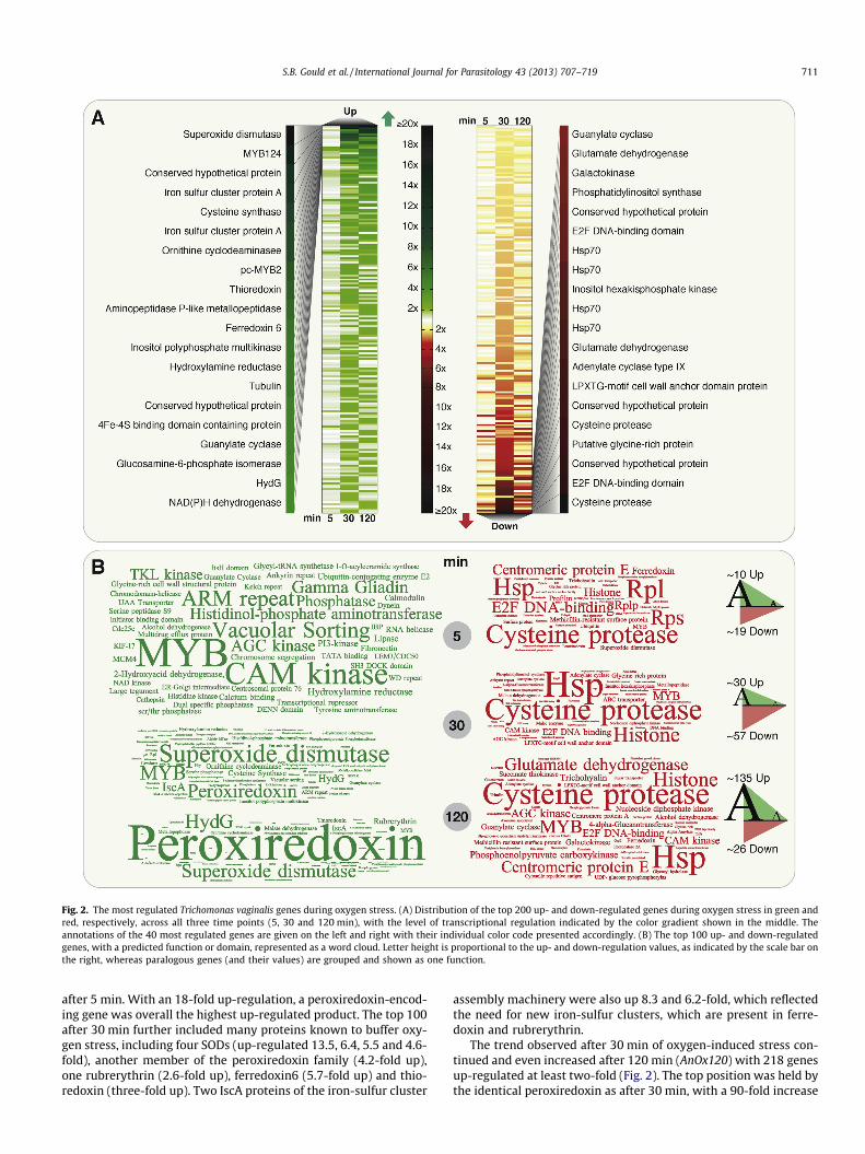
In the AnOxInf experiments, in which MS74 was infected withanaerobically grown parasites, an overall similar pattern of up-reg-ulated genes was observed among the Eukaryotic OrthologousGroups (KOG) categories as in the oxygen-stress experiment(Fig. 3). At all three time points the top positions were held bythe same genes as in the oxygen-only experiments, a MYB tran-scription factor (TVAG_076270) after 5 min, and a peroxiredoxin(TVAG_455310) after 30 and 120 min. Many other genes sharedidentical positions among the set of genes with increased expres-sion, and the intensity of regulation was comparable. Generallythe up-regulated factors were again dominated by proteins in-volved in dealing with oxygen stress, also including the enzymesof the iron-sulfur cluster generating machinery (SupplementaryTable S3). Among the top 200 results, 31.5% of the genes whoseexpression was increased, were identical between just oxygenstress and oxygen stress combined with infection, and 40.1% ofthe down-regulated genes (Fig. 5B). In comparison, the AdOxInfset shared only 0.7% among the up-regulated genes with the AnInfset (see below), and 9.2% of the down-regulated genes. One obvi-ous difference was observed among the down-regulated genes: anoticeable amount of genes associated with the core-carbonmetabolism, in particular ME, malate dehydrogenase and fruc-
Fig. 3. Transcriptional shift of eukaryotic orthologous group categories. Regulated genes were mapped to the eukaryotic orthologous group database and each gene assignedto one of the 25 categories according to the best Basic Local Alignment Search Tool (BLAST) hit. In blue, information storage and processing; in yellow to red, cellular processesand signaling; in green, metabolism; and in grey to black, general or unknown function. Categories of particular interest are highlighted by the corresponding letter of theindividual eukaryotic orthologous group category (shown on the right).
712 S.B. Gould et al. / International Journal for Parasitology 43 (2013) 707–719

tose-bisphosphate aldolase, are increasingly down-regulated overthe time of 2 h, an effect that does not occur to this degree duringoxygen stress alone (Supplementary Table S3).
The transcriptome of parasites adapted to the growth condi-tions of the CO2 incubator and then exposed to MS74 differed sig-nificantly at all time points from both other experimental sets,which immediately becomes apparent from the KOG categories
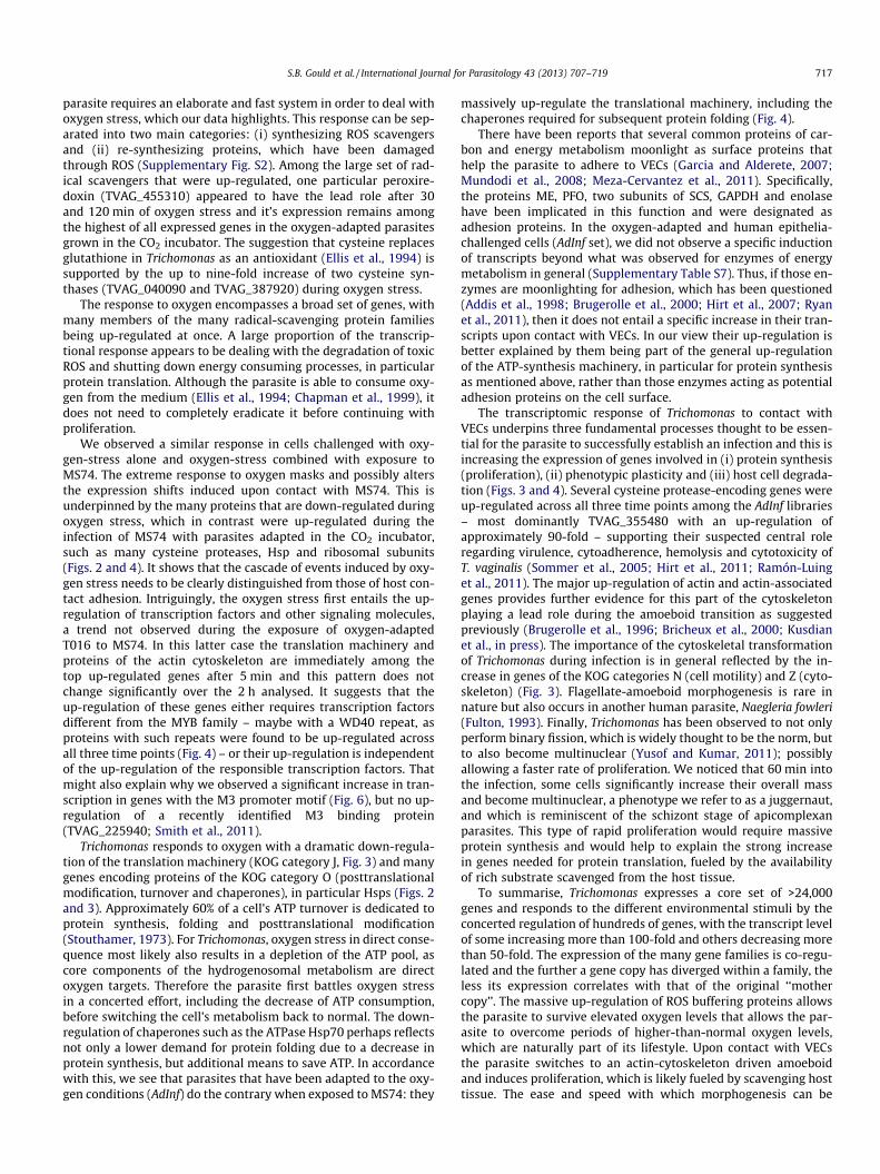
that were up-regulated (Fig. 3 and Supplementary Table S4). After30 and 120 min, approximately half of the top 200 genes with in-creased expression belong to category J (translation, ribosomalstructure and biogenesis) and many of them encode ribosomalsubunits, elongation factors and tRNA synthetases. Other genefamilies noticeably up-regulated included many encoding cys-teine-like proteases, actin and the actin-binding protein profilin
Fig. 4. The top regulated genes of oxygen adapted Trichomonas vaginalis after exposure to human vaginal epithelial cells. (A) Distribution of the top 200 up- and down-regulated genes during oxygen stress in green and red, respectively, across all three time points (5, 30 and 120 min). The annotations of the top 40 most regulated genes aregiven on the left and right. (B) The word cloud representation shows the majority of up-regulated genes during infection is rather stable over the three time points (see alsoFig. 5A) and guided by genes encoding actin, cysteine proteases and ribosomal proteins. For details refer to Fig. 2.
S.B. Gould et al. / International Journal for Parasitology 43 (2013) 707–719 713

(Fig. 4). Noteworthy, the set of up-regulated genes during the first2 h of the infection was extremely stable in comparison with theother experimental sets (Fig 5A), i.e. across all three time pointsapproximately 200 of the genes were identical.
Next the AdInf data set was screened for gene families previ-ously suggested to play a role when infection is established. Forexample, the large family of BspA-like proteins are thought to beimportant for various aspects of T. vaginalis pathobiology at thehost-pathogen interface (Noël et al., 2010). We found expressionevidence for 721 of the reported 911 putative BspA genes, albeitthe vast majority was represented by a low number of mappedtranscripts, with a median of 3.7 and an average of 37.2 reads/gene(max: 5446, min: 1) and compared with a general median of 19and an average of 457 reads/gene when considering the entire data
in this study. More BspA genes were up-regulated during oxygenstress (AnOx), and in particular when combined with exposure toVECs (AnOxInf), then when oxygen-adapted parasites were exposedto VECs (AdInf). In the latter set the vast majority of BspA geneswere observed to be down-regulated and only seven were up-reg-ulated more than two-fold (Supplementary Table S5). We searchedfor further potential host-interacting and surface bound proteins ofTrichomonas by screening for up-regulated proteins with signalpeptides or a single transmembrane domain. These included previ-ously suggested and potentially important protein families such asthe leishmanolysin-like, legume-like or polymorphic membraneprotein-like proteins (Carlton et al., 2007). However, apart from afew individual cases (e.g. TVAG_265530; saposin, TVAG_388060;a polymorphic membrane protein, TVAG_140850) no striking
Cor
rela
tion
coef
ficie
nt o
f exp
ress
ion
!1
-0.8
-0.6
-0.4
-0.2
0
0.2
0.4
0.6
0.8
1
Global nucleotide identity300 40 50 60 70 80 90 100
D Gene identity vs. coexpression
> 10 5-10
3-5 2
Family size
Linear Regression
2010Median correlation among gene family
Freq
uenc
y
Distribution of Coexpression
Randomized Gene Families
C Distribution of gene family coexpression250
100
200
150
50
00 10.5-0.5-1
100
200
300
400
500
600
700
800
900
1000
1: AnOx2: AnOxInf3: AdInf
1 2 3 1 2 3
B
Freq
uenc
y
Concurrence of genes among conditions
Up Down
5 30 120
O2 Stress(AnOx)
O2 Stress/Infection(AnOxInf)
Infection(AdInf)
450
400
350
300
250
200
150
100
50
5 30 120 5 30 12
0 5 30 120 5 30 12
0 5 30 120
A
321
DownUp
Freq
uenc
y
Concurrence of genes among time points
min
Fig. 5. Concurrence and correlated expression of Trichomonas vaginalis genes and gene families. (A) Concurrence of regulated genes among the individual expression sets isindicated by a color gradient. If a gene is up-regulated within only one time point this is indicated by light green; if a gene is up-regulated at all three time points, it isindicated by dark green. The highest amount of concurrence was observed among the up-regulated genes induced through the infection of MS74 with oxygen-adaptedparasites (AdInf). (B) Co-occurrence of identical genes among the three conditions (AnOx, AnOxInf, AdInf), which demonstrates that oxygen alone (AnOx) and in combinationwith vaginal epithelial cells (AnOxInf) leads to the identical up- and down-regulation of approximately 200 genes. They differ significantly from the transcriptomes of theparasites adapted to oxygen before their exposure to vaginal epithelial cells (AdInf). (C) Displayed are the median expression correlations of gene family members (brown)and as a reference compared with a randomized set of gene families (grey). Expression levels of members of the same gene family correlate to a higher degree than expectedby chance (Kolmogorov–Smirnow test P value <0.05). (D) Median co-expression per gene family plotted against median nucleotide identity. The linear regression line isshown in red (r = !0.25; P value <0.05).
714 S.B. Gould et al. / International Journal for Parasitology 43 (2013) 707–719

up-regulations were detected, especially not for entire families(Supplementary Table S6). In contrast, a protein of the tetraspaninfamily, TvTSP6, involved in parasite migration and potential sensorreception (de Miguel et al., 2012), was up-regulated in all of ourinfection libraries, as well as some hydrogenosomal proteins andproteins of the glycolytic pathway, which were reported to havea moonlighting function and serve as potential adhesion proteins,too, as mentioned in the Introduction (see also SupplementaryTable S7). A surface proteome analysis revealed that 11 proteinswere found more commonly present on the surface of more adher-ent strains (de Miguel et al., 2010). All of these genes were foundexpressed in T016 (a strain that was not part of the proteome anal-ysis), with TVAG_239650 (unknown function) having the highestabsolute expression. The transcriptomic response of the 11 genesis mixed and to a certain degree mirrors that of the BspA family.Genes up- or down-regulated during infection of VECs with para-sites not adapted to oxygen (AnOxInf) did not necessarily matchthose up- or down-regulated during infection of VECs with oxy-gen-adapted parasites (Supplementary Table S8). It is noteworthythat oxygen stress alone (AnOx) had very little to no effect, whereas
the presence of VECs in both experimental sets (AnOxInf and AdInf)induced more significant transcriptional changes.
3.4. Regulation and co-expression of gene families
One interesting aspect of trichomonad biology is the massiveexpansion of gene families, whose coordinated expression hasnot been examined hitherto in detail due to the lack of availablesequencing data. The correlation of expression among 2,509 genefamilies was analysed, for which we found at least two membersto be identified by 100 mapped reads. A higher correlation of reg-ulation between the members of gene families was observed whencompared with randomised samples (Fig. 5C), but for many –including important gene families – the patterns were non-uni-form. Whereas in some families a considerable correlation be-tween the regulations of expression was observed, in othersindividual copies could be very differentially expressed. For exam-ple, for ribosomal protein-encoding genes or the actin gene family,all genes are generally expressed at high levels, the co-expressioncoefficient was high and expression variance was low. On the
Fig. 6. Trichomonas vaginalis core promoter distribution. We screened the 60 bp upstream region from the start codon, for the five core promoters identified in Trichomonas.(A) In light grey (Total) the distribution of the promoter motifs among all genes annotated at TrichDB and medium grey (Expressed) the distribution among all genes, forwhich there is expression evidence. The Inr motif was found to occur at the same rate among scrambled upstream sequences (Randomized), albeit this did not take intoaccount the distance to the start codon. The occurrence of the M3 and M5motif is slightly increased among the genes up-regulatedP two-fold during oxygen stress, but evenmore significantly among the AdInf set (asterisk). (B) Table of the values from (A) with the frequency of analyzed sequences given in the first row and the percentage of theindividual motifs (shown in the second column) identified among the sets analyzed.
S.B. Gould et al. / International Journal for Parasitology 43 (2013) 707–719 715

contrary, we found expression evidence for 30 genes of the Hsp70family, within which one gene copy (TVAG_092490) was repre-sented in total by 216,000 normalised reads, another copy(TVAG_130280) by only two. The same extreme difference was ob-served, but was not limited to, a glucosylceramidase and a synap-togamin family (Supplementary Table S9). Importantly, theexpression correlation within an individual gene family was notrandom but was correlated with sequence identity, i.e. the moresimilar the sequences of two genes, the greater the chance theirexpression significantly correlates (Fig. 5D).
To elucidate whether certain promoter elements might be pre-dominantly associated with one of the introduced environmentalchanges, we extracted the 50 UTRs of all annotated genes, screenedthem for the known core promoter regions and compared themwith those genes whose expression within our libraries was regu-lated at least two-fold and for which at least 100 reads were iden-tified. First, to generally elucidate promoter motif significance, werandomised the 60 bp upstream regions of all expressed genes andnoticed that only the Inr motif ((A/T/C)(T/C)A(A/T/C)(T/A)), as de-fined by Smith et al. (2011), to be as frequently encoded amongthe scrambled sequences as it was among true promoter regions(Fig. 6). However, the latter does not take into account the distanceto the start codon of the motif and which is likely of importance.
All five promoter motifs are enriched among the >33,000 ex-pressed genes in comparison with the approximately 60,000 anno-tated at TrichDB. While the Inr motif, which mediates correcttranscription initiation, was present in 98% of all the upstream re-gions of genes expressed, the M3 was found within only 4.5% andthe M5 in 5.7% of the sequences (Fig. 6). The Inr motif experiencedthe lowest amount of change, the most significant being a decreaseto 93.4% among the up-regulated genes of the infection set. Theamount of M3 and M5 motifs changed significantly among all setsof genes but most significantly among those up-regulated duringthe 2 h of exposure to VECs. Twenty-five point five percent of theupstream regions of all genes up-regulated harbored a M3 motifand 34% a M5 motif, which approximately corresponded to a five-and six-fold increase, respectively. Even after subtraction of ribo-somal subunit encoding genes, which are known to harbor thesemotifs (Smith et al., 2011), the elevation of M3 and M5 remainshigh at 14.9% and 7.5%, respectively.
4. Discussion
Eukaryotic genomes vary in size and protein-coding capacityranges from approximately 2,000 in Encephalitozoon cuniculi topossibly even 60,000 in T. vaginalis (Katinka et al., 2001; Carltonet al., 2007). In the parabasalian parasite, the massive codingcapacity is the result of the duplication of at least large sectionsof the genome and additional, partly massive, expansion of somegene families (Carlton et al., 2007). However, even if it is consid-ered that the duplication of genes can also lead to the expressionof redundant copies and proteins with identical functions, the factthat almost 28,000 remain after clustering genes with 90% nucleo-tide identity, demonstrates the large arsenal of proteins that theparasite can tap. Our transcriptomes encompass >30,000 expressedgenes and cover 98% of the 714 genes recently discussed in themicroarray-based analysis of Horváthová et al. (2012) that focusedon iron-regulated genes in Trichomonas. The difference demon-strates that deep-sequencing transcriptomes from different condi-tions will expand this list even further. The overall response ofT. vaginalis to oxygen stress and upon contact with human tissuewas extensive, with hundreds of at least two-fold up- and down-regulated genes, and with individual genes experiencing a 100-foldincrease in gene expression.
It has been suggested that the expansion of certain gene fami-lies is not random, but rather favors those that aid the special life-style of Trichomonas (Carlton et al., 2007). However, these genefamilies can only then act on the parasite, if they are all expressed.By comparing the correlation of expression within gene families,we found evidence that indeed many hundreds of the expandedfamilies, including paralogous groups of Tyrosine Kinase-Like(TKL) kinases, actin, rabs or tubulin for example, are co-regulated(Fig. 5C). What is puzzling is why so many gene families clusteraround the 45–50% identity mark when plotting global nucleotideidentity versus expression correlation (Fig. 5C). They do not onlycorrespond to a limited set of certain gene families, nor do theycorrelate with the size of gene families. This ‘cloud’ might repre-sent a footprint of an evolutionary event (genome duplication?),or maybe at this level of identity a certain threshold level isreached where any two genes that are compared can have any typeof co-regulation imaginable; i.e. it could also be random. It indi-cates that the extension of specific gene families – or the retentionthereof in comparison with others – might be related to their bio-logical significance for the parasite’s specialised lifestyle. Yet, theexpansion of a gene family does not always result in high expres-sion, as can be seen for example from the BspA family, and not al-ways does a low or high expression level of an individual genedirectly translate into a high or low amount of protein (Mairet al., 2006; Gry et al., 2009). Horváthová and colleagues (2012)also noted that among genes regulated under different iron con-centrations, not all genes of a family experienced identical changes.In some cases all or the majority of paralogous copies were simul-taneously regulated, in others it was only one gene of a family. Ourresults are congruent with those findings, for a larger sample ofgenes, different strains and different conditions, suggesting thatthis trend is general for Trichomonas.
The results for some genes such as those of the BspA family andfor example for some of the surface-associated genes identified byde Miguel et al., 2010) are peculiar. For instance, our results for theentire BspA family confirm earlier findings for a few BspA genes,that they are up-regulated during amoeboid transition and simul-taneous oxygen exposure (Noël et al., 2010). However, we foundthat oxygen-adapted cells do not up-regulate BspA genes uponexposure to VECs, which suggests either an oxygen-dependentcomponent might be involved in the up-regulation of this, and per-haps other, gene families or that the strong up-regulation of, inparticular, the translation machinery, in our case ‘masks’ lessstrongly regulated genes.
The many different strains of T. vaginalis isolated and examinedto date disclose very different behaviors upon environmental stim-uli (Ellis et al., 1992; Pereira-Neves and Benchimol, 2007; de Mig-uel et al., 2012). As the different behaviors result from differentgene expression – demonstrated for example by the increasedexpression rate of TSP in highly-adherent strains (de Miguelet al., 2012) – this needs to be considered when comparing the setsof expression data from different Trichomonas strains. Also, albeitquantitative real time PCRs on individual genes can mirror patternsobserved among large-scale generated data, these two techniquescannot always be directly compared. Due to the required normali-sation of deep-sequencing data, genes with a very high expressionwill tend to mask the expression values of those with very lowexpression.
The parasite’s core energy metabolism harbors many proteinswith iron-sulfur clusters and is hence very sensitive to oxygen.High levels of oxygen therefore directly hamper substrate levelphosphorylation, the only pathway for ATP synthesis within thehydrogenosomes of Trichomonas. However, the natural habitat ofTrichomonas is never absolutely oxygen free and, during transmis-sion and menstruation, the parasite experiences high levels of oxy-gen fluctuation (Ellis et al., 1994; Hill et al., 2005). Therefore the
716 S.B. Gould et al. / International Journal for Parasitology 43 (2013) 707–719
parasite requires an elaborate and fast system in order to deal withoxygen stress, which our data highlights. This response can be sep-arated into two main categories: (i) synthesizing ROS scavengersand (ii) re-synthesizing proteins, which have been damagedthrough ROS (Supplementary Fig. S2). Among the large set of rad-ical scavengers that were up-regulated, one particular peroxire-doxin (TVAG_455310) appeared to have the lead role after 30and 120 min of oxygen stress and it’s expression remains amongthe highest of all expressed genes in the oxygen-adapted parasitesgrown in the CO2 incubator. The suggestion that cysteine replacesglutathione in Trichomonas as an antioxidant (Ellis et al., 1994) issupported by the up to nine-fold increase of two cysteine syn-thases (TVAG_040090 and TVAG_387920) during oxygen stress.
The response to oxygen encompasses a broad set of genes, withmany members of the many radical-scavenging protein familiesbeing up-regulated at once. A large proportion of the transcrip-tional response appears to be dealing with the degradation of toxicROS and shutting down energy consuming processes, in particularprotein translation. Although the parasite is able to consume oxy-gen from the medium (Ellis et al., 1994; Chapman et al., 1999), itdoes not need to completely eradicate it before continuing withproliferation.
We observed a similar response in cells challenged with oxy-gen-stress alone and oxygen-stress combined with exposure toMS74. The extreme response to oxygen masks and possibly altersthe expression shifts induced upon contact with MS74. This isunderpinned by the many proteins that are down-regulated duringoxygen stress, which in contrast were up-regulated during theinfection of MS74 with parasites adapted in the CO2 incubator,such as many cysteine proteases, Hsp and ribosomal subunits(Figs. 2 and 4). It shows that the cascade of events induced by oxy-gen stress needs to be clearly distinguished from those of host con-tact adhesion. Intriguingly, the oxygen stress first entails the up-regulation of transcription factors and other signaling molecules,a trend not observed during the exposure of oxygen-adaptedT016 to MS74. In this latter case the translation machinery andproteins of the actin cytoskeleton are immediately among thetop up-regulated genes after 5 min and this pattern does notchange significantly over the 2 h analysed. It suggests that theup-regulation of these genes either requires transcription factorsdifferent from the MYB family – maybe with a WD40 repeat, asproteins with such repeats were found to be up-regulated acrossall three time points (Fig. 4) – or their up-regulation is independentof the up-regulation of the responsible transcription factors. Thatmight also explain why we observed a significant increase in tran-scription in genes with the M3 promoter motif (Fig. 6), but no up-regulation of a recently identified M3 binding protein(TVAG_225940; Smith et al., 2011).
Trichomonas responds to oxygen with a dramatic down-regula-tion of the translation machinery (KOG category J, Fig. 3) and manygenes encoding proteins of the KOG category O (posttranslationalmodification, turnover and chaperones), in particular Hsps (Figs. 2and 3). Approximately 60% of a cell’s ATP turnover is dedicated toprotein synthesis, folding and posttranslational modification(Stouthamer, 1973). For Trichomonas, oxygen stress in direct conse-quence most likely also results in a depletion of the ATP pool, ascore components of the hydrogenosomal metabolism are directoxygen targets. Therefore the parasite first battles oxygen stressin a concerted effort, including the decrease of ATP consumption,before switching the cell’s metabolism back to normal. The down-regulation of chaperones such as the ATPase Hsp70 perhaps reflectsnot only a lower demand for protein folding due to a decrease inprotein synthesis, but additional means to save ATP. In accordancewith this, we see that parasites that have been adapted to the oxy-gen conditions (AdInf) do the contrary when exposed to MS74: they
massively up-regulate the translational machinery, including thechaperones required for subsequent protein folding (Fig. 4).
There have been reports that several common proteins of car-bon and energy metabolism moonlight as surface proteins thathelp the parasite to adhere to VECs (Garcia and Alderete, 2007;Mundodi et al., 2008; Meza-Cervantez et al., 2011). Specifically,the proteins ME, PFO, two subunits of SCS, GAPDH and enolasehave been implicated in this function and were designated asadhesion proteins. In the oxygen-adapted and human epithelia-challenged cells (AdInf set), we did not observe a specific inductionof transcripts beyond what was observed for enzymes of energymetabolism in general (Supplementary Table S7). Thus, if those en-zymes are moonlighting for adhesion, which has been questioned(Addis et al., 1998; Brugerolle et al., 2000; Hirt et al., 2007; Ryanet al., 2011), then it does not entail a specific increase in their tran-scripts upon contact with VECs. In our view their up-regulation isbetter explained by them being part of the general up-regulationof the ATP-synthesis machinery, in particular for protein synthesisas mentioned above, rather than those enzymes acting as potentialadhesion proteins on the cell surface.
The transcriptomic response of Trichomonas to contact withVECs underpins three fundamental processes thought to be essen-tial for the parasite to successfully establish an infection and this isincreasing the expression of genes involved in (i) protein synthesis(proliferation), (ii) phenotypic plasticity and (iii) host cell degrada-tion (Figs. 3 and 4). Several cysteine protease-encoding genes wereup-regulated across all three time points among the AdInf libraries– most dominantly TVAG_355480 with an up-regulation ofapproximately 90-fold – supporting their suspected central roleregarding virulence, cytoadherence, hemolysis and cytotoxicity ofT. vaginalis (Sommer et al., 2005; Hirt et al., 2011; Ramón-Luinget al., 2011). The major up-regulation of actin and actin-associatedgenes provides further evidence for this part of the cytoskeletonplaying a lead role during the amoeboid transition as suggestedpreviously (Brugerolle et al., 1996; Bricheux et al., 2000; Kusdianet al., in press). The importance of the cytoskeletal transformationof Trichomonas during infection is in general reflected by the in-crease in genes of the KOG categories N (cell motility) and Z (cyto-skeleton) (Fig. 3). Flagellate-amoeboid morphogenesis is rare innature but also occurs in another human parasite, Naegleria fowleri(Fulton, 1993). Finally, Trichomonas has been observed to not onlyperform binary fission, which is widely thought to be the norm, butto also become multinuclear (Yusof and Kumar, 2011); possiblyallowing a faster rate of proliferation. We noticed that 60 min intothe infection, some cells significantly increase their overall massand become multinuclear, a phenotype we refer to as a juggernaut,and which is reminiscent of the schizont stage of apicomplexanparasites. This type of rapid proliferation would require massiveprotein synthesis and would help to explain the strong increasein genes needed for protein translation, fueled by the availabilityof rich substrate scavenged from the host tissue.
To summarise, Trichomonas expresses a core set of >24,000genes and responds to the different environmental stimuli by theconcerted regulation of hundreds of genes, with the transcript levelof some increasing more than 100-fold and others decreasing morethan 50-fold. The expression of the many gene families is co-regu-lated and the further a gene copy has diverged within a family, theless its expression correlates with that of the original ‘‘mothercopy’’. The massive up-regulation of ROS buffering proteins allowsthe parasite to survive elevated oxygen levels that allows the par-asite to overcome periods of higher-than-normal oxygen levels,which are naturally part of its lifestyle. Upon contact with VECsthe parasite switches to an actin-cytoskeleton driven amoeboidand induces proliferation, which is likely fueled by scavenging hosttissue. The ease and speed with which morphogenesis can be
S.B. Gould et al. / International Journal for Parasitology 43 (2013) 707–719 717

induced in T. vaginalis offers opportunities to study locomotionshifts from tubulin-based swimming to actin-based gliding.
Acknowledgements
This work was funded by the Deutsche Forschungsgemeinschaft(DFG, Germany) grants (GO 1825/3-1 and MA1426/19-1) to S.B.G.and W.F.M. and the support of the ‘‘Stiftung zur Erforschung infe-ktiös-immunologischer Erkrankungen’’ to SBG.
Appendix A. Supplementary data
Supplementary data associated with this article can be found, inthe online version, at http://dx.doi.org/10.1016/j.ijpara.2013.04.002.
References
Addis, M.F., Rappelli, P., Delogu, G., Carta, F., Cappuccinelli, P., Fiori, P.L., 1998.Cloning and molecular characterization of a cDNA clone coding for Trichomonasvaginalis alpha-actinin and intracellular localization of the protein. Infect.Immun. 66, 4924–4931.
Altschul, S.F., Madden, T.L., Schäffer, A.A., Zhang, J., Zhang, Z., Miller, W., Lipman, D.J.,1997. Gapped BLAST and PSI-BLAST: a new generation of protein databasesearch programs. Nucleic Acids Res. 25, 3389–3402.
Aurrecoechea, C., Brestelli, J., Brunk, B.P., Carlton, J.M., Dommer, J., Fischer, S., Gajria,B., Gao, X., Gingle, A., Grant, G., Harb, O.S., Heiges, M., Innamorato, F., Iodice, J.,Kissinger, J.C., Kraemer, E., Li, W., Miller, J.A., Morrison, H.G., Nayak, V.,Pennington, C., Pinney, D.F., Roos, D.S., Ross, C., Stoeckert Jr., C.J., Sullivan, S.,Treatman, C., Wang, H., 2009. GiardiaDB and TrichDB: integrated genomicresources for the eukaryotic protist pathogens Giardia lamblia and Trichomonasvaginalis. Nucleic Acids Res. 37, D526–D530.
Benchimol, M., 2008. The hydrogenosome as a drug target. Curr. Pharm. Des. 14,872–881.
Bricheux, G., Coffe, G., Pradel, N., Brugerolle, G., 1998. Evidence for an uncommonalpha-actinin protein in Trichomonas vaginalis. Mol. Biochem. Parasitol. 95, 241–249.
Bricheux, G., Coffe, G., Bayle, D., Brugerolle, G., 2000. Characterization, cloning andimmunolocalization of a coronin homologue in Trichomonas vaginalis. Eur. J. CellBiol. 79, 413–422.
Brugerolle, G., Bricheux, G., Coffe, G., 1996. Actin cytoskeleton demonstration inTrichomonas vaginalis and in other trichomonads. Biol. Cell 88, 29–36.
Brugerolle, G., Bricheux, G., Coffe, G., 2000. Immunolocalization of twohydrogenosomal enzymes of Trichomonas vaginalis. Parasitol. Res. 86, 30–35.
Carlton, J.M., Hirt, R.P., Silva, J.C., Delcher, A.L., Schatz, M., Zhao, Q., Wortman, J.R.,Bidwell, S.L., Alsmark, U.C., Besteiro, S., Sicheritz-Ponten, T., Noel, C.J., Dacks, J.B.,Foster, P.G., Simillion, C., Van de Peer, Y., Miranda-Saavedra, D., Barton, G.J.,Westrop, G.D., Müller, S., Dessi, D., Fiori, P.L., Ren, Q., Paulsen, I., Zhang, H.,Bastida-Corcuera, F.D., Simoes-Barbosa, A., Brown, M.T., Hayes, R.D., Mukherjee,M., Okumura, C.Y., Schneider, R., Smith, A.J., Vanacova, S., Villalvazo, M., Haas,B.J., Pertea, M., Feldblyum, T.V., Utterback, T.R., Shu, C.L., Osoegawa, K., de Jong,P.J., Hrdy, I., Horvathova, L., Zubacova, Z., Dolezal, P., Malik, S.B., Logsdon Jr., J.M.,Henze, K., Gupta, A., Wang, C.C., Dunne, R.L., Upcroft, J.A., Upcroft, P., White, O.,Salzberg, S.L., Tang, P., Chiu, C.H., Lee, Y.S., Embley, T.M., Coombs, G.H., Mottram,J.C., Tachezy, J., Fraser-Liggett, C.M., Johnson, P.J., 2007. Draft genome sequenceof the sexually transmitted pathogen Trichomonas vaginalis. Science 315, 207–212.
Chapman, A., Linstead, D.J., Lloyd, D., 1999. Hydrogen peroxide is a product ofoxygen consumption by Trichomonas vaginalis. J. Biosci. 24, 339–344.
Cong, P., Luo, Y., Bao, W., Hu, S., 2010. Genomic organization and promoter analysisof the Trichomonas vaginalis core histone gene families. Parasitol. Int. 59, 29–34.
Coombs, G.H., Westrop, G.D., Suchan, P., Puzova, G., Hirt, R.P., Embley, T.M.,Mottram, J.C., Müller, S., 2004. The amitochondriate eukaryote Trichomonasvaginalis contains a divergent thioredoxin-linked peroxiredoxin antioxidantsystem. J. Biol. Chem. 279, 5249–5256.
de Miguel, N., Lustig, G., Twu, O., Chattopadhyay, A., Wohlschlegel, J.A., Johnson, P.J.,2010. Proteome analysis of the surface of Trichomonas vaginalis reveals novelproteins and strain-dependent differential expression. Mol. Cell. Proteomics 9,1554–1566.
de Miguel, N., Riestra, A., Johnson, P.J., 2012. Reversible association of tetraspaninwith Trichomonas vaginalis flagella upon adherence to host cells. Cell. Microbiol.14, 1797–1807.
Ellis, J.E., Cole, D., Lloyd, D., 1992. Influence of oxygen on the fermentativemetabolism of metronidazole-sensitive and resistant strains of Trichomonasvaginalis. Mol. Biochem. Parasitol. 56, 79–88.
Ellis, J.E., Yarlett, N., Cole, D., Humphreys, M.J., Lloyd, D., 1994. Antioxidant defencesin the microaerophilic protozoan Trichomonas vaginalis: comparison ofmetronidazole-resistant and sensitive strains. Microbiology 140, 2489–2494.
Enright, A.J., Van Dongen, S., Ouzounis, C.A., 2002. An efficient algorithm for large-scale detection of protein families. Nucleic Acids Res. 30, 1575–1584.
Fulton, C., 1993. Naegleria – a research partner for cell and developmental biology. J.Eukaryot. Microbiol. 40, 520–532.
Garcia, A.F., Alderete, J., 2007. Characterization of the Trichomonas vaginalis surface-associated AP65 and binding domain interacting with trichomonads and hostcells. BMC Microbiol. 7, 116.
Gold, D., Ofek, I., 1992. Adhesion of Trichomonas vaginalis to plastic surfaces:requirement for energy and serum constituents. Parasitology 105, 55–62.
Gretes, M.C., Poole, L.B., Karplus, P.A., 2012. Peroxiredoxins in parasites. Antioxid.Redox Signaling 17, 608–633.
Gry, M., Rimini, R., Strömberg, S., Asplund, A., Pontén, F., Uhlén, M., Nilsson, P., 2009.Correlations between RNA and protein expression profiles in 23 human celllines. BMC Genomics 10, 365.
Hill, D.R., Brunner, M.E., Schmitz, D.C., Davis, C.C., Flood, J.A., Schlievert, P.M., Wang-Weigand, S.Z., Osborn, T.W., 2005. In vivo assessment of human vaginal oxygenand carbon dioxide levels during and post menses. J. Appl. Physiol. 99, 1582–1591.
Hirt, R., Noel, C.J., Sicheritz-Ponten, T., Tachezy, J., Fiori, P., 2007. Trichomonasvaginalis surface proteins: a view from the genome. Trends Parasitol. 23, 540–547.
Hirt, R., de Miguel, N., Nakjang, S., Dessi, D., Liu, Y.C., Diaz, N., Rappelli, P., Acosta-Serrano, A., Fiori, P.L., Mottram, J.C., 2011. Trichomonas vaginalis pathobiologynew insights from the genome sequence. Adv. Parasitol. 77, 87–140.
Horváthová, L., !afaríková, L., Basler, M., Hrdy, I., Campo, N.B., Shin, J.W., Huang, K.Y.,Huang, P.J., Lin, R., Tang, P., Tachezy, J., 2012. Transcriptomic identification ofiron-regulated and iron-independent gene copies within the heavily duplicatedTrichomonas vaginalis genome. Genome Biol. Evol. 4, 1017–1029.
Hrdy, I., Müller, M., 1995. Primary structure of the hydrogenosomal malic enzymeof Trichomonas vaginalis and its relationship to homologous enzymes. J.Eukaryot. Microbiol. 42, 593–603.
Katinka, M.D., Duprat, S., Cornillot, E., Méténier, G., Thomarat, F., Prensier, G., Barbe,V., Peyretaillade, E., Brottier, P., Wincker, P., Delbac, F., El Alaoui, H., Peyret, P.,Saurin, W., Gouy, M., Weissenbach, J., Vivarès, C.P., 2001. Genome sequence andgene compaction of the eukaryote parasite Encephalitozoon cuniculi. Nature 414,450–453.
Kulda, J., 1999. Trichomonads, hydrogenosomes and drug resistance. Int. J. Parasitol.29, 199–212.
Kusdian, G., Woehle, C., Martin, W.F., Gould, S.B., in press. The actin-basedmachinery of Trichomonas vaginalis mediates flagellate-amoeboid transitionand migration across host tissue. Cell. Microbiol. doi:10.1111/cmi.12144.
Lal, K., Noel, C., Field, M., Goulding, D., Hirt, R., 2006. Dramatic reorganisation ofTrichomonas endomembranes during amoebal transformation: a possible rolefor G-proteins. Mol. Biochem. Parasitol. 148, 99–102.
Langmead, B., Salzberg, S.L., 2012. Fast gapped-read alignment with Bowtie 2. Nat.Methods 9, 357–359.
Li, H., Handsaker, B., Wysoker, A., Fennell, T., Ruan, J., Homer, N., Marth, G., Abecasis,G., Durbin, R.Genome Project Data Processing Subgroup, 2009. The sequencealignment/map format and SAMtools. Bioinformatics 25, 2078–2079.
Linstead, D., Bradley, S., 1988. The purification and properties of two solublereduced nicotinamide: acceptor oxidoreductases from Trichomonas vaginalis.Mol. Biochem. Parasitol. 27, 125–133.
Mair, G.R., Braks, J.A., Garver, L.S., Wiegant, J.C., Hall, N., Dirks, R.W., Khan, S.M.,Dimopoulos, G., Janse, C.J., Waters, A.P., 2006. Regulation of sexual developmentof Plasmodium by translational repression. Science 313, 667–669.
Mentel, M., Zimorski, V., Haferkamp, P., Martin, W., Henze, K., 2008. Protein importinto hydrogenosomes of Trichomonas vaginalis involves both N-terminal andinternal targeting signals: a case study of thioredoxin reductases. Eukaryot. Cell7, 1750–1757.
Meza-Cervantez, P., González-Robles, A., Cárdenas-Guerra, R.E., Ortega-López, J.,Saavedra, E., Pineda, E., Arroyo, R., 2011. Pyruvate:ferredoxin oxidoreductase(PFO) is a surface-associated cell-binding protein in Trichomonas vaginalis and isinvolved in trichomonal adherence to host cells. Microbiology 157, 3469–3482.
Müller, M., 1988. Energy metabolism of protozoa without mitochondria. Annu. Rev.Microbiol. 42, 465–488.
Müller, M., Mentel, M., van Hellemond, J.J., Henze, K., Woehle, C., Gould, S.B., Yu,R.Y., van der Giezen, M., Tielens, A.G., Martin, W.F., 2012. Biochemistry andevolution of anaerobic energy metabolism in eukaryotes. Microbiol. Mol. Biol.Rev. 76, 444–495.
Mundodi, V., Kucknoor, A.S., Alderete, J.F., 2008. Immunogenic and plasminogen-binding surface-associated alpha-enolase of Trichomonas vaginalis. Infect.Immun. 76, 523–531.
Noël, C.J., Diaz, N., Sicheritz-Ponten, T., Safarikova, L., Tachezy, J., Tang, P., Fiori, P.L.,Hirt, R.P., 2010. Trichomonas vaginalis vast BspA-like gene family: evidence forfunctional diversity from structural organisation and transcriptomics. BMCGenomics 11, 99.
Okumura, C.Y., Baum, L.G., Johnson, P.J., 2008. Galectin-1 on cervical epithelial cellsis a receptor for the sexually transmitted human parasite Trichomonas vaginalis.Cell. Microbiol. 10, 2078–2090.
Page-Sharp, M., Behm, C.A., Smith, G.D., 1996. Tritrichomonas foetus andTrichomonas vaginalis: the pattern of inactivation of hydrogenase activity byoxygen and activities of catalase and ascorbate peroxidase. Microbiology 142,207–211.
Paget, T.A., Lloyd, D., 1990. Trichomonas vaginalis requires traces of oxygen and highconcentrations of carbon dioxide for optimal growth. Mol. Biochem. Parasitol.41, 65–72.
Pal, C., Bandyopadhyay, U., 2012. Redox-active antiparasitic drugs. Antioxid. RedoxSignal. 17, 555–582.
718 S.B. Gould et al. / International Journal for Parasitology 43 (2013) 707–719
Pereira-Neves, A., Benchimol, M., 2007. Phagocytosis by Trichomonas vaginalis: newinsights. Biol. Cell 99, 87–101.
Petrin, D., Delgaty, K., Bhatt, R., Garber, G., 1998. Clinical and microbiologicalaspects of Trichomonas vaginalis. Clin. Microbiol. Rev. 11, 300–317.
Pruitt, K.D., Tatusova, T., Maglott, D.R., 2007. NCBI reference sequences (RefSeq): acurated non-redundant sequence database of genomes, transcripts andproteins. Nucleic Acids Res. 35, D61–D65.
Pütz, S., Dolezal, P., Gelius-Dietrich, G., Bohacova, L., Tachezy, J., Henze, K., 2006. Fe-hydrogenase maturases in the hydrogenosomes of Trichomonas vaginalis.Eukaryot. Cell 5, 579–586.
Quinlan, A.R., Hall, I.M., 2010. BEDTools: a flexible suite of utilities for comparinggenomic features. Bioinformatics 26, 841–842.
Ramón-Luing, L.D., Rendón-Gandarilla, F.J., Puente-Rivera, J., Ávila-González, L.,Arroyo, R., 2011. Identification and characterization of the immunogeniccytotoxic TvCP39 proteinase gene of Trichomonas vaginalis. Int. J. Biochem.Cell Biol. 43, 1500–1511.
Rasoloson, D., Tomková, E., Cammack, R., Kulda, J., Tachezy, J., 2001. Metronidazole-resistant strains of Trichomonas vaginalis display increased susceptibility tooxygen. Parasitology 123, 45–56.
Rice, P., Longden, I., Bleasby, A., 2000. EMBOSS: the European Molecular BiologyOpen Software Suite. Trends Genet. 16, 276–277.
Robinson, M.D., McCarthy, D.J., Smyth, G.K., 2010. EdgeR: a Bioconductor packagefor differential expression analysis of digital gene expression data.Bioinformatics 26, 139–140.
Ryan, C.M., de Miguel, N., Johnson, P.J., 2011. Trichomonas vaginalis: currentunderstanding of host-parasite interactions. Essays Biochem. 51, 161–175.
Schumacher, M.A., Lau, A.O., Johnson, P.J., 2003. Structural basis of core promoterrecognition in a primitive eukaryote. Cell 115, 413–424.
Schwebke, J.R., Hobbs, M.M., Taylor, S.N., Sena, A.C., Catania, M.G., Weinbaum, B.S.,Johnson, A.D., Getman, D.K., Gaydos, C.A., 2011. Molecular testing for
Trichomonas vaginalis in women: results from a prospective US clinical trial. J.Clin. Microbiol. 49, 4106–4111.
Smith, A., Johnson, P., 2011. Gene expression in the unicellular eukaryoteTrichomonas vaginalis. Res. Microbiol. 162, 646–654.
Smith, A.J., Chudnovsky, L., Simoes-Barbosa, A., Delgadillo-Correa, M.G., Jonsson,Z.O., Wohlschlegel, J.A., Johnson, P.J., 2011. Novel core promoter elements and acognate transcription factor in the divergent unicellular eukaryote Trichomonasvaginalis. Mol. Cell. Biol. 31, 1444–1458.
Smutná, T., Gonçalves, V.L., Saraiva, L.M., Tachezy, J., Teixeira, M., Hrdy, I., 2009.Flavodiiron protein from Trichomonas vaginalis hydrogenosomes: the terminaloxygen reductase. Eukarot. Cell 8, 47–55.
Sommer, U., Costello, C.E., Hayes, G.R., Beach, D.H., Gilbert, R.O., Lucas, J.J., Singh,B.N., 2005. Identification of Trichomonas vaginalis cysteine proteases that induceapoptosis in human vaginal epithelial cells. J. Biol. Chem. 280, 23853–23860.
Stouthamer, A.H., 1973. A theoretical study on the amount of ATP required forsynthesis of microbial cell material. Antonie Van Leeuwenhoek 39, 545–565.
Tatusov, R.L., Fedorova, N.D., Jackson, J.D., Jacobs, A.R., Kiryutin, B., Koonin, E.V.,Krylov, D.M., Mazumder, R., Mekhedov, S.L., Nikolskaya, A.N., Rao, B.S., Smirnov,S., Sverdlov, A.V., Vasudevan, S., Wolf, Y.I., Yin, J.J., Natale, D.A., 2003. The COGdatabase: an updated version includes eukaryotes. BMC Bioinformatics 4, 41.
Upcroft, P., Upcroft, J.A., 2001. Drug targets and mechanisms of resistance in theanaerobic protozoa. Clin. Microbiol. Rev. 14, 150–164.
Wagner, G., Levin, R., 1978. Oxygen tension of the vaginal surface during sexualstimulation in the human. Fertil. Steril. 30, 50–53.
Williams, K.P., Leadlay, P.F., Lowe, P.N., 1990. Inhibition of pyruvate:ferredoxinoxidoreductase from Trichomonas vaginalis by pyruvate and its analogues.Comparison with the pyruvate decarboxylase component of the pyruvatedehydrogenase complex. Biochem. J. 268, 69–75.
Yusof, A., Kumar, S., 2011. Ultrastructural changes during asexual multiplereproduction in Trichomonas vaginalis. Parasitol. Res. 110, 1823–1828.
S.B. Gould et al. / International Journal for Parasitology 43 (2013) 707–719 719