International Doctorate Program in Molecular … · Molecular Oncology and Endocrinology Doctorate...
114
International Doctorate Program in Molecular Oncology and Endocrinology Doctorate School in Molecular Medicine XXII cycle - 2006–2009 Coordinator: Prof. Giancarlo Vecchio “The crosstalk between Dendritic and Natural Killer cells together with the function of Tumor Associated Macrophages (TAM) represent novel cancer-related targets for Human Immunodeficiency Type-1 Protease Inhibitors” Elena Ciaglia University of Naples Federico II Dipartimento di Biologia e Patologia Cellulare e Molecolare “L. Califano”
Transcript of International Doctorate Program in Molecular … · Molecular Oncology and Endocrinology Doctorate...

International Doctorate Program in Molecular Oncology and Endocrinology Doctorate School in Molecular Medicine
XXII cycle - 2006–2009 Coordinator: Prof. Giancarlo Vecchio
“The crosstalk between Dendritic and Natural
Killer cells together with the function of
Tumor Associated Macrophages (TAM)
represent novel cancer-related targets for
Human Immunodeficiency Type-1 Protease
Inhibitors”
Elena Ciaglia
University of Naples Federico II Dipartimento di Biologia e Patologia Cellulare e Molecolare
“L. Califano”
Administrative Location
Dipartimento di Biologia e Patologia Cellulare e Molecolare “L. Califano” Università degli Studi di Napoli Federico II
Partner Institutions
Italian Institutions
Università degli Studi di Napoli “Federico II”, Naples, Italy Istituto di Endocrinologia ed Oncologia Sperimentale “G. Salvatore”, CNR, Naples, Italy Seconda Università di Napoli, Naples, Italy Università degli Studi di Napoli “Parthenope”, Naples, Italy Università del Sannio, Benevento, Italy Università di Genova, Genoa, Italy Università di Padova, Padua, Italy Università degli Studi “Magna Graecia”, Catanzaro, Italy Università degli Studi di Firenze, Florence, Italy Università degli Studi di Bologna, Bologna, Italy Università degli Studi del Molise, Campobasso, Italy Università degli Studi di Torino, Turin, Italy Università di Udine, Udine, Italy
Foreign Institutions
Université Libre de Bruxelles, Brussels, Belgium Universidade Federal de Sao Paulo, Brazil University of Turku, Turku, Finland Université Paris Sud XI, Paris, France University of Madras, Chennai, India University Pavol Jozef Šafàrik, Kosice, Slovakia Universidad Autonoma de Madrid, Centro de Investigaciones Oncologicas (CNIO), Spain Johns Hopkins School of Medicine, Baltimore, MD, USA Johns Hopkins Krieger School of Arts and Sciences, Baltimore, MD, USA National Institutes of Health, Bethesda, MD, USA Ohio State University, Columbus, OH, USA Albert Einstein College of Medicine of Yeshiwa University, N.Y., USA
Supporting Institutions
Ministero dell’Università e della Ricerca Associazione Leonardo di Capua, Naples, Italy

Dipartimento di Biologia e Patologia Cellulare e Molecolare “L. Califano”, Università degli Studi di Napoli “Federico II”, Naples, Italy Istituto Superiore di Oncologia (ISO), Genoa, Italy Università Italo-Francese, Torino, Naples, Italy Università degli Studi di Udine, Udine, Italy Agenzia Spaziale Italiana Istituto di Endocrinologia ed Oncologia Sperimentale “G. Salvatore”, CNR, Naples, Italy
Italian Faculty
Giancarlo Vecchio, MD, Co-ordinator
Salvatore Maria Aloj, MD
Francesco Saverio Ambesi Impiombato,
MD
Francesco Beguinot, MD
Maria Teresa Berlingieri, MD
Angelo Raffaele Bianco, MD
Bernadette Biondi, MD
Francesca Carlomagno, MD
Gabriella Castoria, MD
Angela Celetti, MD
Mario Chiariello, MD
Lorenzo Chiariotti, MD
Vincenzo Ciminale, MD
Annamaria Cirafici, PhD
Annamaria Colao, MD
Alma Contegiacomo, MD
Sabino De Placido, MD
Gabriella De Vita, MD
Monica Fedele, PhD
Pietro Formisano, MD
Alfredo Fusco, MD
Michele Grieco, MD
Massimo Imbriaco, MD
Paolo Laccetti, PhD
Antonio Leonardi, MD
Paolo Emidio Macchia, MD
Barbara Majello, PhD
Rosa Marina Melillo, MD
Claudia Miele, PhD
Francesco Oriente, MD
Roberto Pacelli, MD
Giuseppe Palumbo, PhD
Silvio Parodi, MD
Nicola Perrotti, MD
Giuseppe Portella, MD
Giorgio Punzo, MD
Antonio Rosato, MD
Guido Rossi, MD
Giuliana SalvatoreMD,
Massimo Santoro, MD
Giampaolo Tortora, MD
Donatella Tramontano, PhD
Giancarlo Troncone, MD
Giuseppe Viglietto, MD
Roberta Visconti, MD
Mario Vitale, MD
5
Foreign Faculty
Johns Hopkins School of Medicine, USAVincenzo Casolaro, MD Pierre A. Coulombe, PhD James G. Herman MD Robert P. Schleimer, PhD
Johns Hopkins Krieger School of Arts and Sciences, USAEaton E. Lattman, MD
National Institutes of Health, Bethesda, MD, USA Michael M. Gottesman, MD J. Silvio Gutkind, PhD Genoveffa Franchini, MD Stephen J. Marx, MD Ira Pastan, MD Phillip Gorden, MD
Ohio State University, Columbus, OH, USA Carlo M. Croce, MD Ginny L. Bumgardner, MD PhD
Albert Einstein College of Medicine of Yeshiwa University, N.Y., USA Luciano D’Adamio, MD Nancy Carrasco, MD
Université Libre de Bruxelles, BelgiumGilbert Vassart, MD Jacques E. Dumont, MD
Universidade Federal de Sao Paulo, BrazilJanete Maria Cerutti, PhD Rui Monteiro de Barros Maciel, MD PhD
University of Turku, Turku, Finland Mikko Laukkanen, PhD
Université Paris Sud XI, Paris, FranceMartin Schlumberger, MD Jean Michel Bidart, MD
University of Madras, Chennai, IndiaArasambattu K. Munirajan, PhD
University Pavol Jozef Šafàrik, Kosice, Slovakia Eva Cellárová, PhD Peter Fedoro ko, PhD
Universidad Autonoma de Madrid - Instituto de Investigaciones Biomedicas, Spain Juan Bernal, MD, PhD Pilar Santisteban, PhD
Centro de Investigaciones Oncologicas, Spain Mariano Barbacid, MD

6
“The crosstalk between Dendritic and
Natural Killer cells together with the
function of Tumor Associated Macrophages
(TAM) represent novel cancer-related
targets for Human Immunodeficiency Type-
1 Protease Inhibitors”
7
TABLE OF CONTENTS
LIST OF PUBLICATIONS .............................................................................9
ABSTRACT.....................................................................................................10
1. BACKGROUND......................................................................................12
1.1 Mechanisms that link inflammation and cancer ....................................12
1.2 An overview of inflammation ................................................................20
1.3 Tumor –Associated-Macrophages (TAM).............................................23
1.4 Dendritic Cells. ......................................................................................27
1.5 Dendritic Cell-Natural Killer crosstalk ..................................................29
1.6 Emerging aspects of NK cell biology ....................................................31
1.7 HIV protease inhibitors: antiretroviral agents........................................32
1.8 Antitumour effects of antiretroviral therapy ..........................................33
2. AIM OF THE STUDY. ...........................................................................38
3. MATERIALS AND METHODS............................................................39
3.1 Media and Reagents. ..............................................................................39
3.2 Isolation and culture of NK cells, DCs and Tumour-Associated-Macrophage (TAM). .........................................................................................39
3.3 Flow Cytometry. ....................................................................................40
3.4 Proliferation Assay.................................................................................41
3.5 Analysis of NK-cell cytotoxicity by chromium release.........................42
3.6 NK-DC cocolture. ..................................................................................42
8
3.7 CD107a degranulation assay..................................................................42
3.8 Cytokine secretion and mediators quantification...................................43
4. RESULTS AND DISCUSSION..............................................................44
4.1 HIV-1 protease inhibitor treatment affects the immune phenotype and LPS-induced terminal differentiation of DC.....................................................44
4.2 HIV-1 protease inhibitor treatment affects the cytokine production of LPS-induced terminal differentiated DC. .........................................................46
4.3 Ritonavir impairs the polarization of CD4+ T cells toward a Th1 phenotype. .........................................................................................................47
4.4 Inhibitory effects of Ritonavir on the capacity of mDC to activate autologous NK cells. .........................................................................................50
4.5 Impact of Ritonavir on the ability of mDC to improve tumor-directed cytotoxicity of NK cells. ...................................................................................52
4.6 Impaired NK cell-mediated killing of Ritonavir treated mDC. .............53
4.7 Ritonavir reduces the surface level of the inhibitory molecule ILT3 and upregulates that of the costimulatory molecule CD86 in established TAM.....55
4.8 RTV alters the secretion profile of different human cytokine and protumoral mediators by TAM. ........................................................................57
4.9 RTV-treated TAM lose their protumoral properties. .............................60
5. CONCLUSIONS......................................................................................63
7. REFERENCES ........................................................................................67
9
LIST OF PUBLICATIONS
This dissertation is based upon the following publications:
1. Ciaglia E, Giardino Torchia ML, Masci AM, Vitiello L, Mavilio D, La
Sala A and Racioppi L.. Dendritic cells/Natural Killer cross-talk: a
novel target for Human Immunodeficiency Type-1 Protease
Inhibitors. (Submitted for publication).
2. Illario M, Giardino-Torchia ML, Sankar U, Ribar TJ, Galgani M,
Vitiello L, Masci AM, Bertani FR, Ciaglia E, Astone D, Maulucci G,
Cavallo A, Vitale M, Cimini V, Pastore L, Means AR, Rossi G,
Racioppi L. Calmodulin-dependent kinase IV links Toll-like
receptor 4 signaling with survival pathway of activated dendritic
cells. Blood 2008 Jan 15;111(2):723-31.
10
ABSTRACT
Inflammatory conditions and infections in selected organs increase the
risk of cancer. In the tumor microenvironment, smoldering inflammation
contributes to proliferation and survival of malignant cells, angiogenesis,
metastasis and subversion of adaptive immunity.
The human immunodeficiency virus (HIV) infection is characterized by
increased risk of several solid tumors due to its inherent nature of weakening
the immune system. Recent observations point to a lower incidence of some
cancers in patients treated with protease inhibitor (PI) cocktail such as HAART
(Highly Active Anti-Retroviral Therapy).
Human Immunodeficiency Type-1 Virus protease inhibitors (HIV-1-PIs)
originally designed to block selectively the aspartic protease of HIV-1, also
shown the ability to modulate a variety of biological functions, including the
immune response, by mechanisms largely independent from their anti-viral
activity. Herein, we investigate the effects of PIs on differentiation programs of
monocytes toward: (a) dendritic cells (DC); (b) Tumor Associated
Macrophages-like cells (TAM-like).
Differentiation of human circulating monocytes in the presence of PIs led
to generation of DC with atypical phenotype, including low level of Cd1a, and
DC-SIGN, a receptor that enables DC to bind HIV-1 virions in tissues, and
carry them to lymphonodes. Moreover, DC generated in the presence of
ritonavir also fail to terminally differentiate, and secrete lower amounts of IL-
12 and IL-15, in response to bacterial endotoxin (LPS). This phenomenon
parallels their inability to prime NK cells, and become resistant to NK-
mediated cytotoxicity.
The exposure of monocytes to certains PIs determines generation of
Tumour Associated Macrophages-Like cells with an atypical phenotype,
including higher level of the co-stimulatory molecules CD86, and lower
11
expression of ILT3, a receptor playing an imunosuppressive role. Accordingly,
in response to LPS, TAM-like cells generated in the presence of PIs, secrete
lower amount of MM9 and VEGF, a phenomenon accompanying their ability
to release more GM-CSF.
Altogether, these findings demonstrate the ability of PIs to modulate the
differentiation programs of human monocytes. The remakable property of
certain PIs to modulate phenotypes and functionalities of DC and TAM, might
open novel perspectives for immune-intervention aimed to manipulate the
cancer inflammatory milieu.
12
1. BACKGROUND
1.1 Mechanisms that link inflammation and cancer
It was in 1863 that Rudolf Virchow noted leucocytes in neoplastic tissues and
made a connection between inflammation and cancer. On these bases, he
suggested that the “lymphoreticular infiltrate” reflected the origin of cancer at
sites of chronic inflammation. Over the past ten years our understanding of the
inflammatory microenvironment of malignant tissues has supported Virchow’s
hypothesis, and the links between tumour and inflammation have implications
for prevention and treatment. Furthermore, usage of non-steroidal anti
inflammatory agents is associated with protection against various tumors, a
finding that to a large extent mirrors that of inflammation as a risk factor for
certain cancers.
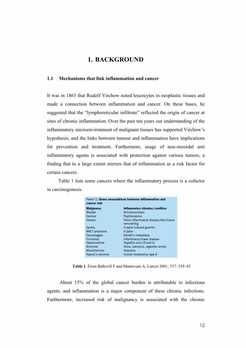
Table 1 lists some cancers where the inflammatory process is a cofactor
in carcinogenesis.
Table 1. From Balkwill F and Mantovani A, Lancet 2001; 357: 539–45
About 15% of the global cancer burden is attributable to infectious
agents, and inflammation is a major component of these chronic infections.
Furthermore, increased risk of malignancy is associated with the chronic
13
inflammation caused by chemical and physical agents, and autoimmune and
inflammatory reactions of uncertain aetiology (Balkwill and Mantovani 2001).
For example, there are strong associations between alcohol abuse, which leads
to inflammation of the liver and pancreas, and cancers of these organs.
Cigarette smoking, asbestos and silica exposure are each associated with
inflammation of the lung and lung carcinoma; inflammatory bowel disease
(IBD) is associated with colon cancer; infections with Helicobacter pylori is
associated with gastric carcinoma; chronical viral hepatitis is associated with
liver cancer; infections with Schistosoma spp. is associated with bladder and
colon carcinoma; infection with some strains of HPV is associated with
cervical cancer; and infection with EBV is associated with Burkitt lymphoma
and nasopharyngeal carcinoma. (Coussens et Werb 2002, Shacter and
Weitzman, 2002, Hussain et al. 2003, Fox and Wang 2007, Dobrovolskaia and
Kozlov, 2005).
Key features of cancer-related inflammation (CRI) include the infiltration
of white blood cells such as DCs, T cells, NK cells and prominently tumor-
associated macrophages (TAMs); the presence of polypeptide messengers of
inflammation [cytokines such as tumor necrosis factor (TNF), interleukin (IL)-
1, IL-6, IL-12, chemokines such as CCL2 and CXCL8] and the occurrence of
tissue remodeling and angiogenesis. (Colotta et al. 2009).
Recent efforts have shed new light on molecular and cellular circuits
linking inflammation and cancer (Mantovani et al. 2008). Two pathways have
been schematically identified; in the intrinsic pathway, genetic events causing
neoplasia initiate the expression of inflammation-related programs that guide
the construction of an inflammatory microenvironment. Of note, it is reported
that the rearrangements of the RET receptor tyrosine kinase gene generating
RET-PTC oncogenes, specific to papillary thyroid carcinoma (PTC), when
exogenously expressed in primary normal human thyrocytes, induce the
expression of a large set of genes involved in inflammation and tumor
invasion, including those encoding chemokines (CCL2, CCL20, CXCL8, and
14
CXCL12), chemokine receptors (CXCR4), cytokines (IL1B, CSF-1, GM-CSF,
and G-CSF), matrix-degrading enzymes (metalloproteases and urokinase- type
plasminogen activator and its receptor), and adhesion molecules (L-selectin).
Selected relevant genes (CCL20, CCL2, CXCL8, CXCR4, L-selectin, GM-
CSF, IL1B, MMP9, UPA, and SPP1-OPN) were found up-regulated also in
clinical samples of PTC, particularly those characterized by RET-PTC
activation, local extrathyroid spread, and lymph node metastases, when
compared with normal thyroid tissue or follicular thyroid carcinoma,
demonstrating that the RET-PTC1 oncogene activates a proinflammatory
program and provide a direct link between a transforming human oncogene,
inflammation, and malignant behaviour (Borrello et al. 2005). It is reported
that other oncogenes representative of different molecular classes and mode of
action (tyrosine kinases, ras–raf, nuclear oncogenes and tumor suppressors
such as von Hippel-Lindau tumour suppressor (VHL), and phosphatase and
tensin homologue (PTEN) share the capacity to orchestrate proinflammatory
circuits (e.g. angiogenetic switch; recruitment of myelo-monocytic cells)
(Mantovani et al. 2008) predisposing to cancer.
In the extrinsic pathway, are the inflammatory conditions cited before to
facilitate cancer development by a variety of mechanisms (Balkwill and
Mantovani 2001). I will now look in more detail at the mechanisms by which
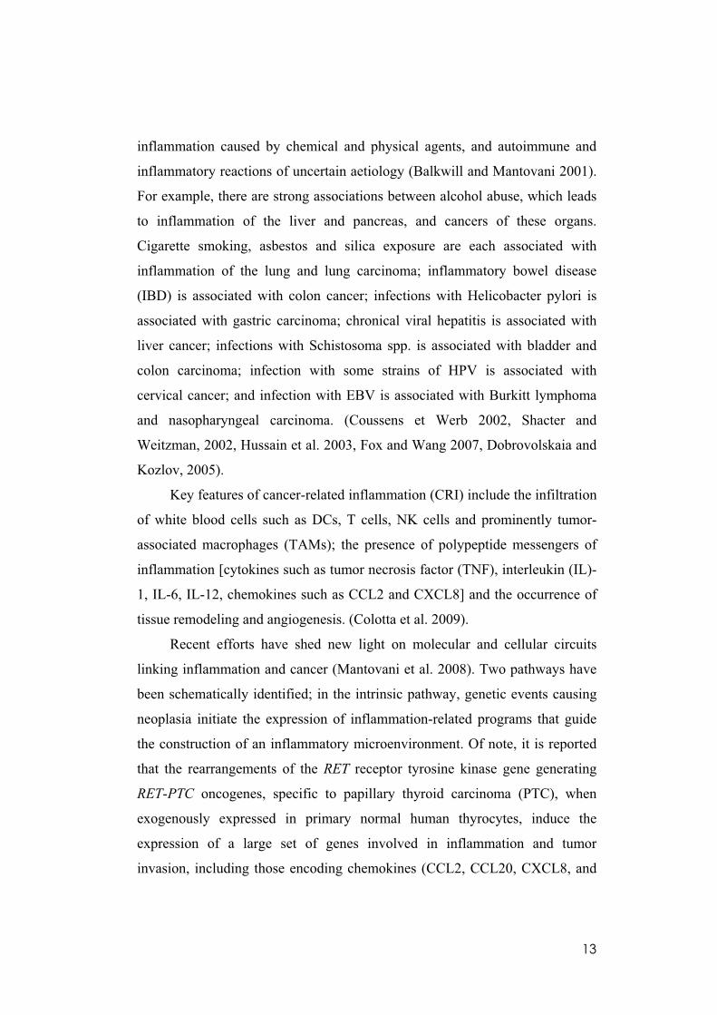
cytokines and chemokines might act to promote cancer also summerized in
next figure 1.
15
Figure 1: Chronic inflammation, tissue damage, and chronic infection may stimulate
cytokines and chemokines that contribute to development of malignant disease ( Balkwill
F and Mantovani A, 2001).
Mediators of inflammation as growth and survival factors
Cytokines and chemokines have the potential to stimulate tumour-cell
proliferation and survival and some of them may also act as autocrine growth
and survival factors for malignant cells. IL-6 is a growth factor for
haematological malignancies (Tricot 2000); IL-1 has growth stimulating
activity for gastric carcinoma that may be related to genetic predisposition (B-
Omar 2000) and for myeloid leukaemias; and growth of melanomas is
promoted by IL-8 and related chemokines (Hanghnegahdar et al. 2000) .
Angiogenesis
The inflammatory cell infiltrate, particularly TAM, may contribute to
tumour angiogenesis, and there are many reports of associations between
macrophage infiltration, vascularity, and prognosis (Leek et al. 1999).
Moreover TNF, IL-1, and IL-6 can stimulate production of angiogenic factors
such as VEGF. Inflammatory macrophages also produce TGF- 1 that is itself
angiogenic and induces production of VEGF. Chemokines also have a role.
16
Some CXC chemokines (eg, IL-8) are proangiogenic whereas others such as
IP-10 (CXCL10) have antiangiogenic activity (Keane and Strieter 1999). In
addition, CC chemokines may inhibit or stimulate angiogenesis indirectly, via
their influence on TAM.
Invasion and metastasis
Cytokines and chemokines affect various stages in the process of
metastasis. TNF- and CC chemokines can induce production of proteases
important for invasion in both tumour cells and macrophages. In one skin
tumour model, paracrine matrix metalloproteinase-9 production by
inflammatory cells was implicated in epithelial hyperproliferation,
angiogenesis and increased malignant potential, and skin tumour development
was reduced in mice genetically “knocked out” for this protein. TNF- and IL-
1 increase the expression of adhesion molecules on endothelial cells (Ekbom et
al. 1990, Negus et al. 1997). IL- 18 derived from the endothelium may be the
ultimate mediator of one tumour cytokine-induced adhesion molecule (Tricot
2000) 10. Certain cells have receptors for adhesion molecules and use these
molecular tools, typical of migrating leucocytes, to seed at distant anatomical
sites (Martin Padura et al. 1991) .
Furthermore, chemokine agonists induce migration or proliferation of
some tumour cells (Hanghnegahdar et al. 2000). Receptors that are essential for
lymphocyte and dendritic cell homing to lymph nodes (Allavena et al. 2000),
could play a role in lymphatic dissemination of certain carcinomas. Thus,
tumour cells use the same molecular tools (adhesion molecules, cytokines,
chemokines, chemokine receptors) and pathways as leucocytes to spread to
distant anatomical sites during inflammation.
Subversion of immunity
The prevalence of Th2 cells is common to tumours, suggesting that this
polarization may be a general strategy to subvert immune responses against
tumours. Chronic exposure to high cytokine concentrations (IL-4, IL-13, IL-

17
10,) in the tumour microenvironment may set in motion a vicious cycle leading
to skewing towards a type II inflammatory response (Sica et al. 2000).
Some viruses encode chemokines and their inhibitors and receptors. Of
particular interest is human herpesvirus type 8, which is involved in the
pathogenesis of Kaposi’s sarcoma. The virus genome codes for three
chemokines which are selective attractants of polarized Th2 cells. The virus-
encoded chemokines might subvert immunity by activating type 2 responses
and diverting effective Th1 defence mechanisms (Sozzani et al. 1998, Endres
et al. 1999).
Genome Instability
Recent data suggest that an additional mechanism involved in cancer-
related- inflammation (CRI) is the induction of genetic instability by
inflammatory mediators either directly inducing DNA damage (via reactive
oxygen) or affecting DNA repair systems and altering cell cycle checkpoint.
For example HIF-1 , which is induced in cancer cells by inflammatory
cytokines (TNF and IL-1 ), PGE2 (Jung et al. 2003) and reactive oxygen and
nitrogen species (Sandau et al. 2000) downregulate mismatch repair (MMR)
proteins MSH2 and MSH6 by displacing c-Myc from MSH2/MSH6 promoters
(Koshiji et al. 2005).
Hydrogen peroxide produced by inflammatory cells inactivates MMR
members by damaging the enzymes at the protein level; NO-induced
upregulation of DNA methyltransferase results in extensive methylation of the
cytosine bases, which is associated with promoter silencing and loss of gene
expression of the MMR member hMLH1 (Fleisher et al. 2000). In fact by
immunohistochemistry, decreased levels of hMLH1 proteins are seen in gastric
epithelial cells in H.pylori-positive patients (Mirzaee et al. 2008) and in colitis-
associated cancers, hMLH1 hypermethylation is observed in a substantial
proportion of patients (Fleisher et al. 2000). The nucleotide excision repair
pathway, which serves to repair a variety of DNA lesions caused by UV
radiation, mutagenic chemicals and chemotherapeutic agents, appears to be

18
affected by IL-6 that in multiple myeloma cells induces hypermethylation, and
thus defective function, of the key nucleotide excision repair component
hHR23B (Hodge et al. 2005). HIF-1 induces the microRNA-373 that
downregulates the expression of the nucleotide excision repair component
RAD23B (Crosby et al. 2009). Chromosomal instability (CI) results in
abnormal segregation of chromosomes and aneuploidy. In most cancers with
CI, proteins of the mitotic checkpoints are disregulated (Rajagopalan et al.
2003). As a consequence, cancer cells fail to halt the cell cycle until DNA
repair can be executed. Inactivation of p53 may play a role in CI (Tomasini et
al. 2008). The p53 pathway protects cells from transformation by inducing
apoptosis upon DNA damage and CI. p53 deficiency and a defective mitotic
checkpoint in T lymphocytes increase CI through aberrant exit from mitotic
arrest (Baek et al. 2003). Loss of p53 and p73 are associated with increased
aneuploidy in mouse embryonic fibroblasts (Talos et al. 2007). The
proinflammatory cytokine migration inhibitory factor suppresses p53 activity
as a transcriptional activator (Hudson et al. 1999). NO and its derivatives
inhibit the function of p53 (Calmels et al. 1997, Cobbs et al.2003) and are
associated with p53 mutations (Ambs et al. 1999, Marshall et al. 2000, Jaiswa
et al. 2001, Wink et al. 1994). NO (Hmadcha et al. 1999) and the inflammatory
cytokine IL-6 (Hodge et al. 2005) increase the activity of DNA
methyltransferase, resulting in CpG island methylation. Malignant cells
employ matrix metalloproteinases (MMPs) to penetrate the extracellular matrix
and basement membrane and to invade distant tissues. Recent data suggest that
MMPs produced by tumours and by the inflamatory cells may also function as
oncogenes by promoting CI. MT1-MMP, which is present also in the
pericentrosomal compartment, compromises normal cytokinesis inducing
aneuploidy. A potential target of MT1-MMP is pericentrin, an integral
centrosomal/midbody protein required for centrosome performance and
chromosome segregation (Golubkov et al. 2007). MMP-3, which is
upregulated in many breast cancers (Stemlicht et al. 2001), also mediates CI in
19
cultured cells and in transgenic mice (Stemlicht et al. 1999, Lochter et al.
1997). Expression of MMP-3 in cells stimulates the production of Rac1b
(Matos et al. 2003), an hyperactive alternative splicing form of Rac1, which
stimulates ROS production which can cause oxidative DNA damage and CI.
This cancer genetic instability through accelerated somatic evolution
leads to a genomically heterogenous population of expanding cells naturally
selected for their ability to proliferate, invade distant tissues and evading host
defenses (Colotta et al. 2009).
The intrinsic and extrinsic pathways converge on the inhibitor of NF-kB
kinase/ NF-kB (IKK/ NF-kB) signaling pathway, which is activated by many
proinflammatory cytokines. NF-kB is a transcription factor that regulates the
expression of many genes whose products can suppress tumor cell death;
stimulate tumor cell cycle progression; enhance epithelial-to-mesenchymal
transition, which has an important role in tumor invasiveness; and provide
newly emerging tumors with an inflammatory microenvironment that supports
their progression, invasion of surrounding tissues, angiogenesis, and metastasis
(Dobrovolskaia and Kozlov 2005, Karin 2006).
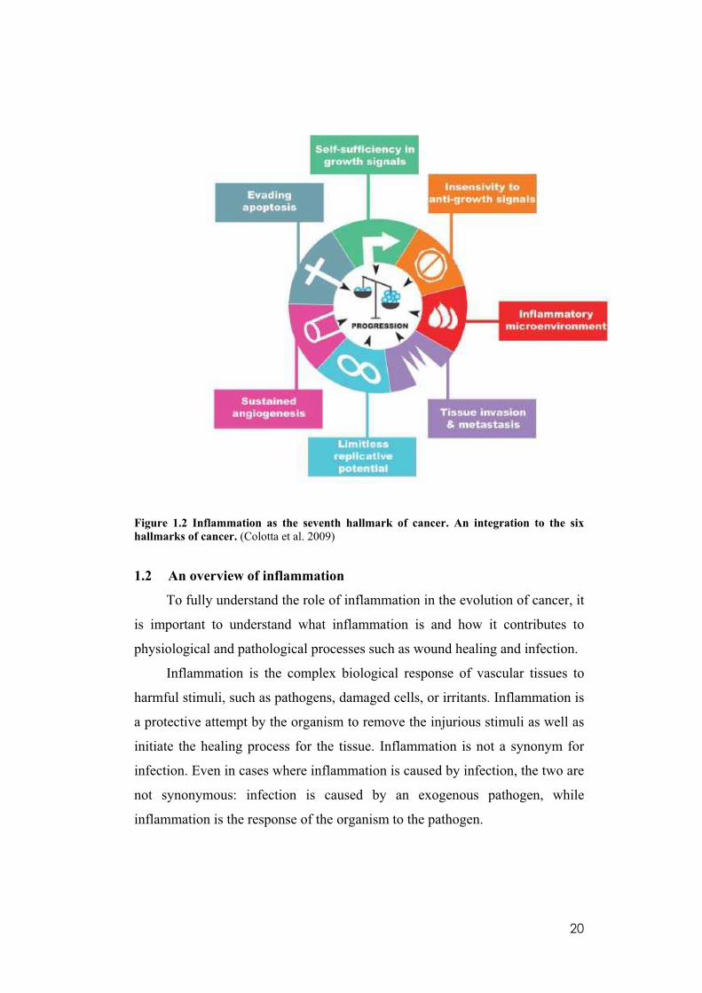
In conclusion, CRI is a key component of tumors and may represent the
seventh hallmark of cancer, providing further impetus for studies targeted to
the inflammatory microenvironment of tumors (Colotta et al. 2009) (Figure
1.2).
20
Figure 1.2 Inflammation as the seventh hallmark of cancer. An integration to the six
hallmarks of cancer. (Colotta et al. 2009)
1.2 An overview of inflammation
To fully understand the role of inflammation in the evolution of cancer, it
is important to understand what inflammation is and how it contributes to
physiological and pathological processes such as wound healing and infection.
Inflammation is the complex biological response of vascular tissues to
harmful stimuli, such as pathogens, damaged cells, or irritants. Inflammation is
a protective attempt by the organism to remove the injurious stimuli as well as
initiate the healing process for the tissue. Inflammation is not a synonym for
infection. Even in cases where inflammation is caused by infection, the two are
not synonymous: infection is caused by an exogenous pathogen, while
inflammation is the response of the organism to the pathogen.
21
In the absence of inflammation, wounds and infections would never heal
and progressive destruction of the tissue would compromise the survival of the
organism (Ferrero-Miliani et al. 2007, Coussens and Werb 2002).
Inflammation can be classified as either acute or chronic. Acute
inflammation is the initial response of the body to harmful stimuli and is
achieved by the activation and directed migration of leukocyte (neutrophils,
monocytes and eosinophils) from the blood into the injured tissues. For
neutrophils, a four-step mechanism is believed to coordinate recruitment of
these inflammatory cells to sites of tissue injury and to the provisional
extracellular matrix (ECM) that forms a scaffolding upon which fibroblast and
endothelial cells proliferate and migrate, thus providing a nidus for
reconstitution of the normal microenvironment (Chettibi et al. 1999). These
steps involve: activation of members of the selectin family of adhesion
molecules (L- P-, and E-selectin) that facilitate rolling along the vascular
endothelium; triggering of signals that activate and upregulate leukocyte
integrins mediated by cytokines and leukocyte-activating molecules;
immobilization of neutrophils on the surface of the vascular endothelium by
means of tight adhesion through 4 1 and 4 7 integrins binding to
endothelial vascular cell-adhesion molecule-1 (VCAM-1) and MadCAM-1,
respectively; and transmigration through the endothelium to sites of injury,
presumably facilitated by extracellular proteases, such as matrix
metalloproteinases (MMPs).
A number of chemokines, which possess a relatively high degree of
specificity for chemoattraction of specific leukocyte populations (Rossi and
Zlotnik 2000, Homey et al. 2002), recruits downstream effector cells and
dictates the natural evolution of the inflammatory response.
Neutrophils (and sometimes eosinophils) are the first recruited effectors
of the acute inflammatory response. Monocytes, which differentiate into
macrophages in tissues, are next to migrate to the site of tissue injury, guided
by chemotactic factors. Once activated, macrophages are the main source of
22
growth factors and cytokines, which profoundly affect endothelial, epithelial
and mesenchymal cells in the local microenvironment. The process of acute
inflammation is initiated also by cells already present in all tissues, mainly
resident macrophages, dendritic cells, histiocytes, Kuppfer cells and
mastocytes. At the onset of an infection, burn, or other injuries, these cells
undergo activation and release inflammatory mediators responsible for the
clinical signs of inflammation.
Prolonged inflammation, known as chronic inflammation, leads to a
progressive shift in the type of cells which are present at the site of
inflammation and is characterized by simultaneous destruction and healing of
the tissue from the inflammatory process. (Cotran et al. 1998).
In tumor development, the major driving force is chronic inflammation
secondary to persistent infection with a parasite, bacterium, or virus (Karin and
Greten 2005).
Infectious organisms (parasite, bacterium or virus) trigger inflammation
through activation of receptors that recognize pathogen-associated molecular
patterns (PAMPs), such as cell wall components and nucleic acids (Medzhitov
2001). At least four families of mammalian innate immune receptors that
recognize PAMPs have been identified; these are known as pattern recognition
receptors (PRRs) and include TLRs, nucleotide-binding oligomerization
domain–like (NOD-like) receptors (NLRs), C-type lectin receptors (CLRs),
and triggering receptors expressed on myeloid cells (TREMs) (Akira et
al.2006, Fritz et al. 2006, Robinson et al. 2006, Klesney-Tait et al. 2006). The
interaction between PAMPs and PRRs results in the activation of innate
immune cells and initiation of host responses whose major purpose is to
eliminate and kill invading organisms (Karin and Greten 2005). However,
inadequate pathogen eradication, prolonged inflammatory signaling, and
defects in antinflammatory mechanisms can all lead to chronic inflammation
and benefit tumor development (Han and Hulvetich 2005).
23
Inflammatory cells in tumour microenvironment.
The inflammatory microenvironment of tumours is characterized by the
presence of host leucocytes both in the supporting stroma and in tumour areas
(Negus et al. 1997). Tumour infiltrating leucocytes may contribute to cancer
growth and spread, and to the immunosuppression associated with malignant
disease.
Macrophages and dendritic cells infiltrate tumours (Scarpino et al. 2000,
Mantovani et al. 2002). In the tumour microenvironment many signals polarize
these mononuclear phagocytes which can express different functional
programmes. Fully polarized type I and type II (or alternatively activated)
macrophages are the extremes of a continuum of functional states. Tumor-
derived and T cell-derived cytokines stimulate tumor associated macrophages
(TAM) to acquire a polarized type II phenotype. These functionally polarized
cells, and similarly oriented or dysfunctional and immature dendritic cells
present in tumors, play a key role in the subversion of adaptive immunity and
in inflammatory circuits which promote tumor growth and progression (Solinas
et al. 2009, Fricke and Gabrilovich 2006).
1.3 Tumor –Associated-Macrophages (TAM)
The tumor mass is undoubtedly a multifaceted show, where different cell
types, including neoplastic cells, fibroblasts, endothelial, and immune-
competent cells, interact with one another continuously. Macrophages
represent up to 50% of the tumor mass, and they certainly operate as
fundamental actors. (Solinas et al. 2009).
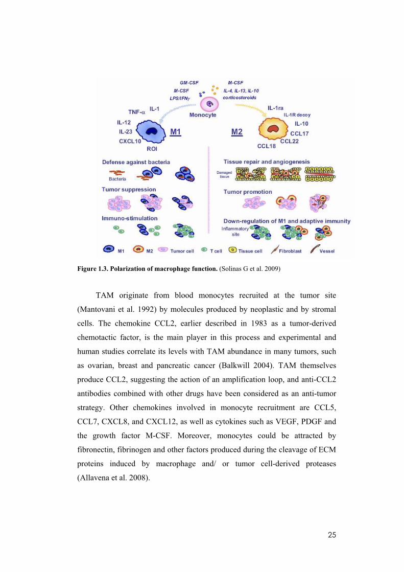
Polarization of macrophage function.
Macrophages constitute an extremely heterogeneous population, which
could be divided schematically into two main classes: M1 and M2 (Figure 1.3).
Blood monocytes differentiating in the presence of LPS/IFN- mature into M1-
polarized cells (classically activated macrophages). They produce high levels
of IL-12, IL-1, IL-23, TNF- , and CXCL10 and are characterized by cytotoxic
24
activity against microorganisms and neoplastic cells, expression of high levels
of ROI, and capability as APCs with high expression of the co-stimulatory
molecule CD86. On the other hand, when monocytes differentiate in the
presence of IL-4, IL-13, IL-10, or corticosteroids, they mature into M2
macrophages (alternatively activated macrophage) which secrete IL-10,
CCL17, CCL22, CCL18, IL-1ra, and IL-1R decoy. M2 cells are active workers
of the host, promoting scavenging of debris, angiogenesis, remodeling, and
repair of wounded/damaged tissues. Within the tumor mass, they exert the
same functions favoring tumor promotion. In addition, M2 macrophages
control the inflammatory response by down-regulating M1-mediated functions
and adaptive immunity (Solinas et al. 2009). They are in fact the major source
of the soluble and membrane-bound imunoglobulin-like transcript 3 (ILT3)
which may be responsible for the immuno-escape mechanisms of tumors. Both
membrane-bound ILT3 (mILT3) and soluble ILT3(sILT3) inhibited T cell
proliferation in mixed lymphocyte culture (MLC), anergizing CD4+ Th cells,
suppressing the differentiation of IFN-gamma producing CD8+ cytotoxic T
cells, and inducing the differentiation of alloantigen-specific CD8+ T
suppressors in primary 7-day MLC (Kim-Schulze et al. 2006). Furthermore, it
is reported that in a humanized severe combined mmunodeficiency (SCID)
mouse model, soluble and membrane ILT3 induce CD8+ T suppressor cells
and prevent rejection of allogeneic tumor transplants. Furthermore, patients
with carcinoma of the pancreas produce the soluble ILT3 protein, which
induces the differentiation of CD8+ T suppressor cells and impairs T cell
responses in mixed lymphocyte culture. These responses are restored by anti-
ILT3 mAb or by depletion of sILT3 from the serum suggesting that ILT3
depletion or blockade is crucial to the success of immunobiotherapy (Cortesini
2007).
25
Figure 1.3. Polarization of macrophage function. (Solinas G et al. 2009)
TAM originate from blood monocytes recruited at the tumor site
(Mantovani et al. 1992) by molecules produced by neoplastic and by stromal
cells. The chemokine CCL2, earlier described in 1983 as a tumor-derived
chemotactic factor, is the main player in this process and experimental and
human studies correlate its levels with TAM abundance in many tumors, such
as ovarian, breast and pancreatic cancer (Balkwill 2004). TAM themselves
produce CCL2, suggesting the action of an amplification loop, and anti-CCL2
antibodies combined with other drugs have been considered as an anti-tumor
strategy. Other chemokines involved in monocyte recruitment are CCL5,
CCL7, CXCL8, and CXCL12, as well as cytokines such as VEGF, PDGF and
the growth factor M-CSF. Moreover, monocytes could be attracted by
fibronectin, fibrinogen and other factors produced during the cleavage of ECM
proteins induced by macrophage and/ or tumor cell-derived proteases
(Allavena et al. 2008).
26
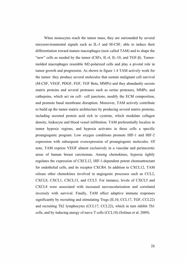
When monocytes reach the tumor mass, they are surrounded by several
microenvironmental signals such as IL-3 and M-CSF, able to induce their
differentiation toward mature macrophages (now called TAM) and to shape the
“new” cells as needed by the tumor (CSFs, IL-4, IL-10, and TGF- ). Tumor-
molded macrophages resemble M2-polarized cells and play a pivotal role in
tumor growth and progression. As shown in figure 1.4 TAM actively work for
the tumor: they produce several molecules that sustain malignant cell survival
(M-CSF, VEGF, PDGF, FGF, TGF Beta, MMPs) and they abundantly secrete
matrix proteins and several proteases such as serine proteases, MMPs, and
cathepsins, which act on cell– cell junctions, modify the ECM composition,
and promote basal membrane disruption. Moreover, TAM actively contribute
to build up the tumor matrix architecture by producing several matrix proteins,
including secreted protein acid rich in cysteine, which modulate collagen
density, leukocyte and blood vessel infiltration. TAM preferentially localize in
tumor hypoxic regions, and hypoxia activates in these cells a specific
proangiogenic program. Low oxygen conditions promote HIF-1 and HIF-2
expression with subsequent overexpression of proangiogenic molecules. Of
note, TAM express VEGF almost exclusively in a vascular and perinecrotic
areas of human breast carcinomas. Among chemokines, hypoxia tightly
regulates the expression of CXCL12, HIF-1-dependent potent chemoattractant
for endothelial cells, and its receptor CXCR4. In addition to CXCL12, TAM
release other chemokines involved in angiogenic processes such as CCL2,
CXCL8, CXCL1, CXCL13, and CCL5. For instance, levels of CXCL5 and
CXCL8 were associated with increased neovascularization and correlated
inversely with survival. Finally, TAM affect adaptive immune responses
significantly by recruiting and stimulating Tregs (IL10, CCL17, TGF, CCL22)
and recruiting Th2 lymphocytes (CCL17, CCL22), which in turn inhibit Th1
cells, and by inducing anergy of naıve T cells (CCL18) (Solinas et al. 2009).
27
Figure 1.4 Protumoral functions of tumor-associated macrophages (TAM) and interplay
with tumor cells. (Sica A et al. 2008)
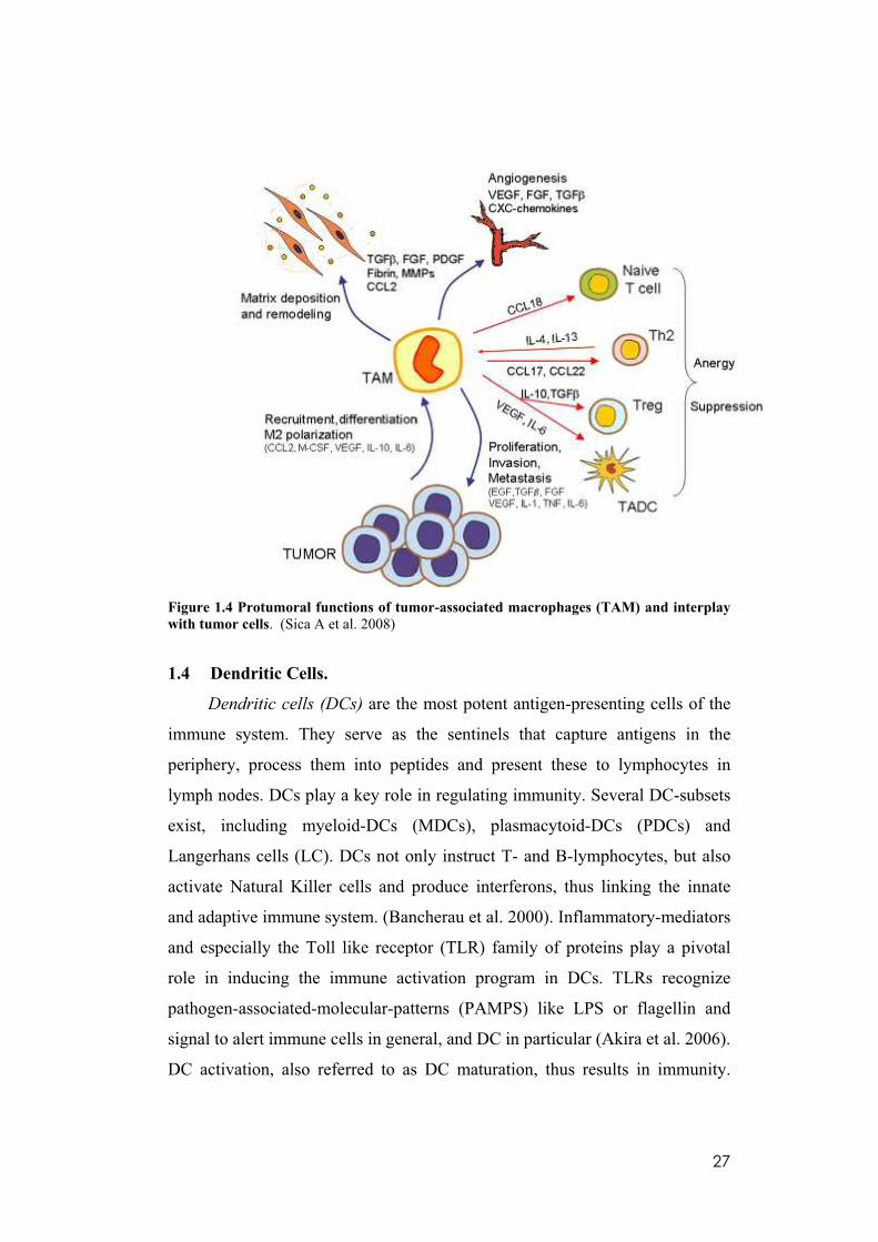
1.4 Dendritic Cells.
Dendritic cells (DCs) are the most potent antigen-presenting cells of the
immune system. They serve as the sentinels that capture antigens in the
periphery, process them into peptides and present these to lymphocytes in
lymph nodes. DCs play a key role in regulating immunity. Several DC-subsets
exist, including myeloid-DCs (MDCs), plasmacytoid-DCs (PDCs) and
Langerhans cells (LC). DCs not only instruct T- and B-lymphocytes, but also
activate Natural Killer cells and produce interferons, thus linking the innate
and adaptive immune system. (Bancherau et al. 2000). Inflammatory-mediators
and especially the Toll like receptor (TLR) family of proteins play a pivotal
role in inducing the immune activation program in DCs. TLRs recognize
pathogen-associated-molecular-patterns (PAMPS) like LPS or flagellin and
signal to alert immune cells in general, and DC in particular (Akira et al. 2006).
DC activation, also referred to as DC maturation, thus results in immunity.
28
Mature DC produce pro-inflammatory cytokines. In contrast, resting DC or DC
receiving immune-inhibitory signals, such as IL-10 and/or corticosteroids,
induce immune tolerance via T cell deletion and induction of suppressive T
cells, now termed regulatory T cells. Several mouse models have demonstrated
that the immunological outcome depends upon the DC activation state; mature
immune-activating DC protect mice from a tumor or pathogen, whereas
tolerogenic DC induce tolerance against transplanted tissues. Hence, DC act at
the interface of immunity and peripheral tolerance (Steinman et al. 2003).
Among the inflammatory cytokines produced by DC, it is reported that
IL-12 has a dual opposite effect: antitumor property relying on its ability to
promote Th1 adaptive immunity and CTL responses by stimulating the
production of IFN- from T and natural killer (NK) cells, and reducing IL-4
mediated suppression of IFN- which results in enhancement of the cytotoxic
activity of NK cells and CD8+ cytotoxic T lymphocytes (Trinchieri 2003), but
also tumour- promoting role. In fact, it is reported that IL-12 plays a major role
in sustaining the chronic phase of several inflammatory conditions that often
degenarate in carcinoma such as colitis (Leach and Rennick 1998).
IL-15 is an other cytokine produced by activated DC, that has been
shown to play a pivotal role in orchestrating immune-mediated tissue
destruction in inflammatory disease (Mention et al. 2003, Kovesdy and
Kalantar-Zadeh 2008). IL-15 is indispensable for the generation, maintenance,
and homeostasis of local T and NK cell. IL-15 also induces proliferation of
CD8+ T and NK lymphocytes in addition to enhancing their effector functions,
including those associated with cytolysis and cytokine secretion. IL-15 also
promotes perpetuation of chronic inflammation by mediating activation of
monocyte and neutrophils and by preventing activation-induced cell death of
activated CD8+ T cells (Waldmann 2006, Huntington et al. 2007). In addition
to these positive modulatory effects on the activation pathways leading to
persistent inflammation, IL-15 can block the negative regulatory pathways
critical in maintaining immune homeostasis by inhibiting the anti-

29
inflammatory Smad-dependent signaling of TGF- thereby, further
aggravating ongoing inflammation (Benahmed et al. 2007).
The presence of DCs in human carcinomas has been largely
documented, (Yang and Carbone 2004) and has been proposed to correlate
with a more favorable prognosis (Tsujitani et al. 1990, Ishigami et al. 1998,
Iwamoto et al. 2003). Ideally, DC should be recruited to the tumour site to
initiate the immune response, and promote tumour rejection. In breast cancer,
immature TADC are interspersed in the tumour mass, whereas mature dendritic
cells are confined to the peritumoral area (Treiilleux et al. 2004). In papillary
thyroid carcinoma TADC are also immature, but they tend to localise at the
invasive edge of the tumour (Scarpino et al. 2000).
However, although DC can engulf tumor cells debrites, process and cross
present tumor-associated antigens to cytotoxic T lymphocytes (CTLs) (Chan
and Housseau 2008), the tumours microenvironment conteract this
phenomenon by releasing a number of immunosuppressive factors, including
IL6, VEGF, IL8 and IL10, that contributes to DC malfunction (Fricke and
Gabrilovich 2006). Thus, tumour-associated-denditic cells (TADC) usually
have an immature phenotype, with defective ability to stimulate T cells, a
phenotype suggesting a controversial role for TADC in the immune response
toward cancer cells.
1.5 Dendritic Cell-Natural Killer crosstalk
Natural killer (NK) cells are a population of large granular lymphocytes
with a CD56+/CD3-phenotype. They are distinguishable from B and T
lymphocytes by lack of antigen receptors. NK cells kill a variety of tumor cells,
virus-infected cells and allogeneic cells in a non-major histocompatibility
complex restricted manner, and provide the first line of immune defense
(Trinchieri 1989, Moretta et al. 2002).
30
Although NK cells lack the antigen-specific receptors, they distinguish
between normal cells and abnormal cells by their cell surface receptors. After
binding to potential target cell, NK cell activating and inhibitory receptors
interact with ligands and transmit signals, and then all the signals are integrated
to determine whether NK cell stays and responds (Bottino et al. 2005, Long
1999, Moretta et al. 2001). The effector function of NK cells is regulated by a
balance between the inhibitory signals delivered by the MHC class I-specific
inhibitory receptors and the activating signals transmitted by activating
receptors (Lanier 2005). NK cell effector function is mainly mediated through:
1) releasing cytoplasmic cytotoxic granules (granzyme and perforin) by
exocytosis; 2) secreting proinflammatory cytokines (IFN- , TNF- , etc.); and
3) the engagement of death receptors on target cells by their cognate ligands
(e.g., FasL and TRAIL) on NK cells (Janeway and Medzhitov 2002). In course
of DC-NK interplay, myeloid DCs by secreting NK-cells activating cytokines
(IL-12, IL-15, type I IFNs), promote the secretion of pro-inflammatory
cytokines and cytotoxicity of NK cells (Walzer et al. 2005).
Reciprocally, NK cells, traditionally considered to be major innate
effector cells, have also recently been shown to play immunoregulatory 'helper'
functions, being able to activate DCs and to enhance their ability to produce
pro-inflammatory cytokines. (Walzer et al. 2005, Moretta et al. 2006, Degli-
Esposti and Smyth 2005). In addition, once activated, NK cells acquire the
capability of killing immature myeloid DCs (Moretta et al. 2002, Zitvogel
2002). This effect is due to the fact that immature DCs typically underexpress
HLA-class I molecules that would protect from NK-mediated lysis. On the
other hand, DCs that, after Ag uptake, undergo maturation, upregulate MHC-
class I expression becoming essentially resistant to NK cells (Ferlazzo et al.
2003). It has been suggested that the NK-mediated killing of DCs may serve to
keep in check the quality and the quantity of DCs undergoing maturation
(‘editing’ process). According to this view, DCs that fail to express sufficient
amounts of MHC molecules would be removed. Thanks to this mechanism NK
31
cells may prevent the survival of faulty DCs that after expression of CCR7 and
migration to lymph nodes, would induce inappropriate, low-affinity, T-cell
priming resulting either in Th2 responses or in a state of tolerization
(Langenkamp 2000).
Indeed, only DCs undergoing this NK-mediated quality control would
become fully mature and capable of inducing priming of protective and
cytotoxic Th1 responses.
1.6 Emerging aspects of NK cell biology
In contrast to their protective role in various inflammatory conditions,
NK cells can also act as mediators of innate immunopathology.In patients with
chronic hepatitis B virus infection, a subset of NK cells contributes to liver
inflammation by inducing hepatocyte death through a TRAIL-dependent
mechanism (Dunn et al. 2007). In hepatitis B virus transgenic mice, NK cells
also promote liver injury through NKG2D (Chen et al. 2007). Moreover, NK
cells act detrimentally in experimental sepsis induced by Streptococcus
pneumoniae or Escherichia coli by exacerbating inflammatory responses (Kerr
et al. 2005, Badgwell et al. 2002). Consistent with these data, a potential
contribution of NK cells has also been postulated in human inflammatory
diseases such as arthritis (de Matos et al. 2007) and sarcoidosis (Katchar et al.
2005) .
Transgenic mice that express human NK cells-activating IL-15
specifically in enterocytes (T3b-hIL-15 Tg mice) develop villous atrophy and
severe duodeno-dejunal inflammation with massive accumulation of NK-like
CD8-lymphocytes in the affected mucosa that leads to a major propensity for
the development of enteropathy associated CD8 T cell lymphoma. Finally, in
humans NK cells have been shown to home to inflamed skin in various
conditions, such as vernal keratoconjunctivitis (Lambiase et al. 2007), atopic
dermatitis (Buentke 2002) and psoriasis (Ottaviani et al. 2006).
32
Thus another interesting mechanism of action of drugs that target cancer-
related inflammation might be to prevent deleterious NK cell-driven
inflammatory response.
1.7 HIV protease inhibitors: antiretroviral agents
Immunodeficiency is a hallmark of human immunodeficiency virus type
1 (HIV-1) disease and is characterized by a progressive decrease in CD4 T
cells. The advent of new antiretroviral drugs, most notably HIV protease
inhibitors, has generated new hope in the fight against AIDS. Development of
HIV-protease inhibitors in the early 1990s followed the characterization of the
crystal structure of HIV protease in 1989 (Navia MA et al. 1989). Inhibitors of
HIV protease are peptidomimetics that generally contain a synthetic analogue
of the peptide bond between phenylalanine and proline at positions 167 and
168 of the gag-pol polyprotein, which is the target of the HIV aspartyl protease
(Flexner C 1998). This action prevents production of infectious viral particles.
The first inhibitor of HIV protease developed that received FDA approval was
saquinavir, followed by ritonavir, indinavir, nelfinavir, and amprenavir. Drugs
approved more recently include lopinavir (in combination with ritonavir),
atazanavir, fosamprenavir (a prodrug of amprenavir), tipranavir, and darunavir.
Used in combination with nucleoside inhibitors of HIV reverse
transcriptase, protease inhibitors have led to impressive clinical outcomes.
Such combined therapeutic regimens, known as highly active antiretroviral
therapies (HAART), work by suppressing HIV replication and can lead to a
large reduction in HIV plasma viraemia, restoration of normal numbers of
CD4-positive T lymphocytes, immunological recovery, and reduction of
morbidity and mortality related to HIV and opportunistic infections. The
increase in CD4-positive T-cell counts and the immune restoration that occurs
with HAART is most likely to depend on the following mechanisms: increased
peripheral CD4-positive T-cell survival and proliferation, central renewal of
lymphocytes, improvement of T-cell responses, and restoration of the T-cell
33
repertoire. Therefore, protease-inhibitor-based HAART owes its success to the
ability to block HIV replication and promote subsequent immunological
recovery (Sgadari et al. 2003).
1.8 Antitumour effects of antiretroviral therapy
Infection by human immunodeficiency virus (HIV) is associated with an
increased risk of certain tumours, particularly Kaposi’s sarcoma, non-
Hodgkin’s lymphomas and cervical cancer. However, the incidence of these
cancers and the general tumour burden in HIV-infected patients has decreased
significantly since the widespread use of highly active antiretroviral therapy
(HAART). This effect cannot be solely explained by the ability of these drugs
to suppress HIV replication and thereby reconstitute the immune system; in
fact tumour development is not always correlated with a patient’s viral load or
level of immune reconstitution. Recent studies have shown that inhibitors of
the HIV aspartyl protease, which are widely used in HAART, have direct anti-
angiogenic and antitumour effects that are unrelated to their antiviral activity
(Monini et al. 2004).
These direct antitumour effects of HAART could be related to specific
actions of the protease inhibitors included in this therapeutic cocktail, such as
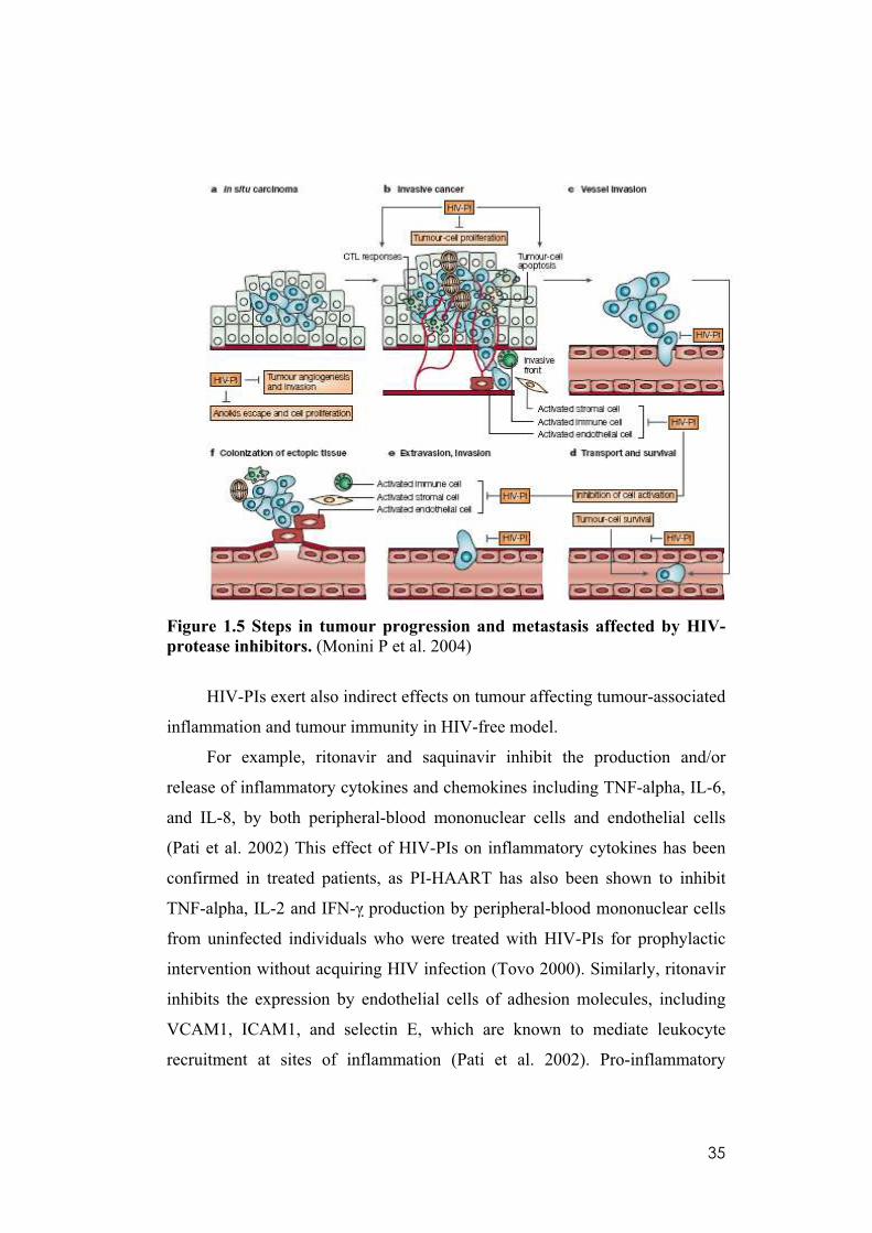
ritonavir, saquinavir, indinavir and nelfinavir. Figure 1.5 summarizes the
various steps in tumour progression and metastasis affected by HIV-protease
inhibitors. These steps usually lead to progression of in situ carcinoma (a) to
invasive cancer (b) and to metastasis formation and dissemination (c–f).
Tumour outgrowth (a,b) is dependent on tumour neoangiogenesis and its net
rate is determined by the balance between tumour cell proliferation versus
apoptosis, invasive behaviour and the ability of tumour cells to evade the
immune response. At concentrations similar or above therapeutic peak levels,
HIV-PIs promote apoptosis and inhibit proliferation of tumour cells with little
or no effects on survival and proliferation of normal cells (Gaedicke et al.
2002), whereas at therapeutic steady-state concentrations they inhibit tumour
34
angiogenesis and tumour-cell invasion. Furthermore, HIV-PIs have anti-
inflammatory effects (b). As metastatic cell clones emerge, tumour cells loosen
their contact with surrounding cells and the extracellular matrix (ECM). This
leads to invasion of blood or lymphatic vessels and to extravasation of tumour
cells at distant sites (c–e). These steps require the degradation of basement
membranes and, at the same time, inhibition of apoptosis following loss of cell
anchorage (anoikis), processes that are also inhibited by HIV-PIs. Finally,
colonization of ectopic tissue by tumour cells (f) is required for establishment
of metastases, and this process is similarly affected by HIV-PIs (Sgadari et al.
2002). During tissue invasion and establishment of metastases (b,f), activated
endothelial cells, stromal cells and immune cells cooperate in basement
membrane and ECM degradation, modify the ECM composition, release ECM-
bound growth and angiogenic factors, and produce cytokines and chemokines
that stimulate tumour-cell growth and migration, and recruit all these cell types
at the invasive front. These processes are all affected by HIV-PIs through their
ability to inhibit cytokine and chemokine production, cell activation, and basal
membrane and ECM degradation and remodelling. The ability of these drugs to
prevent tumour growth and progression might be mediated by their ability to
inhibit proteasome function, resulting in the inhibition of NF-kB activity, and
the activity of matrix metalloproteinases (Pajonk et al. 2002).
35
Figure 1.5 Steps in tumour progression and metastasis affected by HIV-
protease inhibitors. (Monini P et al. 2004)
HIV-PIs exert also indirect effects on tumour affecting tumour-associated
inflammation and tumour immunity in HIV-free model.
For example, ritonavir and saquinavir inhibit the production and/or
release of inflammatory cytokines and chemokines including TNF-alpha, IL-6,
and IL-8, by both peripheral-blood mononuclear cells and endothelial cells
(Pati et al. 2002) This effect of HIV-PIs on inflammatory cytokines has been
confirmed in treated patients, as PI-HAART has also been shown to inhibit
TNF-alpha, IL-2 and IFN- production by peripheral-blood mononuclear cells
from uninfected individuals who were treated with HIV-PIs for prophylactic
intervention without acquiring HIV infection (Tovo 2000). Similarly, ritonavir
inhibits the expression by endothelial cells of adhesion molecules, including
VCAM1, ICAM1, and selectin E, which are known to mediate leukocyte
recruitment at sites of inflammation (Pati et al. 2002). Pro-inflammatory
36
cytokines, chemokines and adhesion molecules are crucial in the development
of Kaposi’s sarcoma, as they mediate local inflammatory and immune
responses to Kaposi’s-sarcoma cells and to other KSHV-infected cells.
Furthermore, they regulate survival, growth, invasion and eradication of most
tumours. In fact, they lead to local stroma activation, basement-membrane
and/or extracellular-matrix perturbation angiogenesis, and regulate local
tumour immunity. In this context, HIV-PIs directly modulate antigen
processing, T-cell survival (by the inhibition of T-cell apoptosis) and
proliferative responses (Lu and Andrieu 2000, Sloand et al. 1999), and they
might even affect T-cell priming, as they can inhibit dendritic-cell maturation
and function (Gruber et al. 2001).
The most prominent mechanism underlying these last antitumour effects
of HIV-Pis is likely to be MMP inhibition, that is not only responsible for the
blockage of cell invasion and angiogenesis but it is also involved in several
crucial immune and immunomodulatory functions, and in cancer-mediated
immune suppression. (Sgadari et al. 2002, Lopez et al. 2000). Moreover,
MMPs, including MMP2, act as potent modulators of local inflammation by
activating or degrading inflammatory cytokines and chemokines present on the
cell membrane, such as TNF-alpha, monocyte chemoattractant protein 3 nd IL-
8 (Gearing et al. 1994, Ito et al. 2000, Schonbeck et al. 1998). Importantly,
MMPs activate transforming growth factor-beta which, in turn, inhibits T cell
responses against tumours (Gorelik and Flavell 2001, Yu and Stamenkovic
2000). MMPs can also cleave IL-2 receptor (Sheu et al. 2001), which is
required for T-cell proliferation following antigen stimulation. These effects of
MMPs are important determinants of tumour immune evasion, but might also
explain the strong stimulatory effect of low HIV-PIs concentrations on T-cell
proliferation and survival. Furthermore, as MMPs are required for leukocyte
transmigration and tissue infiltration by inflammatory cells, the capability of
ritonavir to inhibit CTL-dependent inflammatory responses could be mediated
not only following the modulation of CTL-epitope processing by the

37
proteasome, but also through the inhibition of MMP activation or function in
transmigrating lymphocytes (Kelleher et al. 2001).

38
2. AIM OF THE STUDY.
On last decade, an increasing number of reports have shown that PIs,
originally designed to block the HIV-1 protease, can also exert remarkable
immunomodulatory effects on multiple cell types by mechanisms not related to
their anti-viral activity. Since these drugs have been widely used in HIV-1
therapy, prove their capability to intercept the inflammatory response, and
identify their cell targets, might generate valuable information for their
“offlabel” use in disorders where a modulation of inflammatory response is
required.
A plethora of evidences identify the inflammatory response has a key
component of mechanisms responsible for cell transformation, tumour growth
and metastatic process, in human cancer. Hematopoietic cells, mostly from
myeloid lineage, play a pivotal role in these processes by driving the immune
response toward a beneficial anti-tumour pathway, or providing support to
cancer cells. In this context, urges to propose novel immunomodulatory
strategies aimed to modify cancer milieu, and drive the anti-tumors response
toward most favorable routes.
Monocytes are the circulating progenitors of different cell types that can
infiltrate cancer lesions, and has a powerful ability to shape the inflammatory
response. For this reason, monocytes and their activation programs are
attractive targets for immune therapies aimed to subvert the immune response
in cancer-bearing individuals. The present study has been aimed to verify the
capability of PIs to interfere with differentiation and activation programs of
human peripheral monocytes.
Specifically, I verified the ability of a panel of PIs, widely used in HIV-1+
patients, to interfere with differentiation of monocytes toward:
(a) Dendritic cell lineage
(b) Tumor associated macrophage-like lineage.
39
3. MATERIALS AND METHODS
3.1 Media and Reagents.
The regular medium used throughout the study was RPMI 1640
(Invitrogen) supplemented with 2 mM L-glutamine, 50 ng/ml streptomycin, 50
units/ml penicillin, and 10% heat-inactivated fetal calf serum (Hyclone
Laboratories, Logan, UT). Granulocytes monocytes-colony stimulating factor
(GM-CSF) was purchased from Schering-Plough (Kenilworth, NJ) and used at
a concentration of 50 ng/ml. Interleukin-4 (IL-4) was obtained from
ImmunoTools and used at 1000U/ml.
Saquinavir, Ritonavir, Nelfinavir, Indinavir, Amprenavir were dissolved
in dimethyl sulfoxide (Me2SO) and used at 20uM. As controls, cells were either
left untreated or were treated with a comparable concentration of Me2SO but
without HIV-1 protease inhibitor. Saquinavir, Ritonavir, Nelfinavir, Indinavir
sulfate, Amprenavir were obtained through the NIH AIDS Research and
Reference Reagent Program, Division af AIDS, NIAID, NIH.
3.2 Isolation and culture of NK cells, DCs and Tumour-Associated-
Macrophage (TAM).
Peripheral blood mononuclear cells (PBMCs) from healthy donors were
isolated by density gradient on Ficoll Lymphoprep (Axis-Shield PoC AS, Oslo,
Norway). Blood samples were obtained in accordance with the ethical
committee requirements. NK cells were negatively selected by depleting by
using an antibodies cocktail against lineages specific markers and magnetic
beads (StemCell Technologies Inc.). According to cytometry, typical purified
NK cells were 97% pure. Purified NK cells contained 3% contamination with
other PBMC subsets as determined by the expression of CD3, TCR- / , TCR-
/ , CD19, or CD14. Polyclonal NK cells and NK cell subsets were activated
in vitro with recombinant IL-2 (rIL-2; Roche) at 200 UI/m for 6 days.
40
To generate iDCs, monocytes were purified by positive selection with anti-
CD14 conjugated magnetic microbeads (Miltenyi Biotec, Bologna, Italy).
CD14+ cells were than cultured at a concentration of 0.5–1 x 10⁶ cells/ml in
regular medium supplemented with GM-CSF (50 ng/ml) and IL-4 (1000U/ml)
for 4–5 days to obtain cells with typical phenotype of iDCs. After 6 d of
stimulation in culture, CD14dim-neg and CD1apos iDCs were induced to
undergo maturation by incubation with LPS at 1 g/ml (Sigma-Aldrich) for 24
h.
In order to generate Macrophage and TAM, myeloid cells were
maintained in complete medium (CM) consisting of culture medium
supplemented with 20 ng/ml GM-CSF. Macrophages (M ) were differentiated
from CD14+ monocytes cultured for 5 d in CM at 106 cells/ml. TA-M were
differentiated from CD14+ monocytes cultured for 5 d in CM at 106 cells/ml
with tumour ascites (diluted 1:10).
All cell culture was conducted at 37°C in humidified 5 % CO2
atmosphere.
3.3 Flow Cytometry.
Cell phenotypes of DCs were analyzed by flow cytometry by using the
following monoclonal antibodies conjugated: anti-HLA-I and anti-CD14 from
Sigma; anti-CD1a, anti-CD86, anti-CD80, anti-CD83, anti-CD40, anti-HLA-
DR, anti-HLA-ABC, anti-CD11c, anti-CD36, anti-CD54 from BD
Biosciences, anti DC-SIGN from NIH research and reference reagent program.
To analyze T-cell programming, DCs generated in presence or absence of
HIV-PIs (1×10⁴ cells/well) were cocultered with allogeneic naïve CD45RA+
CD4+ Tcells (1×10⁵ cells/well) in the presence of LPS for 10 days. Thereafter,
T cells were stimulated with 10 ng/ml phorbol myristate acetate (PMA, Sigma-
Aldrich) and 1 µg/ml ionomycin (Sigma-Aldrich) for 4 h and evaluated for
IFN- and IL-4. For intracellular cytokine detection, Brefeldin A (5 µg/ml;
Sigma) was added to the culture medium. Cells were then fixed and

41
permeabilized by using a cytokine staining kit following the manufacturer's
instructions (Caltag Laboratories, Burlingame, CA). Antibodies against, IFN-
and IL-4 were purchased from BD Biosciences. FACSCalibur cytometer and
Cellquest software were used for these analyses (BD Biosciences).
Human NK cells analysis was performed with: anti–TCR- / (IgG1),
anti–TCR- / (IgG1), anti-CD19 (IgG1), FITC-anti-CD14, PE-anti-CD107a
purchased from Becton Dickinson, USA, FITC anti -CD3/PE-Cy5 –anti-CD56
purchased from Beckman-Coulter-Immunotech, Marseille, France.
Data were collected using a FACSCAlibur flow cytometer (Becton
Dickinson, USA) and analyzed using FlowJo v6.3.3 (Treestar, Palo Alto, CA,
USA).
Cell phenotypes of Macrophages (M ) and TA-M were analyzed using
FITC-labeled anti- ILT3 (R&D Systems), PE-labeled anti-CD86 (BD
Pharmingen, San Diego, CA) and Per-CP-labeled anti-CD14 mAbs (Becton
Dickinson, USA). Isotype control mAbs were from BD Pharmingen and
Beckman Coulter. Results are expressed as mean fluorescence intensities
(MFI) after subtraction of the value obtained with the control mAb.
FACSCalibur cytometer and Cellquest software were used for these
analyses (BD Biosciences).
3.4 Proliferation Assay.
Freshly purified NK cells were cryopreserved until required as
responders. Experiments were performed in triplicate in 96-well round plates
with complete medium. NK cells were cocultured at a constant concentration of
2 x 10⁵ NK cells/well with autologous mDCs (stimulators) in serial dilutions
(10–1.50 x 10³ cells/well). [3H]Thymidine (0.037 Mbq per well; PerkinElmer
Life Sciences) was added 18 h before harvest cell cultures, and incorporation
of [3H]thymidine into the cells was quantified using a b-counter.
42
3.5 Analysis of NK-cell cytotoxicity by chromium release.
After 6 d of activation with rIL-2, NK cells were tested for cytolytic
activity in a 4-h 51Cr release assay. A total of 1×10⁶ target cells (K562 or
autologous DC) were labeled with 1mCi of Na51CrO4 for 1 h at 37°C.
Cells were then washed twice with complete medium and incubated with
effector cells at an E:T ratio of 20:1. After incubation for 4 h at 37°C, a sample
of supernatant was counted on a Microbeta Trilux Scintillation counter
(PerkinElmer). Percentage of cytotoxicity was calculated using the formula
(experimental-spons)/(maximum-spons) ×100%, where spons = release from
targets incubated with medium alone and maximum = release from targets
induced by 10% SDS (Sigma-Aldrich).
Saturating concentrations (10 g/ml) of specific mAbs blocking NK cell
receptors were added for the masking experiments performed with autologous
DCs.
3.6 NK-DC cocolture.
NK-DCs were cocoltured at 1:1 ratio (2X 10⁵ /well) in presence of LPS
(10 g/ml) in 48-well cell culture plates. After 16-h incubation, cell culture
supernatants were collected and stored at -20° until analyzed for cytokine
production and NK cells were collected and analyzed for CD107a
degranulation assay.
3.7 CD107a degranulation assay.
r-IL2–activated purified NK cells were cocultered alone (no target
control) or with K562 target cells at a 1:1 E:T ratio (2×10⁵ effector cells: 2×10⁵
target cells in a volume of 200 l) in the presence of 20 l of PE-CD107a mAb
for 3 h at 37°C in total. After the first 1 h 5 l of the secretion inhibitor 2mM
monensin (Sigma Aldrich, Munich, Germany) in 100% ethanol was added. At
the end of coincubation, cells were washed in PBS and stained with mAbs
(CD56, CD3) for flow cytometric analysis. NK cells were gated by

43
CD56+/CD3- staining, and CD107a expression was determined based on
background level of staining exhibited by no target control cells.
3.8 Cytokine secretion and mediators quantification.
The levels of IL-12p70 (IL-12) , TNF- and IL-15 secreted by mDCs
and that of MMP9, VEGF and GM-CSF secreted by 48h LPS- activated TA-
M were quantified by ELISA in the cell-free supernatants. (R&D Systems
and Biosource International).
To detect the production of IFN- , freshly purified NK cells were
cryopreserved until required and cocultured with autologous LPS matured DCs
in 96-well round-bottom plates with complete medium. The mDC/NK cell ratio
was 1:10. The supernatant of the cultures was collected after 24 h and assayed
by ELISA (BD Biosciences).
To simultaneosly profile the relative levels of multiple cytokines and
angiogenesis-related proteins in a single sample of TA-M culture supernates
were used the R&D Systems Human Cytokine Array Panel A and the R&D
Systems Human Angiogenesis Array .Briefly 500 L of conditioned media
was used for each array shown. Cell density was 1 x 10⁶ cells/mL.
Array signals from scanned X-ray film images were analyzed using
image analysis software. Array images are from one minute exposures to X-ray
film.

44
4. RESULTS AND DISCUSSION
4.1 HIV-1 protease inhibitor treatment affects the immune phenotype
and LPS-induced terminal differentiation of DC.
To investigate the ability of HIV-1 PIs to interfere with the differentiation
program of human DCs, I generated monocyte-derived DCs in the presence or
absence of 20uM of Saquinavir (SQV), Ritonavir (RTV), Indinavir (IDV),
Amprenavir (APV) or Nelfinavir (NFV). After 7 days, cell surface expression
of typical differentiation markers (CD14, CD1a, CD11c, CD83), adhesion
molecules (CD54, CD11a, CD11c), co-stimulatory molecules (CD80, CD86)
and scavenger receptors (CD209, CD36) was tested by flow cytometry.
DC generated in regular medium with or without PIs (DC-PI and DC,
respectively) showed comparable levels of CD54, CD11a, CD11c, MHC-I and
-II, and CD80 (fig. 4.1A). Monocyte differentiation program toward DC
lineage includes downregulation of CD14 and de novo synthesis of CD1a,
events that were unaffected by the presence in the culture medium of the
majority of tested PIs. Although the loss of CD14 was unaffected in DC
generated in the presence of sqv and rtv, these cells showed an atypical
phenotype including a barely detectable expression of CD1a, and a low
expression of CD86 (fig. 4.1A). Of note, DC generated in the presence of each
individual PIs showed a marked decrease in the level of CD209 (DC-SIGN), a
molecule involved in the binding and spreading of the HIV-1 virions to T
lymphocytes.
To investigate the capability of DC-PI to secrete cytokines and terminally
differentiate, we exposed DC generated in the presence or absence of PIs to
LPS, and after 24 hours we evaluated phenotype by flow cytometry and
cytokines accumulated in supernatants. iDC generated in the presence of ind,
ampr or nlf showed a comparable ability to terminally differentiate compared
to DC generated in regular medium (fig. 4.1 B). On the contrary, iDC-sqv and
iDC-rtv show a marked defect to up-regulate CD86 and de novo express CD83,

45
changes typically associated with terminal differentiation process, in response
to LPS.
Figure 4.1 Influence of HIV-PIs on immunophenotype and maturation of DCs.
(A) iDCs generated in presence or absence of HIV-1 PIs were analyzed by flow cytometry. The histograms represent the percentage of cells positive for the indicated molecule. (B) iDCsgenerated in presence or absence of HIV-1 PIs were washed and then left in regular medium or exposed to LPS (1mg/ml) for 24 hours. Subsequently the expression levels of CD86 and CD83 at the surface of DCs were determined by flow cytometry. The histograms represent the percentage of cells positive for the indicated molecule.Asterisks indicate a statistically significant difference (p< 0.05).
* * * * * * *
*
* * *
*
*
B
A
46
4.2 HIV-1 protease inhibitor treatment affects the cytokine production
of LPS-induced terminal differentiated DC.
To further investigate the effect of the HIV-PIs on terminal
differentiation of DC, I examined by ELISA the amount of pro-inflammatory
cytokines (TNF-alpha, IL-12, IL-15) produced in response to LPS. I focused
my study on RTV rather than on SQV, considering that the latter is less used in
clinical practice, because of the plethora of adverse effects and the low
biodisponibility. In addition, I compared RTV effects to APV, the less
effective drug in my experimental system. While LPS-dependent TNF-alpha
and IL-12p40 induction were both unchanged, RTV treatment completely
blocked the secretion of bioactive IL-12p70 and IL-15 (Figure 4.2). These
findings further substantiate the results of previous studies demonstrating that,
for example, ritonavir and saquinavir inhibit the production and/or release of
inflammatory cytokines and chemokines including TNF-alpha, IL-6, and IL-8,
by both peripheral-blood mononuclear cells and endothelial cells (Pati et al.
2002). This effect of HIV-PIs on inflammatory cytokines has been confirmed
in treated patients, as PI-HAART has also been shown to inhibit TNF-alpha,
IL-2 and IFN- production by peripheral-blood mononuclear cells from
uninfected individuals who were treated with HIV-PIs for prophylactic
intervention without acquiring HIV infection (Tovo 2000). Because of the
great importance of DC in the control of the inflammatory response, it was
conceivable that HIV-PIs might exert their anti-inflammatory activity by
impairing the immunostimulatory properties of this cell type, with a potential
perturbation of the inflammatory circuits supporting tumor growth and
progression.

47
Figure 4.2 Impact of HIV-PIs on cytokine expression of DCs. iDCs generated in presence or absence of HIV-1 PIs were washed and then left in regular medium or exposed to LPS (1mg/ml) for 24 hours.Supernatants were collected and the levels of secreted cytokines were evaluated by ELISA. The results are presented as mean± s.e. of duplicate determinations.Asterisks indicate a statistically significant difference (p<0.05) between the indicated cytokines production of Ritonavir-treated DCs in comparison to untreated DCs.
4.3 Ritonavir impairs the polarization of CD4+ T cells toward a Th1
phenotype.
LPS-activated DCs efficiently direct the differentiation of na ve CD4+ T
lymphocytes into IFN- -producing Th1 cells. To determine whether RTV
inhibits this functional property, DCs that had been previously generated in
presence or absence of the indicated drugs were cocultured with allogeneic
naïve CD45RA+ CD4+ T cells in the presence of LPS. Thereafter, T cells were
stimulated with PMA and ionomycin, and evaluated for IFN- and IL-4
production. As depicted in figure 4.3, Ritonavir profoundly impaired the
capacity of LPS-activated DCs to induce differentiation into IFN- -producing
***
TNF-alpha IL-12p40
IL-12p70 IL-15
48
CD4+ T helper cells. This effect can be explained by the RTV-induced
impairment of IL-12 secretion, since it has been shown that the differentiation
of naïve CD4+ T cells into Th1 cells by LPS-stimulated DCs is critically
dependent on this cytokine (Hsieh CS et al. 1993). These results reveal that the
treatment with Ritonavir can substantially affect the potential of human DCs to
induce programming of CD4+ T helper cells into Th1 cells which if on one
hand may contribute to antitumor immunity, on the other by substainig the
inflammatory network, can also have pro-tumour activity. Evidence indicates
that NF- B is important in determining this balance between the protumour
and antitumour properties of different inflammatory cell type (Saccani A et al.
2006), thus NF- B could be targeted to ‘re-educate’ tumour-promoting
immune cell towards an antitumour function.
The obtained results are in line with other studies indicating that
Ritonavir can significantly inhibit CTL-dependent inflammatory response
(Kelleher AD at al. 2001).
Overall the effects of this class of drugs on eukaryotic cells can be
explained supposing their inhibitory effect on endogen proteases that share
amino acid sequence homology and structure with that of the proteases of HIV.
Several authors highlighted the role of proteases in the regulation of the
differentiation and functional activity of DCs.
Recent studies reported indeed that ritonavir, by inhibiting the
chymotrypsin-like activity of the proteasome impairs in a murine model the
major histocompatibility complex class I restricted presentation of several viral
antigens (Schmidtke G et al. 1999). Furthermore, it has been suggested that the
proteasome inhibitor Bortezomib inhibited cytokine-induced maturation of
human monocyte derived immature dendritic cells (DCs) (Subklewe et al.
2007). Even if the immunosuppresion by proteasome inhibition has been
demonstrated in a murine model, this last work suggests that the effect might
be mediated by the inhibition of DCs maturation (Subklewe et al. 2007).
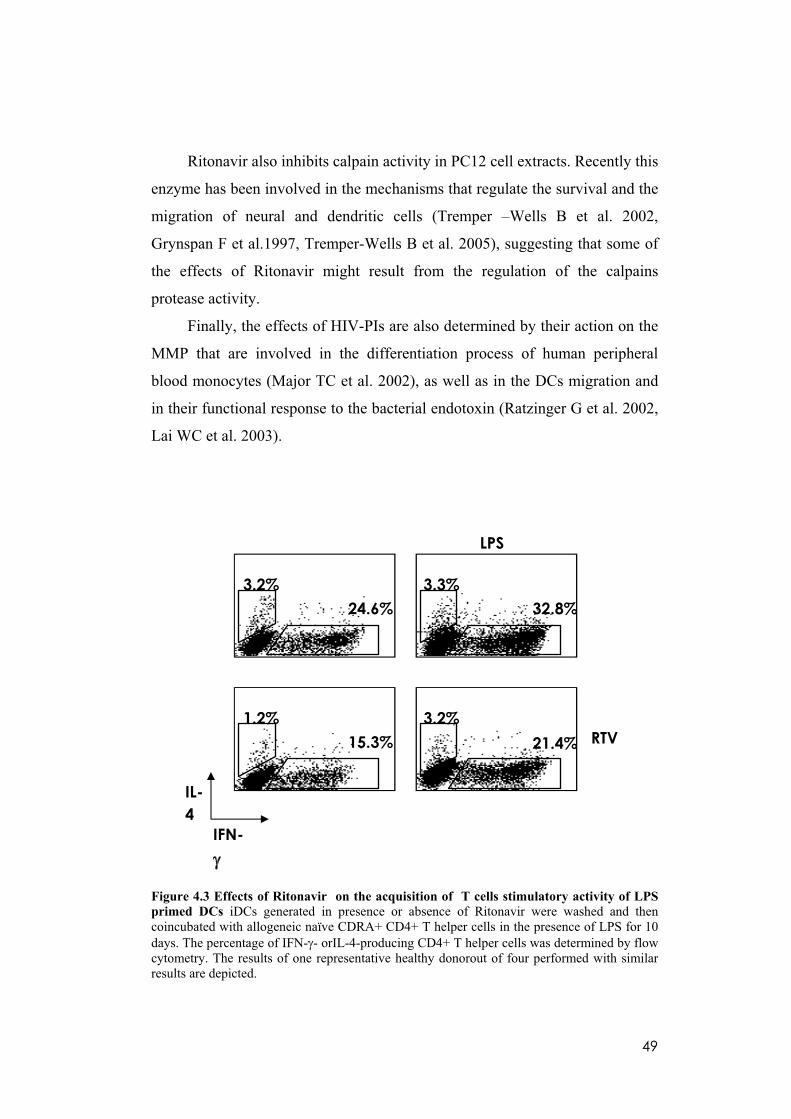
49
Ritonavir also inhibits calpain activity in PC12 cell extracts. Recently this
enzyme has been involved in the mechanisms that regulate the survival and the
migration of neural and dendritic cells (Tremper –Wells B et al. 2002,
Grynspan F et al.1997, Tremper-Wells B et al. 2005), suggesting that some of
the effects of Ritonavir might result from the regulation of the calpains
protease activity.
Finally, the effects of HIV-PIs are also determined by their action on the
MMP that are involved in the differentiation process of human peripheral
blood monocytes (Major TC et al. 2002), as well as in the DCs migration and
in their functional response to the bacterial endotoxin (Ratzinger G et al. 2002,
Lai WC et al. 2003).
Figure 4.3 Effects of Ritonavir on the acquisition of T cells stimulatory activity of LPS
primed DCs iDCs generated in presence or absence of Ritonavir were washed and then coincubated with allogeneic naïve CDRA+ CD4+ T helper cells in the presence of LPS for 10 days. The percentage of IFN- - orIL-4-producing CD4+ T helper cells was determined by flow cytometry. The results of one representative healthy donorout of four performed with similar results are depicted.
RTV
IFN-
IL-
4
LPS
3.2%
24.6%
3.3%
32.8%
1.2%
15.3%
3.2%
21.4%
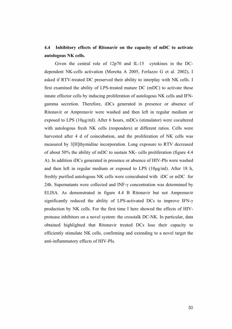
50
4.4 Inhibitory effects of Ritonavir on the capacity of mDC to activate
autologous NK cells.
Given the central role of 12p70 and IL-15 cytokines in the DC-
dependent NK-cells activation (Moretta A 2005, Ferlazzo G et al. 2002), I
asked if RTV-treated DC preserved their ability to interplay with NK cells. I
first examined the ability of LPS-treated mature DC (mDC) to activate these
innate effector cells by inducing proliferation of autologous NK cells and IFN-
gamma secretion. Therefore, iDCs generated in presence or absence of
Ritonavir or Amprenavir were washed and then left in regular medium or
exposed to LPS (10 g/ml). After 6 hours, mDCs (stimulator) were cocultered
with autologous fresh NK cells (responders) at different ratios. Cells were
harvested after 4 d of coincubation, and the proliferation of NK cells was
measured by 3[H]thymidine incorporation. Long exposure to RTV decreased
of about 50% the ability of mDC to sustain NK- cells proliferation (figure 4.4
A). In addition iDCs generated in presence or absence of HIV-PIs were washed
and then left in regular medium or exposed to LPS (10 g/ml). After 18 h,
freshly purified autologous NK cells were coincubated with iDC or mDC for
24h. Supernatants were collected and INF- concentration was determined by
ELISA. As demonstrated in figure 4.4 B Ritonavir but not Amprenavir
significantly reduced the ability of LPS-activated DCs to improve IFN-
production by NK cells. For the first time I here showed the effects of HIV-
protease inhibitors on a novel system: the crosstalk DC-NK. In particular, data
obtained highlighted that Ritonavir treated DCs lose their capacity to
efficiently stimulate NK cells, confirming and extending to a novel target the
anti-inflammatory effects of HIV-PIs.
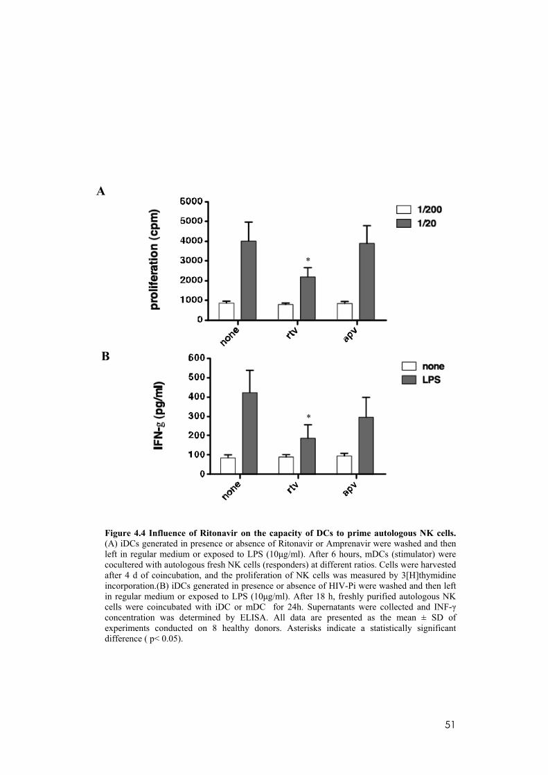
51
Figure 4.4 Influence of Ritonavir on the capacity of DCs to prime autologous NK cells.
(A) iDCs generated in presence or absence of Ritonavir or Amprenavir were washed and then left in regular medium or exposed to LPS (10 g/ml). After 6 hours, mDCs (stimulator) were cocultered with autologous fresh NK cells (responders) at different ratios. Cells were harvested after 4 d of coincubation, and the proliferation of NK cells was measured by 3[H]thymidine incorporation.(B) iDCs generated in presence or absence of HIV-Pi were washed and then left in regular medium or exposed to LPS (10 g/ml). After 18 h, freshly purified autologous NK cells were coincubated with iDC or mDC for 24h. Supernatants were collected and INF-concentration was determined by ELISA. All data are presented as the mean ± SD of experiments conducted on 8 healthy donors. Asterisks indicate a statistically significant difference ( p< 0.05).
*
*
A
B
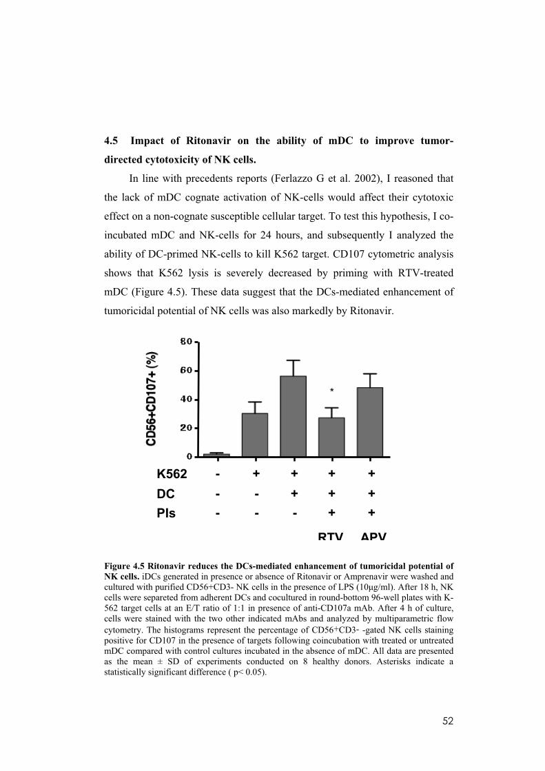
52
4.5 Impact of Ritonavir on the ability of mDC to improve tumor-
directed cytotoxicity of NK cells.
In line with precedents reports (Ferlazzo G et al. 2002), I reasoned that
the lack of mDC cognate activation of NK-cells would affect their cytotoxic
effect on a non-cognate susceptible cellular target. To test this hypothesis, I co-
incubated mDC and NK-cells for 24 hours, and subsequently I analyzed the
ability of DC-primed NK-cells to kill K562 target. CD107 cytometric analysis
shows that K562 lysis is severely decreased by priming with RTV-treated
mDC (Figure 4.5). These data suggest that the DCs-mediated enhancement of
tumoricidal potential of NK cells was also markedly by Ritonavir.
Figure 4.5 Ritonavir reduces the DCs-mediated enhancement of tumoricidal potential of
NK cells. iDCs generated in presence or absence of Ritonavir or Amprenavir were washed and cultured with purified CD56+CD3- NK cells in the presence of LPS (10 g/ml). After 18 h, NK cells were separeted from adherent DCs and cocultured in round-bottom 96-well plates with K-562 target cells at an E/T ratio of 1:1 in presence of anti-CD107a mAb. After 4 h of culture, cells were stained with the two other indicated mAbs and analyzed by multiparametric flow cytometry. The histograms represent the percentage of CD56+CD3- -gated NK cells staining positive for CD107 in the presence of targets following coincubation with treated or untreated mDC compared with control cultures incubated in the absence of mDC. All data are presented as the mean ± SD of experiments conducted on 8 healthy donors. Asterisks indicate a statistically significant difference ( p< 0.05).
*
K562
DC
PIs
- + + + +
- - + + +
- - - + +
RTV APV

53
4.6 Impaired NK cell-mediated killing of Ritonavir treated mDC.
I next asked if NK-cells dependent killing of autologous iDCs were
active in my system. This function depends on the engagement of NKp30 by
still-undefined cellular ligands expressed by DCs. Indeed Cr 51+ release assay
showes that non treated autologous iDC are efficiently killed by NK cells in an
NKp30-dependent manner, as demonstrated by the ability of the specific mAb
masking NKp30 (F252) but not of the specific mAb masking NKG2D
(BAT221) to inhibit the NK killing activity (Figure 4.6 A). As expected
untreated mDCs that, upregulate MHC-class I expression become essentially
resistant to NK cells (Figure 4.6 B).
On the contrary, the treatment with Ritonavir increases the susceptibility
to NK-cells mediated lysis of mDCs whereas iDC lysis was high irrespective to
the treatment (Figure 4.6 A and B). This apparent paradox might be explained
by the fact that RTV-treated DC failed to fully mature in response to LPS and
even upon its stimulation RTV-treated DCs fail to express sufficient amounts
of HLA-class I molecules that would protect from NK-mediated lysis (Ferlazzo
G 2003).
All this set of results suggest that RTV can act on DC to disrupt their
crosstalk with NK cells, leading to a deficient proliferation and activation. In
addition, the major sensitivity to NK cells dependent lysis may lead to DC
deficiency, thus amplifying the inhibitory effect on the adaptive immune
response. These observations should be considered not only with respect to
immunomodulatory aspects of HIV-1 treatment, but also to design new
therapies for other inflammatory conditions that often degenerate in cancer.

54
Figure 4.6 Impaired NK cell-mediated killing of Ritonavir treated mDC(A) Autologous iDC cytolysis exerted by rIL-2–activated NK cells purified from a healthy donor . (B) Autologous mDC cytolysis exerted by rIL-2–activated NK cells purified from a healthy donor NK cells were incubated either in the absence (baseline lysis) or presence of a specific mAb masking NKp30 (F252) or NKG2D (BAT221). The NK cell/DC ratio in all experiments was 10:1. All data are presented as the mean ± SD of experiments conducted on 8 healthy donors. Asterisks indicate a statistically significant difference ( p< 0.05).
iDCA)
B)
*
mDC
*
mDC

55
4.7 Ritonavir reduces the surface level of the inhibitory molecule ILT3
and upregulates that of the costimulatory molecule CD86 in
established TAM.
Besides investigating the ability of DCs to sustain the pro-inflammatory
circuits predisposing to cancer, I also evaluated whether RTV and APV may
reverse the immunosuppressive properties of Tumor-associated macrophages
(TAM), that are the major players of the cancer-related inflammation. To this
purpose, I first tested RTV and APV for their ability to restore
immunostimulatory molecule expression on established TAM. TAM were
stimulated for 48 hr with LPS, and then CD86 expression was analyzed.
As a result, RTV- as well as APV-treated TAM recovered the ability to
express higher levels of CD86 in response to LPS (Figure 4.7 A). Opposingly,
analysis of surface molecules by flow cytometry also revealed that RTV-
treated TAM expressed lower levels of the cell surface inhibitory molecule
ILT3 than untreated TAM (Figure 4.7 B). Also the treatment with APV
decreased ILT3 expression by TAM, but to a lower extent than RTV.
Up to now little is known about the regulation of ILT3 gene and protein
expression. It is selectively expressed by myeloid antigen presenting cells such
as monocytes, macrophages and dendritic cells, in which it displays a dual
function of inhibitory receptor and antigen-capturing molecule. (Cella M et al.
1997). Recent data underline the critical role of ILT3 in the control of
inflammation because the silencing of ILT3 expression in DC increase TLR
responsiveness to their specific ligand as reflected in increased synthesis and
secretion of proinflammatory cytokines (Chang CC et al. 2009). I can’t
hypothesize the mechanisms by which RTV might reduce ILT3 surface
expression, however the reduction of its level by RTV really correlates with an
antinflammatory condition (see later results). ILT3 signaling results in the
inhibition of NF-kappaB pathway and of the transcription of costimulatory
molecules (Suciu-Foca N and Cortesini R, 2007). On the other hand activation
stimuli such as inflammatory conditions downregulate inhibitory ILT3 receptor
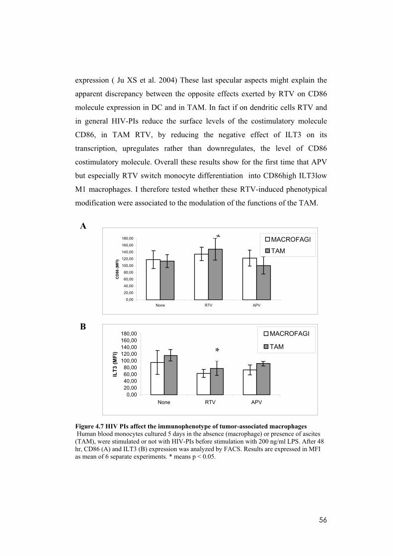
56
expression ( Ju XS et al. 2004) These last specular aspects might explain the
apparent discrepancy between the opposite effects exerted by RTV on CD86
molecule expression in DC and in TAM. In fact if on dendritic cells RTV and
in general HIV-PIs reduce the surface levels of the costimulatory molecule
CD86, in TAM RTV, by reducing the negative effect of ILT3 on its
transcription, upregulates rather than downregulates, the level of CD86
costimulatory molecule. Overall these results show for the first time that APV
but especially RTV switch monocyte differentiation into CD86high ILT3low
M1 macrophages. I therefore tested whether these RTV-induced phenotypical
modification were associated to the modulation of the functions of the TAM.
0,00
20,00
40,00
60,00
80,00
100,00
120,00
140,00
160,00
180,00
None RTV APV
CD
86
(M
FI)
MACROFAGI
TAM
0,00
20,00
40,00
60,00
80,00
100,00
120,00
140,00
160,00
180,00
None RTV APV
ILT
3 (
MF
I)
MACROFAGI
TAM
Figure 4.7 HIV PIs affect the immunophenotype of tumor-associated macrophages
Human blood monocytes cultured 5 days in the absence (macrophage) or presence of ascites (TAM), were stimulated or not with HIV-PIs before stimulation with 200 ng/ml LPS. After 48 hr, CD86 (A) and ILT3 (B) expression was analyzed by FACS. Results are expressed in MFI as mean of 6 separate experiments. * means p < 0.05.
A
B
**
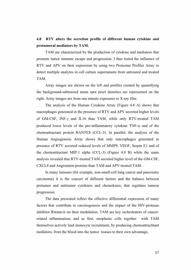
57
4.8 RTV alters the secretion profile of different human cytokine and
protumoral mediators by TAM.
TAM are characterized by the production of cytokine and mediators that
promote tumor immune escape and progression. I thus tested the influence of
RTV and APV on their expression by using two Proteome Profiler Array to
detect multiple analytes in cell culture supernatants from untreated and treated
TAM.
Array images are shown on the left and profiles created by quantifying
the background-subtracted mean spot pixel densities are represented on the
right. Array images are from one minute exposures to X-ray film.
The analysis of the Human Cytokine Array (Figure 4.8 A) shows that
macrophages generated in the presence of RTV and APV secreted higher levels
of GM-CSF, INF- and IL16 than TAM, while only RTV-treated TAM
produced lower levels of the pro-inflammatory cytokine TNF- and of the
chemoattractant protein RANTES (CCL-5). In parallel, the analysis of the
Human Angiogenesis Array shows that only macrophages generated in
presence of RTV secreted reduced levels of MMP9, VEGF, Serpin E1 and of
the chemoattractant MIP-1 alpha (CCL-3) (Figure 4.8 B) while the same
analysis revealed that RTV-treated TAM secreted higher level of the GM-CSF,
CXCL8 and Angiostatin proteins than TAM and APV-treated TAM.
In many tumours (for example, non-small-cell lung cancer and pancreatic
carcinoma) it is the concert of different factors and the balance between
protumor and antitumor cytokines and chemokines, that regulates tumour
progression.
The data presented reflect the effective differential expression of many
factors that contribute to carcinogenesis and the impact of the HIV-protease
inhibitor Ritonavir on their modulation. TAM are key orchestrators of cancer-
related inflammation, and as first, neoplastic cells together with TAM
themselves actively lead monocyte recruitment, by producing chemoattrachtant
mediators, from the blood into the tumor tissues to their own advantage,

58
Several “anti-macrophage” approaches are under evaluation currently.
Interesting observations come from studies performed with chemokines and
chemokine receptors as anti-cancer targets. For instance, in a breast cancer
murine model, malignant cells recruit macrophages via the chemokine
RANTES (CCL5), and treatment of murine breast cancers with Met-CCL5
(receptor antagonist) leads to a decreased number of infiltrating macrophages
associated with a significantly reduced tumor size (Robinson et al. 2003). From
the literature it is clear that protease inhibitors therapy reduced the high
production of inflammatory cytokines (TNF- alpha, MIP-1alpha, MIP-1beta,
RANTES, and INF-gamma) (De Luca A et al. 2000, Tovo PA 2000) in patients
with more advanced HIV infection vivo. All this support the observations done
in my experimental model, where ritonavir exerts an antitumor function by
reducing the protein levels of inflammatory molecules linked to the recruitment
of macrophages such as RANTES (CCL-5), MIP-1 alpha (CCL-3) and VEGF
as well as that of TNF-alpha which exert a key role in the inflammatory
circuits supporting tumor growth and progression. However, I also found an
increase rather than a decrease in the level of the other pro-inflammatory
cytokine, INF-gamma. This is just in apparent contrast with the antitumour
potential of Ritonavir. It is enough to think that blood monocytes
differentiating in the presence of INF-gamma mature into M1-polarized cells
(classically activated macrophages) that offers defence against bacteria, tumor
suppression and immuno-stimulation (Solinas G et al. 2009).
TAM contribute to tumor progression also by producing several factors
which enhance neo-angiogenesis (VEGF), and the dissolution and remodeling
of the interstitial matrix (metalloproteases, MMPs). I then investigated the
effect of RTV and APV on the detected ELISA VEGF and MMP9. (see next
results). On the other hand this process is counteracted by the production of
factors that limit the tumour spread like endostatin. My data reveale that RTV
reduces the protein levels of VEGF and MMP9, increasing that of the
endostatin that is the most potent inhibitor of tumor anngiogenesis.
59
Finally, data showed that RTV can also affect the protein level of PAI-
1/Serpin E1, whose expression is increased in primary tumor tissue of breast
cancer patients, and correlate with tumor aggressiveness and poor clinical
outcome (Annecke K et al. 2008).
Figure 4.8. The human Cytokine and Angiogenesis arrays detects multiple analytes in TAM culture supernates generated in presence or absence of the indicated HIV-Pis. Array images are shown on the left and profiles created by quantifying the background-subtracted mean spot pixel densities are identified on the right. Array images are from one minute exposures to X-ray film.
Human Cytokine Array
0
1000
2000
3000
4000
5000
6000
7000
8000
9000
10000
POS
CTR C
5a
CD40
liga
nd
GM
-CSF
CXC
L1
CCL1
CD54
Type
II-IN
F
IL1-
ra IL6
CXC
L8IL
13IL
16
CXC
L10
CXC
L11
CCL2 M
IF
CCL3
/ MIP
-1al
pha
CCL4
/MIP
-1 b
eta
Serpin
E1/
PAI
RAN
TES
TNF-
alph
a
Me
an
Pix
el
De
ns
ity
None
Ritonavir
Amprenavir
1 2 3 4 5 6 7 8 9 10
11
12
13
14
15
16
17
18 19
20
A
BCDE
APVlps
RTV lps
None
lps
1 2 3 4 5 6 7 8 9 10
11
12
13
14
15
16
17
18 19
20
A
BCDE
1 2 3 4 5 6 7 8 9 10
11
12
13
14
15
16
17
18 19
20
A
BCDE
A
BCDE
APVlps
RTV lps
None
lps
Human Angiogenesis Array
0
1000
2000
3000
4000
5000
6000
7000
8000
9000
10000
11000
12000
13000
14000
POS C
TR
ANGIO
GENIN
PLA
SMIN
OGEN
CXCL16
CD26
ENDO
STATIN
GM
-CSF
IGFBP
-1
IGFBP
-2
IGFBP
-3
CXCL8
CCL2
HB-E
GF
MIP
-1ALPH
A
MM
P-9
PEN
TRAXIN
3
PLATE
LET F
ACTO
R 4
PROLA
CTI
N
SER
PIN E
1
TIMP-1
uPA
VEG
F
THROM
BOSPONDIN
-1
Mean
Pix
el D
en
sit
y
NONE
RITONAVIR
AMPRENAVIR
AMP lps
None
lps
1 2 3 4 5 6 7 8 9 10 11
12
13
14
15
16 17
18
19
20
ABCDE
21
22
23
24
F
RTVlps
AMP lps
None
lps
AMP lps
None
lps
1 2 3 4 5 6 7 8 9 10 11
12
13
14
15
16 17
18
19
20
ABCDE
21
22
23
24
F
RTVlps
1 2 3 4 5 6 7 8 9 10 11
12
13
14
15
16 17
18
19
20
ABCDE
21
22
23
24
F
1 2 3 4 5 6 7 8 9 10 11
12
13
14
15
16 17
18
19
20
ABCDE
1 2 3 4 5 6 7 8 9 10 11
12
13
14
15
16 17
18
19
20
ABCDE
ABCDE
21
22
23
24
F
RTVlps
A
B
60
4.9 RTV-treated TAM lose their protumoral properties.
To confirm the ability of RTV and APV to affect TAM-like cell
functions, I next focused the attention on specific mediators seen modulated
before and now assayed by ELISA in the cell-free supernatants. Thus, TAM
generated in presence or absence of RTV and APV were stimulated for 48 hr
with 200 ng/ml LPS and then MMP-9 (A), VEGF (B) and GM-CSF (C) were
quantified in supernatants. Data showed that the macrophages generated in the
presence of RTV secreted lower levels of MMP9 and VEGF than TAM and
APV-treated TAM while RTV enhanced that of the immunostimulatory
cytokine GM-CSF (Figure 4.9).
The data below confirmed the previous observations and contribute to
better define the anti-angiogenic and anti-tumour activity of the Human
Immunodeficiency Type-1 Protease Inhibitors, Ritonavir.
GM-CSF might be relevant to cancer patients in two broad areas: first, it
drives hematopoietic precursor cells to mature granulocytes, macrophages, or
dendritic cells (Wognum et al. 1994) and for this it is used clinically to
accelerate bone marrow recovery and to increase the production of white blood
cells to facilitate host defense and second, because of its pletora of effects on
cancer cells. If on one hand it is reported that GM-CSF is aberrantly expressed
in a number of different solid tumors (for example osteosarcoma (Rochet N et
al, 1999), gliomas (Mueller MM et al., 1999) and pulmonary adenocarcinoma
(Wislez M et al., 2001), where it correlates with an enhanced and metastatic
potential and where it increases the number of granulocytes and macrophages
in the tumor vicinity (Obermueller E et al., 2004) as well as that of the
immunosuppressive immature myeloid cells (Serafini P, 2004); on the other
hand recent data suggest that GM-CSF reduces VEGF activity by inducing
secretion of the soluble form of VEGFR-1 by human monocytes/macrophages
and thus by reducing biologically active VEGF available for angiogenesis
(Eubank TD et al., 2004). Because Ritonavir increased the levels of GM-CSF
produced by TAM, this result can give more details on the unpredicted anti-
61
tumor actions of this class of compounds, suggesting that Ritonavir in
particular is effective in inhibiting tumour-associated angiogenesis and tumour
cell invasion. This statement is corroborated also by the finding that in my
experimental model RTV reduced the release by TAM of the angiogenic and
monocyte chemoattrachtant factor VEGF and of the metalloprotease, MMP-9
which is involved in the degradation and remodelling of the matrix but also in
the modulation of local inflammation (Sgadari C et al. 2002, Lopez et al.
2000); that could explain the ability of Ritonavir to reduce a set of
inflammatory cytokine by TAM but also by dendritic cell. Data presented in
the literature confirm that two other HIV-PIs, Indinavir and Saquinavir,
effectively reduced the number of neo-formed vessels in a murine model of
Kaposi Sarcoma (Sgadari C et al. 2002). HIV-Pis are also effective in
inhibiting tumor-associated angiogenesis and tumor cell invasion in other
xenograft human tumor models, including lung and breast adenocarcinoma and
tumor of haematopoietic cell origin such as myelomonocytic or T-cell
leukaemia and Burkitt Lymphoma (Monini P et al. 2003). In a separate set of
experiments it is also reported that Saquinavir and Indinavir affect pathways
involved in cell invasion and MMP activity, particularly MMP-2 proteolytic
activation. (Sgadari C et al. 2002). The data shown extend the anti-
angiogenetic activity of the HIV-PIs also to the Ritonavir, and indicated for the
first time the specific cell type, TAM, a critical supplier of mediators to the
carcinogenesis pathway, that is the target of the action of the drug.

62
Figure 4.9 Ritonavir treated TAM loose their protumoral properties. TAM generated in presence or absence of the indicated HIV-PIs were stimulated 48 hr with 200 ng/ml LPS. MMP9 (a),VEGF (b) and GM-CSF (c) were quantified by ELISA in the cell-free supernatants.
0,00
500,00
1000,00
1500,00
2000,00
2500,00
TAM none TAM RTV TAM AMP TAM none
LPS
TAM RTV
LPS
TAM AMP
LPS
MM
P-9
Concentr
ati
(ng/m
l)
0,00
50,00
100,00
150,00
200,00
250,00
300,00
350,00
400,00
450,00
TAM none TAM RTV TAM AMP TAM none
LPS
TAM RTV
LPS
TAM AMP
LPS
VE
GF C
oncentr
ation (pg
*
*
A
B
C
0,00
1000,00
2000,00
3000,00
4000,00
5000,00
6000,00
7000,00
8000,00
TAM none TAM RTV TAM AMP TAM none
LPS
TAM RTV
LPS
TAM AMP
LPS
GM
-CS
F (
pg
/ml)
*
0,00
1000,00
2000,00
3000,00
4000,00
5000,00
6000,00
7000,00
8000,00
TAM none TAM RTV TAM AMP TAM none
LPS
TAM RTV
LPS
TAM AMP
LPS
GM
-CS
F (
pg
/ml)
*
63
5. CONCLUSIONS
A plethora of evidences identify the inflammatory response as a key
component of mechanisms responsible for cell transformation, tumour growth
and metastatic process, in human cancer. Thus, it urges to identify novel
therapies aimed to revert the inflammatory status, and subvert
immunosuppressive mechanisms operating in established cancer lesions.
In the present study, I have identified PIs used in the therapy of HIV-1
infection, as a promising class drugs with unexpected immunomodulatory
activity. I demonstrated that PIs, and in particular ritonavir, interfere with
differentiation programs of monocyte by modulating their ability to generate
DC, and most notably, TAM.
The findings I have generated in the first part of the present study
document the differential ability of PIs to affect phenotype and functionality of
immature DC. Ritonavir has been identified as the PIs with the most
remarkable immunomodulatory activity: DC generated in the presence of this
drug fail to differentiate, secrete lower amount of pro-inflammatory cytokines,
and lack the ability to polarize T-cells toward a Th1 phenotype. Ritonavir also
impair DC-NK crosstalk, by reducing the ability of DCs to promote secretion
of IFN-g, and cytotoxicity of NK cells.
The results obtained in the second part of my study, highlight for the first
time the ability of ritonavir to interfere with phenotype and functions of
monocyte derived TAM, by promoting the expression of co-stimulatory
molecules, and decreasing the level of immunosuppressive ones. In parallel,
TAM generated in the presence of ritonavir secrete lower level of mediators
promoting tumor growth.
Altogether, these findings identify the differentiation programs of human
monocytes as relevant targets of PIs activity. In this context, the remarkable
property of certain PIs to modulate phenotypes and functionalities of DC, and
64
most notably TAM, might open novel perspectives for immune-intervention
aimed to manipulate the cancer inflammatory milieu.
Finally the anti-angiogenic, anti-tumour and anti-inflammatory effects of
PIs, their relatively low toxicity and the large body of data on their
pharmacokinetics and tissue distribution, make them a suitable candidate for
their rapid clinical ‘repositioning’ in the oncological field: in combination with
the traditional antiblastic therapies, they might significantly contribute to the
achievement of improved clinical goals without requiring imponent additional
research efforts.

65
6. ACKNOWLEDGMENTS
I thank Prof. Guido Rossi for giving me the opportunity to join his group, and
for supporting me during the last period of my Doctorate.
Thanks to Prof. Giancarlo Vecchio, who allowed me to attend the doctorate
program in Molecular Oncology and Endocrinology under his supervision.
Thanks to Prof. Luigi Racioppi who transmitted to me the love for science.
A special thank to Doctor Maddalena Illario, who followed my work in the last
period of my Doctorate, and helped me during the development of the
experiments and the elaboration of this thesis. I’m grateful to her enthusiasm,
and to her confidence in my ability.
Thanks to all my colleagues of the Rossi’s and the Zappacosta’s lab for their
friendship. In particular, I would like to thank Doctor Maria Letizia Giardino
Torchia, whose collaboration was fundamental for this thesis, and I would like
to thank also Laura Vitiello for her teachings, and for being always there for
me.
I’m really grateful to Doctor Domenico Mavilio for the opportunity he gave me
to spend three months in his laboratory of Immunoregulation at National
Institutes of Allergy and Infectious Diseases (NIAID/National Institutes of
Health). I thank him for his teachings as well as for his criticisms, and for
allowing me to work in a very stimulating scientific environment.
Many thanks to all my colleagues and friends from Bethesda, who made my
stay the US really unforgettable.
I cannot end without thanking my family for the constant encouragement and
love I have relied on throughout my life.
Thank to my father and my mother, for supporting my dreams and aspirations;

66
Thanks to my brother Vincenzo for his criticism and support.
Thanks to my husband Gaetano without whose love, patience, encouragement and editing assistance, I would not have finished this thesis.
Lastly, and most importantly I wish to thank my son, Stefano for the revolution he brought into my life. Now you don’t understand why I had to be away some times, but some day you will. The success of finishing this thesis is my first present to my son.
I thank God for standing by me
67
7. REFERENCES
Akira S, Uematsu S and Takeuchi O. Pathogen recognition and innate
immunity. Cell 2006; 124:783–801.
Allavena P, Sica A, Garlanda C, Mantovani A. The Yin-Yang of tumor-
associated macrophages in neoplastic and immune surveillance. Immunol. Rev.
2008; 222:155–161.
Allavena P, Sica A,Vecchi A, Locati M, Sozzani S, Mantovani A. The
chemokine receptor switch paradigm and dendritric cell migration: its
significance in tumor tissues. Immunol. Rev. 2000; 177:141–49.
Ambs S, Hussain SP, Marrogi AJ, Harris CC. Cancer-prone oxyradical
overload disease. IARC Sci Publ 1999; 150:295–302.
Annecke K, Schmitt M, Euler U, Zerm M, Paepke D, Paepke S, von
Minckwitz G, Thomssen C, Harbeck N. uPA and PAI-1 in breast cancer:
review of their clinical utility and current validation in the prospective NNBC-
3 trial. Adv Clin Chem. 2008; 45:31-45. Review.
Badgwell B, Parihar R, Magro C, Dierksheide J, Russo T, Carson WE
3rd.. Natural killer cells contribute to the lethality of a murine model of
Escherichia coli infection. Surgery 2000; 132:205–212.
Baek KH, Shin HJ, Yoo JK, Cho JH, Choi YH, Sung YC, McKeon F,
Lee CW. p53 deficiency and defective mitotic checkpoint in proliferating T
lymphocytes increase chromosomal instability through aberrant exit from
mitotic arrest. J. Leukoc. Biol. 2003; 73:850–861.
Balkwill F, Mantovani A. Inflammation and cancer: back to Virchow?.
Lancet 2001 Feb ; 17:539-45.
Balkwill F. Cancer and the chemokine network. Nat. Rev. Cancer 2004;
4:540–550.
68
Banchereau J, Briere F, Caux C, Davoust J, Lebecque S, Liu YJ ,
Pulendran B, Palucka K. Immunobiology of dendritic cells. Annu Rev.
Immunol. 2000; 18: 767–811.
Benahmed M, Meresse B, Arnulf B, Barbe U, Mention JJ, Verkarre V,
Allez M, Cellier C, Hermine O, Cerf-Bensussan N. Inhibition of TGF-beta
signaling by IL-15:A new role for IL-15 in the loss of immune homeostasis in
celiac disease. Gastroenterology 2007; 132:994–1008.
Bleharski JR, Kiessler V, Buonsanti C, Sieling PA, Stenger S, Colonna
M, and Modlin RL. A role for triggering receptor expressed on myeloid cells-1
in host defense during the early- induced and adaptive phases of the immune
response. J. Immunol. 2003 170, 3812–3818.
Borrello MG, Alberti L, Fischer A, Degl'innocenti D, Ferrario C,
Gariboldi M, Marchesi F, Allavena P, Greco A, Collini P, Pilotti S, Cassinelli
G, Bressan P, Fugazzola L, Mantovani A, Pierotti MA. Induction of a
proinflammatory program in normal human thyrocytes by the RET/PTC1
oncogene. Proc Natl Acad Sci U S A. 2005 Oct 11; 102(41):14825-30.
Bottino C, Castriconi R, Moretta L, Moretta A. Cellular ligands of
activating NK receptors. Trends Immunol. 2005; 26: 221–226.
Buentke E, Heffler LC, Wilson JL, Wallin RP, Löfman C, Chambers
BJ, Ljunggren HG, Scheynius A. Natural killer and dendritic cell contact in
lesional atopic dermatitis skin – Malassezia-influenced cell interaction. J.
Invest. Dermatol. 2002; 119: 850–857.
Calmels S, Hainaut P, Ohshima H. Nitric oxide induces conformational
and functional modifications of wild-type p53 tumor suppressor protein.
Cancer Res. 1997; 57:3365–3369.
Cella M, Döhring C, Samaridis J, Dessing M, Brockhaus M,
Lanzavecchia A, Colonna M. A novel inhibitory receptor (ILT3) expressed on
monocytes, macrophages, and dendritic cells involved in antigen processing. J
Exp Med. 1997 May 19; 185(10):1743-51.
69
Chan CW, Housseau F. The 'kiss of death' by dendritic cells to cancer
cells. Cell Death Differ. 2008 Jan;15(1):58-69.
Chang CC, Liu Z, Vlad G, Qin H, Qiao X, Mancini DM, Marboe CC,
Cortesini R, Suciu-Foca N. Ig-like transcript 3 regulates expression of
proinflammatory cytokines and migration of activated T cells. J Immunol.
2009 May 1; 182(9):5208-16.
Chen Y, Wei H, Sun R, Dong Z, Zhang J, Tian Z. Increased susceptibility
to liver injury in hepatitis B virus transgenic mice involves NKG2D-ligand
interaction and natural killer cells. Hepatology 2007; 46,:706–715.
Chettibi S & Ferguson MW. In: Gallin JI & Snyderman R, editors.
Inflammation: Basic Principles and Clinical Correlates. Philadelphia:
Lippincott, Williams, and Wilkins; 1999. p. 865–881.
Cobbs CS, Whisenhunt TR, Wesemann DR, Harkins LE, Van Meir EG,
Samanta M. Inactivation of wild type p53 protein function by reactive oxygen
and nitrogen species in malignant glioma cells. Cancer Res. 2003; 63:8670–
8673.
Colotta F, Allavena P, Sica A, Garlanda C, Mantovani A.Cancer-related
inflammation, the seventh hallmark of cancer: links to genetic instability.
Carcinogenesis 2009 Jul; 30(7):1073-81.
Cortesini R. Pancreas cancer and the role of soluble immunoglobulin-like
transcript 3 (ILT3). JOP 2007 Nov 9; 8(6):697-703.
Coussens LM, Werb Z. Inflammation and cancer. Nature 2002; 420:860–
867.
Crosby ME, Kulshreshtha R, Ivan M, Glazer PM. MicroRNA regulation
of DNA repair gene expression in hypoxic stress. Cancer Res. 2009;69:1221–
1229.
De Luca A, Giancola ML, Cingolani A, Ammassari A, Murri R, Antinori
A. Circulating levels and ex vivo production of beta-chemokines, interferon
gamma, and interleukin 2 in advanced human immunodeficiency virus type 1
70
infection: the effect of protease inhibitor therapy.AIDS Res Hum Retroviruses.
2000 Jun 10;16(9):835-43.
de Matos CT, Berg L, Michaëlsson J, Felländer-Tsai L, Kärre K,
Söderström K. Activating and inhibitory receptors on synovial fluid natural
killer cells of arthritis patients: role of CD94/NKG2A in control of cytokine
secretion. Immunology 2007; 122: 291–301.
Degli-Esposti MA & Smyth MJ. Close encounters of different kinds:
dendritic cells and NK cells take centre stage. Nat. Rev. Immunol 2005; 5:
112–124.
Dobrovolskaia MA, Kozlov SV. Inflammation and cancer: when NF-
kappaB amalgamates the perilous partnership. Curr. Cancer Drug Targets
2005; 5:325–344.
Dunn C, Brunetto M, Reynolds G, Christophides T, Kennedy PT,
Lampertico P, Das A, Lopes AR, Borrow P, Williams K, Humphreys E, Afford
S, Adams DH, Bertoletti A, Maini MK. Cytokines induced during chronic
hepatitis B virus infection promote a pathway for NK cell-mediated liver
damage. J. Exp. Med. 2007; 204: 667–680.
Ekbom A, Helmick C, Zack M, Adami H-O. Ulcerative colitis and
colorectal cancer. N. Engl. J. Med. 1990; 323:1228–33.
El-Omar EM, Carrington M, Chow WH, McColl KE, Bream JH, Young
HA, Herrera J, Lissowska J, Yuan CC, Rothman N, Lanyon G, Martin M,
Fraumeni JF Jr, Rabkin CS.. Interleukin-1 polymorphisms associated with
increased risk of gastric cancer. Nature 2000; 404: 398–402.
Endres MJ, Garlisi CJ, Xiao H, Shan L, Hendrick JA. The Kaposi’s
sarcoma-related herpes virus (KSHV)-encoded chemokine vMIP-1 is a specific
agonist for the CC receptor (CCR)8. J. Exp. Med. 1999;189:1993–98.
Eubank TD, Roberts R, Galloway M, Wang Y, Cohn DE, Marsh CB.
GM-CSF induces expression of soluble VEGF receptor-1 from human
monocytes and inhibits angiogenesis in mice. Immunity. 2004 Dec; 21(6):831-
42.
71
Ferlazzo G, Morandi B, D’Agostino A, Meazza R, Melioli G, Moretta A,
Moretta L. The interaction between NK cells and dendritic cells in bacterial
infections results in rapid induction of NK cell activation and in the lysis of
uninfected dendritic cells. Eur. J. Immunol. 2003; 33: 306–313.
Ferlazzo G, Tsang ML, Moretta L, Melioli G, Steinman RM, Münz C.
Human dendritic cells activate resting natural killer (NK) cells and are
recognized via the NKp30 receptor by activated NK cells. J Exp Med. 2002
Feb 4; 195(3):343-51.
Ferrero-Miliani L, Nielsen OH, Andersen PS, Girardin SE .Chronic
inflammation: importance of NOD2 and NALP3 in interleukin-1beta
generation". Clin. Exp. Immunol. February 2007;147(2): 227–35.
Fleisher AS, Esteller M, Harpaz N, Leytin A, Rashid A, Xu Y, Liang J,
Stine OC, Yin J, Zou TT, Abraham JM, Kong D, Wilson KT, James SP,
Herman JG, Meltzer SJ. Microsatellite instability in inflammatory bowel
disease-associated neoplastic lesions is associated with hypermethylation and
diminished expression of the DNA mismatch repair gene, hMLH1. Cancer Res.
2000; 60:4864–4868.
Flexner C. HIV-protease inhibitors. N Engl J Med 1998; 338: 1281–92.
Fox JG, Wang TC. Inflammation, atrophy, and gastric cancer. J. Clin.
Invest. 2007; 117:60–69.
Fricke I, Gabrilovich DI. Dendritic cells and tumor microenvironment: a
dangerous liaison. Immunol. Invest. 2006; 35(3-4):459-83.
Fritz JH, Ferrero RL, Philpott DJ and Girardin SE. Nod-like proteins in
immunity, inflammation and disease. Nat. Immunol. 2006; 7:1250–1257.
Gaedicke S, Firat-Geier E, Constantiniu O, Lucchiari-Hartz M,
Freudenberg M, Galanos C, Niedermann G. Antitumor effect of the human
immunodeficiency virus proteas inhibitor ritonavir: induction of tumor-cell
apoptosis associated with perturbation of proteasomal proteolysis. Cancer Res.
2002; 62:6901–6908.
72
Gearing AJ, Beckett P, Christodoulou M, Churchill M, Clements J,
Davidson AH, Drummond AH, Galloway WA, Gilbert R, Gordon JL, et al.
Processing of tumour necrosis factor- precursor by metalloproteinases. Nature
1994;370:555–557.
Golubkov VS, Strongin AY. Proteolysis-driven oncogenesis. Cell Cycle,
6:147–150.
Gorelik L & Flavell RA. Immune-mediated eradication of tumors
through the blockade of transforming growth factor- signaling in T cells.
Nature Med. 2001; 7:1118–1122.
Gruber A, Wheat JC, Kuhen KL, Looney DJ & Wong-Staal F.
Differential effects of HIV-1 protease inhibitors on dendritic cell
immunophenotype and function. J. Biol. Chem. 2001;276:47840–47843.
Grynspan F, Griffin WB, Mohan PS, Shea TB, Nixon RA. Calpains and
calpastatin in SH-SY5Y neuroblastoma cells during retinoic acid-induced
differentiation and neurite outgrowth: comparison with the human brain
calpain system. J Neurosci Res. 1997 May 1;48(3):181-91.
Haghnegahdar H, Du J, Wang D, Strieter RM, Burdick MD, Nanney LB,
Cardwell N, Luan J, Shattuck-Brandt R, Richmond A. The tumorigenic and
angiogenic effects of MGSSA/GRO proteins in melanoma. J. Leukoc. Biol.
2000; 67: 53–62.
Han J and Ulevitch RJ. Limiting inflammatory responses during
activation of innate immunity. Nat. Immunol. 2005; 6:1182–1189.
Hmadcha A, Bedoya FJ, Sobrino F, Pintado E. Methylation-dependent
gene silencing induced by interleukin 1beta via nitric oxide production. J. Exp.
Med. 1999;190:1595–1604.
Hodge DR, Peng B, Cherry JC, Hurt EM, Fox SD, Kelley JA, Munroe
DJ, Farrar WL. Interleukin 6 supports the maintenance of p53 tumor
suppressor gene promoter methylation. Cancer Res. 2005; 65:4673–4682.
Homey B, Muller A. & Zlotnik A. Chemokines: agents for the
immunotherapy of cancer? Nature Rev. Immunol. 2002; 2: 175–184.
73
Hsieh CS, SE Macatonia, CS Tripp, SF Wolf, A O'Garra, K M Murphy.
Development of TH1 CD4+ T cells through IL-12 produced by Listeria-
induced macrophages. Science 1993; 260: 547-549.
Hudson JD, Shoaibi MA, Maestro R, Carnero A, Hannon GJ, Beach DH.
A proinflammatory cytokine inhibits p53 tumor suppressor activity. J. Exp.
Med. 1999; 190:1375–1382.
Huntington ND, Puthalakath H, Gunn P, Naik E, Michalak EM, Smyth
MJ, Tabarias H, Degli-Esposti MA, Dewson G, Willis SN, Motoyama N,
Huang DC, Nutt SL, Tarlinton DM, Strasser A. Interleukin 15-mediated
survival of natural killer cells is determined by interactions among Bim, Noxa
and Mcl-1. Nat. Immunol. 2007; 8:856–863.
Hussain SP, Hofseth LJ, Harris CC. Radical causes of cancer. Nat. Rev.
Cancer 2003; 3:276–285.
In Cotran RS, Kumar V, Collins T, editors. Robbins Pathologic Basis of
Disease. 6th ed. Philadelphia: WB Saunders Company; 1999. p. 1330-1334.
Ishigami S, Aikou T, Natsugoe S, Hokita S, Iwashige H, Tokushige M,
Sonoda S. Prognostic value of HLA-DR expression and dendritic cell
infiltration in gastric cancer. Oncology 1998; 55: 65–69.
Ito A, Mukaiyama A, Itoh Y, Nagase H, Thogersen IB, Enghild JJ,
Sasaguri Y, Mori Y. Degradation of interleukin 1 by matrix
metalloproteinases. J. Biol. Chem. 1996; 271:14657–14660.
Iwamoto M, Shinohara H, Miyamoto A, Okuzawa M, Mabuchi H,
Nohara T, Gon G, Toyoda M, Tanigawa N. Prognostic value of tumor-
infiltrating dendritic cells expressing CD83 in human breast carcinomas. Int. J.
Cancer 2003; 104: 92–97.
Jaiswal M, LaRusso NF, Gores GJ. Nitric oxide in gastrointestinal
epithelial cell carcinogenesis: linking inflammation to oncogenesis. Am. J.
Physiol.Gastrointest. Liver Physiol. 2001; 281:G626–G34.
Janeway Jr CA, Medzhitov R. Innate immune recognition. Annu. Rev.
Immunol. 2002; 20: 197–216.
74
Ju XS, Hacker C, Scherer B, Redecke V, Berger T, Schuler G, Wagner
H, Lipford GB, Zenke M. Immunoglobulin-like transcripts ILT2, ILT3 and
ILT7 are expressed by human dendritic cells and down-regulated following
activation. Gene. 2004 Apr 28; 331:159-64.
Jung YJ, Isaacs JS, Lee S, Trepel J, Neckers L. IL-1beta-mediated up-
regulation of HIF-1alpha via an NFkappaB/COX-2 pathway identifies HIF-1 as
a critical link between inflammation and oncogenesis. FASEB J 2003; 17:
2115–2117.
Karin M and Greten FR. NF-kB: linking inflammation and immunity to
cancer development and progression. Nat. Rev. Immunol. 2005; 5:749–759.
Karin M. Nuclear factor-kB in cancer development and progression.
Nature 2006; 441:431–436.
Katchar K, Soderstrom K, Wahlstrom J, Eklund A & Grunewald J.
Characterisation of natural killer cells and CD56+ T-cells in sarcoidosis
patients. Eur. Respir. J. 2005; 26: 77–85.
Keane MP, Strieter RM. In:Mantovani A, editor. Chemokines: chemical
immunology: vol 72. Basel: Karger 1999;86–101.
Kelleher AD, Booth BL Jr, Sewell AK, Oxenius A, Cerundolo V,
McMichael AJ, Phillips RE, Price DA. Effects of retroviral protease inhibitors
on proteasome function and processing of HIV-derived MHC class I-restricted
cytotoxic T lymphocyte epitopes. AIDS Res. Hum. Retroviruses 2001;
17:1063–1066.
Kerr AR, Kirkham LA, Kadioglu A, Andrew PW, Garside P, Thompson
H, Mitchell TJ.Identification of a detrimental role for NK cells in
pneumococcal pneumonia and sepsis in immunocompromised hosts. Microbes
Infect. 2005; 7:845–852.
Kim-Schulze S, Scotto L, Vlad G, Piazza F, Lin H, Liu Z, Cortesini R,
Susciu-Foca N. Recombinant Ig-like transcript 3-Fc modulates T cell
responses via induction of Th anergy and differentiation of CD8+ T suppressor
cells. J Immunol. 2006; 176:2790-8.
75
Klesney-Tait J, Turnbull IR and Colonna M. The TREM receptor family
and signal integration. Nat. Immunol. 2006; 7:1266–1273.
Koshiji M, To KK, Hammer S, Kumamoto K, Harris AL, Modrich P,
Huang LE. HIF-1alpha induces genetic instability by transcriptionally
downregulating MutSalpha expression. Mol .Cell. 2005; 17:793–803.
Kovesdy CP, Kalantar-Zadeh K. Novel targets and new potential:
developments in the treatment of inflammation in chronic kidney disease.
Expert Opin. Investig. Drugs 2008 Apr;17(4):451-67.
Lai WC, Zhou M, Shankavaram U, Peng G, Wahl LM. Differential
regulation of lipopolysaccharide-induced monocyte matrix metalloproteinase
(MMP)-1 and MMP-9 by p38 and extracellular signal-regulated kinase 1/2
mitogen-activated protein kinases. J Immunol. 2003 Jun 15;170(12):6244-9.
Lambiase A, Normando EM, Vitiello L, Micera A, Sacchetti M, Perrella
E, Racioppi L, Bonini S, Bonini S. Natural killer cells in vernal
keratoconjunctivitis. Mol. Vis. 2007; 13: 1562–1567.
Langenkamp A, Messi M, Lanzavecchia A, Sallusto F. Kinetics of
dendritic cell activation: impact on priming of TH1, TH2 and nonpolarized T
cells. Nat. Immunol. 2000; 1: 311–316.
Lanier LL. NK cell recognition. Annu. Rev. Immunol. 2005; 23:225–274
Leach MW and Rennick DM. IL-12, but not IFNgamma, plays a major
role in sustaining the chronic phase of colitis in IL-10–deficient mice. J.
Immunol. 1998; 161:3143–3149.
Leek RD, Landers RJ, Harris AL, Lewis CE. Necrosis correlates with
high vascular density and focal macrophage infiltration in invasive carcinoma
of the breast. Br. J. Cancer 1999;79: 991–95.
Lochter A, Srebrow A, Sympson CJ, Terracio N, Werb Z, Bissell MJ.
Misregulation of stromelysin-1 expression in mouse mammary tumor cells
accompanies acquisition of stromelysin-1-dependent invasive properties. J.
Biol. Chem. 1997;272:5007–5015.
76
Long EO. Regulation of immune responses through inhibitory receptors.
Annu. Rev. Immunol. 1999; 17: 875–904.
Lopez D, Gil-Torregrosa BC, Bergmann C & Del VM. Sequential
cleavage by metallopeptidases and proteasomes is involved in processing HIV-
1 ENV epitope for endogenous MHC class I antigen presentation. J. Immunol.
2000;164:5070–5077.
Lu W & Andrieu JM. HIV protease inhibitors restore impaired T-cell
proliferative response in vivo and in vitro: a viral-suppression-independent
mechanism. Blood 2000;96:250–258.
Major TC, Liang L, Lu X, Rosebury W, Bocan TM. Extracellular matrix
metalloproteinase inducer (EMMPRIN) is induced upon monocyte
differentiation and is expressed in human atheroma. Arterioscler Thromb Vasc
Biol. 2002 Jul 1;22(7):1200-7.
Mantovani A, Allavena P, Sica A, Balkwill F. Cancer-related
inflammation. Nature 2008; 454:436–444.
Mantovani A, Sozzani S, Locati M, Allavena P, Sica A. Macrophage
polarization: Tumor-associated macrophages as a paradigm for polarized M2
mononuclear phagocytes. Trends Immunol. 2002; 23:549–55.
Mantovani A., Bottazzi B, Colotta F, Sozzani S, Ruco LP.The origin and
function of tumor-associated macrophages. Immunol. Today 1992; 13:265–
270.
Marshall HE, Merchant K, Stamler JS. Nitrosation and oxidation in the
regulation of gene expression. FASEB J 2000;14:1889–1900.
Martìn-Padura I, Mortarini R, Lauri D, Bernasconi S, Sanchez-Madrid F,
Parmiani G, Mantovani A, Anichini A, Dejana E. Heterogeneity in human
melanoma cell adhesion to cytokine activated endothelia cells correlates with
VLA-4 expression. Cancer Res. 1991; 51:2239–41.
Matos P, Collard JG, Jordan P. Tumor-related alternatively spliced Rac1b
is not regulated by Rho-GDP dissociation inhibitors and exhibits selective
downstream signaling. J. Biol. Chem. 2003; 278:50442–50448.
77
McQuibban GA, Gong JH, Tam EM, McCulloch CA, Clark-Lewis I,
Overall CM. Inflammation dampened by gelatinase A cleavage of monocyte
chemoattractant protein-3. Science 2000; 289:1202–1206.
Medzhitov R.Toll-like receptors and innate immunity. Nat. Rev.
Immunol.2001; 1:135–145.
Mention JJ, Ben Ahmed M, Bègue B, Barbe U, Verkarre V, Asnafi V,
Colombel JF, Cugnenc PH, Ruemmele FM, McIntyre E, Brousse N, Cellier C,
Cerf-Bensussan N. Interleukin 15: A key to disrupted intraepithelial
lymphocyte homeostasis and lymphomagenesis in celiac disease.
Gastroenterology 2003; 125:730–745.
Mirzaee V, Molaei M, Shalmani HM, Zali MR. Helicobacter pylori
infection and expression of DNA mismatch repair proteins. World J.
Gastroenterol. 2008; 14:6717–6721.
Monini P, Sgadari C, Toschi E, Barillari G, Ensoli B.Antitumour effects
of antiretroviral therapy . Nat. Rev. Cancer. 2004 Nov;4(11):861-75 .
Moretta A, Bottino C, Mingari MC, Biassoni R, Moretta L. What is a
natural killer cell? Nat . Immunol. 2002; 3: 6–8.
Moretta A, Bottino C, Vitale M, Pende D, Cantoni C, Mingari MC,
Biassoni R, Moretta L. Activating receptor and coreceptors involved in human
natural killer cell-mediated cytolysis. Annu. Rev. Immunol. 2001; 19: 197–
223.
Moretta A. Natural killer cells and dendritic cells: rendezvous in abused
tissues. Nat .Rev. Immunol. 2002; 2: 957–964.
Moretta A. The dialogue between human natural killer cells and dendritic
cells. Curr Opin Immunol. 2005 Jun; 17(3):306-11.
Moretta L, Ferlazzo G, Bottino C, Vitale M, Pende D, Mingari MC,
Moretta A. Effector and regulatory events during natural killer-dendritic cell
interactions. Immunol. Rev.2006; 214: 219–228.
Mueller MM, Herold-Mende CC, Riede D, Lange M, Steiner HH,
Fusenig NE. Autocrine growth regulation by granulocyte colony-stimulating

78
factor and granulocyte macrophage colony-stimulating factor in human
gliomas with tumor progression. Am J Pathol. 1999 Nov; 155(5):1557-67.
Navia MA, Fitzgerald PMD, McKeever BM, et al. Three-dimensional
structure of aspartyl protease from human immunodeficiency virus HIV-1.
Nature 1989; 337: 615–20.
Negus RPK, Stamp GWQ, Hadley J, Balkwill FR. Quantitative
assessment of the leucocyte infiltrate in ovarian cancer and its relationship to
the expression of C-C chemokines. Am. J. Pathol.1997; 150:723–34.
Obermueller E, Vosseler S, Fusenig NE, Mueller MM . Cooperative
autocrine and paracrine functions of granulocyte colony-stimulating factor and
granulocyte-macrophage colony-stimulating factor in the progression of skin
carcinoma cells. Cancer Res. 2004 Nov 1; 64(21):7801-12.
Ottaviani C, Nasorri F, Bedini C, de Pità O, Girolomoni G, Cavani A.
CD56brightCD16– NK cells accumulate in psoriatic skin in response to
CXCL10 and CCL5 and exacerbate skin inflammation. Eur. J. Immunol. 2006;
36:118–128.
Pajonk F, Himmelsbach J, Riess K, Sommer A & McBride WH. The
human immunodeficiency virus (HIV)-1 protease inhibitor saquinavir inhibits
proteasome function and causes apoptosis and radiosensitization in non
associated human cancer cells. Cancer Res. 2002; 62:5230–5235.
Pati S, Pelser CB, Dufraine J, Bryant JL, Reitz MS Jr, Weichold FF.
Antitumorigenic effects of HIV protease inhibitor ritonavir: inhibition of
Kaposi sarcoma. Blood 2002;99:3771–3779.
Rajagopalan H, Nowak MA, Vogelstein B, Lengauer C. The significance
of unstable chromosomes in colorectal cancer. Nat. Rev. Cancer 2003; 3:695–
701.
Ratzinger G, Stoitzner P, Ebner S, Lutz MB, Layton GT, Rainer C,
Senior RM, Shipley JM, Fritsch P, Schuler G, Romani N. Matrix
metalloproteinases 9 and 2 are necessary for the migration of Langerhans cells
79
and dermal dendritic cells from human and murine skin. J Immunol. 2002 May
1; 168(9):4361-71.
Robinson MJ, Sancho D, Slack EC, Leibundgut- Landmann S. and
Sousa CR. Myeloid C-type lectins in innate immunity. Nat. Immunol. 2006;
7:1258–1265.
Robinson SC, Scott KA, Wilson JL, Thompson RG, Proudfoot AE,
Balkwill FR. A chemokine receptor antagonist inhibits experimental breast
tumor growth. Cancer Res. 2003 Dec 1;63(23):8360-5.
Rochet N, Dubousset J, Mazeau C, Zanghellini E, Farges MF, de Novion
HS, Chompret A, Delpech B, Cattan N, Frenay M, Gioanni J. Establishment,
characterisation and partial cytokine expression profile of a new human
osteosarcoma cell line (CAL 72). Int J Cancer. 1999 Jul 19;82(2):282-5.
Rossi D & Zlotnik A. The biology of chemokines and their receptors.
Annu. Rev. Immunol. 2000; 18: 217–242.
Saccani A, Schioppa T, Porta C, Biswas SK, Nebuloni M, Vago L,
Bottazzi B, Colombo MP, Mantovani A, Sica A. p50 nuclear factor-kappaB
overexpression in tumor-associated macrophages inhibits M1 inflammatory
responses and antitumor resistance.Cancer Res. 2006 Dec 1;66(23):11432-40.
Samper E, Nicholls DG, Melov S. Mitochondrial oxidative stress causes
chromosomal instability of mouse embryonic fibroblasts. Aging Cell
2003;2:277–285.
Sandau KB, Faus HG, Brüne B. Induction of hypoxia-inducible-factor 1
by nitric oxide is mediated via the PI 3K pathway. Biochem. Biophys Res
Commun 2000; 278:263–267.
Scarpino S, Stoppacciaro A, Ballerini F, Marchesi M, Prat M, Stella MC,
Sozzani S, Allavena P, Mantovani A, Ruco LP.Papillary carcinoma of the
thyroid: hepatocyte growth factor (HGF) stimulates tumor cells to release
chemokines active in recruiting dendritic cells. Am. J. Pathol. 2000; 156:831–
837.
80
Schmidtke G, Holzhütter HG, Bogyo M, Kairies N, Groll M, de Giuli R,
Emch S, Groettrup M.How an inhibitor of the HIV-I protease modulates
proteasome activity. J Biol Chem. 1999 Dec 10; 274(50):35734-40.
Schonbeck U, Mach F & Libby P. Generation of biologically active IL-
1 by matrix metalloproteinases: a novel caspase-1-independent pathway of
IL-1 processing. J. Immunol. 1998; 161:3340–3346.
Serafini P, Carbley R, Noonan KA, Tan G, Bronte V, Borrello I. High-
dose granulocyte-macrophage colony-stimulating factor-producing vaccines
impair the immune response through the recruitment of myeloid suppressor
cells Cancer Res. 2004 Sep 1;64(17):6337-43.
Sgadari C, Barillari G, Toschi E, Carlei D, Bacigalupo I, Baccarini S,
Palladino C, Leone P, Bugarini R, Malavasi L, Cafaro A, Falchi M, Valdembri
D, Rezza G, Bussolino F, Monini P, Ensoli B. HIV protease inhibitors are
potent antiangiogenic molecules and promote regression of Kaposi sarcoma.
Nature Med. 2002;8:225–232.
Sgadari C, Monini P, Barillari G, Ensoli B. Use of HIV protease
inhibitors to block Kaposi's sarcoma and tumour growth. Lancet Oncol. 2003
Sep;4(9):537-47.
Shacter E, Weitzman SA. Chronic inflammation and cancer. Oncology
2002; 16:217–226.
Sheu BC, Hsu SM, Ho HN, Lien HC, Huang SC, Lin RH. A novel role of
metalloproteinase in cancer mediated immunosuppression. Cancer Res. 2001;
61:237–24.
Sica A, Saccani A, Bottazzi B, Polentarutti N, Vecchi A, van Damme J,
Mantovani A. Autocrine production of IL-10 mediates defective IL-12
production and NF-kB activation of tumor-associated macrophages. J.
Immunol. 2000; 164:762–67.
Sloand EM, Kumar PN, Kim S, Chaudhuri A, Weichold FF, Young NS.
Human immunodeficiency virus type 1 protease inhibitor modulates activation
81
of peripheral blood CD4+ T cells and decreases their susceptibility to apoptosis
in vitro and in vivo. Blood 1999; 94:1021–1027.
Solinas G, Germano G, Mantovani A, Allavena P. Tumor-associated
macrophages (TAM) as major players of the cancer-related inflammation..J
Leukoc Biol 2009; 86: 000–000 .
Sozzani S, Luini W, Bianchi G, Allavena P, Wells TN, Napolitano M,
Bernardini G, Vecchi A, D'Ambrosio D, Mazzeo D, Sinigaglia F, Santoni A,
Maggi E, Romagnani S, Mantovani A. The viral chemokine macrophage
inflammatory protein-II is a selective Th2 chemoattractant. Blood 1998;
92:4036–39.
Steinman RM, Hawiger D, Liu K, Bonifaz L, Bonnyay D, Mahnke K et
al. Dendritic cell function in vivo during the steady state: a role in peripheral
tolerance. Ann. NY Acad. Sci. 2003; 987:15–25.
Sternlicht MD, Lochter A, Sympson CJ, Huey B, Rougier JP, Gray JW,
Pinkel D, Bissell MJ, Werb Z. The stromal proteinase MMP3/stromelysin-1
promotes mammary carcinogenesis. Cell 1999; 98:137–146.
Sternlicht,M.D. et al. (2001) How matrix metalloproteinases regulate cell
behavior. Annu. Rev. Cell Dev. Biol. 2007;17:463–516.
Subklewe M, Sebelin-Wulf K, Beier C, Lietz A, Mathas S, Dörken B,
Pezzutto A. Dendritic cell maturation stage determines susceptibility to the
proteasome inhibitor bortezomib. Hum Immunol. 2007 Mar;68(3):147-55.
Epub 2007 Jan 4.
Suciu-Foca N, Cortesini R. Central role of ILT3 in the T suppressor cell
cascade. Cell Immunol. 2007 Jul; 248(1):59-67. Epub 2007 Oct 17. Review.
Talos F, Nemajerova A, Flores ER, Petrenko O, Moll UM. p73
suppresses polyploidy and aneuploidy in the absence of functional p53. Mol.
Cell. 2007; 27:647–659.
Tomasini R, Mak TW, Melino G. The impact of p53 and p73 on
aneuploidy andcancer. Trends Cell. Biol. 2008; 18:244–252.
82
Tovo PA. Highly active antiretroviral therapy inhibits cytokine
production in HIV-uninfected subjects. AIDS 2000;14:743–744.
Treilleux I, Blay JY, Bendriss-Vermare N, Ray-Coquard I, Bachelot T,
Guastalla JP, Bremond A, Goddard S, Pin JJ, Barthelemy-Dubois C, Lebecque
S. Dendritic cell infiltration and prognosis of early stage breast cancer. Clin
Cancer Res. 2004 Nov 15; 10(22):7466-74.
Tremper-Wells B, Mathur A, Beaman-Hall CM, Vallano ML .Trophic
agents that prevent neuronal apoptosis activate calpain and down-regulate
CaMKIV. J Neurochem. 2002 Apr;81(2):314-24.
Tremper-Wells B, Vallano ML .Nuclear calpain regulates Ca2+-
dependent signaling via proteolysis of nuclear Ca2+/calmodulin-dependent
protein kinase type IV in cultured neurons. J Biol Chem. 2005 Jan
21;280(3):2165-75 .
Tricot G. New insights into role of microenvironment in multiple
myeloma. Lancet 2000; 355: 248–50.
Trinchieri G. Biology of natural killer cells. Adv. Immunol. 1989; 47,
187–376.
Trinchieri G. Interleukin-12 and the regulation of innate resistance and
adaptive immunity. Nat. Rev. 2003; 3:133–146.
Tsujitani S, Kakeji Y, Watanabe A, Kohnoe S, Maehara Y, Sugimachi K.
Infiltration of dendritic cells in relation to tumor invasion and lymph node
metastasis in human gastric cancer. Cancer 1990; 66: 2012–2016.
Waldmann TA) The biology of interleukin-2 and interleukin-15:
Implications for cancer therapy and vaccine design. Na.t Rev. Immunol. 2006;
6:595–601.
Walzer T, Dalod M., Robbins SH, Zitvogel L & Vivier E. Natural-killer
cells and dendritic cells: “l’union fait la force.” Blood 2005; 106: 2252–2258.
Wan W, DePetrillo PB. Ritonavir inhibition of calcium-activated neutral
proteases. Biochem Pharmacol. 2002 Apr 15; 63(8):1481-4.

83
Wink DA, Laval J. The Fpg protein, a DNA repair enzyme, is inhibited
by the biomediator nitric oxide in vitro and in vivo. Carcinogenesis 1994;
15:2125–2129.
Wislez M, Fleury-Feith J, Rabbe N, Moreau J, Cesari D, Milleron B,
Mayaud C, Antoine M, Soler P, Cadranel J. Tumor-derived granulocyte-
macrophage colony-stimulating factor and granulocyte colony-stimulating
factor prolong the survival of neutrophils infiltrating bronchoalveolar subtype
pulmonary adenocarcinoma. Am J Pathol. 2001 Oct; 159(4):1423-33.
Wognum AW, Westerman Y, Visser TP, and Wagemaker G. Distribution
of receptors for granulocyte-macrophage col ony-stimulating factor on
immature CD34 bone marrow cells, dif- ferentiating monomyeloid progenitors,
and mature blood cell subsets. Blood 1994; 84: 764–774.
Yang L, Carbone DP. Tumor-host immune interactions and dendritic cell
dysfunction. Adv. Cancer Res. 2004; 92: 13–27.
Yu Q & Stamenkovic I. Cell surface-localized matrix metalloproteinase-
9 proteolytically activates TGF- and promotes tumor invasion and
angiogenesis. Genes Dev.2000;14: 163–176.
Zitvogel L. Dendritic and natural killer cells cooperate in the
control/switch of innate immunity. J. Exp. Med. 2002; 195: F9–14.
doi:10.1182/blood-2007-05-091173Prepublished online Oct 1, 2007;2008 111: 723-731
Luigi Racioppi Anna Cavallo, Mario Vitale, Vincenzo Cimini, Lucio Pastore, Anthony R. Means, Guido Rossi andVitiello, Anna Maria Masci, Francesca R. Bertani, Elena Ciaglia, Dalila Astone, Giuseppe Maulucci, Maddalena Illario, Maria L. Giardino-Torchia, Uma Sankar, Thomas J. Ribar, Mario Galgani, Laura survival pathway of activated dendritic cellsCalmodulin-dependent kinase IV links Toll-like receptor 4 signaling with
http://bloodjournal.hematologylibrary.org/cgi/content/full/111/2/723Updated information and services can be found at:
(1930 articles)Signal Transduction � (3997 articles)Immunobiology �
collections: BloodArticles on similar topics may be found in the following
http://bloodjournal.hematologylibrary.org/misc/rights.dtl#repub_requestsInformation about reproducing this article in parts or in its entirety may be found online at:
http://bloodjournal.hematologylibrary.org/misc/rights.dtl#reprintsInformation about ordering reprints may be found online at:
http://bloodjournal.hematologylibrary.org/subscriptions/index.dtlInformation about subscriptions and ASH membership may be found online at:
.Hematology; all rights reservedCopyright 2007 by The American Society of 200, Washington DC 20036.semimonthly by the American Society of Hematology, 1900 M St, NW, Suite Blood (print ISSN 0006-4971, online ISSN 1528-0020), is published
For personal use only. by on November 29, 2009. www.bloodjournal.orgFrom
IMMUNOBIOLOGY
Calmodulin-dependent kinase IV links Toll-like receptor 4 signaling with survivalpathway of activated dendritic cells
Maddalena Illario,1 Maria L. Giardino-Torchia,1 Uma Sankar,2 Thomas J. Ribar,2 Mario Galgani,1 Laura Vitiello,1,3
Anna Maria Masci,1,3 Francesca R. Bertani,4 Elena Ciaglia,1 Dalila Astone,5,6 Giuseppe Maulucci,7 Anna Cavallo,1
Mario Vitale,8 Vincenzo Cimini,9 Lucio Pastore,5,6 Anthony R. Means,2 Guido Rossi,1,10 and Luigi Racioppi1,10
1Department of Molecular and Cellular Biology and Pathology, Federico II University of Naples, Naples, Italy; 2Department of Pharmacology and Cancer Biology,
Duke University Medical Center, Durham, NC; 3Laboratory of Immunobiology of Cardiovascular Diseases, Department of Medical Science and Rehabilitation,
and 4Laboratory of Cellular and Molecular Pathology, Scientific Institute for Research, Hospitalization, and Health Care (IRCCS) San Raffaele Pisana, Rome,
Italy; 5Center for Genetic Engineering (CEINGE) Advanced Biotechnology, Naples, Italy; 6Department of Biochemistry and Medical Biotechnology, Federico II
University of Naples, Naples, Italy; 7Institute of Physics, University Sacro Cuore, Rome, Italy; 8Department of Endocrinology and Molecular and Clinical
Oncology, 9Department of Biomorphological and Functional Science, and 10Interdepartimental Center for Immunological Science, Federico II University of
Naples, Naples, Italy
Microbial products, including lipopolysac-
charide (LPS), an agonist of Toll-like re-
ceptor 4 (TLR4), regulate the lifespan of
dendritic cells (DCs) by largely undefined
mechanisms. Here, we identify a role for
calcium-calmodulin–dependent kinase IV
(CaMKIV) in this survival program. The
pharmacologic inhibition of CaMKs as
well as ectopic expression of kinase-
inactive CaMKIV decrease the viability of
monocyte-derived DCs exposed to bacte-
rial LPS. The defect in TLR4 signaling
includes a failure to accumulate the phos-
phorylated form of the cAMP response
element-binding protein (pCREB), Bcl-2,
and Bcl-xL. CaMKIV null mice have a
decreased number of DCs in lymphoid
tissues and fail to accumulate mature
DCs in spleen on in vivo exposure to LPS.
Although isolated Camk4�/� DCs are able
to acquire the phenotype typical of ma-
ture cells and release normal amounts of
cytokines in response to LPS, they fail to
accumulate pCREB, Bcl-2, and Bcl-xL and
therefore do not survive. The transgenic
expression of Bcl-2 in CaMKIV null mice
results in full recovery of DC survival in
response to LPS. These results reveal a
novel link between TLR4 and a calcium-
dependent signaling cascade comprising
CaMKIV-CREB-Bcl-2 that is essential for
DC survival. (Blood. 2008;111:723-731)
© 2008 by The American Society of Hematology
Introduction
Dendritic cells (DCs) are antigen presenting cells (APCs) that
circulate in the blood and are also present in peripheral tissues and
lymphoid organs. They are able to sustain and polarize the primary
adaptive immune response and are involved in the mechanisms of
tolerance toward self-antigens.1-3 These cells recognize microbial
products by using a variety of molecules expressed on their surface
that enable them to detect infections in the periphery. Among these
molecules, the Toll-like receptors (TLRs) bind pathogen-derived
molecules to trigger the activation programs of DC, thus inducing
the release of cytokines and driving DC migration to the T-cell
zone.4-6 Visualization of cellular interactions in intact lymphoid
tissues reveals that DC–T cell conjugates must remain stable for up
to 2 days for lymphocytes to become fully activated.7 Therefore,
the lifespan of DC is an essential factor in controlling the number of
viable antigen-bearing DC in the T-cell zone, and in turn, to
regulate the quality and magnitude of the adaptive immune
response.
Agonists of TLR, including the Gram-negative bacterial lipo-
polysaccharide (LPS), control survival of DC by mechanisms only
partially defined.8 LPS signals DC via TLR4, an interaction that
requires the lipopolysaccharide-binding protein and MD2, a TLR4-
associated molecule.4,6 Two distinct biochemical pathways are
activated by this interaction. The “MyD88-dependent” cascade,
involving Toll-interleukin-1 receptor domain adaptors MyD88 and
Mal, regulates activation of the NF-k�B transcription factor and
drives the synthesis of cytokines and the terminal differentiation
program. The triggering of the “MyD88-independent” pathway
requires TRIF and TRAM (a second set of Toll-interleukin-1
receptor domain adaptors) and stimulates phosphorylation and
dimerization of IRF-3, a key event regulating the synthesis of
interferon-�. Several reports have suggested that TLR4 agonists
activate antiapoptotic as well as proapoptotic pathways.8-12 Re-
cently, it has been proposed that LPS controls accumulation of both
proapoptotic and antiapoptotic members of the Bcl-2 family of
proteins and in so doing regulates the lifespan of DC.8
Calcium (Ca2�) is a pervasive intracellular second messenger
that initiates signaling cascades, leading to essential biologic
processes such as secretion, cell proliferation, differentiation,
and movement.13 In DCs, many critical functions involve Ca2�
signaling. For example, apoptotic body engulfment and process-
ing are accompanied by a rise in intracellular Ca2� and are
dependent on external Ca2�.14 In addition, chemotactic mol-
ecules produce Ca2� increases in DC,15-18 suggesting the
involvement of a Ca2�-dependent pathway in the regulation of
DC migration. The role of a Ca2�-dependent pathway in the
mechanism regulating DC maturation is suggested by the
Submitted May 18, 2007; accepted September 25, 2007. Prepublished online as
Blood First Edition paper, October 1, 2007; DOI 10.1182/blood-2007-05-091173.
The publication costs of this article were defrayed in part by page charge
payment. Therefore, and solely to indicate this fact, this article is hereby
marked ‘‘advertisement’’ in accordance with 18 USC section 1734.
© 2008 by The American Society of Hematology
723BLOOD, 15 JANUARY 2008 � VOLUME 111, NUMBER 2
For personal use only. by on November 29, 2009. www.bloodjournal.orgFrom

opposite effects induced by Ca2� ionophores or chelation of
extracellular Ca2� on this process.19-21
Many of the effects of Ca2� are mediated via Ca2�-induced
activation of the ubiquitous Ca2� receptor calmodulin (CaM).22 In
turn, Ca2�/CaM stimulates a plethora of enzymes including those
that comprise the family of multifunctional, serine-threonine
kinases (CaMKs), 2 of which are CaMKII and CaMKIV.23 These
protein kinases have different tissue distributions, as CaMKII is
ubiquitous24 whereas CaMKIV is tissue-selective, and expressed
primarily in brain, thymus, testis, ovary, bone marrow, and adrenal
glands.25 Whereas CaMKIV is expressed in immature thymocytes
and mature T cells, it is absent in B cells. Previous studies have
revealed roles for CaMKIV in regulating thymic selection as well
as activation of naive and memory T cells. Moreover, CaMKIV
plays a role in regulating the survival of hematopoietic progenitor
cells.26 Importantly, in addition to a rise in intracellular Ca2�,
activation of CaMKIV requires phosphorylation by an upstream
CaMKK, leading to the suggestion that these 2 Ca2�/CaM-
dependent enzymes constitute a “CaM kinase cascade.”
In this study, we demonstrate that CaMKIV is expressed in DC
and plays a key role in the pathway linking the TLR4 with the
control of DC lifespan by regulating the temporal expression of
Bcl-2. These findings, which have been confirmed in human
monocyte–derived DCs as well as in DCs derived from mice null
for CaMKIV, reveal the importance of a CaMK cascade in
mediating DC survival.
Methods
Mice and DCs
Mice were housed and maintained in the Levine Science Research Center
Animal Facility located at Duke University under a 12-hour light, 12-hour
dark cycle. Food and water were provided ad libitum, and all care was given
in compliance within National Institutes of Health (NIH) and institutional
guidelines on the use of laboratory and experimental animals under an
approved Duke Institutional Animal Care and Use Committee protocol.
Camk4�/� mice were generated as previously described.27 The BCL-2
transgenic mice (a kind gift from Dr Tannishtha Reya, Duke University)
have been previously described.28
BCL-2tg/tg/Camk4�/� mice were generated by crossing BCL-2tg/tg with
Camk4�/� mice to generate BCL-2tg/tg/Camk4�/� hybrids. These hybrids
were crossed to generate the BCL-2tg/tg/Camk4�/� mice used in our
experiments. All mice were screened by PCR to confirm the presence of the
BCL-2 transgene and the absence of the Camk4 gene.
Mouse DCs were isolated from spleen, thymus, and lymph nodes of
4- to 8-week-old mice. CD11c� cells were positively selected using an
anti-CD11 antibody (Miltenyi Biotech, Calderara di Reno, Italy). The purity
of DC determined by flow cytometry was 80%-92%.
Human DCs were generated from CD14� monocytes isolated from
peripheral blood of healthy donors (Miltenyi Biotech) cultured for 5 days in
RPMI 1640 (Invitrogen, Carlsbad, CA), 10% fetal calf serum, 50 ng/mL
granulocyte macrophage colony stimulating factor (GM-CSF; Schering-
Plough, Kenilworth, NJ), and 250 ng/mL interleukin-4 (PeproTech, Rocky
Hill, NJ). Phenotype was evaluated by cytometry. LPS was from Sigma
(St Louis, MO).
Measurement of viability
The percentage of apoptotic cells was quantified using annexin V fluores-
cein isothiocyanate (FITC) kits (Bender MedSystem, Vienna, Austria)
according to the manufacturer’s instructions. Viable cells were evaluated by
the exclusion of Trypan blue using a kit from Invitrogen.
Protein and RNA analyses
Immunoblots were performed as described.29 Calpain inhibitors ALLM and
ALLN were obtained from Calbiochem (San Diego, CA). Primary antibod-
ies were: anti-CaMKII (Santa Cruz Biotech, Santa Cruz, CA), anti-
CaMKIV (BD, San Jose, CA and Acris, Hidden Hausen, Germany),
anti-actin (Sigma), anti-pCREB (phosphorylated form of the cAMP re-
sponse element-binding protein), anti-pAkt, anti-Bcl-2 family proteins
(Cell Signaling, Danvers, MA), anti-human Bcl-2 (BD). Binding was
detected by horseradish peroxidase-conjugated secondary antibody and
chemiluminescence (Amersham Pharmacia Biotech, Chalfont, United King-
dom). NIH Scion Image software version 1.61 (Bethesda, MD) was used to
quantify bands.
RNA was isolated by using Trizol kits (Invitrogen), and first strand
cDNA prepared by using SuperScript III (Invitrogen), according to the
manufacturer’s directions. PCR-based gene expression analysis was per-
formed as reported elsewhere.27 The sequences of all the primers used in
this study are available on request.
Immunocytochemistry
CD14� monocytes were resuspended at 106 cells/mL in regular medium
supplemented with IL-4 (1000 IU/mL, Immunotools, Friesoythe, Ger-
many) and GM-CSF (50 ng/mL, Schering-Plough) and adhered to
microscope slides coated with 0.05 mg/mL of poly-L-lysine in 24-well
plates. DCs were fixed and permeabilized with the Cytofix/Cytoperm
reagent (Becton Dickinson, Milan, Italy) according to the manufactur-
er’s instruction and left in 3% bovine serum albumin solution in
phosphate-buffered saline for 30 minutes at room temperature. DCs
were then incubated with a rabbit polyclonal antibody to CaMKIV
(0.5 �g/mL Acris Antibodies), stained with Alexa Fluor 594 goat
anti-rabbit IgG (0.5 �g/mL Molecular Probes, Eugene, OR) and
counterstained with Hoechst 33342 (Vector). Images were acquired with
a DMIRE2 inverted confocal microscope (Leica Microsystems, Wetzlar,
Germany) using a 40� lens at 40�/1.25 NA oil objective and processed
using LCS software version 2.61 (Leica Microsystems). Internal photon
multiplier tubes collected images in 8-bit, unsigned images at a 400-Hz
scan speed. Hoechst 33342 fluorescence (Invitrogen) was excited with a
mode-locked titanium-sapphire laser (Chameleon; Coherent, Santa
Clara, CA; excitation wavelength: 740 nm, emission range:
410-470 nm). Two-photon intensity input was regulated with an
amplitude modulator linked to the Leica Software System. Alexa Fluor
594 (Invitrogen) was excited by a helium-neon laser line (excitation
wavelength: 543 nm, emission range: 600-700 nm). Line profiles of
acquired images were performed with LCS 2.61 image analysis software
(Leica Microsystems).
Flow cytometry
Antibodies used for human DC analysis: FITC-anti-CD14, phycoerythrin
(PE)-anti-CD86, PE-CD1a, FITC-anti-CD83. Mouse DC staining were
performed with: FITC-anti-I-A, PE-anti-CD8�,APC-anti-CD11c, FITC-anti-
CD86, PE-anti-tumor necrosis factor (TNF), FITC-anti-IL-6. All of these
antibodies were purchased from BD Pharmingen.
Lentiviral infection
The lentiviral constructs were generated and characterized by Kitsos et al.26
Briefly, CaMKIV-WT and CaMKIV-K71M cDNA were cloned into Lenti-
IRES-GFP vectors, and high titer control and recombinant viruses were
prepared by pseudo-typing with VSV.G using a quadruple transfection
protocol in 293T cells according to Follenzi et al.30 Approximately
5 � 106 of immature monocyte-derived DCs were infected with the
appropriate lentivirus at a multiplicity of infection of 5.2 days after
infections GFP� cells were sorted by flow cytometry and cultured for an
additional 18 hours in the presence of LPS (1 �g/mL), or left in regular
medium.
724 ILLARIO et al BLOOD, 15 JANUARY 2008 � VOLUME 111, NUMBER 2
For personal use only. by on November 29, 2009. www.bloodjournal.orgFrom
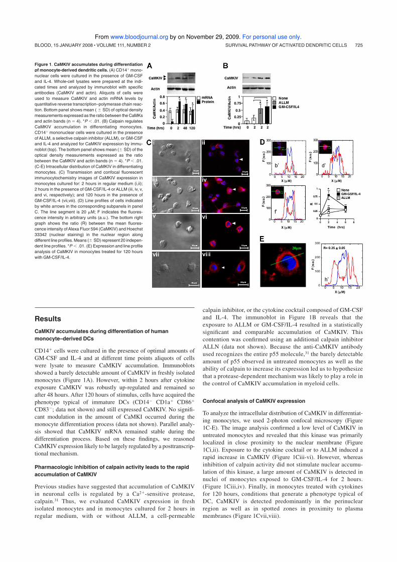
Results
CaMKIV accumulates during differentiation of human
monocyte–derived DCs
CD14� cells were cultured in the presence of optimal amounts of
GM-CSF and IL-4 and at different time points aliquots of cells
were lysate to measure CaMKIV accumulation. Immunoblots
showed a barely detectable amount of CaMKIV in freshly isolated
monocytes (Figure 1A). However, within 2 hours after cytokine
exposure CaMKIV was robustly up-regulated and remained so
after 48 hours. After 120 hours of stimulus, cells have acquired the
phenotype typical of immature DCs (CD14� CD1a� CD86�
CD83�; data not shown) and still expressed CaMKIV. No signifi-
cant modulation in the amount of CaMKI occurred during the
monocyte differentiation process (data not shown). Parallel analy-
sis showed that CaMKIV mRNA remained stable during the
differentiation process. Based on these findings, we reasoned
CaMKIV expression likely to be largely regulated by a posttranscrip-
tional mechanism.
Pharmacologic inhibition of calpain activity leads to the rapid
accumulation of CaMKIV
Previous studies have suggested that accumulation of CaMKIV
in neuronal cells is regulated by a Ca2�-sensitive protease,
calpain.31 Thus, we evaluated CaMKIV expression in fresh
isolated monocytes and in monocytes cultured for 2 hours in
regular medium, with or without ALLM, a cell-permeable
calpain inhibitor, or the cytokine cocktail composed of GM-CSF
and IL-4. The immunoblot in Figure 1B reveals that the
exposure to ALLM or GM-CSF/IL-4 resulted in a statistically
significant and comparable accumulation of CaMKIV. This
contention was confirmed using an additional calpain inhibitor
ALLN (data not shown). Because the anti-CaMKIV antibody
used recognizes the entire p55 molecule,31 the barely detectable
amount of p55 observed in untreated monocytes as well as the
ability of calpain to increase its expression led us to hypothesize
that a protease-dependent mechanism was likely to play a role in
the control of CaMKIV accumulation in myeloid cells.
Confocal analysis of CaMKIV expression
To analyze the intracellular distribution of CaMKIV in differentiat-
ing monocytes, we used 2-photon confocal microscopy (Figure
1C-E). The image analysis confirmed a low level of CaMKIV in
untreated monocytes and revealed that this kinase was primarily
localized in close proximity to the nuclear membrane (Figure
1Ci,ii). Exposure to the cytokine cocktail or to ALLM induced a
rapid increase in CaMKIV (Figure 1Ciii-vi). However, whereas
inhibition of calpain activity did not stimulate nuclear accumu-
lation of this kinase, a large amount of CaMKIV is detected in
nuclei of monocytes exposed to GM-CSF/IL-4 for 2 hours.
(Figure 1Ciii,iv). Finally, in monocytes treated with cytokines
for 120 hours, conditions that generate a phenotype typical of
DC, CaMKIV is detected predominantly in the perinuclear
region as well as in spotted zones in proximity to plasma
membranes (Figure 1Cvii,viii).
Figure 1. CaMKIV accumulates during differentiation
of monocyte-derived dendritic cells. (A) CD14� mono-
nuclear cells were cultured in the presence of GM-CSF
and IL-4. Whole-cell lysates were prepared at the indi-
cated times and analyzed by immunoblot with specific
antibodies (CaMKIV and actin). Aliquots of cells were
used to measure CaMKIV and actin mRNA levels by
quantitative reverse transcription–polymerase chain reac-
tion. Bottom panel shows mean ( � SD) of optical density
measurements expressed as the ratio between the CaMKs
and actin bands (n 4). *P .01. (B) Calpain regulates
CaMKIV accumulation in differentiating monocytes.
CD14� mononuclear cells were cultured in the presence
of ALLM, a selective calpain inhibitor (ALLM), or GM-CSF
and IL-4 and analyzed for CaMKIV expression by immu-
noblot (top). The bottom panel shows mean (� SD) of the
optical density measurements expressed as the ratio
between the CaMKIV and actin bands (n 4). *P .01.
(C-E) Intracellular distribution of CaMKIV in differentiating
monocytes. (C) Transmission and confocal fluorescent
immunocytochemistry images of CaMKIV expression in
monocytes cultured for: 2 hours in regular medium (i,ii);
2 hours in the presence of GM-CSF/IL-4 or ALLM (iii, iv, v,
and vi, respectively); and 120 hours in the presence of
GM-CSF/IL-4 (vii,viii). (D) Line profiles of cells indicated
by white arrows in the corresponding subpanels in panel
C. The line segment is 20 �M; F indicates the fluores-
cence intensity in arbitrary units (a.u.). The bottom right
graph shows the ratio (R) between the mean fluores-
cence intensity of Alexa Fluor 594 (CaMKIV) and Hoechst
33342 (nuclear staining) in the nuclear region along
different line profiles. Means (� SD) represent 20 indepen-
dent line profiles. *P .01. (E) Expression and line profile
analysis of CaMKIV in monocytes treated for 120 hours
with GM-CSF/IL-4.
.
SURVIVAL PATHWAY OF ACTIVATED DENDRITIC CELLS 725BLOOD, 15 JANUARY 2008 � VOLUME 111, NUMBER 2
For personal use only. by on November 29, 2009. www.bloodjournal.orgFrom
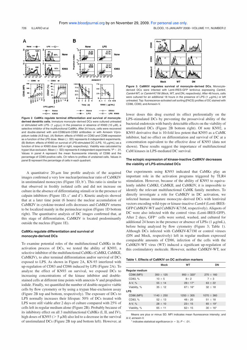
A quantitative 20-�m line profile analysis of the acquired
images confirmed a very low nuclear/perinuclear ratio of CaMKIV
in unstimulated monocytes (Figure 1D, b�). This ratio is similar to
that observed in freshly isolated cells and did not increase on
culture in the absence of differentiating stimuli or in the presence of
calpain inhibitors (Figure 1D, c� and d�). Kinetic analysis showed
that at a later time point (6 hours) the nuclear accumulation of
CaMKIV in cytokine-treated cells decreases and CaMKIV returns
to be localized mainly in the perinuclear region (Figure 1D bottom
right). The quantitative analysis of DC images confirmed that, at
this stage of differentiation, CaMKIV is located predominantly
outside the nucleus (Figure 1E).
CaMKs regulate differentiation and survival of
monocyte-derived DCs
To examine potential roles of the multifunctional CaMKs in the
activation process of DCs, we tested the ability of KN93, a
selective inhibitor of the multifunctional CaMKs (CaMKI, CaMKII,
CaMKIV), to alter terminal differentiation and/or survival of DCs
exposed to LPS. As shown in Figure 2A, KN-93 interfered with
up-regulation of CD83 and CD86 induced by LPS (Figure 2A). To
analyze the effect of KN93 on survival, we exposed DCs to
increasing concentrations of the kinase inhibitor and double-
stained cells at different time points with annexin-V and propidium
iodide. Finally, we quantified the number of double-negative viable
cells by flow cytometry or by using a trypan blue-exclusion assay
(Figure 2B top and bottom, respectively). The exposure of DCs to
LPS normally increases their lifespan: 50% of DCs treated with
LPS were still viable after 2 days of culture compared with 25% of
cells left in regular medium alone (Figure 2B). Probably because of
its inhibitory effect on all 3 multifunctional CaMKs (I, II, and IV),
high doses of KN93 (� 5 �M) also led to a decrease in the survival
of unstimulated DCs (Figure 2B top and bottom left). However, at
lower doses this drug exerted its effect preferentially on the
LPS-stimulated DCs by preventing the prosurvival ability of the
bacterial endotoxin with barely detectable effects on the viability of
unstimulated DCs (Figure 2B bottom right). Of note KN92, a
KN93 derivative that is 10-fold less potent that KN93 as a CaMK
inhibitor, had no effect on differentiation and survival of DC at a
concentration equivalent to the effective dose of KN93 (data not
shown). These results suggest the importance of multifunctional
CaM kinases in LPS-mediated DC survival.
The ectopic expression of kinase-inactive CaMKIV decreases
the viability of LPS-stimulated DCs
Our experiments using KN93 indicated that CaMKs play an
important role in the activation programs triggered by TLR4
stimulation. However, because of the ability of KN93 to equiva-
lently inhibit CaMKI, CaMKII, and CaMKIV, it is impossible to
identify the relevant multifunctional CaMK family members. To
directly investigate a role for CaMKIV in DC activation, we
infected human immature monocyte–derived DCs with lentiviral
vectors encoding wild-type or kinase-inactive Camk4 (Lenti-IRES-
GFP CaMKIV-WT and CaMKIV-K71M, respectively). Aliquots of
DC were also infected with the control virus (Lenti-IRES-GFP).
After 2 days, GFP� cells were sorted, washed, and cultured for
additional 24 hours in the presence or absence of LPSs (1 �g/mL),
before being analyzed by flow cytometry (Figure 3; Table 1).
Although DCs infected with CaMKIV-K71M or control viruses
(DN and Mock, respectively) left in regular medium expressed
comparable amounts of CD86, infection of the cells with the
CaMKIV-WT virus (WT) induced a significant up-regulation of
this costimulatory molecule. However, neither CaMKIV-WT, nor
Figure 2. CaMKs regulate terminal differentiation and survival of monocyte-
derived dendritic cells. Immature monocyte–derived DCs were cultured untreated
or stimulated with LPS- (1 �g/�L) in the presence or absence of KN93 (10 �M), a
selective inhibitor of the multifunctional CaMKs. After 24 hours, cells were recovered
and double-stained with anti-CD86/anti-CD83 antibodies or with Annexin V/pro-
pidium iodide (A,B top). (A) Bottom: effects of KN93 on CD83 and CD86 expression
as a function of the LPS dose. Mean (� SD) represents 6 independent experiments.
(B) Bottom: effects of KN93 on survival of LPS-stimulated DC (LPS, 10 �g/mL) as a
function of time or KN93 dose (left or right, respectively). Viability was calculated by
trypan blue exclusion. Mean (� SD) represents 6 independent experiments.*P .01.
Values in panel A represent the mean fluorescente intensity of CD86 and the
percentage of CD83 positive cells. Ctr refers to profiles of unstained cells. Values in
panel B represent the percentage of cells in each quadrant.
Figure 3. CaMKIV regulates survival of monocyte–derived DCs. Monocyte-
derived DCs were infected with Lenti-IRES-GFP lentivirus expressing Camk4,
Camk4-WT, or Camk4-K71M (Mock, WT, and DN, respectively). After 48 hours, cells
were cultured for an additional 18 hours in the presence of LPS (1 �g/mL) or left
untreated. Top: fluorescence-activated cell sorting (FACS) profiles of DC stained with
CD86, CD83, and Annexin-V.
Table 1. Effects of CaMKIV on DC activation markers
Mock WT DN
Regular medium
CD86 (MFI) 350 � 125 950 � 320* 270 � 160
CD83, % 10 � 5 8� 2 7 � 3
A-V, % 55 � 14 28� 17* 63 � 22
Viability, % 35 � 12 67� 16* 32 � 18
LPS
CD86 (MFI) 1140 � 250 1250 � 305 1070 � 360
CD83, % 52 � 13 48� 20 51 � 18
A-V, % 28 � 10 23� 15 65 � 19*
Viability, % 65 � 11 62� 15 30 � 16*
Means are plus or minus SD. MFI indicates mean fluorescence intensity; and
A-V, annexin-V.
* indicates statistical significance (n 3); P .01.
726 ILLARIO et al BLOOD, 15 JANUARY 2008 � VOLUME 111, NUMBER 2
For personal use only. by on November 29, 2009. www.bloodjournal.orgFrom
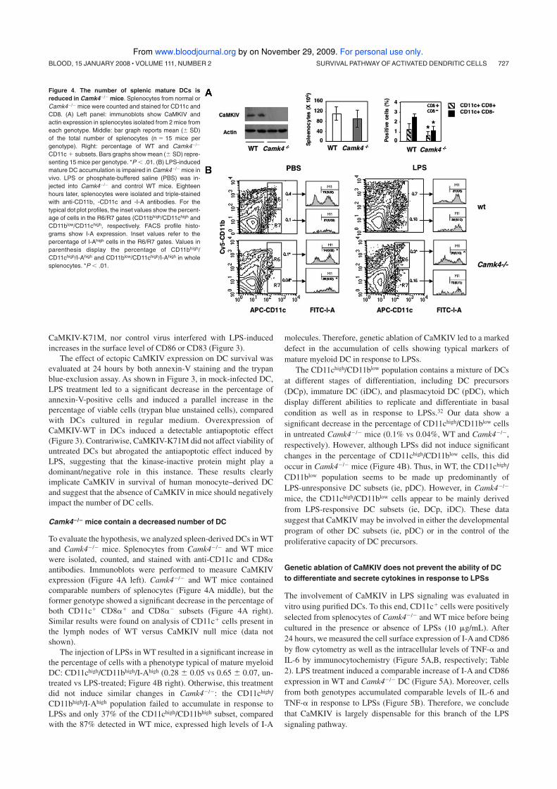
CaMKIV-K71M, nor control virus interfered with LPS-induced
increases in the surface level of CD86 or CD83 (Figure 3).
The effect of ectopic CaMKIV expression on DC survival was
evaluated at 24 hours by both annexin-V staining and the trypan
blue-exclusion assay. As shown in Figure 3, in mock-infected DC,
LPS treatment led to a significant decrease in the percentage of
annexin-V-positive cells and induced a parallel increase in the
percentage of viable cells (trypan blue unstained cells), compared
with DCs cultured in regular medium. Overexpression of
CaMKIV-WT in DCs induced a detectable antiapoptotic effect
(Figure 3). Contrariwise, CaMKIV-K71M did not affect viability of
untreated DCs but abrogated the antiapoptotic effect induced by
LPS, suggesting that the kinase-inactive protein might play a
dominant/negative role in this instance. These results clearly
implicate CaMKIV in survival of human monocyte–derived DC
and suggest that the absence of CaMKIV in mice should negatively
impact the number of DC cells.
Camk4�/� mice contain a decreased number of DC
To evaluate the hypothesis, we analyzed spleen-derived DCs in WT
and Camk4�/� mice. Splenocytes from Camk4�/� and WT mice
were isolated, counted, and stained with anti-CD11c and CD8�
antibodies. Immunoblots were performed to measure CaMKIV
expression (Figure 4A left). Camk4�/� and WT mice contained
comparable numbers of splenocytes (Figure 4A middle), but the
former genotype showed a significant decrease in the percentage of
both CD11c� CD8�� and CD8�� subsets (Figure 4A right).
Similar results were found on analysis of CD11c� cells present in
the lymph nodes of WT versus CaMKIV null mice (data not
shown).
The injection of LPSs in WT resulted in a significant increase in
the percentage of cells with a phenotype typical of mature myeloid
DC: CD11chigh/CD11bhigh/I-Ahigh (0.28 � 0.05 vs 0.65 � 0.07, un-
treated vs LPS-treated; Figure 4B right). Otherwise, this treatment
did not induce similar changes in Camk4�/�: the CD11chigh/
CD11bhigh/I-Ahigh population failed to accumulate in response to
LPSs and only 37% of the CD11chigh/CD11bhigh subset, compared
with the 87% detected in WT mice, expressed high levels of I-A
molecules. Therefore, genetic ablation of CaMKIV led to a marked
defect in the accumulation of cells showing typical markers of
mature myeloid DC in response to LPSs.
The CD11chigh/CD11blow population contains a mixture of DCs
at different stages of differentiation, including DC precursors
(DCp), immature DC (iDC), and plasmacytoid DC (pDC), which
display different abilities to replicate and differentiate in basal
condition as well as in response to LPSs.32 Our data show a
significant decrease in the percentage of CD11chigh/CD11blow cells
in untreated Camk4�/� mice (0.1% vs 0.04%, WT and Camk4�/�,
respectively). However, although LPSs did not induce significant
changes in the percentage of CD11chigh/CD11blow cells, this did
occur in Camk4�/� mice (Figure 4B). Thus, in WT, the CD11chigh/
CD11blow population seems to be made up predominantly of
LPS-unresponsive DC subsets (ie, pDC). However, in Camk4�/�
mice, the CD11chigh/CD11blow cells appear to be mainly derived
from LPS-responsive DC subsets (ie, DCp, iDC). These data
suggest that CaMKIV may be involved in either the developmental
program of other DC subsets (ie, pDC) or in the control of the
proliferative capacity of DC precursors.
Genetic ablation of CaMKIV does not prevent the ability of DC
to differentiate and secrete cytokines in response to LPSs
The involvement of CaMKIV in LPS signaling was evaluated in
vitro using purified DCs. To this end, CD11c� cells were positively
selected from splenocytes of Camk4�/� and WT mice before being
cultured in the presence or absence of LPSs (10 �g/mL). After
24 hours, we measured the cell surface expression of I-A and CD86
by flow cytometry as well as the intracellular levels of TNF-� and
IL-6 by immunocytochemistry (Figure 5A,B, respectively; Table
2). LPS treatment induced a comparable increase of I-A and CD86
expression in WT and Camk4�/� DC (Figure 5A). Moreover, cells
from both genotypes accumulated comparable levels of IL-6 and
TNF-� in response to LPSs (Figure 5B). Therefore, we conclude
that CaMKIV is largely dispensable for this branch of the LPS
signaling pathway.
Figure 4. The number of splenic mature DCs is
reduced in Camk4�/� mice. Splenocytes from normal or
Camk4�/� mice were counted and stained for CD11c and
CD8. (A) Left panel: immunoblots show CaMKIV and
actin expression in splenocytes isolated from 2 mice from
each genotype. Middle: bar graph reports mean (� SD)
of the total number of splenocytes (n 15 mice per
genotype). Right: percentage of WT and Camk4�/�
CD11c � subsets. Bars graphs show mean (� SD) repre-
senting 15 mice per genotype. *P .01. (B) LPS-induced
mature DC accumulation is impaired in Camk4�/� mice in
vivo. LPS or phosphate-buffered saline (PBS) was in-
jected into Camk4�/� and control WT mice. Eighteen
hours later, splenocytes were isolated and triple-stained
with anti-CD11b, -CD11c and -I-A antibodies. For the
typical dot plot profiles, the inset values show the percent-
age of cells in the R6/R7 gates (CD11bhigh/CD11chigh and
CD11blow/CD11chigh, respectively. FACS profile histo-
grams show I-A expression. Inset values refer to the
percentage of I-Ahigh cells in the R6/R7 gates. Values in
parenthesis display the percentage of CD11bhigh/
CD11chigh/I-Ahigh and CD11blow/CD11chigh/I-Ahigh in whole
splenocytes. *P .01.
SURVIVAL PATHWAY OF ACTIVATED DENDRITIC CELLS 727BLOOD, 15 JANUARY 2008 � VOLUME 111, NUMBER 2
For personal use only. by on November 29, 2009. www.bloodjournal.orgFrom
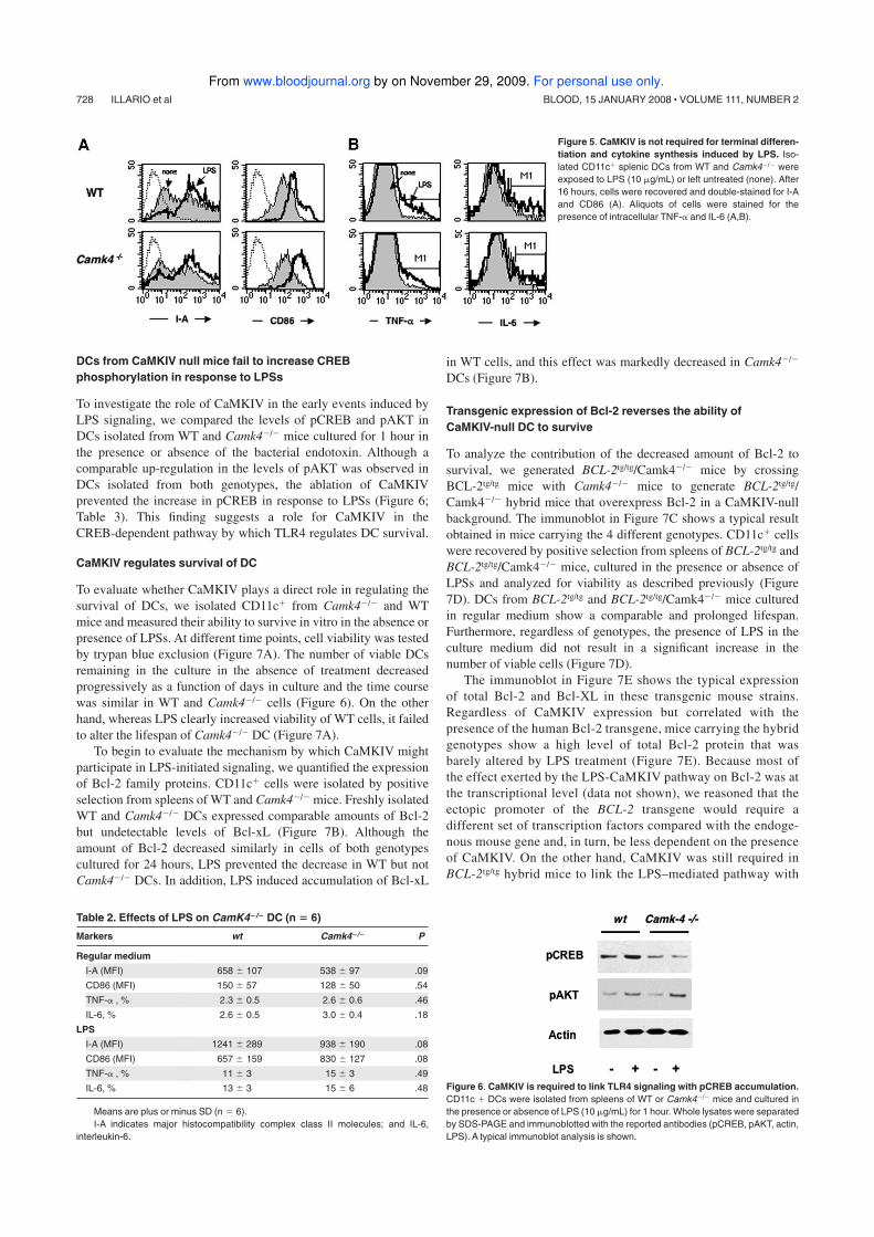
DCs from CaMKIV null mice fail to increase CREB
phosphorylation in response to LPSs
To investigate the role of CaMKIV in the early events induced by
LPS signaling, we compared the levels of pCREB and pAKT in
DCs isolated from WT and Camk4�/� mice cultured for 1 hour in
the presence or absence of the bacterial endotoxin. Although a
comparable up-regulation in the levels of pAKT was observed in
DCs isolated from both genotypes, the ablation of CaMKIV
prevented the increase in pCREB in response to LPSs (Figure 6;
Table 3). This finding suggests a role for CaMKIV in the
CREB-dependent pathway by which TLR4 regulates DC survival.
CaMKIV regulates survival of DC
To evaluate whether CaMKIV plays a direct role in regulating the
survival of DCs, we isolated CD11c� from Camk4�/� and WT
mice and measured their ability to survive in vitro in the absence or
presence of LPSs. At different time points, cell viability was tested
by trypan blue exclusion (Figure 7A). The number of viable DCs
remaining in the culture in the absence of treatment decreased
progressively as a function of days in culture and the time course
was similar in WT and Camk4�/� cells (Figure 6). On the other
hand, whereas LPS clearly increased viability of WT cells, it failed
to alter the lifespan of Camk4�/� DC (Figure 7A).
To begin to evaluate the mechanism by which CaMKIV might
participate in LPS-initiated signaling, we quantified the expression
of Bcl-2 family proteins. CD11c� cells were isolated by positive
selection from spleens of WT and Camk4�/� mice. Freshly isolated
WT and Camk4�/� DCs expressed comparable amounts of Bcl-2
but undetectable levels of Bcl-xL (Figure 7B). Although the
amount of Bcl-2 decreased similarly in cells of both genotypes
cultured for 24 hours, LPS prevented the decrease in WT but not
Camk4�/� DCs. In addition, LPS induced accumulation of Bcl-xL
in WT cells, and this effect was markedly decreased in Camk4�/�
DCs (Figure 7B).
Transgenic expression of Bcl-2 reverses the ability of
CaMKIV-null DC to survive
To analyze the contribution of the decreased amount of Bcl-2 to
survival, we generated BCL-2tg/tg/Camk4�/� mice by crossing
BCL-2tg/tg mice with Camk4�/� mice to generate BCL-2tg/tg/
Camk4�/� hybrid mice that overexpress Bcl-2 in a CaMKIV-null
background. The immunoblot in Figure 7C shows a typical result
obtained in mice carrying the 4 different genotypes. CD11c� cells
were recovered by positive selection from spleens of BCL-2tg/tg and
BCL-2tg/tg/Camk4�/� mice, cultured in the presence or absence of
LPSs and analyzed for viability as described previously (Figure
7D). DCs from BCL-2tg/tg and BCL-2tg/tg/Camk4�/� mice cultured
in regular medium show a comparable and prolonged lifespan.
Furthermore, regardless of genotypes, the presence of LPS in the
culture medium did not result in a significant increase in the
number of viable cells (Figure 7D).
The immunoblot in Figure 7E shows the typical expression
of total Bcl-2 and Bcl-XL in these transgenic mouse strains.
Regardless of CaMKIV expression but correlated with the
presence of the human Bcl-2 transgene, mice carrying the hybrid
genotypes show a high level of total Bcl-2 protein that was
barely altered by LPS treatment (Figure 7E). Because most of
the effect exerted by the LPS-CaMKIV pathway on Bcl-2 was at
the transcriptional level (data not shown), we reasoned that the
ectopic promoter of the BCL-2 transgene would require a
different set of transcription factors compared with the endoge-
nous mouse gene and, in turn, be less dependent on the presence
of CaMKIV. On the other hand, CaMKIV was still required in
BCL-2tg/tg hybrid mice to link the LPS–mediated pathway with
Figure 6. CaMKIV is required to link TLR4 signaling with pCREB accumulation.
CD11c � DCs were isolated from spleens of WT or Camk4�/� mice and cultured in
the presence or absence of LPS (10 �g/mL) for 1 hour. Whole lysates were separated
by SDS-PAGE and immunoblotted with the reported antibodies (pCREB, pAKT, actin,
LPS). A typical immunoblot analysis is shown.
Table 2. Effects of LPS on CamK4�/� DC (n � 6)
Markers wt Camk4�/� P
Regular medium
I-A (MFI) 658 � 107 538 � 97 .09
CD86 (MFI) 150 � 57 128 � 50 .54
TNF-� , % 2.3 � 0.5 2.6 � 0.6 .46
IL-6, % 2.6 � 0.5 3.0 � 0.4 .18
LPS
I-A (MFI) 1241 � 289 938 � 190 .08
CD86 (MFI) 657 � 159 830 � 127 .08
TNF-� , % 11 � 3 15 � 3 .49
IL-6, % 13 � 3 15 � 6 .48
Means are plus or minus SD (n 6).
I-A indicates major histocompatibility complex class II molecules; and IL-6,
interleukin-6.
Figure 5. CaMKIV is not required for terminal differen-
tiation and cytokine synthesis induced by LPS. Iso-
lated CD11c� splenic DCs from WT and Camk4�/� were
exposed to LPS (10 �g/mL) or left untreated (none). After
16 hours, cells were recovered and double-stained for I-A
and CD86 (A). Aliquots of cells were stained for the
presence of intracellular TNF-� and IL-6 (A,B).
728 ILLARIO et al BLOOD, 15 JANUARY 2008 � VOLUME 111, NUMBER 2
For personal use only. by on November 29, 2009. www.bloodjournal.orgFrom

Bcl-xL expression (Figure 7D). This finding provides an addi-
tional evidence for a role for CaMKIV in the pathway respon-
sible for Bcl-xL expression and also documents the dominant
role played by Bcl-2 in modulating the lifespan of LPS-activated
DC in a manner that involves CaMKIV signaling.
Discussion
Stimulation of TLR4 has been associated with the initiation of both
apoptotic and antiapoptotic pathways, the balance of which deter-
mines the outcome of innate and adaptive immune responses.4-6
Here we describe a novel CaMK cascade-dependent antiapoptotic
pathway responsible for the survival of LPS-activated DCs. The
results obtained, using pharmacologic inhibition of CaMKs, ec-
topic expression of CaMKIV, and a CaMKIV kinase-inactive
mutant as well as mice null for CaMKIV, demonstrate that a
CaMKIV signaling cascade controls the phosphorylation of CREB
and accumulation of Bcl-2 necessary to support the antiapoptotic
branch of the TLR4 pathway.
The multifunctional CaMK family proteins are involved in the
control of differentiation and survival of several cell types, including
neurons and hematopoietic stem cells.26,27 Analysis of mouse embryos
Figure 7. CaMKIV regulates lifespan and Bcl-2 family
protein accumulation. CD11c � DCs were positively
selected from WT (Camk4�/�), Camk4�/�, BCL-2 tg/tg
transgenic, and Camk4�/�/BCL-2 tg/tg hybrid mice cul-
tured in the presence or absence of LPS (10 �g/mL).
(A,D) Viability was assayed by trypan blue exclusion at
daily intervals. The results represent mean and SD of
6 independent experiments. (B,E) Typical results ob-
tained by immunoblot analysis. Bar graphs show mean
(� SD) of the optical density measurements expressed
as the ratio between Bcl-2 or Bcl-xL and actin bands
(n 6). (C) Immunoblot shows the typical expression of
CaMKIV and hu-Bcl-2 detected in WT (Camk4�/�),
Camk4�/�, BCL-2 tg/tg transgenic, and Camk4�/�/BCL-
2tg/tg hybrid mice (lanes 1, 2, 3, and 4, respectively).
*P .01.
Table 3. TLR4 signaling in CamK4�/� DC
wt Camk4�/�
Regular medium
pCREB 0.32 � 0.1 0.19 � 0.05*
pAKT 0.09 � 0.04 0.07 � 0.06
LPS
pCREB 0.51 � 0.12 0.15 � 0.03*
pAKT 0.17 � 0.05 0.23 �0.06
Means are plus or minus SD of the optical density measurements expressed as
the ratio between pCREB or pAKT.
* indicates statistical significance (n 3); P .01.
SURVIVAL PATHWAY OF ACTIVATED DENDRITIC CELLS 729BLOOD, 15 JANUARY 2008 � VOLUME 111, NUMBER 2
For personal use only. by on November 29, 2009. www.bloodjournal.orgFrom
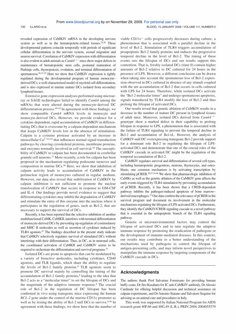
revealed expression of CaMKIV mRNA in the developing nervous
system as well as in the hematopoietic-related tissues.25,26 These
developmental patterns coincide temporally with periods of significant
cellular differentiation in the nervous system, axonal migration and
neuron survival. Correlation of CaMKIV expression with differentiation
is also evident in adult animals as Camk4�/� mice show major defects in
maintenance of hematopoietic stem cells, postnatal maturation of
Purkinje cells, thymopoiesis, ovulation, and terminal differentiation of
spermatozoa.26,33-39 Here we show that CaMKIV expression is tightly
regulated during the developmental program of human monocyte–
derived DCs, a well-characterized model of myeloid cell differentiation,
and is also expressed in murine mature DCs isolated from secondary
lymphoid tissues.
Extensive gene expression analyses performed using microar-
ray or SAGE technologies failed to identify Camk4 among the
mRNAs that were altered during the monocyte-derived DC
differentiation process.40-42 In agreement with these findings, we
show comparable Camk4 mRNA levels in monocyte and
monocyte-derived DCs. However, we provide evidence for a
cytokine-dependent, rapid accumulation of CaMKIV in differen-
tiating DCs that is overcome by a calpain-dependent mechanism
that keeps CaMKIV levels low in the absence of stimulation.
Calpain is a cysteine protease activated by an increase in
intracellular Ca2�43,44 that influences normal signal transduction
pathways by cleaving cytoskeletal proteins, membrane proteins,
and enzymes normally involved in cell survival.45 The suscepti-
bility of CaMKIV to calpain has been documented in cerebellar
granule cell neurons.31 More recently, a role for calpain has been
proposed in the mechanism regulating podosome turnover and
composition in murine DCs.46 Here, we show that inhibition of
calpain activity leads to accumulation of CaMKIV in the
perinuclear region of monocytes cultured in regular medium.
However, our data also reveal that stabilization of CaMKIV by
calpain inhibition is not sufficient to promote the nuclear
translocation of CaMKIV that occurs in response to GM-CSF
and IL-4. Our findings provide novel evidence to suggest that
differentiating cytokines may inhibit the degradation of CaMKIV
and stimulate the entry of this enzyme into the nucleus where it
participates in the regulation of genes, such as Bcl-2, that are
necessary to support the survival of DCs.
Recently, it has been reported that the selective inhibition of another
multifunctional CaMK, CaMKII, interferes with terminal differentiation
of monocyte-derived DCs by preventing up-regulation of costimulatory
and MHC II molecules as well as secretion of cytokines induced by
TLR4 agonists.47 The findings described in the present study indicate
that CaMKIV selectively regulates survival of stimulated DCs without
interfering with their differentiation. Thus, in DC, as in neuronal cells,
the coordinated activation of CaMKII and CaMKIV seems to be
required to orchestrate the differentiation and survival programs.33
Isolated DCs are prone to apoptosis that can be modulated by
a variety of bioactive molecules, including cytokines, CD40
agonists, and TLR ligands, which share the ability to regulate
the levels of Bcl-2 family proteins.8 TLR agonists seem to
promote DC survival mainly by controlling the timing of the
accumulation of Bcl-2 family proteins,8 leading to the idea that
Bcl-2 acts as a “molecular timer” to set the lifespan of DCs and
the magnitude of the adaptive immune response.8 The crucial
role of Bcl-2 in the regulation of DC lifespan has been
confirmed in vivo using transgenic mice expressing the human
BCL-2 gene under the control of the murine CD11c promoter as
well as by testing the ability of Bcl-2 null DCs to survive.8,46 In
agreement with these findings, we show here that the number of
viable CD11c� cells progressively decreases during culture, a
phenomenon that is associated with a parallel decline in the
level of Bcl-2. Stimulation of TLR4 triggers accumulation of
proapoptotic Bcl-2 family proteins and induces the progressive
temporal decline in the level of Bcl-2. The timing of these
events sets the lifespan of DCs and our results support this
contention. That is, freshly isolated DCs (time 0) contain higher
amounts of Bcl-2 relative to DC cultured for 24 hours in the
presence of LPS. However, a different conclusion can be drawn
when taking into account the spontaneous loss of Bcl-2 expres-
sion observed in DCs cultured in absence of any stimuli coupled
with the net accumulation of Bcl-2 that occurs in cells cultured
with LPS for 24 hours. Therefore, while isolated DCs activate
the “Bcl-2 molecular timer” and undergo spontaneous apoptosis,
signals transduced by TLR4 modify the loss of Bcl-2 and thus
prolong the lifespan of activated DCs.
Our results reveal that genetic ablation of CaMKIV results in a
decrease in the number of mature DC present in lymphoid tissues
of adult mice. Moreover, isolated DCs derived from Camk4�/�
genotype show a marked defect in their capability to prolong
lifespan in response to LPS, a phenomenon that is associated with
the failure of TLR4 signaling to prevent the temporal decline in
Bcl-2 and accumulation of Bcl-xL. However, the analysis of
CaMKIV-null DC overexpressing transgenic Bcl-2 provide support
for a dominant role Bcl-2 in regulating the lifespan of LPS-
activated DCs and demonstrate that one of the crucial roles of the
CaMKIV cascade in activated DC might be the regulation of the
temporal accumulation of Bcl-2.
CaMKIV regulates survival and differentiation of several cell types,
including hematopoietic progenitors, neurons, thymocytes, and osteo-
blasts; one common mechanism is by activating transcription by
stimulating pCREB.26,27,34,48 We show that pharmacologic inhibition of
CaMKs as well as the genetic ablation of the CaMKIV gene affects the
early events triggered by TLR4 stimulation by preventing accumulation
of pCREB. Recently, it has been shown that a CREB-dependent
pathway inhibits the pathogen-induced apoptosis of bone marrow–
derived macrophages.12 Our data confirm the relevance of pCREB in the
survival program and document its involvement in the molecular
mechanisms regulating the lifespan of LPS-activated DCs. Furthermore,
they identify the CaMKIV/CREB signaling cascade as a novel pathway
that is essential in the antiapoptotic branch of the TLR4 signaling
pathway.
Genetic or microenvironmental factors may control the
lifespan of activated DCs and in turn regulate the adaptive
immune response by promoting the eradication of pathogens or
the development of immune-mediated diseases. In this context,
our results may contribute to a better understanding of the
mechanisms used by pathogens to control the lifespan of
antigen-presenting cells, and may inform novel perspectives to
manipulate the immune response by targeting components of the
CaMKIV cascade in DCs.
Acknowledgments
The authors thank Prof Salvatore Formisano for providing human
buffy-coats, Dr Jiro Kasahara for IC anti-CaMKIV antibody, Dr Alessio
Cardinale for offering helpful discussion and technical assistance on
calpain experiments, and Dr Antonio Staiano and Salvatore Sequino for
advising us on animal care and procedures in Italy.
This work was supported by Italian National Program for AIDS
research grant 40F.66 and 40G.49 (L.R.), PRIN 2004-2004055579
730 ILLARIO et al BLOOD, 15 JANUARY 2008 � VOLUME 111, NUMBER 2
For personal use only. by on November 29, 2009. www.bloodjournal.orgFrom

and 2006-051402 (L.R.), NIH grant DK074701 (A.R.M.), ACS
fellowship F-05-171-01-LIB (U.S.), and PRIN 2004-2004069479
and Telethon GGP04039 (L.P.).
Authorship
Contribution: M.I. designed and performed research and drafted
the manuscript; M.G., L.V., A.M.M., F.B., E.C., D.A., A.C., V.C.,
and G.M. performed research on human monocyte–derived DCs;
L.M.G.-T., U.S., and T.J.R. performed research on murine models;
L.P. designed research using lentivaral vectors; A.R.M. interpreted
data and drafted the manuscript; G.R. and M.V. interpreted data and
drafted the manuscript; and L.R. designed and performed research,
analyzed and interpreted data, and drafted the manuscript.
Conflict-of-interest disclosure: The authors declare no compet-
ing financial interests.
Correspondence: Luigi Racioppi, Departmnent of Cellular and
Molecular Biology and Pathology, Via S Pansini 5, 80131 Naples,
Italy; e-mail: [email protected].
References
1. Banchereau J, Steinman RM. Dendritic cells and
the control of immunity. Nature. 1998;392:245-
252.
2. Steinman RM. The dendritic cell system and its
role in immunogenicity. Annu Rev Immunol. 1991;
9:271-296.
3. Steinman RM, Bonifaz L, Fujii S, et al. The innate
functions of dendritic cells in peripheral lymphoid
tissues. Adv Exp Med Biol. 2005;560:83-97.
4. Akira S, Takeda K. Toll-like receptor signalling.
Nat Rev Immunol. 2004;4:499-511.
5. De Smedt T, Pajak B, Muraille E, et al. Regulation
of dendritic cell numbers and maturation by lipo-
polysaccharide in vivo. J Exp Med. 1996;184:
1413-1424.
6. Takeda K, Akira S. TLR signaling pathways. Se-
min Immunol. 2004;16:3-9.
7. Stoll S, Delon J, Brotz TM, Germain RN. Dynamic
imaging of T cell-dendritic cell interactions in
lymph nodes. Science. 2002;296:1873-1876.
8. Hou WS, Van Parijs L. A Bcl-2–dependent mo-
lecular timer regulates the lifespan and immuno-
genicity of dendritic cells. Nat Immunol. 2004;5:
583-589.
9. Bae JS, Jang MK, Hong S, et al. Phosphorylation
of NF-kappa B by calmodulin–dependent kinase
IV activates anti-apoptotic gene expression. Bio-
chem Biophys Res Commun. 2003;305:1094-
1098.
10. Franchi L, Condo I, Tomassini B, Nicolo C, Testi
R. A caspaselike activity is triggered by LPS and
is required for survival of human dendritic cells.
Blood. 2003;102:2910-2915.
11. Rescigno M, Martino M, Sutherland CL, Gold MR,
Ricciardi-Castagnoli P. Dendritic cell survival and
maturation are regulated by different signaling
pathways. J Exp Med. 1998;188:2175-2180.
12. Park JM, Greten FR, Wong A, et al. Signaling
pathways and genes that inhibit pathogen-in-
duced macrophage apoptosis: CREB and NF-
kappaB as key regulators. Immunity. 2005;23:
319-329.
13. Berridge MJ, Bootman MD, Roderick HL. Calcium
signalling: dynamics, homeostasis and remodel-
ling. Nat Rev Mol Cell Biol. 2003;4:517-529.
14. Rubartelli A, Poggi A, Zocchi MR. The selective
engulfment of apoptotic bodies by dendritic cells
is mediated by the alpha(v)beta3 integrin and re-
quires intracellular and extracellular calcium. Eur
J Immunol. 1997;27:1893-1900.
15. Chan VW, Kothakota S, Rohan MC, et al. Sec-
ondary lymphoid-tissue chemokine (SLC) is che-
motactic for mature dendritic cells. Blood. 1999;
93:3610-3616.
16. Delgado E, Finkel V, Baggiolini M, Mackay CR,
Steinman RM, Granelli-Piperno A. Mature dendritic
cells respond to SDF-1, but not to several beta-che-
mokines. Immunobiology. 1998;198:490-500.
17. Dieu MC, Vanbervliet B, Vicari A, et al. Selective re-
cruitment of immature and mature dendritic cells by
distinct chemokines expressed in different anatomic
sites. J Exp Med. 1998;188:373-386.
18. Yanagihara S, Komura E, Nagafune J, Watarai H,
Yamaguchi Y. EBI1/CCR7 is a new member of
dendritic cell chemokine receptor that is up-regu-
lated upon maturation. J Immunol. 1998;161:
3096-3102.
19. Czerniecki BJ, Carter C, Rivoltini L, et al. Calcium
ionophore-treated peripheral blood monocytes
and dendritic cells rapidly display characteristics
of activated dendritic cells. J Immunol. 1997;159:
3823-3837.
20. Koski GK, Schwartz GN, Weng DE, et al. Calcium
mobilization in human myeloid cells results in ac-
quisition of individual dendritic cell-like character-
istics through discrete signaling pathways. J Im-
munol. 1999;163:82-92.
21. Koski GK, Schwartz GN, Weng DE, et al. Calcium
ionophore-treated myeloid cells acquire many den-
dritic cell characteristics independent of prior differen-
tiation state, transformation status, or sensitivity to
biologic agents. Blood. 1999;94:1359-1371.
22. Chin D, Means AR. Calmodulin: a prototypical
calcium sensor. Trends Cell Biol. 2000;10:322-
328.
23. Hook SS, Means AR. Ca2�/CaM–dependent ki-
nase: from activation to function. Annu Rev Phar-
macol Toxicol. 2001;41:471-505.
24. Yamauchi T. Neuronal Ca2�/calmodulin–depen-
dent protein kinase II–discovery, progress in a
quarter of a century, and perspective: implication
for learning and memory. Biol Pharm Bull. 2005;
28:1342-1354.
25. Wang SL, Ribar TJ, Means AR. Expression of
Ca(2�)/calmodulin–dependent protein kinase IV
(CaMKIV) messenger RNA during murine embryo-
genesis. Cell Growth Differ. 2001;12:351-361.
26. Kitsos CM, Sankar U, Illario M, et al. Calmodulin–
dependent protein kinase IV regulates hemato-
poietic stem cell maintenance. J Biol Chem.
2005;280:33101-33108.
27. Ribar TJ, Rodriguiz RM, Khiroug L, et al. Cerebel-
lar defects in Ca2�/calmodulin kinase IV-defi-
cient mice. J Neurosci. 2000;20,RC107.
28. Nopora A, Brocker T. Bcl-2 controls dendritic cell
longevity in vivo. J Immunol. 2002;169;3006-
3014.
29. Galgani M, De Rosa V, De Simone S, et al. Cyclic
AMP modulates the functional plasticity of imma-
ture dendritic cells by inhibiting Src-like kinases
through protein kinase A-mediated signaling.
J Biol Chem. 2004;279:32507-32514.
30. Follenzi A, Ailles LE, Bakovic S, Geuna M, Naldini
L. Gene transfer by lentiviral vectors is limited by
nuclear translocation and rescued by HIV-1 pol
sequences. Nat Genet. 2000;25:217-222.
31. Tremper-Wells B, Vallano ML. Nuclear calpain
regulates Ca2�–dependent signaling via proteol-
ysis of nuclear Ca2�/calmodulin–dependent pro-
tein kinase type IV in cultured neurons. J Biol
Chem. 2005;280:2165-2175.
32. Diao J, Winter E, Cantin C, et al. In situ replica-
tion of immediate dendritic cell (DC) precursors
contributes to conventional DC homeostasis in
lymphoid tissue. Immunol. 2006,176:7196-7206
33. Hansen MR, Bok J, Devaiah AK, Zha XM, Green
SH. Ca2�/calmodulin–dependent protein ki-
nases II and IV both promote survival but differ in
their effects on axon growth in spiral ganglion
neurons. J Neurosci Res. 2003;72:169-184.
34. Raman V, Blaeser F, Ho N, Engle DL, Williams
CB, Chatila TA. Requirement for Ca2�/calmodu-
lin–dependent kinase type IV/Gr in setting the
thymocyte selection threshold. J Immunol. 2001;
167:6270-6278.
35. Blaeser F, Toppari J, Heikinheimo M, et al.
CaMKIV/Gr is dispensable for spermatogenesis
and CREM-regulated transcription in male germ
cells. Am J Physiol Endocrinol Metab. 2001;281:
E931-E937.
36. Wu JY, Ribar TJ, Cummings DE, et al. Spermio-
genesis and exchange of basic nuclear proteins
are impaired in male germ cells lacking Camk4.
Nat Genet. 2000;25:448.
37. Wu JY, Gonzalez-Robayna IJ, Richards JS,
Means AR. Female fertility is reduced in mice
lacking Ca2�/calmodulin–dependent protein ki-
nase IV. Endocrinology. 2000;141:4777-4783.
38. Wu JY, Means AR. Ca2�/calmodulin–dependent
protein kinase IV is expressed in spermatids and
targeted to chromatin and the nuclear matrix.
J Biol Chem. 2000;275:7994-7999.
39. Wu JY, Ribar TJ, Means AR. Spermatogenesis
and the regulation of Ca2�-calmodulin–depen-
dent protein kinase IV localization are not depen-
dent on calspermin. Mol Cell Biol. 2001;21:6066-
6070.
40. Le Naour F, Hohenkirk L, Grolleau A, et al. Profiling
changes in gene expression during differentiation
and maturation of monocyte–derived dendritic cells
using both oligonucleotide microarrays and proteom-
ics. J Biol Chem. 2001;276:17920-17931.
41. Messmer D, Messmer B, Chiorazzi N. The global
transcriptional maturation program and stimuli-spe-
cific gene expression profiles of human myeloid den-
dritic cells. Int Immunol. 2003;15:491-503.
42. Wilson HL, O’Neill HC. Identification of differen-
tially expressed genes representing dendritic cell
precursors and their progeny. Blood. 2003;102:
1661-1669.
43. Croall DE, DeMartino GN. Calcium-activated neu-
tral protease (calpain) system: structure, function,
and regulation. Physiol Rev. 1991;71:813-847.
44. Saido TC, Sorimachi H, Suzuki K. Calpain: new
perspectives in molecular diversity and physio-
logical-pathological involvement. FASEB J. 1994;
8:814-822.
45. Huang Y, Wang KK. The calpain family and hu-
man disease. Trends Mol Med. 2001;7:355-362.
46. Herrmann TL, Morita CT, Lee K, Kusner DJ. Cal-
modulin kinase II regulates the maturation and
antigen presentation of human dendritic cells.
J Leukoc Biol. 2005;78:1397-1407.
47. Anderson KA, Ribar TJ, Illario M, Means AR. De-
fective survival and activation of thymocytes in
transgenic mice expressing a catalytically inac-
tive form of Ca2�/calmodulin–dependent protein
kinase IV. Mol Endocrinol. 1997;11:725-737.
48. Sato K, Suematsu A, Nakashima T, et al. Regula-
tion of osteoclast differentiation and function by
the CaMK-CREB pathway. Nat Med. 2007;12:
1410-1416.
SURVIVAL PATHWAY OF ACTIVATED DENDRITIC CELLS 731BLOOD, 15 JANUARY 2008 � VOLUME 111, NUMBER 2
For personal use only. by on November 29, 2009. www.bloodjournal.orgFrom
1
Dendritic cells/Natural Killer cross-talk: a novel target for Human Immunodeficiency Type-1 Protease
Inhibitors.
Running title
HIV-1 Protease Inhibitors and DC
by
Ciaglia Elena1, Giardino Torchia Maria Letizia1*, Masci Anna Maria1-2, Vitiello Laura1-2, Fogli Manuela3, Mavilio
Domenico3, La Sala Andrea4 and Racioppi Luigi1-5
1Department of Molecular and Cellular Biology and Pathology, Federico II University of Naples, Naples, Italy
2Laboratory of Immunobiology of Cardiovascular Diseases, Department of Medical Science and Rehabilitation,
Scientific Institute for Research, Hospitalization, and Health Care (IRCCS) San Raffaele Pisana, Rome, Italy
3Laboratory of Immunoregulation, National Institute of Allergy and Infectious Diseases, National Institutes of Health,
Bethesda, MD; 4Laboratory of Molecular and Cellular Immunology, Istituto di Ricovero e Cura a Carattere Scientifico
(IRCCS) San Raffaele, Rome, Italy
5Interdepartimental Center for Immunological Science, Federico II University of Naples, Naples, Italy
*present address: Laboratory of Immune Cell Biology, National Cancer Institute, National Institutes of Health,
Bethesda, MD
*Corresponding author: Racioppi Luigi, Dipartimento di Biologia e Patologia Cellulare e Molecolare,
Università di Napoli Federico II, Via Pansini, 5, 80131 Naples. E-mail: [email protected]. ^ G-T ML and
EC equally contributed.
2
Abstract
HIV-1 protease inhibitors (PIs) are designed to specifically inhibit the aspartic protease of HIV-1, but
several evidences shown that are also able to exert various effects on immune cells. Here we investigate the
capability of HIV-1 PIs to interfere with Monocyte-Derived DCs (MDCs) lifecycle. We shown that
prolonged exposure of PIs decrease the yield of MDCs; decrease the expression of typical MDCs
differentiation markers; decrease ability to secrete IL-15 and IL-12 p40 in response to bacterial endotoxin
stimulus. In turn, these cells fail to prime the proliferation, interferon gamma secretion and cytotoxicity of
autologous NK cells. In addition, MDCs generated in the presence of PIs are more prone to NK-dependent
lysis. These findings reveal DC-NK as a novel target of the immunomodulatory activity of PIs opening
novel therapeutical perspective for using this category of drug to manipulate the immune response.

3
Introduction
Dendritic cells (DC) are well described as sentinels of the immune system, able to recognize
danger signals mainly via Toll-Like Receptors, undergo maturation process and efficiently prime naïve T
cells, inducing the adaptive immune response (Steinman RM 2006). Actually, there is a growing body of
evidences about the interplay of DC and Natural Killer (NK)-cells (Walzer T et al. 2005). NK-cells can
induce terminal differentiation of DC, and, reciprocally, DC can secrete NK-cells activating cytokines (IL-
12, IL-15, type I IFNs), leading to proliferation, IFN-gamma secretion and increase of cytotoxic activity.
High NK - DC (DC) ratio, results in the killing of DC which express low levels of MHC I molecules and/or
human leukocyte antigen-E molecule. This “switch-off” mechanism of the immune response is mediated by
triggering NKp30 NKAR and CD40L on NK cells and TNF-related apoptosis-inducing ligand (TRAIL)
death receptors on DC (Walzer et al. 2005, Moretta L et al. 2006).
HIV-1 Protease Inhibitors (PIs), that are included in the Highly Active Antiretroviral Therapy
(HAART) of AIDS, are specifically designed to inhibit HIV-1 aspartic protease and have significantly
improved the clinical management of HIV-1 infected patients (Mastrolorenzo et al. 2007, Dash C et al.
2003). However, a number of evidences proved that HIV-1 PIs are able to inhibit mitochondrial-mediated
apoptosis in uninfected T lymphocytes, in an antiviral-independent fashion. This effect can be directly
mediated by inhibition of mitochondrial-membrane permeabilization, or by neutralization of pro-apoptotic
effects of HIV-1 proteins (Vpr) (Hisatomi T et al. 2008). While there are more evidences about the
antiviral-unrelated effects of HIV-1 PIs on T lymphocytes, poor is known about uninfected DC. Previous
study report that exposure to different HIV-1 PIs (Saquinavir, Ritonavir, Indinavir, Nelfinavir) during DC
differentiation, induce decrease ability to undergo maturation process and to sustain T lymphocyte
proliferation (Gruber et al.2001). In addition, it is reported that HIV-1 PI Ritonavir can impairs
proteasome-activity in cell lines, leading to defects in antigen processing and presentation (Schmidtke G et
al. 1999). In light of this findings, we looked more extensively at the effects of HIV-1 PIs on DC
differentiation and function. We describe that DC generated in the presence of Ritonavir show a marked
decrease in their ability to activate NK-cells and are more prone to NK-cells mediated lysis. These results
reveal for the first time that HIV-1 PIs can impairs DC-NK cells cross-talk, shedding light on their
immunomodulatory activity in HIV-1 infection and opening novel therapeutical perspectives to manipulate
the immune response.
Materials and Methods
Media and Reagents. The regular medium used throughout was RPMI 1640 (Invitrogen) supplemented with
2 mM L-glutamine, 50 ng/ml streptomycin, 50 units/ml penicillin, and 10% heat-inactivated fetal calf serum
(Hyclone Laboratories, Logan, UT). Granulocytes monocytes-colony stimulating factor (GM-CSF) was
purchased from Schering-Plough (Kenilworth, NJ) and used at a concentration of 50 ng/ml. Interleukin-4
4
(IL-4) was obtained from ImmunoTools and used at 1000U/ml. Interleukin-2 ( IL-2 ) was purchased from
Roche and used at a concentration of 200 UI/ml .
Saquinavir, Ritonavir, Nelfinavir, Indinavir, Amprenavir were dissolved in ethanol and added at cells
cultures at a concentration of 20mM. Saquinavir, Ritonavir, Nelfinavir, Indinavir sulfate, Amprenavir were
obtained through the NIH AIDS Research and Reference Reagent Program, Division af AIDS, NIAID,
NIH.
Isolation and culture of NK cells and generation of MDDCs. PBMCs were isolated from healthy donors by
density gradient on Ficoll Lymphoprep (Axis-Shield PoC AS, Oslo, Norway). NK cells were freshly
isolated by negative selection (StemCell Technologies Inc.). Purified NK cells contained 3%
contamination with other PBMC subsets as determined by the expression of CD3, TCR- / , TCR- / ,
CD19, or CD14. Polyclonal NK cells and NK cell subsets were activated in vitro with recombinant IL-2
(rIL-2; Roche) at 200 UI/m for 6 d .
To generate iDCs, monocytes were purified by positive selection with anti-CD14 conjugated magnetic
microbeads (Miltenyi Biotec, Bologna, Italy). CD14+ cells were than cultured at a concentration of 0.5–1 x
106 cells/ml in regular medium supplemented with GM-CSF (50 ng/ml) and IL-4 (1000U/ml) for 4–5 days
to obtain cells with typical phenotype of iDCs. To generate mDCs, iDCs were further cultured for 24–48 h
in the presence of 1 µg/ml LPS (Sigma-Aldrich).
All cell culture was conducted at 37°C in humidified 5 % CO2 atmosphere.
Flow Cytometry. Phenotype of DC were analysed by flow cytometry by using the following monoclonal
antibodies conjugated: anti-HLA-I and anti-CD14 from Sigma; anti-CD1a, anti-CD86, anti-CD80, anti-
CD83, anti-CD40, anti-HLA-DR, anti-HLA-ABC, anti-CD11c, anti-CD36, anti-CD54 from BD
Biosciences, anti DC-SIGN from NIH research and reference reagent program.
For intracellular cytokine detection, Brefeldin A (5 µg/ml; Sigma) was added to the culture
medium. Cells were then fixed and permeabilized by using a cytokine staining kit following the
manufacturer's instructions (Caltag Laboratories, Burlingame, CA). Antibodies against TNF-�, IL-12, IL-
10, IFN- and IL-4 were purchased from BD Biosciences. FACSCalibur cytometer and Cellquest software
were used for these analyses (BD Biosciences).
Human NK cells analysis was performed with: anti–TCR- / (IgG1), anti–TCR- / (IgG1), anti-CD19
(IgG1), FITC-anti-CD14, PE-anti-CD107a purchased from Becton Dickinson, USA, FITC anti -CD3/PE-
Cy5 –anti-CD56 purchased from Beckman-Coulter-Immunotech, Marseille, France.
Data were collected using a FACSCAlibur flow cytometer (Becton Dickinson, USA) and analyzed using
FlowJo v6.3.3 (Treestar, Palo Alto, CA, USA).
5
Proliferation Assay. Freshly purified NK cells were cryopreserved until required as responders.
Experiments were performed in triplicate in 96-well round plates with complete medium. NK cells were
cocultured at a constant concentration of 2 x 10⁵ NK cells/well with autologous mDCs (stimulators) in serial
dilutions (10–1.50 x 10³ cells/well). [3H]Thymidine (0.037 Mbq per well; PerkinElmer Life Sciences) was
added 18 h before harvest cell cultures, and incorporation of [3H]thymidine into the cells was quantified
using a b-counter.
Analysis of NK-cell cytotoxicity by chromium release. After 6 d of activation with rIL-2, NK cells were
tested for cytolytic activity in a 4-h 51Cr release assay. A total of 1×10⁶ target cells (K562 or autologous
DC) were labeled with 1mCi of Na51CrO4 for 1 h at 37°C.
Cells were then washed twice with complete medium and incubated with effector cells at an E:T ratio of
20:1. After incubation for 4 h at 37°C, a sample of supernatant was counted on a Microbeta Trilux
Scintillation counter (PerkinElmer). Percentage of cytotoxicity was calculated using the formula
(experimental-spons)/(maximum-spons) ×100%, where spons = release from targets incubated with
medium alone and maximum = release from targets induced by 10% SDS (Sigma-Aldrich).
Saturating concentrations (10 g/ml) of specific mAbs blocking NK cell receptors were added for the
masking experiments performed with autologous DCs.
NK-DC cocolture. NK-DCs were cocoltured at 1:1 ratio (2X 10⁵ /well) in presence of LPS (10 g/ml) in
48-well cell culture plates. After 16-h incubation, cell culture supernatants were collected and stored at -
20° until analyzed for cytokine production and NK cells were collected and analyzed for CD107a
degranulation assay.
CD107a degranulation assay. r-IL2–activated purified NK cells were cocultered alone (no target control)
or with K562 target cells at a 1:1 E:T ratio (2×10⁵ effector cells: 2×10⁵ target cells in a volume of 200 l) in
the presence of 20 l of PE-CD107a mAb for 3 h at 37°C in total. After the first 1 h 5 l of the secretion
inhibitor 2mM monensin (Sigma Aldrich, Munich, Germany) in 100% ethanol was added. At the end of
coincubation, cells were washed in PBS and stained with mAbs (CD56, CD3) for flow cytometric analysis.
NK cells were gated by CD56+/CD3- staining, and CD107a expression was determined based on
background level of staining exhibited by no target control cells.

6
Cytokine secretion. The levels of IL-12p70 (IL-12) and IL-15 secreted by mDCs were measured from cell
culture supernatant by ELISA (R&D Systems and Biosource International). To detect the production of
IFN- , freshly purified NK cells were cryopreserved until required and cocultured with autologous
LPS matured DCs in 96-well round-bottom plates with complete medium . The mDC/NK cell ratio was
1:10. The supernatant of the cultures was collected after 24 h and assayed by ELISA (BD Biosciences).

7
Results
HIV-1 protease inhibitor treatment affects the immune phenotype and LPS-induced terminal
differentiation of DC.
To gain novel insights into the impact of HIV-1 PIs on the differentiation program and immunostimulatory
properties of human DCs, we investigated whether this class of drugs influences the maturation and
cytokine production of DCs. Therefore, freshly isolated CD14+ monocytes were cultured for 7 days in
optimal amount of GM-CSF an IL-4 to obtain iDC, and in the presence or absence of 20uM of Saquinavir
(SQV), Ritonavir (RTV), Indinavir (IDV), Amprenavir (APV) or Nelfinavir (NFV). At this time point, the
expression of typical differentiation markers of iDC was evaluated by flow cytometry. We found that SQV
and RTV exerted the strongest effect on iDC immunophenotype, leading to low expression of CD1a and
CD86. Moreover, SQV and IDV treatment decreased CD36 expression, while APV exposure did not affect
any of the markers analyzed. Notably, all the drugs tested, with the exception of APV, significantly
decreased CD209 (DC-SIGN) expression (Figure 1 A). Overall, this results demonstrated that the majority
of HIV-1PIs tested are able to impair iDC differentiation.
Next, we examined the terminal maturation of DC following PIs exposure. To this end iDC generated in
presence or absence of 20uM of Saquinavir (SQV), Ritonavir (RTV), Indinavir (IDV), Amprenavir (APV)
or Nelfinavir (NFV) were collected, washed and exposed for additional 24 hours to TLR-4 agonist, namely
Lipopolysaccharide (LPS). As expected, LPS induced a marked increase in double positive CD86/CD83
percentage in DC growth in regular medium. On the contrary, and in line with the immunophenotype
findings, the cells that had been exposed to PIs were unable to up-regulate CD86 and CD83, whereas the
other drugs did not alter the maturation markers analyzed (Figure 1 B).
This finding seems to missmatch with the previous study by Gruber et al. in which they did not
observe any effect on iDC differentiation. The discordance can be due to the different protocol applied to
generate DC. Indeed, conversely to CD14+monocyte-derived DC, plastic-adherent PBMC-derived DC
contain lymphocytes populations which could impair the outcome of the differentiation process. Overall,
the results we obtained demonstrated that the majority of HIV-1PIs tested are able to impair iDC
differentiation.
HIV-1 protease inhibitor treatment affects the cytokine production of LPS-induced terminal
differentiated DC.
To further investigate the effect of the HIV-PIs on terminal differentiation of DC, we examined the amount
of pro-inflammatory cytokines (TNF-alpha, IL-12, IL-15) produced in response to LPS by ELISA. We
focused my study on RTV rather than on SQV, considering that the latter is less used in clinical practice,
because of the plethora of adverse effects and the low biodisponibility. Furthermore, we compared RTV
effects to APV, that was the less effective drug in my experimental system. Whereas LPS-dependent TNF-
8
alpha and IL-12p40 induction were both unchanged, RTV treatment completely blocked the secretion of
bioactive IL-12p70 and IL-15 (Figure 2). These findings further substantiate the results of previous studies
demonstrating that, for example, ritonavir and saquinavir inhibit the production and/or release of
inflammatory cytokines and chemokines including TNF-alpha, IL-6, and IL-8, by both peripheral-blood
mononuclear cells and endothelial cells (Pati et al. 2002). This effect of HIV-PIs on inflammatory
cytokines has been confirmed in treated patients, as PI-HAART has also been shown to inhibit TNF-alpha,
IL-2 and IFN- production by peripheral-blood mononuclear cells from uninfected individuals who were
treated with HIV-PIs for prophylactic intervention without acquiring HIV infection (Tovo 2000). Because
of the great importance of DC in the control of the inflammatory response, it was conceivable that HIV-PIs
might exert their anti-inflammatory activity by impairing the immunostimulatory properties of this cell
type.
Inhibitory effects of Ritonavir on the capacity of mDC to activate autologous NK cells.
Given the central role of 12p70 and IL-15 cytokines in the DC-dependent NK-cells activation (Moretta A
2005, Ferlazzo G et al. 2002), we asked if RTV-treated DC preserved their ability to interplay with NK
cells. We first examined the ability of LPS-treated mature DC (mDC) to activate these innate effector cells
by inducing proliferation of autologous NK cells and IFN-gamma secretion.Therefore, iDCs generated in
presence or absence of Ritonavir or Amprenavir were washed and then left in regular medium or exposed
to LPS (10 g/ml). After 6 hours, mDCs (stimulator) were cocultered with autologous fresh NK cells
(responders) at different ratios. Cells were harvested after 4 d of coincubation, and the proliferation of NK
cells was measured by 3[H]thymidine incorporation. Long exposure to RTV decreased of about 50% the
ability of mDC to sustain NK- cells proliferation (figure 3 A). In addition iDCs generated in presence or
absence of HIV-PIs were washed and then left in regular medium or exposed to LPS (10 g/ml). After 18 h,
freshly purified autologous NK cells were coincubated with iDC or mDC for 24h. Supernatants were
collected and INF- concentration was determined by ELISA. As demonstrated in figure 3 B Ritonavir but
not Amprenavir significantly reduced the ability of LPS-activated DCs to improve IFN- production by NK
cells. For the first time here we showed the effects of HIV-protease inhibitors on a novel system: the
crosstalk DC-NK. In particular, data obtained highlighted that Ritonavir treated DCs lose their capacity to
efficiently stimulate NK cells, confirming and extending to a novel target the anti-inflammatory effects of
HIV-PIs.
Impact of Ritonavir on the ability of mDC to improve tumor-directed cytotoxicity of NK cells.
In line with precedents reports (Ferlazzo G et al. 2002), we reasoned that the lack of mDC cognate
activation of NK-cells would affect their cytotoxic effect on a non-cognate susceptible cellular target. To
test this hypothesis, we co-incubated mDC and NK-cells for 24 hours, and subsequently we analyzed the
ability of DC-primed NK-cells to kill K562 target. CD107 cytometric analysis shows that K562 lysis is

9
severely decreased by priming with RTV-treated mDC (Figure 4 ). These data suggest that the DCs-
mediated enhancement of tumoricidal potential of NK cells was also markedly by Ritonavir.
Impaired NK cell-mediated killing of Ritonavir treated mDC.
We next asked if NK-cells dependent killing of autologous iDCs were active in our system. This function
depends on the engagement of NKp30 by still-undefined cellular ligands expressed by DCs. Indeed Cr 51+
release assay showes that non treated autologous iDC are efficiently killed by NK cells in an NKp30-
dependent manner, as demonstrated by the ability of the specific mAb masking NKp30 (F252) but not of
the specific mAb masking NKG2D (BAT221) to inhibit the NK killing activity (Figure 5 A). As expected
untreated mDCs that, upregulate MHC-class I expression become essentially resistant to NK cells (Figure 5
B).
On the contrary, the treatment with Ritonavir increases the susceptibility to NK-cells mediated lysis of
mDCs whereas iDC lysis was high irrespective to the treatment (Figure 5 A and B). This apparent paradox
might be explained by the fact that RTV-treated DC failed to fully mature in response to LPS and even
upon its stimulation RTV-treated DCs fail to express sufficient amounts of HLA-class I molecules that
would protect from NK-mediated lysis (Ferlazzo G 2003).
10
Discussion
In our study we describe new evidences by which HIV-1 PI, targeting DCs, modulate NK cell function.
We describe that Ritonavir (RTV) is able to impair MDDC differentiation and function, based on the
expression of the typical markers and cytokine production. These alterations were responsible for a broad
defects in NK cells activity. The proliferation, IFN-gamma production, killing ability was impaired and the
same RTV-treated mDC was more susceptibile to NK cell induced-lysis.
The cross-talk DC-NK is an area of intense investigation in immunology and HIV research. DC are central
players of the immune response for their ability to prime naïve T cells and to stimulate, by cytokines and/or
by cell-cell contact, the majority of the cells involved in the immune response, including NK cells. On the
other hand, NK cells are the first defense line again extra-cellular pathogen and virus, they can interact with
DCs regulating their maturation and function, thereby linking the innate and the adaptive immune
responses. Long-term RTV exposure results deleterious directly to the innate-immune response. IL-12p70
and Il-15 are multifunction cytokines that act on different cell types to induce and /or enhance
inflammatory response. We focused on NK cells because they are directly dependent to both these
cytokines for activation and function.
One of the major obstacles to the HIV eradication from infected patients is the persistence of reservoirs in
witch the virus can survive latently and be protected by the drugs. DCs are one of the most important viral
reservoirs. One of the DC-molecules involved in the process is CD209 (DC-SIGN). CD209 show ability to
bind with high affinity the HIV-1 envelope protein gp120, and DC-SIGN+ DCs can become a vehicle for
the in trans infection of CCR5 or CCR7 positive T cells (Kwon DS et al. 2002)
In our experimental system we found that, despite the strongest effect on the inhibition of iDC immuno-
phenotype exerted by SQV and RTV, all the drugs tested, with the exception of APV, significantly
decreased CD209 (DC-SIGN) expression. This implies that DC would be no more able to carry-on virus to
lymph-nodes within their migrations and the spread of the infection may be reduced.
Moreover, long-term treatment with RTV makes mDC more susceptible to NK-induced lysis. The effect of
RTV on the reservoirs equilibrium is therefore double: from one side it reduces the ability of DC to be
infected by HIV, from the other side it favors the direct killing of the same reservoirs, creating a synergy,
or at least an additive effect, on the reduction of the infection spread.
The impairment of DCs differentiation affects the crosstalk between the formers and NK cells.
The crosstalk between DC and NK is responsible for the correct homeostasis of the innate immune
response compartment and for a correct inflammatory response. Our study offers a better understanding of
the well-known anti-inflammatory activity of the HIV-PIs and reveals DC-NK functional units as a novel
potential target for this class of antiviral drug. This might open new fields in drug discovery aimed at the
synthesis of more specific molecules with a different anti-viral and anti-inflammatory activity.
11
References
Dash, C. et al. (2003) Aspartic peptidase inhibitors: implications in drug development. Crit Rev
Biochem Mol Biol 38 (2), 89-119
Ferlazzo G, Morandi B, D’Agostino A, Meazza R, Melioli G, Moretta A, Moretta L. The
interaction between NK cells and dendritic cells in bacterial infections results in rapid induction of
NK cell activation and in the lysis of uninfected dendritic cells. Eur. J. Immunol. 2003; 33: 306–
313.
Ferlazzo G, Tsang ML, Moretta L, Melioli G, Steinman RM, Münz C. Human dendritic cells
activate resting natural killer (NK) cells and are recognized via the NKp30 receptor by activated
NK cells.J Exp Med. 2002 Feb 4;195(3):343-51.
Gruber, A. et al. (2001) Differential effects of HIV-1 protease inhibitors on dendritic cell
immunophenotype and function. The Journal of biological chemistry 276 (51), 47840-47843.
Hisatomi, T. et al. (2008) HIV protease inhibitors provide neuroprotection through inhibition of
mitochondrial apoptosis in mice. The Journal of clinical investigation 118 (6), 2025-2038.
Kwon, D.S. et al. (2002) DC-SIGN-mediated internalization of HIV is required for trans-
enhancement of T cell infection. Immunity 16 (1), 135-144.
Mastrolorenzo, A. et al. (2007) Inhibitors of HIV-1 protease: current state of the art 10 years after
their introduction. From antiretroviral drugs to antifungal, antibacterial and antitumor agents based
on aspartic protease inhibitors. Curr Med Chem 14 (26), 2734-2748.
Moretta A. The dialogue between human natural killer cells and dendritic cells. Curr Opin
Immunol. 2005 Jun;17(3):306-11.
Moretta, L. et al. (2006) Effector and regulatory events during natural killer-dendritic cell
interactions. Immunological reviews 214, 219-228.
Pati S, Pelser CB, Dufraine J, Bryant JL, Reitz MS Jr, Weichold FF. Antitumorigenic effects of
HIV protease inhibitor ritonavir: inhibition of Kaposi sarcoma. Blood 2002;99:3771–3779.
12
Schmidtke G, Holzhütter HG, Bogyo M, Kairies N, Groll M, de Giuli R, Emch S, Groettrup
M.How an inhibitor of the HIV-I protease modulates proteasome activity. J Biol Chem. 1999 Dec
10;274(50):35734-40.
Steinman, R.M. and Hemmi, H. (2006) Dendritic cells: translating innate to adaptive immunity.
Current topics in microbiology and immunology 311, 17-58.
Tovo PA. Highly active antiretroviral therapy inhibits cytokine production in HIV-uninfected
subjects. AIDS 2000;14:743–744.
Walzer, T. et al. (2005) Natural-killer cells and dendritic cells: "l'union fait la
force". Blood. 2005 Oct 1;106(7):2252-8. Review .
Whelan, K.T. et al. (2003) The HIV protease inhibitor indinavir reduces immature dendritic cell
transendothelial migration. European journal of immunology 33 (9), 2520-2530
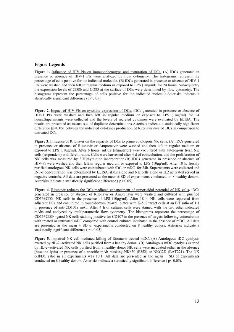
13
Figure Legends
Figure 1. Influence of HIV-PIs on immunophenotype and maturation of DCs. (A) iDCs generated in
presence or absence of HIV-1 PIs were analyzed by flow cytometry. The histograms represent the
percentage of cells positive for the indicated molecule. (B) iDCs generated in presence or absence of HIV-1
PIs were washed and then left in regular medium or exposed to LPS (1mg/ml) for 24 hours. Subsequently
the expression levels of CD86 and CD83 at the surface of DCs were determined by flow cytometry. The
histograms represent the percentage of cells positive for the indicated molecule.Asterisks indicate a
statistically significant difference (p< 0.05).
Figure 2. Impact of HIV-PIs on cytokine expression of DCs. iDCs generated in presence or absence of
HIV-1 PIs were washed and then left in regular medium or exposed to LPS (1mg/ml) for 24
hours.Supernatants were collected and the levels of secreted cytokines were evaluated by ELISA. The
results are presented as mean± s.e. of duplicate determinations.Asterisks indicate a statistically significant
difference (p<0.05) between the indicated cytokines production of Ritonavir-treated DCs in comparison to
untreated DCs.
Figure 3. Influence of Ritonavir on the capacity of DCs to prime autologous NK cells. (A) iDCs generated
in presence or absence of Ritonavir or Amprenavir were washed and then left in regular medium or
exposed to LPS (10 g/ml). After 6 hours, mDCs (stimulator) were cocultered with autologous fresh NK
cells (responders) at different ratios. Cells were harvested after 4 d of coincubation, and the proliferation of
NK cells was measured by 3[H]thymidine incorporation.(B) iDCs generated in presence or absence of
HIV-Pi were washed and then left in regular medium or exposed to LPS (10 g/ml). After 18 h, freshly
purified autologous NK cells were coincubated with iDC or mDC for 24h. Supernatants were collected and
INF- concentration was determined by ELISA. iDCs alone and NK cells alone or IL2 activated served as
negative controls. All data are presented as the mean ± SD of experiments conducted on 8 healthy donors.
Asterisks indicate a statistically significant difference ( p< 0.05).
Figure 4. Ritonavir reduces the DCs-mediated enhancement of tumoricidal potential of NK cells. iDCs
generated in presence or absence of Ritonavir or Amprenavir were washed and cultured with purified
CD56+CD3- NK cells in the presence of LPS (10 g/ml). After 18 h, NK cells were separeted from
adherent DCs and cocultured in round-bottom 96-well plates with K-562 target cells at an E/T ratio of 1:1
in presence of anti-CD107a mAb. After 4 h of culture, cells were stained with the two other indicated
mAbs and analyzed by multiparametric flow cytometry. The histograms represent the percentage of
CD56+CD3- -gated NK cells staining positive for CD107 in the presence of targets following coincubation
with treated or untreated mDC compared with control cultures incubated in the absence of mDC. All data
are presented as the mean ± SD of experiments conducted on 8 healthy donors. Asterisks indicate a
statistically significant difference ( p< 0.05).
Figure 5. Impaired NK cell-mediated killing of Ritonavir treated mDC. (A) Autologous iDC cytolysis
exerted by rIL-2–activated NK cells purified from a healthy donor . (B) Autologous mDC cytolysis exerted
by rIL-2–activated NK cells purified from a healthy donor NK cells were incubated either in the absence
(baseline lysis) or presence of a specific mAb masking NKp30 (F252) or NKG2D (BAT221). The NK
cell/DC ratio in all experiments was 10:1. All data are presented as the mean ± SD of experiments
conducted on 8 healthy donors. Asterisks indicate a statistically significant difference ( p< 0.05).
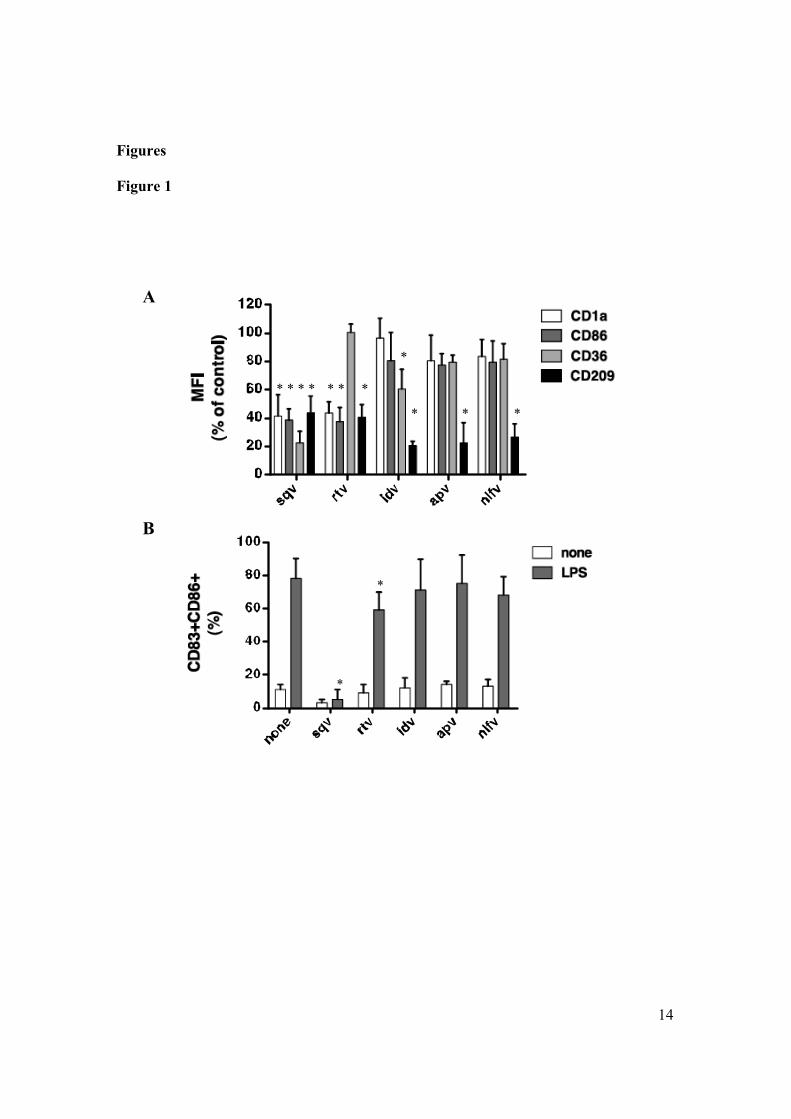
14
Figures
Figure 1
* * * * * * *
*
* * *
*
*
A
B

15
Figure 2
***
TNF-alpha IL-12p40
IL-12p70 IL-15
16
Figure 3
*
*
A
B
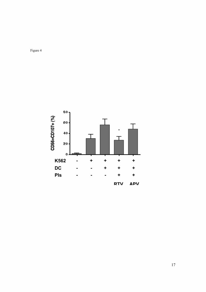
17
Figure 4
*
K562
DC
PIs
- + + + +
- - + + +
- - - + +
RTV APV

18
Figure 5
iDCA)
*
mDC
*
mDCB)

19

20

21