INTERNATIONAL COMMISSION ON RADIOLOGICAL PROTECTION...
28
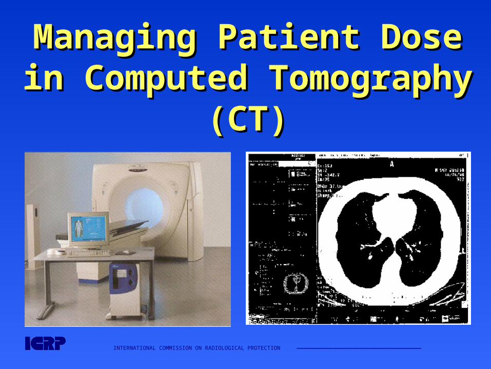
INTERNATIONAL COMMISSION ON RADIOLOGICAL PROTECTION —————————————————————————————————————— Managing Patient Dose Managing Patient Dose in Computed Tomography in Computed Tomography (CT) (CT)
-
Upload
aubrie-gardner -
Category
Documents
-
view
221 -
download
0
Transcript of INTERNATIONAL COMMISSION ON RADIOLOGICAL PROTECTION...
INTERNATIONAL COMMISSION ON RADIOLOGICAL PROTECTION ——————————————————————————————————————
Managing Patient Dose in Managing Patient Dose in Computed Tomography (CT)Computed Tomography (CT)

INTERNATIONAL COMMISSION ON RADIOLOGICAL PROTECTION ——————————————————————————————————————
International Commission on Radiological Protection
Information abstracted from ICRP Publication 87
Available at www.icrp.org
Task Group: M.M. Rehani, G. Bongartz, S.J. Golding, L.Gordon, W. Kalender, T. Murakami, P. Shrimpton,
R. Albrecht, K. Wei
INTERNATIONAL COMMISSION ON RADIOLOGICAL PROTECTION ——————————————————————————————————————
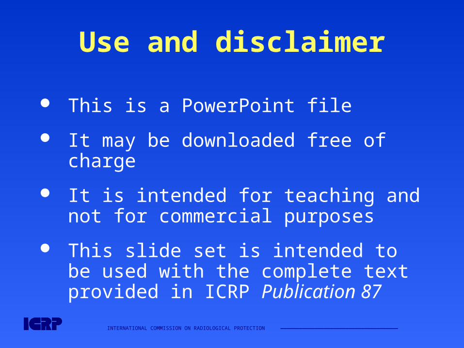
Use and disclaimer
This is a PowerPoint file
It may be downloaded free of charge
It is intended for teaching and not for commercial purposes
This slide set is intended to be used with the complete text provided in ICRP Publication 87
INTERNATIONAL COMMISSION ON RADIOLOGICAL PROTECTION ——————————————————————————————————————
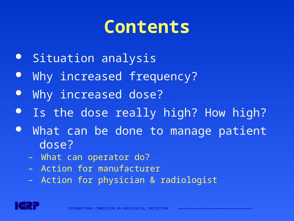
Contents
Situation analysis Why increased frequency? Why increased dose? Is the dose really high? How high? What can be done to manage patient dose?
– What can operator do?– Action for manufacturer– Action for physician & radiologist
INTERNATIONAL COMMISSION ON RADIOLOGICAL PROTECTION ——————————————————————————————————————
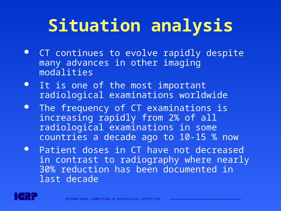
Situation analysis
CT continues to evolve rapidly despite many advances in other imaging modalities
It is one of the most important radiological examinations worldwide
The frequency of CT examinations is increasing rapidly from 2% of all radiological examinations in some countries a decade ago to 10-15 % now
Patient doses in CT have not decreased in contrast to radiography where nearly 30% reduction has been documented in last decade
INTERNATIONAL COMMISSION ON RADIOLOGICAL PROTECTION ——————————————————————————————————————
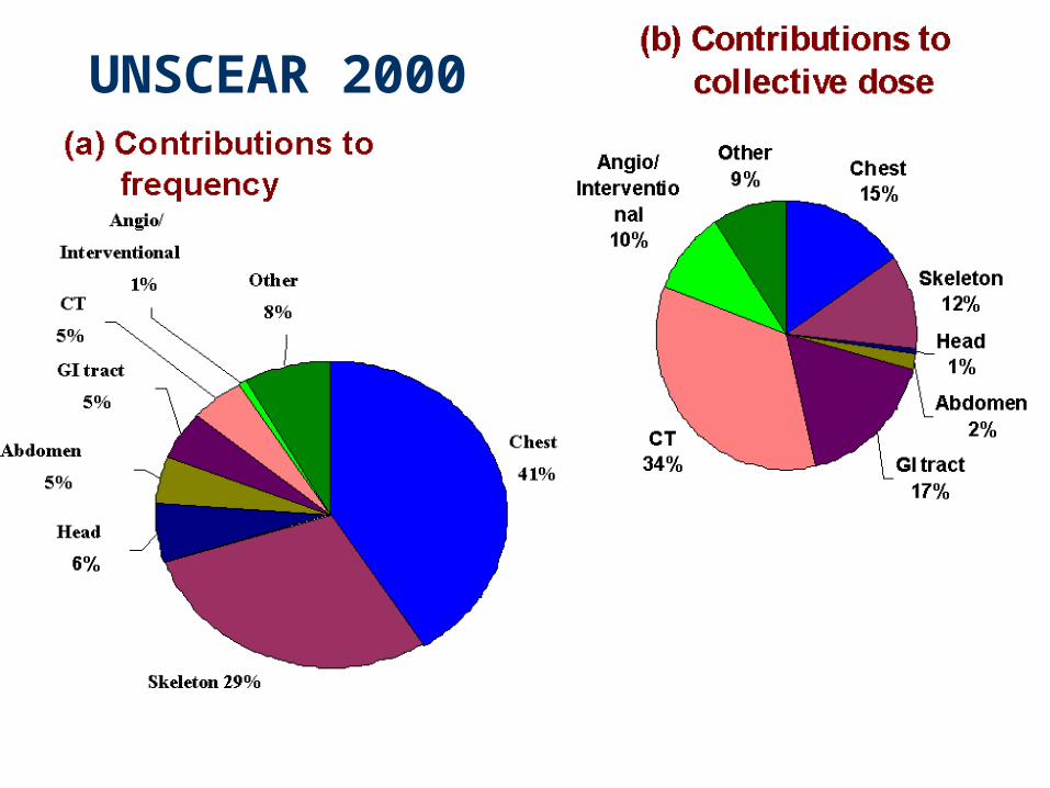
UNSCEAR 2000
INTERNATIONAL COMMISSION ON RADIOLOGICAL PROTECTION ——————————————————————————————————————
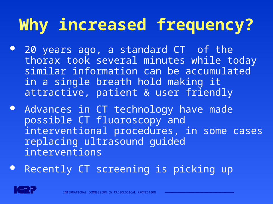
Why increased frequency? 20 years ago, a standard CT of the thorax took
several minutes while today similar information can be accumulated in a single breath hold making it attractive, patient & user friendly
Advances in CT technology have made possible CT fluoroscopy and interventional procedures, in some cases replacing ultrasound guided interventions
Recently CT screening is picking up
INTERNATIONAL COMMISSION ON RADIOLOGICAL PROTECTION ——————————————————————————————————————
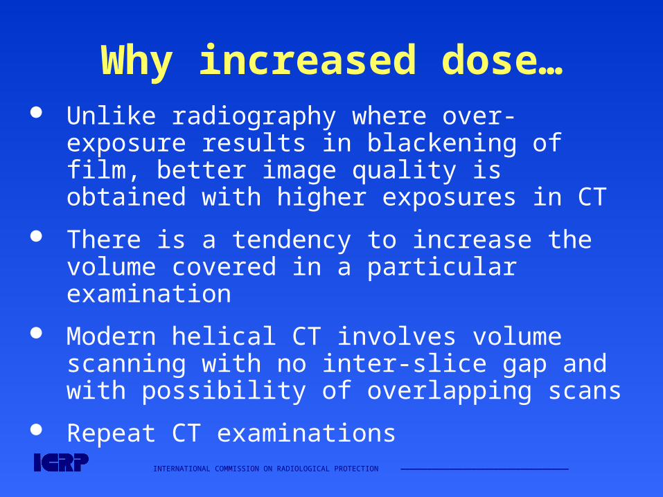
Why increased dose… Unlike radiography where over-exposure
results in blackening of film, better image quality is obtained with higher exposures in CT
There is a tendency to increase the volume covered in a particular examination
Modern helical CT involves volume scanning with no inter-slice gap and with possibility of overlapping scans
Repeat CT examinations
INTERNATIONAL COMMISSION ON RADIOLOGICAL PROTECTION ——————————————————————————————————————
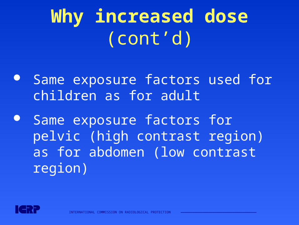
Why increased dose (cont’d)
Same exposure factors used for children as for adult
Same exposure factors for pelvic (high contrast region) as for abdomen (low contrast region)
INTERNATIONAL COMMISSION ON RADIOLOGICAL PROTECTION ——————————————————————————————————————
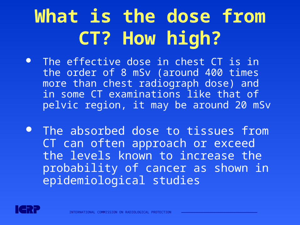
What is the dose from CT? How high?
The effective dose in chest CT is in the order of 8 mSv (around 400 times more than chest radiograph dose) and in some CT examinations like that of pelvic region, it may be around 20 mSv
The absorbed dose to tissues from CT can often approach or exceed the levels known to increase the probability of cancer as shown in epidemiological studies
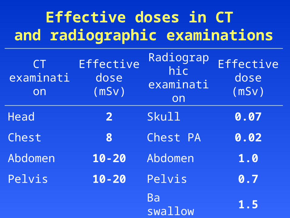
Effective doses in CT and radiographic examinations
CT examination
Effective dose (mSv)
Radiographic examination
Effective dose (mSv)
Head 2 Skull 0.07
Chest 8 Chest PA 0.02
Abdomen 10-20 Abdomen 1.0
Pelvis 10-20 Pelvis 0.7
Ba swallow 1.5
Ba enema 7
INTERNATIONAL COMMISSION ON RADIOLOGICAL PROTECTION ——————————————————————————————————————
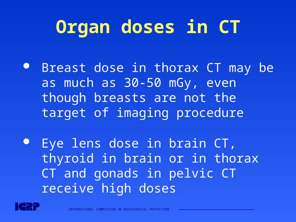
Organ doses in CT
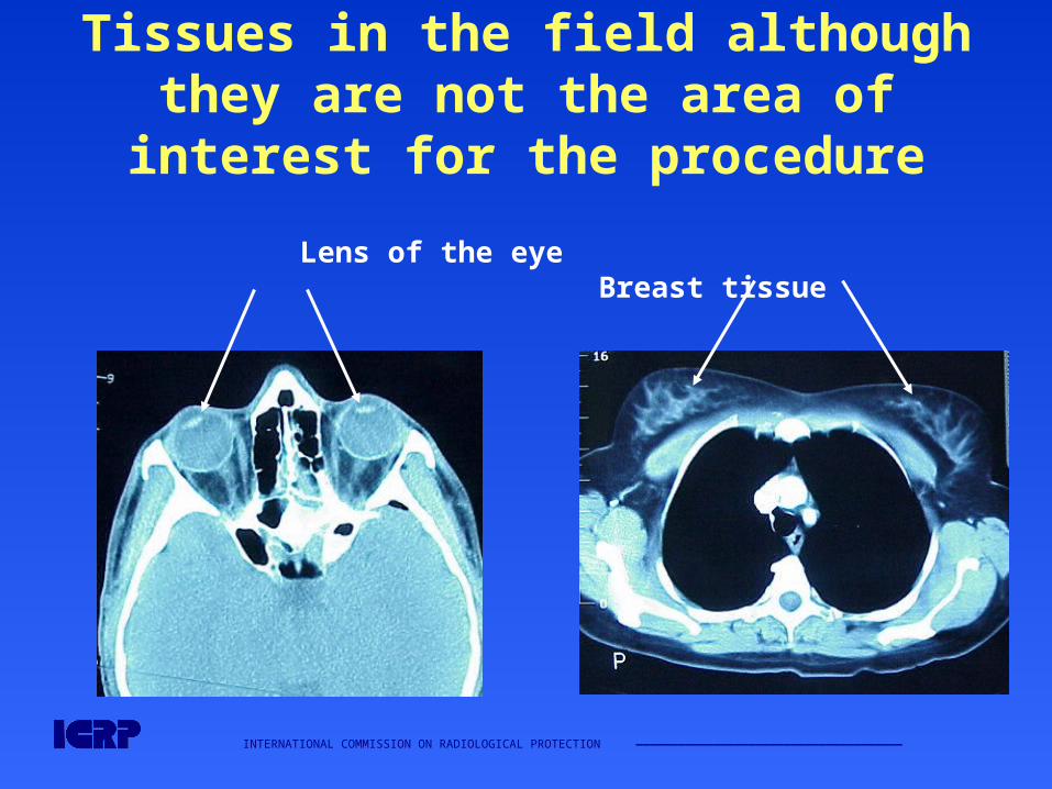
Breast dose in thorax CT may be as much as 30-50 mGy, even though breasts are not the target of imaging procedure
Eye lens dose in brain CT, thyroid in brain or in thorax CT and gonads in pelvic CT receive high doses
INTERNATIONAL COMMISSION ON RADIOLOGICAL PROTECTION ——————————————————————————————————————
Tissues in the field although they are not the area of interest for the procedure
Lens of the eye Breast tissue
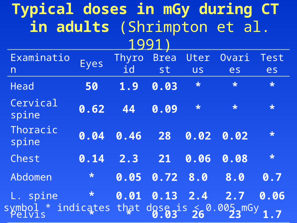
Typical doses in mGy during CT in adults (Shrimpton et al. 1991)
Examination Eyes Thyroid Breast Uterus Ovaries Testes
Head 50 1.9 0.03 * * *
Cervical spine 0.62 44 0.09 * * *
Thoracic spine 0.04 0.46 28 0.02 0.02 *
Chest 0.14 2.3 21 0.06 0.08 *
Abdomen * 0.05 0.72 8.0 8.0 0.7
L. spine * 0.01 0.13 2.4 2.7 0.06
Pelvis * * 0.03 26 23 1.7
The symbol * indicates that dose is < 0.005 mGy
INTERNATIONAL COMMISSION ON RADIOLOGICAL PROTECTION ——————————————————————————————————————
Does spiral CT give more or less radiation dose?
It depends upon the choice of factors
Even though it is possible to perform a spiral CT with lower radiation dose than slice-by-slice CT, in practice the patient gets higher dose due to the factors chosen (scan volume, mAs, pitch, slice width
INTERNATIONAL COMMISSION ON RADIOLOGICAL PROTECTION ——————————————————————————————————————
Does multi-slice CT impart more or less radiation dose?
An increase by 10-30% may occur with multi-slice detector array
INTERNATIONAL COMMISSION ON RADIOLOGICAL PROTECTION ——————————————————————————————————————
Some observations
Most doctors including many radiologists have a feeling that modern CT scanners which are very fast give lesser radiation dose
Unfortunately ‘time’ and ‘radiation dose’ are not proportional in such a situation
Over the years the x-ray tubes are becoming more and more powerful such that they can give high bursts of x-rays which can give satisfactory image in shorter exposure time
INTERNATIONAL COMMISSION ON RADIOLOGICAL PROTECTION ——————————————————————————————————————
What can be done to What can be done to manage patient dose in CT?manage patient dose in CT?
INTERNATIONAL COMMISSION ON RADIOLOGICAL PROTECTION ——————————————————————————————————————
What can operators do…?
Limit the scanned volume Reduce mAs values Use automatic exposure control by adapting
the scanning parameters to the patient cross section. 10-50% reduction in dose documented, without any loss of image quality
INTERNATIONAL COMMISSION ON RADIOLOGICAL PROTECTION ——————————————————————————————————————
What can operators do (cont’d)
Use of spiral CT with pitch factor>1 and calculation of overlapping images instead of acquiring overlapping single scans
Shielding of superficial organs such as thyroid, breast, eye lens and gonads particularly in children and young adults. This results in 30-60% dose reduction to the organ
INTERNATIONAL COMMISSION ON RADIOLOGICAL PROTECTION ——————————————————————————————————————
What can operators do (cont’d)
Separate factors for children. Can reduce dose by a factor of 5 or more
Use of partial rotation e.g. 270 degree in Head CT (refer figure on next slide)
Adequate selection of image reconstruction parameters
Use of z-filtering with multi-slice CT systems Record of dose, exposure factors
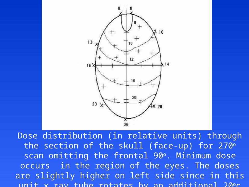
Dose distribution (in relative units) through the section of the skull (face-up) for 270o scan omitting the frontal 90o. Minimum dose
occurs in the region of the eyes. The doses are slightly higher on left side since in this unit x ray tube rotates by an additional 20oc
(clockwise) for patient movement (adapted from Robinson 1996).
INTERNATIONAL COMMISSION ON RADIOLOGICAL PROTECTION ——————————————————————————————————————
Actions for manufacturers
Introduce automatic exposure control Be conscious of high doses in CT Include safety features to avoid unnecessary
dose Display of dose Convenience in using low dose protocols Draw attention of users to selecting separate
protocols for paediatric patients

INTERNATIONAL COMMISSION ON RADIOLOGICAL PROTECTION ——————————————————————————————————————
Actions for physician & radiologist…
Justification: Ensure that patients are not irradiated unjustifiably
Request for CT examination should be generated only by properly qualified medical or dental practitioners depending upon national educational and qualification system. The physician is responsible for weighing the benefits against risks
Clinical guidelines advising which examinations are appropriate and acceptable should be available to clinicians and radiologists
INTERNATIONAL COMMISSION ON RADIOLOGICAL PROTECTION ——————————————————————————————————————
Actions for physician & radiologist (cont’d)
Consider whether the required information be obtained by MRI, ultrasonography
Consider value of contrast medium enhancement prior to commencing examination
CT scanning in pregnancy may not be contraindicated, particularly in emergency situations, although examinations of the abdomen or pelvis should be carefully justified
INTERNATIONAL COMMISSION ON RADIOLOGICAL PROTECTION ——————————————————————————————————————
Actions for physician & radiologist (cont’d)
CT examination should not be repeated without clinical justification and should be limited to the area of interest
Clinician has the responsibility to communicate to the radiologist about previous CT examination of the patient
CT examination for research purpose that do not have clinical justification (immediate benefit to the person undergoing the examination) should be subject to critical evaluation by an ethics committee

INTERNATIONAL COMMISSION ON RADIOLOGICAL PROTECTION ——————————————————————————————————————
Actions for physician & radiologist (cont’d)
CT examination of chest in young girls and young females needs to be justified in view of high breast dose
Once the examination has been justified, radiologist has the primary responsibility for ensuring that the examination is carried out with good technique
INTERNATIONAL COMMISSION ON RADIOLOGICAL PROTECTION ——————————————————————————————————————
Web sites for additional information on radiation sources and effects
European Commission (radiological protection pages):
europa.eu.int/comm/environment/radprot
International Atomic Energy Agency: www.iaea.org
International Commission on Radiological Protection: www.icrp.org
United Nations Scientific Committee on the Effects of Atomic Radiation: www.unscear.org
World Health Organization: www.who.int