Intermediate and fine filaments of vascular leiomyomas (angiomyoma), leiomyoma and leiomyosarcomas...
9
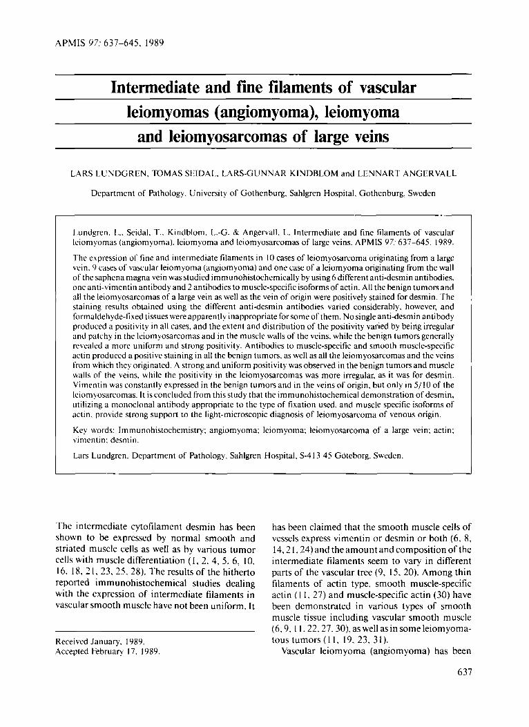
APMIS 07: 637-645. 1989 Intermediate and fine filaments of vascular leiom yomas (angiomyoma), leiom yoma ~ ~~ and leiomyosarcomas of large veins LARS LUNDGREN. TOMAS SEIDAL. LARS-GUNNAR KINDBLOM and LENNART ANGERVALL Department of Pathology. University of Gothenburg. Sahlgren Hospital, Gothenburg. Sweden Lundgren, L.. Seidal. T.. Kindblom, L.-G. & Angervall. L. Intermediate and fine filaments of vascular leiom yomas (angiomyoma). leiomyoma and leiomyosarcomas of large veins. APMIS 97: 637-645, 1989. The expression of tine and intermediate filaments in 10 cases of leiomyosarcoma originating from a large vein. 9 cases of vascular leiomyoma (angiomyoma) and one case of a leiomyoma originating from the wall of the saphena magna vein was studied immunohistochemically by using 6 different anti-desmin antibodies. one anti-vimentin antibody and 2 antibodies to muscle-specific isoforms of actin. All the benign tumors and all the leiomyosarcomas of a large vein as well as the vein of origin were positively stained for desmin. The staining results obtained using the different anti-desmin antibodies varied considerably, however, and formaldehyde-fixed tissues were apparently inappropriate for some of them. No single anti-desmin antibody produced a positivity in all cases, and the extent and distribution of the positivity vaned by being irregular and patchy in the leiomyosarcomas and in the muscle walls of the veins. while the benign tumors generally revealed a more uniform and strong positivity. Antibodies to muscle-specific and smooth muscle-specific actin produced a positive staining in all the benign tumors, as well as all the leiomyosarcomas and the veins from which they originated. A strong and uniform positivity was observed in the benign tumors and muscle walls of the veins. while the positivity in the leiomyosarcomas was more irregular. as it was for desmin. Vimentin was constantly expressed in the benign tumors and in the veins of origin, but only in 5/10 of the leiomyosarcomas. It is concluded from this study that the immunohistochemical demonstration of desmin, utilizing a monoclonal antibody appropriate to the type of fixation used, and muscle specific isoforms of actin. provide strong support to the light-microscopic diagnosis of leiom yosarcoma of venous origin. Key words: Immunohistochemistry: angiomyoma; leiom yoma: leiomyosarcoma of a large vein: actin; vimentin: desmin. Lars Lundgren. Department of Pathology, Sahlgren Hospital. S-4 13 45 Goteborg, Sweden. The intermediate cytofilament desmin has been shown to be expressed by normal smooth and striated muscle cells as well as by various tumor cells with muscle differentiation ( 1, 2, 4, 5, 6, 10, 16, 18, 2 I, 23, 25, 28). The results of the hitherto reported immunohistochemical studies dealing with the expression of intermediate filaments in vascular smooth muscle have not been uniform. It Received January. 1989. Accepted February 17, 1989. has been claimed that the smooth muscle cells of vessels express vimentin or desmin or both (6, 8, 14,2 1.24) and the amount and composition of the intermediate filaments seem to vary in different parts of the vascular tree (9, 15, 20). Among thin filaments of actin type, smooth muscle-specific actin ( 1 1, 27) and muscle-specific actin (30) have been demonstrated in various types of smooth muscle tissue including vascular smooth muscle (6,9. 1 1.22.27,30), as well as in some leiomyoma- tous tumors ( 1 1, 19, 23, 3 1 ). Vascular leiomyoma (angiomyoma) has been 637
-
Upload
lars-lundgren -
Category
Documents
-
view
212 -
download
0
Transcript of Intermediate and fine filaments of vascular leiomyomas (angiomyoma), leiomyoma and leiomyosarcomas...
APMIS 07: 637-645. 1989
Intermediate and fine filaments of vascular leiom yomas (angiomyoma), leiom yoma
~ ~~
and leiomyosarcomas of large veins
LARS LUNDGREN. TOMAS SEIDAL. LARS-GUNNAR KINDBLOM and LENNART ANGERVALL
Department of Pathology. University of Gothenburg. Sahlgren Hospital, Gothenburg. Sweden
Lundgren, L.. Seidal. T.. Kindblom, L.-G. & Angervall. L. Intermediate and fine filaments of vascular leiom yomas (angiomyoma). leiomyoma and leiomyosarcomas of large veins. APMIS 97: 637-645, 1989.
The expression of tine and intermediate filaments in 10 cases of leiomyosarcoma originating from a large vein. 9 cases of vascular leiomyoma (angiomyoma) and one case of a leiomyoma originating from the wall of the saphena magna vein was studied immunohistochemically by using 6 different anti-desmin antibodies. one anti-vimentin antibody and 2 antibodies to muscle-specific isoforms of actin. All the benign tumors and all the leiomyosarcomas of a large vein as well as the vein of origin were positively stained for desmin. The staining results obtained using the different anti-desmin antibodies varied considerably, however, and formaldehyde-fixed tissues were apparently inappropriate for some of them. No single anti-desmin antibody produced a positivity in all cases, and the extent and distribution of the positivity vaned by being irregular and patchy in the leiomyosarcomas and in the muscle walls of the veins. while the benign tumors generally revealed a more uniform and strong positivity. Antibodies to muscle-specific and smooth muscle-specific actin produced a positive staining in all the benign tumors, as well as all the leiomyosarcomas and the veins from which they originated. A strong and uniform positivity was observed in the benign tumors and muscle walls of the veins. while the positivity in the leiomyosarcomas was more irregular. as it was for desmin. Vimentin was constantly expressed in the benign tumors and in the veins of origin, but only in 5/10 of the leiomyosarcomas. It is concluded from this study that the immunohistochemical demonstration of desmin, utilizing a monoclonal antibody appropriate to the type of fixation used, and muscle specific isoforms of actin. provide strong support to the light-microscopic diagnosis of leiom yosarcoma of venous origin.
Key words: Immunohistochemistry: angiomyoma; leiom yoma: leiomyosarcoma of a large vein: actin; vimentin: desmin.
Lars Lundgren. Department of Pathology, Sahlgren Hospital. S-4 13 45 Goteborg, Sweden.
The intermediate cytofilament desmin has been shown to be expressed by normal smooth and striated muscle cells as well as by various tumor cells with muscle differentiation ( 1, 2, 4, 5 , 6, 10, 16, 18, 2 I , 23, 25, 28). The results of the hitherto reported immunohistochemical studies dealing with the expression of intermediate filaments in vascular smooth muscle have not been uniform. It
Received January. 1989. Accepted February 17, 1989.
has been claimed that the smooth muscle cells of vessels express vimentin or desmin or both (6, 8, 14,2 1.24) and the amount and composition of the intermediate filaments seem to vary in different parts of the vascular tree (9, 15, 20). Among thin filaments of actin type, smooth muscle-specific actin ( 1 1, 27) and muscle-specific actin (30) have been demonstrated in various types of smooth muscle tissue including vascular smooth muscle (6,9. 1 1.22.27,30), as well as in some leiomyoma- tous tumors ( 1 1 , 19, 23, 3 1 ).
Vascular leiomyoma (angiomyoma) has been
637
LUNDGREN CI a/.
reported to express desmin and sometimes also vimentin ( 6 , 18, 19). In an immunohistochemical study of 19 leiomyosarcomas of the external soft tissues, desmin was demonstrated in 9 of the tumors (12) and in a recent report a desmin positivity was observed in 39/32 cases of leiomyosarcoma with varying location ( 19). Six of the 19 tumors reported by Hashimoto and co- workers and 2 of the 32 reported by Miettinen involved a large vein, but it is not possible to evaluate from their reports whether any of the desmin-positive tumors were of venous origin. There are to our knowledge no reports in the literature which particulary deal with the interme- diate and fine filaments of smooth muscle tumors originating from large veins.
The aim of the present investigation was to study the expression of intermediate and fine cytofilaments in vascular leiomyoma (angiomy- oma) and in leiomyoma and leiomyosarcoma originating from large veins, and to compare the findings on the two latter tumors with those on the smooth muscle of the vein of origin.
MATERIALS AND METHODS
The material for the immunohistochemical study com- prised 9 cases of vascular leiomyoma, 10 cases of leiom yosarcoma and one case of a leiomyoma originat- ing from the wall of a large vein. All the tumors were accrued at the Department of Pathology, Sahlgren Hos-
pital. Goteborg. Sweden. The surgical specimens ob- tained were fixed in 4 percent formaldehyde.
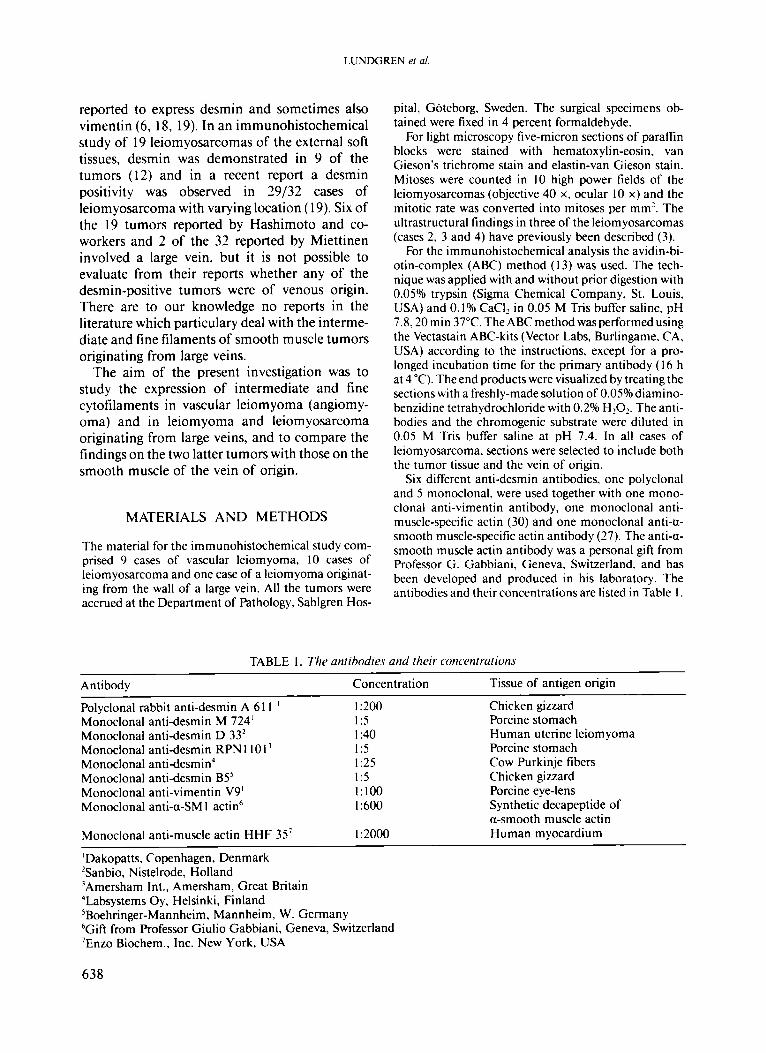
For light microscopy five-micron sections of paraffin blocks were stained with hematoxylin-eosin, van Gieson's trichrome stain and elastin-van Gieson stain. Mitoses were counted in 10 high power fields of the leiomyosarcomas (objective 40 x. ocular 10 x) and the mitotic rate was converted into mitoses per mm'. The ultrastructural findings in three of the leiomyosarcomas (cases 2, 3 and 4) have previously been described (3).
For the immunohistochemical analysis the avidin-bi- otin-complex (ABC) method ( I 3) was used. The tech- nique was applied with and without prior digestion with 0.05% trypsin (Sigma Chemical Company, St. Louis. USA) and 0.1% CaCl, in 0.05 M Tris buffer saline, pH 7.8,20 min 37°C. The ABC method was performed using the Vectastain ABC-kits (Vector Labs, Burlingame. CA, USA) according to the instructions. except for a pro- longed incubation time for the primary antibody (16 h at 4 "C). The end products were visualized by treating the sections with a freshly-made solution of 0.05% diamino- benzidine tetrahydrochloride with 0.296 HzOz. The anti- bodies and the chromogenic substrate were diluted in 0.05 M Tris buffer saline at pH 7.4. In all cases of leiomyosarcoma, sections were selected to include both the tumor tissue and the vein of origin.
Six different anti-desmin antibodies, one polyclonal and 5 monoclonal, were used together with one mono- clonal anti-vimentin antibody, one monoclonal anti- muscle-specific actin (30) and one monoclonal anti-u- smooth muscle-specific actin antibody (27). The anti-a- smooth muscle actin antibody was a personal gift from Professor G. Gabbiani, Geneva, Switzerland, and has been developed and produced in his laboratory. The antibodies and their concentrations are listed in Table 1 .
TABLE 1. The antibodies and their concentrations
Anti body Concentration Tissue of antigen origin
Polyclonal rabbit anti-desmin A 6 1 1 ' Chicken gizzard Monoclonal anti-desmin M 724' 1:5 Porcine stomach
1 :200
Monoclonal anti-desmin D 33' Monoclonal anti-desmin RPN 1 10 l 3 Monoclonal anti-desmin4 Monoclonal anti-desmin B5' Monoclonal anti-vimentin V9' Monoclonal anti-a-SM 1 actid
:40 :5 :25 :5 : 100 :600
Human uterine leiomyoma Porcine stomach Cow Purkinje fibers Chicken gizzard Porcine eye-lens Synthetic decapeptide of a-smooth muscle actin
Monoclonal anti-muscle actin HHF 35' 1 :2000 Human myocardium
'Dakopatts, Copenhagen, Denmark 'Sanbio, Nistelrode, Holland 'Amersham Int., Amersham, Great Britain 'Labsystems Oy, Helsinki, Finland 5Boehringer-Mannheim, Mannheim, W. Germany 'Gift from Professor Giulio Gabbiani, Geneva, Switzerland 'En20 Biochem.. Inc. New York, USA
638
INTERMEDIATE FILAMENTS IN LEIOMYOMA A N D LEIOMYOSARCOMA
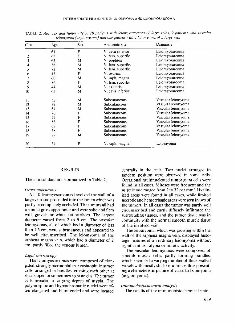
TABLE 3. Age, .W.Y and tiirnor .sit[> in 10 paticnts with liiomvosarcoma of larg(1 wins, 9 patients bvith vasciilar It%)m voma (angiomvoma) and nnc patient with a leiomyoma (?f a Iurp vein
Case Ane Sex Anatomic site Diagnosis
I
3 4 5 h 7 8 9
10
7
I I 12 13 14 15 16 17 I8 I9
20
61 63 63 58 73 45 60 86 44 63
52 79 64 76 77 58 67 58 27
34
F F M M M F M F M M
M M M F F F F F M
F
V. cava inferior V. fem. superfic. V. poplitea V. fem. superfic. V. fem. superfic. V. ovarica V. saph. magna V. fem. superfic V. axillaris V. cava inferior
Subcutaneous Subcutaneous Subcutaneous Subcutaneous Subcutaneous Subcutaneous Subcutaneous Subcutaneous Subcutaneous
V. saph. magna
Leiom yosarcoma Leiom yosarcoma Leiom yosarcoma Leiom yosarcoma Leiom yosarcoma Leiom yosarcoma Leiom yosarcoma Leiom yosarcoma Leiom yosarcoma Leiom yosarcoma
Vascular leiomyoma Vascular leiom yoma Vascular leiomyoma Vascular leiomyoma Vascular leiomyoma Vascular leiom yoma Vascular leiom yoma Vascular leiomyoma Vascular leiom yoma
Leiom yoma
RESULTS
The clinical data are summarized in Table 2.
Gross uppourunce All 10 leiomyosarcomas involved the wall of a
large vein and protruded into the lumen which was partly or completely occluded. The tumors all had a similar gross appearance and were solid and firm with greyish or white cut surfaces. The largest diameter varied from 2 to 9 cm. The vascular leiomyomas, all of which had a diameter of less than 1.5 cm, were subcutaneous and appeared to be well circumscribed. The leiomyoma of the saphena magna vein, which had a diameter of 2 cm, partly filled the venous lumen.
Light microscopy The leiomyosarcomas were composed of elon-
gated, strongly picrinophilic or eosinophilic tumor cells. arranged in bundles, crossing each other at sharp, open or sometimes right angles. The tumor cells revealed a varying degree of atypia. The polymorphic and hyperchromatic nuclei were of- ten elongated and blunt-ended and were located
centrally in the cells. Two nuclei arranged in tandem position were observed in some cells. Occasional multinucleated tumor giant cells were found in all cases. Mitoses were frequent and the mitotic rate ranged from 2 to 32 per mm’. Hyalin- ized areas were found in all cases, while limited necrotic and hemorrhagic areas were seen in two of the tumors. In all cases the tumor was partly well circumscribed and partly diffusely infiltrated the surrounding tissues. and the tumor tissue was in continuity with the normal smooth muscle tissue of the involved vein.
The leiomyoma, which was growing within the wall of the saphena magna vein, displayed histo- logic features of an ordinary leiomyoma without significant cell atypia or mitotic activity.
The vascular leiomyomas were composed of smooth muscle cells, partly forming bundles, which encircled a varying number of thick-walled vessels with mostly slit-like luminae, thus present- ing a characteristic picture of vascular leiom yoma (angiomyoma).
Immtinohistochemicul una1r.si.s The results of the immunohistochemical stain-
639

LUNDGREN e! a/
+ + + + + + + + + +
+ + + + + + + + + +
+ + + + + + + + + +
LL I + + l I l + l I I
+ + + + + + + + + +
L L I I I l + l + l I I
LL L L L L L I + l I + l + + l +
+ + + l + l + + + +
M C C cd
.d
.- i- m
0
6) 3 -0
c
II II L * +
II II + I
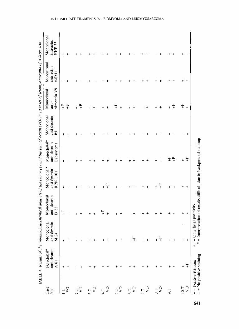
ings are summarized in Tables 3 and 4. The staining results obtained using the 6 differ-
ent anti-desmin antibodies varied considerably. From the results obtained in control studies it was obvious that formaldehyde-fixed tissues were in- appropriate for 4 of the anti-desmin antibodies, three of which produced a disturbing background stain (see Tables 3 and 4). The monoclonal anti- body D 33 was found to produce the most consis- tent staining without any disturbing background. With this antibody 9/10 of the benign tumors, 7/10 of the leiomyosarcomas and 9/10 of the muscle walls of veins were positively stained. With the D 33 antibody the staining was strong and homogeneous in the benign tumors. while the positivity was irregularly and patchily distributed both in the leiomyosarcomas and in the muscle wall of the veins. The muscle-specific and the smooth muscle-specific actin antibodies produced a positive staining in all the benign and malignant tumors, as well as in the veins, although the staining was stronger and more prominent in the benign tumors than in the leiomyosarcomas, as it was for desmin. Vimentin was constantly found in all the benign tumors, while only 5/10 of the leiomyosarcomas of a large vein were positively stained.
DISCUSSION
Leiomyosarcomas constitute a heterogeneous group of tumors in terms of clinical presentation, morphologic appearance and type of smooth mus- cle of origin (7). Leiomyosarcomas of a large vein, however, are considered to represent a hornoge- neousgroupof tumors(3). In the 10 leiomyosarco- mas of a large vein included in this study, the smooth muscle differentiation was obvious light- microscopically, as is characteristically found in such tumors. In a previous study including 3 of the leiomyosarcomas described in this study the leiomyomatous differentiation has been further established by the electronmicroscopic findings ( 3 ) . The leiomyoma of the saphena magna vein and the vascular leiomyomas were also chosen to represent light-microscopically characteristic ex- amplesof these tumorentities. Thus, the imrnuno- histochemical analysis was performed on a well- defined group of leiomyomatous tumors.
Desmin. which is found in both smooth and striated muscle cells. has been shown to be ex-
640
TAB
LE 4
. Res
ults
q/'
the
imm
unoh
i.sto
c~ie
mic
al an
aly.v
is of
the
tum
or (T
) and
the
vein
of
orig
in (V
O) i
n 10
case
s of
leio
myo
sarc
oma
of a
larg
e ve
in
Cas
e Po
lycl
onal
* M
onoc
lona
l M
onoc
lona
l M
onoc
lona
l*
Mon
oclo
nal*
M
onoc
lona
l M
onoc
lona
l M
onoc
lona
l M
onoc
lona
l N
O
anti-
desm
in
anti-
desm
in
anti-
desm
in
anti-
desm
in
anti-
desm
in
anti-
desm
in
anti-
an
ti-ac
tin
2 A
61
1
M 2
4 D
33
RPN
1 1
01
Labs
yste
ms
B5
vim
entin
V9
a-SM
1 H
HF
35
2 z +
F
+ +
o_
anti-
actin
;a
- I .
T
+ +F
-
- -
+F
+ +
- -
-
- -
vo
+ -
- 3
3.T
-
- +
+ -
+ +
3
vo
- -
+ +
+ -
+F
+ +
r
rn
3.T
+
+ +
+ +
+ +
z -3 vo
+
+ +
+ +
+ +
+ v) $
- -
-
4.T
vo
+ + +F
+
- +F
+ + + +
+ + + +
5.T vo
+F
+
+ + + +
+ + - +
- + - +
+ +
6.T
vo
+ +
+ + + +
+ + + +
- +F
- +
7.T
vo
+ +
+ + + +
+ + - +
+ + + +
- + - +
+ +
- +
+ +
8.T
+
vo
+ +F
+
+F
- -
+ +
+
9.T
+
- +
+ +
F
- -
+ +
+ -
+ -
+F
-
+F
+
+
10.T
+
+ +
+ +
- +F
+
+ vo
+
F
- +
+ +
F
- +
+ +
Q\
+ =
Pos
itive
sta
inin
g +
F =
Onl
y fo
cal p
ositi
vity
5
- =
No
posi
tive
stai
ning
* =
Int
erpr
etat
ion
of re
sults
diff
icul
t due
to b
ackg
roun
d st
aini
ng

LUNDGREN c! a!.
642

INTERMEDIATE FILAMENTS IN LEIOMYOMA A N D LEIOMYOSARCOMA
pressed in benign and malignant tumors with striated muscle differentiation ( 1 ~ 10, 19, 2 I , 25, 28) and has been found to be a useful marker in the recognition of poorly differentiated rhabdomyo- sarcomas (26). The usefulness of desmin expres- sion in the recognition of leiomyosarcomas in general and of venous origin in particular has not been investigated to the same degree. In this study desmin was demonstrated in all the vascular leiomyomas, in the leiomyoma and in all the leiomyosarcomas of venous origin when using the entire battery of anti-desmin antibodies. The re- sults obtained by the different anti-desmin anti- bodies varied considerbly. however, and none of the antibodies used alone produced a positivity in all cases. In previous studies of rhabdomyosarco- ma the anti-desmin antibody which was found to produce the most consistent positive staining with- out disturbing background staining (D 33) has also been found to be highly reliable when formalde- hyde-fixed tissues are used (25). The poor results obtained by most of the other anti-desmin anti- bodies. with either disturbing background staining making the interpretation of positive results diffi- cult or the complete lack of positivity (in both benign and malignant tumors as well as in ordinary veins), are probably explained by the fact that formaldehyde-fixed tissues are inappropriate for these antibodies.
In the present study the smooth muscle cells of all the large veins of the extremities and pelvis as well as the 2 inferior cava veins were found to
F/g. I :I Leiomyosarcoma protruding into the lumen of the vein of origin. Elastin-van Gieson stain, x 16. B. Leiomyosarcoma composed of picrinophilic cells arranged in fascicles crossing each other at sharp or right angles. The tumor cells reveal an elongated nucleus with blunt ends. van Gieson stain, x 80. C & D. Leiomyosarcoma of a large vein. Positive immunohistochemical staining for a-smooth muscle actin (C) and desmin (D) in tumor cells as well as in the smooth muscle cells of the vein of origin. T =tumor, VO = vein of origin. x 40. E & F. Leiomyosarcoma of a large vein. Positive staining of the tumor cells for a-smooth muscle-specific (E) and muscle-specific (F) actin. x 80. G & H. Vascular leiomyoma with tumor cells positively stained for desmin (G) and a-smooth muscle actin (H). x 40. (Fig. I C to 1 H: ABC-technique with hematoxylin coun- terstain)
express desmin. The positive staining was, howev- er, frequently irregularly distributed in the wall of the veins. With only one exception, all the benign tumors were strongly and homogeneously positive for desmin using the D 33 antibody, while 3 of the leiomyosarcomas were completely negative and 2 were only partially positively stained with this antibody. The results thus indicate a quantitative and/or qualitative difference between ordinary vascular smooth muscle and benign tumors on the one hand and the vascular leiomyosarcomas on the other.
Vimentin was found to be expressed in smooth muscle of the veins of origin and in all the benign tumors, while only 5 / I0 of the leiom yosarcomas contained tumor cells which expressed vimentin. Although double staining for desmin and vimentin was not performed it was obvious that there was a co-expression of these filaments in the cells of the benign tumors and most probably also in some of the leiomyosarcomas. Our findings thus contrast with those reported by Schurch and cu-workers ( 1987), who found a positive staining for vimentin in lo/ 10 leiomyosarcomas, none of which was of proven venous origin.
Both the muscle-specific and the smooth mus- cle-specific isoforms of actin were found to be present homogeneously throughout the smooth muscle walls of the veins and the benign tumors. All the leiomyosarcomas were also found to ex- press these actins, although the stainings were not always homogeneous. These muscle-specific actins seem to be more regularly expressed in leiom yosarcomas than desmin, and are considered to be a valuable diagnostic tool. However, the value of demonstrating the isoform of actin common to both smooth and striated muscle in the diagnosis of leiomyosarcoma is somewhat limited by the fact that a positive staining is also obtained in rhab- domyoblastic tumors ( 19, 30). Furthermore, this actin and the smooth muscle-specific actin are found in myofibroblasts of reactive lesions such as proliferative myositis and fasciitis and of fibro- matoses such as desmoid. palmar and plantar fibromatosis (17, 29, 31, 32). In a recent study Schurch and co-workers (1987) reported that the expression of a-smooth muscle-specific actin was related to the degree of cell differentiation in the leiomyosarcomas. The results of the present study did not clearly indicate any relationship between the degree of light microscopic differentiation in the tumor cells of the leiomyosarcomas and the
643

LUNDGREN 1" ul
expression of muscle actins. As has been pointed out, however, a stronger and more homogeneous staining was more often seen in the benign tumors than in the leiomyosarcomas.
It can be concluded from the results of the present investigation that desmin is expressed by the smooth muscle cells of veins and vascular leiomyomas, as well as of leiomyomas and most leiom yosarcomas originating from large veins. The lack of desmin positivity in a single section of a spindle cell sarcoma of venous origin does not, however, exclude the diagnosis of leiomyosarco- ma. at least as long as formaldehyde-fixed tissues are used. The demonstration of different isoforms of muscle-specific actins is considered to provide strong support for the light microscopic diagnosis of leiomyosarcoma. despite their lack of smooth muscle speficity. The combination of an anti- desmin antibody, chosen as appropriate for the type of fixation used, and antibodies against mus- cle-specific actins seems to be a good way of obtaining support for the diagnosis of a vascular leiom yosarcoma. Whether the results and conclu- sions of this study are also valid for leiomyoma- tous tumors with a different origin still remains to be shown.
REFERENCES
I .
2.
3.
4.
5.
6.
.41rmannsherger, M., Weber. ti.. horn. M.; Desmin is a specific
Droste, R. & Os- marker for rhab-
domyosarcomas of human and rat origin. Am. J.
.1Itmunn.sb~~r~~c~r, M., Dirk. T., Oshorn. M . & Weher, K.: Immunohistochemistry of cytoskeletal fila- ments in the diagnosis of soft tissue tumors. Sem. Diagn. Pathol. 3: 306-3 13. 1986. Berlin, 0, Stener: B., tiindblom. L.-G. & Angervall. L.; Leiomyosarcomas of venous origin in the ex- tremities: A correlated clinical, roentgenologic and morphologic study with diagnostic and surgical implictions. Cancer 54: 2 147-2 159, 1984. Brooks, J . J.: The significance of double phenotypic patterns and markers in human sarcomas. Am. J.
Denk. €1.. Krepler. R.. Artlieb* U. , Gabbiani. G., Rungger-Brundles E., Leoncini, P. & Franke, W. U'.: Proteins of intermediate filaments: An immunohis- tochemical and biochemical approach to the classifi- cation of soft tissue tumors. Am. J. Pathol. 110:
Evuns, D. J.. Lumperl. I . A . & Jacobs, M.: Interme- diate filaments in smooth muscle tumors. J. Clin.
Pathol. 118: 85-95, 1985.
Pathol. 125; 113-123. 1986.
193-208, 1983.
Pathol. 36: 57-61. 1983.
7. Enzingcy F. M. & M.'eiss, S. U'.; Soft tissue tumors. St. Louis, C. V. Mosby, 1988, pp. 402, 417-419.
8. Frank, E. D. & Warren, L.: Aortic smooth muscle cellscontain vimentin insteadof desmin. Proc. Natl. Acad. Sci. 78: 3020-3024, I98 I .
9. Gubbiani. G., Schmid, E.. Ri'nrer, S. , Cliuponnicy C.. de CIIastonay. C., I andt~rkrrckliovi~, J.. M'ehcr, ti. & Franke. M'. M'.: Vascular smooth muscle cells differ from other smooth muscle cells: Predomi- nance of vimentin filaments and a specific a-type actin. Proc. Natl. Acad. Sci. 78: 298-302. 198 1.
10. Gubbiani. G.. tiupanci, Y., Bara::one. P. & Franke. U', W'.; lmmunohistochemical identification of in- termediate-sized filaments in human neoplastic cells: A diagnostic aid for the surgical pathologist. Am. J. Pathol. 104: 206-216, 1981.
I I . Gown, A . M., Vogel, A . M . . Gordon, D. & Lic. P. L.: A smooth muscle-specific monoclonal antibody rec- ognizes smooth muscle actin isozymes. J. Cell. Biol.
12. liashimoro, H . . Daimurii, Y.. Tsiincy).rhi, M . & Enjoji, .&I.; Leiomyosarcoma of the external soft tissues. Cancer 57: 2077-2088, 1986.
13. €l.sii, S. M., Ruinc. L. & Funget: €I.: The use of antiavidin antibody and avidin-biotin-peroxidase complex in immunoperoxidase technics. Am. J. Clin. Pathol. 75: 816-821, 1981.
14. Kocher: O., Skulli. 0.. Bloom, M'. S. & Gahhiuni. G.: Cytoskeleton of rat aortic smooth muscle cells: Normal conditions and experimental intimal thick- ening. Lab. Invest. SO; 645-652, 1984.
15. tiocher, 0. & Gahbiuni, G.: Cytoskeletal fetures of normal and atheromatous human arterial smooth muscle cells. Hum. Pathol. 17: 875-880, 1986.
16. Lazarides, E.: Intermediate filaments: A chemically heterogeneous. developmentally regulated class of proteins. Ann. Rev. Biochem. 51: 219-250. 1982.
17. Lundgren. L., Kindhlom, L.-G., M'illems, J. & Angenwll, L.: Proliferative myositis and fasciitis: A light- and electron-microscopic, cytologic and im- munohistochemical study and a quantitative DNA- analysis. Submitted 1989.
18. Miettinen. M.. Lehto, V.-P. & l'irranen. I . : Anti- bodies to intermediate filament proteins in the diagnosis and classification of human tumors. Ul- trastruct. Pathol. 7: 83-107, 1985.
19. Miettinen, M.: Antibody specific to muscle actins in the diagnosis and classification of soft tissue tumors. Am. J. Pathol. 130: 205-215, 1988.
20. Osborn, M.. Caselit:. J . & Weher, K.: Heterogeneity of intermediate filament expression in vascular smooth muscle: A gradient in desmin positive cells from the rat aortic arch to the level of the arteria iliaca communis. Differentiation 20: 196-202, 198 I .
2 I . Osborn, M . & Weber, ti.: Tumor diagnosis by inter- mediate filament typing: A novel tool for surgical pathology. Lab. Invest. 48: 372-394, 1983.
22. Owens, G. ti., Loeb, A , Gordon. D. & Thompson, M . M.: Expression of smooth muscle-specific a-isoactin in cultured vascular smooth muscle cells: Relation-
100: 807-813. 1985.
644
INTERMEDIATE FILAMENTS IN LEIOMYOMA A N D LEIOMYOSARCOMA
ship between growth and cytodifferentiation. J . Cell. Biol. 102: 343-352, 1986.
23. Sehiirdi. U.,. Skulli, 0.. Seemajw T. & Gahhiani, G.: Intermediate filament proteins and actin isoforms as markers for soft tissue tumor differentiation and origin. I. Smooth muscle tumors. Am. J . Pathol. 128:
24. Sckmid. E.. Oshorn, jkf.s Ritngger-Brundle, E.. Gah- hiani. G., H'ehcr, K. & Frunke, M'. M..: Distribution of vimentin and desmin filaments in smooth muscle of mammalian and avian aorta. Exp. Cell. Res. 137:
25. Seidal. T.. Kindhlom. L.-G. & Angendl, L.: Myo- globin, desmin and vimentin in ultrastructurally proven rhabdomyomas and rhabdomyosarcomas.
26. Seiikd 71, Kindhlom. L.-G. & . . l ngmd . L.: Alve- olar and poorly differentiated rhabdomyosarcoma. A clinicopathologic, light-microscopic, ultrastruc- tural and immunohistochemical analysis. APMIS
27. Skalli, 0.. Ropru:. P., Trzeciuk. .4 . . Benzonuna. G.. Gillc.ssen. D. & Guhhiani. G.: A monoclonal anti- body against a-smooth muscle actin: A new probe for smooth muscle differentiation. J . Cell. Biol. 103:
91-103. 1987.
329-340. 1982.
Appl. Pathol. 5: 201-219. 1987.
96: 839-848. 1988.
2787-2796. 1986.
28. Skalli. 0.. Gabhimi. G.. Bahui. F., Seemaj.er. T. '4.. Pizzolaro, 6. & SchiircIi, MI.: Intermediate filament proteins and actin isoforms as markers for soft tissue tumor differentiation and origin. 11. Rhabdomyosar- comas. Am. J. Pathol. 130: 515-531, 1988.
29. Skulli. 0.. Sdii ir th, U'.. Seemayer, T., Lagact', R., .ilonlundon, D. & Guhhiuni, G.: Myofibroblasts from diverse pathologic settings are heterogenous in their content of actin isoforms and intermediate filament proteins. Lab. Invest. vol. 60 1989. In press.
30. Tsitkudu, T., Tipprns, D., Gordon. D.. Ross, R. & Go\vn, A . M . : HHF 35, a muscle-actin-specific mon- oclonal antibody. Am. J. Pathol. 126: 51-60, 1987.
3 1. Tnrkudu. T., M c h'iitt, M. 11.. Ross. R . & Gown. A . M . : HHF35: A muscle specific monoclonal anti- body: 11. Reactivity in normal, reactive and neoplas- tic human tissues. Am. J . Pathol. 127: 389-402, 1987.
32. I'iulc. ti.. Doglioni, C.? Iiixolino. P.. Boniempini. L., C'olomhi, R., Coggi. G. & Dell'Orto. P.: Infantile digital fibromatosis-like tumour (inclusion body fibromatosis)of adulthood: Report of twocaseswith ultrastructural and immunocytochemical findings. Histopathology I.?: 415-424. 1988.
645