Intergranular Corrosion-Fatigue Failure of Cobalt-Alloy ...
6
Copyright 1994 by The Journal ofBone and Joint Surgery. Incorporated 110 THE JOURNAL OF BONE AND JOINT SURGERY Intergranular Corrosion-Fatigue Failure of Cobalt-Alloy Femoral Stems A FAILURE ANALYSIS OF TWO IMPLANTS* BY JEREMY L. GILBERT, PH.D.t. CHRISTINE A. BUCKLEY, M.S.t. JOSHUA J. JACOBS. M.D.. KIM C. BERTIN. MDL AND MICHAEL R. ZERNICH. MDI. CHICAGO, ILLINOIS Investigation performed at the Division of Biological Materials. North western University, Chicago ABSTRACT: Two modular hip implants with a cobalt- alloy head and a cobalt-alloy stem were retrieved after a fracture had occurred in the neck region of the fem- oral component, eighty-five and seventy months after implantation. Both implants failed less than one milli- meter distal to the taper junction between the head and the stem (outside of the taper). The fracture surfaces of the implant were investigated with the use of scanning electron microscopy, to determine the nature of the failure process. The fractures occurred at the grain boundaries of the microstructure and appeared to be the result of three factors: porosity at the grain bound- aries; intergranular corrosive attack, initiated both at the head-neck taper and at the free surface; and cyclic fatigue-loading of the stem. The corrosive attack of the free surface was initiated, in part, by the egression of surface grains and by the ingression of fluid into the intergranular regions. Sectioned surfaces showed ex- tensive intergranular corrosive attack in the prosthetic neck localized in the region of the head-neck taper junction and penetrating deeply into the microstructure. Recent reports’346’ have indicated that a corrosive attack can occur in the taper crevice of modular im- plants made of similar metals or of mixed-metal combi- nations. On the basis of these observations, it has been speculated that the corrosive attack, while resulting in *One or more of the authors have received or will receive ben- efits for personal or professional use from a commercial party re- lated directly or indirectly to the subject of this article. In addition, benefits have been or will be directed to a research fund, founda- tion, educational institution, or other non-profit organization with which one or more of the authors are associated. Funds were re- ceived in total or partial support of the research or clinical study presented in this article. The funding source was National Institutes of Health Grant AR 39310. tDivision of Biological Materials, Northwestern University, Chicago, Illinois 60611. Please address requests for reprints to Dr. Gilbert. Rush Medical College Joint Replacement Program, Depart- ment of Orthopedic Surgery, Rush-Presbyterian-St. Luke’s Medical Center, Chicago, Illinois 60612. §Department of Orthopedic Surgery, University of Utah, Latter Day Saints Hospital, 440 D Street, Suite 206, Salt Lake City. Utah 84103. 9lDepartment of Orthopedic Surgery, University of Pittsburgh, Aliquippa Medical Center, Hospital Drive, Aliquippa, Pennsylvania 15001. metal release9, may lead to mechanical failure of the component. Gilbert et al.6 and Buckley et al. reported that one of several corrosion mechanisms observed in the taper region was intengranular attack in implants that had a cobalt-alloy head coupled with a cobalt-alloy stem. In one prosthesis, this intengranulam attack had penetrated as much as five millimeters deep from the surface of the neck region of the taper. It is possible that this corrosive attack may have ramifications with regard to the structural integrity of implants and may contnib- ute to fracture in the neck region of hip prostheses. This observation has not been reported or demonstrated in the literature to our knowledge. Recently, two modular total hip prostheses, consist- ing of a wrought cobalt-alloy (ASTM F799) head cou- pled with a wrought cobalt-alloy stem, were received in the implant retrieval laboratory at Rush-Presbyterian- St. Luke’s Medical Center after revisions had been per- formed to replace the fractured components. Both of the fractures had occurred in the neck. just distal to the head-neck taper. We present the results of a detailed fractographic and electron microscopic analysis of the region in which these prostheses had failed. Case Reports CASE 1. A 112-kilogram. 175-centimeter-tall man had a primary total hip replacement with the insertion of a PCA prosthesis (How- medica, Rutherford, New Jersey) without cement, at the age of sixty- seven years, for the treatment of severe osteoarthrosis (Fig. I-A). Seventy months postoperatively. the patient was seen by one of us (M. R. Z.) because of the sudden onset of pain in the hip. Before this, the hip had been asymptomatic. The patient had had an active lifestyle and had frequently played golf. Radiographs revealed a fractured prosthetic neck (Fig. 1-B). At the revision. the prosthetic fracture was easily identified, and the prosthetic head was free in the joint. The femoral component was extremely well fixed to the adjacent bone and was difficult to extract. On removal, an extensive amount of bone was adherent to the device, and a fracture of the femoral shaft was sustained intraoperatively. The acetabular component was well fixed. A long-stem prosthesis was inserted without cement and with the use of strut allografts. The patient was doing well two years postoperatively. CASE 2. A 111-kilogram. 178-centimeter-tall man was managed. at the age of forty-nine years. with a primary total hip replacement with the insertion of a PCA prosthesis (Howmedica) without cement, for end-stage osteoarthrosis of the right hip. The postoperative course was uneventful with the exception of mild pain in the lateral aspect of
Transcript of Intergranular Corrosion-Fatigue Failure of Cobalt-Alloy ...

Copyright 1994 by The Journal ofBone and Joint Surgery. Incorporated
110 THE JOURNAL OF BONE AND JOINT SURGERY
Intergranular Corrosion-Fatigue Failure of
Cobalt-Alloy Femoral StemsA FAILURE ANALYSIS OF TWO IMPLANTS*
BY JEREMY L. GILBERT, PH.D.t. CHRISTINE A. BUCKLEY, M.S.t. JOSHUA J. JACOBS. M.D.�. KIM C. BERTIN. MDL
AND MICHAEL R. ZERNICH. MDI. CHICAGO, ILLINOIS
Investigation performed at the Division of Biological Materials. North western University, Chicago
ABSTRACT: Two modular hip implants with a cobalt-
alloy head and a cobalt-alloy stem were retrieved aftera fracture had occurred in the neck region of the fem-oral component, eighty-five and seventy months afterimplantation. Both implants failed less than one milli-
meter distal to the taper junction between the head and
the stem (outside of the taper). The fracture surfaces of
the implant were investigated with the use of scanningelectron microscopy, to determine the nature of thefailure process. The fractures occurred at the grainboundaries of the microstructure and appeared to bethe result of three factors: porosity at the grain bound-
aries; intergranular corrosive attack, initiated both at
the head-neck taper and at the free surface; and cyclic
fatigue-loading of the stem. The corrosive attack of thefree surface was initiated, in part, by the egression of
surface grains and by the ingression of fluid into theintergranular regions. Sectioned surfaces showed ex-tensive intergranular corrosive attack in the prosthetic
neck localized in the region of the head-neck taperjunction and penetrating deeply into the microstructure.
Recent reports’346’ have indicated that a corrosive
attack can occur in the taper crevice of modular im-
plants made of similar metals or of mixed-metal combi-
nations. On the basis of these observations, it has been
speculated that the corrosive attack, while resulting in
*One or more of the authors have received or will receive ben-
efits for personal or professional use from a commercial party re-
lated directly or indirectly to the subject of this article. In addition,
benefits have been or will be directed to a research fund, founda-
tion, educational institution, or other non-profit organization with
which one or more of the authors are associated. Funds were re-
ceived in total or partial support of the research or clinical study
presented in this article. The funding source was National Institutes
of Health Grant AR 39310.
tDivision of Biological Materials, Northwestern University,
Chicago, Illinois 60611. Please address requests for reprints to Dr.
Gilbert.
�Rush Medical College Joint Replacement Program, Depart-
ment of Orthopedic Surgery, Rush-Presbyterian-St. Luke’s Medical
Center, Chicago, Illinois 60612.
§Department of Orthopedic Surgery, University of Utah, Latter
Day Saints Hospital, 440 D Street, Suite 206, Salt Lake City. Utah
84103.9lDepartment of Orthopedic Surgery, University of Pittsburgh,
Aliquippa Medical Center, Hospital Drive, Aliquippa, Pennsylvania
15001.
metal release9, may lead to mechanical failure of the
component. Gilbert et al.6 and Buckley et al. reported
that one of several corrosion mechanisms observed in
the taper region was intengranular attack in implants
that had a cobalt-alloy head coupled with a cobalt-alloy
stem. In one prosthesis, this intengranulam attack had
penetrated as much as five millimeters deep from the
surface of the neck region of the taper. It is possible that
this corrosive attack may have ramifications with regard
to the structural integrity of implants and may contnib-
ute to fracture in the neck region of hip prostheses. This
observation has not been reported or demonstrated in
the literature to our knowledge.
Recently, two modular total hip prostheses, consist-
ing of a wrought cobalt-alloy (ASTM F799) head cou-
pled with a wrought cobalt-alloy stem, were received in
the implant retrieval laboratory at Rush-Presbyterian-
St. Luke’s Medical Center after revisions had been per-
formed to replace the fractured components. Both of
the fractures had occurred in the neck. just distal to the
head-neck taper. We present the results of a detailed
fractographic and electron microscopic analysis of the
region in which these prostheses had failed.
Case Reports
CASE 1. A 112-kilogram. 175-centimeter-tall man had a primary
total hip replacement with the insertion of a PCA prosthesis (How-
medica, Rutherford, New Jersey) without cement, at the age of sixty-
seven years, for the treatment of severe osteoarthrosis (Fig. I-A).
Seventy months postoperatively. the patient was seen by one of us
(M. R. Z.) because of the sudden onset of pain in the hip. Before this,
the hip had been asymptomatic. The patient had had an active lifestyle
and had frequently played golf. Radiographs revealed a fractured
prosthetic neck (Fig. 1-B).
At the revision. the prosthetic fracture was easily identified, and
the prosthetic head was free in the joint. The femoral component was
extremely well fixed to the adjacent bone and was difficult to extract.
On removal, an extensive amount of bone was adherent to the device,
and a fracture of the femoral shaft was sustained intraoperatively. The
acetabular component was well fixed. A long-stem prosthesis was
inserted without cement and with the use of strut allografts. The
patient was doing well two years postoperatively.
CASE 2. A 111-kilogram. 178-centimeter-tall man was managed.
at the age of forty-nine years. with a primary total hip replacement
with the insertion of a PCA prosthesis (Howmedica) without cement,
for end-stage osteoarthrosis of the right hip. The postoperative course
was uneventful with the exception of mild pain in the lateral aspect of

FIG. 1-A FIG. 1-B
FIG. 2
VOL. 76-A. NO. I. JANUARY 1994
INTERGRANULAR CORROSION-FATIGUE FAILURE OF COBALT-ALLOY FEMORAL STEMS
Figs. 1-A and 1-B: Case I. Clinical anteroposterior radiographs.
Fig. I-A: Postoperative radiograph.
Fig. 1-B: Seventy months postoperatively, the fractured prosthetic neck is clearly seen just distal to the head-neck junction.
111
the thigh since the arthroplasty. The patient was active and performed
moderately strenuous manual labor that required him to ascend and
descend stairs frequently.
Eighty-five months postoperatively, the patient was seen in the
emergency room by one of us (K. C. B.). because of an acute onset of
severe pain in the right hip and an inability to bear weight on the right
lower extremity. Just before the onset of the pain. the patient had
noted a loud snap when he was bending over while mowing his lawn.
He stated that for three weeks before this incident the pain in the thigh
had been slightly worse. He recalled no specific traumatic event
leading to the current. acute symptoms. Radiographs revealed a frac-
tured prosthetic femoral neck (Fig. 2).
At the revision, the fracture of the femoral component was
readily identified. There was chronic granulomatous tissue within the
joint capsule. but there was no evidence of metallic tissue-staining. The
femoral component was well fixed and difficult to extract.The acetab-
ular component was also well fixed, but. because of substantial poly-
ethylene wear, it was also removed. A revision total hip replacement
was done without cement. The patient was minimally symptomatic
seven months postoperatively.
Materials and Methods
After retrieval, the implants were cleaned and pre-
pared for analysis. The prostheses were sectioned with
a water-cooled, high-speed cut-off wheel to expose the
modular taper junction and so that the microstructune
and intergmanulan attack in the region of the neck could
be observed. The fracture surfaces were not altered in
Case 2, but they were sectioned in Case 1.
The sectioned surfaces were ground and polished,
with the use of a water suspension of alumina, down to
0.3 micrometer. No etching was used at any time. The
fracture surfaces were cleaned ultrasonically in ethanolCase 2. Clinical anteroposterior radiograph showing the fractured
prosthesis eighty-five months postoperatively.

FIG. 3
FIG. 4
I 12 J. L. GILBERT ET AL.
THE JOURNAL OF BONE AND JOINT SURGERY
Case 1 . Low-magnification micrograph of the edge of the fracture surface. showing the intergranular nature of the fracture. The fracture
surface at the superior initiation point is at the upper left. and the free surface of the neck is at the lower right. lntergranular pores are present
on the fracture surface (x 41.8).
or a dilute sodium-hypochlorite solution, or both, fol-
lowed by an acetone rinse. These solutions did not alter
the metal surface in any way. Scanning electron micros-
copy was then performed on the surfaces.
Results
Both fractures had occurred in the neck region, just
outside of the taper junction between the head and neck
components. The fractures had begun at or near the
most superior region just outside of the taper-free sun-
face junction and had propagated toward the infemo-
medial side, parallel to the taper junction.
Analysis ofthe Fracture Surfaces
For Case 1, a low-magnification scanning electron
micrograph of the superolatenal fracture surface (Fig. 3)
showed that the fracture surface had a smooth appear-
ance, with small pores on craters. As will be subsequently
shown, this appearance was due to interconnected and
isolated porosity as well as a corrosive attack at the
Case 1 . Low-magnification micrograph of the free surface, showing pits that have formed because of egression of small surface grains. These
small grains are located at the larger grain boundaries (x 40.8).
Fl;. 5-A
INTERGRANULAR CORROSION-FATIGUE FAILURE OF COBALI-ALLOY FEMORAL S � EMS 113
VOL. 76-A. NO. I. JANUARY 1994
Fl;. 5-B
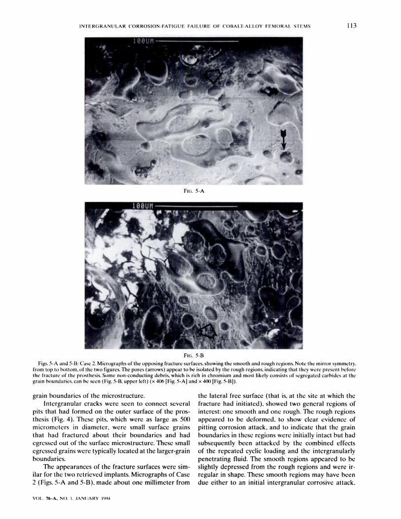
Figs. 5-A and 5-B: Case 2. Micrographs of the opposing fracture surfaces. showing the smooth and rough regions. Note the mirror symmetry.
from top to bottom. of the two figures. The pores (arrows) appear to be isolated by the rough regions. indicating that they were present before
the fracture of the prosthesis. Some non-conducting debris. which is rich in chromium and most likely consists of segregated carhides at the
grain boundaries, can he seen (Fig. 5-B. upper left) (x 406 [Fig. 5-A] and x 4(X) [Fig. 5-Bj).
grain boundaries of the microstructure.
Intergranular cracks were seen to connect several
pits that had formed on the outer surface of the pros-
thesis (Fig. 4). These pits. which were as large as 500micrometers in diameter, were small surface grains
that had fractured about their boundaries and had
egressed out of the surface microstructure. These small
egressed grains were typically located at the larger-grain
boundaries.
The appearances of the fracture surfaces were sim-
ilar for the two retrieved implants. Micrographs of Case
2 (Figs. 5-A and 5-B), made about one millimeter from
the lateral free surface (that is, at the site at which the
fracture had initiated), showed two general regions of
interest: one smooth and one rough. The rough regions
appeared to be deformed. to show clear evidence of
pitting corrosion attack, and to indicate that the grain
boundaries in these regions were initially intact hut had
subsequently been attacked by the combined effects
of the repeated cyclic loading and the intergranulamly
penetrating fluid. The smooth regions appeared to be
slightly depressed from the rough regions and were in-
regular in shape. These smooth regions may have been
due either to an initial intergnanular corrosive attack,

F
k,
- ___
.�4::
This suggests that the pores were not created by fluid
penetrating along the grain boundaries but were instead
generated during the manufacture of the prosthesis.
Analysis ofthe Sectioned Surfaces
The sections from the necks revealed clean evidence
of corrosive penetration of fluid intergmanulamly into the
microstructure. This intengranulam attack was adjacent
to the fracture surface and was continuous with an inter-
granular attack emanating from the head-neck taper
region. In a montage of a sectioned surface from Case 1
(Fig. 6), th’� outline of the grains was seen clearly and
was the result of intemgranular attack and porosity. For
Case 2, the neck-head taper junction was seen clearly
and had been extensively attacked at the grain bound-
,,- .�
I �.i.o.,�i.
.....‘-
r---� ‘
THE JOURNAL OF BONE AND JOINT SURGERY
114
FI;. 6
J. L. GILBERT ET AL.
Case 1. Montage of the cross section, with the fracture surface at
the top and the laterosupcrior surface at the left. showing intergranu-
lar cracks throughout the neck. The fracture surface follows the grain
boundaries of the material.
during which the penetrating fluid created smooth path-
ways or, alternatively. to porosity at the grain bound-
aries that had existed before the corrosive attack and
thus was the result of the manufacturing conditions used
to make the prosthesis.
In order to determine if the smooth regions were
the result of formation of pores during the manufac-
tune of the prosthesis on the result of fluid penetration,
it was necessary to find the corresponding regions on
each fracture surface from one implant (that is, to locate
the mirror image of one fracture site on the opposite
fracture surface). Then, if there were regions in which
pores were not interconnected but rather isolated by
the rough regions (indicating that the grain regions had
been initially intact). it could be assumed that the pores
probably had been present before the intengranular at-
tack and were therefore the result of porosity formation
during the manufacture of the prosthesis.
This analysis was carried out for Case 2. Micro-
graphs with mirror symmetry, from top to bottom, of the
same region on opposing fracture surfaces (Figs. 5-A
and 5-B) showed several isolated, roughly circular pores
that had no signs of being interconnected but were,
instead, only surrounded by a rough fracture surface.
FI3. 7
Case 2. Optical photograph showing intergranular cracking at the
taper-free surface junction. This cracking. which is localized at the
taper junction. appears to travel down into the neck toward the
region of the fracture surface.
aries (Fig. 7). The intengranulam attack seemed to be
concentrated at the superior and inferior surfaces of the
neck at the taper-free surface junction.
Discussion
The scanning electron microscopy revealed evi-
dence of corrosion-fatigue fracture of the femomal neck
in two similar-metal (cobalt-alloy head and cobalt-alloy
stem) modular hip prostheses. In both patients. con-
sidenable time had elapsed before failure (seventy and
eighty-five months), indicating a process that required
time to evolve to the failure point. In addition, both
patients were heavy and quite active; thus, they placed
considerable mechanical demands on the prostheses.

INTERGRANULAR CORROSION-FATIGUE FAILURE OF COBALT-ALLOY FEMORAL STEMS 115
VOL. 76-A, NO. I. JANUARY 1994
The granulomatous reaction seen in Case 2 was sim-
ilar to that reported by Mathiesen et al., in a severely
corroded modular hip prosthesis consisting of a cobalt-
alloy stem and a cobalt-alloy head, and the reaction
could be the result of metal-ion release.
Furthermore, an intergranular corrosive attack was
shown to have occurred in the neck region, at the lateral
and medial sites of the head-neck taper junction, and
this intengranular attack had penetrated several milli-
meters into the microstructume. In Case 2, the inter-
granular cracking was focused at the taper junction and
had penetrated into the microstructume.
Several factors may have contributed to this failure
process. Porosity was present at the grain boundaries in
both implants, and this porosity may have resulted from
manufacturing conditions; most likely, it had not been
created by corrosion. This was determined by observa-
tion of the opposite sides of the same fracture site and
the location of regions with isolated pores that could not
have been the result of a penetrating corrosive attack
(if they had, they would have been interconnected).
During the manufacture of these prostheses, a po-
mous coating of beads is sintened to the surface of the
femonal component. To do this, the temperature of the
device has to be raised to near the melting point. This
incipient melting condition makes it very likely that
diffusional processes are ongoing, so that it is possible
for vacancy motion and void formation to occur at the
grain boundaries. Also, the sinteming process facilitates
alloy segmegations’267, which may further weaken the
grain boundary regions.
Mechanically assisted crevice corrosion” also ap-
pears to have contributed to the fracture process. In
the two implants, the intengranular corrosion, which
had started both in the head-neck taper region and
at the free surface where egression of grains had oc-
cumned5, had extended intergranularly throughout the
microstructume near the fracture region and had weak-
ened the grain-boundary material further. When this
was compounded by crack propagation induced by the
cyclic loading stresses, the combination of factors even-
tually created a large enough crack to fracture the neck.
In conclusion, a combination of factors appears to
have contributed to the intengranulan fractures of the
prosthetic necks. An intergnanulam corrosive attack of
the microstructume of the neck starting at the taper and
at sites of grain egression, in conjunction with fatigue
loading, resulted in the intengranular corrosion-fatigue
fractures. Also, conditions for manufacturing of the fem-
oral component may have resulted in intergnanulam po-
nosity, which may have further weakened the prosthesis
and provided a pathway for intergmanular attack.
No,r ThC authors acknowledge Robert M. Urban. Ray C. Wasickwski. M.D.. and LeslieManion. RN.. for their technical assistance in the preparation of this manuscript.
References
1. Buckley, C. A.; Gilbert, J. J.; Urban, R. M.; Sumner, D. R.; Jacobs, J. J.; and Lautenschlager, E. P.: Mechanically assisted corrosion of
modular hip prosthesis components in mixed and similar metal combinations. Trans. Soc. Biomater., Imp/a,it Retrieval Symposium, 15:
58. 1992.
2. Clemow, A. J., and Daniell, B. L.: Solution treatment behavior of Co-Cr-Mo alloy. J. Biomed. Mater. Res., 13: 265-279. 1979.
3. Collier, J. P.; Surprenant, V. A.; Jensen, R. E.; and Mayor, M. B.: Corrosion at the interface of cobalt-alloy heads on titanium-alloy stems.
C/in. Orthop.. 271: 305-312, 1991.
4. Collier, J. P.; Surprenant, V. A.; Jensen, R. E.; Mayor, M. B.; and Surprenant, H. P.: Corrosion between the components of modular
femoral hip prostheses.J. BoneandJointSurg., 74-B(4):511-517, 1992.
5. Gilbert, J. L., and Piehler, H. R.: Grain egression: a new mechanism of fatigue crack initiation in Ti-6A1-4V. Metal/org. Trans. 20-A:
1715-1725. 1989.
6. Gilbert, i. L.; Buckley, C. A.; Jacobs, J. .L; Urban, R. M.; Lautenschlager, E. P.; and Galante, J. 0.: Mechanically assisted corrosive attack
in the Morse taper of modular hip prostheses. In Transactions of the Fourth World Biomaterials Congress, p. 267. Berlin, European
Society for Biomaterials. 1992.
7. Jacobs, J. J.; Latanision, R. M.; Rose, R. M.; and Veeck, S. J.: The effect of porous coating processing on the corrosion behavior of cast
Co-Cr-Mo surgical implant alloys.J. Orthop. Res., 8: 874-882, 1990.
8. Mathiesen, E. B.; Lindgren, J. U.; Blomgren, G. G. A.; and Reinholt, F. P.: Corrosion of modular hip prostheses. J. Bone and Joint Surg.,
73-B(4): 569-575. 1991.
9. Urban, R. M.; Jacobs, J. J.; Gilbert, J. L.; and Galante, J. 0.: Corrosion products of modular hip prostheses: microchemical identification
and histopathological significance. Trans. Orthop. Res. Soc., 18: 81, 1993.