Interferon Escape of Respiratory Syncytial Virus: Functional … · 2012-10-16 · 2.1.2 Genome...
123
Max-von Pettenkofer-Institute and Gene Center, Ludwig Maximilians University, Munich Prof. Dr. Karl-Klaus Conzelmann Institute of Molecular Animal Breeding and Biotechnology, Gene Center Faculty of Veterinary Medicine of the Ludwig Maximilian University, Munich Prof. Dr. Eckhard Wolf Interferon Escape of Respiratory Syncytial Virus: Functional Analysis of the Nonstructural Proteins NS1 and NS2 Thesis for the attainment of the title Doctor in Veterinary Medicine from the Faculty of Veterinary Medicine of the Ludwig Maximilian University, Munich by Sabrina Marozin from Milan, Italy Munich, 2005
Transcript of Interferon Escape of Respiratory Syncytial Virus: Functional … · 2012-10-16 · 2.1.2 Genome...
Max-von Pettenkofer-Institute and Gene Center,
Ludwig Maximilians University, Munich
Prof. Dr. Karl-Klaus Conzelmann
Institute of Molecular Animal Breeding and Biotechnology, Gene Center
Faculty of Veterinary Medicine of the Ludwig Maximilian University, Munich
Prof. Dr. Eckhard Wolf
Interferon Escape of Respiratory Syncytial Virus:
Functional Analysis
of the Nonstructural Proteins NS1 and NS2
Thesis for the attainment of the title Doctor in Veterinary Medicine
from the Faculty of Veterinary Medicine of the Ludwig Maximilian University, Munich
by
Sabrina Marozin
from Milan, Italy
Munich, 2005
Aus der
Abteilung für Virologie des Max-von Pettenkofer-Institutes,
Genzentrum, München
Arbeitsgruppe Prof. Dr. Karl-Klaus Conzelmann
Leiter Prof. Dr. Ulrich Koszinowski
Vorgelegt über
das Institut für Tierzucht der Tierärztlichen Fakultät
der Ludwig-Maximilians-Universität München
Lehrstuhl für Molekulare Tierzucht und Biotechnologie, Genzentrum
Univ.-Prof. Dr. Eckhard Wolf
Unterdrückung der Interferon-vermittelten Immunantwort
durch das Respiratorische Synzitial Virus:
Funktionelle Analyse der Nicht-Strukturproteine
NS1 und NS2
Inaugural-Dissertation
zur Erlangung der tiermedizinischen Doktorwürde
der Tierärztlichen Fakultät
der Ludwig-Maximilians-Universität München
von
Sabrina Marozin
aus Mailand, Italien
München 2005
Gedruckt mit Genehmigung der Tierärztlichen Fakultät der
Ludwig-Maximilians-Universität München
Dekan: Univ.-Prof. Dr. E. P. Märtlbauer
Referent: Univ.-Prof. Dr. E. Wolf
Korreferent: Priv.-Doz. Dr. H.-Ch. Siebert
Tag der Promotion: 10. Februar 2006
Part of this work is published:
Bossert B., Marozin S., Conzelmann K.K., 2003, Non structural proteins NS1 and NS2 of bovine
respiratory syncytial virus block activation of interferon regulatory factor 3, J. Virol., 77: 8661-
8668
Schlender J., Hornung V., Finke S., Gunthner-Biller M., Marozin S., Brzozka K., Moghim S.,
Endres S., Hartmann G. and Conzelmann K.K. , 2005, Inhibition of toll-like receptor 7- and 9-
mediated alpha/beta interferon production in human plasmacytoid dendritic cells by respiratory
syncytial virus and measles virus, J. Virol., 79: 5507-5519
Other publications:
Gregory V., Lim W., Cameron K., Bennett M., Marozin S., Klimov A., Hall H., Cox N., Hay
A. and Lin Y.P., 2001, Infection of a child in Hong Kong by an influenza A H3N2 virus closely
related to viruses circulating in European pigs, J. Gen. Virol., 82: 1397-1406
Marozin S., Gregory V., Vallette M., Aymard M., Barigazzi G., Y. P. Lin and Hay A., 2002,
Recent evolution of influenza A H1N1 and H1N2 subtypes in European pigs, J. Gen. Virol., 83:
735-745
Marozin S., Prank U. and Sodeik B., 2004, Herpes simplex type I infection of polarized epithelial
cells requires microtubules and access to receptors present at cell-cell contact sites, J. Gen. Virol.,
85: 775- 786

If a man will begin with certainties, he shall end in doubts,
but if he will content to begin with doubts, he shall end in certainties.
Francis Bacon
I
CONTENTS
Page
1 INTRODUCTION AND OBJECTIVES 1
2 REVIEW OF THE LITERATURE 3
2.1 Structure and genome 3
2.1.1 Virion structure 3
2.1.2 Genome organization 4
2.1.3 Viral proteins 5
2.2 Replicative cycle 7
2.3 Epidemiology and Pathogenesis 10
2.4 Immunity 12
2.4.1 Humoral immunity 12
2.4.2 Cell-mediated immunity 13
2.4.3 Innate immunity 14
2.4.4 The Interferon / system 15
2.5 Viral antagonists of IFN type I response 18
2.6 The RSV nonstructural proteins NS1 and NS2 19
3 MATERIALS AND METHODS 21
3.1 Cell culture and viruses 21
3.1.1 Cells 21
3.1.2 RSV propagation and titer determination 21
3.1.3 Rabies virus (RV) stocks and titer determination 22
3.2 General cloning procedures 22
3.2.1 Restriction enzyme digest 22
3.2.2 Extraction of DNA fragments from agarose gel 23
3.2.3 DNA ligation 23
3.2.4 Transformation into competent bacteria 24
3.2.5 Preparation of DNA minipreps and midipreps 25
3.3 Reverse Transcription and PCR conditions 26
3.3.1 Extraction of RNA from cells and reverse transcription 26
3.3.2 PCR conditions 27
II
3.4 Generation of recombinant BRSV viruses expressing
HRSV NS1 and NS2 proteins 29
3.4.1 Construction of rBRSV viruses expressing NS proteins of HRSV
strain Long (rBRSVh1/2) 29
3.4.2 Construction of rBRSV expressing a mutated HRSV NS2 protein
(rBRSVh1/2*T26I) 29
3.4.2.1 Quick site-directed mutagenesis of HRSV Long NS2 gene 29
3.4.2.2 Generation of rBRSVh1/2*T26I
virus 30
3.5 Recovery of recombinant BRSV viruses 31
3.6 Generation of recombinant SAD eGFP-P viruses expressing
HRSV NS proteins 31
3.6.1.Generation of recominant SAD eGFP-P viruses 31
3.6.2 Rescue of recombinant eGFP-P viruses expressing RSV NS proteins 32
3.7 Growth characteristics of recombinant BRSV and eGFP-P viruses 32
3.7.1 rBRSV viruses expressing HRSV NS proteins 32
3.7.2 Recombinant rabies eGFP-P viruses expressing RSV NS proteins 33
3.8 IFN treatment of rBRSV viruses 33
3.9 Cloning of the human TANK-binding kinase 1 (TBK1) and
TRAF-binding protein (TANK) 33
3.10 Gene Reporter Assay 35
3.10.1 Modified Gene Reporter Assay in Vero-p125Luc cell line 35
3.10.2 Dual luciferase assay and transfection of reporter plasmids 35
3.10.3 Transcription factors activation and TBK1 inhibition by RSV infection 36
3.11 Protein expression analysis 36
3.11.1 Extraction of proteins from cells 36
3.11.2 SDS-polyacrylamide gel electrophoresis (SDS-PAGE) 37
3.11.3 Electroblotting 38
3.11.4 Western blotting 39
3.12 Immunofluorescence 40
3.13 Immunoprecipitation of IgTANK and IgTBK1 40
3.14 Materials and equipment 41
3.14.1 Serological reagents 41
3.14.2 Chemicals 42
3.14.3 Enzymes 43
III
3.14.4 Kits 43
3.14.5 Miscellaneous 44
3.14.6 Tissue culture reagents 45
3.14.7 Equipment 45
3.14.8 Bacteria and plasmids 46
4 RESULTS 49
4.1 Nonstructural proteins (NS) of BRSV block activation of
interferon regulatory factor 3 49
4.2 HRSV prototypes, A2 and Long, differ in their ability
to induce type I IFN 50
4.2.1 Detection of IFN- induction 50
4.2.2 Analysis of activation of IFN- transcription factors
AP-1, NF-kB and IRF3 51
4.3 Sequence comparison of RSV NS1 and NS2 proteins 52
4.3.1 Sequence analysis of HRSV A2 and HRSV Long nonstructural proteins 52
4.4 Chimeric BRSV viruses expressing HRSV NS genes 54
4.4.1 Construction of recombinant BRSV virus harbouring Long-derived NS1
and a mutated form of NS2 54
4.4.2 HRSV NS protein expression and cDNA restriction analysis 55
4.4.3 Growth kinetics of recombinant BRSV viruses 56
4.4.4 Sequence comparison of HRSV NS genes 58
4.4.5 rBRSVh1/2*T26I
but not rBRSVh1/2 inhibits IFN- induction 60
4.4.6 Recombinant BRSVh1/2 *T26I
selectively reduces activation of
transcriptional factor IRF3 62
4.4.7 Residue 26 of NS2 protein does not influence interferon resistance of
recombinant BRSV viruses 63
4.5 IkappaB kinase-related complex: TBK1, IKK- and TANK 64
4.5.1 Cloning of TANK-Binding Kinase 1 (TBK1) and TRAF family
member-associated NF-kappa B activator (TANK) 64
4.5.2 TBK1 activates IFN- production 66
4.5.3 HRSV strain A2 inhibits TBK1 activation 68
4.5.4 rBRSVh1/2*T26I
inhibits TBK1 activation similarly to HRSV A2 69
IV
4.6 Recombinant RV expressing RSV nonstructural proteins 72
4.6.1 Construction of recombinant SAD eGFP-P viruses harbouring
RSV NS proteins 72
5 DISCUSSION 75
5.1 Human respiratory syncytial virus: strains A2 and Long 75
5.1.1 Suppression of IFN- induction differs in HRSV A2 and Long 75
5.1.2 Activation of IFN- transcription factors 76
5.1.3 Sequence analysis of nonstructural NS proteins in HRSV 77
5.2 A mutated Long-derived NS2 protein prevents induction of IFN- 78
5.2.1 rBRSV expressing HRSV nonstructural protein 78
5.2.2 rBRSVh1/2*T26I
infection inhibits IFN- induction by blocking
IRF3 activation 79
5.3 The novel IkB-related kinases (IKK): IKK- and TBK1 80
5.3.1 Cloning TANK-binding kinase 1 (TBK1) and TRAF family member–
associated NF-kB activator (TANK) 80
5.3.2 TBK1-dependent IFN- induction 82
5.4 Inhibition of TBK1 by RSV 82
5.4.1 HRSV A2 blocks TBK1-induced IFN- expression 82
5.4.2 Residue 26 of NS2 protein is important for TBK1 inhibition 83
5.5 Recombinant Rabies Viruses expressing RSV nonstructural proteins 86
5.5.1 Generation of SAD eGFP-P viruses harbouring RSV NS1 and
NS2 proteins 86
5.5.2 Recombinant eGFP-PNS2/NS1 viruses do not counteract IFN production 86
5.6 Final considerations 87
6 SUMMARY 89
7 ZUSAMMENFASSUNG 91
8 BIBLIOGRAPHY 93
V
Abbreviations
A Ampere
APS ammonium persulfate
ATP adenosintriphosphate
b bovine
BRSV Bovine respiratory syncytial virus
bp base pair
BSA bovine serum albumin
cDNA complementary DNA
cm centimeters
CMV cytomegalovirus
CoA coenzyme A
CPE cytopathic effect
CS newborn calf serum
C-terminus carboxy terminus
DEPC diethylpyrocarbotate
DL Dual luciferase assay
DMEM Dulbecco’s Modified Eagle Medium
DNA desoxyribonucleic acid
dNTP desoxyribonucleoside triphosphates
DTT dithiothreitol
EDTA ethylene diamine tetraacetic acid
e.g example given
Et al. Et alii
FCS fetal calf serum
FITC fluoresceinisothiocyanate
FFU focus forming units
g gram
GFP green fluorescent protein
h human
h, hrs hour(s)
HRSV Human respiratory syncytial virus
IFN interferon
Ig immunoglobulin
IRF Interferon regulatory factor
IU international units
kb kilobase
kDa kilodalton
l liter
LB Luria Broth
LPS lipopolysaccharides
Luc luciferase
M molar, marker
VI
m milli
mA milliampere
mg milligram
min minute(s)
ml milliliter
mM millimolar
mm millimeter
MOI multiplicity of infection
mRNA messenger RNA
MvP Max von Pettenkofer
ng nanogram
nm nanometers
N nucleoprotein
N-terminus amino terminus
NS nonstructural protein
nt nucleotide
P phosphoprotein
p plasmid
PAGE polyacrylamide gel elctrophoresis
PBS phosphate-buffered saline
PBS-T phosphate-buffered saline/0.05% Tween
PCNA proliferating cell nuclear antigen
PCR polymerase chain reaction
p.i. post infection
PKR RNA-dependent protein kinase
Poly I/C polyriboinosinic acid/polyribocytidilic acid
PRD positive regulatory domain
p.t. post transfection
OD optical density
ORF open reading frame
RLU relative light unit
RNA ribonucleic acid
RNase ribonuclease
Rnasin Rnase inhibitor
RNP ribonucleoprotein
rpm revolution per minute
RT-PCR reverse transcription PCR
RSV respiratory syncytial virus
RT reverse transcriptase
RV Rabies virus
SDS sodium dodecyl sulfate
STAT signal transducer and activator of transcription
TAE Tris-acetate-EDTA
TBS Tris-buffered saline
VII
TEMED N,N,N’,N’-tetramethylethylendiamine
U Unit
UV ultraviolet
V volt
wt wild-type
g micrograms
l microliters
Introduction and Objectives
___________________________________________________________________________
1
1 INTRODUCTION AND OBJECTIVES
Respiratory Syncytial Virus (RSV) is recognised as the most frequent cause of severe lower
respiratory tract infections in infants and cattle worldwide. Human Respiratory Syncytial
Virus (HRSV) infects around 50% of infants during the first year and almost all the children
by age of two, resulting in the most common factor of paediatric hospitalizations in particular
of subjects between 2 and 6 months of age (Collins P.L. et al., 2001; Hoffman S.J. et al.,
2004). Bovine Respiratory Syncytial Virus (BRSV)-associated disease has been observed in
young calves at less than 6 months of age and also associated with outbreaks in dairy cows
(Elvander M., 1996). Seroprevalence among the adult human population and cattle is around
70% (Van der Poel W.H. et al., 1994). BRSV and HRSV are closely antigenically related and
in both cases serological subgroups have been identified (Mallipeddi S.K. et al., 1993; Furze
J.M. et al., 1994; Mufson M.A. et al., 1985). Human and bovine respiratory syncytial viruses
share common epidemiological, clinical and pathological characteristics. RSV is spread from
respiratory secretion via close contact with infected persons or contaminated materials.
Infections follow a seasonal periodicity and typical pathological manifestations of RSV-
related illness are tracheobronchitis, peri-bronchiolitis, bronchiolitis and pneumonia.
Bronchiolitis is associated with long term impairment of pulmonary functions and histamine
hyper responsiveness can last for many years after RSV-infection in infancy probably leading
to the development of asthma and general allergic sensitisation in children (Sigurs N. et al.,
2000).
The presence of maternal antibodies gives neither efficacious protection nor reduces viral-
shedding after infection. RSV infection does not lead to a complete and durable immunity and
human and cattle of all ages can be re-infected throughout life. Current treatments of RSV are
based on supportive care and antiviral therapy. An antiviral compound, which is approved for
RSV treatment in humans, is Ribavirin, a nucleoside analog. Ribavirin aereosol can be used in
the treatment of some patients with severe disease. However, limited clinical benefits have
been observed. Controversy on the significance of passive RSV prophylaxis, using RSV
Immunoglobulin (RespiGam) or genetically engineered humanized monoclonal antibodies
against F glycoprotein (Palivizumab), is also stated. However, combination of
immunoglobulin intravenously (IGIV) with neutralizing RSV antibody (RSV IGIV) and
Ribavirin has been used to treat patients with compromised immune system. Development of
Introduction and Objectives
___________________________________________________________________________
2
an RSV vaccine is a high research priority, but none is yet available. In conclusion, no
effective treatments for RSV infection are available at the moment. Moreover the
development of a successful vaccine has been hampered by the fact that natural infection does
not provide complete protection against re-infections. Besides, previous immunization can
enhance the severity of the disease in naturally infected individuals. A safe effective RSV
vaccine and/or the development of efficient therapeutic interventions is crucial and relies on a
proper understanding of RSV disease pathogenesis and virus host-cell interactions. In this
respect, studies of the innate immunity, which plays a critical role at the time of maximum
severity of illness and influences the subsequent adaptive response, as well as viral counter
measurements must be elucidated.
RSV, as well as many negative strand RNA viruses (e.g. Measles, Mumps, Parainfluenza and
Influenza virus) has evolved several strategies to hinder IFN response.
IFN treatment of RSV infected cells does not inhibit viral replication, indicating RSV ability
to circumvent the action of IFN. RSV NS1 and NS2 proteins are responsible for the
pronounced resistance to exogenous interferon and their ability to inhibit activity of cellular
IFN-induced antiviral proteins is exerted in a host-adapted manner (Schlender J. et al., 2000;
Bossert B. et al., 2002). RSV nonstructural proteins (NS) as other viral accessory proteins in
Influenza or Bunyamwera viruses, are also strong inhibitors of IFN / production.
Interferon type I synthesis is regulated at a transcriptional level. In response to viral infection,
the transcription factors AP-1, NF-kB and Interferon Regulatory Factor 3 (IRF3) become
activated following protein phosphorylation, bind to designated positive regulatory domains
(PRD) present in the IFN- promoter and form a transcriptional enhancer complex which
stimulates transient activation of IFN- transcription. Nonstructural proteins of RSV
specifically impair IRF3 activation by preventing its phosphorylation (Bossert B. et al., 2003)
but how this occurs is still unclear.
My attempt is to gain more insight into the mechanisms leading to NS protein-mediated
inhibition of IRF3 factor. My approach includes analysis of both cellular and viral elements
that play an active role in this complex mechanism. The aim of my work is to identify
possible cellular targets within the signalling pathway activating IFN production and to
determine specific amino acid motifs in the HRSV NS proteins involved in their interferon-
inhibitory activity.
Review of the Literature ___________________________________________________________________________
3
2 REVIEW OF THE LITERATURE
2.1 Structure and genome
2.1.1 Virion structure
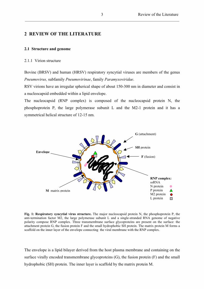
Bovine (BRSV) and human (HRSV) respiratory syncytial viruses are members of the genus
Pneumovirus, subfamily Pneumovirinae, family Paramyxoviridae.
RSV virions have an irregular spherical shape of about 150-300 nm in diameter and consist in
a nucleocapsid embedded within a lipid envelope.
The nucleocapsid (RNP complex) is composed of the nucleocapsid protein N, the
phosphoprotein P, the large polymerase subunit L and the M2-1 protein and it has a
symmetrical helical structure of 12-15 nm.
G (attachment)
SH protein
F (fusion)
M matrix protein
Envelope
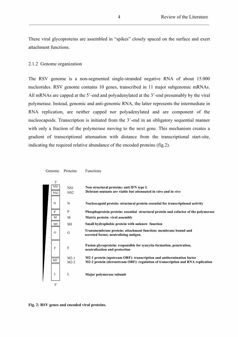
RNP complex:ssRNAN protein P protein M2 protein L protein
Fig. 1: Respiratory syncytial virus structure. The major nucleocapsid protein N, the phosphoprotein P, the anti-termination factor M2, the large polymerase subunit L and a single-stranded RNA genome of negative polarity compose RNP complex. Three transmembrane surface glycoproteins are present on the surface: the attachment protein G, the fusion protein F and the small hydrophobic SH protein. The matrix protein M forms a scaffold on the inner layer of the envelope connecting the viral membrane with the RNP complex.
The envelope is a lipid bilayer derived from the host plasma membrane and containing on the
surface virally encoded transmembrane glycoproteins (G), the fusion protein (F) and the small
hydrophobic (SH) protein. The inner layer is scaffold by the matrix protein M.
Review of the Literature ___________________________________________________________________________
4
These viral glycoproteins are assembled in “spikes” closely spaced on the surface and exert
attachment functions.
2.1.2 Genome organization
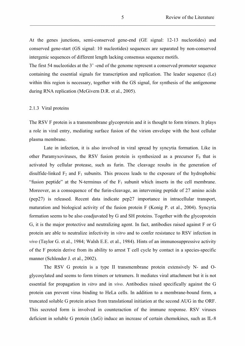
The RSV genome is a non-segmented single-stranded negative RNA of about 15.000
nucleotides. RSV genome contains 10 genes, transcribed in 11 major subgenomic mRNAs.
All mRNAs are capped at the 5’-end and polyadenylated at the 3’-end presumably by the viral
polymerase. Instead, genomic and anti-genomic RNA, the latter represents the intermediate in
RNA replication, are neither capped nor polyadenylated and are component of the
nucleocapsids. Transcription is initiated from the 3’-end in an obligatory sequential manner
with only a fraction of the polymerase moving to the next gene. This mechanism creates a
gradient of transcriptional attenuation with distance from the transcriptional start-site,
indicating the required relative abundance of the encoded proteins (fig.2).
NS1
Matrix protein: viral assembly
Non structural proteins: anti IFN type I. Deletant mutants are viable but attenuated in vitro and in vivo
Nucleocapsid protein: structural protein essential for transcriptional activity
Phosphoprotein protein: essential structural protein and cofactor of the polymerase
Small hydrophobic protein with unknow function
Transmembrane protein: attachment function; membrane bound and secreted forms; neutralizing antigen.
Fusion glycoprotein: responsible for syncytia formation, penetration, neutralization and protection
M2-1 protein (upstream ORF): transcription and antitermination factor M2-2 protein (downstream ORF): regulation of transcription and RNA replication
Major polymerase subunit
NS1NS2
N
PM
SH
G
F
M2-1M2-2
L
NS2
N
P
M
SH
G
F
M2
L
3’
5’
Genome Proteins Functions
Fig. 2: RSV genes and encoded viral proteins.
Review of the Literature ___________________________________________________________________________
5
At the genes junctions, semi-conserved gene-end (GE signal: 12-13 nucleotides) and
conserved gene-start (GS signal: 10 nucleotides) sequences are separated by non-conserved
intergenic sequences of different length lacking consensus sequence motifs.
The first 54 nucleotides at the 3’ -end of the genome represent a conserved promoter sequence
containing the essential signals for transcription and replication. The leader sequence (Le)
within this region is necessary, together with the GS signal, for synthesis of the antigenome
during RNA replication (McGivern D.R. et al., 2005).
2.1.3 Viral proteins
The RSV F protein is a transmembrane glycoprotein and it is thought to form trimers. It plays
a role in viral entry, mediating surface fusion of the virion envelope with the host cellular
plasma membrane.
Late in infection, it is also involved in viral spread by syncytia formation. Like in
other Paramyxoviruses, the RSV fusion protein is synthesized as a precursor F0 that is
activated by cellular protease, such as furin. The cleavage results in the generation of
disulfide-linked F2 and F1 subunits. This process leads to the exposure of the hydrophobic
“fusion peptide” at the N-terminus of the F1 subunit which inserts in the cell membrane.
Moreover, as a consequence of the furin-cleavage, an intervening peptide of 27 amino acids
(pep27) is released. Recent data indicate pep27 importance in intracellular transport,
maturation and biological activity of the fusion protein F (Konig P. et al., 2004). Syncytia
formation seems to be also coadjuvated by G and SH proteins. Together with the glycoprotein
G, it is the major protective and neutralizing agent. In fact, antibodies raised against F or G
protein are able to neutralize infectivity in vitro and to confer resistance to RSV infection in
vivo (Taylor G. et al., 1984; Walsh E.E. et al., 1984). Hints of an immunosuppressive activity
of the F protein derive from its ability to arrest T cell cycle by contact in a species-specific
manner (Schlender J. et al., 2002).
The RSV G protein is a type II transmembrane protein extensively N- and O-
glycosylated and seems to form trimers or tetramers. It mediates viral attachment but it is not
essential for propagation in vitro and in vivo. Antibodies raised specifically against the G
protein can prevent virus binding to HeLa cells. In addition to a membrane-bound form, a
truncated soluble G protein arises from translational initiation at the second AUG in the ORF.
This secreted form is involved in counteraction of the immune response. RSV viruses
deficient in soluble G protein ( sG) induce an increase of certain chemokines, such as IL-8
Review of the Literature ___________________________________________________________________________
6
and Rantes in comparison to wild-type strains (Arnold et al., 2004). Moreover, an activity in
trapping RSV-neutralizing antibodies cannot be excluded (Collins P.L. et al., 2001). Deletion
mutant RSV viruses for the G protein ( G) are viable but display a host range restriction in
growth.
The small hydrophobic SH protein is a short integral transmembrane protein and it is
present in glycosylated and non-glycosylated species in form of oligomers. Its function is so
far unknown. Recombinant RSV viruses lacking SH protein fully replicate in vitro but
attenuation in the lower respiratory tract in vivo has been reported (Jin H. et al., 2000). Since
expression of SH protein in bacteria increased permeability of small molecular-weight
compounds, it has been speculated its involvement in membrane channels formations (Perez
M. et al., 1997).
The matrix protein M is a non-glycosylated protein forming a sheet on the inner side
of the viral envelope. It plays an important role in virus assembly and budding by mediating
the association between viral nucleoprotein (vRNP) and cell plasma membrane (Peeples M.E.,
1991; Ghildyal R. et al., 2002). Apart from inactivating transcription activity of the
nucleoprotein before packaging, early in infection M protein it localises into the nucleus
where it possibly inhibits host-cell transcription (Ghildyal R. et al., 2003). RSV M protein has
also RNA-binding capacity as recently described (Rodriguez L. et al., 2004) but the real
function of this interaction is still unclear.
RSV N, P and L proteins co-purify with nucleocapsid. They are necessary and
sufficient for RNA replication. The major nucleocapsid protein is the nucleoprotein N. It
binds to genomic and antigenomic RNA conferring RNase resistance to the nucleocapsids.
The phosphoprotein P is the major phosphorylated species. It functions as a chaperonin for
soluble N and it is essential together with N protein for encapsidation activity. P protein is
also a polymerase cofactor. It seems to convert initiated polymerase into a stable complex and
its phosphorylation is mandatory for this function (Dupuy L.C. et al., 1999). The L protein is
the major RNA-dependent RNA polymerase subunit and it is bound to its cofactors, the
phosphoprotein P and M2-1 by the N protein.
The M2-1 protein is a transcription processivity factor and it is essential for viral
replication. It prevents premature termination during transcription (Fearns R. and Collins P.L.,
1999; Zhou et al., 2003) and enhances read-through at the gene junctions (Hardy R.W. and
Wertz G.W., 1998; Hardy R.W. et al., 1999). The M2-1 protein interacts with the
nucleocapsid N protein through RNA mediation (Cuesta I. et al., 2000; Cartee T.L. and Wertz
Review of the Literature ___________________________________________________________________________
7
G.W, 2001) and with the P protein (Mason S.W. et al., 2003). Phosphorylation of M2-1
appears to be indispensable for the interaction with the P protein. RSV infection is
characterised by persistent NF-kB activation and recently M2-1 protein has been identified as
inducer of Rel A (p65), a protein of the mammalian NF-kB complex (Reimers K. et al.,
2005). The M2 mRNA encodes for the M2-1 protein by the 5’-proximal ORF, while the M2-2
protein originates by the 3’-proximal open reading frame. M2-2 protein has a possible role in
RNA synthesis regulation mediating the switch from transcription to genome replication
(Collins P.L. et al., 1996). Recombinant viruses lacking M2-2 protein expression ( M2-2) are
attenuated in vitro and in vivo compared to wild type (Jin H. et al., 2000 b).
NS1 and NS2 are proteins with an estimated mass of about 14-15 kDa. They are found
only in pneumoviruses and they have been classified as nonstructural proteins since they are
found only in traces in purified virions (Evans J.E. et al., 1996). Their subgenomic mRNAs
are the most abundant among the transcripts due to the typical gradient of transcription being
their promoter the most proximal in the RSV genome. From the functional point of view, NS1
and NS2 proteins, despite enhancing growth, are not essential. Single and double deletion
mutants are viable although displaying an attenuated phenotype both in vitro and in vivo
(Buchholz U.J. et al., 1999; Jin H. et al., 2000; Teng M.N. and Collins P.L., 1999; Teng M.N.
et al., 2000). NS1 protein has been reported to be a potent negative regulatory factor of
transcription and synthesis of genome and antigenome RNA, most likely acting at early stages
of promoter initiation by the viral polymerase (Atreya P.L. et al, 1998).
2.2 Replicative cycle
Binding and entry of RSV into targeted cells are mediated by the interaction between the G
and the F proteins with host cell molecules. The specific RSV cellular-receptor has not been
identified so far but there are data indicating an interaction of the RSV G protein with cell
surface glycosaminoglycans (GAGs), such as heparan sulphate and chondroitin sulphate B
(Feldman S.A. et al., 1999). Cell surface GAGs are essential for RSV binding in vitro and are
therefore important for infectivity (Martinez I. and Melero J.A., 2000). Interestingly,
recombinant RSV viruses lacking the G protein are infectious in cell cultures, however they
show attenuation in vivo, both in human and mouse airway cells (Karron R.A. et al., 1997;
Teng M.N. et al., 2001). These results imply that G protein is dispensable for cell attachment
but it has other functions that might influence the efficiency of the process. For example, a
Review of the Literature ___________________________________________________________________________
8
putative binding domain to CX3C receptor (CX3CR1), which could favour infection, has
been shown (Tripp R.A et al., 2000 and 2001).
Protein F alone can mediate attachment. It is responsible for fusion of the virus envelope with
the host plasmame mbrane and for syncytium formation, therefore it is absolutely required
during the virus life cycle.
Assembly
Budding of a mature virion
Nucleus
Cell to cell fusion and syncytia formation
Ribosomes
mRNA transcription Genomic RNAsynthesis
RSV attachment
dsRNA
RSV fusion
.
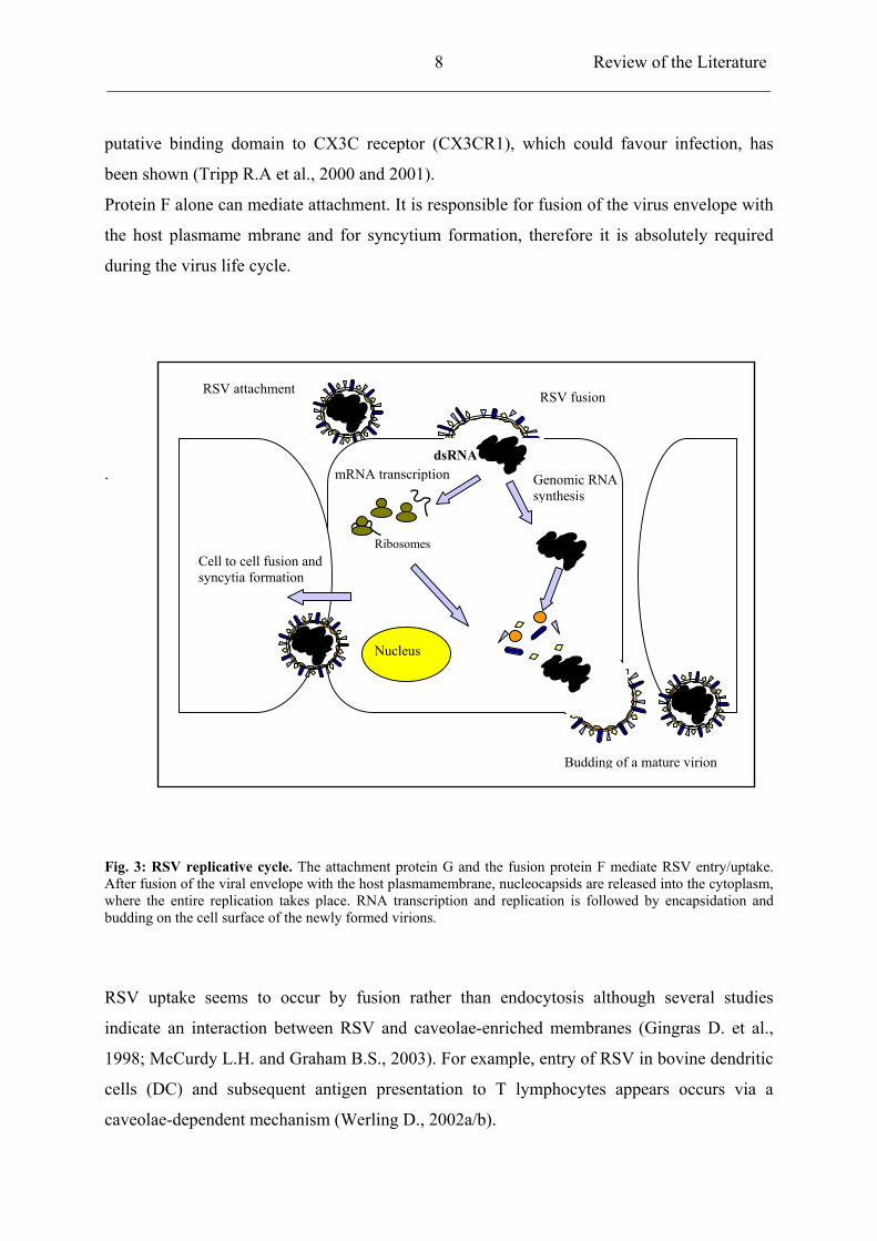
Fig. 3: RSV replicative cycle. The attachment protein G and the fusion protein F mediate RSV entry/uptake. After fusion of the viral envelope with the host plasmamembrane, nucleocapsids are released into the cytoplasm, where the entire replication takes place. RNA transcription and replication is followed by encapsidation and budding on the cell surface of the newly formed virions.
RSV uptake seems to occur by fusion rather than endocytosis although several studies
indicate an interaction between RSV and caveolae-enriched membranes (Gingras D. et al.,
1998; McCurdy L.H. and Graham B.S., 2003). For example, entry of RSV in bovine dendritic
cells (DC) and subsequent antigen presentation to T lymphocytes appears occurs via a
caveolae-dependent mechanism (Werling D., 2002a/b).
Review of the Literature ___________________________________________________________________________
9
RSV requires cytoskeletal elements. The microtubule and actin networks play a role in several
aspects of the viral life cycle. Perturbation of cytoskeletal functions is negatively reflected on
RSV entry, release, cell-cell spreading and syncytia formation (Kallewaard N.L. et al., 2005).
The entire RSV replicative cycle takes place in the cytoplasm and begins with transcription of
the genome into 5’-capped and 3’-polyadenylated mRNAs by the viral RNA-dependent
polymerase complex. RNA synthesis occurs in a sequential manner from the 3’-end of the
genome with the polymerase complex terminating and reinitiating mRNA transcription at
each junction. Reinitiating can be occasionally inefficient and this results in a gradient of
mRNA decreasing proportionally to the distance of the gene from the 3’-end of the genome.
The polymerase complex is also responsible for antigenomic RNA synthesis. In this case, all
the junction signals are completely ignored and the result is a complementary positive-sense
copy of the viral genome. The antigenomic RNA represents a replicative intermediate and it is
less abundant in comparison to the genomic RNA of about 10-20 fold. Both RNAs are packed
into virions in an equal proportion. Antigenomic and genomic RNA synthesis correlates with
protein translation indicating a need for cosynthetic encapsidation. In contrast to other
Paramyxoviruses, the switch between RSV transcription and RNA replication seems to
implicate M2-2 protein and not to depend on the intracellular levels of N and P proteins
(Fearns R. et al., 1997). At the early stages of infection, low levels of M2-2 correlates to a
high transcriptional rate. Afterwards, when the intracellular levels of M2-2 protein increased,
transcription is inhibited in favour of replication (Bermingham A. and Colllins P.L., 1999).
Assembly of the nucleocapsids is entirely cytoplasmatic. N protein associates first with
genomic and antigenomic RNA followed by P and L. Once assembled, nucleocapsids are
transported close to the cell surface and bud at specific plasma membrane patches where
glycoprotein G clusters. Association of the nucleocapsids with the nascent envelope is
mediated by M protein, which makes contacts with the cytoplasmatic tails of the viral
glycoproteins. However, efficient viral formation in vitro is not affected by deletion of G and
SH proteins. On the contrary F protein seems to be essential in this context. Assembly is also
related to an intact cellular cytoskeleton. (Ulloa L. et al., 1998; Burke E. et al., 2000; Gower
T.L. et al., 2001).
Review of the Literature ___________________________________________________________________________
10
2.3 Epidemiology and Pathogenesis
Respiratory syncytial virus (RSV) infection is one of the most important health problems in
infancy accounting for about 85% of cases of bronchiolitis and approximately 20% of cases of
childhood pneumonia (Wright P.F. and Cutts F.T., 2000). It significantly contributes to
hospitalisation of infants in developed countries. Only in the United States, it has been
estimated that more than 120.000 children younger than 1 year are hospitalised annually, with
about 200 deaths as a results of this illness (Shay D.K. et al., 2001). Of course the scenario in
countries with less well-developed medical care programs is even more serious. Bronchiolitis
and pneumonia occur most frequently between the 6 weeks and 9 months of age, showing a
peak in coincidence of the dropping of maternal antibody titers around the second-seventh
month. Fortunately, the risk of severe illness related to RSV is quite low in developed
countries. However, several groups of infants might be more predisposed to a severe outcome,
like in the case of infants with chronic lung disease of prematurity, congenital heart disease or
compromised immunity. In addition RSV is an important pathogen in the elderly.
RSV has a worldwide distribution and it is a seasonal infection, with peaks around winter
and/or spring (Stensballe L.G. et al., 2003). Persistence, in vivo, has been postulated to
explain the apparent absence of the virus between epidemics. There are experimental
indications demonstrating that, for example BRSV-infected B lymphocyte can be isolated in
calves 10 weeks after infection and that B-lymphocytes cell-lines show persistent infection in
vitro for 6 months (Streckert H.J. et al., 1996; Valarcher J.F. et al., 2001).
Transmission occurs via contact with respiratory secretions. The incubation period can vary
between 2-8 days and it is followed by symptoms related to upper and lower respiratory tract
infection.
Veterinary pathogens, belonging to the same subfamily of the human respiratory syncytial
virus (HRSV), have been identified. The avian metapneumovirus (APV) is the causative agent
of the turkey rhinotracheitis (Njenga M.K. et al., 2003) and probably of the swollen head
syndrome in chickens (Cook J.K., 2000). Outbreaks follow a seasonal pattern and wild
migratory birds might be involved in virus spreading. APV causes severe upper respiratory
infections with high mortality and big economic loss for the industry as seen in the late
nineties in USA (Panigrahy B. et al., 2000; Jirjis F.F. et al., 2002). The pneumonia virus of
mice (PMV) was isolated for the first time in 1938 and it seems not to be an important disease
Review of the Literature ___________________________________________________________________________
11
in wild mice, with the exception of immunocompromised subjects. PMV represents an
alternative model to study pneumovirus-related disease in rodents (Cook P.M. et al., 1998)
and recently its full genome has been published (Krempl C.D. et al., 2005). APV is closely
related to a newly described human metapneumovirus (HMPV), which is responsible for a
clinical syndrome practically indistinguishable from the one related to HRSV (van den
Hoogen B.G. et al., 2001; van den Hoogen B.G. et al., 2002; Kahn J.S., 2003).
Sheep and goats have also distinct RSV pathogens (Trudel M. et al., 1989; Brogden K.A. et
al., 1998). Bovine pneumovirus (BRSV) has been isolated in the 70s and research on this
virus has been encouraged by the fact that BRSV represents the most close phylogenetic-
relative of HRSV. BRSV shares with the human counterpart the same way of transmission
and the seasonality of the outbreaks, but, unlike HRSV, bovine pneumovirus infections are
often complicated by concomitant bacterial infection, e.g. Pasteurella multocida,
Haemophilus somnus (Woolums A.R. et al., 2004; Gershwin L.R. et al., 2005).
RSV-illness usually begins with infection of the upper respiratory tract and it is characterised
by non-specific symptoms: fever and rhinorrhea that can last several days. When the lower
respiratory tract gets involved, the outcome can be pneumonia with tachypnea, difficulties in
breathing, wheezing upon auscultation or bronchiolitis that might impair pulmonary functions
for long time and/or predispose to asthma (Psarras S. et al., 2004). Morbidity can be increased
by many predisposing conditions like premature birth, heart diseases, immunodeficiency
(Welliwer R.C., 2002). Host factors, especially related to aberrant inflammatory response,
have been associated to severe RSV disease. Particular emphasis has been paid to the
potential role of proinflammatory chemokines and cytokines, whose expression is up-
regulated in RSV-related disease and, in some cases, contributes to exacerbate the detrimental
effects of the primary infection. High levels of IL-1 , IL-6, IL-8, TNF- , MIP1
(macrophage inflammatory protein), RANTES and adhesion molecule ICAM-1 have been
detected in respiratory secretions. The productions of these factors follow a biphasic pattern
with an early peak during RSV active infection and a later peak not related to viral replication
(Miller A.L. et al., 2004; Kong X. et al., 2005).
Prominent pathological features of severe RSV infections are necrosis of the airway
epithelium, interstitial inflammation with lymphocytes, plasma cell and macrophages
infiltrations, mucus secretion leading to obstruction of the airways and a general respiratory
compromise.
Review of the Literature ___________________________________________________________________________
12
Similarly, BRSV induces mild to severe respiratory signs especially among calves and it has
shown to be involved in the paroxystic respiratory distress syndrome (PRDS) (Jolly S. et al.,
2004).
2.4 Immunity
Natural infection with RSV does not provide efficient protection against reinfections,
indicating that the acquired immunity in this case is neither complete nor durable.
Ineffectiveness of the immune response against RSV infection has hampered the development
of effective vaccines against BRSV and HRSV. The other major problem is that prior
vaccination can enhance the severity of the disease during subsequent natural infection. This
has been already observed in children in the 60s during trials with formalin-inactivated (FI) or
alum-adjuvant vaccines (Kim H.W. et al., 1969). Similarly, in animals inactivated vaccines
were responsible for immunopathological states in calves infected with BRSV (Schreiber P. et
al., 2000; Antonis A.F. et al., 2003; Kalina W.V. et al., 2005).
Severe RSV-disease following natural infection or immunization can be attribute to
impairment of type 1/2 phenotype balance with a dominance of Th2 lymphocyte response.
Infants and mice with severe bronchiolitis show an augmented eosinophilia and an increase in
type 2 cytokine levels, e.g. IL-4, IL-5, and IL-13 (Roman M. et al., 1997; Boelen A. et al.,
2002; Johnson T.R.et al., 2005).
Other studies have emphasised more the potential role of chemokine production than the Th2
cytokines levels as a key factor of RSV immunopathogenesis. In particular the beta
chemokine macrophages inflammatory protein-1alpha (MIP1 ), the monocyte chemotactic
protein 1 (MCP-1) and the regulated on activation normal T lymphocyte expressed and
secreted (RANTES) are known to attract lymphocytes, basophils and eosinophils and they are
associated with greater inflammatory response in severe RSV-illness (Hornsleth A. et al.,
2001; Garofalo R.P. et al., 2001 and 2005).
2.4.1 Humoral immunity
Partially protective antibodies against F and G proteins are produced during natural infection.
Passive immunization with immunoglobulin preparations containing human RSV-specific Ig
Review of the Literature ___________________________________________________________________________
13
or monoclonal anti-F antibodies have shown to be able to attenuate the severity of the disease
(Prince G.A. et al., 2000; Simoes E.A. and Groothuis J.R., 2002; Sastre P. et al., 2004).
Unfortunately not all humoral responses are favourable. RSV-specific IgE for example may
also contribute to increase the severity of the disease (Welliver R.C. et al., 1981; Tumas D.B.
et al., 2001; Dakhama A. et al., 2004). RSV is able to infect infants in the presence of
moderate titers of maternal antibodies. Secretory antibodies (IgA) are defective in neutralizing
the virus in vitro (McIntosh K. et al., 1978), which could explain the failure of natural
immunity during early ages. In adults, higher levels of IgA are produced as a result of
reinfections and in experimental trials immunity showed to better correlation with RSV-
neutralizing secretory antibodies than with serum neutralizing immunoglobulin. Nevertheless
secretory IgA gives only partial protection and do not confer resistance to reinfections
(Gleeson M. et al., 2004; Walsh E.E. and Falsey A.R., 2004). Humoral immunity seems not to
provide complete protection against RSV infection and current hypotheses point the
importance of the cell-mediated immune response in viral clearance.
2.4.2 Cell-mediated immunity
Infants with a primary RSV infection develop a cellular immune response within 10 days. In
the BALB/c mouse model the first to appear are natural killer (NK) cells followed by CD8+
cytotoxic T cells (CTL), which can further modulate the immunity by secretion of
lymphokines, especially IFN (Chiba Y. et al., 1989; Graham B.S. et al., 1991; Johnson T.R.
et al., 2002). Human cytotoxic T lymphocytes recognize mainly HRSV N protein and also
SH, F, M, NS1, M2, and NS2 but not G protein (Cherrie A.H. et al., 1992; Heidema J. et al.,
2004). In a mouse model, where BALB/c mice were infected with HRSV, CTLs major target
was M2 followed by F and N proteins (Openshaw P.J. et al., 1990; Jiang S. et al., 2002).
Similar studies have been carried out also in cattle. The recognition pattern of bovine CD8+ T
lymphocytes includes F, N, M2 proteins and, differently from human and mice, the G protein
may also elicit CTL activation (Gaddum R.M. et al. 1996 and 2003).
Vaccination with recombinant vaccinia virus (rVV) expressing RSV proteins or with
recombinant viral proteins prime CD8+ T cell response and can mediate protection (Connors
M. et al, 1992; Taylor G. et al., 1997; Zeng R.H. et al., 2005). However, the derived immunity
has a short duration and this is due to the capacity of RSV to interfere with T-cell receptor
(TCR)-mediated signalling (Connors M. et al, 1991; Chang J. and Braciale T.J., 2001). In
Review of the Literature ___________________________________________________________________________
14
conclusion CD8+ and also CD4+ T-lymphocytes appear to play important roles in virus
clearance but, also may also contribute to lung pathology (Alwan W.H. et al., 1991; Taylor G.
et al., 1995; Rutigliano J.A. and Graham B.S., 2004).
2.4.3 Innate immunity
Due to the difficulties to induce an efficient and safe protection towards RSV-infection via
induction of the adaptive immune response, many recent studies have been focusing on the
innate antiviral host defences. Respiratory epithelial cells, as well as being the principal target
of RSV-infection, represent also the first line of defence of the innate immune response
before the involvement of professional antigen presenting cells (APC), namely macrophages
(M ) and dendritic cells (DC).
Respiratory epithelial cells release nitric oxide (NO) upon infection, produce opsonins and
collectins, which are important in virus clearance, and secrete inflammatory mediators, such
as chemokines, leucotrienes and cytokines (Olszewska Pazdrak B. et al., 1998; Barr F.E. et
al., 2000; Hacking D. et al., 2002; LeVine A. et al., 2004). Release of such inflammatory
factors initiates maturations of neutrophils, eosinophils, macrophages and CD4+ T helper
chemiotaxis. Alveolar macrophages are very important in the innate immune defence against
RSV. They regulate the ensuing immune response by releasing proinflammatory cytokines:
tumor necrosis factor (TNF) and interleukin 10 (IL-10) which synergistically enhance
opsonization; IL-6 and IL-8. Alteration of macrophages (M ) and dendritic cells (DC) upon
RSV infection has been observed. In particular, RSV-induced release of IL-10 is responsible
for a local immunosuppressive activity and a Th2 bias shift. Thus, increased production of IL-
10 concomitant with a reduction in IL-12 levels, which instead supports Th1-type immune
response, leads to a reduced production of interferon gamma by T cells (Bartz H. et al., 2003;
Schauer U. et al., 2004).
In macrophages and epithelial cells, RSV induces activation of NF-kB, which in turn
stimulates transcription of genes linked to antiviral response (Bitko V. et al., 1997; Tian B. et
al., 2004). NF-kB is an ubiquitously expressed transcription factor that is present in the cell
cytoplasm as a complex of homo- and heterodimers of Rel family members (p65/RelA; RelB;
c-Rel; p100/p52 and p105/p50). In unstimulated cells, NF-kB is held into an inactive state by
the inhibitory IkB proteins. Phosphorylation and proteasomic degradation of IkB mediates
Review of the Literature ___________________________________________________________________________
15
NF-kB activation. Activation consists in NF-kB translocation to the nucleus and binding to
the promoter/enhancer of targeted genes (Baldwin A.S. et al., 1996).
NF-kB regulates expression of cytokines in response to ligation of many receptors involved in
immunity. Numerous pathways lead to NF-kB activation. The so-called “classical” pathway
has inputs from tumor necrosis factor receptors (TNFR1/2), T and B cell receptors, Toll-like
and IL-1 receptors (TRL/IL-1R). The “alternative or noncanonical” pathway, which goes
through NIK (NF-kB inducing kinase) activation, responds to ligands to lymphotoxin-
receptor and CD40 (Bonizzi G. and Karin M., 2004). Interestingly, early in infection RSV
induces NIK activity and consequent activation of the “noncanonical” NF-kB activation
pathway. This pathway is independent of activation of IKK- , which occurres only later in the
course of the infection with involvement of the “classical” pathway (Choudhary S. et al.,
2005). NF-kB plays an essential role in early stages of innate immune response especially via
the Toll-like receptor (TLR) signalling pathway (Haeberle H. et al., 2002; Cusson-Hermance
N. et al., 2005). Toll-like receptors are evolutionary conserved pattern recognition receptors,
which are responding to pathogen-associated molecular patterns (PAMPs). PAMPs include
lipopolysaccharids (LPS), nonmethylated CpG DNA and dsRNA (Medzhitov R., 2001;
Barton G.M. and Medzhitov R., 2003; Gelman A.E. et al., 2004).
Cytokine and chemokine production in RSV-infected cells involves the Toll-like receptor
signalling pathway. RSV is known to express potent activators of Toll-like receptors and the role
of several TLRs is currently under extensive examination. Up-regulation of TLR3 in human lung
fibroblasts and epithelial cells has been shown, while involvement of TLR4 is still under
controversy (Haynes L.M. et al., 2001; Ehl S. et al., 2004 and Rudd B.D et al., 2005). Recently,
RSV has been reported to switch off the activation of TLR-7 and -9 in PDCs. (Schlender J. et al.,
2005).
2.4.4 The interferon / system
The interferons (IFN) are heterogeneous family of inducible cytokines, originally identified
on the basis of their biological activity in determining antiviral resistance in cell culture.
Interferons are commonly classified into two types, which are functionally not redundant in
host antiviral defence. The type II interferon is known as IFN- and it is considered to be a
regulator of the adaptive immune response. IFN is induced upon mitogenic or antigenic
stimuli mainly by haematopoietic-derived stemm cells, like T cells (CD4+ Th1 and CD8+) or
Review of the Literature ___________________________________________________________________________
16
natural killer cells (NK).Type I interferons are the main cytokines for innate immune
responses against viral infection. They are produced by many types of cells, from leucocytes
to fibroblasts, in response to different viruses and their induction is primarily controlled at the
transcriptional level. The type I interferons include IFN- I-II, IFN- , IFN- and IFN- and
their genes cluster on chromosome 9 in humans and chromosome 4 in the mouse. Most of the
type I interferons are posttranslationally glycosilated, except human IFN- , and they can
function as mono- or homodimers (reviewed in Samuel C.E., 2001). Spontaneous production
of IFN- / in absence of viral infection has been reported. There are indications that
interferonmight be involved in antitumoral activities and cell-growth regulation (Gresser I.,
1990).
NF-kBIkB
PRD IPRD IIIPRD IV PRD II IFN ß
NF-kBCBP/p300
P
IFN
IFN
IRF-3IkB
P P
Fas, TNFreceptor
IL-1receptor
IKK
IKK
IKK
TBK
1
IKK
TANK
IRF-3IRF-3P
MAPKK
TLR-3
TRIF-TICAM
nucleus
dsRNA
PKR
dsRNA
RIG-IMDA5
IPS-1 VISA
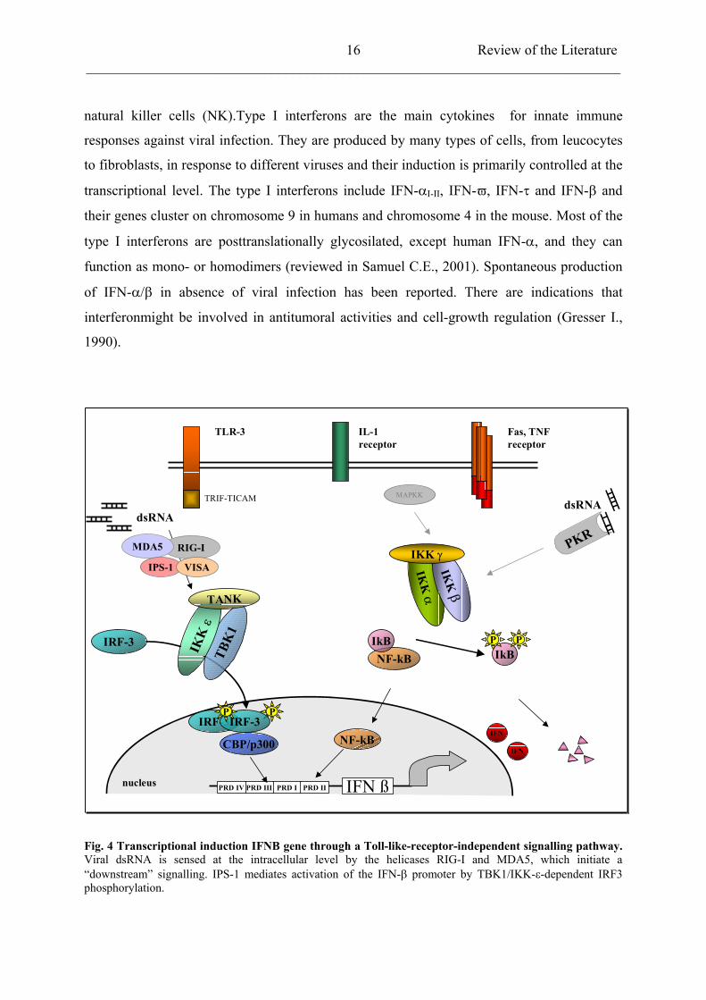
Fig. 4 Transcriptional induction IFNB gene through a Toll-like-receptor-independent signalling pathway. Viral dsRNA is sensed at the intracellular level by the helicases RIG-I and MDA5, which initiate a “downstream” signalling. IPS-1 mediates activation of the IFN- promoter by TBK1/IKK- -dependent IRF3 phosphorylation.
Review of the Literature ___________________________________________________________________________
17
Many viruses can induce activation of IFNA/B genes transcription. Activation occurs when
members of the IRF (Interferon Regulatory Factors) family bind to regulatory sequences in
the IFN- / gene promoters. The IRF family consists in 9 transcriptional activators of which
IRF3 and IRF7 are essential for IFN- / expression. IRF3 is constitutively expressed in all
cells and resides into the cytoplasm of unstimulated cells in a latent form. IRF3 activation is
mediated by dsRNA and virus infection. Upon activation, IRF3 undergoes serine/threonine
phosphorylation, dimerization, nuclear translocation, and after association with p300/CBP
coactivator, DNA-binding at consensus sites (Lin R. et al., 1998). Transcriptional activity of
IRF3 is controlled by C-terminal phosphorylation, which is carried out by a newly identified
virus-activated kinase complex, whose components are the IKK-related kinases TBK1 and
IKK-i/ (Fitzgerald K.A. et al., 2003; Sharma S.et al., 2003; McWhirter S.M. et al., 2004).
Activation of the TBK1/IKK- -mediated IFN- signalling pathway is mediated by the
retinoic-acid inducible gene I (RIG-I) and the melanoma differentiation associated gene 5
(MDA5). RIG-I and MDA5 are cytoplasmatic RNA elicases responsible for dsRNA
recognition. By gene targeting, it has been shown that RIG-I is essential for induction of type
I interferons after infection of fibroblasts and conventional dendritic cells (DCs). RIG-I
activates IRF3 via TBK1 and IKK- (Yoneyama M et al., 2004; Hiroki K. et al., 2005).
Moreover, RIG-I and MDA5 interact with the newly identified interferon-beta promoter
stimulator 1 (IPS-1) and the virus-induced signalling adaptor VISA in sensing viral infection
and in the activation of IFN- / induction signalling pathway (Kawai T. et al., 2005; Xu L.G.
et al., 2005).
IFN- / signalling pathway is mediated by a common receptor complex IFNAR, which
consists in two subunits, IFNR1 and IFNR2. Upon ligand-induced stimulation of IFNAR, two
receptor-associated Janus protein tyrosine kinases, Jak1 and Tyk2, become cross-activated.
This is followed by tyrosine phosphorylation of two members of the family of signal
transducers and activators of transcription (STATs), namely STAT1 and STAT2. Activation
of STATs leads to the formation of two transcriptional-activator complexes, IFN- -activated
factor (AAF) and IFN-stimulated gene factor 3 (ISGF3). ISGF3 consists of activated STAT1,
STAT2 and the interferon regulatory factor (IRF) 9. This trimeric complex locates into the
nucleus and binds to a cis-acting DNA element, designated ISRE, which is present in IFN- /
inducible genes. Among these IFN-inducible genes, some encode for proteins implicated in
Review of the Literature ___________________________________________________________________________
18
antiviral activities: the RNA-dependent protein kinase (PKR); the 2’, 5’-oligoadenylate
synthetase (OAS); RNase L and the Mx protein GTPases (Lau J.F. and Horvarth C.M., 2002).
2.5 Viral antagonists of IFN type I response
Both DNA and RNA viruses encode for proteins and have developed strategies to impair IFN
response. Circumventing or blocking IFN activity delays the generation of an acquired
immunity and allows viruses to successfully establish infection. Viral countermeasures can
affect IFN production or target IFN signalling. Several viruses have been reported to prevent
IFN induction by sequestration of dsRNA activators of PKR or 2’,5’oligoadenylate
synthetase/Rnase L system. dsRNA-binding proteins are encoded by vaccinia virus (E3L),
reovirus (capsid protein 3), rotavirus (NSP3) and influenzavirus (NS1) (Chang H.W. et al.,
1992; Lu Y. et al. 1995; Bergeron J. et al., 1998). A direct antagonism of PKR has been also
observed in poliovirus, adenovirus, SV40 and hepatitis C virus (HCV) (Black T.L. et al.1993;
Gale M. and Katze M.G., 1998). Encephalomyocarditis virus (EMCV) and HIV instead
downregulate RNase L (Martinand C.et al., 1998 and 1999). Another common strategy to
block IFN production consists in repressing transcriptional activation of IFN- / promoter.
For example, human herpesvirus 8 (HHV8) synthesizes an IRF homologue that is able to
block the transcriptional complex CBP/p300-IRF3 (Gao S.J. et al., 1997). The E6 protein of
human papillomavirus type 16 (HPV-16) binds IRF3 and therefore inhibits its activity (Ronco
L.V. et al., 1998). The nonstructural proteins (NS1 and NS2) of BRSV antagonize IRF3
phosphorylation (Bossert B. et al., 2003).
The IFN signalling pathway can be targeted as well by several viruses and its block can be
achieved by multiple mechanisms. Poxviruses, for example, encode soluble IFN receptor
homologues (vIFN-Rc). These secreted viral proteins sequester cellular IFNs, antagonizing
their binding to natural receptors (Smith G.L. et al., 1998). Simian 5 virus (SV5) or mumps
virus (MV) induce degradation of STAT1, while parainfluenza virus type 2 elicits degradation
of STAT2 , thereby preventing the formation of ISGF3 complexes (Dideock L. et al., 1999;
Young D.F. et al., 2000). Adenovirus affects DNA-binding of ISGF3 via the E1A protein
(Leonard G.T. and Sen G.C., 1997). Among Herpesviruses, varicella-zoster virus (VZV)
inhibits expression of STAT1 and JAK2, whereas human cytomegalovirus (HCMV) blocks
STATs phosphorylation by inducing degradation of JAK1 and IRF9 (Miller D.M.et al., 1998;
Abendroth A. et al., 2000).
Review of the Literature ___________________________________________________________________________
19
Respiratory syncytial virus is resistant to type I IFN but conflicting results about its ability to
affect type I IFN-signalling have been reported. Most of the data indicate that inhibition of the
IFN-signalling pathway is not involved. IFN-mediated MxA expression is maintained in
epithelial cells infected with HRSV or BRSV. Besides no decrease of STAT1 and STAT2
levels has been observed (Atreya P.L. and Kulkarni S.,1999; Young D.F. et al., 2000; Bossert
B. et al., 2003). Other groups have been arguing that RSV does block JAK/STAT pathway by
decreasing STAT2 expression (Ramaswamy M. et al., 2004; Lo M.S. et al., 2005).
2.6 The RSV nonstructural proteins NS1 and NS2
Pneumovirus is the only genus in the Paramyxoviridae family whose members encode the
NS1 and NS2 proteins. The two NS genes are located at the 3’ end of the negative-strand
RNA genome and, due to a characteristic transcriptional gradient, the transcripts of these
genes are abundantly expressed in infected cells. NS proteins are not essential for RNA
replication, however recombinant RSV lacking NS genes are severely attenuated in vitro and
in vivo (Teng M.N. and Collins P.L., 1999; Schlender J. et al., 2000; Valarcher J.F. et al.,
2004). The NS1 and NS2 genes of HRSV subgroup A are 528 and 499 nucleotides long with
single open reading frames encoding polypeptides of 139 and 124 amino acids, respectively.
Similarly, NS1 and NS2 genes of BRSV strain A51908 have 524 and 489 nucleotides which
encode for polypeptides of 136 and 124 amino acids, respectively. Comparison of the
sequences of HRSV NS proteins with the corresponding BRSV revealed amino acid identity
of 69% for NS1 and 84% for NS2 protein (Collins P.L. and Wertz G.W., 1985; Pastey M.K.
and Samal S.K., 1995). HRSV NS1 protein coprecipitates with M protein and interacts with
the C-terminal region of phosphoprotein P. The NS2 protein, despite colocalizing with N and
P proteins in cytosolic “inclusion bodies”, does not coprecipitate with any viral protein (Evans
J.E. et al., 1996; Hengst U. and Kiefer P., 2000; Bossert B. et al., personal observation).
Nonstructural proteins of pneumoviruses do not show common features of other known
proteins or functional domains that would suggest their function. NS proteins are abundantly
transcribed in RSV-infected cells and can block induction of IFN- / . Conzelmann and
colleagues generated BRSV mutants in which NS1 and NS2 genes have been deleted singly
( NS1; NS2) or in combination ( NS1/NS2). All three mutant viruses display a slight
attenuation in their growth in BSR T7/5 and Vero cells. Similar results have been also
obtained with the human counterpart HRSV. These results suggest that NS proteins are
Review of the Literature ___________________________________________________________________________
20
dispensable for viral growth but they also indicate their involvement in viral replication
(Buchholz U. et al., 1999; Teng M. and Collins P.L., 1999). In cultured cell lines that are
competent for type I IFN production, as in vivo, deletion HRSV and BRSV viruses are
severely impeded in their replication providing evidence that NS1 and NS2 proteins
independently or cooperatively subvert IFN- / -mediated antiviral state (Jin H. et al., 2000
and 2003; Valarcher J.F. et al., 2003). NS proteins can therefore be considered as potent
antagonists of IFN induction.
Our studies demonstrate that BRSV impairs type I IFN production by preventing the
activation of IRF3. In wild-type BRSV-infected cells, but not in cells infected with the double
deletion mutant BRSV NS1/NS2, phosphorylation of IRF3 is selectively blocked. This leads
to the suppression of IRF3 transcriptional activity in infected cells and compromises the
subsequent establishment of an IFN-mediated immune response (Bossert B. et al., 2003).
Similarly to BRSV, wild-type HRSV poorly induces type I interferons, in contrast
recombinant viruses lacking NS1 and NS2 genes do increase dramatically expression levels of
IFN- (Spann K.M. et al., 2004).
Materials and Methods
___________________________________________________________________________
21
3 MATERIALS AND METHODS
3.1 Cells culture and viruses
3.1.1 Cells
Vero and HEp2 cells were obtained by the American Type Culture Collection (ATCC;
Rockville, MD) and maintained in Dulbecco’s modified Eagle’s medium (DMEM, Gibco
BRL) supplemented with 5% fetal calf serum (FCS) and 1% Penicillin/Streptomycin 100x
(Sigma cell culture). For Vero-p125Luc cells, stably transfected with the IFN-
promoter/enhancer, 1 mg/ml of G418 were added to the medium. 293 and A549 cells were
cultured in DMEM containing 10% FCS and antibiotics. For 293 cells the medium was
additionally supplemented with 1% L-glutamine (Gibco BRL) . Baby hamster kidney cells
stably expressing T7 RNA polymerase (BSR T7/5) were propagated in BHK-21 medium
(Glasgow MEM, Gibco) containing 10% newborn calf serum (CS), 2% MEM amino acids
(Gibco BRL), 2% Tryptose phosphate broth 50x (Gibco BRL), 1% Penicillin/Streptomycin
and 1 mg/ml of G418 .
3.1.2 RSV propagation and titer determination
Human respiratory syncytial virus (HRSV) subgroup A strains A2 and Long, were obtained
by the American Type Culture Collection. Recombinant bovine respiratory syncytial virus
(rBRSV) was derived from BRSV strain A51908 (American Type Culture Collection) variant
Atue51908 (GeneBank accession no AF092942). For production of RSV stocks, 80%
confluent Vero cells grown in a 75 cm2tissue culture flask were infected at a multiplicity of
infection (MOI) of 0.1 in serum-free DMEM. After absorption for 1-1.5 hours, the inoculum
was removed and cells were incubated in DMEM supplemented with 2.5% FCS. When an
extensive cytopathic effect (syncytia formation) was observed, virus was released by freezing
and thawing. After centrifugation at 3,500 rpm (Heraeus Varifuge 3R) for 5 min to remove
cellular debris, the supernatant was aliquoted and stored at 70°C. Virus titers were
determined by limiting dilution in microwell plates. A confluent 75 cm2flask of Vero cells
was trypsinized and resuspended in 20-30 ml of DMEM containing 5% FCS, 100 µl were
distributed in each well of a 96-well microtiter plate. Virus stocks were stepwise 10-fold
Materials and Methods
___________________________________________________________________________
22
diluted in serum-free DMEM and 100 µl of each dilution were pipetted into the wells. After 4
days, cells were fixed with 80% acetone for 20 min at 4°C and air-dried. Infected cell foci
were stained with a monoclonal antibody recognizing the RSV nucleoprotein N (Serotec,
diluted 1:75 in PBS) for 60 min at room temperature. After washing with PBS for three times,
a FITC-conjugated anti-mouse antibody (dilution 1:100 in PBS) was applied for 60 min at
RT. Wells were washed three times with PBS and once with dH2O; foci were counted using a
fluorescent microscope (Olympus, IX71).
3.1.3 Rabies virus (RV) stocks and titer determination
A recombinant rabies virus carrying nucleotide sequence of Street Alabama Dufferin B19
(rRV SAD L16), a recombinant RV where the P coding sequence was replaced with eGFP-P
fusion protein (SAD eGFP-P) and mutant SAD eGFP-Ps harbouring the HRSV NS1 and NS2
genes (SAD eGFP-Ph2/1 and SAD eGFP-Ph2*T26I/1) were propagated in BSR T7/5 cells.
A 25 cm2tissue culture flasks 80% confluent were infected at an MOI of 0.1 for 1 hour. Cells
were then incubated in Glasgow MEM supplemented with 10% CS and supernatants were
harvested after 3 and, when possible, 6 days post infection. Supernatants were centrifuged 5
min at 3.500 rpm, aliquoted and freezed at 70°C. Determination of virus titers was carried
out on BSR T7/5 cells by limiting dilutions as described for RSV. Virus foci were visualised
by immunostaining with a fluorescein isothiocyanate conjugate (Centocor ) recognizing RV
N protein. For recombinant SAD eGFP-P viruses, fluorescence of infected cells was detected
directly by fluorescence microscopy.
3.2 General cloning procedures
3.2.1 Restriction enzyme digest
Restriction endonuclease digests were performed according to the supplier’s manual using the
recommended buffer and 10 units (U) of the chosen enzyme for each microgram of DNA.
Materials and Methods
___________________________________________________________________________
23
3.2.2 Extraction of DNA fragments from agarose gel
Restriction fragments were separated by electrophoresis on 1%-1.5% agarose gel using 1 x
TAE buffer containing 0.1 µg/ml of ethidium bromide. Addition of ethidium bromide solution
permitted visualization of nucleic acids under UV light.
x TAE buffer: Tris 40 mM
CH3COONa x 3 H2O 5 mM
EDTA 1 mM
Samples were diluted in 1x DNA loading buffer, loaded into the slots and electrophoresis was
performed for 45-60 min at 120 V.
DNA loading buffer: Ficoll 400 15%
TAE 5%
Orange G
Fragments were visualised under UV light at 366 nm (BIO-RAD, Universal Hood II) and
DNA fragments were recovered by gel excision. DNA was purified using QIAquick gel
extraction kit (Qiagen) following the supplier’s manual. Concentration of purified DNA
(µg/µl) was estimated by measuring the OD of 100 µl of a 1:50 dilution of the sample at 260
and 280 nm in a spectrophotometer (BioPhotometer, Eppendorf). Only samples with a ratio
between 1.7 and 2.0 were considered as appropriately pure.
3.2.3 DNA ligation
A ratio of vector/insert DNA of 1:5 or 1:10 was used in any ligation. 100 ng of vector were
mixed, in a sterile 1.5 ml centrifuge tube, with the appropriate amount of insert DNA, 2 µl of
10x ligation buffer, 2 U/µl of T4 DNA ligase (MBI) and bidistilled water up to 20 µl. The
ligation mix was incubated at room temperature for 3-4 hours or alternatively over night at
16°C.
Materials and Methods
___________________________________________________________________________
24
3.2.4 Transformation into competent bacteria
Competent XL-1 Escherichia coli were prepared by calcium chloride method. Bacteria were
thawed on ice and 50 µl were transferred to a semisterile centrifuge tube. About 10 ng of
plasmid or up to 20 µl of a ligation mix were added. After mixing by pipetting, bacteria were
incubated on ice for 20 min. The tube was than transferred for 2 min to a heating block,
preheated at 42°C and then rapidly returned to ice for 1-2 min. 250 µl of LB++ medium were
added to each tube and cultures were incubated at 37°C for 1 hour on a shaker.
LB++Medium: LB medium 1 l
MgSO4 19 ml of 1 M solution
KCl 3,2 ml of 3 M solution
An appropriate volume of culture (10-50 µl for plasmids and 250 µl for ligation) was
distributed onto agarose-LB plates containing 100 µg/ml of ampicillin. Transformed bacteria
were spread over the agarose plate by using a sterile metal rod and incubated over night at
37°C.
Agarose-LB: LB medium 1 l
Agar, in granules 15 g
After autoclaving the solution, swirl carefully to dissolved the agarose. When the solution
cools down to 50°C, add the antibiotic and pour directly in 90 mm Petri dishes. Wait until the
medium has solidified completely, turn the plates up side down and store at 4°C.
3.2.5 Preparation of minipreps and midipreps
To screen for positive clones, colonies were picked with pipette tips from the agarose-LB
plate and cultured in 1 ml of LB media supplemented with 100 µg/ml of ampicillin overnight
under constant agitation.
Materials and Methods
___________________________________________________________________________
25
LB++Medium: NaCl 5 g
Yeast extract 5 g
Bactotrypton 10 g
MgSO4 1 ml of 1 M solution
Bidistilled water up to 1 l
Dissolved the solute, adjust the pH at 7.5 and autoclave
Cultures were centrifuged for 5 min at 4000 x g (Ependorf table centrifuge). The supernatant
was discharged and the pellet resuspended in 0.2 ml of Flexi I buffer. Flexi II buffer (0.2 ml)
was added to lyse the cells for 5 min at room temperature, followed by the addition of 0.2 ml
of Flexi III buffer to precipitate chromosomal DNA and cellular debris. After an incubation
on ice for 5 min, tubes were centrifuged for 10 min at 8000 x g and the supernatant was
transferred to a fresh 1.5 ml. A volume of 0.42 ml of isopropanol was added to precipitate
plasmid DNA and the solution was mixed by pipetting before centrifugation at 8000 x g for
10 min at room temperature. The supernatant was removed and the pellet was washed with 1
ml of 70% ethanol, centrifuged as before and, after being air-dried, dissolved in 50 µl of
dH2O. Restriction enzyme analysis was performed. Larger amounts of plasmid DNA were
prepared by midipreps. Overnight cultures of 100 ml of LB medium supplemented with 100
µg/ml of ampicillin were used and DNA was purified with Nucleobond plasmid purification
kit AX 100 (Macherey-Nagel) according to the manifacturer’s instructions.
Flexi I: Tris-HCl 100 mM [pH 7.5]
EDTA 10 mM
Rnase I 400 g /ml
Flexi II: NaOH 200 mM
EDTA 1%
Flexi III: KCH3COO (aq) 300 mM [pH 5.75]
Materials and Methods
___________________________________________________________________________
26
3.3 Reverse Transcription and PCR conditions
3.3.1 Extraction of RNA from cells and reverse transcription
For reverse transcription (RT) reactions, RNA was isolated from confluent mock and infected
cells seeded into 6-well plates using Qiagen Rneasy kit (Qiagen). For IFN- mRNA isolation
cells were infected at MOI of 1 for 16-18 hours. A DNase digest was performed during the
RNA extraction process using RNase-free DNase set (Qiagen). RNA was resuspended in 50
µl of DEPC-H2O and the concentration was determined as described for the DNA
concentration. 1 µg of DNase-treated RNA was mixed with 3 µl (30 pmol) of the desired
antisense primer and 0.5 µl of Rnasin (Amersham-Pharmacia) in a total volume of 42 µl.
After an incubation of 10 min at 65 °C followed by 10 min at 37°C, 2 µl of dNTP mix (25
mM each), 5 µl of 10x RT buffer (provided by the manufacturer) and 1 µl of StrataScript
reverse transcriptase were added. The reaction was incubated for 1 hour at 37°C followed by
inactivation of enzyme activity at 95 °C for 5 min. 5 µl were used in a PCR reaction.
DNase digested RNA x µl (1 µg)
Antisense primer 3 µl (30 pmol)
dNTPs (25 mM) 2 µl
10x reaction buffer 5 µl
Rnasin 0.5 µl
reverse transcriptase 1 µl
bidistilled water up to 50 µl
The following primers were used to amplify:
Human IFN-
h 3’ (antisense): 5’-aag atg ttc tgg agc atc tga tag atg-3’
Actin
-actin 3’ (antisense): 5’-ccg cca gac agc act gtg ttg gcg ta-3’
HRSV NS1:
hNS1-EcoRI (antisense): 5’-att gag aat tct tat gga tta aga tca aa-3’
HRSV NS2:
Materials and Methods
___________________________________________________________________________
27
hNS2-EcoRI (antisense): 5’-att gag aat tct tat gga tta aga tca aa-3’
BRSV NS2:
BNS2HA-EcoRI (antisense): 5’-gca ata gaa ttc cta ttt atc gtc atc atc ttt ata atc tgg att
taa atc ata ctt ata-3
3.3.2 PCR conditions
Standard PCR reactions were prepared with 100 ng of template DNA, 25 pmol of each sense
and antisense primers, 1 µl of dNTPS mix (25 mM of each dATP, dCTP, dGTP and dTTP),
10 µl of DMSO, 10 µl of 10x buffer (supplied by the manufacturer and containing 50mM of
MgSO4) and 1 µl of Taq Polymerase in a total volume of 100 µl.
For detection of human IFN- , the following primers were used:
h 5’ (sense): 5’-ctc ctc caa att gct ctc ctg ttg tg-3’
h 3’ (antisense) 5’-aag atg ttc tgg agc atc tga tag atg-3’
To confirm integrity of the DNA and to verify infection, sequences of the -actin and RSV
NS genes were amplified, respectively, using the primers listed below:
-actin 5’ (sense): 5’-ggc atc gtg atg gac tcc-3’
-actin 3’ (antisense): 5’-ccg cca gac agc act gtg ttg gcg ta-3’
hNS1-NcoI (sense): 5’-att gac cat ggg cag caa ttc att-3’
hNS1-EcoRI (antisense): 5’-att gag aat tct tat gga tta aga tca aa-3’
bNS2-BamHI (sense): 5’-aag cgg atc ccc aac cag cca tga gca cc-3’
bNS2FL-EcoRI (antisense): 5’-gca ata gaa ttc cta ttt atc gtc atc atc ttt ata atc tgg att
Materials and Methods
___________________________________________________________________________
28
taa atc ata ctt ata-3’
hNS2-NcoI (sense): 5’-att gac cat gga cac aac cca ca -3’
hNS2-EcoRI (antisense): 5’-gga att cga atc ttg tgt tga aat t-3’
The reactions were prepared on ice in a sterile 0.5 ml tube and contained:
DNA sample 5 µl
Sense primer 2.5 µl (10 pmol)
Antisense primer 2.5 µl (10 pmol)
dNTPs (25mM) 2.0 µl
10x reaction buffer + MgSO4 10 µl
DMSO 10 µl
Taq Polimerase (5U) 1 µl
Bidistilled water 64 µl
PCR reaction was carried out in a Biometra T3 thermocycler including the following steps:
1ststep: denaturation (95 °C, 5 min)
2ndstep: denaturation (94 °C, 1 min)
3rdstep: annealing ( x °C, 1min)
4thstep: extension (72 °C, 1 min 30 sec)
5thstep: extension (72 °C, 10 min)
6thstep: cooling/pause to 4°C
Step 2 to 4 were repeated 35 times, before step 5 and 6, for a total number of 36 cycles. The
temperature of the annealing step depended on the length and GC-content of the primers:
62°C were used for IFN- and for -actin, 58°C for HRSV NS2, 52°C for and 63°C for
HRSV and BRSV NS1 gene, respectively. The resulting DNA fragments were mixed with
DNA loading buffer and run on a 1.5 % agarose TAE gel with ethidium bromide and
visualised with UV light. If required, purification of PCR products was performed by Qiagen
PCR purification kit or by QIAquick gel extraction kit (Qiagen).

Materials and Methods
___________________________________________________________________________
29
3.4 Generation of recombinant BRSV viruses expressing HRSV NS1 and NS2 proteins
3.4.1 Construction of rBRSV viruses expressing NS proteins of HRSV strain Long
(rBRSVh1/2)
Construction of the recombinant BRSV virus harbouring HRSV Long NS1 and NS2
(rBRSVh1/2) has been previously described (Bossert B. and Conzelmann K.K., 2002).
Briefly, cDNA encompassing both HRSV Long NS genes was obtained by RT-PCR of
infected Vero cells and subcloned in a plasmid, generating the phNS1hNS2cass. This plasmid
was digested with NotI and Acc65I and the resulting fragment containing the HRSV Long NS
genes was inserted in a full length BRSV cDNA, resulting in rBRSVh1/2.
3.4.2 Construction of rBRSV expressing a mutated HRSV NS2 protein (rBRSVh1/2*T26I)
3.4.2.1 Quick site-directed mutagenesis of HRSV Long NS2 gene
phNS1hNS2 was used for mutagenesis. Threonine (T) at position 26 of NS2 protein was
substituted with an Isoleucine (I) residue, mimicking the situation in HRSV A2 strain. An
additional restriction site for the endonuclease SpeI was inserted for subsequent restriction
digest analysis. Mutagenesis was performed by using a modified Quickchange-Mutagenesis
protocol. PCR was done using the following primers:
hNS2*T26I
(sense): 5’-ttg tca ctt gag act att ata act agt cta acc aga-3’
hNS2*T26I
(antisense): 5’-agt ctc aag tga caa tgg tct cat g-3’
The restriction recognition site is underlined and the mutated codon is in bold.
Materials and Methods
___________________________________________________________________________
30
PCR reaction was performed as previously described (3.3.2):
DNA sample x µl (about 100 ng)
Sense primer 2.5 µl (10pmol)
Antisense primer 2.5 µl (10pmol)
dNTPs (25mM) 1.0 µl
10x reaction buffer 10 µl
Pfu Polimerase 1 µl (2.5 U)
Bidistilled water up to 100 µl
The following steps were used:
1ststep: denaturation (95 °C, 30 sec)
2ndstep: denaturation (95 °C, 30 sec)
3rdstep: annealing (50 °C, 1 min)
4thstep: extension (68 °C, 8 min)
5thstep: extension (68 °C, 10 min)
6thstep: cooling/pause to 4°C
PCR product was digested over night at 37°C with DpnI (10 U) endonuclease to eliminate
residual DNA template and purified by Qiagen PCR purification kit. 5 µl of purified DNA
were transformed in competent XL-1 bacteria, which were spread on agarose plates over night
at 37°C as described in paragraph 3.2.4. Positive clones were screened by restriction digest,
using in combination the following endonucleases: SpeI with NotI and NotI with Acc65I.
Positive clones were sequenced to confirm the presence of the desired mutation and large
scale amount of DNA were prepared at least from two different positive clones by midipreps.
3.4.2.2 Generation of rBRSVh1/2*T26I
virus
The plasmid phNS1hNS2*T26I
was digested with NotI and Acc65I. The resulting fragment of
1094 nt was inserted by DNA ligation into the full length BRSV cDNA, previously digested
with the same restriction enzymes, resulting in pBRSVhNS1hNS2*T26I
(rBRSVh1/2*T26I)
18 cycles
Materials and Methods
___________________________________________________________________________
31
3.5 Recovery of recombinant BRSV viruses
Recombinant rBRSVh1/2*T26I, as well as rBRSVh1/2 and BRSV wild-type, were rescued as
described before (Buchholz U.J. et al., 1999). BSR T7/5 cells were transfected with the
corresponding full length cDNA plasmid after calcium phosphate precipitation (Mammalian
transfection kit, Stratagene) as specified by the supplier. 1x 10⁶cells/dish were seeded in 35
mm-diameter dishes in BHK-21 medium supplemented with 10% CS and grown over night.
Cells were then washed with serum-free DMEM and 1 ml of the medium was added to each
dish. After 1 hour, transfection was performed using a plasmid mixture containing 10 µg of
pBRSVhNS1hNS2*T26I
(or pBRSVwt or pBRSVhNS1hNS2) and the plasmids pTITB-N
(4 µg), pTITB-P (4 µg), pTITB-L (2 µg) and pTITB-M2 (2 µg). Four hours post transfection,
the medium was removed and cells were incubated in BHK-21 medium containing 10% FCS.
Three days later cells were split (ratio 1:3) and they were maintained in 2.5% FCS BHK-21
medium. Cells were further split every 4-5 days until the formation of syncytia was observed.
Virus stocks were obtained as described previously in 3.1.2.
3.6 Generation of recombinant SAD eGFP-P viruses expressing HRSV NS proteins
3.6.1 Generation of recombinant SAD eGFP-P viruses
Recombinant rabies virus (SAD eGFP-P), coding for a eGFP-P fusion protein and carrying
RSV NS1 and NS2 genes of different origin were constructed. RSV NS genes were inserted
into the intergenic region between G and L genes.
For BRSV NS genes, a full length RV cDNA containing an extra transcriptional stop-restart
sequence in the 3’ non coding region of G gene and harbouring BRSV NS1 and NS2 genes
(pSAD VB NS2FL/NS1HA) was used (Mebatsion T. et al., 1996; Bossert B. and Conzelmann
K.K., 2002). The plasmid was digested with PpMI and MluI endonucleases and the large
fragment of about 5,000 bp was inserted in the full length SAD eGFP-P cDNA resulting in
pSAD eGFP-PbNS2flbNS1ha (eGFP-Pb2/1). Similarly, the full length cDNA pSAD eGFP-
PhNS2hNS1 (eGFP-Ph2/1) was constructed using in this case a RV cDNA plasmid
harbouring HRSV Long NS genes (pSAD VBhNS2hNS1). In the case of the recombinant RV
expressing a mutated HRSV NS2 gene (eGFP-Ph2*T26I/1), I used a different strategy as
follows. A plasmid containing HRSV-Long NS genes (phNS2hNS1) was mutagenised as
Materials and Methods
___________________________________________________________________________
32
described in 3.4.2.1. The resulting plasmid phNS2*T26IhNS1 was digested with NotI and
Acc65I and treated with Klenow polymerase to generate blunt ends. The DNA fragment,
which encompassed the NS genes, was inserted into the unique SmaI site of pSAD VB
resulting in pSAD VBhNS2*T26IhNS1. Positive clones were screened by restriction analysis
and sequenced to confirm the presence of the inserted mutation and the correct orientation. At
this point the procedure was the same used for the previous two recombinant RVs. pSAD VB
hNS2T26IhNS1 was digested with PpMI and MluI and the purified DNA fragment was
inserted in the full length SAD eGFP-P cDNA resulting in pSAD eGFP-PhNS2*T26I/hNS1
(eGFP-Ph2*T26I/1).
3.6.2 Rescue of recombinant eGFP-P viruses expressing RSV NS proteins
Recovery of recombinant eGFP-Pb2/1, eGFP-Ph2/1, eGFP-Ph2*T26I/1 was performed using
the CaPO4-method as previously described for recovery of RSV. In this case plasmids
encoding for RV protein N (pTIT-N, 5 µg), P and L (pTIT-P and pTIT-L, 2.5 µg each) with
10 µg the respective cDNA (pSAD eGFP-PbNS2flbNS1ha, pSAD eGFP-PhNS2hNS1 or
pSAD eGFP-PhNS2*T26I/hNS1). After transfection, cells were maintained in BHK-21
medium supplemented with 10% CS and cell-culture supernatants were harvested 3 and 6
days post transfection. Fluorescence of the recombinant eGFP-P viruses was monitored to
detect virus infection and to determine viral titers of the stocks.
3.7 Growth characteristics of recombinant BRSV and eGFP-P viruses
3.7.1 rBRSV viruses expressing HRSV NS proteins
Recombinant rBRSVh1/2 and rBRSVh1/2*T26I
were analysed for their ability to grow in
different cells systems and compared to the HRSV prototypes Long and A2. 4x 10⁵Vero
(interferon-deficient) and human HEp2 (interferon-active) cells were infected with the
recombinant BRSVh1/2 and BRSVh1/2*T26I
and with Long and A2 strains at an MOI of 0.3.
Infection was performed in suspension, in serum-free DMEM medium for 1-1.5 hours in 15
ml falcon tubes. Cells were kept in suspension by gently shaking each 15 min. Afterwards,
cells were transferred into 12-well plate in 1 ml of DMEM containing 2.5% of FCS.
Materials and Methods
___________________________________________________________________________
33
Supernatants were harvested daily for 3 days, centrifuged to remove cell debris and stored at
–70°C before virus titers were determined on Vero cells as already described. Cell lysates
were also collected to monitor viral protein expression at the different time points and to
verify the correct translation of NS proteins in the recombinant BRSVs.
3.7.2 Recombinant rabies eGFP-P viruses expressing RSV NS proteins
Growth characteristics of eGFP-Pb2/1, eGFP-Ph2/1 and eGFP-Ph2*T26I/1 were determined in
BRS T7/5 cells, which are defective the type I IFN induction pathway, and in Hep2 cells.
Wild-type SAD L16 and SAD eGFP-P were included as control viruses, since they have been
shown to differ in their ability to induce IFN- .
BSR T7/5 and HEp2 cells, at a concentration of 1x 10⁶, were infected with the viruses
mentioned above at an MOI of 0.01 and maintained in BHK-21 medium containing 10%CS
or DMEM with 5% FCS. Supernatants were collected for 3 days and virus titers were
determined on BSR T7/5 as described before.
3.8 IFN treatment of rBRSV viruses
To analyse the effect of IFN type I on the replication of HRSVs and rBRSVs, HEp2 cells
were infected at an MOI of 0.3 as described for growth kinetic analysis and IFN- A/D (PBL
Biomedical Laboratories) was added to the culture medium directly after seeding.
Concentrations of 500, 1000 and 5000 IU/ml were assayed using a working dilution of 100
IU/µl IFN- in PBS. Virus titers were determined 2 or 3 days post infection depending on the
extent of the observed CPE.
3.9 Cloning of the human TANK-binding kinase 1 (TBK1) and TRAF-binding protein
(TANK)
The human kinase TBK1 and TANK genes were amplified by PCR reaction from a human
lung cDNA library. PCR reaction was performed as previously described using the following
oligonucleotide primers:
hTBK1 BamHI (sense): 5’-atg gat ccc atg cag agc act tct aat cat ctg-3’
Materials and Methods
___________________________________________________________________________
34
hTBK1 NotI (antisense): 5’-ata tgc ggc cgc cta aag aca gtc aac gtt gcg aag gcc-3’
hTANK BamHI (sense): 5’-atg gat ccc atg gat aaa aac att ggc gag-3’
hTANK NotI (antisense): 5’-ata tgc ggc cgc tta agt ctc tcc att gaa gtg-3’
Inserted restriction sites are underlined
Restriction sites for the endonucleases BamHI and NotI were inserted at the 5’ and 3’ ends of
the amplified ORF respectively to allow subsequent cloning in the expression vector pCR3-
Ig.
The reaction was carried out according to the following steps:
1ststep: denaturation (95 °C, 5 min)
2ndstep: denaturation (94 °C, 1 min)
3rdstep: annealing ( °C, 1min)
4thstep: extension (72 °C, 1 min 30 sec)
5thstep: extension (72 °C, 10 min)
6thstep: cooling/pause to 4°C
The obtained DNA fragments were subjected to restriction digest with BamHI and NotI and
purified by gel extraction (QIAquick gel extraction kit, Qiagen). The sequence of DNA
fragment obtained by PCR was confirmed by DNA sequencing. To construct the expression
vectors expressing the wild type TBK1 and TANK genes, the cDNA fragments were ligated
into pCR3-Ig vector plasmid, previously digested with the appropriate restriction enzymes. In
this way, N-terminal Ig-tagged TBK1 and TANK fusion proteins were generated (IgTBK1
and IgTANK). Correct expression of the recombinant IgTBK1 and IgTANK were assayed by
Western blot and immunofluorescence analysis. Functional activity was tested by gene
reporter assay.
35 cycles
Materials and Methods
___________________________________________________________________________
35
3.10 Gene Reporter Assay
3.10.1 Modified Gene Reporter Assay in Vero-p125Luc cell line
Vero cells, stably transfected with the IFN- gene promoter/enhancer (p125-Luc), were
seeded in 24-well-plates, using 2x 10⁵cells/well, and infected with RSV viruses at an MOI of
0.3. After an absorption of 90 min in serum-free DMEM, cells were incubated in DMEM
supplemented with 5% FCS. At 48 hours post infection cells were harvested in 200 µl of
luciferase lysis buffer [20 mM Tris HCl pH (7.8), 2 mM DTT, 2 mM CDTA, 10% glycerol,
1% triton X-100; incubation 10 min at 37°C]. Samples were than assayed for reporter gene
activity in a luciferase assay, that was performed as follows. Aliquots were shortly
centrifuged to remove unsoluble parts and 20 µl of each cell lysate were diluted in 80 µl of
luciferase lysis buffer. 100 µl of luciferase substrate buffer [20 mM Tricine, 2.67 mM MgSo4,
0.1 mM EDTA, 33.3 mM DTT, 1.07 mM MgCO3 x Mg(OH)2 x 5 H2O] supplemented with
470 µM AcetylCoA and 530 µM ATP were added to the cell lysates directly through the
luminometer (LUMAT LB9501, Perkin Helmer Wallac GmbH). The light emission was
measured as relative light units (RLU). Infection was verified by Western blot analysis using
the same cell lysates.
3.10.2 Dual luciferase assay and transfection of reporter plasmids
For experiments aimed to verify the biological activity of the non-canonical kinases TBK1
and IKK- to induce IFN, BSR T7/5 cells were transfected with a reporter plasmid expressing
the firefly luciferase gene under the control of the IFN- gene promoter/enhancer (p125Luc)
and a plasmid containing a Renilla luciferase gene under the control of the CMV promoter
was used as internal control (pCMV-RL). Additionally, plasmids containing wild type human
IRF3 (pEF-haIRF3), human TBK1 (pCR3-IgTBK1) or IKK- (pFlagIKK- ) or the kinase
inactive form of IKK- (pFlagIKK- K38A) were cotransfected. BSR T7/5 cells were seeded
in a 24-well plate using 1x 10⁵cells/well and grown over night. Transfection was performed
with FuGENE 6 reagent (Roche) as indicated by the manifacturer using 0.1 µg of the reporter
construct p125Luc, 0.02 µg pCMV-RL, 0.1 µg of the transcription factor IRF3 and 0.1 µg of
the indicated kinases. Luciferase activity was measured 48 hours post transfection.
Materials and Methods
___________________________________________________________________________
36
The relative light units (RLU) were standardised based on the Renilla luciferase activity and
converted in fold induction.
Similar experiments were also conducted in 293 cells, but in this case cotransfections were
carried out using reporter plasmids for IRF3 promoter (pCIB55Luc), pCMV-RL and TBK1 or
IKK- .
.
3.10.3 Transcription factor activation and TBK1 inhibition by RSV infection
To assay the activities of the transcription factors AP-1, NF-kB and IRF3, 2x 105293 cells
were transfected with 0.1 µg of the respective reporter construct (pAP-1Luc, pNF-kBLuc or
p55CIBLuc) and 0.02 µg pCMV-RL using FuGENE 6. At 24 hours post transfection, cells
were infected with the indicated viruses at an MOI of 0.3. At 24 hours post infection,
luciferase activity was measured by DL (Dual Luciferase Reporter Assay System, Promega)
as indicated by the supplier. To investigate the capacity of the different RSVs to inhibit
TBK1-dependent induction of the IFN- gene promoter, 2x 105293 cells were infected in
serum-free DMEM medium with the corresponding viruses at different MOIs. After 90 min of
incubation on gently shaking, cells were seeded in 24-well plate and maintained in DMEM
containing 10% of FCS. At 24 hours post infection, cells were transfected with 0.1 µg of
p125Luc, 0.02 µg of pCMV-RL and 0.1 µg of pCR3-IgTBK1. Viral protein expression was
then assessed by Western blot on the remaining cell lysates used to perform the luciferase
assay. The same experiment was performed in BSR T7/5 cells with the only difference that
infected cells were transfected with the above mention plasmids plus pEF-haIRF3, being the
endogenous IRF3 being defective in this cell line.
3.11 Protein expression analysis
3.11.1 Extraction of proteins from cells
Extraction of proteins from cells was performed using an appropriate amount of lysis buffer
(500 µl for a 6-well plate, or 250 µl for a 12-well plate). Cell extracts were incubated at 95°C
for 5 min, centrifugated at maximum speed (8000 x g) in a Eppendorf table centrifuge for 5
min and equal amounts were loaded onto a sodium dodecyl sulfate-polyacrylamide gel (SDS).
Materials and Methods
___________________________________________________________________________
37
Protein extraction buffer: Tris pH 6.8 6.25 mM
sodium dodecyl sulfate (SDS) 2%
glycerol 10%
urea 6 M
methanol 5%
bromophenol blue 0.01%
bromophenol red 0.01%
2-mercaptoethanol 0.5%
3.11.2 SDS-polyacrylamide gel electrophoresis (SDS-PAGE)
Proteins were separated using the PenguinTMModel P9DS (OWL scientific). The separating
gel, containing 10% or alternatively 12% acrylamid, was poured into the gap between the two
glass plates of the system and a space of about 3 cm was left to allow the adding of the
stacking gel. The gel was overlaid with 2-iso-propanol to ensure a levelled surface and left for
polymerisation for about two hours.
Separating gel: 10% gel 12% gel
Polyacrylamide 9.0 ml 10.8 ml
Gel buffer 12.0 ml 12.0 ml
dH2O 12.9 ml 11.1 ml
glycerol 2.0 ml 2.0 ml
TEMED 0.017 ml 0.017 ml
APS (ammonium persulfate) 0.175 ml 0.175 ml
The indicated amounts of gel mix were sufficient for one 14x 16 cm gel
Gel Buffer: Tris 364 g
SDS 15 ml of a 20% solution
After complete polymerisation the 2-iso-propanol was discarded and the stacking gel was
prepared and added on the top of the separating gel. The comb was immediately inserted
carefully avoiding to trap air bubbles under the teeth.
Materials and Methods
___________________________________________________________________________
38
Stacking gel: Polyacrylamide 1.4 ml
(for two gels) Gel buffer 3.5 ml
dH2O 9 ml
TEMED 0.116 ml
APS 0.018 ml
After polymerisation (1-2 hours), the comb was removed and the plates mounted in the
electrophoresis apparatus, which was filled with anode and cathode SDS-PAGE
electrophoresis buffer.
Jagow-Anode buffer: Tris 242 g
dH2O 1 l
The pH was adjusted to a value of 8.9 by HCl addition
Jagow-Cathode buffer: Tris 121 g
pH [8.25] Tricine 179.2 g
SDS 10 g
dH2O 1 l
Samples were loaded and the electrophoretic run was performed at 40-60 V until the
bromophenol blue left the separating gel at the bottom (about 15-16 hours) depending on the
gel percentage. A molecular weight standard (Precision Plus ProteinTMStandards, BIO-RAD)
was pipetted onto the gel to estimate protein size.
3.11.3 Electroblotting
When the electrophoretic run was completed, gels were removed from the glass plates and
incubated in 1x semi-dry buffer for 10-15 min. The separated proteins were transferred to a
nitrocellulose membrane in a semidry transfer apparatus (The PantherTMModel HEP-1; OWL
Scientific).
Materials and Methods
___________________________________________________________________________
39
Semi-dry transfer buffer (10x): Tris 58 g
Glycine 29 g
SDS 2.5 ml of a 20% solution
dH2O 1 l
For electrophoretic blotting 1x Semi-dry buffer was used; 180 ml of methanol were added to
1 l of fresh 1x solution
One sheet of Whatman blotting paper of the same size as the gel was soaked in transfer buffer
and placed on the bottom electrode and pressed to remove air bubbles. The gel and the
nitrocellulose membrane, which were also soaked in transfer buffer, were positioned exactly
over the bottom paper and covered with another soaked blotting paper. After having placed
the upper electrode the system was connected to a power supply (Standard power Pack P25,
BIOMETRA) and the transferred was performed at 400 mA for each gel for 120 min.
3.11.4 Western blotting
After the transfer, the membrane containing the separated proteins was incubated at room
temperature for 60 min in constant agitation with a blocking solution.
Blocking solution: 5% instant skimmed milk
0.05% Tween 20
1x PBS
After the blocking, the membrane was washed 3 times for about 15 min at RT with PBS
containing 0.05% Tween 20 (PBS-T). Incubation with the primary antibody diluted in PBS-T
took place over night at 4°C in constant agitation. The membrane was then washed 3 times for
about 15 min at RT with PBS-T and the incubation with peroxide-conjugated secondary
antibody diluted 1:10000 in PBS-T occurred for 90 min at RT. Finally the membrane was
washed 3 times with PBS-T and once in PBS for 10 min at RT. For detection, the membrane
was incubated with 1 ml of ECL Western blotting detection reagent (Perkin Helmer) and
exposed to an Hyperfilm ECL (Amersham) for varied times depending on the strength of the
signal.
Materials and Methods
___________________________________________________________________________
40
3.12 Immunofluorescence
BSR T7/5 cells were seeded overnight on coverslips in 6-well plate and transfected the day
after with FuGENE 6 (Roche). 4 µg of each expressing plasmid where used. At 48 hours post-
transfection, cells were washed with phosphate-buffered saline (PBS) and fixed with 3%
(w/v) paraformaldehyde in PBS for 30 min followed by treatment with 50 mM NH4Cl in PBS
for 20 min. Cells were permeabilized with 0.5% Triton X-100 for 10 min and blocked for 1
hour at room temperature (RT) with 1% goat serum and 0.5% BSA in PBS. Cells transfected
with FlagIKK- were incubated for 40 min at RT with the primary antibody anti-mouse Flag
(M2, Sigma) diluted 1:1000 in blocking reagent. In the case of Ig-TANK and Ig-TBK1 goat
FITC-tagged anti-human Ig were used at dilution 1:50. After primary-antibody incubation, for
cells transfected with FlagIKK- , a Cy3-conjugated anti-mouse antibody was used at dilution
1:200 in PBS. After 40 min incubation at RT, cells were washed with PBS and nuclei were
stained with DAPI (Hoechst) at the concentration of 1 µl/ml. Coverslips were mounted in
with Mounting Medium-Vectashield for fluorescence (LINARIS) and examined with a
fluorescence microscope equipped for laser-scanning confocal light microscopy (Zeiss,
LSM510). Digitalized images were further processed using Adobe Photoshop version 7.0.
3.13 Immunoprecipitation of IgTANK and IgTBK1
BSR T7/5 cells were seeded overnight in 60 mm petri dishes at a concentration of 1x 10⁶cells
per dish and transfected the day after with 15 µg of each of the following expression
plasmids: pCR3-Ig (empty vector), pCR3-IgTANK and pCR3-IgTBK1 by CaPO4 method
(Mammalian Transfection Kit, Stratagene). At 48 hours post transfection, cells were lysed for
30 min on ice with 300 µl of immunoprecipitation buffer (IP buffer). Lysates were
centrifuged at 14.000 rpm for 15 min at 4°C to precipitate nuclei and cell debris, followed by
transfer of the clean supernatants to fresh 1.5 ml Eppendorf tubes.
Materials and Methods
___________________________________________________________________________
41
IP buffer: TRIS 50 mM pH 7.6
NaCl 150 mM
NP-40 1%
EDTA 1 mM
EGTA 2 mM
NaF 20 mM
Protease inhibitor cocktail (Complete, Roche) was freshly added to the buffer before use
100 µl of Sepharose A beads (Protein A SepharoseTM 4 Fast Flow, Amersham), previously
washed 3 times in IP buffer, were added and the lysates were incubated overnight at 4°C in
continuous rotation. After incubation, lysates were centrifuged at 8000 x g at 4°C for 15 min
to precipitate the beads. The pellets were then washed 3 times in IP buffer. Once the washing
step was completed, the beads were pelleted down and resuspended in 100 µl of protein lysing
buffer. The lysates were analysed by Western blotting.
3.14 Materials and equipment
3.14.1 Serological reagents
Primary antibodies:
abCam, Rabbit -human IFN- , polyclonal
Cambridge, UK
BFAV
Insel Reims, Germany Rabbit -RV N and P proteins (S50), polyclonal
Biogenesis, Goat -RSV serum, polyclonal
c/o Quartett GmbH,
Berlin, Germany
Dianova, Normal goat serum
Hamburg,Germany
Santa Cruz Biotec. Inc. Rabbit -TBK1, polyclonal
Heidelberg, Germany Rabbit -PCNA, polyclonal
Rabbit -IRF-3, polyclonal
Rabbit -HA (Y11), polyclonal
Materials and Methods
___________________________________________________________________________
42
Serotec, Mouse -RSV N protein, monoclonal
Düsseldorf, Germany
Sigma-Aldrich, Rabbit -actin (20-33), polyclonal
Taufkirchen, Germany Mouse -FLAG (M2), monoclonal
J.A. Melero, Rabbit -HRSV NS1/NS2 ( -IC/C), polyclonal
University of Madrid, Spain
Freiburg, Germany Mouse -Mx A, monoclonal
All secondary antibodies (FITC-, Cy3-, peroxidase-conjugated anti-mouse, anti-rabbit and
anti-human Igs) were obtained from Dianova ()
3.14.2 Chemicals
Boehringer /Roche DTT (1,4-dithioerythreitol)
Mannheim, Germany
Fluka, NP40 (nonidet P40)
Deisenhofen, Germany tricine
ICN Biochemical Inc APS (ammonium persulfate)
Cleveland, Ohio-USA
Merk, CaCl2Darmstadt, Germany EDTA
Ethidium bromide
CH3COOK
KCl
MgSO4MgCl2CH3COONa
NaCl
NH4Cl
Orange G
Paraformaldehyde
Phenol red
Propidium iodide
Triton X-100
Materials and Methods
___________________________________________________________________________
43
Amersham-Pharmacia, Ficoll
Freiburg, Germany
Riedel-de-Haen, Ethanol
Deisenhofen, Germany Methanol
NaOH
Carl Roth GmbH, Acetone (tech)
Karlsruhe, Germany DMSO (dimethylsulfoxide)
Glycerol
Glycine
HCl
Hepes
Methanol
SDS
Tris
Tween 20
Urea
Sigma, ATP
Taufkirchen, Germany bromophenol blue
CDTA
MgCO3xMg(OH)2TEMED
3.14.3 Enzymes
MBI Fermentas, Klenow polymerase
St. Leon-Rot, Germany
New England Biolabs, Restriction endonucleases
Frankfurt am Main, Germany T4 DNA ligase
endonucleases
Roche Biochemicals, Shrimp alkaline phosphatase
Mannheim, Germany
Stratagene, pfu polymerase
Amsterdam, Netherland Reverse transcriptase
Materials and Methods
___________________________________________________________________________
44
3.14.4 Kits
Machrey & Nagel, Nucleobond AX100
Düren, Germany
Qiagen, Rneasy kit
Hilden Germany QIAquick PCR purification/Gel
extraction/Nucleotide removal
Perkin Elmer, Western lightning Chemiluminescence Reagent
Plus
Freiburg, germany
Promega GmbH, Dual Luciferase Reporter Assay System
Mannheim, Germany
Stratagene, Mammalian transfection kit
Amsterdam, Holland
3.14.5 Miscellaneous
Amersham Bioscience Gmbh, Hyperfilma ECL
Freiburg, Germany Protein A SepharoseTM4 Fast Flow
BD, Bacto yeast extract
Heidelberg, Germany
BIO-RAD, Precision Plus ProteinTMStandards
Munich, Germany
Difco, Bacto Tryptone peptone
Hamburg, Germany
Gibco BRL, Agarose
Karlsruhe, Germany Ampicillin
DNA 1kb ladder
LINARIS GmbH, Mounting Medium-Vectashield for fluorescence
Wertheim, Germany
Merck, Acrylamid/Bisacrylamid solution (29:1)
Darmstadt, Germany Skimmed milk powder
Materials and Methods
___________________________________________________________________________
45
New England Biolabs, dNTPs
Frankfurt am Main, Germany
PBL Biomedical Lab, IFN- A/D
NJ, USA
Amersham Pharmacia, Rnasin
Freiburg, Germany
Roche Diagnostics GmbH, FuGENE 6
Mannheim, Germany Complete (protease inhibitor cocktail)
Carl Roth GmbH, Whatman paper
Karlsruhe, Germany
Schleicher & Schuell, Nitrocellulose membrane (0.5 µm)
Dassel, Germany
Sigma, AcetylCoA
Taufkirchen, Germany Luciferin
Poly I/C
3.14.6 Tissue culture reagents
Roche Biochemicals, Foetal calf serum (FCS)
Mannheim, Germany
Gibco BRL, BHK-21 medium
Karlsruhe, Germany Dulbecco´s modified medium (DMEM)
L-Glutamine 20 mM 100x
MEM amino acids (AA)
Newborn calf serum (CS)
Phosphate-buffered saline (PBS)
Trypsin-EDTA 1x
Tryptose phosphate broth 50x (TP)
Nunc GmbH, Tissue culture flasks and plates
Wiesbaden, Germany
Sigma, Penicillin/Streptomycin 100x (P/S)
Taufkirchen, Germany
Materials and Methods
___________________________________________________________________________
46
3.14.7 Equipment
Biometra, T3 Thermocycler
Goettingen, Germany Standard Power Pack P25
BIO-RAD, Universal Hood II
Munich, Germany
Eppendorf, BioPhotometer
Hamburg, Germany Centrifuge 5417R
Table centrifuge
Olympus, Microscope, IX71
Hamburg, Germany
OWL Scientific/Nunc GmbH, The PantherTMModel HEP-1
Wiesbaden, Germany
Perkinelmer Wallach Gmbh, Luminometer LUMAT LB9501
Freiburg, Germany
Zeiss GmbH, Confocal laser microscope, LSM 510
Jena, Germany
3.14.8 Bacteria and plasmids
E.coli XL1 (blue) were used for preparation of plasmid DNA
All constructs and full-length cDNAs of rabies and BRSV are based on the Bluescript SKII-
vector from Pharmacia and subsequently cloned into pTIT vectors controlled by a T7
promoter.
pTIT-N, pTIT-P, pTIT-L plasmids containing RV N, P or L genes
pTITB-N, pTITB-P, pTITB-M2, pTITB-L plasmids containing BRSV N, P, M2 or L
genes
Materials and Methods
___________________________________________________________________________
47
pSAD VB plasmid harbouring the full-length RV
cDNA under T7-promoter control with
additional stop-restart sequence between
the G and L gene
pSAD VBhNS2hNS1 pSAD VB harbouring HRSV-Long NS1
and NS2 genes between G and L gene.
phNS1hNS2 plasmid harbouring HRSV-Long NS1 and
NS2 genes
phNS1hNS2*T26I
plasmid harbouring HRSV-Long NS1 and
NS2 genes, where Threonine 26 of NS2
was mutated in an Isoleucin
phNS2*T26I
hNS1 plasmid harbouring HRSV-Long NS2 and
NS1 genes, where Threonine 26 of NS2
was mutated in an Isoleucin
pSAD VBhNS2*T26I/ hNS1 pSAD VB harbouring HRSV-Long NS1
and NS2* (T26I) genes between G and L
gene.
pSAD GFP-P plasmid harbouring the full-length SAD
L16 expressing eGFP fusion P protein
pSAD GFP-PhNS2hNS1 pSAD GFP-P harbouring HRSV-Long
NS1 and NS2 genes.
pSAD GFP-PhNS2*T26I
/hNS1 pSAD GFP-P harbouring HRSV-Long
NS1 and NS2*(T26I) genes
prBRSVhNS1hNS2 plasmid harbouring the full-length BRSV
cDNA and HRSV-Long non structural
protein genes.
prBRSVhNS1hNS2*T26I
prBRSV hNS1hNS2 where Threonine at
position 26 of NS2 was mutated to an
Isoleucine
p125Luc Firefly luciferase gene under the control of
the IFN- gene promoter/enhancer
(kindly provided by by T. Fujita,
University of Kyoto, Japan)
Materials and Methods
___________________________________________________________________________
48
p55CIBLuc Luciferase gene under the control of
positive regulatory domain (PRDI/IRF3
binding site) of the IFN- gene
promoter/enhancer (kindly provided by by
T. Fujita, University of Kyoto, Ja
pCMV-RL Renilla luciferase gene under the control
of the CMV promoter/enhancer
pNF-kBLuc luciferase gene controlled by a synthetic
promoter containing 5 direct repeats of
NF-kB binding sites (Stratagene)
pAP-1Luc luciferase gene controlled by a synthetic
promoter containing 7 direct repeats of
NF-kB binding sites (Stratagene)
pEF-haIRF3 plasmid encoding human IRF3 gene
(kindly provided by T. Fujita, University
of Kyoto, Japan)
pFlag IKK- plasmid encoding the N-terminus Flag-
tagged kinase IKK- gene(kindly provided
by Prof. R. Ruckdeschel, Max von
Pettenkofer Institute, Munich)
pFlag IKK- K38A plasmid encoding kinase-dead mutant of
IKK- gene with an amino acid
substitution at position 38 (K38 A)
(kindly provided by Prof. R. Ruckdeschel,
Max von Pettenkofer Institute, Munich)
pIg-TBK1 plasmid encoding an N-terminus Ig-tagged
version of the human TANK-binding
kinase1 (TBK1)
pIg-TANK plasmid encoding N-terminus Ig-tagged
gene of the human TRAF family member-
associated NF-kB activator (TANK)
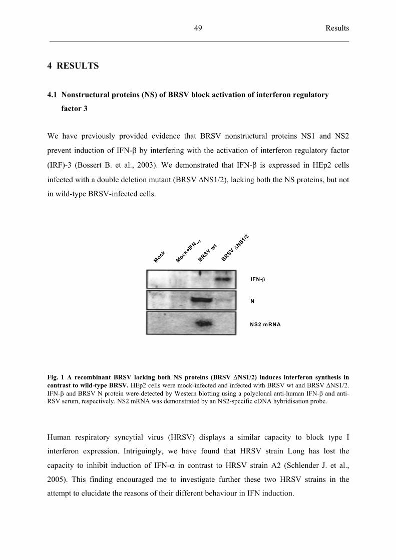
Results ___________________________________________________________________________
49
4 RESULTS
4.1 Nonstructural proteins (NS) of BRSV block activation of interferon regulatory
factor 3
We have previously provided evidence that BRSV nonstructural proteins NS1 and NS2
prevent induction of IFN- by interfering with the activation of interferon regulatory factor
(IRF)-3 (Bossert B. et al., 2003). We demonstrated that IFN- is expressed in HEp2 cells
infected with a double deletion mutant (BRSV NS1/2), lacking both the NS proteins, but not
in wild-type BRSV-infected cells.
Mock
Mock+IFN -BRSV wtBRSV
NS1/2
IFN-
N
NS2 mRNA
Fig. 1 A recombinant BRSV lacking both NS proteins (BRSV NS1/2) induces interferon synthesis in contrast to wild-type BRSV. HEp2 cells were mock-infected and infected with BRSV wt and BRSV NS1/2.IFN- and BRSV N protein were detected by Western blotting using a polyclonal anti-human IFN- and anti-RSV serum, respectively. NS2 mRNA was demonstrated by an NS2-specific cDNA hybridisation probe.
Human respiratory syncytial virus (HRSV) displays a similar capacity to block type I
interferon expression. Intriguingly, we have found that HRSV strain Long has lost the
capacity to inhibit induction of IFN- in contrast to HRSV strain A2 (Schlender J. et al.,
2005). This finding encouraged me to investigate further these two HRSV strains in the
attempt to elucidate the reasons of their different behaviour in IFN induction.
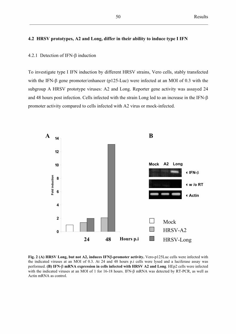
Results ___________________________________________________________________________
50
4.2 HRSV prototypes, A2 and Long, differ in their ability to induce type I IFN
4.2.1 Detection of IFN- induction
To investigate type I IFN induction by different HRSV strains, Vero cells, stably transfected
with the IFN- gene promoter/enhancer (p125-Luc) were infected at an MOI of 0.3 with the
subgroup A HRSV prototype viruses: A2 and Long. Reporter gene activity was assayed 24
and 48 hours post infection. Cells infected with the strain Long led to an increase in the IFN-
promoter activity compared to cells infected with A2 virus or mock-infected.
0
2
4
6
8
10
12
14
Fold
indu
ctio
n
A B
A2 LongMock
IFN-
w /o RT
Actin
MockHRSV-A2
24 48 Hours p.i HRSV-Long
Fig. 2 (A) HRSV Long, but not A2, induces IFN -promoter activity. Vero-p125Luc cells were infected withthe indicated viruses at an MOI of 0.3. At 24 and 48 hours p.i cells were lysed and a luciferase assay was performed. (B) IFN- mRNA expression in cells infected with HRSV A2 and Long. HEp2 cells were infected with the indicated viruses at an MOI of 1 for 16-18 hours. IFN- mRNA was detected by RT-PCR, as well as Actin mRNA as control.
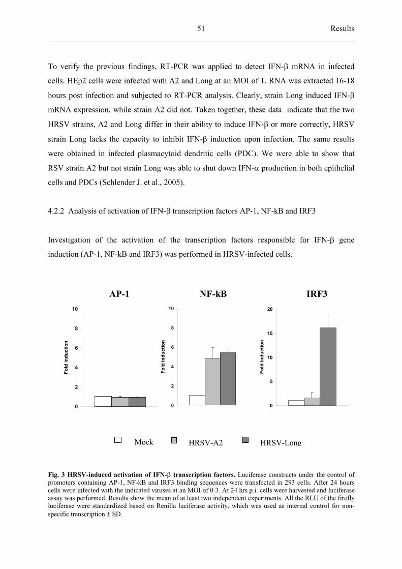
Results ___________________________________________________________________________
51
To verify the previous findings, RT-PCR was applied to detect IFN- mRNA in infected
cells. HEp2 cells were infected with A2 and Long at an MOI of 1. RNA was extracted 16-18
hours post infection and subjected to RT-PCR analysis. Clearly, strain Long induced IFN-
mRNA expression, while strain A2 did not. Taken together, these data indicate that the two
HRSV strains, A2 and Long differ in their ability to induce IFN- or more correctly, HRSV
strain Long lacks the capacity to inhibit IFN- induction upon infection. The same results
were obtained in infected plasmacytoid dendritic cells (PDC). We were able to show that
RSV strain A2 but not strain Long was able to shut down IFN- production in both epithelial
cells and PDCs (Schlender J. et al., 2005).
4.2.2 Analysis of activation of IFN- transcription factors AP-1, NF-kB and IRF3
Investigation of the activation of the transcription factors responsible for IFN- gene
induction (AP-1, NF-kB and IRF3) was performed in HRSV-infected cells.
AP-1 NF-kB IRF3
0
5
10
15
20
Fold
indu
ctio
n
0
2
4
6
8
10
Fold
indu
ctio
n
0
2
4
6
8
10
Fold
indu
ctio
n
Mock HRSV-A2 HRSV-Long
Fig. 3 HRSV-induced activation of IFN- transcription factors. Luciferase constructs under the control of promoters containing AP-1, NF-kB and IRF3 binding sequences were transfected in 293 cells. After 24 hours cells were infected with the indicated viruses at an MOI of 0.3. At 24 hrs p.i. cells were harvested and luciferase assay was performed. Results show the mean of at least two independent experiments. All the RLU of the fireflyluciferase were standardized based on Renilla luciferase activity, which was used as internal control for non-specific transcription SD.
Results ___________________________________________________________________________
52
Reporter constructs, harbouring the binding sequences for each transcription factors
controlling the expression of the downstream firefly luciferase gene were transfected in 293
cells together with the Renilla (pCMV-RL). Renilla gene is under the control of a CMV
promoter and it was used as a control for non specific (IRF3-independent) regulation of gene
expression in the reporter assays. Twenty four hours later, cells were mock-infected and
infected with HRSV A2 and Long at MOI of 0.3. HRSV A2 and Long strains activated NF-
kB reporter activity of 4.9-fold and 5.4-fold, respectively, while AP-1 was not significantly
induced. Interestingly, infection with HRSV-Long resulted in a 16-fold increase of the
luciferase activity in cells transfected with IRF3 reporter construct. In contrast, HRSV A2
caused an 1.5-fold induction. Thus virus-specific activation of IRF3 appeared to be selectively
blocked in the presence of HRSV A2, resembling the situation in BRSV wild-type infected-
cells previously described (Bossert B. et al., 2003).
4.3 Sequence comparison of RSV NS1 and NS2 proteins
4.3.1 Sequence analysis of HRSV A2 and HRSV Long nonstructural proteins
Since our previous studies identified RSV nonstructural proteins as crucial antagonists of type
I IFN induction, nucleotide sequences of the complete NS1 and NS2 genes from HRSV A2
and Long were determined and compared with those of two, out of seven, clinical isolates,
namely patients 86 and 112. Clinical isolates were shown to block induction of IFN- and for
this reason they were considered comparable to strain A2 in their inhibitory capacity
(Schlender J. et al., 2005). The NS1 protein sequence was highly conserved among the
different HRSV viruses analysed. Only two amino acid differences were detected: one in the
clinical isolate number 86 (N76S) and the other in HRSV A2 (I115L). None of these changes
was considered significant. At the N-terminus of NS2 protein variable residues were
identified. At position 7, the clinical isolates displayed a Glycine (G) instead of an Aspartic
acid (D). HRSV A2 differed from the other viruses by the presence of an Asparagine (N) and
a Lysine (K) at position 8 and 38. The only amino acid which distinguished HRSV Long
strain from clinical isolates and HRSV A2 was a Threonine at position 26 instead of an
Isoleucine. Threonine is a neutral amino acid like Isoleucine but, differently from the latter,
its side chain can undergo O-linked glycosylation and it can become phosphorylated through
the action of a threonine kinase. For these peculiar characteristics and its unique presence in
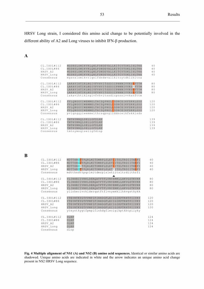
Results ___________________________________________________________________________
53
HRSV Long strain, I considered this amino acid change to be potentially involved in the
different ability of A2 and Long viruses to inhibit IFN- production.
A
40CL.ISOL#112 40CL.ISOL#86 40HRSV_A2 40HRSV_Long
Consensus
MGSNSLSMIKVRLQNLFDNDEVALLKITCYTDKLIHLTNAMGSNSLSMIKVRLQNLFDNDEVALLKITCYTDKLIHLTNAMGSNSLSMIKVRLQNLFDNDEVALLKITCYTDKLIHLTNAMGSNSLSMIKVRLQNLFDNDEVALLKITCYTDKLIHLTNAm
MMMMg
GGGGs
SSSSn
NNNNs
SSSSl
LLLLs
SSSSm
MMMMi
IIIIk
KKKKv
VVVVr
RRRRl
LLLLq
QQQQn
NNNNl
LLLLf
FFFFd
DDDDn
NNNNd
DDDDe
EEEEv
VVVVa
AAAAl
LLLLl
LLLLk
KKKKi
IIIIt
TTTTc
CCCCy
YYYYt
TTTTd
DDDDk
KKKKl
LLLLi
IIIIh
HHHHl
LLLLt
TTTTn
NNNNa
AAAA
80CL.ISOL#112 80CL.ISOL#86 80HRSV_A2 80HRSV_Long
Consensus
LAKAVIHTIKLNGIVFVHVITSSDICPNNNIVVKSNFTTMLAKAVIHTIKLNGIVFVHVITSSDICPNNNIVVKSSFTTMLAKAVIHTIKLNGIVFVHVITSSDICPNNNIVVKSNFTTMLAKAVIHTIKLNGIVFVHVITSSDICPNNNIVVKSNFTTMl
LLLLa
AAAAk
KKKKa
AAAAv
VVVVi
IIIIh
HHHHt
TTTTi
IIIIk
KKKKl
LLLLn
NNNNg
GGGGi
IIIIv
VVVVf
FFFFv
VVVVh
HHHHv
VVVVi
IIIIt
TTTTs
SSSSs
SSSSd
DDDDi
IIIIc
CCCCp
PPPPn
NNNNn
NNNNn
NNNNi
IIIIv
VVVVv
VVVVk
KKKKs
SSSSn
N
NNf
FFFFt
TTTTt
TTTTm
MMMM
120CL.ISOL#112120CL.ISOL#86120HRSV_A2120HRSV_Long
Consensus
PVLQNGGYIWEMMELTHCSQPNGLIDDNCEIKFSKKLSDSPVLQNGGYIWEMMELTHCSQPNGLIDDNCEIKFSKKLSDSPVLQNGGYIWEMMELTHCSQPNGLLDDNCEIKFSKKLSDSPVLQNGGYIWEMMELTHCSQPNGLIDDNCEIKFSKKLSDSp
PPPPv
VVVVl
LLLLq
QQQQn
NNNNg
GGGGg
GGGGy
YYYYi
IIIIw
WWWWe
EEEEm
MMMMm
MMMMe
EEEEl
LLLLt
TTTTh
HHHHc
CCCCs
SSSSq
QQQQp
PPPPn
NNNNg
GGGGl
LLLLi
IILId
DDDDd
DDDDn
NNNNc
CCCCe
EEEEi
IIIIk
KKKKf
FFFFs
SSSSk
KKKKk
KKKKl
LLLLs
SSSSd
DDDDs
SSSS
139CL.ISOL#112139CL.ISOL#86139HRSV_A2139HRSV_Long
Consensus
TMTNYMNQLSELLGFDLNPTMTNYMNQLSELLGFDLNPTMTNYMNQLSELLGFDLNPTMTNYMNQLSELLGFDLNPt
TTTTm
MMMMt
TTTTn
NNNNy
YYYYm
MMMMn
NNNNq
QQQQl
LLLLs
SSSSe
EEEEl
LLLLl
LLLLg
GGGGf
FFFFd
DDDDl
LLLLn
NNNNp
PPPP
B 40CL.ISOL#112 40CL.ISOL#86 40HRSV_A2 40HRSV_Long
Consensus
MDTTHNGTTPQRLMITDMRPLSLETIITSLTRDIITHRFIMDTTHNGTTPQRLMITDMRPLSLETIITSLTRDIITHRFIMDTTHNDNTPQRLMITDMRPLSLETIITSLTRDIITHKFIMDTTHNDTTPQRLMITDMRPLSLETTITSLTRDIITHRFIm
MMMMd
DDDDt
TTTTt
TTTTh
HHHHn
NNNNd
GGDDt
TT
Tt
TTTTp
PPPPq
QQQQr
RRRRl
LLLLm
MMMMi
IIIIt
TTTTd
DDDDm
MMMMr
RRRRp
PPPPl
LLLLs
SSSSl
LLLLe
EEEEt
TTTTi
III
i
IIIIt
TTTTs
SSSSl
LLLLt
TTTTr
RRRRd
DDDDi
IIIIi
IIIIt
TTTTh
HHHHr
RRKRf
FFFFi
IIII
80CL.ISOL#112 80CL.ISOL#86 80HRSV_A2 80HRSV_Long
Consensus
YLINHECIVRKLDERQATFTFLVNYEMKLLHKVGSTKYKKYLINHECIVRKLDERQATFTFLVNYEMKLLHKVGSTKYKKYLINHECIVRKLDERQATFTFLVNYEMKLLHKVGSTKYKKYLINHECIVRKLDERQATFTFLVNYEMKLLHKVGSTKYKKy
YYYYl
LLLLi
IIIIn
NNNNh
HHHHe
EEEEc
CCCCi
IIIIv
VVVVr
RRRRk
KKKKl
LLLLd
DDDDe
EEEEr
RRRRq
QQQQa
AAAAt
TTTTf
FFFFt
TTTTf
FFFFl
LLLLv
VVVVn
NNNNy
YYYYe
EEEEm
MMMMk
KKKKl
LLLLl
LLLLh
HHHHk
KKKKv
VVVVg
GGGGs
SSSSt
TTTTk
KKKKy
YYYYk
KKKKk
KKKK
120CL.ISOL#112 120CL.ISOL#86 120HRSV_A2 120HRSV_Long
Consensus
YTEYNTKYGTFPMPIFINHDGFLECIGIKPTKHTPIIYKYYTEYNTKYGTFPMPIFINHDGFLECIGIKPTKHTPIIYKYYTEYNTKYGTFPMPIFINHDGFLECIGIKPTKHTPIIYKYYTEYNTKYGTFPMPIFINHDGFLECIGIKPTKHTPIIYKYy
YYYYt
TTTTe
EEEEy
YYYYn
NNNNt
TTTTk
KKKKy
YYYYg
GGGGt
TTTTf
FFFFp
PPPPm
MMMMp
PPPPi
IIIIf
FFFFi
IIIIn
NNNNh
HHHHd
DDDDg
GGGGf
FFFFl
LLLLe
EEEEc
CCCCi
IIIIg
GGGGi
IIIIk
KKKKp
PPPPt
TTTTk
KKKKh
HHHHt
TTTTp
PPPPi
IIIIi
IIIIy
YYYYk
KKKKy
YYYY
124CL.ISOL#112 124CL.ISOL#86 124HRSV_A2 124HRSV_Long
Consensus
DLNPDLNPDLNPDLNPd
DDDDl
LLLLn
NNNNp
PPPP
Fig. 4 Multiple alignment of NS1 (A) and NS2 (B) amino acid sequences. Identical or similar amino acids areshadowed. Unique amino acids are indicated in white and the arrow indicates an unique amino acid change present in NS2 HRSV Long sequence.
Results ___________________________________________________________________________
54
4.4 Chimeric BRSV viruses expressing HRSV NS genes
4.4.1 Construction of recombinant BRSV virus harbouring Long-derived NS1 and a mutated
form of NS2
To study the functional implication in IFN- antagonism by the residue 26 (Threonin vs
Isoleucine) of NS2 protein, a recombinant BRSV viruses harbouring Long-derived or A2-like
NS proteins were generated. Recombinant BRSV (rBRSVh1/2) possessing NS1 and NS2
genes from HRSV strain Long was previously constructed (Bossert B. and Conzelmann K.K.,
2002). In addition, a recombinant BRSV (rBRSVh1/2*T26I) carrying an Isoleucine at position
26 of Long-NS2 protein was created by site-directed mutagenesis as described in Materials
and Methods. Chimeric viruses were recovered by cotransfection of BSR T7/5 cells with the
rBRSV full length cDNAs and the support plasmids encoding for BRSV N, P, L and M2
genes as described previously. Viruses were viable and comparable in growth.
Spe I
proteinsvRNA
rBRSVh1/2 (Long)
...SLETTITSLT...
...SLET SLT...IIT
Not I Acc65 I
hNS1 hNS2 NN
Not I Acc65 I
hNS1 hNS2 NN
NS1NS1 NS2NS2 NN PP MM SHSH GG FF M2-1M2-1M2-2M2-2
LL
rBRSVwt
SIETEIISLT
rBRSVh1/2* (T26I)
Fig. 5 Chimeric rBRSVs harbouring HRSV NS genes. Viral genome (vRNA) and gene ORF encoded proteinsare shown. In the enlargement: BRSV NS proteins were replaced by HRSV Long NS proteins (rBRSVh1/2). An amino acid exchange was subsequently introduced into the NS2 protein at position 26 (T26I) generating therecombinant virus rBRSVh1/2*T26I
.
Results ___________________________________________________________________________
55
4.4.2 HRSV NS protein expression and cDNA restriction analysis
Western blot analysis was used to confirm the correct translation of NS proteins in Vero cells
infected with HRSV prototypes, A2 and Long, and with the chimeric rBRSVs.
Restriction digest analysis was performed on HRSV NS2 cDNA obtained by RT-PCR
reaction of virus-infected HEp2 cells. The NS2 ORF of the rBRSVh1/2*T26I contained an
additional restriction site for Spe I endonuclease, which is absent in the wild-type sequences.
After the digest, only NS2 cDNA derived from rBRSVh1/2*T26I showed a lower migrating
band.
A
HRSVLong
BRSVh1/2
BRSVh1/2*(T26I)
Mock HRSVA2
NS2NS1
BHRSVLong
BRSVh1/2
BRSVh1/2*(T26I)
MarkerHRSVA2
Fig. 6 Analysis of chimeric BRSVs expressing HRSV NS genes. (A) Western blot analysis for NS proteinexpression in Vero infected cells. NS proteins were detected using a polyclonal antibody ( -IC/C) recognizingHRSV NS proteins (kindly provided by J.A. Melero, Spain). (B) Restriction analysis of NS2 cDNA obtained from HEP2 cells infected with HRSV A2, HRSV Long and recombinant BRSVh1/2 and BRSVh1/2T26I .
Results ___________________________________________________________________________
56
4.4.3 Growth kinetics of recombinant BRSV viruses
Growth properties of rBRSVh1/2 and rBRSVh1/2*T26I and HRSV strains were analysed in
parallel in Vero and HEp2 cells.
1 , 0 0 E + 0 2
1 , 0 0 E + 0 4
1 , 0 0 E + 0 6
1 , 0 0 E + 0 8
0 2 4 4 8 7 2
FFU
/ml
1 , 0 0 E + 0 2
1 , 0 0 E + 0 4
1 , 0 0 E + 0 6
1 , 0 0 E + 0 8
0 2 4 4 8 7 2
FFU
/ml
Vero cells
Hours p.i
HEp2 cells
Hours p.i
BRSV-h1/2HRSV-A2
HRSV-Long BRSV- h1/2* (T26I)
Fig. 7 Analysis of chimeric BRSVs expressing HRSV NS genes. Growth kinetics of chimeric rBRSVh1/2 and rBRSVh1/2*T26I, and HRSV strains A2 and Long in Vero and HEp2 cells. Cells were infected with MOI of 0.3 and viruses were harvested at the indicated time points.
Results ___________________________________________________________________________
57
In Vero cells, which do not possess an intact IFN system, the two chimeric rBRSV viruses
behaved similarly. After 3 days of infection at a MOI of 0.3, they reached titers of about 7
x104 FFU, close to those of HRSV A2 and Long. In HEp2 cells, a human cell line competent
for type I IFN expression, growth characteristics of rBRSVh1/2 and rBRSVh1/2*T26I were
once again comparable. After 2 days of infection, both viruses reached similar titers of about
1.8-2.2 x10⁴ FFU/ml , respectively. When compared to HRSV strains, which grew to highest
titers of about 1 x10⁶ FFU/ml 3 days post infection, the recombinant BRSV viruses displayed
a slightly attenuated phenotype of approximately 1 log. This might be due to the fact that
recombinant BRSV viruses are less efficient in the context of a human cell line as a
consequence of viral host-specific factors.
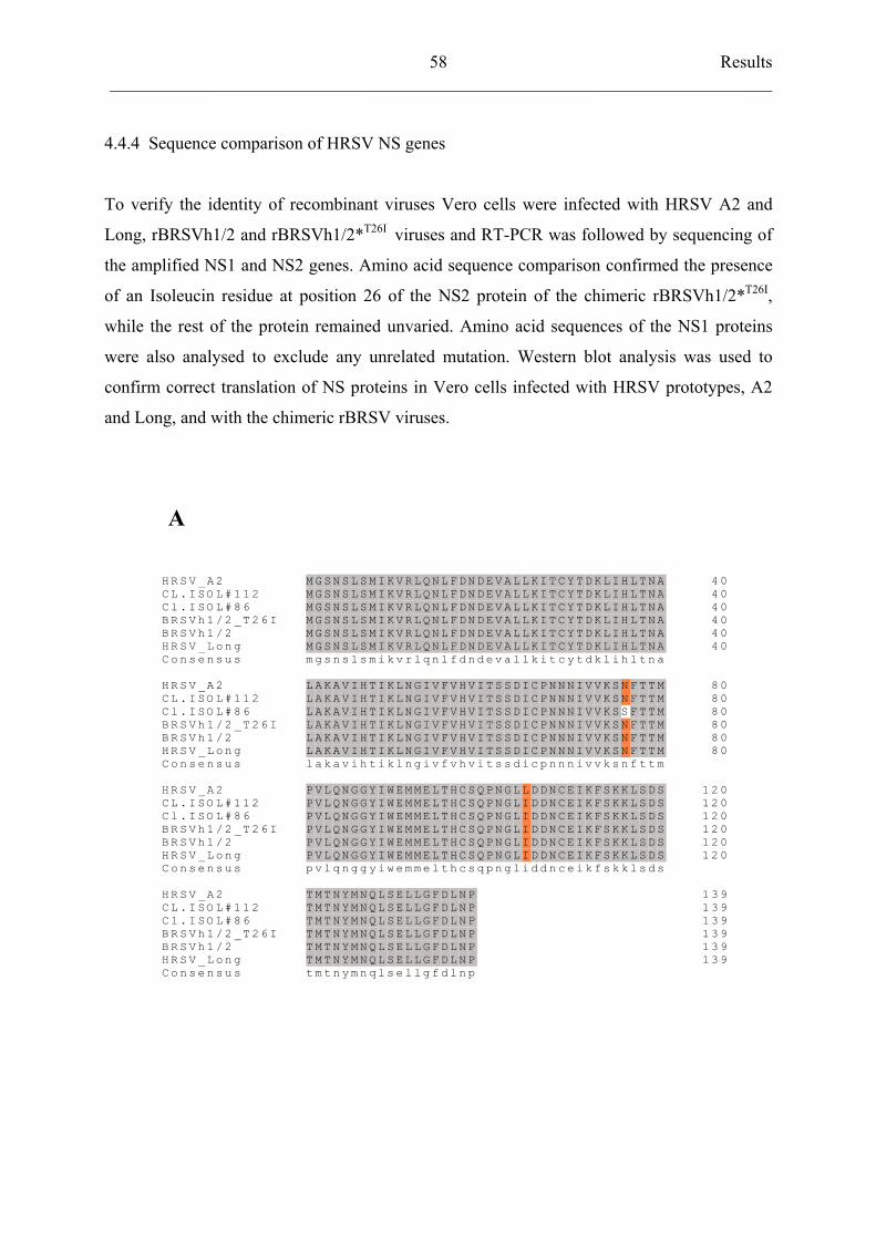
Results ___________________________________________________________________________
58
4.4.4 Sequence comparison of HRSV NS genes
To verify the identity of recombinant viruses Vero cells were infected with HRSV A2 and
Long, rBRSVh1/2 and rBRSVh1/2*T26I viruses and RT-PCR was followed by sequencing of
the amplified NS1 and NS2 genes. Amino acid sequence comparison confirmed the presence
of an Isoleucin residue at position 26 of the NS2 protein of the chimeric rBRSVh1/2*T26I,
while the rest of the protein remained unvaried. Amino acid sequences of the NS1 proteins
were also analysed to exclude any unrelated mutation. Western blot analysis was used to
confirm correct translation of NS proteins in Vero cells infected with HRSV prototypes, A2
and Long, and with the chimeric rBRSV viruses.
A
4 0H R S V _ A 2 4 0C L . I S O L# 1 1 2 4 0C l . I S O L# 8 6 4 0B R S V h 1 /2 _ T 2 6 I 4 0B R S V h 1 /2 4 0H R S V _ L on g
C o n s e n su s
M GS N S L S M I K V R L QN L F D N D E V A L LK I T C Y T D K L I HL T N AM GS N S L S M I K V R L QN L F D N D E V A L LK I T C Y T D K L I HL T N AM GS N S L S M I K V R L QN L F D N D E V A L LK I T C Y T D K L I HL T N AM GS N S L S M I K V R L QN L F D N D E V A L LK I T C Y T D K L I HL T N AM GS N S L S M I K V R L QN L F D N D E V A L LK I T C Y T D K L I HL T N AM GS N S L S M I K V R L QN L F D N D E V A L LK I T C Y T D K L I HL T N Am
MMMMMMg
GGGGGGs
SSSSSSn
NNNNNNs
SSSSSSl
LLLLLLs
SSSSSSm
MMMMMMi
IIIIIIk
KKKKKKv
VVVVVVr
RRRRRRl
LLLLLLq
QQQQQQn
NNNNNNl
LLLLLLf
FFFFFFd
DDDDDDn
NNNNNNd
DDDDDDe
EEEEEEv
VVVVVVa
AAAAAAl
LLLLLLl
LLLLLLk
KKKKKKi
IIIIIIt
TTTTTTc
CCCCCCy
YYYYYYt
TTTTTTd
DDDDDDk
KKKKKKl
LLLLLLi
IIIIIIh
HHHHHHl
LLLLLLt
TTTTTTn
NNNNNNa
AAAAAA
8 0H R S V _ A 2 8 0C L . I S O L# 1 1 2 8 0C l . I S O L# 8 6 8 0B R S V h 1 /2 _ T 2 6 I 8 0B R S V h 1 /2 8 0H R S V _ L on g
C o n s e n su s
L AK A V I H T I K L N G IV F V H V I T S S D IC P N N N I V V K S NF T T ML AK A V I H T I K L N G IV F V H V I T S S D IC P N N N I V V K S NF T T ML AK A V I H T I K L N G IV F V H V I T S S D IC P N N N I V V K S SF T T ML AK A V I H T I K L N G IV F V H V I T S S D IC P N N N I V V K S NF T T ML AK A V I H T I K L N G IV F V H V I T S S D IC P N N N I V V K S NF T T ML AK A V I H T I K L N G IV F V H V I T S S D IC P N N N I V V K S NF T T Ml
LLLLLLa
AAAAAAk
KKKKKKa
AAAAAAv
VVVVVVi
IIIIIIh
HHHHHHt
TTTTTTi
IIIIIIk
KKKKKKl
LLLLLLn
NNNNNNg
GGGGGGi
IIIIIIv
VVVVVVf
FFFFFFv
VVVVVVh
HHHHHHv
VVVVVVi
IIIIIIt
TTTTTTs
SSSSSSs
SSSSSSd
DDDDDDi
IIIIIIc
CCCCCCp
PPPPPPn
NNNNNNn
NNNNNNn
NNNNNNi
IIIIIIv
VVVVVVv
VVVVVVk
KKKKKKs
SSSSSSn
NN
NNNf
FFFFFFt
TTTTTTt
TTTTTTm
MMMMMM
1 2 0H R S V _ A 2 1 2 0C L . I S O L# 1 1 2 1 2 0C l . I S O L# 8 6 1 2 0B R S V h 1 /2 _ T 2 6 I 1 2 0B R S V h 1 /2 1 2 0H R S V _ L on g
C o n s e n su s
P VL Q N G G Y I W E M M EL T H C S Q P N G L LD D N C E I K F S K KL S D SP VL Q N G G Y I W E M M EL T H C S Q P N G L ID D N C E I K F S K KL S D SP VL Q N G G Y I W E M M EL T H C S Q P N G L ID D N C E I K F S K KL S D SP VL Q N G G Y I W E M M EL T H C S Q P N G L ID D N C E I K F S K KL S D SP VL Q N G G Y I W E M M EL T H C S Q P N G L ID D N C E I K F S K KL S D SP VL Q N G G Y I W E M M EL T H C S Q P N G L ID D N C E I K F S K KL S D Sp
PPPPPPv
VVVVVVl
LLLLLLq
QQQQQQn
NNNNNNg
GGGGGGg
GGGGGGy
YYYYYYi
IIIIIIw
WWWWWWe
EEEEEEm
MMMMMMm
MMMMMMe
EEEEEEl
LLLLLLt
TTTTTTh
HHHHHHc
CCCCCCs
SSSSSSq
QQQQQQp
PPPPPPn
NNNNNNg
GGGGGGl
LLLLLLi
LIIIIId
DDDDDDd
DDDDDDn
NNNNNNc
CCCCCCe
EEEEEEi
IIIIIIk
KKKKKKf
FFFFFFs
SSSSSSk
KKKKKKk
KKKKKKl
LLLLLLs
SSSSSSd
DDDDDDs
SSSSSS
1 3 9H R S V _ A 2 1 3 9C L . I S O L# 1 1 2 1 3 9C l . I S O L# 8 6 1 3 9B R S V h 1 /2 _ T 2 6 I 1 3 9B R S V h 1 /2 1 3 9H R S V _ L on g
C o n s e n su s
T MT N Y M N Q L S E L L GF D L N PT MT N Y M N Q L S E L L GF D L N PT MT N Y M N Q L S E L L GF D L N PT MT N Y M N Q L S E L L GF D L N PT MT N Y M N Q L S E L L GF D L N PT MT N Y M N Q L S E L L GF D L N Pt
TTTTTTm
MMMMMMt
TTTTTTn
NNNNNNy
YYYYYYm
MMMMMMn
NNNNNNq
QQQQQQl
LLLLLLs
SSSSSSe
EEEEEEl
LLLLLLl
LLLLLLg
GGGGGGf
FFFFFFd
DDDDDDl
LLLLLLn
NNNNNNp
PPPPPP
Results ___________________________________________________________________________
59
B 40HRSV-A2 40CL.ISOL#112 40CL.ISOL#86 40BRSVh1/2_T26I 40BRSVh1/2 40HRSV-Long
Consensus
MDTTHNDNTPQRLMITDMRPLSLETIITSLTRDIITHKFIMDTTHNGTTPQRLMITDMRPLSLETIITSLTRDIITHRFIMDTTHNGTTPQRLMITDMRPLSLETIITSLTRDIITHRFIMDTTHNDTTPQRLMITDMRPLSLETIITSLTRDIITHRFIMDTTHNDTTPQRLMITDMRPLSLETTITSLTRDIITHRFIMDTTHNDTTPQRLMITDMRPLSLETTITSLTRDIITHRFIm
MMMMMMd
DDDDDDt
TTTTTTt
TTTTTTh
HHHHHHn
NNNNNNd
DGGDDDt
TTTTTt
TTTTTTp
PPPPPPq
QQQQQQr
RRRRRRl
LLLLLLm
MMMMMMi
IIIIIIt
TTTTTTd
DDDDDDm
MMMMMMr
RRRRRRp
PPPPPPl
LLLLLLs
SSSSSSl
LLLLLLe
EEEEEEt
TTTTTTi
IIII
i
IIIIIIt
TTTTTTs
SSSSSSl
LLLLLLt
TTTTTTr
RRRRRRd
DDDDDDi
IIIIIIi
IIIIIIt
TTTTTTh
HHHHHHr
KRRRRRf
FFFFFFi
IIIIII
80HRSV-A2 80CL.ISOL#112 80CL.ISOL#86 80BRSVh1/2_T26I 80BRSVh1/2 80HRSV-Long
Consensus
YLINHECIVRKLDERQATFTFLVNYEMKLLHKVGSTKYKKYLINHECIVRKLDERQATFTFLVNYEMKLLHKVGSTKYKKYLINHECIVRKLDERQATFTFLVNYEMKLLHKVGSTKYKKYLINHECIVRKLDERQATFTFLVNYEMKLLHKVGSTKYKKYLINHECIVRKLDERQATFTFLVNYEMKLLHKVGSTKYKKYLINHECIVRKLDERQATFTFLVNYEMKLLHKVGSTKYKKy
YYYYYYl
LLLLLLi
IIIIIIn
NNNNNNh
HHHHHHe
EEEEEEc
CCCCCCi
IIIIIIv
VVVVVVr
RRRRRRk
KKKKKKl
LLLLLLd
DDDDDDe
EEEEEEr
RRRRRRq
QQQQQQa
AAAAAAt
TTTTTTf
FFFFFFt
TTTTTTf
FFFFFFl
LLLLLLv
VVVVVVn
NNNNNNy
YYYYYYe
EEEEEEm
MMMMMMk
KKKKKKl
LLLLLLl
LLLLLLh
HHHHHHk
KKKKKKv
VVVVVVg
GGGGGGs
SSSSSSt
TTTTTTk
KKKKKKy
YYYYYYk
KKKKKKk
KKKKKK
120HRSV-A2 120CL.ISOL#112 120CL.ISOL#86 120BRSVh1/2_T26I 120BRSVh1/2 120HRSV-Long
Consensus
YTEYNTKYGTFPMPIFINHDGFLECIGIKPTKHTPIIYKYYTEYNTKYGTFPMPIFINHDGFLECIGIKPTKHTPIIYKYYTEYNTKYGTFPMPIFINHDGFLECIGIKPTKHTPIIYKYYTEYNTKYGTFPMPIFINHDGFLECIGIKPTKHTPIIYKYYTEYNTKYGTFPMPIFINHDGFLECIGIKPTKHTPIIYKYYTEYNTKYGTFPMPIFINHDGFLECIGIKPTKHTPIIYKYy
YYYYYYt
TTTTTTe
EEEEEEy
YYYYYYn
NNNNNNt
TTTTTTk
KKKKKKy
YYYYYYg
GGGGGGt
TTTTTTf
FFFFFFp
PPPPPPm
MMMMMMp
PPPPPPi
IIIIIIf
FFFFFFi
IIIIIIn
NNNNNNh
HHHHHHd
DDDDDDg
GGGGGGf
FFFFFFl
LLLLLLe
EEEEEEc
CCCCCCi
IIIIIIg
GGGGGGi
IIIIIIk
KKKKKKp
PPPPPPt
TTTTTTk
KKKKKKh
HHHHHHt
TTTTTTp
PPPPPPi
IIIIIIi
IIIIIIy
YYYYYYk
KKKKKKy
YYYYYY
124HRSV-A2 124CL.ISOL#112 124CL.ISOL#86 124BRSVh1/2_T26I 124BRSVh1/2 124HRSV-Long
Consensus
DLNPDLNPDLNPDLNPDLNPDLNPd
DDDDDDl
LLLLLLn
NNNNNNp
PPPPPP
Fig. 8 Multiple alignment of amino acid sequences of NS1 (A) and NS2 (B) proteins in recombinant BRSV(rBRSVh1/2 and rBRSVh1/2*T26I ), HRSV strains A2 and Long and HRSV-clinical isolates. Sequenceidentity for NS2 and NS1 was 99,19% and 99,82% respectively. The arrow indicates the presence of the insertedmutation in the NS2 sequence of rBRSVh1/2*T26I
Results ___________________________________________________________________________
60
4.4.5 rBRSVh1/2*T26I but not rBRSVh1/2 inhibits IFN induction
To examine the ability of the two chimeric rBRSVs to induce IFN- , a modified reporter gene
assay was used. Vero cells harbouring the IFN- gene promoter/enhancer (p125Luc) were
mock-infected and infected with wild-type BRSV, rBRSVh1/2, rBRSVh1/2*T26I, HRSV A2
and HRSV Long at an MOI of 0.3. Luciferase activity was measured 24 and 48 hours post
infection. An induction of luciferase activity of about 9- and 12- fold was observed in cells
infected with HRSV Long and rBRSVh1/2, respectively. A minor induction of the IFN-
gene promoter, compared to mock-infected cells, was detected for the wild-type BRSV,
HRSV A2 and the recombinant rBRSVh1/2*T26I.
Fig. 9 Virus-induced activation of IFN- gene promoter. (A) Vero cells expressing the IFN- gene promoter/enhancer (p125Luc) were infected with the indicated viruses at an MOI of 0.3 24 and 48 hours postinfection activity of the luciferase reporter gene was measured and the relative light units were expressed as foldinduction relative to the mock-infected control. Results represent the mean value of three independentexperiments with error bars indicating standard deviation. (B) Western blot analysis of cell lysates used in thegene reporter assay. Blots were probed with a polyclonal -RSV serum
0
5
10
15
20
Mock BRSV BRSVh1.2
BRSVh1/2*T26I
HRSVA2
HRSVLong
fold
indu
ctio
n
24 hr p.i 48 hr p.iA B
48 hour p.i
HRSV-Lo
ngBRSVh
1/2
Mock
BRSVwt
24 hour p.i
RSV N
Actin
HRSV-A2
BRSVh1/2
*(T26
I)
RSV N
Actin
HRSV-A2
HRSV-Lo
ngBRSVh
1/2
Mock
BRSVwt
BRSVh1/2*
(T26I)
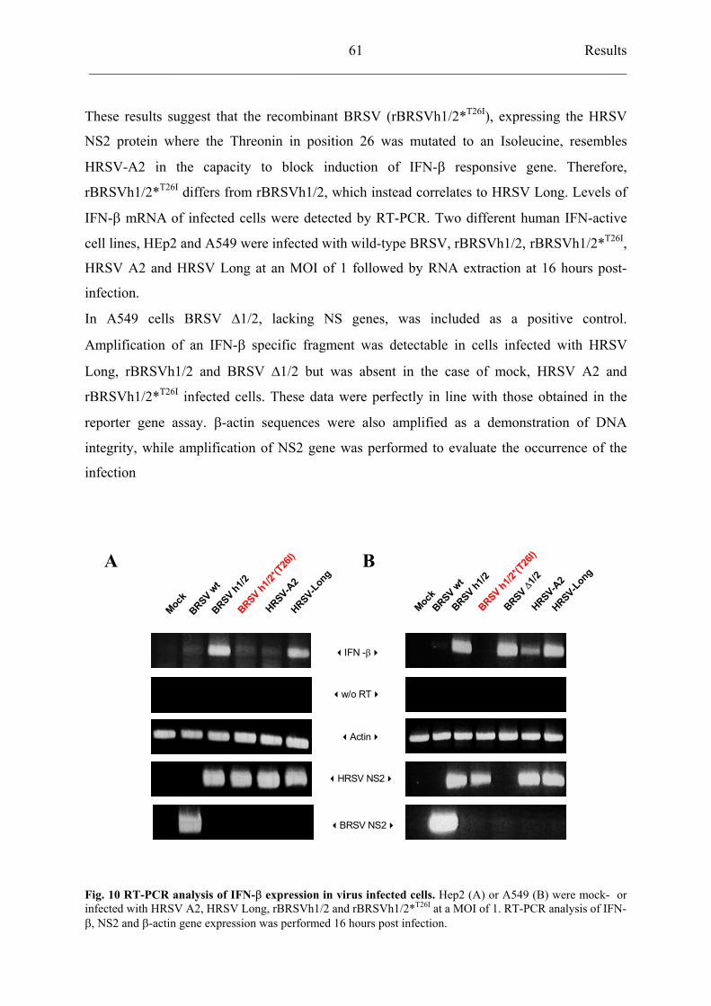
Results ___________________________________________________________________________
61
These results suggest that the recombinant BRSV (rBRSVh1/2*T26I), expressing the HRSV
NS2 protein where the Threonin in position 26 was mutated to an Isoleucine, resembles
HRSV-A2 in the capacity to block induction of IFN- responsive gene. Therefore,
rBRSVh1/2*T26I differs from rBRSVh1/2, which instead correlates to HRSV Long. Levels of
IFN- mRNA of infected cells were detected by RT-PCR. Two different human IFN-active
cell lines, HEp2 and A549 were infected with wild-type BRSV, rBRSVh1/2, rBRSVh1/2*T26I,
HRSV A2 and HRSV Long at an MOI of 1 followed by RNA extraction at 16 hours post-
infection.
In A549 cells BRSV 1/2, lacking NS genes, was included as a positive control.
Amplification of an IFN- specific fragment was detectable in cells infected with HRSV
Long, rBRSVh1/2 and BRSV 1/2 but was absent in the case of mock, HRSV A2 and
rBRSVh1/2*T26I infected cells. These data were perfectly in line with those obtained in the
reporter gene assay. -actin sequences were also amplified as a demonstration of DNA
integrity, while amplification of NS2 gene was performed to evaluate the occurrence of the
infection
HRSV-A2
HRSV-Lon
g
BRSV h1/2
BRSV h1/2*
(T26I)
BRSV h1/2*(T
26I)
Mock
BRSVwt
IFN -
w/o RT
Actin
HRSV NS2
BRSV NS2
HRSV-A2
HRSV-Lon
g
BRSV h1/2
Mock
BRSVwt
BRSV1/2
A B
Fig. 10 RT-PCR analysis of IFN- expression in virus infected cells. Hep2 (A) or A549 (B) were mock- or infected with HRSV A2, HRSV Long, rBRSVh1/2 and rBRSVh1/2*T26I at a MOI of 1. RT-PCR analysis of IFN-
, NS2 and -actin gene expression was performed 16 hours post infection.
Results ___________________________________________________________________________
62
4.4.6 Recombinant BRSVh1/2 *T26I selectively reduces activation of transcriptional factor
IRF3
Expression of IFN- / genes is strongly induced at the transcriptional level following viral
infection. IFN- promoter contains binding sites for AP-1, NF-kB and IRF3 factors, which
form a multiprotein transcriptional-promoting complex. In order to investigate whether the
activation of these transcriptional factors is impaired upon RSV infection, 293 cells were first
transfected with the corresponding reporter plasmids, as previously described. At 24 hours
post transfection, cells were mock-infected and infected at an MOI of 0.3 with HRSV A2 and
Long, BRSV wild-type and the recombinant BRSVs: BRSVh1/2 and BRSVh1/2*T26I .
AP-1 NF-kB IRF3
0
2
4
6
8
10
Fold
indu
ctio
n
0
5
1 0
1 5
2 0
2 5
Fold
indu
ctio
n
0
2
4
6
8
10
Fold
indu
ctio
n
Mock rBRSV h1/2*T26IHRSV-Long
HRSV-A2 rBRSV h1/2
Fig. 11 Induced activation of IFN- transcription factors. Luciferase constructs under the control of promoters containing AP-1, NF-kB and IRF3 binding sequences were transfected in 293 cells. After 24 hours cells were infected with the indicated viruses at an MOI of 0.3. At 24 hrs p.i. cells were harvested and luciferaseassay was performed. Results show the mean of at least two independent experiments
Results ___________________________________________________________________________
63
Infection did not result in detectable activation of AP-1 reporter in either case. NF-kB-related
luciferase activity showed instead a comparable increase between 4.2 and 6.7-fold in infected
cells, with no major differences among the described viruses. Infection with HRSV Long
increased 18-fold IRF3-promoter activity, while a very small induction was observed in
HRSV A2 and BRSV wt infected cells, as already described. As expected, rBRSVh1/2
promoted IRF3 activation; luciferase activity was lower than in HRSV Long infected cells but
still an increase of about 7-fold was observed. When cells were infected with rBRSVh1/2*T26I
only a 3.2-fold increase was detected indicating a possible block of IRF3 specific activation in
the case NS2 protein contains an Isoleucine at residue 26.
4.4.7 Residue 26 of NS2 protein does not influence interferon resistance of recombinant
BRSV viruses
Type I IFNs do not inhibit productive infection of RSV in human epithelial cells suggesting
the existence of viral mechanisms for evasion of the airway defence-response.
The behaviour of HRSV strains and recombinant BRSV viruses was analysed in IFN-treated
cells. HEp2 cells, infected at an MOI of 0.3 with HRSVs or BRSV chimeras, were treated
with increasing amounts of IFN- A/D and viral titers were determined 3 days post infection.
Despite protection was not complete, all four viruses were significantly resistant to IFN-
treatment. After application of 5000 IU of IFN, titers of rBRSVh1/2 and rBRSVh1/2*T26I
declined by about 1 log. Thus, both chimeric BRSV viruses were only slightly sensitive to
IFN and in a comparable way, indicating that the exchange of Threonin 26 with an Isoleucine
did not play a role in RSV IFN-resistance.
Results ___________________________________________________________________________
64
IFN IU/ml
HRSV A2
HRSV Long
rBRSV h1/2
1,00E+02
1,00E+03
1,00E+04
1,00E+05
1,00E+06
1,00E+07
1,00E+08
0 500 1000 5000
FFU
/ml
HRSV-A2
HRSV-Long
rBRSVh1/2
IFN-
rBRSVh1/2*T26I
0 500 1000 5000 IU/ml
rBRSV h1/2*T26I
Fig. 12 Recombinant BRSV are only slightly IFN type I sensitive. (A) HEp2 cells infected at an MOI of 0.3 with HRSV A2, HRSV Long, rBRSVh1/2 and rBRSVh1/2*T26I were incubated with the indicated amounts of recombinant IFN- . Infectious viral titers were determined 3 days post infection. Bars show standard deviationof two independent experiments. (B) Western blot analysis of RSV N protein synthesis in IFN-treated HEp2 cells
4.5 IkappaB kinase-related complex: TBK1, IKK- and TANK
4.5.1 Cloning of TANK-Binding Kinase 1 (TBK1) and TRAF family member-associated NF-
kappa B activator (TANK)
The inducible I B kinase (IKK-i or IKK- ) and the TANK-Binding Kinase 1 (TBK1) act as
IRF3 kinases and they are involved in IFN- expression (see Introduction).
The complete reading frame of TBK1 and TANK (which is a structural component of the
kinase complex) were amplified by PCR from a human lung cDNA library. The TBK1 and
TANK complete ORFs were then cloned into an expression vector, where they were coupled
at their N-termini with a human Ig fragment.
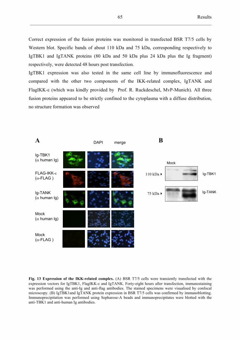
Results ___________________________________________________________________________
65
Correct expression of the fusion proteins was monitored in transfected BSR T7/5 cells by
Western blot. Specific bands of about 110 kDa and 75 kDa, corresponding respectively to
IgTBK1 and IgTANK proteins (80 kDa and 50 kDa plus 24 kDa plus the Ig fragment)
respectively, were detected 48 hours post transfection.
IgTBK1 expression was also tested in the same cell line by immunofluorescence and
compared with the other two components of the IKK-related complex, IgTANK and
FlagIKK- (which was kindly provided by Prof. R. Ruckdeschel, MvP-Munich). All three
fusion proteins appeared to be strictly confined to the cytoplasma with a diffuse distribution,
no structure formation was observed
DAPI mergeA B
Ig-TBK1( human Ig)
75 kDa
110 kDa
Mock
FLAG-IKK-( -FLAG )
Ig-TBK1
Ig-TANKIg-TANK( human Ig)
Mock( human Ig)
Mock( -FLAG )
Fig. 13 Expression of the IKK-related complex. (A) BSR T7/5 cells were transiently transfected with theexpression vectors for IgTBK1, FlagIKK- and IgTANK. Forty-eight hours after transfection, immunostainingwas performed using the anti-Ig and anti-flag antibodies. The stained specimens were visualised by confocalmicroscopy. (B) IgTBK1and IgTANK protein expression in BSR T7/5 cells was confirmed by immunoblotting.Immunoprecipitation was performed using Sepharose-A beads and immunoprecipitates were blotted with theanti-TBK1 and anti-human Ig antibodies.
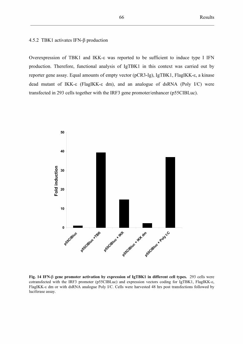
Results ___________________________________________________________________________
66
4.5.2 TBK1 activates IFN- production
Overexpression of TBK1 and IKK- was reported to be sufficient to induce type I IFN
production. Therefore, functional analysis of IgTBK1 in this context was carried out by
reporter gene assay. Equal amounts of empty vector (pCR3-Ig), IgTBK1, FlagIKK- , a kinase
dead mutant of IKK- (FlagIKK- dm), and an analogue of dsRNA (Poly I/C) were
transfected in 293 cells together with the IRF3 gene promoter/enhancer (p55CIBLuc).
0
10
20
30
40
50
p55CIB
luc
p55CIB
luc +TBK
p55CIB
luc + IK
K
p55CIB
luc + IK
K dm
p55CIB
luc + Poly
I:C
Fold
indu
ctio
n
Fig. 14 IFN- gene promoter activation by expression of IgTBK1 in different cell types. 293 cells were cotransfected with the IRF3 promoter (p55CIBLuc) and expression vectors coding for IgTBK1, FlagIKK- ,FlagIKK- dm or with dsRNA analogue Poly I/C. Cells were harvested 48 hrs post transfections followed byluciferase assay.
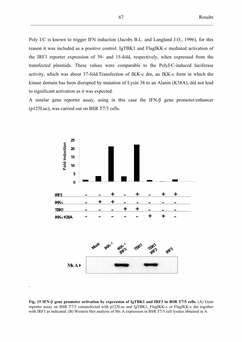
Results ___________________________________________________________________________
67
Poly I/C is known to trigger IFN induction (Jacobs B.L. and Langland J.O., 1996), for this
reason it was included as a positive control. IgTBK1 and FlagIKK- mediated activation of
the IRF3 reporter expression of 39- and 15-fold, respectively, when expressed from the
transfected plasmids. These values were comparable to the PolyI/C-induced luciferase
activity, which was about 37-fold.Transfection of IKK- dm, an IKK- form in which the
kinase domain has been disrupted by mutation of Lysin 38 to an Alanin (K38A), did not lead
to significant activation as it was expected.
A similar gene reporter assay, using in this case the IFN- gene promoter/enhancer
(p125Luc), was carried out on BSR T7/5 cells.
.
0
5
10
15
20
25
Fold
indu
ctio
n
IRF3
IKK-
TBK1
- - - ---
- ---
- - -- -
IKK- K38A - - - - - --
Mx A
MockIKK-
IKK-
IRF3 TBK1TBK1
IRF3 IRF3
Fig. 15 IFN- gene promoter activation by expression of IgTBK1 and IRF3 in BSR T7/5 cells. (A) Gene reporter assay on BSR T7/5 cotransfected with p125Luc and IgTBK1, FlagIKK- or FlagIKK- dm togetherwith IRF3 as indicated. (B) Western blot analysis of Mx A expression in BSR T7/5 cell lysates obtained in A
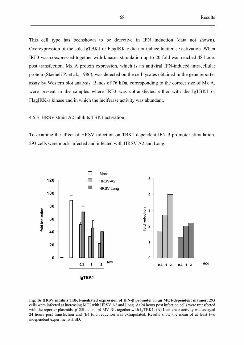
Results ___________________________________________________________________________
68
This cell type has beenshown to be defective in IFN induction (data not shown).
Overexpression of the sole IgTBK1 or FlagIKK- did not induce luciferase activation. When
IRF3 was coexpressed together with kinases stimulation up to 20-fold was reached 48 hours
post transfection. Mx A protein expression, which is an antiviral IFN-induced intracellular
protein (Staeheli P. et al., 1986), was detected on the cell lysates obtained in the gene reporter
assay by Western blot analysis. Bands of 76 kDa, corresponding to the correct size of Mx A,
were present in the samples where IRF3 was cotransfected either with the IgTBK1 or
FlagIKK- kinase and in which the luciferase activity was abundant.
4.5.3 HRSV strain A2 inhibits TBK1 activation
To examine the effect of HRSV infection on TBK1-dependent IFN- promoter stimulation,
293 cells were mock-infected and infected with HRSV A2 and Long.
Mock
0
1
2
3
4
5
fold
redu
ctio
n
0
20
40
60
80
100
120
fold
indu
ctio
n
HRSV-Long
HRSV-A2
MOI MOI0.3 1 2 0.3 1 2 0.3 1 2
IgTBK1
Fig. 16 HRSV inhibits TBK1-mediated expression of IFN- promoter in an MOI-dependent manner. 293cells were infected at increasing MOI with HRSV A2 and Long. At 24 hours post infection cells were transfectedwith the reporter plasmids, p125Luc and pCMV-RL together with IgTBK1. (A) Luciferase activity was assayed24 hours post transfection and (B) fold reduction was extrapolated. Results show the mean of at least twoindependent experiments SD.
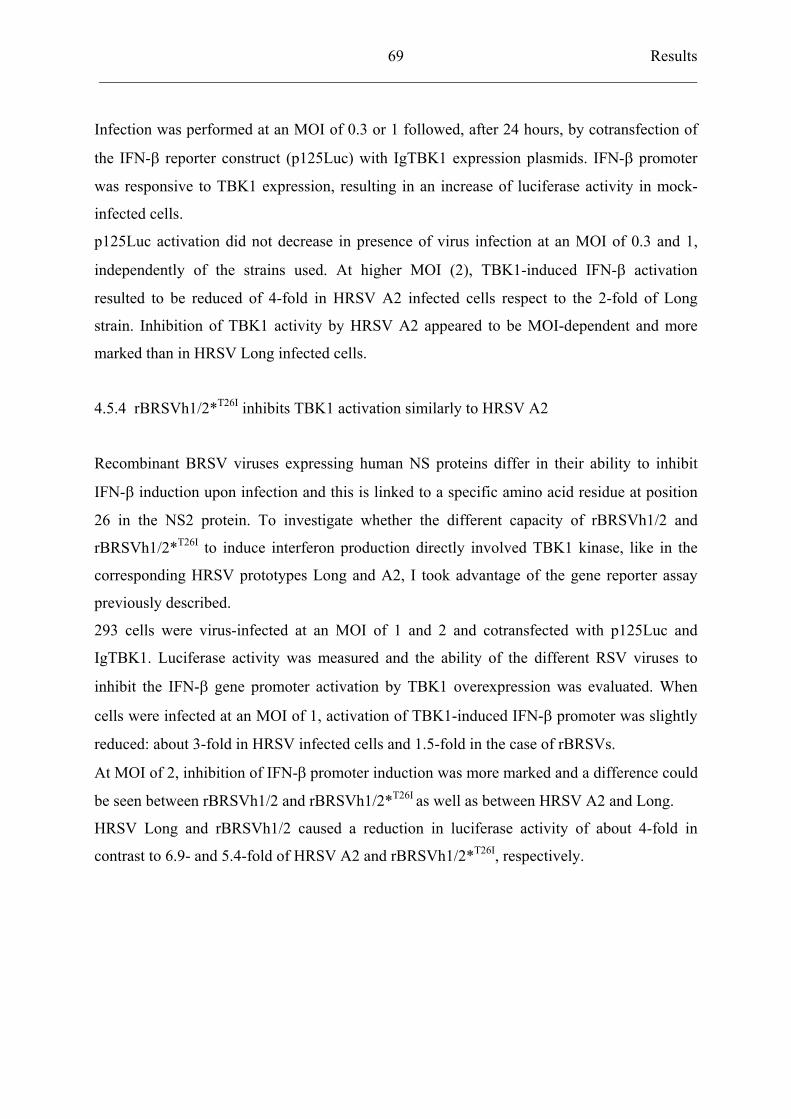
Results ___________________________________________________________________________
69
Infection was performed at an MOI of 0.3 or 1 followed, after 24 hours, by cotransfection of
the IFN- reporter construct (p125Luc) with IgTBK1 expression plasmids. IFN- promoter
was responsive to TBK1 expression, resulting in an increase of luciferase activity in mock-
infected cells.
p125Luc activation did not decrease in presence of virus infection at an MOI of 0.3 and 1,
independently of the strains used. At higher MOI (2), TBK1-induced IFN- activation
resulted to be reduced of 4-fold in HRSV A2 infected cells respect to the 2-fold of Long
strain. Inhibition of TBK1 activity by HRSV A2 appeared to be MOI-dependent and more
marked than in HRSV Long infected cells.
4.5.4 rBRSVh1/2*T26I inhibits TBK1 activation similarly to HRSV A2
Recombinant BRSV viruses expressing human NS proteins differ in their ability to inhibit
IFN- induction upon infection and this is linked to a specific amino acid residue at position
26 in the NS2 protein. To investigate whether the different capacity of rBRSVh1/2 and
rBRSVh1/2*T26I to induce interferon production directly involved TBK1 kinase, like in the
corresponding HRSV prototypes Long and A2, I took advantage of the gene reporter assay
previously described.
293 cells were virus-infected at an MOI of 1 and 2 and cotransfected with p125Luc and
IgTBK1. Luciferase activity was measured and the ability of the different RSV viruses to
inhibit the IFN- gene promoter activation by TBK1 overexpression was evaluated. When
cells were infected at an MOI of 1, activation of TBK1-induced IFN- promoter was slightly
reduced: about 3-fold in HRSV infected cells and 1.5-fold in the case of rBRSVs.
At MOI of 2, inhibition of IFN- promoter induction was more marked and a difference could
be seen between rBRSVh1/2 and rBRSVh1/2*T26I as well as between HRSV A2 and Long.
HRSV Long and rBRSVh1/2 caused a reduction in luciferase activity of about 4-fold in
contrast to 6.9- and 5.4-fold of HRSV A2 and rBRSVh1/2*T26I, respectively.
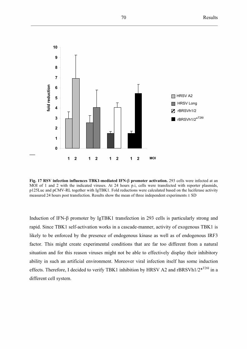
Results ___________________________________________________________________________
70
0
1
2
3
4
5
6
7
8
9
10fo
ld re
duct
ion
HRSV A2
HRSV Long
rBRSVh1/2
rBRSVh1/2*T26I
1 2 1 2 1 2 1 2 MOI
Fig. 17 RSV infection influences TBK1-mediated IFN- promoter activation. 293 cells were infected at an MOI of 1 and 2 with the indicated viruses. At 24 hours p.i, cells were transfected with reporter plasmids,p125Luc and pCMV-RL together with IgTBK1. Fold reductions were calculated based on the luciferase activitymeasured 24 hours post transfection. Results show the mean of three independent experiments SD
Induction of IFN- promoter by IgTBK1 transfection in 293 cells is particularly strong and
rapid. Since TBK1 self-activation works in a cascade-manner, activity of exogenous TBK1 is
likely to be enforced by the presence of endogenous kinase as well as of endogenous IRF3
factor. This might create experimental conditions that are far too different from a natural
situation and for this reason viruses might not be able to effectively display their inhibitory
ability in such an artificial environment. Moreover viral infection itself has some induction
effects. Therefore, I decided to verify TBK1 inhibition by HRSV A2 and rBRSVh1/2*T26I in a
different cell system.
Results ___________________________________________________________________________
71
0
20
40
60
80
100
120
140
160
180
fold
indu
ctio
n
0
5
10
15
20
25
fold
redu
ctio
n
IgTBK1 + IRF3
Mock HRSV-A2 HRSV-Long rBRSV h1/2*T26IrBRSV h1/2
Fig. 18 TBK1-mediated IFN- promoter activation in BSR T7/5 infected cells. BSR T7/5 cells were infectedat an MOI of 1 with HRSV A2, Long, rBRSVh1/2 and rBRSVh1/2*T26I. At 24 hours p.i., cells were transfectedwith reporter plasmids p125Luc and pCMV-RL together with IgTBK1 and pEF–haIRF3. Fold reductions werecalculated based on the luciferase activity of mock-infected cells stimulated with IgTBK1.
I performed the same experiment previously described but in BSR T7/5 cells. This cell line is
faulty in IFN-induction signalling pathway and this may be attribute to a defective IRF3
(overexpression of IgTBK1 does in fact not activate p125Luc) and more likely also to TBK1
itself. In this way, I could exclude residual IFN induction by viral infection, which was
performed before transfection, and I could induce IFN- promoter just by exogenous TBK1
and IRF3. BSR T7/5 cells were infected with the indicated viruses at an MOI of 1 followed 24
hours later by transfection with the following plasmids: p125Luc, pCMV-RL, pEF-haIRF3
and pCR3-IgTBK1. Stimulation of mock-infected cells with IgTBK1 and IRF3 led to an
increase of luciferase activity of 113-fold. Upon infection with HRSV Long and rBRSVh1/2
Results ___________________________________________________________________________
72
a slight reduction of about 2.5-fold was observed In contrast HRSV A2 and rBRSVh1/2*T26I
decreased significantly TBK1-dependent activation of IFN- promoter of 21- and 15- fold,
respectively.
4.6 Recombinant RV expressing RSV nonstructural proteins
4.6.1 Construction of recombinant SAD eGFP-P viruses harbouring RSV NS proteins
To determine whether NS proteins were sufficient to block IFN induction by an unrelated
virus, I generated RV viruses expressing Long-derived NS1 protein with either NS2 from
strain Long (SAD eGFP-Ph2/1) or a mutated form of NS2 where Threonine at position 26
was exchanged to an Isoleucine (SAD eGFP-Ph2*T26I/1).
e G F P - Ph 2 * T 2 6 I / 1
e G F P - P h 2 / 1( L o n g )N G F P - P M G Lh N S 2 h N S 1
N G F P - P M G Lh N S 2 * h N S 1
e G F P - P hb 2 /1 ( B R S V )N G F P - P M G Lb N S 2 b N S 1
Fig. 19 SAD eGFP-P viruses expressing RSV non structural proteins. Schematic genome organization ofeGFP-Ph2/1 expressing NS proteins from HRSV Long; eGFP-Ph2*T26I/1 harbouring the NS1 gene from Long strain and an NS2 gene derived from Long but mutated at position 26 (T26I); eGFP-Pb2/1 with insertion of NS1 and NS2 genes from BRSV wt.
For this purpose, I used a recombinant fluorescent RV in which the fusion of eGFP to P
protein (SAD eGFP-P) unabled the virus to block IFN induction upon infection (Brzozka K.
et al., 2005). The additional NS genes were introduced between G and L genes of SAD eGFP-
P as described in Materials and Methods and recombinant viruses were rescued from cDNA
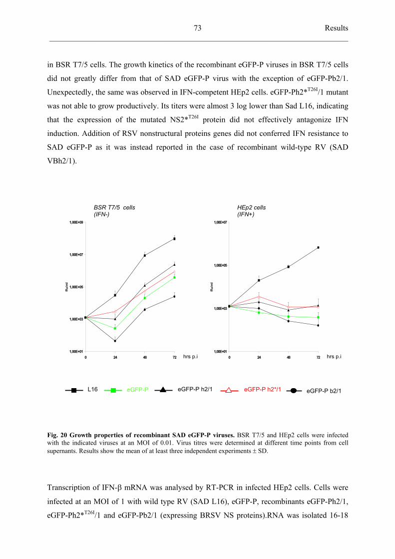
Results ___________________________________________________________________________
73
in BSR T7/5 cells. The growth kinetics of the recombinant eGFP-P viruses in BSR T7/5 cells
did not greatly differ from that of SAD eGFP-P virus with the exception of eGFP-Pb2/1.
Unexpectedly, the same was observed in IFN-competent HEp2 cells. eGFP-Ph2*T26I/1 mutant
was not able to grow productively. Its titers were almost 3 log lower than Sad L16, indicating
that the expression of the mutated NS2*T26I protein did not effectively antagonize IFN
induction. Addition of RSV nonstructural proteins genes did not conferred IFN resistance to
SAD eGFP-P as it was instead reported in the case of recombinant wild-type RV (SAD
VBh2/1).
BSR T7/5 cells(IFN-)
HEp2 cells (IFN+)
hrs p.i hrs p.i 1,00E+01
1,00E+03
1,00E+05
1,00E+07
1,00E+09
0 24 48 72
ffu/m
l
1,00E+01
1,00E+03
1,00E+05
1,00E+07
0 24 48 72
ffu/m
l
eGFP-P h2/1 L16 eGFP-P h2*/1eGFP-P eGFP-P b2/1
Fig. 20 Growth properties of recombinant SAD eGFP-P viruses. BSR T7/5 and HEp2 cells were infected with the indicated viruses at an MOI of 0.01. Virus titres were determined at different time points from cellsupernants. Results show the mean of at least three independent experiments SD.
Transcription of IFN- mRNA was analysed by RT-PCR in infected HEp2 cells. Cells were
infected at an MOI of 1 with wild type RV (SAD L16), eGFP-P, recombinants eGFP-Ph2/1,
eGFP-Ph2*T26I/1 and eGFP-Pb2/1 (expressing BRSV NS proteins).RNA was isolated 16-18
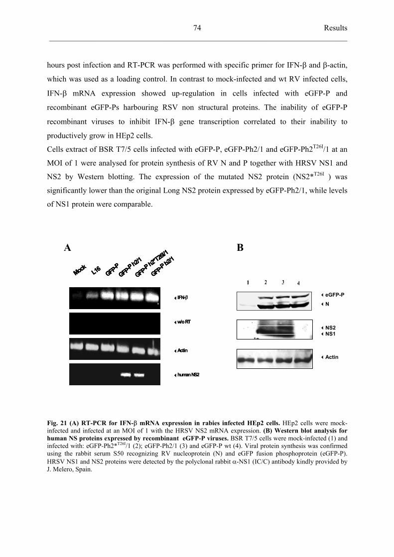
Results ___________________________________________________________________________
74
hours post infection and RT-PCR was performed with specific primer for IFN- and -actin,
which was used as a loading control. In contrast to mock-infected and wt RV infected cells,
IFN- mRNA expression showed up-regulation in cells infected with eGFP-P and
recombinant eGFP-Ps harbouring RSV non structural proteins. The inability of eGFP-P
recombinant viruses to inhibit IFN- gene transcription correlated to their inability to
productively grow in HEp2 cells.
Cells extract of BSR T7/5 cells infected with eGFP-P, eGFP-Ph2/1 and eGFP-Ph2T26I/1 at an
MOI of 1 were analysed for protein synthesis of RV N and P together with HRSV NS1 and
NS2 by Western blotting. The expression of the mutated NS2 protein (NS2*T26I ) was
significantly lower than the original Long NS2 protein expressed by eGFP-Ph2/1, while levels
of NS1 protein were comparable.
Mock L16 GFP-PGFP-P
h2/1
GFP-Ph2*T
26I/1
GFP-Pb2/1
IFN-
w/o RT
Actin
human NS2
Mock L16 GFP-PGFP-P
h2/1
GFP-Ph2*T
26I/1
GFP-Pb2/1
IFN-
w/o RT
Actin
human NS2
Mock L16 GFP-PGFP-P
h2/1
GFP-Ph2*T
26I/1
GFP-Pb2/1
Mock L16 GFP-PGFP-P
h2/1
GFP-Ph2*T
26I/1
GFP-Pb2/1
Mock L16 GFP-PGFP-P
h2/1
GFP-Ph2*T
26I/1
GFP-Pb2/1
IFN-
w/o RT
Actin
human NS2
A B
4321
eGFP-P
N
NS2NS1
Actin
Fig. 21 (A) RT-PCR for IFN- mRNA expression in rabies infected HEp2 cells. HEp2 cells were mock-infected and infected at an MOI of 1 with the HRSV NS2 mRNA expression. (B) Western blot analysis forhuman NS proteins expressed by recombinant eGFP-P viruses. BSR T7/5 cells were mock-infected (1) and infected with: eGFP-Ph2*T26I/1 (2); eGFP-Ph2/1 (3) and eGFP-P wt (4). Viral protein synthesis was confirmedusing the rabbit serum S50 recognizing RV nucleoprotein (N) and eGFP fusion phosphoprotein (eGFP-P). HRSV NS1 and NS2 proteins were detected by the polyclonal rabbit -NS1 (IC/C) antibody kindly provided byJ. Melero, Spain.
Discussion___________________________________________________________________________
75
5 DISCUSSION
5.1 Human respiratory syncytial virus: strains A2 and Long
5.1.1 Suppression of IFN- induction differs in HRSV A2 and Long
Human respiratory virus (HRSV) is the major cause of viral bronchiolitis and pneumonia in
infants worldwide. Although RSV is commonly known as a paediatric pathogen, it can cause
life-threatening lung disease in bone marrow recipients and elderly. The World Health
Organization has estimated a global annual infection of something like 64 millions
individuals, with an overall claiming of 3-5 millions human lives annually.
RSV isolates can be classified into two subgroups, designated A and B, which exhibit
antigenic and genetic differences, with a major divergence in the G glycoprotein. Group A
and B can co-circulate during epidemics, which occur during winter and early spring in
temperate climates and during the rainy season in tropical areas.
By 2 years of age, essentially 100% of the children get infected with RSV at least once.
Moreover re-infections can occur throughout life and are usually symptomatic. Severe RSV
infection in infancy is often followed by recurrent childhood wheezing probably leading to
asthma exacerbations in older children and adults. RSV-associated disease is in large part due
to excessive immune response, and the immunopathology of RSV includes the relatively
unique phenomenon of vaccine-enhanced disease or “immunopotentiation”. These particular
features of RSV infection, susceptibility to repeat infections and enhanced disease during a
recall/memory adaptive immune response to infection after vaccination, indicate that RSV
employs several novel mechanisms of immune dysregulation to propagate within the human
population. Understanding of RSV effects on the development and expression of both
innative and adaptive immune response may provide important information concerning the
formulation of effective therapies. IFN- / (type I interferons) are inducible cytokines
secreted by most eukaryotic cells in response to viral pathogens and they are of particular
importance as an early line of innate immune defence. Induction of type I IFN by dsRNA or
by virus infection is controlled at the transcriptional level. IFN- mRNA expression level
peaks around 15-18 hours post stimulation and dramatically decreases afterwards due to a
negative-feedback loop.
RSV expresses two putative nonstructural proteins, NS1 and NS2. We have previously shown
that the NS proteins of the bovine RSV (BRSV) exert an inhibitory activity on the induction
of IFN- / suppressing interferon-mediated antiviral state (Bossert B. et al., 2003).
Discussion___________________________________________________________________________
76
Recently, this has been shown also for HRSV, the human counterpart of BRSV (Spam K.M et
al., 2004). HRSV strongly inhibits activation of interferon regulatory factor 3, whereas high
levels of activation were detected in cells infected with deletion mutants for NS1 and/or NS2
genes (Spann K.M., et al., 2005).
We have demonstrated that BRSV interferes with IRF3 activation by blocking its
phosphorylation, which is an essential step for its nuclear import. It is likely that the same
mechanism is true for HRSV. We could show that two commonly used HRSV laboratory
strains, A2 and Long, differ fin their capacity to interfere with IFN induction. Long has been
found to induce IFN- in epithelial cells and plasmacytoid dendritic cells (PDC) in contrast to
A2 and clinical isolates from hospitalised children (Schlender J. et al., 2005). In this study, I
have analysed IFN- induction in epithelial cells infected with the two HRSV prototypes. The
first evaluation was performed using a Vero cell-line stably transfected with IFN-
promoter/enhancer controlling the expression of the luciferase gene (p125Luc). While
promoter activity increased more than 12-fold after infection with Long, A2 did not elicit a
significant increase. Interestingly, the difference in luciferase activity was greatest at 48 hours
post infection. This might depend on accumulation of NS proteins in the cell, which is
necessary to counteract induction by dsRNA. Spann and colleagues have in fact found that
HRSV resembles the NS gene deletion mutants in their ability to activate IRF3 at early stages
of infection. Later on, HRSV wt, opposite to the deletion mutants, blocks further activation
probably when accumulation of NS1 and NS2 proteins reaches sufficient levels in the cell.
RT-PCR for detection of IFN- mRNA expression has been performed in HEp2 cells mock-
infected and infected with HRSV A2 and Long. Infection of HEp2 cells with HRSV Long
strain resulted in a appreciable increase in the expression level of IFN- mRNA compared to
that of mock- or A2-infected cells. Thus, it was possible to confirm the results obtained in the
luciferase assay and I concluded that Long strain is a potent IFN- inducer in contrast to A2.
5.1.2 Activation of IFN- transcription factors
The IFN- promoter contains binding sites for three distinct transcriptional factors: ATF-2/c-
Jun (AP-1), NF-kB, and IRF3. These factors form a multiprotein complex, called
“enhanceosome”, which drives to transcription of the IFN- gene.
Discussion___________________________________________________________________________
77
Using reporter constructs expressing the luciferase gene under the control of either AP-1, NF-
kB or IRF3 binding sites, activation of all three transcriptional factors by infection with
HRSV A2 and Long was assayed.
AP-1 was not significantly activated in response to infection with both strains. The limited
responsiveness of AP-1 to RSV infection has been already documented and it has been
attributed to sensitivity to the cell culture redox state (Mastronarde J.G. et al., 1998).
NF-kB plays an essential role in early events of innate immune response regulating the
expression of inflammatory and immunomodulatory genes. RSV infection has been shown to
potently and specifically activate NF-kB in vivo (Haeberle H.A. et al., 2002).
Since HRSV-A2 and Long vary considerably in their ability to induce IFN- in vitro, the
magnitude of NF-kB activation has been also investigated. The results indicate that the levels
of NF-kB-responsive reporter gene activation were essentially the same in the case of both A2
and Long infected cells.
Interferon regulatory factor (IRF)-3 is the critical transcription factor regulating
immediate/early alpha-beta IFN genes. Various pathogens activate cell signals resulting in
phopshorylation of specific Serine and threonine residues in theIRF3 C-terminal region by
recently identified kinases (IKK- and TBK1). Several viral proteins have been reported to
interfere with IRF3 activation. These proteins are encoded for example by Bunyamwera,
Influenza A, Ebola, Bovine respiratory syncytial and Measles virus (Kohl A. et al., 2003;
Talon J. et al., 2000; Basler C.F. et al., 2003; Bossert B. et al., 2003). In the present study, I
investigated IRF3 activation by HRSV. In contrast to A2, Long correlates to increased
activation of IRF3 promoter, therefore indicating a selective block of this factor in A2
infected cells. This clearly explains the reason why IFN- induction is compromised by
infection with A2 virus and not with Long strain.
5.1.3 Sequence analysis of nonstructural NS proteins in HRSV
In an effort to correlate the ability to antagonize IFN- expression with specific viral factors, I
have analysed and compared NS protein amino acid sequences derived from different HRSV
strains, which had proved to differ in the achievement of a full inhibitory effect.
In the analysis, HRSV A2 and Long and two representative clinical isolates obtained from
hospitalised children were included. Our previous work had shown that the clinical isolates
were able to counteract IFN- production similarly to the laboratory strain A2. Since NS1 and
Discussion___________________________________________________________________________
78
NS2 proteins have been already described as potent interferon antagonists, I searched for
relevant differences in their amino acid sequences.
NS1 protein of BRSV shares 69% identity at amino acid level with the corresponding
sequence of HRSV subgroup A NS1. Comparison analysis of NS1 gene of HRSV laboratory
strains A2 and Long and the clinical isolates number 86 and 112 revealed amino acid
sequences highly similar to each other and no residue changes appeared to be peculiar for one
or more strains.
Only a very limited number of differences were apparent in the NS2 protein sequence as well.
Four amino acid changes clustered at its N-terminus and among these, I could identify at
position 26 the unique presence of a Threonine distinguishing HRSV Long sequence. A2 and
the clinical isolates displayed at the same position an Isoleucine residue instead. This
mutation was considered particularly interesting because the two residues greatly differ in
terms of polarity: Threonine is a polar amino acid while Isoleucine is hydrophobic. In
addition the presence of a Threonine results in a potential additional O-glycosylation site but,
even more important, represents a potential phosphorylation acceptor. This observation
prompted us to formulate the hypothesis by which the different ability to prevent expression
of IFN- by HRSV A2 and Long strains might be specifically linked to the nature of the
residue at position 26 of the NS2 protein.
5.2 A mutated Long-derived NS2 protein prevents induction of IFN-
5.2.1 Recombinant BRSV expressing HRSV nonstructural proteins
BRSV NS1 and NS2 proteins antagonize the induction of IFN- / interfering, at the early
stages of virus-stimulated IFN pathway, with the activation of IRF3 (Schlender J. et al., 2000;
Bossert B. et al., 2003).
To investigate the interferon inhibitory functions of NS2 proteins derived from A2 and Long
viruses, I used reverse genetics to generate recombinant BRSV expressing nonstructural
protein of HRSV origin. A chimerical BRSV expressing HRSV Long NS proteins (rBRSV
h1/2) has been previously generated, indicating that the HRSV nonstructural proteins are able
to fulfil the functions of the bovine counterparts with respect to IFN resistance and in
supporting viral replication. Notably, rBRSVh1/2 resembled HRSV Long in the ability to
induce IFN- opposite to wild type BRSV. To establish the importance of Threonine at
Discussion___________________________________________________________________________
79
position 26 of the NS2 protein, I mutated this residue in the context of rBRSV h1/2 to an
Isoleucine, resembling the situation in A2 strain and in the clinical isolates. The chimerical
BRSV h1/2*T26I was successfully rescued; NS1 and NS2 gene were sequenced to confirm the
correctness of the inserted mutation and the expression of NS proteins was assessed in Vero
cells. Furthermore, I compared the growth of the recombinant BRSV viruses (rBRSV h1/2
and BRSVh1/2*T26I) to that of HRSV A2 and Long in HEp2 cells, which are competent for
the expression of interferon, and in Vero cells, which instead lack the genes of these
cytokines. The chimerical BRSVs replicated efficiently and comparably to HRSVs in both
cell lines. The virtually identical growth indicated that the mutation in the HRSV NS2 protein
(T26 I) did not affect viral replication.
5.2.2 rBRSVh1/2*T26I infection inhibits IFN- induction by blocking IRF3 activation
Induction of IFN- promoter activity was analysed making use of a Vero cell line stably
transfected with a reporter construct expressing the luciferase gene under the control of the
IFN- promoter/enhancer. A very slight increase in the promoter activity was observed in
cells infected with HRSV A2, BRSV wt and rBRSVh1/2*T26I while HRSV Long and
rBRSVh1/2 displayed an induction around 10-folds.
IFN- mRNA expression was also analysed by RT-PCR in HEp2 infected cells. Even in this
case, while the rBRSVh1/2 led to an increased of expression readily detectable 15-16 hours
post infection, rBRSVh1/2*T26I did not. Taken together these data indicate that the presence of
an Isoleucine instead of a Threonine at position 26 of the NS2 amino acid sequence does
indeed contribute to the IFN inhibitory functions of the chimeric rBRSVs.
Induction of IFN- is controlled by three different transcription factors (AP-1, NF-kB and
IRF3), which become activated following phosphorylation events in response to viral
infection. Therefore I further investigated if the block of IFN- expression by rBRSV
h1/2*T26I was due to impairment of one or more of these transcription factor activities. I used
reporter constructs expressing the luciferase gene under the control of either one or the other
transcription factor binding sites. IRF3 activation was greatly compromised in rBRSV
h1/2*T26I (the same was for HRSV A2 and BRSV wt) but not in rBRSVh1/2 infected cells.
This pointed toward a selective block of IRF3 transcription factor activation by
rBRSVh1/2*T26I ; no differences were in fact observed in the case of AP-1 or NF-kB.
Discussion___________________________________________________________________________
80
BRSV and HRSV are known to be resistant to antiviral responses triggered by exogenous IFN
and this ability once again is attributable to the nonstructural protein NS1 and NS2.
Recombinant BRSV grew identically in HEp2 cells despite in the case of rBRSVh1/2 IFN is
produced. To exclude any negative influence on viral IFN-resistance caused by the mutation
in the NS2 protein, I tested viral replication of rBRSVh1/2 and rBRSVh1/2*T26I in IFN-treated
HEp2 cells. All the viruses tested, including the chimerical BRSVs, were almost completely
protected even against high doses of exogenous IFN- (up to 5000 IU/ml). Therefore, I could
exclude that residue 26 of NS2 protein playes a role in the ability of RSV to counteract IFN-
induced antiviral immune response.
5.3 The novel IkB-related kinases (IKK): IKK- and TBK1
5.3.1 Cloning TANK-binding kinase 1 (TBK1) and TRAF family member–associated NF-kB activator (TANK)
Induction of IFN- expression by viral infection requires activation of the transcription
factors ATF-2/c-Jun (AP1), NF-kB and IRF3. In unstimulated cells, IRF3 is present in the
cytoplasm. Activation of IRF3 by viruses or dsRNA occurs via phosphorylation of a cluster of
Serine and Threonine residues in its carboxyterminus and particular importance has been
attributed to Serine 386 (Mori M. et al., 2003). Phosphorylation leads to dimerization of IRF3
and formation of a complex including CREB-binding protein (CBP) and p300. This complex
after translocation to the nucleus, activates IFN gene transcription. The kinases responsible
for IRF3 phosphorylation have been recently identified in the inducible IKK- and in the
constitutively expressed TANK-binding kinase, TBK1 (Sharma S. et al., 2003). TBK1 is an
80-85 kDa protein which exerts kinase activity upon phosphorylation of specific Serine
residues in its loop region. Involvement of the cytoplasmic helicases RIG-I and MDA5 has
been recently described. RIG-I and MDA5 recognize dsRNA and this leads to downstream
activation of IKK- /TBK1 and IKK / via the adaptor protein IPS-1 (Kawai T. et al.,
2005).Viruses, in their attempt to evade the innate immune response, have elaborated
sophisticated mechanisms to abrogate the production of type I IFNs. The nonstructural protein
NS3/4-A of hepatitis C virus has been described as an inhibitor of RIG-I and MDA5
signalling functions (Yoneyama M. et al., 2005; Breiman A. et al., 2005; Foy E. et al., 2005).
On the same line, hepatitis A virus blocks activation of IRF3 by interaction with RIG-I
(Fernsterl V. et al., 2005).
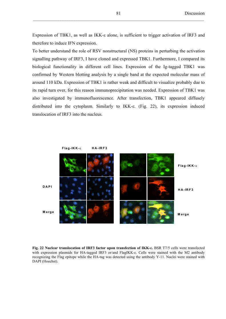
Discussion___________________________________________________________________________
81
Expression of TBK1, as well as IKK- alone, is sufficient to trigger activation of IRF3 and
therefore to induce IFN expression.
To better understand the role of RSV nonstructural (NS) proteins in perturbing the activation
signalling pathway of IRF3, I have cloned and expressed TBK1. Furthermore, I compared its
biological functionality in different cell lines. Expression of the Ig-tagged TBK1 was
confirmed by Western blotting analysis by a single band at the expected molecular mass of
around 110 kDa. Expression of TBK1 is rather weak and difficult to visualize probably due to
its rapid turn over, for this reason immunoprecipitation was needed. Expression of TBK1 was
also investigated by immunofluorescence. After transfection, TBK1 appeared diffusely
distributed into the cytoplasm. Similarly to IKK- . (Fig. 22), its expression induced
translocation of IRF3 into the nucleus.
F la g -IK K -
H A -IR F 3
M erg e
D A P I
F la g -IK K - H A -IR F 3
M erg e
Fig. 22 Nuclear translocation of IRF3 factor upon transfection of IKK- . BSR T7/5 cells were transfected with expression plasmids for HA-tagged IRF3 or/and FlagIKK- . Cells were stained with the M2 antibody recognizing the Flag epitope while the HA-tag was detected using the antibody Y-11. Nuclei were stained with DAPI (Hoechst).
Discussion___________________________________________________________________________
82
5.3.2 TBK1-dependent IFN- induction
As previously described (Fitzgerald K.A. et al., 2003), expression of FlagTBK1 in 293 cells
induced activation of IRF3-regulated IFN- promoter in a luciferase assay.
TBK1 increased luciferase activity several fold in a dose-dependant manner (Marozin S
personal observation), comparable to cells transfected with the dsRNA analog Poly I:C.
Furthermore, I was able to reconstitute the IFN-activation signalling pathway in BSR T7/5
cells. This cell line is defective in expressing type I IFN by viral infection and transfection of
dsRNA analogues. I have identified the defect in the lack of a functional IRF3 and more likely
of TBK1 as well. By cotransfection of TBK1 with IRF3, this function was re-established and
provided a useful tool to analyse IFN induction mechanisms in the absence of endogenous
interferences. Expression of the IFN-induced MxA protein confirmed IFN production.
5.4 Inhibition of TBK1 by RSV
5.4.1 HRSV A2 blocks TBK1-induced IFN- expression
To investigate whether infection with HRSV had some adverse effects on TBK1-dependent
activation of IFN- transcription, I assayed TBK1-dependent IFN- promoter activation in
infected cells. My main interest was to identify variations in the inhibitory ability of HRSV
strain A2 and Long that could be linked to the different nature of the amino acid residue at
position 26 of the NS2 protein.. Initially, 293 cells were infected with the HRSV prototypes at
increasing MOI of 0.3, 1 and 2. At 24 hours post infection, cells were transfected with the
reporter plasmid for IFN-. (p125Luc), pCMV-RL (Renilla) and IgTBK1. When infection
was performed at MOI of 0.3 and 1 a very poor inhibition of the luciferase activity was
observed, in the best case with less than 3-fold reduction for A2 virus. In addition no major
discrepancies were detectable between the two strains. At the first sight this indicated that the
interference with IRF3 activation was not at the level of the kinases responsible for its
phosphorylation but probably the block was lying somewhere upstream. In consideration of
the intrinsic characteristics of RSV, such as difficult growth in tissue culture and being prone
to losing infectivity, I considered that MOI of 1 might have been not sufficient to target every
cells with at least one viral particle. A high cell-infection was a prerequisite for the outcome
of the experiment since artificial stimulation of IFN by transfection with TBK1 was probably
Discussion___________________________________________________________________________
83
more efficient than in natural conditions. Moreover, it seemed that the levels of accumulation
of NS proteins into the cytoplasm of infected cells were decisive for the exertion of a full
inhibitory effect from RSV. For this reason, I then used an MOI of 2 to perform the same
experiment. Unfortunately higher MOI than 2 was no applicable because infected cells after
transfection tend to die. In this experimental condition, I was finally able to demonstrate a
difference between A2 and Long viruses. HRSV A2 was able to significantly reduce TBK1-
induced luciferase activity of p125Luc by about 4-folds. In contrast, strain Long, which itself
does not inhibit IFN- / expression, had a minor effect at the same MOI (about 2-fold
reduction).
5.4.2 Residue 26 of NS2 protein is important for TBK1 inhibition
To better clarify the role of residue 26 (Threonine vs Isoleucine) of HRSV NS2 protein, I
performed the same experiment including the recombinant BRSVs expressing HRSV NS1
and either NS2 protein from Long (T 26) or a mutant form of it (I 26) similar to A2 strain.
Even in this case, MOI of 2 was necessary to reveal a pronounced difference in TBK1
antagonism by these viruses. An obvious higher reduction in luciferase activity was detectable
in HRSV A2 infected cells. rBRSVh1/2*T26I showed a trend to better counteract TBK1
stimulation when compared to rBRSVh1/2. Unfortunately, the difference in folds reduction
was not so prominent as in the case of HRSV, just 5.4-folds versus 4 folds, respectively.
Therefore, I tried to perform the same experiment in BSR T7/5 cells to better clarify the
meaning of the results previously obtained. BSR T7/5 cells were used because they lack at
least a functional IRF3 which compromise the endogenous TBK1-dependent IRF3-activation
signalling pathway. It was reasonable to assume that in this cell context RSV growth was
facilitated by the absence of an endogenous IFN-network and that the following transfection
of exogenous TBK1 compromised less the cell viability. In this case, MOI of 1 was sufficient
to efficiently block IFN- promoter stimulation in the case of HRSV A2 and rBRSVh1/2*T26I
infection. In view of this data, I concluded that NS2 protein was responsible for blocking
TBK1 activity and therefore IFN- induction. Moreover, it was the nature of residue 26 to
play an important role in this function probably being part of a NS2 protein domain
responsible for interaction with the cellular kinase TBK1. It is also feasible that this amino
acid, or the sequence stretch comprising residue 26, is crucial for interaction between NS1
and NS2. It has been shown that these viral proteins not only form heterodimers but need to
Discussion___________________________________________________________________________
84
cooperate to efficiently antagonize interferon induction. I could speculate that the presence of
a Threonine instead of an Isoleucine may lead to a drastic modification of important
recognition motif in NS2 protein and either way, this would explain the loss of inhibitory
ability by HRSV Long or the rBRSVh1/2. To unveil the mechanisms behind the identity of
this apparently “essential” amino acid in NS2 protein, I tried to immunoprecipitate TBK1
with NS2 protein either from A2 or from Long infected cells. Unfortunately, no co-
precipitation was observed in any case. This might indicate that TBK1 inhibition is achieved
indirectly by interaction of NS proteins with a third player, so far not known, or simply the
results were imputable to technical problems. It must be reminded that expression of TBK1 is
quite hard to detect and that expression of NS proteins is also rather difficult to observe. NS1
and NS2 proteins can not be efficiently expressed by plasmid vectors, probably due to their
reach content in A/T bases and NS2 in viral infected cells has a turn over of only 30 min. All
these critical steps might have compromised the success of the experiment. Therefore, NS2
inhibitory mechanisms remain un-revealed so far.
Intracellular signal transduction pathways are regulated through the action of protein kinases.
Protein Ser/Thr-kinases mediate phospho-dependent signalling networks recognizing their
substrates by specific Ser/Thr-containing motifs. TBK1 and IKK- themselves contain a
unique mitogen-activated protein kinase-kinase domains (EXXXS) within their activation
loops functioning as a substrate for upstream kinases (Ehrhardt C. et al., 2004). These IKK-
related kinases, TBK1 and IKK- , are Ser/Thr-kinases as well and they target SerXXXSer
sequences in the amino terminal domain of IkB and in the carboxyterminus of IRF3 and
IRF7. Viral infection or treatment with dsRNA or LPS specifically induces phosphorylation
of Ser 386 of IRF3 by IKK- and TBK1 (Mori M. et al., 2003). Interestingly, the Long NS2
protein also contains a putative Ser/Thr motif. The position of Threonine as the second
residue within a potential consensus recognition motif (SerXXXSer/Thr) for phosphorylation
in Long NS2 protein is consistent with predicted target motifs for Ser/Thr-kinases. Moreover,
the phosphorylation motif in the carboxyterminus of IRF3 is almost identical to the NS2
amino acid stretch containing the Threonine residue.
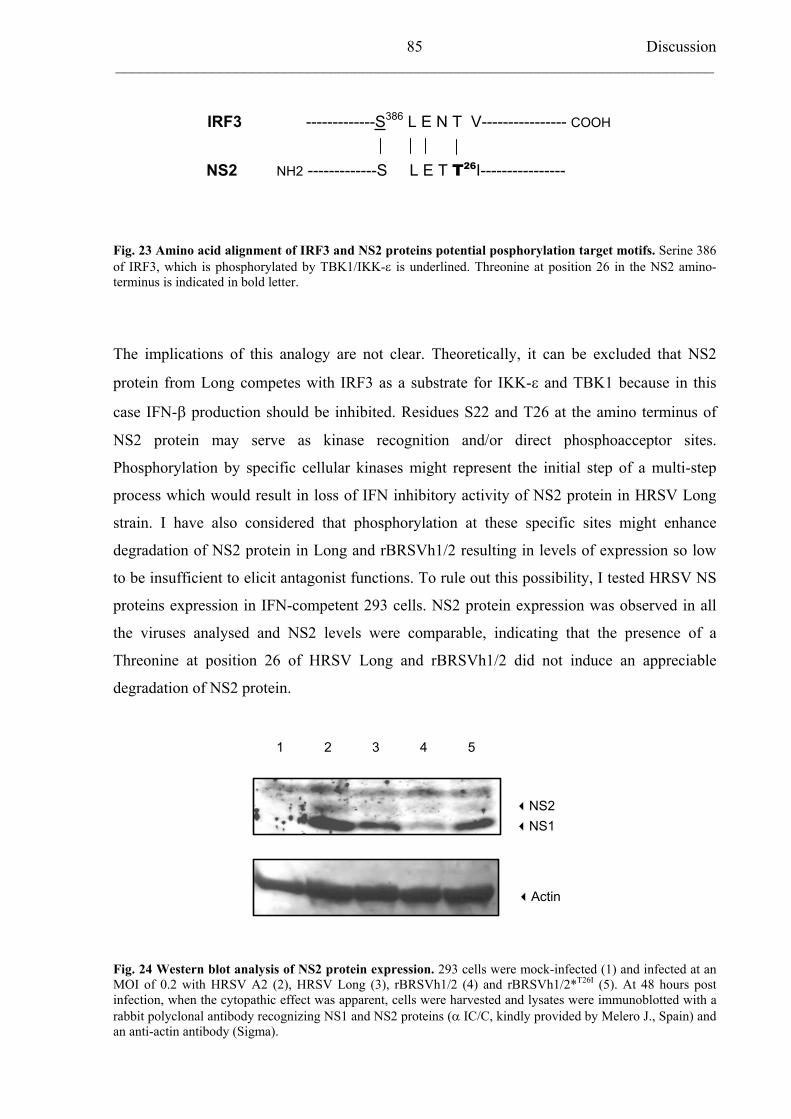
Discussion___________________________________________________________________________
85
IRF3 -------------S386 L E N T V---------------- COOH
NS2 NH2 -------------S L E T T26I----------------
Fig. 23 Amino acid alignment of IRF3 and NS2 proteins potential posphorylation target motifs. Serine 386 of IRF3, which is phosphorylated by TBK1/IKK- is underlined. Threonine at position 26 in the NS2 amino-terminus is indicated in bold letter.
The implications of this analogy are not clear. Theoretically, it can be excluded that NS2
protein from Long competes with IRF3 as a substrate for IKK- and TBK1 because in this
case IFN- production should be inhibited. Residues S22 and T26 at the amino terminus of
NS2 protein may serve as kinase recognition and/or direct phosphoacceptor sites.
Phosphorylation by specific cellular kinases might represent the initial step of a multi-step
process which would result in loss of IFN inhibitory activity of NS2 protein in HRSV Long
strain. I have also considered that phosphorylation at these specific sites might enhance
degradation of NS2 protein in Long and rBRSVh1/2 resulting in levels of expression so low
to be insufficient to elicit antagonist functions. To rule out this possibility, I tested HRSV NS
proteins expression in IFN-competent 293 cells. NS2 protein expression was observed in all
the viruses analysed and NS2 levels were comparable, indicating that the presence of a
Threonine at position 26 of HRSV Long and rBRSVh1/2 did not induce an appreciable
degradation of NS2 protein.
Actin
NS2NS1
21 3 4 5
Fig. 24 Western blot analysis of NS2 protein expression. 293 cells were mock-infected (1) and infected at an MOI of 0.2 with HRSV A2 (2), HRSV Long (3), rBRSVh1/2 (4) and rBRSVh1/2*T26I (5). At 48 hours post infection, when the cytopathic effect was apparent, cells were harvested and lysates were immunoblotted with a rabbit polyclonal antibody recognizing NS1 and NS2 proteins ( IC/C, kindly provided by Melero J., Spain) and an anti-actin antibody (Sigma).
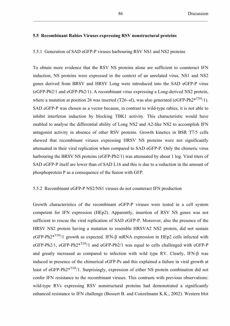
Discussion___________________________________________________________________________
86
5.5 Recombinant Rabies Viruses expressing RSV nonstructural proteins
5.5.1 Generation of SAD eGFP-P viruses harbouring RSV NS1 and NS2 proteins
To obtain more evidence that the RSV NS proteins alone are sufficient to counteract IFN
induction, NS proteins were expressed in the context of an unrelated virus. NS1 and NS2
genes derived from BRSV and HRSV Long were introduced into the SAD eGFP-P virus
(eGFP-Pb2/1 and eGFP-Ph2/1). A recombinant virus expressing a Long-derived NS2 protein,
where a mutation at position 26 was inserted (T26 I), was also generated (eGFP-Ph2*T26I/1).
SAD eGFP-P was chosen as a vector because, in contrast to wild-type rabies, it is not able to
inhibit interferon induction by blocking TBK1 activity. This characteristic would have
enabled to analyse the differential ability of Long NS2 and A2-like NS2 to accomplish IFN
antagonist activity in absence of other RSV proteins. Growth kinetics in BSR T7/5 cells
showed that recombinant viruses expressing HRSV NS proteins were not significantly
attenuated in their viral replication when compared to SAD eGFP-P. Only the chimeric virus
harbouring the BRSV NS proteins (eGFP-Pb2/1) was attenuated by about 1 log. Viral titers of
SAD eGFP-P itself are lower than of SAD L16 and this is due to a reduction in the amount of
phosphoprotein P as a consequence of the fusion with GFP.
5.5.2 Recombinant eGFP-P NS2/NS1 viruses do not counteract IFN production
Growth characteristics of the recombinant eGFP-P viruses were tested in a cell system
competent for IFN expression (HEp2). Apparently, insertion of RSV NS genes was not
sufficient to rescue the viral replication of SAD eGFP-P. Moreover, also the presence of the
HRSV NS2 protein having a mutation to resemble HRSVA2 NS2 protein, did not sustain
eGFP-Ph2*T26I/1 growth as expected. IFN- mRNA expression in HEp2 cells infected with
eGFP-Ph2/1, eGFP-Ph2*T26I/1 and eGFP-Pb2/1 was equal to cells challenged with eGFP-P
and greatly increased as compared to infection with wild type RV. Clearly, IFN- was
induced in presence of the chimerical eGFP-Ps and this explained a failure in viral growth at
least of eGFP-Ph2*T26I/1. Surprisingly, expression of either NS protein combination did not
confer IFN resistance to the recombinant viruses. This contrasts with previous observations:
wild-type RVs expressing RSV nonstructural proteins had demonstrated a significantly
enhanced resistance to IFN challenge (Bossert B. and Conzelmann K.K., 2002). Western blot
Discussion___________________________________________________________________________
87
analysis of RSV NS proteins expressed by RV has then revealed a problem with the
expression of NS2 by the recombinant eGFP-Ph2*T26I/1. While NS1 protein expression was
comparable to the one of eGFP-Ph2/1, the NS2 level was significantly reduced. The reason of
this low efficiency in expression is not yet clear. RT-PCR sequencing revealed that NS2 gene
is correctly inserted in the RV vector and no changes of the sequence were found. Even the
lack of detectable IFN resistance by eGFP-Ph2/1, which did express both nonstructural
proteins, remains unsolved. I can only speculate that the amount on NS proteins produced is
just not enough to protect eGFP-P virus from the IFN-induced antiviral state. In the RSV
genome, the NS genes are located at the most upstream position resulting in a high level of
expression in infected cells. Recombinant wild-type RVs (e.g RV h1/2), which are IFN
resistant, reach very high titres in cell culture, up to 1x 10⁸-109 ffu/ml, comparable to the
parental strain SAD L16 and this is positively influencing the expression levels of the
additional genes inserted in their genome. The fact that chimerical RVs are resistant to
exogenous IFN already ruled out the need of additional RSV proteins to exert this function.
Unfortunately, recombinant RV viruses could not be used for my purposes because wild-type
RV itself is able to counteract IFN induction by the activity of the P protein. Therefore
discrimination of the role played by RSV NS proteins is impossible. On the contrary, SAD
eGFP- P does not block IFN production due to low P levels. The recombinant viruses derived
from SAD eGFP-P (eGFP-Ph2/1and eGFP-Ph2*T26I/1) are attenuated in their growth of about
3 logs in BSR T7/5 cells. The inefficient growth of these viruses and the fact that little NS2
protein is expressed from the current constructs precluded the analysis of NS functions.
In the future recombinant RVs should be generated in which high NS levels of expression are
achieved by inserting the RSV nonstructural genes in a most proximal position in the rabies
genome.
5.6 Final considerations
The results described in this work confirmed thatRSV NS2 protein plays the major role in
blocking the IFN- activation pathway. Hereby, I was able to characterize HRSV evasion of
the innate immune response by identifying in the amino acid residue at position 26 of NS2
protein a crucial determinant for this function. Despite the mechanisms by which NS2 protein
operates its antagonism remain still to be elucidated, the data provided support to the initial
hypothesis that RSV interferes with the very early stages of the IRF3 phosphorylation. RSV
have shown to interfere with the activity of the IKK-related kinase TBK1 but no evidence was
Discussion___________________________________________________________________________
88
found of a direct interaction between this kinase and RSV NS2 protein. When this thesis was
in preparation, several publications have revealed in more detail the identity of some of the
most upstream components of the IFN- activation pathway, which determine TBK1/IKK-
recruitment . This opens the possibility to further elucidate the viral mechanisms involved in
the IFN antagonist functions of HRSV NS2 protein.
Summary
___________________________________________________________________________
89
6 SUMMARY
Interferon Escape of Respiratory Syncytial Virus:
Functional Analysis of Nonstructural Proteins NS1 and NS2
Respiratory syncytial virus (RSV) is recognised as the most frequent cause of severe lung
infections in infants and cattle worldwide. Currently, no effective treatments are available and the
development of a successful vaccine has been hampered by the fact that natural infection does not
provide complete and durable protection. RSV nonstructural proteins, NS1 and NS2, are strong
inhibitors of IFN / -production by specifically preventing interferon regulatory factor (IRF)-3
phosphorylation. However, the exact mechanisms leading to NS protein-mediated inhibition of
IRF3 remain to be unravelled.
One of the objectives of this study was to identify amino acid domains in the human
respiratory syncytial virus (HRSV) nonstructural proteins (NS) responsible for their ability to
ablate the IFN- signalling pathway. Furthermore, I wanted to find out at which level of this
signalling pathway the NS proteins exert their suppressive activity and which are their major
cellular targets. HRSV strains A2 and Long differ in their ability to block interferon type I
synthesis. Sequence analysis of their NS proteins revealed the presence of an amino acid
residue in the NS2 protein with a potential role for RSV IFN-inhibitory functions.
Two recombinant bovine respiratory syncytial (BRSV) viruses harbouring HRSV NS1 and
NS2 genes were generated and tested in their ability to restrict IFN- synthesis. These
recombinant viruses differed only in the identity of the residue at position 26 of the HRSV
NS2 protein: rBRSVh1/2 has a Threonine as in the Long strain, while in rBRS h1/2*T26I
this
amino acid was mutated into an Isoleucin similarly to A2 virus. Sets of in vitro tests revealed
that IFN- induction was impaired by rBRSVh1/2*T26Iwhen compared to rBRSV h1/2.
Analysis of the transcriptional factors (AP-1, NF-kB and IRF3) involved in the activation of
IFN- synthesis provided evidence that the inhibitory ability of rBRSVh1/2*T26I
was
correlated to a selective block of IRF3. The mutation (T26I) in the NS2 protein did neither
effect the NF-kB activation pathway nor perturbed the IFN-resistance characteristics of the
chimeric viruses.
Summary
___________________________________________________________________________
90
IRF3 is activated upon phosphorylation mediated by IKK-related kinases (TBK1 and IKK- ).
TBK1 was therefore cloned from a human lung cDNA library and its biological activities
regarding the induction of IFN- were compared in mock-infected and infected cells.
rBRSVh1/2*T26I
and HRSV A2 precluded virus-induced IRF3 activation by interfering with
TBK1 functions. No direct interaction between TBK1 and NS2 protein was demonstrated
indicating that the kinase TBK1 may not be the sole target involved in RSV mechanisms of
evasion of the innate immune response.
Recombinant IFN-inducible rabies viruses expressing HRSV Long-derived (rGFP-Ph2/1) or
HRSV A2-like (rGFP-Ph2*T26I/1) NS proteins were also generated. The HRSV NS2 protein
expressing an Isoleucin (NS2*T26I) at position 26 was not able to suppress IFN- induction
and to rescue the growth of the recombinant eGFP-Ph2*T26I/1 in interferon-competent cells.
A low expression of the mutated NS2*T26Iprotein was probably the reason of this failure.
In summary, these results show that the HRSV NS2 protein possesses an intrinsic IFN-
inhibitory activity, which is achieved throughout a selective inhibition of the IRF3 activation
pathway. The block appears to be exerted at the level of IRF3-kinase TBK1. Interferon-
antagonist functions of the HRSV NS2 protein are linked to a particular amino acid motif in
the N-terminus of the protein. Identification of this amino acid domain and of TBK1 as the
cellular target provide a better insight of how the HRSV NS2 protein prevents the
establishment of the antiviral innate immune response and therefore it might contribute to the
development of an effective vaccine.
Zusammenfassung___________________________________________________________________________
91
7 ZUSAMMENFASSUNG
Unterdrückung der Interferon-vermittelten Immunantwort durch das Respiratorische
Synzytial Virus: funktionelle Analyse der Nicht-Strukturproteine NS1 und NS2
Das Respiratorische Synzytial Virus (RSV) zählt weltweit zu den häufigsten Auslösern
schwerer Lungenentzündungen bei Kleinkindern und Rindern. Gegenwärtig gibt es dafür
keine effektive Behandlungsmethode und die Entwicklung eines erfolgreichen Impfstoffes
war nicht möglich, da auch eine natürliche Infektion keinen vollständigen und dauerhaften
Schutz gewährleistet.
Die Nicht-Strukturproteine des RSV -NS1 und NS2- wirken stark hemmend auf die
alpha/beta-Interferon ( / IFN)-Produktion, indem sie die Phosphorylierung des Interferon-
Regulierungs-Faktors (IRF)-3 verhindern. Die Mechanismen, die zu einer NS-Protein
vermittelten Hemmung des IRF3 führen, sind bisher unbekannt.
Eines der Ziele dieser Arbeit war es, Aminosäurensequenzen der HRSV Nicht-
Strukturproteine zu finden, die ausschlaggebend für die Hemmung der IFN- Signalkaskade
sind. Außerdem sollte geklärt werden, auf welchem Level der Signalkaskade die Nicht-
Strukturproteine ihre hemmende Wirkung ausüben und auf welche zellulären Komponenten
sie abzielen. Die HSV-Stämme A2 und Long hemmen die Interferon Typ I Synthese auf
unterschiedliche Weise. Mittels Sequenzanalysen ihrer Nicht-Strukturproteine wurde ein
Aminosäurerest im NS2-Protein identifiziert, der eine mögliche Rolle bei der Hemmung von
Interferon durch RSV spielt.
Es wurden zwei rekombinante bovine RS-Viren (BRSV), die Gene für NS1 und NS2 des
humanen RS-Virus beinhalteten, generiert und auf ihre Fähigkeiten untersucht, die IFN- -
Synthese zu unterdrücken. Diese rekombinanten Viren unterscheiden sich nur in der
Aminosäure an Position 26 des HRSV NS2-Proteins: rBRSVh1/2 hat ein Threonin, wie in
dem Long–Stamm, während bei rBRSVh1/2*T26I diese Aminosäure gegen ein Leucin
ausgetauscht wurden, wie es in A2 Viren natürlich vorkommt. In vitro Untersuchungen
ergaben, dass die IFN Induktion durch rBRSVh1/2*T26I im Vergleich mit rBRSVh1/2
abgeschwächt wurde.
Zusammenfassung___________________________________________________________________________
92
Die Analyse der Transkriptionsfaktoren AP-1, NF-kB und IRF3, die an der Aktivierung der
IFN Synthese beteiligt sind ergab, dass die inhibitorische Fähigkeit von rBRSVh1/2*T26I
mit einer selektiven Blockierung von IRF3 zusammenhängt. Die Mutation T26I im NS2
Protein hatte weder einen Effekt auf den NF-kB-Aktivierungs-Weg, noch störte sie die IFN-
Resistenz des Chimären Virus.
IRF3 wird durch Phosphorylierung mittels IKK-verwandten Kinasen (TBK1 und IKK- )
aktiviert. TBK1 wurde daher aus einer humanen Lungen cDNA-Bibliothek kloniert und seine
biologische Aktivität bezüglich der Aktivierung von IFN in „mock“-infizierten Zellen und
Virus-infizierten Zellen verglichen.
rBRSVh1/2*T26I und HRSV A2 verhinderten Virus-induzierte IRF3-Aktivierung durch
Beeinflussung von TBK1-Funktionen. Es wurde gezeigt, dass es keine direkte Interaktion
zwischen TBK1 und NS2 gibt. Dies deutet darauf hin, dass die Kinase TBK1 nicht das
alleinige Zielprotein der RSV-Mechanismen ist, der angeborenen Immunantwort zu entgehen.
Weiterhin wurden rekombinante IFN-induzierbare Rhabdo-Viren generiert, die entweder das
NS2 Protein aus dem HRSV Long-Stamm (rGFP-Ph2/1) oder das A2 ähnliche Protein (rGFP-
Ph2*T26I/1) exprimieren. Das HRSV NS2-Protein, dass an Position 26 ein Isoleucin besitzt,
war nicht fähig die IFN -Induktion zu unterdrücken, beziehungsweise das Wachstum von
rekombinanten eGFP-Ph2*T26I/1 in IFN-kompetenten Zellen zu ermöglichen. Die lag
vermutlich an der geringen Expressionsrate des mutierten NS2*T26I-Proteins.
Zusammenfassend zeigen die Ergebnisse, dass das HRSV NS2-Protein eine intrinsische
Aktivität als IFN-Inhibitor besitzt, die über eine selektive Hemmung des IRF3
Aktivierungs-Weges erfolgt. Die Blockade wirkt auf der Ebene der IRF3-Kinase TBK1. Die
Interferon-antagonistischen Funktionen des HRSV NS2-Proteins begründen sich in einem
speziellen Aminosäure-Motiv am N-Terminus des Proteins. Die Identifizierung dieser
Aminosäure-Domäne sowie von TBK1 als zelluläres Ziel von NS2 ermöglicht einen besseren
Überblick über die Mechanismen, durch die das HRSV NS2-Protein die angeborene antivirale
Immunantwort unterdrückt. Dies könnte ein Ansatzpunkt für die Entwicklung eines effektiven
Impfungsstoffes sein.
Bibliography
___________________________________________________________________________
93
8 BIBLIOGRAPHY
Abendroth A., Slobedman B., Lee E., Mellins E., Wallace M., Arvin A.M., 2000,
Modulation of major histocompatibility complex class II protein expression by varicella-
zoster virus, J. Virol., 74:1900-1907
Alwan W.H., Record F.M., Openshaw P.J., 1991, CD4+T-cells clear virus but augment
disease in mice infected with respiratory syncytial virus. Comparison with the effects of
CD8+T cells, Clin. Exp. Immunol., 88: 527-536
Antonis A.F., Schrijever R.S., Daus F., Steverink P.J., Stockhofe N., Hensen E.J.,
Langedijk J.P., van der Most R.G., 2003, Vaccine-induced immunopathology during
bovine respiratory syncytial virus infection: exploring the parameters of pathogenesis, J.
Virol., 77: 12067-12073
Arnold R., Konig B., Werchau H., Konig W., 2004, Respiratory syncytial virus
deficient in soluble G protein induced an increased proinflammatory response in human
lung epithelial cells, Virology, 330: 384-397
Atreya P.L., Peeples M.E., Collins P.L., 1998, The NS1 protein of human respiratory
syncytial virus is a potent inhibitor of minigenome transcription and RNA replication, J.
Virol., 72: 1452-1461
Atreya P.L. and Kulkarni S., 1999, Respiratory syncytial virus strain A2 is resistant to
the antiviral effect of type I interferons and human MxA, Virology, 261: 227-241
Baldwin A.S. Jr, 1996, The NF-kappa B and I kappa B proteins: new discoveries and
insights, Annu. Rev. Immunol., 14: 649-683
Barr F.E., Pedigo H., Johnson T.R., Shepherd V.L., 2000, Surfactant protein-A
enhances uptake of respiratory syncytial virus by monocytes and U937 macrophages, Am.
J. Respir. Cell. Mol., Biol.,: 586-592
Barton G.M. and Medzhitov R., 2003, Toll-like receptors and their ligands, Curr. Top.
Microbiol. Immunol., 270:81-92.
Bartz H., Turkel O., Hoffjan S., Rothoeft T., Gonschorek A., Schauer U., 2003,
Respiratory syncytial virus decreases the capacity of myeloid dendritic cells to induce
interferon-gamma in naïve T cells, Immunology, 109: 49-57
Basler C.F., Mikulasova A., Martinez-Sobrido L. et al., 2003, The Ebola virus VP35
protein inhibits activation of interferon regulatory factor 3, J. Virol., 77: 7945-7956
Bibliography
___________________________________________________________________________
94
Bergeron J., Mabrouk T., Garzon S., Lemay G., 1998, Characterization of the
thermosensitive ts453 reovirus mutant: increased dsRNA binding of sigma 3 protein
correlates with interferon resistance, Virology, 246: 199-210
Bermingham M. and Collins P.L., 1999, The M2-2 protein of human respiratory virus is
a regulatory factor involved in the balance between RNA replication and transcription,
Proc. Natl. Acad. Sci. USA, 96: 11259-11264
Bitko V., Velazquez A., Yang L., Yang Y.C., Barik S., 1997, Transcriptional induction
of multiple cytokines by human respiratory syncytial virus requires activation of NF-
kappa B and is inhibited by sodium salicylate and aspirin, Virology, 232: 369-378
Black T.L., Barber G.N., Katze M.G., 1993, Degradation of the interferon-induced
68,000 Mr protein kinase by poliovirus requires RNA, J. Virol., 67: 791-800
Boelen A., Kwakkel J., Barends M., de Rond L., Dormans J., Kimman T., 2002,
Effect of lack of Interleukin-4, Interleukin-12, Interleukin-18 or the Interferon-gamma
receptor on virus replication, cytokine response, and lung pathology during respiratory
syncytial virus infection in mice, J. Med. Virol., 66: 552-560
Bonizzi G. and Karin M., 2004, The two NF-kappaB activation pathways and their role
in innate and adaptive immunity, Trends Immunol., 25: 280-288
Bossert B. and Conzelmann K.K., 2002, Respiratory syncytial virus (RSV)
nonstructural (NS) proteins as host range determinants: a chimeric bovine RSV with NS
genes from human RSV is attenuated in interferon-competent bovine cells, J. Virol., 76:
4287-4293
Bossert B., Marozin S., Conzelmann K.K., 2003, Non structural proteins NS1 and NS2
of bovine respiratory syncytial virus block activation of interferon regulatory factor 3, J.
Virol., 77: 8661-8668
Breiman A, grandvaux N., Lin R, Ottone C. et al., 2005, Inhibition of RIG-I-dependent
signalling to the interferon pathway during hepatitis C virus expression and restoration of
signalling by IKKepsilon, J. Virol., 79: 3969-3978
Brogden K.A., Lehmkuhl H.D., Cutlip R.C., 1998, Pasteurella haemolytica
complicated respiratory infections in sheep and goats, Vet. Res., 29: 233-254
Brzozka K., Finke S., Conzelmann K.K., 2005, Identification of the rabies virus
alpha/beta interferon antagonist: phosphoprotein P interferes with phosphorylation of
interferon regulatory factor 3, J. Virol., 79: 7673-7681
Bibliography
___________________________________________________________________________
95
Buchholz U.J., Finke S., Conzelmann K.K., 1999, Generation of bovine respiratory
syncytial virus (BRSV) from cDNA: BRSV NS2 is not essential for virus replication in
tissue culture, and the human RSV leader region acts as a functional BRSV genome
promoter, J. Virol. 73: 251-259
Burke E., Mahoney M.N., Almo S.C., Barik S., 2000, Profilin is required for optimal
actin-dependent transcription of respiratory syncytial virus genome RNA, J. Virol. 74:
660-675
Cartee T.L., Wertz G.W., 2001, Respiratory syncytial virus M2-1 protein requires
phosphorylation for efficient function and binds viral RNA during infection, J Virol., 75:
12188-12197
Chang H.W., Watson J.C., Jacobs B.L., 1992, The E3L gene of vaccinia virus encodes
an inhibitor of the interferon-induced, double-stranded RNA-dependent protein kinase,
Proc. Natl. Acad. Sci. USA 89: 4825-4829
Chang J. and Braciale T.J., 2002, respiratory syncytial virus infection suppresses lung
CD8+T-cell effector activity and peripheral CD8
+T-cell memory in the respiratory tract,
Nat. Med., 8: 54-60
Cherrie A.H., Anderson K., Wertz G.W., Openshaw P.J., 1992, Human cytotoxic T
cells stimulated by antigen on dendritic cells recognize the N, SH, F, M, 22K, and 1b
proteins of respiratory syncytial virus, J. Virol., 66: 2102-2110
Chiba Y., Higashidate Y., Suga K., Honjo K., Tsutsumi H., Ogra P.L., 1989,
Development of cell-mediated cytotoxic immunity to respiratory syncytial virus in human
infants following naturally acquired infection, J. Med. Virol., 28: 133-139
Choudhary S., Boldogh S., Garofalo R., Jamaluddin M., Brasier A.R., 2005,
Respiratory syncytial virus influences NK-kappaB-dependent gene expression through a
novel pathway involving MAP3K14/NIK expression and nuclear complex formation with
NF-kappaB2, J. Virol., 79: 8948-8959
Collins P.L. and Wertz G.W., 1985, Nucleotide sequence of the 1B and 1C non-
structural protein mRNAs of human respiratory syncytial virus, Virology, 143: 442-451
Collins P.L., Hill M.G., Cristina J., Grosfeld H., 1996, Transcription elongation factor
of respiratory syncytial virus, a nonsegmented negative-strand RNA virus, Proc. Natl.
Acad. Sci. USA, 93: 81-85
Collins P.L., Chanock R.M, Murphy B.R., 2001, Respiratory syncytial virus disease,
4th edition, in: Knipe D., Howley P. (Eds), Fields in virology, 1443-1486
Bibliography
___________________________________________________________________________
96
Connors M., Kulkarni A.B., Collins P.L., Firestone C.Y., Holmes K.L., Morse H.C
3rd, Murphy B.R., 1992, Resistance to respiratory syncytial virus (RSV) challenge
induced by infection with a vaccinia virus recombinant expressing the RSV M2 protein is
mediated by CD8+T cells, while that induced by Vac-F or Vac-G recombinants is
mediated by antibodies, J. Virol., 66: 1277-1281
Connors M., Collins P.L., Firestone C.Y., Murphy B.R., 1991, Respiratory syncytial
virus (RSV) F, G, M2 (22K), and N proteins each induce resistance to RSV challenge,
but resistance induced by M2 and N proteins is short-lived, J. Virol., 65: 1634-1637
Cook P.M., Eglin R.P., Easton A.J., 1998, Pathogenesis of pneumovirus infections in
mice: detection of pneumonia virus of mice and human respiratory syncytial virus mRNA
in lungs of infected mice by in situ hybridization, J. Gen. Virol., 79: 2411-241
Cook J.K., 2000, Avian pneumovirus infections of turkeys and chickens, Vet. J., 160:
118-125
Cuesta I., Geng X., Asenjo A., Villanueva N., 2000, Structural phosphoprotein M2-1 of
the human respiratory syncytial virus is an RNA binding protein, J. Virol., 74: 9858-9867
Cusson-Hermance N., Lee T.H., Fitzgerald K.A., Kelliher M.A., 2005, Rip1 mediates
the Trif-dependent toll-like receptor 3 and 4-induced NF-kappa B activation but does not
contribute to IRF-3 activation, J Biol Chem., Aug 22 ahead of print
Dakhama A., Park J.W., Taube C., Chayama K., Balhorn A. et al., 2004, The role of
virus-specific Immunoglobulin E in airway hyperresponsiveness, Am. J. Respir. Crit. Care
Med., 170: 952-959
Didcock L., Young D.F., Goodburn S., Randall R.E., 1999, The V protein of simian
virus 5 inhibits interferon signalling by targeting STAT1 for proteasome-mediated
degradation, J. Virol., 73: 9928-9933
Dupuy L.C., Dobson S., Bitko V., Barik S., 1999, Caseine kinase 2-mediated
phosphorylation of respiratory syncytial virus phosphoprotein P is essential for the
transcription elongation activity of the viral polymerase; phosphorilation by casein kinase
1 occurs mainly at Ser (215) and is without effect, J. Virol., 73: 8384-8392
Ehl S., Bischoff R., Ostler T., Vallbracht S., Schulte-Moenting J., Poltorak A.,
Freundenberg M., 2004, The role of Toll-like receptor 4 versus interleukin-12 in
immunity of respiratory syncytial virus, Eur. J. Immunol., 34: 1146-1153
Elvander M., 1996, Severe respiratory disease in dairy cows caused by infection with
bovine respiratory syncytial virus, Vet. Rec., 138: 101-105
Bibliography
___________________________________________________________________________
97
Evans J.E., Cane P.A., Pringle C.R., 1996, Expression and characterisation of the NS1
and NS2 proteins of respiratory syncytial virus, Virus Res., 155-161
Fearns R., Peeples M.E., Collins P.L., 1997, Increased expression of N protein of
respiratory syncytial virus stimulates minigenome replication but does not alter the
balance between the synthesis of mRNA and antigenome, Virology, 236: 188-201
Fearns R., Collins P.L., 1999, Role of the M2-1 transcription antitermination protein of
respiratory syncytial virus in sequential transcription, J. Virol., 73: 5852-5864
Feldman S.A., Hendry R.M., Beeler J.A., 1999, Identification of a linear heparin
binding domain for respiratory syncytial virus attachment glycoproteins G, J. Virol., 73:
6610-6617
Fensterl V., Grotheer D., Berk I. et al., 2005, hepatitis A virus suppresses RIG-I-
mediated IRF-3 activation to block induction of beta interferon, J. Virol., 79: 10968-
10977
Finke S., Brzozka K., Conzelmann K., 2004, Tracking fluorescence-labeled rabies
virus: enhanced green fluorescent protein-tagged phosphoprotein P supports virus gene
expression and formation of infectious particles, J. Virol., 78. 12333-12343
Fitzgerald K.A., McWhirter S.M., Faia K.L., Rowe D.C., Latz E. et al., 2003,
IKKepsilon and TBK1 are essential components of IRF3 signalling pathway, Nat.
Immunol., 4: 491-496
Foy E., Li k., Sumpter R. Jr., Loo Y.M. et al., 2005, Control of antiviral defenses
through hepatitis C virus disruption of retinoic acid-inducible gene-I signaling, Proc. Natl.
Acad. Sci. USA, 102: 2986-2991
Furze J.M., Wertz G.W., Lerch R., Taylor G., 1994, Antigenic heterogeneity of the
attachment protein of bovine respiratory syncytial virus, J. Gen. Virol., 75: 363-370
Gaddum R.M., Cook R.S., Thomas L.H., Taylor G., 1996, Primary cytotoxic T cell
responses to bovine respiratory syncytial virus in calves, Immunology, 88: 421-427
Gaddum R.M., Cook R.S., Furze J.M., Ellis S.A., Taylor G., 2003, Recognition of
bovine respiratory syncytial virus proteins by bovine CD8+ T lymphocytes, Immunology,
108: 220-229
Gale M. and Katze M.G., 1998, Molecular mechanisms of interferon resistance mediated
by viral-directed inhibition of PKR, the interferon-induced protein kinase, Pharmacol.
Ther., 78: 29-46
Gao S.J., Boshoff C., Jayachandra C. et al., 1997, KSHV ORF K9 (vIRF) is an
oncogene which inhibits the interferon signalling pathway, Oncogene, 15: 1979-1985
Bibliography
___________________________________________________________________________
98
Garofalo R.P., Patti J., Hintz K.A., Hill V., Ogra P.L., Welliver R.C., 2001,
Macrophage inflammatory protein 1-alpha (not T helper type 2 cytokines) is associated
with severe forms of bronchiolitis , J. Infect. Dis., 184: 393-399
Garofalo R.P., Hintz K.A., Hill V., Patti J., Ogra P.L., Welliver R.C., 2005, A
comparison of epidemiologic and immunologic features of bronchiolitis caused by
influenza virus and respiratory syncytial virus, J. Med. Virol., 75: 282-289
Gelman A.E., Zhang J., Choi Y., Turka L.A., 2004, Toll-like receptors ligands directly
promote activated CD4+T cell survival, J. Immunol., 172: 6065-6073
Gershwin L.J., Berghaus L.J., Arnold K., Anderson M.L., Corbeil L.B., 2005,
Immune mechanisms of pathogenetic synergy in concurrent bovine pulmonary infection
with Haemophilus somnus and bovine respiratory syncytial virus, Ve. Immunol.
Immunopathol.,107: 119-30
Ghildyal R., Mills J., Murray M., Vardaxis N., Meanger J., 2002, Respiratory
syncytial virus matrix protein associates with nucleocapsids in infected cells, J. Gen.
Virol., 83: 753-757
Ghildyal R., Baulch-Brown C., Mills J., Meanger J., 2003, The matrix protein of
human respiratory syncytial virus localises to the nucleus of infected cells and inhibits
transcription, Arch. Virol., 148: 1419-1429
Gingras D., Gauthier F., Lamy S., Desrosiers R.R., Beliveau R., 1998, Localization of
RhoA GTPase to endothelial caveolae-enriched membrane domains, Biochem. Biophys.
Res. Commun., 247: 888-893
Gleeson M., Clancy R.L, Cox A., Gulliver S.A., Hall S.T., Cooper D.M., 2004,
Mucosal immune responses to infections in infants with acute life threatening events
classified as 'near-miss' sudden infant death syndrome, FEMS Immunol. Med. Microbiol.,
42 :105-118
Gower T.L., Peeples M.E., Collins P.L., Graham B.S., 2001, RhoA is activated during
respiratory syncytial virus infection, Virology, 283: 188-196
Graham B.S., Bunton L.A., Wright P.F., Karzon D.T., 1991, Role of T-lymphocyte
subsets in the pathogenesis of primary infection and rechallenge with respiratory syncytial
virus in mice, J. Clin. Invest., 88: 1026-1033
Gresser I., 1990, Biological effects of interferons, J. Invest. Dermatol., 95 (Suppl. 6):
S66-71
Jacobs B.L. and Langland J.O., 1996, When two strands are better than one: the
mediators and modulators of the cellular response to double-stranded RNA., Virology,
219: 339-349
Bibliography
___________________________________________________________________________
99
Jiang S., Borthwick N.J., Morrison P., Gao G.F, Steward M.W., 2002, Virus-specific
CTL responses induced by an H-2K(d)-restricted, motif-negative 15-mer peptide from the
fusion protein of respiratory syncytial virus, J. Gen. Virol., 83: 429-438
Jin H., Cheng X., Zhou H.Z., Li S., Seddiqui A., 2000 (b), Respiratory syncytial virus
that lacks open reading frame 2 of the M2 gene (M2-2) has altered growth characteristics
and is attenuated in rodents, J. Virol., 74: 74-82
Jin H., Zhou H., Cheng X., Tang R., Munoz M., Nguyen N., 2000, Recombinant
respiratory syncytial viruses with deletions in NS1, NS2, SH, and M2-2 genes are
attenuated in vitro and vivo, Virology, 273: 210-218
Jin H., Cheng X., Traina-Dotge V.L., Park H.J. et al., 2003, Evaluation of recombinant
respiratory syncytial virus gene deletion mutants in African green monkeys for their
potential as live attenuated vaccine candidates, Vaccine, 21: 3647-3652
Jirjis F.F., Noll S.L., Halvorson D.A., Nagaraja K.V., Shaw D.P., 2002, Pathogenesis
of avian pneumoviruses infection in turkeys., Vet. Pathol., 39: 300-310
Johnson T.R., Hong S., Van Kaer L., Koezuka Y., Graham B.S., 2002, NK T cells
contribute to expansion of CD8(+) T cells and amplification of antiviral immune
responses to respiratory syncytial virus, J. Virol., 76 :4294-303.
Johnson T.R., Mertz S.E., Gitiban N., Hammond S., Legallo R., Durbin R.K., Durbin
J.E., 2005, Role for innate IFNs in determining respiratory syncytial virus
immunopathology, J. Immunol.,174 :7234-41
Jolly S., Detilleux J., Desmecht D., 2004, Exstensive mast cell degranulation in bovine
respiratory syncytial virus-associated paroxystic respiratory distress syndrome, Vet.
Immunol. Immunopathol., 97: 125-136
Kahn J.S., 2003, Human metapneumovirus: a newly emerging respiratory pathogen,
Curr. Opin. Infect. Dis., 16: 255-258
Kalina W.V., Woolums A.R., Berghaus RD., Gershwin L.J., 2004, Formalin-
inactivated bovine RSV vaccine enhances a Th2 mediated immune response in infected
cattle, Vaccine,22: 1465-1474
Kallewaard N.L., Bowen A.L., Crowe J.E. Jr., 2005, Cooperativity of actin and
microtubule elements during replication of respiratory syncytial virus, Virology, 331: 73-
81
Bibliography
___________________________________________________________________________
100
Karron R.A., Buonaugurio D.A., Georgiu A.F. et al., 1997, Respiratory syncytial virus
(RSV) SH and G proteins are not essential for viral replication in vitro: clinical evaluation
and molecular characterization of a cold-passaged, attenuated RSV subgroup B mutant,
Proc. Natl. Acad. Sci. USA, 94: 13961-13966
Kawai T., Takahashi K., Shintaro S., Coban C. et al., 2005, IPS-1, an adaptor
triggering RIG-I and Mda5-mediated type I interferon induction, Nat. Immunol., 6: 981-
988
Kim H.W., Chancola J.G, Brandt C.D., Pyles G., Chanok R.M., Jensen K., Parrott
R.H., 1969, Respiratory syncytial virus disease in infants despite prior administration of
antigenic inactivated vaccine, Am. J. Epidemiol., 89: 422-434
Kohl A.,Clayton R.F., Weber F., Bridgen A., Randall R., Elliott R., 2003,
Bunyamwera virus nonstructural proteins NSs counteracts interferon regulatory factor 3-
mediated induction of early cell death, J. Virol., 77: 7999-8008
Kong X., Hellermann G.R., Patton G., Kumar M., Behera A., Randall T.S., Zhang J.,
Lockey R.F., Mohapatra S.S., 2005, An immunocompromised BALB/c mouse model
for respiratory syncytial virus infection, Virol. J., 2: 3
Konig P., Giesow K., Schuldt K., Buchholz U.J., Keil G.M., 2004, A novel protein
expression strategy using recombinant bovine respiratory syncytial virus (BRSV):
modifications of the peptide sequence between two furin cleavage sites of BRSV fusion
protein yield secreted proteins, but effect processing and function of the BRSV fusion
protein, J. Gen. Virol., 85: 1815-1824
Krempl C.D., Lamirande E.W., Collins P.L., 2005,Complete sequence of the RNA
genome of pneumonia virus of mice (PVM), Virus Genes, 30: 237-249
Hacking D., Rockett K., Hull J., Kwiatkowski D., 2002, Synergistic action of cytokines
and purified respiratory syncytial virus in nitric oxide induction, J. Leukoc. Biol., 71: 729-
730
Hardy R.W., Wertz G.W., 1998, The product of the respiratory syncytial virus M2 gene
ORF1 enhances read-through of intergenic junctions during viral transcription, J. Virol.,
72: 520-526
Haeberle H., Takizawa R., Casola A., Brasier A.R. et al., 2002, Respiratory syncytial
virus-induced activation of Nuclear Factor-kB in the lung involves alveolar macrophages
and Toll-like receptor 4-dependent pathways, J. Infect. Dis., 186: 1199-1206
Hardy R.W., Harmon S.B. Wertz G.W., 1999, Diverse gene junctions of respiratory
syncytial virus modulate the efficiency of transcription termination and respond
differently to M2-mediated termination, J. Virol., 73: 170-176
Bibliography
___________________________________________________________________________
101
Haynes L.M., Moore D.D., Kurt-Jones E.A., Finberg R.W., Anderson L.J., Tripp
R.A., 2001, Involvement of Toll-like receptor 4 in innate immunity to respiratory
syncytial virus, J. Virol., 75: 10730-10737
Heidema J., de Bree G.J., De Graaff P.M., van Maren W.W. et al., 2004, Human
CD8(+) T cell responses against five newly identified respiratory syncytial virus-derived
epitopes, J. Gen. Virol., 85 :2365-2374.
Hengst U., Kiefer P., 2000, Domains of human respiratory syncytial virus P protein
essential for homodimerisation and for binding to N and NS1 protein, Virus genes, 20:
221-22
Hoffman S.J., Laham F.R., Polack F.P., 2004, Mechanisms of illness during respiratory
syncytial virus infection: the lungs, the virus and the immune response, Microbes and
Infections, 6: 767-772
Hornsleth A., Loland L., Larsen L.B., 2001, Cytokines and chemokines in respiratory
secretion and severity of disease in infants with respiratory syncytial virus (RSV)
infection, J. Clin. Virol., 21: 163-170
Lau J.F. and Horvarth C.M., 2002, Mechanisms of type I interferon cell signalling and
STAT-mediated transcriptional response, Mt Sinai J. Med., 69: 156-168
Leonard G.T. and Sen G.C., 1997, Restoration of interferon responses of adenovirus
E1A-expressing HT1080 cell lines by overexpressing p48 protein, J. Virol., 71: 5095-
5101
LeVine A.M., Elliot J., Whitsett A., Srikiatkhachorn A., Crouch E., DeSilva N.,
Korfhagen T., 2004, Surfactant protein-D enhances phagocytosis and pulmonary
clearance of respiratory syncytial virus, Am. J. Respir. Cell. Mol. Biol., 31: 193-199
Lin R., Heylbroeck P.M., Pitha P.M., Hiscott J., 1998, Virus-dependent
phosphorylation of the IRF-3 transcription factor regulates nuclear translocation,
transactivation potential, and proteasome-mediated degradation, Mol. Cell. Biol., 18:
2986-2996
Lo M.S., Brazas R.M., Holtzman M.J., 2005, Respiratory syncytial virus non structural
proteins NS1 and NS2 mediate inhibition of Stat2 expression and alpha/beta interferon
responsiveness, J. Virol., 79: 9315-9319
Lu Y., Wambach M., Katze M.G., Krug R.M., 1995, Binding of the influenza virus NS1
protein to double-stranded RNA inhibits the activation of the protein kinase that
phosphorylates the eIF-2 translation initiation factor, Virology, 214: 222-228
Bibliography
___________________________________________________________________________
102
Mallipeddi S.K., Samal S.K., 1993, Sequence variability of the glycoproteins gene of
bovine respiratory syncytial virus, J. Gen. Virol., 74: 2001-2004
Martinand C., Salehzada T., Silhol M., Lebleu B., Bisbal C., 1998, Rnase L inhibitor
(RLI) antisense constructions block partially the downregulation of the 2-5A/Rnase L
pathway in encephalomyocarditis-virus-(EMCV)-infected cells, Eur. J. Biochem., 254:
248-255
Martinand C., Montavon T., Salehzada T., Silhol M., Lebleu B., Bisbal C., 1999,
Rnase L inhibitor is induced during human immunodeficiency virus type 1 infection and
down regulates the 2-5A/Rnase L pathway in human T cells, J. Virol., 73: 290-296
Martinez I. and Melero J.A., 2000, Binding of human respiratory syncytial virus to
cells: implication of sulfated cell surface proteoglycans, J. Gen. Virol., 81: 2715-2722
Mason S.W., Aberg E., Lawetz C., DeLong R., Whitehead P., Liuzzi M., 2003,
Interaction between human respiratory syncytial virus (RSV) M2-1 protein and P proteins
is required for reconstitution of M2-1-dependent RSV minigenome activity, J Virol., 77:
10670-10676
Mastronarde J.G., Monick M.M., Mukaida N., Matsushima K., Hunninghake G.W.,
1998, Activator protein-1 is the preferred transcription factor for cooperative interaction
with nuclear factor-kappaB in respiratory syncytial virus-induced interleukin-8 gene
expression in airway epithelium, J. Infect. Dis., 177:1275-1281
McCurdy L.H. and Graham B.S., 2003, Role of plasma membrane lipid microdomains
in respiratory syncytial virus filament formation, J Virol., 77: 1747-1756
McGivern D.R., Collins P.L., Fearns R., 2005, Identification of internal sequences in
the 3’ leader region of human respiratory syncytial virus that enhance transcription and
confer replication processivity, J. Virol., 79: 2449-2460
McIntosh K., Maters H.B., Orr I., Chao R.K., Barkin R.M., 1978, The immunologic
response to infection with respiratory syncytial virus in infants, J. Infect. Dis., 138: 24-32
McWhirter S.M., Fitzgerald K.A., Rosains J., Rowe D.C., Golenbock D.T., Maniatis
T., 2004, IFN-regulatory factor 3-dependent gene expression is defective in Tbk1-
deficient mouse embryonic fibroblasts, Proc. Natl. Acad. Sci. USA, 101: 233-238
Mebatsion T., Schnell M.J., Cox J.H., Finke S., Conzelmann K.K., 1996, Highly stable
expression of a foreign gene from rabies virus vector, Proc. Natl. Acad. Sci. USA, 93:
7310-7314
Bibliography
___________________________________________________________________________
103
Medzhitov R., 2001, Toll-like receptors and innate immunity, Nat. Rev. Immunol.,
1:135-145
Miller D.M., Rahill B.M., Boss J.M., Lairmore M.D. et al., 1998, Human
cytomegalovirus inhibits major histocompatibility complex class II expression by
disruption of the Jak/STAT pathway, J. Exp. Med., 187: 675-683
Miller A.L., Bowlin T.L., Luckas N.W., 2004, Respiratory syncytial virus-induced
chemokines production: linking viral replication to chemokine production in vivo and in
vitro, J. Infect. Dis., 189: 1419-1430
Mori M., Yoneyama M., Ito T., Takahashi K. et al., 2004, Identification of Ser-386 of
interferon regulatory factor 3 as critical target for inducible phosphorylation that
determines activation, J. Biol. Chem., 11: 9698-9702
Mufson M.A., Orvell C., Rafnar B., Norrby E., 1985, Two distinct subgroups of human
respiratory syncytial virus, J. Gen. Virol., 66: 2111-2124
Njenga M.K., Lwamba H.M., Seal B.S., 2003, Metapneumoviruses in birds and humans,
Virus Res., 91: 163-169
Olszewska Pazdrak B., Casola A., Saito T, Alam R., Crowe S.E. et al., 1998, Cell-
specific expression of RANTES, MCP-1 and MIP-1alpha by lower airway epithelial cells
and eosinophils infected with respiratory syncytial virus, J. Virol., 72: 4756-4764
Openshaw P.J., Anderson K., Wertz G.W., Askonas B.A., 1990, The 22,000-kilodalton
protein of respiratory syncytial virus is a major target for Kd-restricted cytotoxic T
lymphocytes from mice primed by infection, J. Virol., 64: 1683-1689
Panigrahy B., Senne D.A., Pedersen J.C., Gidlewski T., Edson R.K., 2000,
Experimental and serological observations on avian Pneumovirus
(APV/turkey/Colorado/97) infection in turkeys, Avian Dis., 44: 17-22
Pastey M.K., and Samal S.K., 1995, Nucleotide sequence analysis of the non-structural
NS1 (1C) and NS2 (1B) protein genes of bovine respiratory syncytial virus, J. Gen. Virol.,
76: 193-197
Peeples M.E., 1991, Paramyxovirus M protein: pulling it all together and taking it on the
road, in Kinsbury D.W. (Eds.), The Paramyxoviruses: 427-456
Perez M., Garcia-Barreno B., Melero J.A., Carrasco L., Guinea R., 1997, Membrane
permeability changes induced in Escherichia coli by the SH protein of human respiratory
syncytial virus, Virology, 235: 342-351
Bibliography
___________________________________________________________________________
104
Prince G.A., Mathews A., Curtis S.J., Porter D.D., 2000, Treatment of respiratory
syncytial virus bronchiolitis and pneumonia in cotton rat model with systematically
administered monoclonal antibody (Palivizumab) and glucorticoid., J. Infect. Dis., 182:
1326-1330
Psarras S., Papadopoulos N.G., Johnston S.L., 2004, Pathogenesis of respiratory
syncytial virus bronchiolitis-related wheezing, Paediatr. Respir. Rev., 5 (Suppl.A): S179-
184
Ramaswamy M., Shi L., Monik M.M., Hunnighake G.W., Look D. C., 2004, Specific
inhibition of type I interferon signal transduction by respiratory syncytial virus, Am. J.
Respir. Cell Mol. Biol., 30: 893-900
Reimers K., Buchholz K., Werchau H., 2005, Respiratory syncytial M2-1 protein
induces the activation of nuclear factor kappa B, Virology, 331: 260-268
Rodriguez L., Cuesta I., Asenjo A., Villanueva I., 2004, Human respiratory syncytial
virus matrix protein is an RNA-binding protein: binding properties, location and identity
of the RNA contact residues, J. Gen. Virol., 85: 709-719
Roman M., Calhoun W.J., Hinton K.L., Avendano L.F., Simon V., Escobar A.M,
Gaggero A., Diaz P.V., 1997, Respiratory syncytial virus infection in infants is associated
with predominat Th-2-like response, Am. J. Respir. Crit. Care Med., 156: 190-195
Ronco L.V., Karpova A.Y., Vidal M., Howley P.M., 1998, Human papillomavirus 16
E6 oncoprotein binds to interferon regulatory factor-3 and inhibits its transcriptional
activity, Genes Dev., 12: 2061-2072
Rudd B.D., Burstein E., Duckett C.S., Li X., Lukas N.W., 2005, Differential role of
TLR3 in respiratory syncytial virus-induced chemokine expression, 79: 3350-3357
Rutigliano J.A. and Graham B.S., 2004, Prolonged production of TNF-alpha
exacerbates illness during respiratory syncytial virus infection, J. Immunol., 173: 3408-
3417
Samuel C.E., 2001, Antiviral action of interferons, Clin. Microbiol. Rev., 14: 778-809
Sastre P., Melero J.A., Garcia-Barreno B., Palomo C., 2004, Comparison of antibodies
directed against human respiratory syncytial virus antigens present in two commercial
preparations of human immunoglobulins with different neutralizing activities, Vaccine,
23: 435-43
Bibliography
___________________________________________________________________________
105
Schauer U, Hoffjan S, Rothoeft T, Bartz H, Konig S, Fuchs E, Bittscheidt J,
Kochling A, Stephan V., 2004, Severe respiratory syncytial virus infections and reduced
interferon-gamma generation in vitro, Clin. Exp. Immunol., 138: 102-109
Schlender J., Bossert B., Buchholz U., Conzelmann K.K., 2000, Bovine respiratory
syncytial virus nonstructural proteins NS1 and NS2 cooperatively antagonize alpha/beta
interferon-induced antiviral response, J. Virol., 74: 8234-8242
Schlender J., Walliser G., Fricke J., Conzelmann K.K., 2002, Respiratory syncytial
fusion protein mediates inhibition of mitogen-induced T-cell proliferation by contact, J.
Virol. 76:1163-1170
Schlender J., Hornung V., Finke S., Gunthner-Biller M., Marozin S. et al., 2005,
Inhibition of toll-like receptor 7- and 9-mediated alpha/beta interferon production in
human plasmacytoid dendritic cells by respiratory syncytial virus and measles virus, J.
Virol., 79: 5507-5519
Schreiber P., Matheise J.P., Dessy F., Heimann M., Letessson J.J., Coppe P., Collard
A., 2000, High mortality rate associated with bovine respiratory syncytial virus (BRSV)
infection in Belgian white blue calves previously vaccinated with an inactivated vaccine,
J. Vet. Med. B. Infect. Dis. Vet. Public Health, 47: 535- 550
Sharma S., tenOever B.R., Grandvaux N., Zhou G.P., Lin R., Hischott J., 2003,
Triggering the interferon antiviral response through an IKK-related pathway, Science,
300: 1148-1151
Shay D.K., Holman R.C., Roosevelt G.E., Clarke M.J., Anderson L.J., 2001,
Bronchiolitis-associated mortality and estimates of respiratory syncytial virus-associated
deaths among US children, 1979-1997, J. Infect Dis., 183: 16-22
Siber G.R., Leombruno D., Leszczynski J., McIver J., Bodkin D., Gonin R.,
Thompson C.M. et al., 1994, Comparison of antibody concentration and protective
activity of respiratory syncytial virus immune globulin and conventional immune
globulin, J. Infect. Dis., 169: 1368-1373
Sigurs N., Bjarnason R., Sigurbergsson F., Kjellman B., 2000, Respiratory syncytial
virus bronchiolitis in infancy is an important risk factor for asthma and allergy at age of 7,
Am. J. Respir. Crit. Care Med., 161: 1501-1507
Simoes E.A., Groothuis J.R., 2002, Respiratory syncytial virus prophylaxis-the story so
far, Respir. Med., 96 (Suppl.B): S15-24
Smith G.L., Symons J.A. and Alcami A., 1998, Poxviruses: interfering with interferon,
Semin. Virol., 8: 409-418
Bibliography
___________________________________________________________________________
106
Spann K.M, Tran K.C., Chi B. et al., 2004, Suppression of the induction of alpha, beta
and gamma interferons by the NS1 and NS2 proteins of human respiratory syncytial virus
in human epithelial cells and macrophages, J. Virol., 78: 4363-4369
Spann K.M, Tran K.C., Collins P.L., 2005, Effects of non-structural proteins NS1 and
NS2 of human respiratory syncytial virus on interferon regulatory factor 3, NF-kB, and
proinflammatory cytokines, J. Virol., 79: 5353- 5362
Staeheli P., Haller O., Boll W., Lindenmann J., Weissmann C., 1986, Mx protein:
Constitutive expression in 3T3 cells transformed with cloned Mx cDNA confers
resistance to influenza virus, Cell, 44: 147-158
Stensballe L.G., Devasundaram J.K., Simoes E.A., 2003, Respiratory syncytial virus
epidemics: the ups and downs of a seasonal virus, Pediatr. Infect. Dis J., 22
( Suppl.2): S21-32
Streckert H.J., Philippou S., Reidel F., 1996, Detection of respiratory syncytial virus
(RSV) antigen in the lungs of guinea pigs 6 weeks after experimental infection and despite
of the production of neutralizing antibodies, Arch. Virol., 141: 401-410
Talon J., Horvath M., Polley R., Basler C.F. et al., 2000, Activation of interferon
regulatory factor 3 is inhibited by the influenza A virus NS1 protein, J. Virol., 74:7989-
7996
Taylor G., Stott E.J., Bew M., Fernie B.F., Cote P.J., Collins A.P., Hughes M.,
Jebbett J., 1984, Monoclonal antibodies protect against respiratory syncytial virus
infection in mice, Immunology, 52: 137-142
Taylor G., Thomas L.H., Wyld S.G., Furze J.M., Sopp P., Howard C.J., 1995, Role of
T-lymphocytes subsets in recovery from respiratory syncytial virus infection in calves, J.
Virol., 69: 6658-6664
Taylor G., Thomas L.H., Furze J.M., Cook R.S., Wyld S.G., Lerch R., Hardy R.,
Wertz G.W., 1997, recombinant vaccinia viruses expressing the F, G or N, but not the
M2, protein of bovine respiratory syncytial virus (BRSV) induce resistance to BRSV
challenge in the calf and protect against the development of pneumonic lesions, J. Gen.
Virol., 78: 3195-3206
Teng M.N. and Collins P.L., 1999, Altered growth characteristics of recombinant
respiratory syncytial viruses which do not produce NS2 protein, J. Virol., 73: 466-473
Teng M.N., Whitehead S.S., Bermingham A., St Claire M., Elkins W.R., Murphy
B.R. Collins P.L., 2000, Recombinant respiratory syncytial virus that does not express the
NS1 or M2-2 protein is highly attenuated and immunogenic in chimpanzees, J Virol., 74:
9317-9321
Bibliography
___________________________________________________________________________
107
Teng M.N., Whitehead S.S., Collins P.L., 2001, Contribution of the respiratory syncytial
virus G glycoprotein and its secreted and membrane-bound forms to virus replication in
vitro and in vivo, Virology, 289: 283-296
Tian B., Zhang Y., Luxon B.A., Garofalo R.P., Casola A., Sinha M., Brasier A.R.,
2002, Identification of NF-kappaB-dependent gene networks in respiratory syncytial
virus-infected cells, J Virol., 76: 6800-6814
Tripp R.A., Jones L., Anderson L.J., 2000, Respiratory syncytial virus G and/or SH
glycoproteins modify CC and CXC chemokine mRNA expression in the BALB/c mouse,
J. Virol., 74: 6227-6229
Tripp R.A., Jones L.P., Haynes L.M., Zheng H., Murphy P.M.,Anderson L.J., 2001,
CX3C chemokine mimicry by respiratory syncytial virus G glycoprotein, Nat. Immunol.,
2: 732-738
Trudel M., Nadon F., Simard C., Belanger F., Alain R., Seguin C., Lussier G., 1989,
Comparison of caprine, human and bovine strains of respiratory syncytial virus, Arch.
Virol., 107: 141-149
Tumas D.B., Chan B., Werther W., Wrin T., Vennari J., Desjardin N., Shields R.L.,
Jardieu P., 2001, Anti-IgE efficacy in murine asthma models is dependent on the method
of allergen sensitization, J. Allergy Clin. Immunol., 107: 1025-1033
Ulloa L., Serra R., Asenjo A., Villanueva I., 1998, Interaction between actin and human
respiratory virus (HRSV), Virus Res., 53: 13-25
Valarcher J.F., Bourhy H., Lavenu A., Bourges-Abella N., Roth M., Andreoletti O.,
Ave P., Schlecher F., 2001, Persistent infection of B lymphocytes by bovine respiratory
syncytial virus, Virology, 291: 55-67
Valarcher J.F., Furze J., Wyld S., Cook R., Conzelmann K.K., Taylor G., 2003, Role
of alpha/beta interferons in the attenuation and immunogenecity of recombinant bovine
respiratory syncytial viruses lacking NS proteins, J. Virol., 77: 8426-8439
van den Hoogen B.G., de Jong J.C., Groen J., Kuiken T., de Groot R., Fouchier R.A.,
Osterhaus A.D., 2001, A newly discovered human pneumovirus isolated from young
children with respiratory tract disease, Nat. Med., 7: 719-724
van den Hoogen B.G., Bestebroer T.M., Osterhaus A.D., Fouchier R.A., 2002,
Analysis of the genomic sequence of a human metapneumovirus, Virology, 295: 119-132
Van der Poel W.H, Brand A., Kramps J.A., Van Oirschot J.T., 1994, Respiratory
syncytial virus infections in human beings and in cattle, J. Infect., 29(2): 215-228
Bibliography
___________________________________________________________________________
108
Walsh E.E., Schlesinger J.J., Brandriss M.W., 1984, Protection from respiratory
syncytial virus infection in cotton rats by passive transfer of monoclonal antibodies,
Infect. Immun., 43: 756-758
Walsh E.E.and Falsey A.R., 2004, Humoral and mucosal immunity in protection from
natural respiratory syncytial virus infection in adults, J. Infect. Dis., 190: 373-378
Welliver R.C., Wong D.T., Sun M., Middleton E., Vaughan R.S., Ogra P.L., 1981,
The development of respiratory syncytial virus-specific IgE and the release of histamine
in nasopharyngeal secretions after infections, N. Engl. J. Med., 305: 841-846
Welliwer R.C., 2003, Review of epidemiology and clinical risk factors of severe
respiratory syncytial virus (RSV) infection, J. Paediatr., 143 (Suppl. 5): S112-117
Werling D., Koss M., Howard C.J., Taylor G., Langhans W., Hope C.J., 2002 (a),
Role of bovine chemokines produced by dendritic cells in respiratory syncytial virus-
induced T cell proliferation, Vet. Immunol. Immunophatol., 87: 225-233
Werling D., Collins R.A.., Taylor G., Howard C.J., 2002 (b), Cytokine responses of
bovine dendritic cells and T cells following exposure to live or inactivated bovine
respiratory syncytial virus, J. Leukoc. Biol., 72: 297-304
Woolums A.R., Mason G.L., Hawkins L.L., Brown C.C., Williams S.M., Gould J.A.,
Fox J.J., Sturgeon S.D. et al., Microbiologic findings in feedlot cattle with acute
interstitial pneumonia, Am. J. Vet. Res., 65: 1525-32
Wright P.F. and Cutts F.T., 2000, Generic protocol to examine incidence of RSV-LR1.
Department of Vaccines and Biologicals, WHO Geneva.
Xu L.G., Wang Y.Y., Li L.Y., Zhai Z., Shu H.B., 2005, VISA is an adaptor protein
required for virus-triggered IFN-beta signalling, Moll. Cell., Sep 6 head of print
Yoneyama M., Kikuchi M., Matsumoto K., Imaizumi T. et al., Shared and inique
functions of the DexD/H-box helicases RIG-I, MDA5, and LGP2 in antiviral Innate
immunity, J. Immunol., 175: 2851-2858
Young D.F., Didcock L., Goodburn S., Randall R.E., 2000, Paramyxoviridae use
distinct virus-specific mechanisms to circumvent the interferon response, Virology, 269:
383-390
Zeng R.H., Gong W., Fan C.F., Wang Y.F., Mei X.G., 2005, Induction of balanced
immunity in BALB/c mice by vaccination with a recombinant fusion protein containing a
respiratory syncytial virus G protein fragment and a CTL epitope, Vaccine, Sep 10 ahead
of print

Bibliography
___________________________________________________________________________
109
Zhou H., Cheng X., Jin H., 2003, Identification of amino acids that are critical to the
processivity function of respiratory syncytial virus M2-1 protein, J Virol., 77: 5046-5053
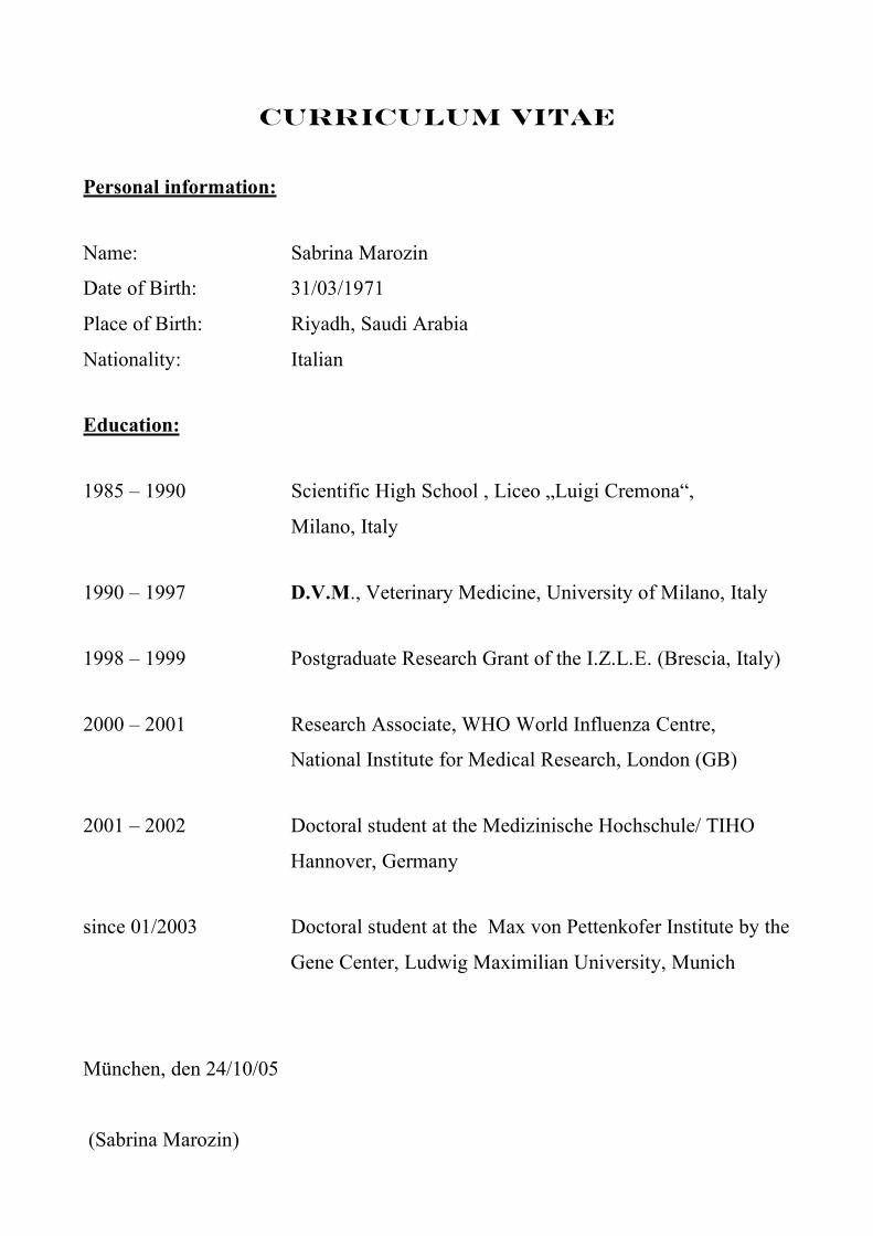
CURRICULUM VITAE
Personal information:
Name: Sabrina Marozin
Date of Birth: 31/03/1971
Place of Birth: Riyadh, Saudi Arabia
Nationality: Italian
Education:
1985 – 1990 Scientific High School , Liceo „Luigi Cremona“,
Milano, Italy
1990 – 1997 D.V.M., Veterinary Medicine, University of Milano, Italy
1998 – 1999 Postgraduate Research Grant of the I.Z.L.E. (Brescia, Italy)
2000 – 2001 Research Associate, WHO World Influenza Centre,
National Institute for Medical Research, London (GB)
2001 – 2002 Doctoral student at the Medizinische Hochschule/ TIHO
Hannover, Germany
since 01/2003 Doctoral student at the Max von Pettenkofer Institute by the
Gene Center, Ludwig Maximilian University, Munich
München, den 24/10/05
(Sabrina Marozin)
ACKNOWLEDGMENTS
I would like to thank Prof. Dr. Karl-Klaus Conzelmann for giving me the opportunity to carry out
this research work at the Max-von-Pettenkofer Institute in the Gene Center and Dr. Stefan Finke for
his patience in answering many of my questions during these years.
My Doktorvater Prof. Dr. Eckhard Wolf for his advices and support, a heartfelt “thank you”!
Dr. Corinna Mörth for her help during decisive stages of this work. My very dear Co without your
big heart I couldn’t have made it…in any sense.
I am particularly grateful to Dr. Sharareh Moghim for providing me the Vero-p125Luc cell line and
for the helpful discussions. Thank you Sheri, your friendship has been a real reward.
A big thank you to Dr. Heidi Feldmann (the sunrise lady), for her warm-hearted support and the
lovely company.
All my appreciation to Dr. Alan Hay, Dr. Keith Camerun, Dr. Yi Pu Lin and Vicky Gregory for all I
learnt from them, for guiding me during my first steps and for your never-ending care.
A te Annalisa, che mi sei cara come una sorella, tutto il mio affetto e la mia gratitudine.
A very special thank to Karen and Natalie, so far away but always so close. Love you deeply!
Massimo e Mose’, vi voglio tanto bene. Cosa ne sarebbe stata della mia anima senza di voi: grazie!
Mein lieber “grumpy” Wolfgang, ich danke Dir für die unterhaltsamen und bereichernden
Gespräche auf unserem roten Sofa
To all of you: Alison and Rob, Claire (the Dolmetscherin), Claudia, Costas, Elena, Gerrit Mörth
(scent of a woman), Jeannette and Sven, Karim (Jones), Katharina, Klaus (the charming jogger),
Isaac and Valerio, Luca, Monica, Philip, Roberto (Mr. Rob), Sabine (Schnecki), Tung.
E alla fine, tenendo il meglio per ultimo, vorrei ringraziare voi Mamma e Papa’, voi che siete il mio
piu’ prezioso tesoro: infinita fonte di sostegno. Vi offro un umile ringraziamento per tutto l´ amore
che mi avete dato in questi anni.