Interdisziplinäre Behandlung von Meningeomen des Sinus ... · between the uniform thick muscular...
84
Aus dem Zentrum für Operative Medizin des Fachbereichs Medizin der Philipps-Universität Marburg Klinik für Neurochirurgie Direktor: Prof. Dr. med. H. Bertalanffy in Zusammenarbeit mit dem Universitätsklinikums Gießen und Marburg GmbH, Standort Marburg Interdisziplinäre Behandlung von Meningeomen des Sinus Cavernosus (Interdisciplinary Management of Meningioma Involving the Cavernous Sinus) Inaugural-Dissertation zur Erlangung des Doktorgrades der gesamten Humanmedizin dem Fachbereich Medizin der Philipps-Universität Marburg vorgelegt von Ahmed Farhoud aus Alexandria, Ägypten Marburg 2006
Transcript of Interdisziplinäre Behandlung von Meningeomen des Sinus ... · between the uniform thick muscular...

Aus dem Zentrum für Operative Medizin des Fachbereichs Medizin der
Philipps-Universität Marburg
Klinik für Neurochirurgie
Direktor: Prof. Dr. med. H. Bertalanffy
in Zusammenarbeit mit dem Universitätsklinikums Gießen und Marburg
GmbH, Standort Marburg
Interdisziplinäre Behandlung von Meningeomen des Sinus
Cavernosus
(Interdisciplinary Management of Meningioma Involving the Cavernous Sinus)
Inaugural-Dissertation zur Erlangung des Doktorgrades der gesamten
Humanmedizin dem Fachbereich Medizin der Philipps-Universität Marburg
vorgelegt von
Ahmed Farhoud aus Alexandria, Ägypten
Marburg 2006

2
Angenommen vom Fachbereich Medizin der Philipps-Universität Marburg
am 01.06.2006
Gedruckt mit Genehmigung des Fachbereichs
Dekan: Prof. Dr. B. Maisch
Referent: Prof. Dr. H. Bertalanffy
Korreferentin: Prof. Dr. R. Engenhart-Cabillic

3
Contents
1. Review of the literature…………………………………………………….. 8
1.1 Terminology………………………………………………………………8
1.2 Anatomy…………………………………………………………………..8
1.2.1 Osseous Relationships……………………………………………...9
1.2.2 Dural Relationships……………………………………………….11
1.2.3 Arterial Relationships……………………………………………..12
1.2.4 Venous Relationships……………………………………………..14
1.2.5 Neural Relationships……………………………………………...15
1.3 Pathology………………………………………………………………...17
1.4 Clinical Presentation……………………………………………………..18
1.5 Radiological Investigations……………………………………………...19
1.5.1 Magnetic resonance Imaging……………………………………...19
1.5.2 Computed Tomography…………………………………………...19
1.5.3 Cerebral Angiography………………………………………….…19
1.5.4 Plain Skull Radiography…………………………………………..19
1.6 Management……………………………………………………………..20
1.7 Surgery ...………………………………………………………………..20
1.7.1. Principles of Cavernous sinus Surgery…………………………...20
1.7.2 Surgical Approaches to the Cavernous Sinus…………...………...21
1.7.3 Complications of Surgery in the parasellar region …………….....22
1.8 Radiation Therapy ...…………………………………………………....23
1.8.1 General Principles………………………………………………...23
1.8.2 Radiation Physics………………………….……………………...23
1.8.3 Fractionated Stereotactic Radiotherapy....………………………...24
1.8.4 Stereotactic Radiosurgery………………………………….……...25
1.8.5 Post-irradiation Complications……………...…………….……....25

4
Part I : Anatomical Study of the Cavernous Sinus
2. Objective………….…………………………………………………………27
3. Material and Methods……………………………………………………...27
3.1 Specimens…….………………………….……………………………....27
3.2 Instruments……………………………………….……………………...27
3.3 Technique…………….…………………………….……………………28
4. Results……………………………………………………………………….31
5. Discussion ……………..……………………………………...…………….35
Part II : Clinical Study of patients with Cavernous Sinus Meningioma
6. Objective………….…………………………………………………………40
7. Patients and Methods………………………………………………………40
7.1 Patients’ Selection………….……...…………………………………….40
7.2 Treatment Planning..…….……………………………………….……...41
7.3 Surgery………………….……………………………………….………41
7.4 Radiation Therapy…….……………………………………….………...41
7.5 Follow-up…………….………………………………………….………42
7.6 Quality of Life Assessment.……………………………………….…….42
7.7 Statistical Analysis.……………………………………………….……..43
8. Results……………………………………………………………………….44
8.1 Patients’Demographics……….…………………………………….……44
8.2 Tumor Characteristics………….…………………..……………………44
8.3 Clinical Findings………………………………………………………...44
8.4 Surgery…………………………………………………………………..44
8.4.1 Extent of resection………………………………………………...47
8.4.2 Surgical approach………….……………………………………...47

5
8.4.3 Post-operative complications……………………………………..47
8.4.4 Outcome of surgery……………………………………………….52
8.5 Fractionated Stereotactic Radiotherapy……………………….…………52
8.5.1 Post-irradiation side effects……………………………………….52
8.5.2 Outcome of radiotherapy………………………………………….52
8.6 Quality of Life Assessment.……………………………….…………….55
9. Discussion…………………………………………………………………...58
9.1 Patients’Demographics……….…………………………………….……58
9.2 Clinical Findings ………….…………………..………………………...58
9.3 Tumor Characteristics. …………..……………………………………...59
9.4 Surgery…………………………………………………………………..60
9.4.1 Extent of resection………………………………………………...60
9.4.2 Surgical approach………….……………………………………...61
9.4.3 Outcome and complications……………………………………....62
9.5 Fractionated Stereotactic Radiotherapy……………………….…………64
9.6 Quality of Life Assessment.……………………………….…………… 65
10. Conclusion....................................................................................................67
11. Zusammenfassung.......................................................................................68
12. References....................................................................................................70
13. Acknowledgement........................................................................................81
14. Lebenslauf....................................................................................................82
15. Verzeichnis der akademischen Lehrer......................................................83
16. Ehrenwörtliche Erklärung.........................................................................84
6
Abbreviations
ACP = Anterior clinoid process
COZ = Cranio-orbital zygomatic approach
CS = Cavernous sinus
CSF = cerebrospinal fluid
CT = Computed tomography
DNA = Deoxyribonucleic acid
Gy = Gray
ICA = Internal carotid artery
ICP = Intracranial pressure
IOF = Inferior orbital fissure
Linac = Linear accelerator
LSC = Lateral sellar compartment
MRI = Magnetic resonance imaging
PCP = Posterior clinoid process
SOF = Superior orbital fissure
SRS = Stereotactic radiosurgery
SRT = Fractionated stereotactic radiotherapy

7
Interdisciplinary Management of Meningioma
Involving the Cavernous Sinus
Ahmed Farhoud, M.D
A Thesis Submitted In Partial Fulfillment of the Requirements
for the Degree of Doctor of Medicine,
Faculty of Medicine
Philipps University, Marburg

8
1. Review of the literature
1.1 Terminology:
The cavernous sinus (CS) can be defined as the dural envelope in which the cavernous
segment of the internal carotid artery (ICA) courses. This dural envelope is also the site of
a venous confluence that receives multiple veins draining the orbit, Sylvian fissure,
anterior and middle cranial fossae. The oculomotor, trochlear, ophthalmic and abducens
nerves course in this sinus. Due to the intricate and compact neurovascular contents of the
CS, it was known as “The anatomical jewel box” [Harris 1976, Parkinson 1990].
It is now well known that the old term “Cavernous Sinus” suggested by Winslow is a
misnomer. It is only used as a historical term referring to this envelope surrounding the
ICA. This compartment, often referred to nowadays as the “lateral sellar compartment”
(LSC), is not a dural sinus nor is it cavernous. There is no histological resemblance
between the uniform thick muscular walled venous sinusoids of the corpora cavernosa that
are devoid of any adipose tissue and the extremely variable, extremely thin walled venous
spaces associated with adipose tissue in LSC [Eisenberg 2000, Parkinson 1998].
There is also a clear difference between the other dural sinuses encased between the
periosteal and meningeal dural layers (with the exception of inferior sagittal and straight
sinuses which lie between opposing layers of meningeal dura) and have no associated
adipose tissue and this extradural venous space with its arterial, nervous and fatty contents
[Krivosic 1987, Parkinson 1998].
Many authors consider this compartment an enlarged segment of the extradural neural
axis compartment extending from the coccyx to the orbit. Through this compartment run
arteries, myelinated and unmyelinated nerves, and valve-less veins. Adipose tissue is
present in different amounts being abundant in the orbit and spinal segments and sparse in
the skull base segment [Parkinson 2000, Weninger 1997].
1.2 Anatomy
The CS is a paired structure of considerable size extending along the lateral aspects of the
body of the sphenoid bone from the superior orbital fissure (SOF) anteriorly to the petrous

9
apex posteriorly. It measures in adults 1.2-1.8 cm in length and 0.7-1.6 cm in width
[Harris 1976, Lang 2004].
1.2.1 Osseous relationships:
The CS sits on the lateral aspect of the body of the sphenoid bone. It extends posteriorly to
the junction between the petrous apex and the body of sphenoid, known as the petro-clival
fissure, and to the lateral edge of the dorsum sellae, which forms the posterior border of
the sella. It extends downwards and laterally to the junction between the body and greater
wing of sphenoid. On either side of the sphenoid body is a groove along which the ICA
courses. This groove, known as the carotid sulcus, begins at the intracranial end of the
carotid canal, turns forward and upward in a serpiginous course to end medial to the
anterior clinoid process (ACP) [Rhoton 2002, Parkinson 1990].
The ACP represents the distal extremity of the lesser wing of sphenoid and also forms the
lateral wall of the intra-cranial end of the optic canal. This process is usually solid but may
contain air cells communicating with the sphenoid sinus. It is formed of weak cancellous
bone surrounded by a dense surface of cortical bone (fig 1).The controlled removal of
ACP is considered a corner stone in the surgical treatment of CS lesions as it covers the
anterior 1/3 of the roof of the sinus [Lang 2004, Rhoton 2002].
Another small prominence, the middle clinoid process, projects upwards from the terminal
part of the carotid sulcus. Sometimes a bridge of bone between the anterior and middle
clinoid processes converts the distal end of the carotid sulcus into an ostium called the
carotico-clinoid foramen [Borba 2000, Erturk 2004]. The dorsum sellae terminates at each
superior angle in a tubercle known as the posterior clinoid process (PCP). This process is
connected to the ACP and petrous apex through the inter-clinoid dural fold and posterior
petroclinoid ligament respectively. An interclinoid osseous bridge between the anterior
and posterior clinoid processes, found in 4-9% of cases, makes it sometimes difficult to
remove ACP extradurally [Borba 2000, Rhoton 2002].
Although other bony landmarks are not directly involved in the formation of the CS, they
constitute important landmarks during surgery as the pterion, foramen ovale, foramen
rotundum, foramen spinosum, cochlea, arcuate eminence and the carotid canal [Borba
2000, Lang 2004].

10
Fig 1. Osseous relationships of the cavernous sinus. A superior view of the sphenoid bone
showing the planum sphenoidale, dorsum sellae, the anterior and middle clinoid
processes, the carotid sulcus and the different foramina in this region. (From Rhoton
2002).

11
1.2.2 Dural relationships:
Each CS has 4 walls: lateral, medial, posterior, and a roof or superior wall. The lateral and
medial walls join anteriorly along the SOF and below along the upper border of the
maxillary nerve to form a narrow edge resembling the keel of a boat. The SOF is situated
between the greater and lesser wings of sphenoid. It is triangular in shape and provides a
communication between the orbit and the middle cranial fossa [Rhoton 2002].
The walls of CS are formed by the dura lining the inner surface of the skull. This dura
consists of 2 layers: an outer endosteal layer lining the bone and an inner meningeal layer
facing the brain. At the upper border of the maxillary nerve (the lower border of CS) these
2 layers separate, the meningeal layer extends upward to form the outer thick layer of the
lateral wall of the sinus, then wraps around the anterior petroclinoid fold, and extends
medially to form the roof of the sinus then the upper layer of the diaphragma sellae. The
endosteal layer divides into 2 layers: the first adheres to the sphenoid bone covering the
carotid sulcus and the floor of the sella while the second extends upward to form the inner
membranous layer of the lateral wall, which can be considered as the epineurium of the
cranial nerves traversing the lateral wall. [Borba 2000, Kawase 1996, Rhoton 2002].
The posterior wall extends from the petroclival fissure inferiorly to the posterior
petroclinoid dural fold superiorly, and between the lateral margin of the dorsum sellae
medially and the ostium of the Meckel’s cave laterally. The posterior wall is the site of a
venous confluence between the cavernous, basilar, superior petrosal and inferior petrosal
sinuses [Rhoton 2002]. The medial wall can be divided into two parts: sellar and
sphenoidal. The sellar part separates the sella and the pituitary gland from the venous
spaces in the sinus. The sphenoidal part is formed by the dura lining the carotid sulcus on
the lateral aspect of the sphenoid body [Yasuda 2004].
These dural walls constitute a natural barrier against tumor invasion. However CS has
some weak points where tumors can penetrate the sinus, namely: the venous plexus
around the SOF, the sellar part of the medial wall, and the meningeal pockets of the
cranial nerves [Kawase 1996].

12
1.2.3 Arterial relationships:
The ICA enters the skull through the carotid canal located in the petrous portion of the
temporal bone. On leaving the carotid canal, the artery ascends to enter the CS under the
petro-lingual ligament. This ligament is a continuation of the periosteum of the carotid
canal connecting the petrous apex to the lingula of the sphenoid bone. It constitutes the
postero-inferior attachment of the lateral wall of the CS and can be considered as the point
where the petrous segment of the carotid artery terminates [Borba 2000, Ziyal 1998].
After penetrating the postero-inferior CS, the artery ascends towards the PCP. This
posterior vertical segment passes under the Gasserian ganglion and ends when the artery
turns anteriorly forming the posterior bend. The ICA then courses anteriorly in a
horizontal direction for approximately 2 cm along the carotid sulcus found on the lateral
aspect of the sphenoid body. This horizontal segment is the longest part of the cavernous
segment of the artery. This segment ends when the artery curves upwards forming the
anterior bend. The ICA passes then upwards medial to ACP to perforate the roof of the CS
forming the anterior vertical segment (fig 2) [Harris 1976, Van Loveren 1996].
This anterior vertical segment, usually known as “clinoid segment”, is obscured by ACP.
It is limited proximally and distally by dural rings. The proximal or lower dural ring is
formed by the medial extension of the dura lining the inferior surface of the ACP. This
dural fold is called the “carotid-oculomotor” membrane because it separates the lower
margin of the ACP from the oculomotor nerve and then extends around the artery. The
upper or distal dural ring is formed by the medial extension of the dura lining the upper
surface of the ACP [Seoane 1998, Van Loveren 1996].
The dura forming the proximal ring turns upward to form a fibrous collar around the
artery (carotid collar), which extends to the upper ring. The proximal ring and the lower
part of the collar are separated from the arterial wall creating a narrow space in which
course some venous channels continuous with the CS. In contrast, the distal ring is
adherent to the surface of the artery and serves as a barrier between intra- and extradural
spaces. Sectioning of these rings allows mobilization of the artery and entry into the
anterior CS [Seoane 1998, Van Loveren 1996].
The arterial branches that arise from the cavernous segment vary in origin and number (fig
2). The largest and most constant is the meningeo-hypophyseal trunk which arises

13
Fig 2. Enlarged view of the intracavernous ICA and its branches. The ophthalmic nerve is
depressed to expose the abducens nerve. The dorsal meningeal artery passes backward
toward Dorello’s canal and the clivus. The inferolateral trunk passes above the 4th nerve
and gives off several branches; one of them is heading to the foramen rotundum. Note that
the petrosphenoid ligament (Gruber’s ligament) roofs the Dorello’s canal in which the 6th
nerve courses. Cav. Seg; cavernous segment, inf. Lat. Tr; inferolateral trunk, dors Men a.;
dorsal meningeal artery. (From Rhoton 2002).

14
typically from the outer wall of the posterior bend and divides shortly into 3 branches: the
tentorial artery, inferior hypophyseal artery, and dorsal meningeal artery [Rhoton 2002].
The tentorial artery (artery of Bernasconi Cassinari) exits the sinus and courses postero-
laterally to supply the tentorium. It represents the main blood supply to tentorial
meningiomas. The inferior hypophyseal artery passes medially to the posterior pituitary
capsule and lobe, and anastomoses with its mate from the opposite side after supplying the
dura of the sellar floor. The dorsal meningeal artery passes posteriorly with the abducens
nerve to supply the dura along the upper clivus (figs 2 & 3) [Taptas 1982].
The second branch of the cavernous carotid is the artery of the inferior cavernous sinus,
also called the infero-lateral trunk. It arises from the central third of the horizontal
segment, and then passes downwards above or below the 4th nerve to supply the dura of
the infero-lateral wall of the CS and the area of foramina rotundum and ovale (fig 2).
McConnell’s capsular arteries arise from the medial side of the horizontal segment and
pass to the capsule of the pituitary gland where they anastomose with the opposite arteries
[Borba 2000, Rhoton 2002].
One or more of these branches can be absent as a normal variation. Another rare variation
is the persistence of the fetal trigeminal artery which arises from the posterior bend and
courses posteriorly to join the basilar artery. In this case the branches of the meningeo-
hypophyseal trunk arise from the trigeminal artery. The ophthalmic artery arises just
above the upper ring outside the CS, but in 8% of individuals it arises within the CS
[Seoane 1998, Suttner 2000].
1.2.4 Venous relationships:
The nature of the venous cavity known as the CS has been a point of debate. Several
authors suggested that the CS is an irregular network of irregular valveless veins forming
a part of the extradural venous network. These thin walled veins are surrounded by
adipose tissue, which is also a part of extradural fat. Others asserted that the CS is not a
venous plexus but a trabeculated venous channel. Recent surgical explorations show that
the CS is more consistent with a venous channel than a venous plexus. [Borba 2000,
Dolenc 1989, Parkinson 1998, Rhoton 2002, Taptas 1982]
The CS is connected to the orbit by the superior and inferior ophthalmic veins, to the
cerebral hemispheres through the middle and inferior cerebral veins, to the retina by the

15
central retinal vein, to the dura by tributaries of the middle meningeal veins, to the
transverse sinus via the superior petrosal sinus, to the jugular bulb through the inferior
petrosal sinus, to the pterygoid venous plexus by emissary veins and to the facial veins
through the ophthalmic veins [Harris 1976, Lang 2004].
Variable venous connections between both cavernous sinuses lie within the sella and are
termed inter-cavernous sinuses. These trans-sellar connections may exist at any point of
the sella and can be classified according to their relation with the pituitary gland into an
anterior group, passing anterior to the hypophysis and a posterior group, passing posterior
to the hypophysis. The anterior sinuses are usually larger than the posterior ones.
However, either the anterior or the posterior connections or both may be absent. If they
co-exist, they form a venous ring around the gland that is sometimes known as “circular
sinus” [Rhoton 2002].
1.2.5 Neural relationships:
The oculomotor, trochlear, trigeminal, and abducens nerves have intimate relations with
the CS (fig 3). The oculomotor, trochlear and ophthalmic nerves course between the two
layers of lateral wall of the CS before entering the SOF. On the other hand the abduscens
nerve passes inside the sinus on the lateral side of the ICA on his way to the SOF [Borba
2000, Dolenc 1989, Umansky 1982]. Another nervous structure inside the sinus is the
bundle of sympathetic fibers travelling on the surface of the carotid artery [Rhoton 1996].
The arterial supply of cranial nerves is frequently overlooked. The portions of cranial
nerves passing through the sinus receive vascular supply from multiple sources. Before
entering the sinus the nerves receive branches from the vertebro-basilar system. Within
the CS, the inferolateral trunk largely supplies the cranial nerves crossing the sinus [Krisht
1994, Rhoton 1996].

16
Fig 3. Anatomical dissection showing the full tortuous course of the intracavernous
carotid as well as the courses of the 3rd, 4th, and 5th cranial nerves coming from the
posterior fossa and passing through the cavernous sinus to their exit foramina. Men. Hyp.
A.; meningeohypophyseal artery, V1; ophthalmic division, V2; maxillary division, V3;
mandibular division (From Rhoton 2002).

17
1.3 Pathology
The complex relation of the neural, endocrine, vascular, meningeal, and osseous tissues in
the parasellar region provides a wide range of pathological probabilities in this small
anatomical compartment. The common neoplastic processes which involve the CS
include: meningioma, pituitary adenoma, neurinoma, and craniopharyngioma. Other
neoplasms are less commonly encountered as chordoma, germinoma, haemangioma,
dermoid and epidermoid cysts, osteocartilagenous tumors, lymphoma, nasopharyngeal
carcinoma, metastases, juvenile angiofibroma, and teratoma [Dolenc 1993, Eisenberg
1999, Tindall 2004, tobias 2001, Vogl 2003, Vrionis 2004].
Meningioma is known to be the most common tumor to involve the CS [Dolenc 1993,
Cusimano 1995]. Meningioma involving the CS can arises from inside the sinus or more
commonly, it takes origin from a neighboring structure and invades the sinus. This can
occur in meningiomas having their origin from medial sphenoid ridge, tuberculum sellae,
petroclival dura, olfactory groove, optic nerve sheath, petrous apex, dorsum sellae and
PCP. The name of each type refers to the site of primary attachment. Most of these tumors
are located both intra- and extradurally [Al-Mefty 2000, Eisenberg 1999].
Meningiomas are extra-axial tumors thought to arise from the arachnoidal cap cells. They
constitute approximately 20% of all primary intracranial tumors. These tumors, known to
be more prevalent in females, grow usually as globular lobulated masses, or less
commonly “en plaque” with a very broad dural attachment. Although meningiomas are
typically benign, they occasionally behave in an aggressive fashion, a variant commonly
referred to as atypical and anaplastic meningioma [Black 1993, Lamszus 2004]. Several
classifications for meningioma are reported in the literature based on histological,
etiological and surgical criteria. A widely used classification for neurosurgeons is the
classification of meningiomas according to the site of origin as proposed by Cushing and
Eisenhardt [Cushing 1938].
Several non-neoplastic pathological processes can present as space occupying lesions
involving the CS and must be included in the differential diagnosis of parasellar masses
[Dolenc 1993, Vogl 2003]. These include:
Vascular lesion as giant cavernous carotid aneurysm and carotid-cavernous fistula
Inflammatory lesions: - infectious
18
- mucocele
- osteomyelitis
- sarcoidosis
- amylodosis
- lymphocytic hypophysitis
- cholesterol granuloma
- histocytosis x
Idiopathic cranial polyneuropathy (Tolosa-Hunt syndrome).
1.4 Clinical presentation
The clinical presentation of CS tumors can be divided into general signs and symptoms of
increased intracranial pressure (ICP) as well as specific signs and symptoms related to the
juxtasellar region. The increased ICP can be the result of the size of the lesion or of the
associated hydrocephalus if the tumor has a suprasellar extension enough to obstruct the
foramen of Monro [Tindall 2004].
Cranial nerves dysfunction is among the common presentations of CS tumors. This results
either from direct compression or infiltration of the nerves. Diplopia due to partial or
complete paralysis of the 3rd, the 4th, or the 6th cranial nerves is a common symptom.
Ptosis and total ophthalmoplegia can also take place. The 1st and 2nd divisions of the
trigeminal nerve may be also affected resulting in numbness or neuralgia [Tindall 2004].
Optic nerve involvement in the form of decreased visual acuity, or visual field defect is
also common. The type of visual impairment depends upon the origin and the direction of
growth of the tumor. For instance, tuberculum sellae meningioma usually causes
bitemporal hemianopsia, whereas a clinoidal meningioma tends to cause an ipsilateral
optic atrophy and visual loss, then contralateral papilledema (Foster Kennedy syndrome).
Other cranial nerves palsies can also be encountered as anosmia in olfactory groove
meningioma, facial, vestibulocochlear, and lower cranial nerves palsies in petroclival
meningioma [Al-Mefty 2000].
Symptoms related to brain compression (e.g. frontal or temporal lobes) and hypothalamic-
pituitary axis dysfunction are also important to be evaluated before management planning
[Al-Mefty 2000].

19
1.5 Radiological investigations
1.5.1 Magnetic Resonance Imaging (MRI)
The inherent ability of MRI to delineate soft tissue makes it the imaging modality of
choice in the diagnosis of parasellar masses. It is superior in demonstrating the brain, the
cranial nerves and the carotid artery especially in T1-weighted images. Paramagnetic
intravenous contrast agents as gadolinium-DTPA are now used routinely in the study of
parasellar pathology. It accumulates in the vascular and interstitial spaces of tissues that
do not have an intact blood brain barrier as the pituitary gland and the CS. Unlike
contrast-enhanced CT scan, rapidly flowing arterial blood does not enhance. Gadolinium
enhancement helps to define the type, extent, and the vascularity of the lesion [Chakeres
1989, Guy 1991, Hirsch 1993].
1.5.2 Computed Tomography (CT) Scan
CT scan is complementary with MRI in the diagnosis of skull base lesions. Although MRI
is superior in delineating soft tissues, CT scan is superior in defining the bones of the skull
base and in detecting calcification and hyperostosis. CT examination of the skull base
requires thin sections (3mm), axial and coronal projections, soft tissue and bone
algorithms, and the administration of an intravenous iodinated contrast agent [Guy 1991,
Hirsch 1993, Angtuaco 2000].
1.5.3 Cerebral Angiography
Despite its invasive nature, angiography remains an important part of the pre-operative
evaluation of patients with CS lesions. It is the gold standard for defining the vascular
lesions. In the evaluation of neoplastic lesions, the carotid angiogram is the best test for
defining the degree and location of involvement of the carotid artery by the neoplasm.
Angiography reveals also the source of vascularity of the neoplasm, allowing for potential
pre-operative embolization [Angtuaco 2000].
1.5.4 Plain Skull Radiography
With the advent of more sophisticated imaging techniques, plain x-ray of the skull became
of limited value in the diagnosis of parasellar tumors. Its main role is the detection of

20
pathological alterations in the bones as ballooning of the sella, hyperostosis, and bony
erosion [Hirsch 1993].
1.6 Management
The CS was considered for many years to be a no-man’s land because of its complex
anatomy and the ominous rate of post-operative morbidity and mortality in this region.
However, this inoperability was seriously challenged in the past 2 decades through the
significant improvements in diagnostic and microsurgical techniques. Since Parkinson’s
trial in the early sixties, the CS has become a surgical hot spot and several surgical
approaches developed since then that made CS lesions surgically accessible [Dolenc 1983,
Hakuba 1989, Kawase 1985, Parkinson 1965, Sekhar 1989].
A similar huge development took place in the past few decades in the techniques of
radiotherapy. The development of stereotactic methods for the planning and delivery of
therapy allowed accurate and focused treatment to reach the tumor in larger doses without
endangering the surrounding normal brain tissue. This permitted the use of high-precision
radiation therapy as an adjuvant post-operative modality or even as an alternative
modality to microsurgery in some cases [Shin 2001, Dufour 2001].
The choice of one treatment modality or the combination of both of them depends upon
several factors as the general condition of the patient, the clinical presentation, the size of
the tumor and the involvement of critical structures as the optic apparatus or the brain
stem [Dufour 2001, O’Sullivan 1997, Shin 2001].
1.7 Surgery
1.7.1 Principles of surgery in the CS
The evolution of the cranial base surgery has led to several general principles that must be
considered in the microsurgical treatment of CS tumors [Al-Mefty 2000, Hakuba 1989,
Inoue 1990, Kawase 1985, Sekhar 1989, Van Loveren 1996]:
1- Wherever possible, bone removal is preferred to avoid brain retraction.
2- The use of preexisting avenues or apertures in the skull reduces the time of surgery
and gives better cosmetic outcome.
21
3- Large craniotomies are preferred because the target area is deep and whenever
possible should be done as a single flap for better cosmetic results.
4- The distance traveled from the skin to the lesion should be kept to a minimum.
5- The cranial nerves inside the CS can be affected by traction or ischemic injury during
surgery. Therefore, these nerves should not be separated anteriorly but dissected as one
nerve bundle. Special care should also be paid to locate the 4th nerve posteriorly under the
tentorial fold and to dissect the 5th nerve from the dura around the foramen rotundum and
ovale.
6- Proximal and distal control of the ICA is an essential step. Proximal control can be
obtained in the neck or petrous bone while the ophthalmic segment is usually used for
distal control.
1.7.2 Surgical Approaches to the Cavernous Sinus
Despite the variety of approaches used nowadays to expose the CS, surgical access to this
area remains a formidable task. Access to the CS has been usually accomplished through
variations of frontal, frontotemporal, or temporal craniotomy. Various modifications to
these basic approaches enable the exposure of different regions of the CS. Advances in
microsurgical techniques and instrumentation have offered another route to access the
central skull base including the CS, namely the transfacial approaches. These include
several approaches, as transsphenoidal, transmaxillary, and mid-facial degloving
approaches, which can offer access to the CS from below [Al-Mefty 2000, Dolenc 1983,
Cavallo 2005, Couldwell 1997, Feiz-Erfan 2005].
Generally, approaches to the CS can be classified into two groups; the conventional
approaches and the skull-base approaches. The traditional conventional approaches
include the subfrontal approach, the frontotemporal transsylvian approach and the
subtemporal-intradural approach, whereas the more recent skull-base approaches include
Dolenc Approach, Kawase approach, cranial-orbitozygomatic approach (COZ), combined
petrosal approach and extended trans-sphenoidal approach. The later group has the
advantage of minimizing brain retraction through removal of some parts of the bones of
the skull base. The choice of the surgical approach is dictated by the tumor type, pattern of
growth, and the aim of surgery [Al-Mefty 2000, Dolenc 1983, Hakuba 1989, Inoue 1990,
Kawase 1985, Sekhar 1989, Taha 1995, Yasargil 1984].

22
1.7.3 Complications of surgery in the parasellar region
In addition to the general complications which could be encountered with any brain
surgery as coagulopathy, sepsis, vasospasm, air embolism and pulmonary embolism, the
following complications can occur during CS surgery:
1- Vascular injury: injury of the ICA or one of its main branches should be dealt with
either by suturing or bypass grafting. Preserving the small perforators and the large
draining veins especially those of the temporal lobe is of utmost importance [Al-Mefty
2000, Sekhar 1987].
2- Cranial nerve injury: the nerves inside the CS are liable to injury during tumor
dissection. Although this complication can occur with any type of tumor, yet it happens
more frequently with meningioma. Repair of these nerves can be carried out either by
direct suture reconstruction or by fibrin glue coaptation of nerves stumps. Other cranial
nerves can be also at risk depending on the pattern of growth and the surgical approach.
On the other hand, post-operative cranial nerve palsies can occur even in the absence of
intra-operative nerve injury. This can be attributed to the effect of intra-operative
manipulation (neuropraxia) or to the disruption of the fine blood supply of the cranial
nerves during dissection [Al-Mefty 2000, Harrison 2000].
3- Injury to the optic apparatus: the more the optic nerve is stretched, the more vulnerable
it becomes even to the least manipulation. It is wiser sometimes to leave a part of the
tumor that it firmly attached to the optic nerve than to cause an irreversible visual loss.
Special care must be paid while drilling the ACP to avoid injury of the optic nerve either
mechanically or thermally [Patterson 1993].
4- Injury to the pituitary gland and pituitary stalk resulting in diabetes insipidus and
pituitary insufficiency [Patterson 1993].
5- Intra-operative brain swelling: this is rarely encountered nowadays due to the use of
skull base approaches which minimize brain retraction. This is preventable by good
anesthesia and by CSF drainage either through a preinstalled lumbar drain or by opening
the cisterns during surgery [Al-Mefty 2000, Patterson 1993].

23
1.8 Radiation Therapy
1.8.1 General principles
External beam radiation therapy has been demonstrated to be an effective adjunctive
therapy after subtotal resection of intracranial tumors and a useful primary treatment for
unresectable or inoperable tumors [Goldsmith 1994, Kokubo 2000, Nutting 1999].
External beam radiation therapy could be delivered in multiple fractions or in a single
fraction [Noel 1996]. Several authors have demonstrated relapse-free survival rates after
external beam radiotherapy for CS meningioma that are equal to the rates achieved by
radical resection. Results of radiotherapy are not instantaneous: several months elapse
before tumor regression takes place. Several months also elapse before the appearance of
radiation-induced complications. It is particularly important to realize that slight tumor
swelling may occur initially so that it can be misdiagnosed as treatment failure [Dufour
2001, Noel 1996, Shin 2001].
The primary cellular target for radiation is the deoxyribonucleic acid (DNA). The extent
of damage to DNA depends upon the radiation dose. Tissue response to irradiation is
related to the rate of proliferation, differentiation, and maturation of this tissue. Neurons,
due to their non-proliferative nature and high differentiation, are radioresistant at
therapeutic dose levels. However, very high doses in a single fraction may cause a direct
effect on neurons (i.e., neuronal degeneration). The endothelial cells may be, even in some
slow-growing benign tumors, the most rapidly proliferating cells. It is therefore quite
possible, but not yet shown, that vascular thrombosis and ischemia secondary to radiation-
induced endothelial damage may also be responsible for or at least contribute to tumor
death [Kondziolka 1999, Niranjan 2004, Noel 1996].
1.8.2 Radiation Physics
Two types of ionizing radiation are used for radiotherapy, either charged particle
irradiation or electromagnetic (photon) irradiation. The former consists of beams of
accelerated nuclei, such as hydrogen (protons), helium, carbon, and silicon generated in
cyclotrons or syncyclotrons. This densely ionizing form of radiation is characterized by a
high ability to transfer energy to the molecules with which it interacts. The amount of
transferred energy is inversely proportional to the velocity of the particle, thus, very fast
particles transfer little energy. As particles interact with tissues and give up energy, they

24
slow down until they reach a state where large amounts of energy are transferred in a short
path length. This effect is known as Bragg peak [Luxton 1993, Suh 2004].
Photon irradiation is formed of beams of high energy x-rays (generated from linear
accelerator) or gamma rays (generated from gamma units). On entering the patient’s body,
any single photon radiation beam begins with a region of lower dose called “build-up
region”, then the dose increases progressively until it reaches the depth of maximal dose.
This criterion allows radiation beams to spare superficial areas as skin and subcutaneous
tissue from the highest dose. After the beam reaches the depth of maximal dose (Dmax)
the dose decreases with any further depth. This is known as “fall off region” and results
from the attenuation of the beam by the intervening tissues [Luxton 1993, Noel 1996, Suh
2004]
The main physical disadvantage of photons, in comparison with charged particles, is that
the dose in photon beams reaches its maximum at relatively shallow depths.
Consequently, a significant dose is delivered to normal tissues, while a suboptimal dose
reaches the target. This disadvantage can be improved but not overcome by the use of
multiple beams [Luxton 1993].
1.8.3 Fractionated stereotactic radiotherapy (SRT)
This technique combines the precision of stereotaxy with the radiobiologic advantages of
dose fractionation. The total dose is divided into fractions delivered during multiple
sessions to reduce the risk of normal tissue injury. The irradiated field includes both the
target lesion and the surrounding parenchyma; however the later is kept to a minimum by
stereotactic measures. This technique takes advantage of the differential susceptibility to
radiation between normal and abnormal tissues. It constitutes an alternative treatment
modality for CS tumors especially those with diameters more than 3 cm [Debus 2001,
Schlegel 1993].
Linear accelerator (linac) is widely used as a delivery system in this type of radiation
therapy. The use of charged particles generators to perform SRT is limited to few centres
worldwide owing to the very high costs required to install and to maintain these systems
[Luxton 1993].

25
1.8.4 Stereotactic radiosurgery (SRS)
This term was first introduced in the 1950s by Lars Leksell [Leksell 1951]. It refers to the
non-invasive destruction of a precisely defined intracranial target using a high dose of
ionizing radiation during a single session. The aim of this computer-assisted procedure is
to induce a highly localized radiobiologic effect within the target while the surrounding
structures receive only an insignificant fraction of this dose. Therefore, while certain
lesions can be radioresistant (equally or less susceptible to radiation than adjacent normal
parenchyma), they will nonetheless respond to the high doses delivered by SRS
[Kondziolka 1999, Noel 1996].
Radiosurgery, in contrast to fractionated SRT, entails the use of a high dose of precisely
focused radiation delivered in a single session. Its effect on the surrounding normal tissue
is limited by the highly focused nature of radiosurgical beams. Whereas fractionated
radiotherapy is generally most effective in killing rapidly dividing cells, radiosurgery is
thought to arrest the cell division capability of target cells irrespective of their mitotic
activity, oxygenation, and inherent radiosensitivity. However, SRS should be reserved to
tumors measuring 2.5-3 cm in their greatest diameter to avoid potential complications
[Kondziolka 1999, Noel 1996].
Radiosurgery is commonly performed using a gamma unit; nevertheless, charged particles
generators and linacs can be also adapted to SRS [Luxton 1993].
1.8.5 Post-irradiation complications:
Complications following irradiation can be divided chronologically into 3 categories
corresponding to chronological brain reactions to therapeutic irradiation. These
complications can be divided according to their time of appearance into acute
complications, which occur during or shortly after treatment (i.e., few days up to 2 weeks
after irradiation), early delayed complications, which occur few weeks up to 3 months
after exposure, and late delayed complications whose onset begins several months up to
several years after exposure [Anscher 1996, McKenzie 1993].
Acute complications are generally of little clinical consequence and are generally
manifested as transient elevation of intracranial pressure with exacerbation of the
presenting symptoms. The most hazardous acute complication is acute radiation necrosis,
which refers to the acute destruction of irradiated brain tissue within hours or days of

26
exposure. This complication is extremely rare with the use of stereotactic techniques and
the proper irradiation dose [Anscher 1996, McKenzie 1993].
Early delayed complications appear few weeks or months after irradiation and may be the
result of irradiation-induced demyelination or abnormalities in myelin renewal and
maintenance resulting from glial cell damage. This is followed by spontaneous recovery
(remyelination). Early delayed effects may be clinically manifested as transient weakness,
lethargy and somnolence [Anscher 1996, McKenzie 1993].
Late delayed complications are the most hazardous sequelae of irradiation. They appear 6
months after exposure up to several years thereafter. Three main late reactions can be
encountered, namely vasogenic edema, radiation necrosis, and vasculopathy. Damage to
the retina, optic nerve and optic chiasm is another uncommon but serious complication of
tumor irradiation in the region of the CS. Damage to the hypothalamic-pituitary axis may
also take place especially with high radiation doses resulting in hormonal disturbances.
Additionally, intellectual deficits have been noted to develop in both adults and children
following cranial irradiation [Anscher 1996, Littley 1991, McKenzie 1993, Mulhern 1988,
Parsons 1983].

27
Part I: Anatomical Study
2. Objective
The aim of this part was to study the anatomical landmarks in and around the CS, their
variations, and the implications of these variations during surgery. Additionally, 3
different skull base approaches commonly used in CS surgery were performed in the
surgical laboratory in order to elaborate the optimal surgical exposure of the CS and to
identify the extent of exposure and limitations of each approach.
3. Material and Methods
3.1 Specimens
To fulfil the first aim of this work, 5 cadaveric heads (i.e. 10 cadaveric sides or
hemicrania) were used for laboratory anatomical dissection. They were provided by the
Anatomy Department, Philipps University, Marburg, Germany. Cadaveric heads were
fixed by a mixed solution of alcohol, glycerine, phenol and formol. Three specimens were
already fixed for a long time and could not be injected with silicon. The other two
specimens were relatively fresh and could be injected with a colored silicone rubber mix.
In this case, the big vessels (common carotid arteries, vertebral arteries and internal
jugular veins) were exposed in the neck and cannulated. Then, each vessel was irrigated
separately with warm tap water until all blood clots were washed out. Injection of colored
silicone follows using a 60 ml syringe (red mixture for the arteries and blue mixture for
the veins). Vessel clamps are used to clamp all vessels other than the one to be injected.
At the end, the silicone compound was allowed to harden overnight [Sanan 1999].
3.2 Instruments
All cadaveric dissections were performed in the surgical laboratory of the Neurosurgery
Department, Philipps University, Marburg, Germany. These procedures were designed to
mimic the actual surgical procedures, starting with head fixation in a Mayfield system,
followed by skin incision, craniotomy, and extra- and intradural dissection to expose the
parasellar region. A microscope (Zeiss), an electric drill (Aesculap) and a complete set of
micro-instruments were used.

28
3.3 Technique
Each cadaveric head was used to perform several skull base approaches commonly used to
reach the CS. The selected approaches were:
1- One piece cranio-orbital zygomatic approach (COZ)
2- Dolenc approach
3- Kawase approach
Each of these approaches was performed 10 times on 10 hemicrania. After the bony work
was performed and the bone flap was excised, we started to identify the deeper structures
using the same microsurgical technique as in live surgery. A special attention was paid to
the course of ICA and the cranial nerves while going in, passing through and going out of
the CS. The steps were documented by gross and microscopic photographs.
The first stage on each side was to perform COZ approach. The skin incision was the
same as a standard pterional approach, except that its lower end extends 1 cm below the
root of the zygomatic arch (fig 4). After subperiosteal dissection and exposure of the
superior and lateral aspects of the orbit, as well as the zygomatic bone, the temporalis
muscle was dissected and retracted inferiorly. The periorbita was bluntly dissected from
the bone with special care not to injure the supraorbital nerve and vessels. The keyhole
and a basal temporal burrhole were placed with 2-3 additional burrholes on the vertex, and
then 6 bony cuts were performed using a craniotome to create the one-piece COZ bone
flap. The first cut extended from the temporal burr hole to a point just lateral to the
supraorbital notch. This cut was also extended from the temporal burr hole to the keyhole
along the squamous temporal bone. The second cut extended from the orbital component
of the keyhole to the inferior orbital fissure (IOF) along the lateral wall of the orbit. The
3rd cut was made across the zygoma from the orbital edge to the IOF. This cut should
remain above the zygomaticofacial foramen to avoid entering the maxillary sinus. The 4th
cut was made across the posterior end of the zygomatic arch, while the 5th cut was an
extension of the 1st cut through the supraorbital ridge. The last cut connected the previous
one with the orbital component of the keyhole through the orbital roof. After completing
the bony cuts, the one- piece bone flap containing the superior and lateral orbital rims and
the zygomatic arch was carefully dissected from the dura (fig 5) [Abdel-Aziz 2002,
Rhoton 2002].

29
Fig 4. Important bony landmarks for COZ and Kawase approaches. EAC; External
auditory meatus
Fig 5. The resulting bone flap (COZ flap) including the traditional frontotemporal
(pterional) flap, a part of the orbital roof and the zygomatic arch.

30
The second stage was to perform Dolenc approach. The remaining part of orbital roof
after COZ was removed, and the orbitomeningeal dural fold was identified and cut. The
frontal and temporal dura were dissected from both sides of the sphenoid ridge, which was
drilled. The optic canal was also unroofed using a diamond burr. The ACP was then cored
by the drill, disconnected from the optic strut, and dissected free from the dura. After
removal of the ACP, the remaining optic strut was also drilled. Dissection of the temporal
dura from the skull base followed with identification of the 2nd and 3rd divisions of the
trigeminal nerve. At this stage, the roof and lateral wall of CS were exposed. The cranial
nerves and the ICA inside the sinus were identified. The artery was followed intradurally
after division of the proximal and distal dural rings. The optic nerve was identified
intradurally as well as the origin of the ophthalmic artery [Dolenc 1983, Van Loveren
1991].
The third stage was to perform Kawase approach. A horse-shoe skin incision was done
around the ear. The previous COZ craniotomy was extended posteriorly till above the
mastoid process (fig 4). The temporal dura was then elevated from the floor of the middle
fossa in a posterior to anterior direction. Division of the middle meningeal artery near the
foramen spinosum allowed further dural retraction till the greater superficial petrosal
nerve was identified and dissected from the dura. The arcuate eminence and foramen
ovale were identified together with the boundaries of Glasscock’s and Kawase triangles.
Glasscock’s triangle is defined by the greater petrosal nerve medially, the trigeminal
ganglion and the mandibular nerve anteriorly, and a line between the foramen spinosum
and the arcuate eminence of the petrous bone laterally. The ICA was exposed in this
triangle. On the other hand, Kawase triangle is bounded by the trigeminal ganglion, the
greater petrosal nerve, and the petrous edge. It is the target of bone removal in Kawase
approach. After drilling the petrous apex, the tentorium was divided to create a window to
the posterior cranial fossa and the ICA was followed inside the CS [Borba 2000, Kawase
1985, Rhoton 2002].
31
4. Results
As regards the results of the microsurgical anatomical dissection, the following findings
were noticed:
1. The supraorbital nerve and vessels, which are important in keeping the viability of
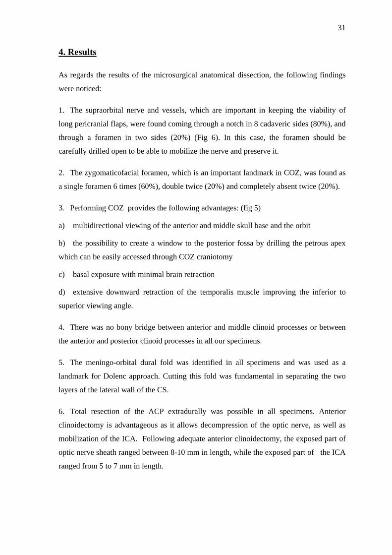
long pericranial flaps, were found coming through a notch in 8 cadaveric sides (80%), and through a foramen in two sides (20%) (Fig 6). In this case, the foramen should be
carefully drilled open to be able to mobilize the nerve and preserve it.
2. The zygomaticofacial foramen, which is an important landmark in COZ, was found as
a single foramen 6 times (60%), double twice (20%) and completely absent twice (20%).
3. Performing COZ provides the following advantages: (fig 5)
a) multidirectional viewing of the anterior and middle skull base and the orbit
b) the possibility to create a window to the posterior fossa by drilling the petrous apex
which can be easily accessed through COZ craniotomy
c) basal exposure with minimal brain retraction
d) extensive downward retraction of the temporalis muscle improving the inferior to
superior viewing angle.
4. There was no bony bridge between anterior and middle clinoid processes or between
the anterior and posterior clinoid processes in all our specimens.
5. The meningo-orbital dural fold was identified in all specimens and was used as a
landmark for Dolenc approach. Cutting this fold was fundamental in separating the two
layers of the lateral wall of the CS.
6. Total resection of the ACP extradurally was possible in all specimens. Anterior
clinoidectomy is advantageous as it allows decompression of the optic nerve, as well as
mobilization of the ICA. Following adequate anterior clinoidectomy, the exposed part of
optic nerve sheath ranged between 8-10 mm in length, while the exposed part of the ICA
ranged from 5 to 7 mm in length.

32
7. Dolenc approach provides access to both the roof and the lateral wall offering a wide
exposure of the CS and allowing the identification of all nervous and vascular structures
in the sinus extradurally.
8. The trochlear nerve crossed over the oculomotor nerve just before entering the SOF in
all cases (fig 7).
9. The abduscens nerve in the CS was observed as one rootlet in all specimens.
10. Identification of the middle meningeal artery and the greater superficial petrosal nerve
is fundamental when performing Kawase approach. The artery should be cut at the
foramen spinosum to allow further dural retraction and the nerve should be dissected from
the dura to avoid excessive facial nerve retraction (fig 9).
11. Kawase approach exposes only the posterior cavernous sinus and Meckel’s cave. It
also offers a window to the posterior fossa up to the level of the 7th and 8th nerves.
12. The meningeohypopheseal trunk could be identified 8 times arising from the posterior
bend of the ICA. The artery of the inferior CS could be identified only four times taking
origin from the horizontal segment of the cavernous carotid, 5-7 mm distal to the
meningeohypophyseal trunk. McConnell’s capsular arteries were not identified in any
specimen.
13. The ophthalmic artery arose in all hemicrania above the dura forming the roof of the
CS. Exposure of the artery required incision of the falciform ligament and mild retraction
of the optic nerve (fig 8).
14. The venous space located behind the posterior bend of ICA (the posterosuperior
venous compartment) was the largest venous compartment in all cases.

33
Fig 6. Periorbita carefully dissected with special care to the supraorbital bundle.
Fig 7. The nerves in the lateral wall of the cavernous sinus going into the superior orbital
fissure. Note that the 4th nerve crosses over the 3rd nerve anteriorly just before entering the
superior orbital fissure. SOF; Superior orbital fissure, Ophth; ophthalmic nerve.
SOF

34
Fig 8. The ophthalmic artery arising from the carotid artery and passing into the optic
foramen underneath the optic nerve. ICA; Internal carotid artery, optic; optic nerve.
Fig 9. Two important landmarks in Kawase approach: first the middle meningeal artery
which should be identified and cut at the foramen spinosum to allow further dural
retraction, and to the side, identification of the greater superficial petrosal nerve (arrow)
and its separation from the dura to avoid excessive traction on the facial nerve. This nerve
separates Glasscock’s and Kawase triangles

35
5. Discussion
Proper understanding of the anatomy of the CS and adjacent structures is the cornerstone
in performing surgery in this region. In order to study the anatomical variations and the
important landmarks in and around the CS, cadaveric dissection was carried out as an
independent part of this work. Knowing these variations and landmarks helps the surgeon
to perform a safer surgical procedure. From the wide variety of approaches which can be
used nowadays to expose the CS, three widely used approaches were chosen for the
anatomical study namely; COZ, Dolenc, and Kawase approaches [Al-Mefty 2000, Dolenc
1983, Kawase 1985]. The extent of exposure and limitations of each approach were
observed in order to optimize the surgical treatment offered to the increasing number of
patients harboring CS meningioma referred to the Neurosurgery department, Philipps
University, Marburg.
Pterional approach has been widely used to resect CS tumors as it is considered a simple
routine approach by most neurosurgeons [Yasargil 1984]. In the past 2 decades, several
authors advocated the use of COZ to approach CS tumors surgically [Al-Mefty 2000,
Inoue 1990]. Removal of the superior and lateral orbital rims and the zygomatic arch
combined with a frontotemporal craniotomy allows extensive mobilization of the
temporalis muscle inferiorly providing a good viewing angle (inferior to superior)
reaching up to the level of the foramina of Monro with minimal brain retraction. This can
not be achieved through a pterional craniotomy except with forceful retraction on the
temporal lobe. COZ provides additionally a good access to the adjoining orbit in case of
intra-orbital tumor extension. It also allows excellent exposure of the anterior and middle
cranial fossae as well as the area of the upper third of the clivus and posterior fossa
(through the petrous apex). Elevation of the whole bony construct as a single piece
ensures a better cosmetic outcome post-operatively [Abdel-Aziz 2002, Inoue 1990].
In any COZ craniotomy, the supraorbital nerves and vessels should be kept intact. This is
essential for keeping the viability of the pericranial flap that is commonly used for
reconstruction of the anterior and middle cranial base and for preventing post-operative
parasthesia of the forehead. The neurovascular bundle should be dissected from the
boundaries of the supraorbital notch while reflecting the pericranial flap, thus preserving
its attachment to the under surface of the pericranium. If the bundle was found to come
out of a foramen (as in 20% of our specimens), the foramen should be opened carefully

36
using a drill or a small chisel to allow retraction of the supraorbital bundle with the flap
[Al-Mefty 2000, Abdel-Aziz 2002].
During COZ, a bony cut across the zygoma extending to the lateral edge of the IOF is
required. It was suggested that the zygomaticofacial foramen, through which a branch of
the maxillary nerve (zygomaticofacial nerve) and its accompanying artery emerge in the
face, could be used as an external landmark for this cut by marking the superior edge of
the IOF. We found a single zygomaticofacial foramen in 6 out of 10 examined
hemicrania, while the foramen was absent in 20% and doubled in another 20% of
specimens. This is almost similar to the results reported by Martins et al., who found a
single foramen referring to the IOF in half of 102 hemicrania, while in 21.6% of examined
hemicrania the foramen was absent and in 28.4% two, three or even four foramina were
found. Accordingly, the zygomaticofacial foramen can be considered a reliable landmark
for locating the IOF and making the cut across the zygoma in only 50-60% of cases
[Martins 2003, Rhoton 2002].
The main step in Dolenc approach is the exradural resection of the ACP. To perform this
step, the surgeon should be aware of the attachments of this process to the sphenoid bone.
The ACP has 3 sites of attachment to the sphenoid bone: the base is attached anteriorly at
the medial edge of the sphenoid ridge and is attached medially to the anterior and
posterior roots of the lesser wing of sphenoid. The anterior root extends medially from the
base of ACP to the body of sphenoid bone above the optic nerve forming the roof of the
optic canal while the posterior root, called the optic strut, extends to the sphenoid body
below the optic nerve forming the floor of the optic canal. The strut separates the optic
canal from the SOF [Dolenc 1983, Hakuba 1989, Harris 1976].
In our study, extradural removal of the ACP was possible in all specimens. Although
some authors recommend the intradural en bloc removal of the ACP to avoid injury of the
optic nerve [Takahashi 2004], we share the opinion of others who consider extradural
anterior clinoidectomy safer, easier and less time consuming than the intradural one
[Bayassi 2005, Dolenc 1983]. There was no bony bridge between the anterior and middle
clinoid processes in any of our specimens. Keyes found this anomaly in 36% of his
specimens but Van Loveren et al. reported it in only 13% of their anatomical dissections
[Keyes 1935, Van Loveren 1996]. We did not also find an interclinoid osseous bridge
between the anterior and posterior clinoid processes in any of our specimens. This

37
anomaly was reported to be present in 4-9% of individuals [Borba 2000, Rhoton 2002].
The presence of one or both anomalies is an indication to continue anterior clinoidectomy
intradurally.
Though it is a relatively small structure, removal of the ACP provides enormous gain in
facilitating the management of lesions in the parasellar region and upper basilar artery
[Yonekawa 1997]. Adequate anterior clinoidectomy exposed 8-10 mm of the optic nerve
sheath and 5-7 mm of the proximal ICA in our dissections. These results go hand in hand
with the results of Evans and colleagues and Takahashi et al. Similarily, Youssef et al.
reported that anterior clinoidectomy and ICA mobilization can increase the carotid
oculomotor window by 44% anteriorly and 28% posteriorly, allowing therefore a much
better exposure of the upper basilar artery compared with the conventional transsylvian
approach [Evans 2000, Takahashi2004, Youssef 2004].
Removal of the ACP exposes a triangular space lined by the dura surrounding the process.
In this space, known as the clinoid space or triangle, the ICA can be identified surrounded
by the carotid collar and the proximal and distal rings. Although this segment of the ICA,
known as the clinoid segment, is considered by many authors extracavernous [Van
Loveren 1996, Ziyal 1998], we share the same opinion of Seoane et al. who consider this
segment a part of the cavernous carotid [Seoane 1998].
After anterior clinoidectomy, the CS can be entered through an incision in the clinoid
triangle extending posteriorly to the posterior clinoid process between the oculomotor
nerve and ICA [Inoue 1990, Van Loveren1996]. The other route of access is to mobilize
the two layers of the lateral wall to reach the space lateral to the artery commonly through
the space between the trochlear and ophthalmic nerves (Parkinson triangle) [Dolenc 1983,
Parkinson 1965].
A guiding landmark in Dolenc approach is the meningo-orbital fold. Incision of this fold
without damaging nervous structures in the SOF is essential to separate both dural layers
of the lateral wall of the CS. The meningo-orbital fold is a small extension of the external
layer of the dura mater of the middle cranial fossa which passes through the SOF and joins
the periorbita. The small artery passing through this fold is considered an important
reference point in recognizing the SOF [ Bayassi 2005, Van Loveren 1991].

38
By inspection of the cranial nerves inside the CS, we found the trochlear nerve crossing
over the oculomotor nerve in all our hemicrania just before entering the SOF. The same
was reported by Van Loveren et al. and Sekhar et al. [Van Loveren 1996, Sekhar 1987].
The later recommended that the incision in the lateral wall of the CS should not extend
more than 8 mm from the point of entry of the oculomotor nerve to the CS to avoid injury
of the trochlear nerve at this point. We also noticed that the abduscens nerve in the CS
consisted of a single rootlet. Kayalioglu et al. reported a variation in which the abduscens
nerve consisted of 2 or more rootlets in more than 20% of their dissections [Kayalioglu
1999].
Originally, Kawase developed his approach to access basilar trunk aneurysms, but he used
it later for tumors extending between the posterior and middle cranial fossae especially in
the region of the Meckel’s cave and posterior CS. Performing this approach, we were not
able to reach the anterior part of the CS. Nevertheless, we were able to reach the level of
the 7th and 8th cranial nerves in all specimens. Identification of some landmarks namely;
the middle meningeal artery, greater superficial petrosal nerve and the arcuate eminence is
essential to determine the target of drilling. The bone of the petrous apex does not contain
any organic structures, and is softer in consistency than the middle ear. This offers an
additional orientation tool during pyramid drilling [Kawase 1985, Kawase 1991].
The most consistent branch of the cavernous portion of ICA is known to be the
meningohypophyseal trunk. We were able to identify the trunk arising from the posterior
bend of the carotid in only 8 hemicrania (80%). Inoue et al. and Van Loveren et al.
reported the presence of the meningohypophyseal trunk in all their specimens while Borba
and Al-Mefty reported its presence in only 84% of their specimens [Borba 2000, Inoue
1990, Van Loveren 1996]. The second branch of the cavernous portion known as the
inferolateral trunk or the inferior artery of the CS was identified in our study only 4 times
(40%) arising from the horizontal segment of the cavernous carotid. This is much less than
the incidence found by Rhoton which was 84% [Rhoton 2002]. This may be due to the
fact that not all our specimens were injected with colored silicone, making it more
difficult to identify small branches. Preservation of the inferolateral trunk during surgery
is important as it represents the main blood supply of the cranial nerves inside the CS
[Krisht 1994]. We found the distance between the origins of the meningeohypophyseal
and inferolateral trunks ranging from 5-7mm, which is similar to the results reported by
Rhoton [Rhoton 2002]. Additionally, McConnell’s capsular arteries were not identified in
39
any of our specimens. Inoue et al. and Van Loveren et al. reported the infrequent presence
of these small branches in 8% and 28% of their specimens, respectively [Inoue 1990, Van
Loveren 1996].
The ophthalmic artery is one of the major branches of the ICA. Knowledge of variations
in the origin and course of this artery is important in performing surgery in this region.
The ophthalmic artery was found to be extracavernous originating distal to the distal dural
ring in all our specimens. This is different from other reports showing the origin of the
ophthalmic artery arising sometimes between both dural rings or from the intracavernous
ICA. However, these variations are known to be rare, occurring only in 3-8% of
individuals [Borba 2000, Rhoton 2002, Seoane 1998]. Exposure of the ophthalmic artery
required removal of the ACP and the posterior part of the orbital roof, incision of the
falciform ligament and slight retraction of the optic nerve.
Harris and Rhoton [Harris 1976] divided the venous space of the CS into four
compartments: medial, lateral, anteroinferior, and posterosuperior according to the
relation of each compartment with the ICA. In all our specimens, the venous space located
behind the posterior bend of ICA was larger than the other spaces offering a roomy space
for dissection. This is similar to other results reported in the literature [Borba 2000, Inoue
1990]. This space is particularly important in case of petroclival meningioma extending to
the CS as this compartment is the first to be filled with tumor.

40
Part II: Clinical Study
6. Objective
This retrospective study was conducted to assess the different modalities used in the
management of meningiomas involving the cavernous sinus in terms of indications,
efficacy and impact on post-treatment quality of life.
7. Patients and Methods
The second section of this work consists of a retrospective study including all patients
treated for CS meningioma between 1997 and 2003 in the Neurosurgery Department
and/or Radiation Oncology Department, Philipps University, Marburg, Germany, in order
to assess the outcome and post-treatment quality of life of these patients. During that
period, 104 patients with CS meningioma were treated in both departments.
7.1 Patients’ selection
The term CS meningioma refers in this study to all meningiomas involving the CS
whether arising from the sinus itself or initially arising from neighbouring structures and
invading the sinus. Meningioma was chosen because it is known to be the most common
tumor involving the CS, and to neutralize the impact of variation in the natural history of
the disease on the outcome if other types of tumors were also included. All cases who did
not receive any form of treatment (only follow-up of a small tumor causing mild or no
symptoms) were excluded from the study.
Histological verification of meningioma was available in all operated cases. In the non-
operated group, the diagnosis of meningioma was based only on radiographic findings,
that is, iso- to hypointense on T1 weighted MRI and hypo- to hyperintense on T2 weighted
MRI, homogenous enhancement, and the “dural tail sign” [Nagele 1994].
All pre-treatment radiographs were retrospectively reassessed and tumors were classified
into three grades according to Hirsch radiological criteria based on the involvement of the
ICA [Hirsch 1993]. In this classification, grade 1 refers to partial encasement of the ICA,
grade 2 refers to complete encasement without narrowing, and grade 3 refers to complete
encasement and narrowing of the artery.

41
7.2 Treatment planning
After thorough evaluation of the patient’s general and neurological condition, an
interdisciplinary treatment plan was determined. Surgery was considered the primary line
of treatment in the presence of large tumor volume exerting mass effect, visual apparatus
compression, or significant brain stem compression. Fractionated stereotactic radiotherapy
was used without previous surgery in patients with high surgical risks, patients having
small tumors causing mild symptoms or patients refusing surgical intervention. It was also
used post-operatively after subtotal resection or in case of proved atypia or anaplasia.
7.3 Surgery
Hospital files of all patients operated in the Neurosurgery Department, Philipps
University, Marburg, were reviewed to determine the surgical approach, the extent of
resection and the occurrence of intra- or post-operative complications. All surgical
procedures were performed using a surgical microscope (Zeiss NC4) and under the
guidance of a neuronavigation system (Vector Vision cranial, Brain Lab). The resulting
biopsies were examined by the same experienced neuropathologist using the following
methods: instant smear preparation, frozen section examination, H&E staining,
immunohistochemistry, and electron microscopy. The meningioma grading was based on
new World Health Organization (WHO) criteria [Kleihues 2002]. The main factor used
for grading was the increased mitotic activity. Grade II meningioma has an increased
mitotic index (4 or more mitoses per 10 high power fields) while Grade III is diagnosed
through a very high mitotic index (20 or more mitoses per 10 high power fields) or by the
presence of an obvious malignant cytology.
7.4 Radiation Therapy
All patients treated with irradiation in this series underwent fractionated stereotactic
radiotherapy. Imaging studies necessary for 3-D treatment planning were performed under
stereotactic conditions with a stereotactic localization system. Patients were immobilized
in an individual precise helmet attached to a stereotactic frame. Accuracy of repositioning
of the patients was better than 2 mm [Gross 2003]. All patients underwent CT with a 3
mm slice thickness and axial contrast enhanced T1 weighted MRI with 3 mm thickness
for treatment planning. After stereotactic registration and image fusion, the planning target

42
volume (PTV) and critical structures were drawn in each slice. 3-D dose distribution was
calculated using the stereotactic treatment planning system “Voxelplan”.
Patients were irradiated with 6 MV photons delivered from a linear accelerator (Siemens
KD2). All tumors were treated with a single isocenter plus a margin of normal
parenchyma varying from 1 to 5 mm (median, 2 mm). In order to optimize dose
distribution, a micro multileaf collimator was used to generate individual radiation fields
(3-7 isocentric beams).
7.5 Follow-up
Complete clinical examination was performed before discharge and 4-6 weeks later with
special emphasis on the evolution of symptomatology and post treatment complications.
Regular clinical examination was carried out afterwards at least once a year.
Regarding the radiological follow-up, all surgical patients underwent immediate
postoperative CT examination. A complete MRI study was carried out for all patients 3-6
months after surgery or after the end of radiotherapy and on a yearly basis afterwards.
Following SRT, tumor dimensions in follow-up MRI were measured to define tumor
response to irradiation. After image fusion, tumor volume was drawn in each slice and
compared with PTV. Patients were classified into 3 levels: regression, no change, or
progression. Regression was defined as more than 2 mm shrinkage in diameter, while
progress meant more than 2 mm increase in diameter.
The mean follow-up period in this study was 40 months. None of these patients was
followed up for less than one year.
7.6 Quality of life assessment
Post-treatment quality of life was assessed using one of the most widely used assessment
instruments, which is the Medical Outcome Study Short Form 36 (SF-36). This
questionnaire comprises 36 items, which measure patients’ responses in 8 different health
domains, i.e., physical functioning, physical role functioning, bodily pain, general health,
vitality, social functioning, emotional role functioning, and mental health. Two additional
sum scores can be also calculated and these are “Physical Component Summary” and
“Mental Component Summary”. We also asked the patients to answer some additional
questions to describe their health status before and after treatment.

43
SF-36 questionnaires were sent to 102 living patients (2 patients were already dead), and
we were able to collect a completely filled questionnaire from 96 patients. These patients
were subdivided for comparison purposes into 3 groups according to their treatment
protocols. The 1st group included patients treated only surgically, the 2nd group included
patients treated uniquely with SRT, and the 3rd group included patients treated with a
combination of microsurgery and SRT.
7.7 Statistical analysis:
SF-36 scores of these three groups were compared with each other and tested for
significance using Kruskal-Wallis test. Results were regarded as statistically significant in
case P < 0.05. Mean values of each group were also compared with the mean values the
normal German population obtained from the scores of more than 2000 normal persons
from different age groups using the multivariant analysis (anova).

44
8. Results
8.1 Patients’ demographics
From 1997 to 2003, one hundred and four patients harboring CS meningiomas were
treated in the University Hospital, Philipps University, Marburg. There were 80 females
and 24 males in this series. The age ranged from 33 to 79 years with a mean age of 56
years. The age distribution of this group of patients is shown in figure 10.
8.2 Tumor characteristics
Different tumor origins were encountered in this series. In most cases, the tumor invaded
the CS by direct extension from a neighboring structure rather than arising from the CS
itself. Figure 11 illustrates the different localizations of the treated Meningiomas.
Histipathological grading was WHO grade I in all operated cases with the exception of
two cases: one was graded as WHO grade II and the other as WHO grade III.
Based on MRI, involvement of the cavernous segment of the ICA was assessed and
graded according to Hirsch grading, 41 tumors (39.4%) were grade 1, 47 tumors (45.2%)
were grade 2, and 16 (15.4%) were grade 3.
8.3 Clinical findings
A wide variety of symptoms was encountered in this series. The most common presenting
symptoms were headache (54.8% of cases) and deterioration of vision (38.5% of cases).
Cranial nerve dysfunctions were also prevalent, especially the 3rd and 6th nerves. The
presenting symptoms are summarized in table 1.
8.4 Surgery
Sixty-eight patients underwent surgery either as the unique treatment, or as initial
treatment followed by post-operative SRT. From these, 37 patients underwent surgery at
our institute and 31 patients were operated upon in other neurosurgical centres before
being referred to our institute for post-operative radiotherapy after subtotal resection. The
mean age in this group was 54 years. It included 14 males and 54 females.

45
Tumor origin
45%
1%8%9%
37%
CS & medial sphenoidridgeOlfactory groove
Optic nerve sheath
Tuberculum sellae
Petroclival
Age Distribution
0
5
10
15
20
25
30
35
40
45
30- 40- 50- 60- 70-79
age
patients
Fig 10. Our patients with CS meningioma divided into different age groups
Fig 11. Pie chart illustrating the incidence of different types of meningioma according to
their origin.

46
Table 1
Presenting symptoms
Symptoms Patients Percentage
Headache 57 54,8%
Visual deterioration 40 38,5%
3rd palsy 21 20,20%
4th palsy 5 4,8%
5th palsy 14 13,5%
6th palsy 18 17,3%
7th palsy 9 8,7%
8th palsy 11 10,6%
Lower cranial nerves 3 2,9%
Epilepsy 8 7,7%
Mental changes 7 6,7%
Ataxia 8 7,7%
Vertigo 12 11,5%
Hemiparesis 8 7,7%
Exophthalmos 6 5,80%
Hormonal imbalance 3 2,9%
Dysphasia 3 2,9%
47
8.4.1 Extent of resection
Among the 37 patients who were operated upon at our institute either as primary cases (27
patients) or recurrent cases (10 patients), gross total resection (defined as complete
excision according to surgeon’s impression and post-operative MRI) was accomplished in
22 patients (59.5%). In the remaining 15 cases, only subtotal resection was possible
followed by SRT for the residual tumor with the exception of 3 patients who refused post-
operative SRT although recommended. The main cause of partial resection was tumor
infiltration of the ICA and cranial nerves in the CS. The incidence of total resection
among recurrent cases was 40%, while in previously non-operated tumors, it was 66.7%.
The degree of encasement of the ICA by the tumor affected the extent of resection (table
2). Total resection was achieved in 7 out of 20 cases (35%) with complete encasement of
the ICA with or without carotid narrowing. On the other hand, total resection was possible
in 15 of 17 cases (88.2%) with partial encasement of the ICA.
8.4.2 Surgical approach
Different surgical approaches were used to access tumors among these 37 cases. These
approaches are shown in table 3. The most commonly used approach was Dolenc
approach, which was used in 18 cases. Each approach was tailored for every case
according to the site and extension of the tumor, the degree of CS involvement, and the
goal of surgery according to the treatment plan.
8.4.3 Post-operative complications
The most common post operative complication was new cranial nerves dysfunction,
encountered in 17 out of 37 patients but it was temporary in most cases. Only 5 patients
(13.5%) had permanent post-operative cranial nerve palsy. Postoperative cerebrospinal
fluid (CSF) leak occurred in 5 patients: in one, the leak resolved after 72 hours of lumbar
drainage; while in the other 4 cases, surgical repair was required and one of these cases
developed meningitis. Only one patient developed an intracerebral hemorrhage requiring
evacuation after resection. No carotid injury or pituitary dysfunction was encountered.
Post-operative complications are summarized in Tables 4 and 5.

48
Table 2
Extent of resection in relation to Hirsch grading
Hirsch grade Subtotal resection Total resection N. of patients
Grade 1 2 15 17
Grade 2 8 5 13
Grade 3 5 2 7
Total 15 22 37
Table 3
Surgical approaches
Approach Number of cases
Dolenc 18
Combined petrosal 6
COZ 5
Kawase 5
Pterional 2
Transcondylar 1
Total 37

49
Table 4
Post-operative complications
Complication Patients Percentage
Cranial nerve dysfunction 17/37 45.9%
CSF leak 5/37 13.5%
Hydrocephalus 2/37 5.4%
Infection 2/37 5.4%
Infarction 1/37 (temporal) 2.7%
Intracerebral haemorrhage 1/37 2.7%
Pneumonia 1/37 2.7%
Table 5
Post-operative individual cranial nerve dysfunction
Cranial nerve Temporary Permanent Total
3rd palsy 8 1 9/37
4th palsy 3 1 4/37
5th palsy 2 1 2/37
6th palsy 5 1 6/37
7th palsy 2 - 2/37
Lower cranial nerves 2 1 3/37
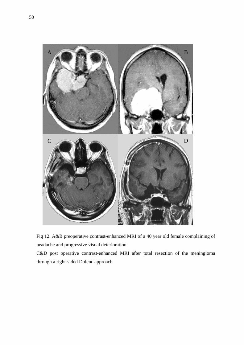
50
Fig 12. A&B preoperative contrast-enhanced MRI of a 40 year old female complaining of
headache and progressive visual deterioration.
C&D post operative contrast-enhanced MRI after total resection of the meningioma
through a right-sided Dolenc approach.
A
C D
B

51
Fig 13. A&B preoperative contrast-enhanced MRI of a 58 year old male complaining of
progressive visual deterioration, exophthalmus and double vision.
C&D post operative contrast-enhanced MRI after subtotal resection. This patient
underwent post-operative SRT.
A B
C D
52
8.4.4 Surgical outcome
There was no mortality related to surgery in this series. Two mortalities occurred during
the follow-up period: the first died one year after surgery from an unrelated cause (cancer
colon); and the other was treated from WHO grade III meningioma by a combination of
surgery and SRT, and died from tumor progress 16 months after the end of treatment.
Nine patients in this group suffered from pre-operative ocular motor nerves palsies and
only two of them showed post-operative recovery.
8.5 Fractionated stereotactic radiotherapy
Seventy-nine patients received SRT either as unique treatment (36 cases) or following
subtotal surgical resection (43 cases). This group of patients comprises 13 males and 66
females with a mean age of 56.5 years. The median target volume was 11.8 ml (range 0.4-
62.2 ml) and the radiation dose ranged from 50.4 Gy to 59.4 Gy with a mean 55.8 Gy.
8.5.1 Post-irradiation side effects
Apart from temporary local alopecia (encountered in 87.3% of cases) and local
radiodermatitis (encountered in 26.6% of cases), symptoms related to acute post-
irradiation toxicity were generally uncommon and easily controlled by steroid medication
(table 6). Late radiation toxicity was uncommon; 3 patients developed late onset
conjunctivitis, one patient developed trigeminal neuralgia, and one patient developed late
onset edema which was treated with corticosteroids. No post-treatment radionecrosis,
visual loss or pituitary dysfunction was encountered.
8.5.2 Post-irradiation outcome
Only 3 patients (3.8%) experienced worsening of their symptoms after SRT, whereas, 35
patients (44.3%) showed improved symptoms and 41 patients (51.9%) remained
unchanged during long-term follow-up. Local tumor control was achieved in all but 2
cases: the first is the case of malignant meningioma previously mentioned (treated with
surgery and SRT), whereas the second case (treated only with SRT without previous
surgery) showed a tumor volume increase of more than 2 mm in the follow-up MRI
without any associated symptoms. This patient refused surgical intervention and is
currently followed up closely for any further tumor progress. Forty patients (50.6%)
showed tumor volume regression and 37 patients (46.8%) had the same tumor volume in
follow-up according to our definition of tumor shrinkage and enlargement.

53
Table 6
Short-term side effects after Radiotherapy
Complication N. of patients Percentage
Alopecia 69/79 87.3%
Radiodermatitis 21/79 26.6%
Increased ICP 19/79 24.1%
Vertigo 10/79 12.7%
Conjunctivitis 8/79 10.1%
Reduced gustation 3/79 3.8%
Reduced hearing 2/79 2.5%
Reduced olfaction 2/79 2.5%
Visual deterioration 1/79 1.3%
Trigeminal neuropathy 1/79 1.3%
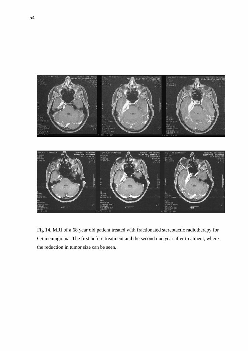
54
Fig 14. MRI of a 68 year old patient treated with fractionated stereotactic radiotherapy for
CS meningioma. The first before treatment and the second one year after treatment, where
the reduction in tumor size can be seen.

55
8.6 Quality of life assessment
Out of a population of 104 patients, questionnaires from a total of 96 patients were
eligible for analysis. These patients were subdivided into 3 groups according to their
treatment protocols. The first group included 22 patients who were treated only surgically,
the second group included 34 patients who were treated uniquely with SRT, and the third
group included 40 patients who were treated with a combination of microsurgery and
SRT.
SF-36 scores were calculated for the eight health domain scales as well as the physical and
mental component summary scales for all 3 groups. By comparing these scores with each
other using the Kruskal-Wallis test, we found that there was no statistically significant
difference in all tested domains of post-treatment life quality between the 3 treatment
protocols; however, patients of the second group had the best values in all domains except
in the physical function and the physical component summary. We also used multivariant
analysis (anova) to compare the mean values of these three patients-groups with the mean
values of the normal German population. There was a statistically significant difference (P
< 0.05) in all eight domains between the mean values obtained from all 3 groups and the
mean values obtained from the normal German population. Results are shown in table 7
and figure 15.
Additionally, 50% of patients in the third group evaluated their present condition as better
or much better than their condition prior to treatment whereas only 12.5% of patients in
this group described their present condition to be worse or much worse than before
treatment. This ratio was the best among all three groups. Results of Patient-evaluated
outcome are shown in Table 8.

56
Table 7
SF-36 Mean Values
Domain Surgery Radiation Combinat. German normal
P-value (ANOVA)
Physical function 72 ± 29.2 71.9 ± 24 66.5 ± 30 85.7 ± 22.1 0.0005
Physical role function 53.6 ± 45.6 56.5 ± 43.3 48.8 ± 42.7 83.7 ± 31.7 0.0005
Body pain 59.5 ± 28.9 71.7 ± 28.2 64.4 ± 31.9 79.1 ± 27.4 0.00052
General health 56.3 ± 23.5 59.3 ± 19.6 57.6 ± 24.5 68.1 ± 20.2 0.00053
Vitality 43.2 ± 26.2 53.1 ± 21 51.3 ± 25.3 63.3 ± 18.5 0.0005
Social function 67.6 ± 34.6 81.5 ± 21.9 76.3 ± 21.6 88.8 ± 18.4 0.0005
Emotional role function 54.5 ± 47.7 77.4 ± 38.9 56.1 ± 47.2 90.4 ± 25.6 0.0005
Mental health 56.7 ± 26.4 72.5 ± 19.7 63.9 ± 23.7 73.9 ± 16.4 0.0005
Physical component sum. 44.4 ± 11.5 43.2 ± 9.4 43 ± 12.1 50.2 ± 10.3 0.0005
Mental component sum. 40 ± 16.6 49.3 ± 10.7 45.8 ± 12.2 51.5 ± 8.2 0.0005
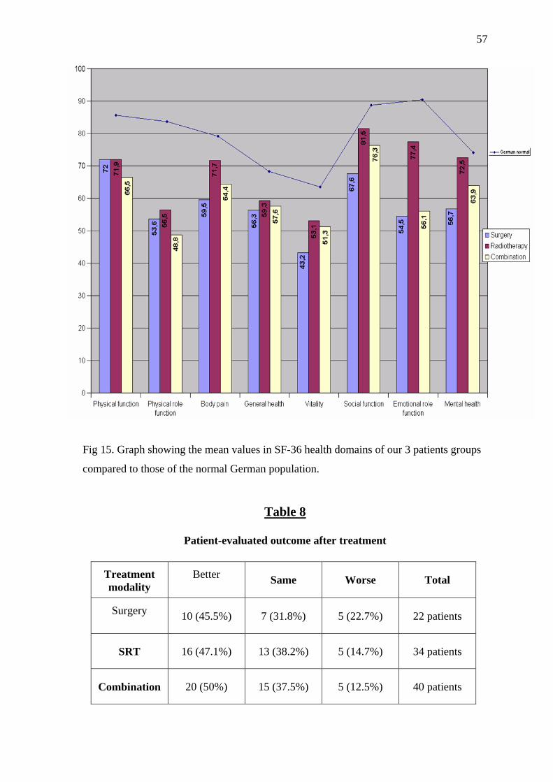
57
Fig 15. Graph showing the mean values in SF-36 health domains of our 3 patients groups
compared to those of the normal German population.
Table 8
Patient-evaluated outcome after treatment
Treatment modality
Better Same Worse Total
Surgery 10 (45.5%) 7 (31.8%) 5 (22.7%) 22 patients
SRT 16 (47.1%) 13 (38.2%) 5 (14.7%) 34 patients
Combination 20 (50%) 15 (37.5%) 5 (12.5%) 40 patients

58
9. Discussion
Tremendous advances have been achieved in the past few decades in all aspects of the
management of meningiomas. However, meningiomas arising at the cranial base,
particularly those involving the cavernous sinus region, still present a formidable surgical
challenge.
9.1 Patients’ demographics
In the present series, 104 patients were treated for CS meningioma between 1997 and
2003 in the Neurosurgery and/or Radiation Oncology Departments, Philipps University,
Marburg. Similar to many other meningioma series, a female preponderance was noticed
in our group of patients [Black 1993, Lamszus 2004].
The age in this series ranged from 33 to 79 years and the most common age group was the
6th decade. The average age was 56 years, which is older than the average age reported in
other series of surgically treated CS meningiomas ranging from 45-49 years [Al-Mefty
2000, Kawase 1991, O’Sullivan 1997, Sekhar 1996]. This discrepancy can be understood
on the basis that all these series include only surgical patients and old age is to some
extent a limitation for performing this type of complicated surgery. On the other hand, this
study includes not only patients undergoing surgical treatment but also patients treated
with SRT. Reciprocally, if we compare our results with the mean age of a series of CS
meningioma treated with SRT [Selch 2004], we will find a younger mean age in our series
compared to their reported mean age of 59 years with more patients in the 7th and 8th
decades.
9.2 Clinical findings
A diversity of symptoms was encountered in this series. The majority of patients
presented with visual symptoms either due to optic nerve compression (diminished visual
acuity and visual field defects) or due to ocular motor nerves compression (ptosis,
diplopia, anisocoria or complete ophthalmoplegia). This is comparable to many series of
CS tumors which reported visual complaints to be the most prevalent clinical feature
[Nicolato 2002, Al-Mefty 1991].
Affection of one or more of the divisions of the trigeminal nerve was encountered in
13.5% of our cases. This is less than the incidence of trigeminal nerve affection reported

59
by Sekhar and colleagues [Sekhar 1989] in a series of 42 patients with CS tumors.
Trigeminal nerve was the most commonly involved cranial nerve in their series being
affected in 14 cases (33.3%).
Other cranial nerves palsies (7th, 8th and lower cranial nerves palsies) were uncommon.
They were only found in large tumors extending to the petroclival area and
cerebellopontine angle. This was similarly found by O’Sullivan et al. [O’Sullivan 1997]
who reported two cases with 7th nerve palsy and two cases with 8th nerve affection among
39 patients with meningiomas involving the CS.
Newman [Newman 2000] allocated the occurrence of cranial nerves dysfunction to one or
more of the following mechanisms: tumor infiltration, compression against rigid structures
and compromise of the blood supply. Individual cranial nerve palsies resulting from a
certain tumor can be explained by the type of this tumor, its size, its site of origin and its
direction of extension.
Headache was a common symptom encountered in more than half of our patients. This is
similar to other series of CS meningioma in which headache was a prevalent symptom
[Cusimano 1995, Chen 2001]. They accredited this to the increased intracranial tension
either from the tumor itself (large size) or from obstruction of CSF pathway or both.
Hormonal disturbance was diagnosed in 3 of our cases in which the tumor was closely
related to the pituitary gland. Other manifestations of brain compression as mental
changes, motor weakness, ataxia and dysphasia were related to the site of the tumor and
its growth pattern.
9.3 Tumor characteristics
The most common tumor origin encountered in this series was medial sphenoid and CS
meningioma followed by petroclival meningioma. The presence of optic or brain stem
compression was in favour of surgical intervention if the patient’s condition allows. The
presence of a large extracavernous part of the tumor was another surgical indication, as
this part can be excised without significant hazards and even if subtotal resection resulted,
this will minimize the risk of a potential post-operative SRT by decreasing the target
volume for irradiation [Milker-Zabel 2005].
According to the new WHO criteria for histopathological diagnosis [Kleihues 2002], all
tumors were WHO grade I except 2 cases: one case had WHO grade II meningioma and

60
the other had WHO grade III meningioma. In this new WHO classification, the mitotic
index is the cornerstone for grading. Counting mitotic figures is a simple, reliable, and
low-cost procedure that requires no additional preparation. It is now well known that the
grade of the meningioma affects significantly the treatment outcome [Mahmood 1993,
Milker-Zabel 2005].
According to Hirsch grading [Hirsch 1993], 39.4% of tumors were grade 1, 45.2% were
grade 2, and 15.4% were grade 3. The relation between the tumor and the cavernous ICA
is a predicting factor for the feasibility of total excision. The more the artery is surrounded
by the tumor, the more is the possibility that the tumor has invaded the arterial wall and
the less is the possibility for complete excision while preserving the artery [Abdel-Aziz
2004, Kotapka 1994, O’Sullivan 1997].
9.4 Surgery
Parkinson and Dolenc created a stimulus to better understand the CS region and to
approach it surgically. Despite the improved orientation by understanding the
microsurgical anatomy and by improvement of standard operative techniques, the surgical
resection of tumors involving the CS remains a significant challenge.
9.4.1 Extent of resection
Among 37 patients operated at our institute, gross total resection was achieved in 22 of
them (59.5%). The incidence of total resection reported in the literature ranged from
20%-76% [Al-Mefty 2000, De Jesus 1996, Kawase 1991, Knosp 1996, O’Sullivan 1997].
The cause of subtotal resection in the remaining 15 cases was tumor adherence to the ICA
and cranial nerves. Several authors proved the histological invasion of the arterial wall
and cranial nerves by CS meningioma [Kotapka 1994, Larson 1995]. Consequently,
dissection of the tumor from these structures without injuring them is in some cases
almost impossible.
Although total resection is important and has been always correlated with better long term
results, improving the functional outcome was always the main goal of surgery. We did
not sacrifice any major vessel or cranial nerve for the sake of total resection in any of our
patients. Preservation of ICA is a top priority in our experience, even if it means leaving a
tumor remnant adherent to the artery. Although saphenous vein bypass procedures have

61
been advocated to achieve a radical resection [Sekhar 1989], we think grafting of the ICA
for the sake of radical resection seems not to be worthwhile considering the possibility of
using post-operative radiotherapy or radiosurgery. This conservative surgical concept was
adopted in several series of CS meningioma [Abdel-Aziz 2004, O’Sullivan 1997].
Several factors increase the difficulty of CS meningioma excision. One of these factors is
the degree of ICA involvement [Hirsch 1993, O’Sullivan 1997]. In the present series, the
rate of total resection in patients with complete carotid encasement was 35% compared to
88.2% in patients with partial carotid encasement. Similarly, Abdel-aziz and colleagues
[Abdel-Aziz 2004] reported total excision in 22 of 24 patients with CS meningioma in
whom the tumor was only partially encasing the ICA, while all 14 patients with complete
ICA encasement underwent subtotal excision.
Previous surgery was another limitation to total resection. The incidence of total resection
among recurrent cases was 40% in this series, while in previously non-operated tumors;
the incidence of total resection was 66.7%. This is in accordance with many meningioma
reports stating that radicality is more difficult in recurrent tumors owing to scaring
resulting from the first surgery and the lack of an intact arachnoid plane between the
tumor and the nervous tissue [Heth 2003, O’Sullivan 1997].
9.4.2. Surgical approach
Since CS tumors are deep in location and surrounded by critical structures, traditional
conventional approaches are, in our opinion, disadvantageous because of the need for
significant brain retraction, poor control of the lesion and adjacent structures, and often
suboptimal exposure. In the past, such operations often resulted in partial resection of the
tumor with high morbidity. On the other hand, modern skull base approaches provide
better exposure of these deeply seated tumors with less brain retraction by combining the
removal of non-critical osseous structures (e.g. ACP, petrous apex) with specialized
operative maneuvers (e.g. tentorium resection, mobilization of neurovascular structures).
With the introduction of these techniques most CS tumors can be safely approached, and
many are radically resected. The choice of the approach should be carefully determined
according to the type and extent of the tumor, the ultimate goal of the procedure and the
benefits and risks in each individual case.
62
In the present study, the traditional pterional approach was used only in two of our early
cases. Dolenc approach was the most commonly used (18 cases) as it has the advantage of
exposing the whole CS and decompressing the optic nerve in case of optic compression.
Although some authors [Al-Mefty 2000] recommend the use of COZ routinely before
performing anterior clinoidectomy and approaching CS tumors, we reserved the use of
COZ craniotomy to large tumors with high suprasellar extension to minimize brain
retraction and to get multiple viewing angles during surgery. Kawase approach was used
for tumors occupying the posterior CS and extending to the posterior fossa. Transcondylar
approach was performed in one case although it can not be considered an approach to the
CS. In this particular case, the tumor was extensive involving both CS as well as the clival
region till the foramen magnum, and surgery was designed to decompress the brain stem
and was followed by SRT.
The same concept of preferring skull base approaches (as COZ, Dolenc and Kawase) for
resection of CS tumors was adopted by many authors [Al-Mefty 2000, De Jesus 1996,
Kawase 1991, O’Sullivan 1997]. Taha et al. [Taha 1995] reported a study comparing the
results of conventional craniotomies to skull base surgical approaches for the removal of
trigeminal schwannomas. They reported a 10% recurrence rate using the modern cranial
base surgical techniques compared with a 65% recurrence rate using conventional
approaches. In addition, they found that the surgeon's estimate of the extent of resection
was less accurate when using the conventional approaches. Although this study involved
patients with trigeminal schwannomas, yet it shows the superiority of skull base
approaches in dealing with this region.
On the other hand, some surgeons still prefer the conventional pterional transsylvian
approach for approaching CS tumors. Yasargil [Yasargil 1984] reported a series of 9
patients with sphenocavernous meningiomas operated through pterional transsylvian
approach with a good outcome in 7 cases and a moderate disability in 2 cases (22.2%).
Tomasello et al. [Tomasello 2003] operarted 13 patients with sphenocavernous
meningiomas via transsylvian approach achieving total resection in 10 cases with 2
mortalities and one moderate disability.
9.4.3 Outcome and complications
Meningiomas involving the CS are difficult lesions to be treated surgically because of the
potential post-operative morbidity. A major concern in CS surgery is cranial nerve

63
morbidity. Temporary cranial nerve deficits are common after tumor removal from the CS
and should be presented as an expected outcome to patients considering surgery. Of
greater importance to the outcome is the incidence of permanent cranial nerve deficits.
O'Sullivan et al. [O’Sullivan 1997] reported an incidence of new permanent post-operative
morbidity of 12% considering only the optic, extraocular, and trigeminal nerves. De
Monte and colleagues [De Monte 1994] reported 10 new neuropathies in 7 patients in
their series of 38 patients. In our series, 5 out of 37 patients suffered from permanent post
operative cranial nerves palsies and only 3 of them had ocular motor nerves palsies.
Skull base techniques entail drilling of some parts of the skull base and sometimes
opening of the air sinuses. Therefore, the chance of CSF leakage from the inadvertently or
intentionally opened air sinuses exists. We had 5 cases of CSF leak in our study (13.5%).
The reported incidence of CSF leak following CS surgery ranged from 8% to 27% [Al-
Mefty 2000, Cusimano 1995, Dolenc 1993].
One of our patients (2.7%) developed a partial middle cerebral infarction post-operatively
manifested with dysphasia which improved afterwards. Cusimano et al. reported a 12%
incidence of post-operative infarction and half of these patients developed permanent
significant neurological deficit [Cusimano 1995]. This relatively high incidence may be
due to the performance of carotid bypass in many cases in this series.
Out of 9 patients with preoperative ocular motor nerves deficits, only two of them (22.2%)
showed improved ocular motility post-operatively. Similarly, O’Sullivan et al.
[O’Sullivan 1997] reported post operative improvement in the ocular motor nerves
function in 21% of their patients with preoperative deficits.
There was no mortality related to surgery in the present study. One of the two mortalities
that took place, was due to the natural history of a malignant tumor (WHO GIII
meningioma) and occurred 16 months after surgery, and the other resulted from a non-
related cause (cancer colon). Mortality rates reported in other series ranged from 0-16%.
The causes of death varied between surgical complications, tumor progress or non-related
causes [Abdel-Aziz 2004, Al-Mefty 2000, De Jesus 1996, Kawase 1991, O’Sullivan
1997].

64
9.5 Fractionated stereotactic radiotherapy
Due to the potential risk of post-operative morbidity following resection of CS tumors,
high-precision radiation therapy has been advocated in the recent years as an alternative to
microsurgery in some cases or as an adjunct to surgery after subtotal excision in other
cases. The published local control rates for radiosurgery are excellent and equivalent local
control rates were also reported with SRT ranging from 92% to 100% [Chen 2001, Debus
2001, Jalali 2002, Nicolato 2002, Selch 2004, Spiegelmann 2002]. In our study, local
control rate was also excellent. Only 2 out of 79 patients (2.5%) who underwent SRT
showed tumor progress and one of these 2 cases had a malignant meningioma.
Additionally, 40 of 79 patients (50.6%) showed tumor shrinkage according to our
definition of tumor volume regression based on 3-D imaging assessment. This incidence is
higher than the incidence of tumor shrinkage reported by Debus et al. and Jalali et al.
following SRT which was 14% and 26.8% respectively [Debus 2001, Jalali 2002].
Radiotherapy was used as a primary treatment in elderly patients, medically infirm
patients, or patients who refused microsurgery. It was also used as an adjuvant treatment
of residual tumor in the CS after subtotal resection in all cases with the exception of 3
cases who refused post operative SRT although recommended. The total dose ranged from
50.4-59.4 Gy with a mean of 55.8 Gy. The total dose was divided into fractions and the
dose in a single fraction was 1.8-2Gy. This is in accordance of other series using SRT for
the treatment of meningiomas [Debus 2001, Milker-Zabel 2005].
The tumor characteristics of the patients in our series frequently exceeded the
recommended limits for radiosurgery. Lee et al. advocated SRS for CS lesions <3 cm in
the greatest dimension and not more than 15 cm3 in volume [Lee 2002]. Another
limitation to the use of SRS in this region is the contact between the tumor and the optic
apparatus. Chen et al. demonstrated a statistically significant correlation of the radiation
dose reaching the optic chiasm and the closest distance of the tumor margin [Chen 2001].
As SRT combines the advantage of stereotactic target localization with the radiobiologic
benefits of dose fractionation, it can be used if the tumor involves some critical structures
as the optic apparatus with a low complication rate [Dufour 2001].
Only 3 patients (3.8%) experienced worsening of their symptoms after SRT. one patient
developed a new visual field defect after SRT. The worsening rates reported in similar
studies were in average between 6-7%. Complications related to SRT were generally mild

65
and easily treated. They were all evaluated as grade 1 or 2 according to the common
toxicity criteria for acute toxicity [Seegenschmiedt 1998] and the LENT-SOMA [LENT-
SOMA 1995] score for late toxicity. This is comparable with other reports of CS
meningiomas treated with high-precision radiation therapy [Debus 2001, Jalali 2002,
Selch 2004].
9.6 Quality of life assessment
The efficacy of any treatment modality used for CS meningioma was generally evaluated
in terms of morbidity and mortality rates, recurrence free survival rates and the extent of
surgical resection or local tumor control after irradiation. The impact of treatment on the
quality of life was commonly assessed by Karnofsky performance scales [Cusimano 1995,
De Jesus 1996, Nicolato 2002]. Cusimano et al. found a difference between the Karnofsky
score reported by the patient himself and that reported by the medical staff of 10 points or
more [Cusimano 1995]
Recent reports suggest that patient outcome as evaluated by surgeons may underestimate
patient’s burden after surgery [Akagami 2002, Lang 1999]. Post-treatment outcome
evaluations are important because a specific deficit may affect one person minimally but
may devastate the career of another; a minimal deficit as perceived by the surgeon may be
debilitating to quality of life from the perspective of the patient.
We used the SF-36, a popular generic outcome measurement instrument, to assess the
post-treatment quality of life for patients with meningiomas involving the CS treated at
our institute. SF-36 is known to be a reliable and valid measure for quality of life and
outcome assessment as proved by several studies [Findler 2001, King 2003]. The SF-36 is
a 36-item questionnaire that measures patient’s responses regarding 8 health domains.
Validated normative data for a German population sample made SF-36 particularly useful
for our study. We divided the patients into 3 groups according to their treatment protocols,
whether surgery, SRT or a combination of both.
Patients in this series exhibited significantly worse scores on all SF-36 domain scales
compared with the average scores of the normal German population. This can be
attributed to the critical site of the tumor and to the fact that meningiomas are more
common in older age groups. Interestingly, the difficulties of these patients extended
beyond physical debility to diminished emotional and mental states. Patients treated
66
surgically, whether alone or in combination with post-operative SRT, had an overall
poorer quality of life than those treated only with SRT. The only exception to this was in
the domain of physical function where surgical patients had better scores. However, the
difference between all 3 groups was statistically insignificant in all 8 domains as well as
the mental and physical summary scores.
Additionally, the majority of patients in each group reported a similar or better condition
compared with their condition before treatment. About half of the patients in each group
described their present health state as better or much better than their pre-treatment health
state. In an outcome assessment of 119 treated for CS meningioma, De Jesus et al. [De
Jesus 1996] reported no statistical difference between pre- and post-operative median
Karnofsky scores. However, the median pre-operative Karnofsky score was 90 and the
post-operative Karnofsky score was 80. In a similar study, Akagami et al. [Akagami 2002]
evaluated the post-operative outcome of 269 patients with basal meningiomas including
CS meningioma using direct simple questions. Only 3% of the patients were not satisfied
with the treatment and 83% were employable. All these results show that resection of CS
tumors is not the prohibitive procedure it once was.
67
10. Conclusion
Proper understanding of the complex anatomy of the CS is the cornerstone in performing
successful surgery in this region. The surgeon has to be aware of the variability of some
commonly used landmarks. Although the skull base approaches are more difficult and
time consuming than the conventional approaches, they are superior in the surgical
excision of CS tumors. However, the approach should be tailored according to the type
and extent of the tumor and the goal of the surgery. The extent of exposure offered by
each approach should be put in consideration during surgical planning.
The clinical part of this study proves that a satisfactory outcome can be achieved after
treatment of CS meningioma in spite of their critical location. SRT seems to be an
efficient and safe initial or adjuvant treatment for meningiomas involving the CS as
proved by tumor control rates and long term quality of life assessment. It offers an
additional treatment option with high efficacy, low risk and good post-treatment life
quality. On the other hand, surgery still plays an important role in the management of
these tumors (especially in the presence of optic compression, brain stem compression and
large tumor volume) with acceptable morbidity and post-operative quality of life.
Complete tumor resection can be achieved in many cases especially with the appropriate
choice of the approach. In certain cases both modalities should be combined in order to
optimize the management. Patients should be aware of management options and risks
associated with every treatment modality. The proper choice of the treatment protocol for
each case minimizes the overall risk of management. Psychosocial and neurobehavioral
findings should not be underestimated in outcome assessment.
68
11. Zusammenfassung
Diese Arbeit besteht aus zwei Teilen: eine anatomische Studie über die komplexe
Beschaffenheit des Sinus Cavernosus (CS) und eine retrospektive Studie der Klinik von
104 Patienten welche in der Universitätsklinik der Phillipps Universität Marburg an
Meningeomen des CS behandelt wurden.
Zum besseren Verständnis der anatomischen Verhältnisse im Bereich des CS sowie zur
Verbesserung der neurochirurgischen Operationstechnik wurden drei allgemein
gebräuchliche Schädelbasiszugänge zur chirurgischen Freilegung des CS an insgesamt 5
Leichenköpfen durchgeführt. Jeder dieser Zugänge wurde zehnmal durchgeführt, jeweils
in Art und Weise des Zugangs bei einer realen Operation. Die Ausdehnung der Freilegung
bei jedem dieser drei Zugänge, die dafür notwendigen anatomischen Landmarken und die
anatomische Beziehung zwischen den im CS gelegenen und diesen umgebenden
Strukturen wurden genaustens identifiziert und dokumentiert.
Der zweite Teil beinhaltet eine retrospektive Studie von insgesamt 104 Patienten mit CS-
Meningeomen, welche zwischen 1997 und 2003 in den Abteilungen für Neurochirurgie
und Strahlentherapie der Phillips Universität Marburg behandelt wurden. Untersucht und
analysiert wurden sowohl die postinterventionelle Lebensqualität als auch die Morbidität
und das Ergebnis der Behandlung. Die postinterventionelle Lebensqualität wurde anhand
der Medical Outcome Study Short Form 36 (SF-36) bemessen. Dieser Fragebogen
vergleicht 36 verschiedene Punkte, welche die Reaktionen der Patienten in 8
verschiedenen gesundheitlichen Bereichen bewerten. Die mittlere Nachuntersuchungszeit
umfasste dabei 40 Monate.
In der Patientengruppe wurden 80 Frauen und 24 Männer im mittleren Alter von 56
Jahren verglichen. Die Mehrheit der Patienten wurde Grad 1 oder 2 des Hirsch
Graduierungssystems für CS Beteiligung zugeordnet. Die häufigste Lage der Tumoren
war die mediale Sphenoid-Kante sowie die petroclivale und die den CS bedeckende Dura.
Die klinischen Auffälligkeiten waren die für CS-Meningeome typischen und bestanden
aus Kopfschmerzen und Gesichtsfeldausfällen als üblichste Symptome.
68 Patienten dieser Gruppe unterliefen mindestens einmal einer operativen Behandlung.
Die Rate der Totalresektionen unter den an unserem Institut behandelten Patienten betrug
59,5%. Der am meisten gebrauchte Zugang war dabei der nach Dolenc. Die häufigste

69
postoperative Komplikation waren Hirnnervendefizite, welche bei 45,9% der Fälle
auftraten, jedoch nur in 13,5% irreversibel waren.
79 Patienten erhielten eine fraktionierte stereotaktische Bestrahlung, welche durch einen
Linearbeschleuniger entweder als alleinige Therapie (36 Fälle) oder im Anschluss an eine
subtotale chirurgische Resektion (43 Fälle) verabreicht wurde. Das mittlere Zielvolumen
betrug 11,8 ml und die mittlere Strahlendosis 55,8 Gy. Abgesehen von lokalem
Haarausfall und Hautreaktionen waren Frühstrahlenschäden gelinde und standen in
Beziehung mit dem entstehenden lokalen Ödem. Spätstrahlenschäden waren ebenso
gelinde und selten. Die Hälfte unserer Patienten zeigte eine Größenregression des
Tumorvolumens, wobei 2 Patienten (2,5%) eine Gößenprogredienz nach der initialen
Bestrahlung aufwiesen.
Zum Zeitpunkt der Studie waren zwei Patienten bereits verstorben: einer aufgrund des
Tumorprogresses, der andere aufgrund einer anderen, nicht in Beziehung stehenden
Ursache. Die SF-36 Fragebögen standen von 96 Patienten zur Analyse zur Verfügung. Die
Einteilung dieser Patienten erfolgte in drei Gruppen welche sich aus dem
Behandlungsprotokoll ergaben: nur operierte Patienten (22), nur stereotaktisch fraktioniert
bestrahlte Patienten (34) und sowohl chirurgisch als auch radiotherapeutisch behandelte
Patienten (40). Es wurde dabei kein statistisch relevanter Unterschied der Lebensqualität
in den drei untersuchten Gruppen gefunden; insgesamt hatten die Patienten der zweiten
Gruppe die beste Lebensqualität in allen gesundheitlichen Bereichen bis auf einen.
Konventionelle stereotaktische fraktionierte Bestrahlung scheint eine effiziente und
sichere, initiale oder adjuvante Methode zur Behandlung von Meningeomen de CS zu
sein. Andererseits spielt auch die operative Behandlung solcher Tumore heute noch eine
große Rolle. In manchen Fällen sollte die Anwendung beider dieser Methoden kombiniert
werden um das Ergebnis zu optimieren. Insgesamt können gute Ergebnisse durch
sorgfältige Auswahl von Patienten erzielt werden.

70
12. References
1. Abdel Aziz KM, Froelich SC, Cohen PL, Sanan A, Keller JT, Van Loveren HR: The
one-piece orbitozygomatic approach: the MacCarty burr hole and the inferior orbital
fissure as keys to technique and application. Acta Neurochir 144:15-24, 2002.
2. Abdel-Aziz KM, Froelich SC, Dagnew E, Jean W, Breneman JC, Zuccarello M, Van
Loveren HR, Tew JM Jr: Large sphenoid wing meningiomas involving the cavernous
sinus: conservative surgical strategies for better functional outcomes. Neurosurgery
54:1375-1383, 2004.
3. Akagami R, Napolitano M, Sekhar LN: Patient-evaluated outcome after surgery for
basal meningiomas. Neurosurgery 50:941-948, 2002.
4. Al-Mefty O, Ayoubi S: Clinoidal meningiomas. Acta Neurochir Suppl (Wien) 53:92-
97, 1991.
5. Al-Mefty O, DeMonte F: Cavernous Sinus meningioma management with carotid
preservation, in: Eisenberg MB, Al-Mefty O (eds): The Cavernous Sinus. A
comprehensive Text. Philadelphia, Lippincott Williams& Wilkins, 2000, pp 251-261.
6. Angtuaco E, Van Hermet R, Jr: Radiology of the Cavernous Sinus including computed
tomography, magnetic resonance imaging, magnetic resonance angiography, magnetic
resonance venography, and angiography, in: Eisenberg MB, Al-Mefty O (eds): The
Cavernous Sinus. A comprehensive Text. Philadelphia, Lippincott Williams& Wilkins,
2000, pp 35-53.
7. Anscher MS, Swift PS, Gaspar LE, Marks LB: Radiation injury of the brain and spinal
cord, in: Wilkins RH, Rengachary S (eds): Neurosurgery, New York, McGraw-Hill, 1996,
pp 1921-1941.
8. Bayassi S: Meningo-orbital fold (MOF) as a guiding point in extradural approach to
the anterior clinoid process. Neurol Neurochir Pol 39:49-55, 2005.
9. Black PM: Meningiomas. Neurosurgery 32:643-657, 1993.
10. Borba L, Al-Mefty O: Normal Anatomy of the Cavernous Sinus, in: Eisenberg MB,
Al-Mefty O (eds): The Cavernous Sinus. A comprehensive Text. Philadelphia, Lippincott
Williams& Wilkins, 2000, pp 21-33.

71
11. Cavallo LM, Cappabianca P, Galzio R, Iaconetta G, de Divitiis E, Tschabitscher M:
Endoscopic transnasal approach to the cavernous sinus versus transcranial route: anatomic
study. Neurosurgery 56 (Suppl):379-389, 2005.
12. Chakeres DW, Curtin A, Ford G: Magnetic resonance imaging of pituitary and
parasellar abnormalities. Radiol Clin North Am 27:265-281, 1989.
13. Chen JC, Giannotta SL, Yu C, Petrovich Z, Levy ML, Apuzzo ML: Radiosurgical
management of benign cavernous sinus tumors: dose profiles and acute complications.
Neurosurgery 48:1022-1030, 2001.
14. Couldwell WT, Sabit I, Weiss MH, Giannotta SL, Rice D: Transmaxillary approach to
the anterior cavernous sinus: a microanatomic study. Neurosurgery 40: 1307-1311, 1997.
15. Cushing HW, Eisenhardt L: Meningiomas: Their Classification, Regional Behavior,
Life History, and Surgical End Results. Springfield, III: Charles C Thomas, 1938, pp 47-
50.
16. Cusimano MD, Sekhar LN, Sen CN, Pomonis S, Wright DC, Biglan AW, Janetta PJ:
The results of surgery for benign tumors of the cavernous sinus. Neurosurgery 37:1-9,
1995.
17. Debus J, Wuendrich M, Pirzkall A, Hoess A, Schlegel W, Zuna I, Engenhart-Cabillic
R, Wannenmacher M: High efficacy of fractionated stereotactic radiotherapy of large
base-of-skull meningiomas: long-term results. J Clin Oncol 19:3547-3553, 2001.
18. De Jesus O, Sekhar LN, Parikh HK, Wright DC, Wagner DP: Long-term follow-up of
patients with meningiomas involving the cavernous sinus: recurrence, progression, and
quality of life. Neurosurgery 39:915-919, 1996.
19. DeMonte F, Smith HK, Al-Mefty O: Outcome of aggressive removal of cavernous
sinus meningiomas. J Neurosurg 81:245-251, 1994.
20. Dolenc V: Direct microsurgical repair of intracavernous vascular lesions. J Neurosurg
58:824-831, 1983.
21. Dolenc V: Anatomy and Surgery of the Cavernous Sinus. New York, Springer-Verlag,
1989, pp 3-37.
72
22. Dolenc V: Cavernous sinus masses, in: Apuzzo ML (ed): Brain Surgery: Complication
Avoidance and Management. New York, Churchill Livingstone, 1993, pp 601-614.
23. Dufour H, Muracciole X, Metellus P, Regis J, Chinot O, Grisoli F: Long-term tumor
control and functional outcome in patients with cavernous sinus meningiomas treated by
radiotherapy with or without previous surgery: is there an alternative to aggressive tumor
removal? Neurosurgery 48:285-294, 2001.
24. Eisenberg MB, Al-Mefty O, DeMonte F, Burson GT: Benign non-meningeal tumors
of the cavernous sinus. Neurosurgery 44:949-954, 1999.
25. Eisenberg MB, Pait T: The history of the Cavernous Sinus, in: Eisenberg MB & Al-
Mefty O (eds), The Cavernous Sinus. A comprehensive Text. Philadelphia, Lippincott
Williams& Wilkins, 2000, pp 3-4.
26. Erturk M, Kayalioglu G, Govsa F: Anatomy of the clinoidal region with special
emphasis on the caroticoclinoid foramen and interclinoid osseous bridge in a recent
Turkish population. Neurosurg Rev 27:22-26, 2004.
27. Evans JJ, Hwang YS, Lee JH: Pre- versus post-anterior clinoidectomy measurements
of the optic nerve, internal carotid artery, and opticocarotid triangle: a cadaveric
morphometric study. Neurosurgery 46:1018-1021, 2000.
28. Feiz-Erfan I, Han PP, Spetzler RF, Porter RW, Klopfenstein JD, Ferreira MA, Beals
SP, Joganic EF: Exposure of midline cranial base without a facial incision through a
combined craniofacial-transfacial procedure. Neurosurgery 56(Suppl):28-35, 2005.
29. Findler M, Cantor J, Haddad L, Gordon W, Ashman T: The reliability and validity of
the SF-36 health survey questionnaire for use with individuals with traumatic brain injury.
Brain Inj 15:715-723, 2001.
30. Goldsmith BJ, Wara WM, Wilson CB, Larson DA: Postoperative irradiation for
subtotally resected meningiomas. A retrospective analysis of 140 patients treated from
1967 to 1990. J Neurosurg 80:195-201, 1994.
31. Gross MW, Spahn U, Engenhart-Cabillic R: Assessment of the accuracy of a
conventional simulation for radiotherapy of head and skull base tumors. Technol Cancer
Res Treat 178:216-222, 2003.

73
32. Guy RL, Benn JJ, Ayers AB, Bingham JB, Lowy C, Cox TC, Sonksen PH: A
comparison of CT and MRI in the assessment of the pituitary and parasellar region. Clin
Radiol 43:156-161, 1991.
33. Hakuba A, Tanaka K, Suzuki T, Nishimura S: A combined orbitozygomatic
infratemporal epidural and subdural approach for lesions involving the entire cavernous
sinus. J Neurosurg 71:699-704, 1989.
34. Harris FS, Rhoton AL, Jr: Anatomy of the cavernous sinus. A microsurgical study. J
Neurosurg 45:169-180, 1976.
35. Harrison MJ, Levine Z, Donald Wright, Sekhar LN: Cranial nerve repair and
reconstruction in Cavernous Sinus surgery, in: Eisenberg MB & Al-Mefty O (eds), The
Cavernous Sinus. A comprehensive Text. Philadelphia, Lippincott Williams& Wilkins,
2000, pp 281-287.
36. Heth JA, Al-Mefty O: Cavernous sinus meningiomas. Neurosurg Focus 14:e3, 2003.
37. Hirsch WL, Sekhar LN, Lanzino G, Pomonis S, Sen CN: Meningiomas involving the
cavernous sinus: value of imaging for predicting surgical complications. AJR Am J
Roentgenol 160:1083-1088, 1993.
38. Inoue T, Rhoton AL, Jr., Theele D, Barry ME: Surgical approaches to the cavernous
sinus: a microsurgical study. Neurosurgery 26:903-932, 1990.
39. Jalali R, Loughrey C, Baumert B, Perks J, Warrington AP, Traish D, Ashley S, Brada
M: High precision focused irradiation in the form of fractionated stereotactic conformal
radiotherapy (SCRT) for benign meningiomas predominantly in the skull base location.
Clin Oncol (R Coll Radiol ) 14:103-109, 2002.
40. Kawase T, Toya S, Shiobara R, Mine T: Transpetrosal approach for aneurysms of the
lower basilar artery. J Neurosurg 63:857-861, 1985.
41. Kawase T, Shiobara R, Toya S: Anterior transpetrosal-transtentorial approach for
sphenopetroclival meningiomas: surgical method and results in 10 patients. Neurosurgery
28:869-875, 1991.
42. Kawase T, Van Loveren H, Keller JT, Tew JM: Meningeal architecture of the
cavernous sinus: clinical and surgical implications. Neurosurgery 39:527-534, 1996.

74
43. Kayalioglu G, Govsa F, Erturk M, Pinar Y, Ozer MA, Ozgur T: The cavernous sinus:
topographic morphometry of its contents. Surg Radiol Anat 21: 255-260, 1999.
44. Keyes JEL: Observations on four thousand optic foramina in human skull of known
origin. Arch Ophthalmo 13:538-568, 1935.
45. King JT, Jr., McGinnis KA, Roberts MS: Quality of life assessment with the medical
outcomes study short form-36 among patients with cervical spondylotic myelopathy.
Neurosurgery 52:113-120, 2003.
46. Kleihues P, Louis DN, Scheithauer BW, Rorke LB, Reifenberger G, Burger PC,
Cavenee WK: The WHO classification of tumors of the nervous system. J Neuropathol
Exp Neurol 61:215-225, 2002.
47. Knosp E, Perneczky A, Koos WT, Fries G, Matula C: Meningiomas of the space of
the cavernous sinus. Neurosurgery 38:434-442, 1996.
48. Kokubo M, Shibamoto Y, Takahashi JA, Sasai K, Oya N, Hashimoto N, Hiraoka M:
Efficacy of conventional radiotherapy for recurrent meningioma. J Neurooncol 48:51-55,
2000.
49. Kondziolka D, Lunsford LD, Flickinger JC: The radiobiology of radiosurgery.
Neurosurg Clin N Am 10:157-166, 1999.
50. Kondziolka D, Nathoo N, Flickinger JC, Niranjan A, Maitz AH, Lunsford LD: Long-
term results after radiosurgery for benign intracranial tumors. Neurosurgery 53:815-821,
2003.
51. Kotapka MJ, Kalia KK, Martinez AJ, Sekhar LN: Infiltration of the carotid artery by
cavernous sinus meningioma. J Neurosurg 81: 252-255, 1994.
52. Krisht A, Barnett DW, Barrow DL, Bonner G: The blood supply of the intracavernous
cranial nerves: an anatomic study. Neurosurgery 34:275-279, 1994.
53. Krivosic I: Histoarchitecture of the Cavernous Sinus. In: Dolenc VV(ed): The
Cavernous Sinus. New York, Springer-Verlag, 1987, pp 117-129.
54. Lamszus K. Meningioma: pathology, genetics, and biology. J Neuropathol Exp Neurol
63:275-286, 2004.

75
55. Lang DA, Neil-Dwyer G, Garfield J: Outcome after complex neurosurgery: the
caregiver's burden is forgotten. J Neurosurg 91:359-363, 1999.
56. Lang J: Sinus Cavernosus, in: Lang J, Wachsmuth W (eds): Praktische Anatomie:
Gehirn und Augenschädel. Berlin Heidelberg New York, Springer-Verlag, 1979, pp 63-
80.
57. Larson JJ, Van Loveren HR, Balko MG, Tew JM, Jr: Evidence of meningioma
infiltration into cranial nerves: clinical implications for cavernous sinus meningiomas. J
Neurosurg 83:596-599, 1995.
58. Lee JYK, Niranjan A, McInerney J, Kondziolka D, Flickinger JC, Lunsford LD:
Stereotactic radiosurgery providing long-term tumor control of cavernous sinus
meningiomas, J Neurosurg 97, 65-72, 2002.
59. Leksell L: The stereotaxic method and radiosurgery of the brain. Acta Chir Scand
102:316-319, 1951.
60. LENT SOMA scales for all anatomic sites. Int J Radiat Oncol Biol Phys 31:1049-
1091, 1995.
61. Littley MD, Shalet SM, Morgenstern GR, Deakin DP. Endocrine and reproductive
dysfunction following fractionated total body irradiation in adults. Q J Med 78:265-274,
1991.
62. Luxton G, Petrovich Z, Jozsef G, Nedzi LA, Apuzzo ML: Stereotactic radiosurgery:
principles and comparison of treatment methods. Neurosurgery 32: 241-259, 1993.
63. Mahmood A, Caccamo DV, Tomecek FJ, Malik GM: Atypical and malignant
meningiomas: a clinicopathological review. Neurosurgery 33:955-963, 1993.
64. Martins C, Li X, Rhoton AL, Jr: Role of the zygomaticofacial foramen in the
orbitozygomatic craniotomy: anatomic report. Neurosurgery 53:168-172, 2003.
65. McKenzie MR, Souhami L, Caron JL, Olivier A, Villemure JG, Podgorsak EB: Early
and late complications following dynamic stereotactic radiosurgery and fractionated
stereotactic radiotherapy. Can J Neurol Sci 20:279-285, 1993.

76
66. Milker-Zabel S, Zabel A, Schulz-Ertner D, Schlegel W, Wannenmacher M, Debus J:
Fractionated stereotactic radiotherapy in patients with benign or atypical intracranial
meningioma: long-term experience and prognostic factors. Int J Radiat Oncol Biol Phys
61:809-816, 2005.
67. Mulhern RK, Wasserman AL, Fairclough D, Ochs J: Memory function in disease-free
survivors of childhood acute lymphocytic leukemia given CNS prophylaxis with or
without 1,800 cGy cranial irradiation. J Clin Oncol 6:315-320, 1988.
68. Nagele T, Petersen D, Klose U, Grodd W, Opitz H, Voigt K. The "dural tail" adjacent
to meningiomas studied by dynamic contrast-enhanced MRI: a comparison with
histopathology. Neuroradiology 36:303-307, 1994.
69. Newman SA: Neuroophthalmic considerations, in: Eisenberg MB, Al-Mefty O (eds):
The Cavernous Sinus. A comprehensive Text. Philadelphia, Lippincott Williams&
Wilkins, 2000, pp 77-110.
70. Nicolato A, Foroni R, Alessandrini F, Bricolo A, Gerosa M: Radiosurgical treatment
of cavernous sinus meningiomas: experience with 122 treated patients. Neurosurgery
51:1153-1159, 2002.
71. Niranjan A, Gobbel GT, Kondziolka D, Flickinger JC, Lunsford LD. Experimental
radiobiological investigations into radiosurgery: present understanding and future
directions. Neurosurgery 55:495-504, 2004.
72. Noell KT, herskovic AM. Principles of Radiotherapy of central nervous system. In:
Rengachary S & Wilkins RH (eds). Neurosurgery, 2nd ed. New York: McGraw-Hill, 1996,
pp 1837-48.
73. Nutting C, Brada M, Brazil L, Sibtain A, Saran F, Westbury C, Moore A, Thomas DG,
Traish D, Ashley S. Radiotherapy in the treatment of benign meningioma of the skull
base. J Neurosurg 90:823-827, 1999.
74. O'Sullivan MG, van Loveren HR, Tew JM, Jr: The surgical resectability of
meningiomas of the cavernous sinus. Neurosurgery 40:238-244, 1997.
75. Parkinson D: A surgical approach to the cavernous portion of the carotid artery.
Anatomical studies and case report. J Neurosurg 23:474-483, 1965.

77
76. Parkinson D: Surgical anatomy of the lateral sellar compartment (cavernous sinus).
Clin Neurosurg 36:219-239, 1990.
77. Parkinson D: Lateral sellar compartment O.T. (cavernous sinus): history, anatomy,
terminology. Anat Rec 251:486-490, 1998.
78. Parkinson D: Extradural neural axis compartment. J Neurosurg 92:585-588, 2000.
79. Parsons JT, Fitzgerald CR, Hood CI, Ellingwood KE, Bova FJ, Million RR: The
effects of irradiation on the eye and optic nerve. Int J Radiat Oncol Biol Phys 9:609-622,
1983.
80. Patterson RH, Jr. Parasellar meningiomas. In: Apuzzo ML (ed): Brain Surgery:
Complications Avoidance and Management. New York, Churchill Livingstone 1993, pp
219-230.
81. Rhoton AL, Jr. The cavernous sinus, the cavernous venous plexus, and the carotid
collar. Neurosurgery 51(suppl 4): 375-410, 2002.
82. Rhoton Al Jr, Natori Y. Neural structures. In Rhoton Al Jr, Natori Y (eds), The Orbit
and Sellar Region: Microsurgical Anatomy and Operative Approaches. New York,
Thieme Medical Publishers, Inc., 1996, pp 28-77.
83. Sanan A, Abdel-Aziz KM, Janjua RM, Van Loveren HR, Keller JT. Colored silicone
injection for use in neurosurgical dissections: anatomic technical note. Neurosurgery
1999; 45(5):1267-71.
84. Schlegel W, Pastyr O, Bortfeld T, Gademann G, Menke M, Maier-Borst W.
Stereotactically guided fractionated radiotherapy: technical aspects. Radiother Oncol
1993, 29(2):197-204.
85. Seegenschmiedt MH: Interdisciplinary documentation of treatment side effects in
oncology: Present status and perspectives. Strahlenther Onkol 174 (Suppl 3):25-29, 1998.
86. Sekhar LN, Burgess J, Akin O. Anatomical study of the cavernous sinus emphasizing
operative approaches and related vascular and neural reconstruction. Neurosurgery 1987;
21: 806–816.

78
87. Sekhar LN, Sen CN, Jho HD, Janecka IP. Surgical treatment of intracavernous
neoplasms: a four-year experience. Neurosurgery 1989; 24:18-30, 1989.
88. Sekhar LN, Patel S, Cusimano M, Wright DC, Sen CN, Bank WO: Surgical treatment
of meningiomas involving the cavernous sinus: evolving ideas based on a ten year
experience. Acta Neurochir Suppl 65:58-62, 1996.
89. Selch MT, Ahn E, Laskari A, Lee SP, Agazaryan N, Solberg TD, Cabatan-Awang C,
Frighetto L, Desalles AA: Stereotactic radiotherapy for treatment of cavernous sinus
meningiomas. Int J Radiat Oncol Biol Phys 59:101-111, 2004.
90. Seoane E, Rhoton AL, Jr., De Oleivera E: Microsurgical anatomy of the dural collar
(carotid collar) and rings around the clinoid segment of the internal carotid artery.
Neurosurgery 1998; 42(4):869-84.
91. Shin M, Kurita H, Sasaki T, Kawamoto S, Tago M, Kawahara N, Morita A, Ueki K,
Kirino T: Analysis of treatment outcome after stereotactic radiosurgery for cavernous
sinus meningiomas. J Neurosurg 95:435-439, 2001.
92. Spiegelmann R, Nissim O, Menhel J, Alezra D, Pfeffer MR: Linear accelerator
radiosurgery for meningiomas in and around the cavernous sinus. Neurosurgery 51:1373-
1379, 2002.
93. Suh JH, Macklis RM: Principles of radiotherapy, in: Richard Winn (ed). Youmans
Neurological Surgery, Philadelphia, Saunders, 2004, pp. 4005-14.
94. Suttner N, Mura J, Tedeschi H, Ferriera MA, Wen HT, De Oliveira E, Rhoton Al Jr.
Persistent trigeminal artery: a unique anatomic specimen-analysis and therapeutic
implications. Neurosurgery 2000; 47(2):428-33.
95. Taha JM, Tew JM, Jr., van Loveren HR, Keller JT, El-Kalliny M. Comparison of
conventional and skull base surgical approaches for the excision of trigeminal
neurinomas. J Neurosurg 82:719-725, 1995.
96. Takahashi JA, Kawarazaki A, Hashimoto N. Intradural en-bloc removal of the anterior
clinoid process. Acta Neurochir (Wien ) 2004; 146(5):505-9.
97. Taptas JN. Cavernous carotid artery. J Neurosurg 1982; 56(2):312-3.

79
98. Taptas JN. The so-called cavernous sinus: a review of the controversy and its
implications for neurosurgeons. Neurosurgery 1982; 11(5):712-7.
99. Tindall GT, Barrow DL. Tumors of the sellar and parasellar area in adults. In: Richard
Winn (ed). Youmans Neurological Surgery, 5th edition. Philadelphia: Saunders, 2004, pp.
2935-69.
100. Tobias S, Valarezo J, Meir K, Umansky F: Giant cavernous sinus teratoma: a clinical
example of a rare entity: case report. Neurosurgery 48:1367-1370, 2001.
101.Tomasello F, de Devitiis O, Angileri FF, Salpietro FM, d'Avella D: Large
sphenocavernous meningiomas: is there still a role for the intradural approach via the
pterional-transsylvian route? Acta Neurochir (Wien) 145: 273-282, 2003.
102.Umansky F, Nathan H: The lateral wall of the cavernous sinus. With special reference
to the nerves related to it. J Neurosurg 56: 228-234, 1982.
103.Van Loveren HR, Keller JT, El-Kalliny M, Scodary DJ, Tew JM, Jr: The Dolenc
technique for cavernous sinus exploration (cadaveric prosection). Technical note. J
Neurosurg 74:837-844, 1991.
104 Van Loveren HR, Guthikonda M, El-Kalliny M, Keller J. Surgical anatomy of the
Cavernous Sinus. In: Rengachary S & Wilkins R (eds): Neurosurgery. New York,
McGraw-Hill, 1996, pp 1673-81.
105.Vogl TJ, Schick C, Mack M, Gstottner W. Diagnostic imaging and differential
diagnosis of pathological processes of the cavernous sinus. Radiologe 43:161-170, 2003.
106.Vrionis FD, Kienstra MA, Rivera M, Padhya TA. Malignant tumors of the anterior
skull base. Cancer Control 11:144-151, 2004.
107.Weninger WJ, Streicher J, Muller GB. Anatomical compartments of the parasellar
region: adipose tissue bodies represent intracranial continuations of extracranial spaces. J
Anat 191: 269-275, 1997.
108.Yasargil MG. General operative techniques. In: Yasargil MG (ed):
Microneurosurgery. New York, Georg Thieme Verlag Stuttgart 1984, pp 208-71.

80
109.Yasuda A, Campero A, Martins C, Rhoton AL, Jr., Ribas GC. The medial wall of the
cavernous sinus: microsurgical anatomy. Neurosurgery 55:179-189, 2004.
110.Yonekawa Y, Ogata N, Imhof HG, Olivecrona M, Strommer K, Kwak TE, Roth P,
Groscurth P. Selective extradural anterior clinoidectomy for supra- and parasellar
processes. Technical note. J Neurosurg 87:636-642, 1997.
111.Youssef AS, Abdel Aziz KM, Kim EY, Keller JT, Zuccarello M, Van Loveren HR.
The carotid-oculomotor window in exposure of upper basilar artery aneurysms: a
cadaveric morphometric study. Neurosurgery 54:1181-1187, 2004.
112.Ziyal IM, Salas E, Wright DC, Sekhar LN. The petrolingual ligament: the anatomy
and surgical exposure of the posterolateral landmark of the cavernous sinus. Acta
Neurochir (Wien ) 140:201-214, 1998.

81
13. Acknowledgment
I would like to express my deep gratitude to Prof. Dr. med. Helmut Bertalanffy for his
unmatched guidance, continuous encouragement and sincere help which made the
achievement of this work possible. I am also very grateful to Prof. Dr. med. Rita
Engenhart-Cabillic for allowing me to collect the data that I needed from the Department
of Radiation Oncology. I would like also to thank Prof. Dr. med. Hans-Dieter Mennel for
his valuable help and P.D. Dr. Helmut Sitter for his innovative ideas in the field of
statistics and quality of life assessment. Great appreciation should be extended to all the
staff members of the Neurosurgery Department, Philipps University of Marburg for the
continuous support they offered me throughout the period I spent between them.
Last but not the least, I cordially dedicate this work to my family whose many sacrifices
have made all my goals easier to attain, with limitless love.
82
14. Lebenslauf
Name: Ahmed Hafez Farhoud
Adresse: Hansenhäuser Weg 11, D-35039 Marburg
Geburtsdatum: 11/9/1972
Geburtsort: Alexandria
Staatangehorigkeit: Ägypter
Familienstand: verheiratet, zwei Kinder
Schulbildung
1978-1984 Grundschule Collège Saint Marc Alexandria
1984-1990 Naturwissenschaftliches Gymnasium Collège Saint Marc Alexandria
1990 Abschluß mit der Allgemeinen Hochschulreife
Studium
1990-1996 Studium der Medizin an der Alexandria Universität
1996 Bachelor der Medizin und Chirurgie (M.B.B.Ch.)
Berufliche Laufbahn
1997-1998 Arzt im Praktikum, Klinikum der Alexandria Universität
1998-2001 Assistenzarzt in der Neurochirurgischen Klinik der Alexandria-Uni
2001 Magister der Chirurgie
seit 2002 Oberarzt in der Neurochirurgischen Klinik der Alexandria Universität
seit Juni 2003 Anstellung: Gastarzt in der Neurochirurgischen Klinik der Phillips
Universitat, Marburg
2004 Angenommen als Doktorand (Philipps-Universität Marburg)
Sprachkenntnisse
Arabisch (Muttersprache)
Englisch, Französisch, Deutsch

83
15. Verzeichnis der akademischen Lehrer
Meine akademischen Lehrer in Alexandria-Universität waren Damen und Herren
Professoren: Abdel-Rahman, Abou-Rayan, Al-Abd, Al-Azouny, Al-Dakak, Al-Desouky,
Al-Mansy, Al-Gebaly, Al-Kenany, Al-Kholy, Al-Kilany, Al-Rahmany, Amara, Awwad,
Borhan, Dokmak, Fathy, Hashem, Hamza, Hamouda, Kamel, Maurice, Nawar, Omar,
Ramadan, Sallam, Seif, Shams.
Meine akademischen Lehrer in Marburg waren Damen und Herren Professoren:
Bertalanffy, Bien, Engenhart-Cabillic, Hellwig, Mennel.

84
16. Ehrenwörtliche Erklärung
Ich erkläre ehrenwörtlich, dass ich die dem Fachbereich Medizin der Philipps-Universität
Marburg eingereichte Arbeit mit dem Titel „Interdisziplinäre Behandlung von
Meningeomen des Sinus Cavernosus“ aus der Klinik für Neurochirurgie unter Leitung von
Herrn Professor Dr. med. Helmut Bertalanffy und ohne sonstige Hilfe selbst durchgeführt
und bei der Abfassung der Arbeit keine anderen als die in der Dissertation angeführten
Hilfsmittel benutzt habe. Ich habe bisher an keinem in- und ausländischen Medizinischen
Fachbereich ein Gesuch um Zulassung zur Promotion eingereicht, noch die vorliegende
oder eine andere Arbeit als Dissertation vorgelegt.
Ahmed Farhoud
Marburg


![Internationaler Prostata-Symptomen-Score (IPSS) · Internationaler Prostata -Symptomenscore (IPSS) geb. C] ohne Behandlung unter Behandlung mit C] nach Operation fast immer fünfmal](https://static.fdocuments.in/doc/165x107/5d4d028d88c993bb6c8b7fb8/internationaler-prostata-symptomen-score-ipss-internationaler-prostata-symptomenscore.jpg)