Interdisciplinary Treatment of a Young Patient with ... · Missing multiple permanent teeth...
5
Citation: Kolokitha OE, Chatzistavrou E and Kotsiomiti E. Interdisciplinary Treatment of a Young Patient with Ectodermal Dysplasia with the Aid of Mini-Implants for Orthodontic Anchorage. Austin Dent Sci. 2018; 3(1): 1015. Austin Dent Sci - Volume 3 Issue 1 - 2018 Submit your Manuscript | www.austinpublishinggroup.com Kolokitha et al. © All rights are reserved Austin Dental Sciences Open Access Abstract Ectodermal Dysplasia (ED) is a rare congenital disease affecting several ectodermal structures such as skin, hair, nail, glands, and teeth. Oligodontia constitutes a problem for the young patient since it affects its functional, esthetic, psychological and social well-being. This clinical report presents the implementation of mini-implants for skeletal anchorage in a 15-year old boy with the characteristic features of ED, and with only 8 permanent teeth present. Missing multiple permanent teeth complicates a usual orthodontic treatment. However, with the aid of mini-implants, Orthodontics assisted in the alignment, levelling and improvement of the bite of the existing teeth, to produce a pre- prosthetic distribution of the available teeth as possible. A multi-disciplinary approach on behalf of the dental specialists contributed to the improvement of the patient’s needs such as mastication, speech, esthetics, as well as self- esteem. The interdisciplinary coordinated treatment proved to be very effective for the benefit of the patient. Keywords: Ectodermal dysplasia; Oligodontia; Orthodontic anchorage; Mini-implants; Interdisciplinary management Introduction Ectodermal Dysplasia (ED) was first described as a syndrome in the medical literature by urnam [1] in 1848, when he reported two maternal first cousins with an unusual condition affecting the skin, hair and teeth. e term was coined by Weech [2] in 1929. ED is a hereditary disorder, characterized by the abnormal development of tissues of ectodermal origin, such as skin, nail, hair and teeth. e triad of nail dystrophy (onychodysplasia), alopecia or hypotrihosis (scanty, fine light hair on the scalp and eyebrows), and palmoplantar hyperkeratosis is usually accompanied by a lack of sweat glands (hypohidrosis) and a partial or complete absence of primary and/or permanent dentition [3]. Two major types of ectodermal dysplasia are described, based on the number and functioning of the sweat glands: a) X-linked anhidrotic or hypohidrotic (HED), where sweat glands are either absent or significantly reduced in number (Christ- Siemens-Touraine syndrome) and b) Hidrotic where sweat glands are normal and the condition is inherited as autosomal dominant (Clouston’s syndrome). e dentition and hair are affected similarly in both types, but the hereditary patterns and nail and sweat gland manifestations tend to differ [4]. Although more than 170 conditions under the umbrella term of “ectodermal dysplasia” have been described, these disorders are considered to be relatively rare [5]. Its estimated incidence is 1:100,000 births, with a mortality rate of 28% in males by 3 years of age due to intermittent hyperthermia. ED is more common and severe in males Special Article - Malocclusion Interdisciplinary Treatment of a Young Patient with Ectodermal Dysplasia with the Aid of Mini-Implants for Orthodontic Anchorage Kolokitha OE 1 *, Chatzistavrou E 2 and Kotsiomiti E 3 1 Department of Orthodontics, School of Dentistry, Aristotle University of Thessaloniki, Greece 2 Private Practice, Thessaloniki, Greece 3 Department of Removable Prosthodontics, School of Dentistry, Aristotle University of Thessaloniki, Greece *Corresponding author: Olga-Elpis Kolokitha, Department of Orthodontics, School of Dentistry, Aristotle University of Thessaloniki, GR-54124, Thessaloniki, Greece Received: January 01, 2018; Accepted: February 13, 2018; Published: February 20, 2018 than in females. e different types of ectodermal dysplasia are caused by the mutation or deletion of certain genes located on different chromosomes. More specifically, mutations in the Ectodysplasin A (EDA), Ectodysplasin A Receptor (EDAR) and Ectodysplasin A Receptor Associated Death-Domain (EDARADD) genes cause HED. e EDA, EDAR and EDARADD genes provide instructions for making proteins (ectodysplasin A) that work together during embryonic development. Defective ectodysplasin A formation prevents normal interactions between the ectoderm and the mesoderm and hence impairs the normal development of hair, sweat glands and teeth [6,7]. Dental anomalies are associated with 80% of cases and may include anodontia, hypodontia, misshapen teeth, taurodontia, Figure 1: Facial frontal (a) and profile (b) view of the patient.
Transcript of Interdisciplinary Treatment of a Young Patient with ... · Missing multiple permanent teeth...

Citation: Kolokitha OE, Chatzistavrou E and Kotsiomiti E. Interdisciplinary Treatment of a Young Patient with Ectodermal Dysplasia with the Aid of Mini-Implants for Orthodontic Anchorage. Austin Dent Sci. 2018; 3(1): 1015.
Austin Dent Sci - Volume 3 Issue 1 - 2018Submit your Manuscript | www.austinpublishinggroup.com Kolokitha et al. © All rights are reserved
Austin Dental SciencesOpen Access
Abstract
Ectodermal Dysplasia (ED) is a rare congenital disease affecting several ectodermal structures such as skin, hair, nail, glands, and teeth. Oligodontia constitutes a problem for the young patient since it affects its functional, esthetic, psychological and social well-being. This clinical report presents the implementation of mini-implants for skeletal anchorage in a 15-year old boy with the characteristic features of ED, and with only 8 permanent teeth present. Missing multiple permanent teeth complicates a usual orthodontic treatment. However, with the aid of mini-implants, Orthodontics assisted in the alignment, levelling and improvement of the bite of the existing teeth, to produce a pre-prosthetic distribution of the available teeth as possible. A multi-disciplinary approach on behalf of the dental specialists contributed to the improvement of the patient’s needs such as mastication, speech, esthetics, as well as self-esteem. The interdisciplinary coordinated treatment proved to be very effective for the benefit of the patient.
Keywords: Ectodermal dysplasia; Oligodontia; Orthodontic anchorage; Mini-implants; Interdisciplinary management
IntroductionEctodermal Dysplasia (ED) was first described as a syndrome in
the medical literature by Thurnam [1] in 1848, when he reported two maternal first cousins with an unusual condition affecting the skin, hair and teeth. The term was coined by Weech [2] in 1929. ED is a hereditary disorder, characterized by the abnormal development of tissues of ectodermal origin, such as skin, nail, hair and teeth. The triad of nail dystrophy (onychodysplasia), alopecia or hypotrihosis (scanty, fine light hair on the scalp and eyebrows), and palmoplantar hyperkeratosis is usually accompanied by a lack of sweat glands (hypohidrosis) and a partial or complete absence of primary and/or permanent dentition [3].
Two major types of ectodermal dysplasia are described, based on the number and functioning of the sweat glands:
a) X-linked anhidrotic or hypohidrotic (HED), where sweat glands are either absent or significantly reduced in number (Christ-Siemens-Touraine syndrome) and
b) Hidrotic where sweat glands are normal and the condition is inherited as autosomal dominant (Clouston’s syndrome).
The dentition and hair are affected similarly in both types, but the hereditary patterns and nail and sweat gland manifestations tend to differ [4].
Although more than 170 conditions under the umbrella term of “ectodermal dysplasia” have been described, these disorders are considered to be relatively rare [5]. Its estimated incidence is 1:100,000 births, with a mortality rate of 28% in males by 3 years of age due to intermittent hyperthermia. ED is more common and severe in males
Special Article - Malocclusion
Interdisciplinary Treatment of a Young Patient with Ectodermal Dysplasia with the Aid of Mini-Implants for Orthodontic AnchorageKolokitha OE1*, Chatzistavrou E2 and Kotsiomiti E3
1Department of Orthodontics, School of Dentistry, Aristotle University of Thessaloniki, Greece2Private Practice, Thessaloniki, Greece3Department of Removable Prosthodontics, School of Dentistry, Aristotle University of Thessaloniki, Greece
*Corresponding author: Olga-Elpis Kolokitha, Department of Orthodontics, School of Dentistry, Aristotle University of Thessaloniki, GR-54124, Thessaloniki, Greece
Received: January 01, 2018; Accepted: February 13, 2018; Published: February 20, 2018
than in females.
The different types of ectodermal dysplasia are caused by the mutation or deletion of certain genes located on different chromosomes. More specifically, mutations in the Ectodysplasin A (EDA), Ectodysplasin A Receptor (EDAR) and Ectodysplasin A Receptor Associated Death-Domain (EDARADD) genes cause HED. The EDA, EDAR and EDARADD genes provide instructions for making proteins (ectodysplasin A) that work together during embryonic development. Defective ectodysplasin A formation prevents normal interactions between the ectoderm and the mesoderm and hence impairs the normal development of hair, sweat glands and teeth [6,7].
Dental anomalies are associated with 80% of cases and may include anodontia, hypodontia, misshapen teeth, taurodontia,
Figure 1: Facial frontal (a) and profile (b) view of the patient.
Austin Dent Sci 3(1): id1015 (2018) - Page - 02
Kolokitha OE Austin Publishing Group
Submit your Manuscript | www.austinpublishinggroup.com
supernumerary teeth, neonatal teeth, natal teeth, retained primary teeth, enamel hypoplasia and lack of an alveolar ridge [8]. According to Shah and Shah (2014) [9], hypodontia resulting from ED appears to follow a definite pattern. Hypohidrotic Ectodermal Dysplasia frequently exhibits the most severe dental anomalies. The average number of missing permanent teeth is reported as 23.7 [10]. The maxillary central incisors, maxillary first molars, and maxillary canines are the teeth most often present while mandibular anterior teeth are the least likely to be present [11,12]. The maxillary central
incisors and the maxillary and mandibular canines are usually peg-shaped conical in shape. The alveolar process fails to develop at edentulous sites and may be poorly developed even at dentate sites [13-16]. The deficient alveolar growth and decreased vertical dimension of occlusion results in a small lower vertical face height [17]. These features give the child a distinctly senile facial appearance
Figure 2: Lateral cephalometric radiograph of the patient.
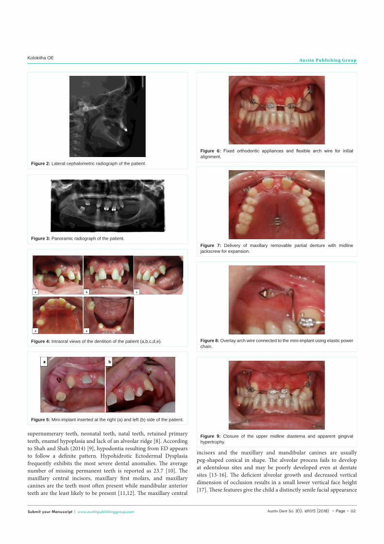
Figure 3: Panoramic radiograph of the patient.
Figure 4: Intraoral views of the dentition of the patient (a,b,c,d,e).
Figure 5: Mini-implant inserted at the right (a) and left (b) side of the patient.
Figure 6: Fixed orthodontic appliances and flexible arch wire for initial alignment.
Figure 7: Delivery of maxillary removable partial denture with midline jackscrew for expansion.
Figure 8: Overlay arch wire connected to the mini-implant using elastic power chain.
Figure 9: Closure of the upper midline diastema and apparent gingival hypertrophy.

Austin Dent Sci 3(1): id1015 (2018) - Page - 03
Kolokitha OE Austin Publishing Group
Submit your Manuscript | www.austinpublishinggroup.com
similar to an edentulous patient [16].
Typically, the face of a patient with ectodermal dysplasia is characterized by square aspect of the forehead, frontal bossing, sunken cheeks, hyper pigmentation of the periorbital skin, prominent supraorbital ridges, saddle nose, large low-set ears and protuberant lips [18]. Other symptoms include usually dry, scaly, and easily irritated skin; sweat glands that may be absent or few in number or non-functioning; eyebrows, eyelashes and other body hair that may be sparse or absent; and hair that may be fragile, dry, and generally disorderly because of the lack of sebaceous glands [19]. Salivary glands, including the intraoral accessory glands are sometimes hypoplastic resulting in xerostomia and dry, cracked lips with pseudorhagades formation [20].
A young patient with ectodermal dysplasia is facing several functional, esthetic, psychosocial and psychological problems [9,16]. Tooth agenesis has been shown to have a significant impact upon the oral health related quality of life. The oral conditions imposed by this syndrome necessitate multidisciplinary dental treatment for balanced development of both the functioning and esthetic aspects of the dentition of an ED patient [21].
Dental management of ectodermal dysplasia traditionally involves the provision of a series of complete or removable partial dentures during the growing years [22-29] and definitive habilitation following completion of jaw growth [30-31]. The role of orthodontics is equally important, starting from the initial visit of the patient to the pediatric dentist. The absence of many primary and permanent teeth, combined with the underdeveloped alveolar processes, leaves the existing, usually conical in shape teeth, often with unfavourable positions (such as rotations and retroclinations) [32]. Alignment and proper positioning of the existing teeth is a prerequisite for a successful rehabilitation of the dentition. However, with the limited number of permanent teeth present, anchorage remains an issue. Mini-implants offer many opportunities in this field. It is the intent of this article to present a young patient with ectodermal dysplasia who benefited from the use of mini-implants for proper positioning of the existing teeth with the simultaneous use of removable prosthesis.
Case PresentationA 15-year old boy with ectodermal dysplasia presented in the
Graduate Orthodontic Clinic of the University with chief complaint the multiple missing teeth. He had sparse, fine silky hair, sparse eyebrows and eyelashes, dry skin, hyper pigmentation around the eyes and mouth, large low-set ears, depressed nasal bridge and protuberant lips (Figure 1a,b). The lateral cephalogram taken at the
time of the initial examination showed a concave hard tissue and soft tissue profile, a posterior position of the maxilla and a forward chin position in relation to the cranial base, an increased labial inclination of the upper incisors in relation to their osseous base and the cranium (Figure 2). The intraoral examination revealed the presence of only 8 permanent teeth (#16,13,11,21,23,33,43,47) and a retained primary tooth (#62). The panoramic x-ray revealed numerous agenesis in the permanent dentition (20 permanent teeth were congenitally absent) and obvious resorption of the root of #62 (Figure 3). His maxillary incisors, already built with composite resin to camouflage their conical shape, were unaesthetically proclined with an enlarged midline diastema (5 mm) and the maxillary canines were conical and labially inclined. Intraorally, the mucosa was dry and the mandible exhibited narrow knife-edge shaped alveolar ridges (Figure 4a,b,c,d,e).
Treatment options considered for this patient included constructing a removable partial denture for the upper arch and a removable complete over denture for the lower. However, the enlarged upper midline diastema and the significant proclination of the maxillary anteriors, as well as the excessive vertical overlap, dictated the implication of Orthodontics in the treatment plan. Due to congenitally multiple missing teeth, mini-implants were used as anchorage units for the orthodontic correction of the position of the upper front teeth.
Initially, two orthodontic mini-implants were placed distal of #13 and 23 (Figure 5a,b). A longer than usual healing duration of six weeks was decided, due to the poor quality of bone at the insertion sites, based on radiologic examination. Fixed orthodontic appliances (0.022’’ slot) were bonded on #13,11,21,23 and initial alignment was achieved with the use of flexible arch wires (Figure 6). While the leveling was continued, successive maxillary removable partial dentures equipped with midline jackscrews were used for maxillary expansion and temporary rehabilitation of the patient (Figure 7). With the mini-implants been healed and the maxillary anteriors
Figure 10: Gingivectomy performed for the improvement of the esthetic result.
Figure 11: Close-up view of the healing of the soft tissues following gingivectomy.
Figure 12: Effort for cuspid intrusion (#13) shortly prior to mini-implant removal.

Austin Dent Sci 3(1): id1015 (2018) - Page - 04
Kolokitha OE Austin Publishing Group
Submit your Manuscript | www.austinpublishinggroup.com
nicely aligned, an overlay arch wire was inserted and connected to the mini-implants using elastic power chain (Figure 8). Progressively in treatment, upper midline diastema closed and maxillary anterior teeth were retracted successfully with the aid of skeletal anchorage (Figure 9). Due to a gingival hypertrophy that occurred as a result of poor oral hygiene and closure of the midline diastema, and in order to achieve optimum esthetic results, gingivectomy was also decided and successfully performed at all the anterior area (Figure 10,11). The overall orthodontic treatment lasted 5 months.
The patient all this time was wearing an interim partial denture for the maxilla and an interim over denture for the mandible. The final improvement of the orthodontic treatment consisted of some intrusion of #13, taking advantage of the skeletal anchorage of the mini-implant on the right side (Figure 12). The fixed appliances were removed and a removable upper Hawley appliance was delivered to the patient (Figure 13a,b). The result was also retained with a bonded lingual fixed retainer between the upper central incisors. At that point, it was decided to refer the patient to the Department of Operative Dentistry for esthetic improvement of the shape of the upper cuspids. Re-evaluation of the removable prostheses will follow at the Department of Removable Prosthodontics. Once growth is completed, evaluation for new removable, fixed or implant based prostheses will take place.
DiscussionPatients with ectodermal dysplasia present to the dentist with
multiple restorative issues. Early prosthetic management is necessary in conjunction with the orthodontic intervention to correct the positioning of the existing, possible misshapen teeth. Oligodontia and, in general, the compromised dental status of these young patients affected from ED can have implications on their psychosocial health. Children with severe hypodontia generally have low oral health-related quality of life [33] and have been shown to suffer from psychosocial depression, especially around the age of nine, as they begin to realize that their condition is quite different from that of other children [34]. This is the reason why it is suggested to these patients to start treatment while in childhood and adolescence.
Individuals with ED are routinely managed by a series of removable complete or partial dentures during the growing years and final rehabilitation following the completion of jaw growth [16]. In the case presented, dental prostheses improved the tone of the muscles of mastication and compensated for the reduced vertical dimension. During treatment time and thereafter, higher self-esteem and a better social acceptance were promoted for the benefit of the young patient.
Figure 13: Patient wearing an Upper Hawley appliance with acrylic teeth for the replacement of all missing posterior teeth (a,b).
The treatment objective of this case presentation was to report a clinical case of ectodermal dysplasia with oligodontia where the contribution of mini-implants proved to be very effective in the anchorage needed for the orthodontic approach. Compared with other skeletal anchorage devices, the versatility, minimal surgical invasiveness and low cost make the orthodontic mini-implants a viable option in the multiple missing teeth situation [35-39]. And although, mini-implants are ready to load and do not usually require any longer healing period, in our case, due to poor bone quality at the insertion sites, it was decided to wait for 6 weeks to heal before loading the segmental arch wire engaged to the anterior maxillary teeth. As was shown, the anchorage via the mini-implants proved effective for the leveling and correction of the inclination of the maxillary front teeth as well as the vertical dimension.
To determine the ideal time for implant treatment in children seems quite difficult, as many different aspects have to be considered [40]. Treatment guidelines issued by the National Foundation for Ectodermal Dysplasias indicate that implants are only recommended for the mandible’s anterior portion in children older than school age [41]. Due to serious financial issues of the family of our case, no treatment approach involving future conventional implants as fixed prostheses could be proposed. In so, the patient was left with his removable prostheses fabricated at the end of the orthodontic treatment.
ConclusionIn multiple tooth agenesis, as is the case with the hereditary
condition of ectodermal dysplasia, orthodontic treatment where the anchorage is obviously deficient, is an issue and the implementation of mini-implants is critical. The treatment results of the case presented showed that orthodontic and orthopedic treatment (via mini-implants) in coordination with prosthetic restoration benefited and significantly improved the function, growth, esthetics and psychological status of the patient. The purpose of this case presentation was to highlight the use of mini-implants as a powerful treatment tool, in young patients with oligodontia. The need for interdisciplinary treatment approach and the cooperation between dental specialists in cases with multiple missing teeth is mandatory for the benefit of the patient.
References1. Thurnam J. Two cases in which the skin, hair and teeth were very imperfectly
developed. Med Chir Trans. 1848; 31: 71-82.
2. Weech AA. Hereditary ectodermal dysplasia (congenital ectodermal defect). Am J Dis Child. 1929; 37: 766-790.
3. Bani M., Tezkirecioglu AM, Akal N, Tuzuner T. Ectodermal dysplasia with anodontia: A report of two cases. Eur J Dent. 2010; 4: 215-222.
4. Tarjan I, Gabris K, Rozsa N. Early prosthetic treatment of patients with ectodermal dysplasia: a clinical report. J Prosthet Dent. 2005; 93: 419-424.
5. Salinas CF, Jorgenson RJ, Wright JT, DiGiovanna JJ, Fete MD. 2008 International Conference on Ectodermal Dysplasias Classification: Conference Report. Am J Med Genet A. 2009; 149A: 1958-1969.
6. Deshmukh S, Prashanth S. Ectodermal Dysplasia: A Genetic Review. Int J Clin Pediatr Dent. 2012; 5: 197-202.
7. Clauss F, Waltmann E, Barriere P, Hadj-Rabia S, Manière MC, Schmittbuhl M. Dento-maxillo-facial phenotype and implants-based oral rehabilitation in Ectodermal Dysplasia with WNT10A gene mutation: Report of a case and literature review. Journal of Craniomaxillofac Surg. 2014; 42: e346-e351.

Austin Dent Sci 3(1): id1015 (2018) - Page - 05
Kolokitha OE Austin Publishing Group
Submit your Manuscript | www.austinpublishinggroup.com
8. Witkop CJ, Brearly LJ, Gentry WC. Hypoplastic enamel, onycholysis, and hypohidrosis inherited as an autosomal dominant trait. A review of ectodermal dysplasia syndromes. Oral Surg Oral Med Oral Pathol. 1975; 39: 71-86.
9. Shah R, Shah S. Oral rehabilitation of a patient with ectodermal dysplasia: A multidisciplinary approach. J Nat Sci Biol Med. 2014; 5: 462-466.
10. Crawford PJ, Aldred MJ, Clarke A. Clinical and radiographic dental findings in X linked hypohidrotic ectodermal dysplasia. J Med Genet. 1991; 28: 181-185.
11. Nakata M, Koshiba H, Eto K, Nance WE. A genetic study of anodontia in X-linked hypohidrotic ectodermal dysplasia. Am J Hum Genet. 1980; 32: 908-919.
12. Guckes AD, Roberts MW, McCarthy GR. Pattern of permanent teeth present in individuals with ectodermal dysplasia and severe hypodontia suggests treatment with dental implants. Pediatr Dent. 1998; 20: 278-280.
13. Everett FG, Jump EB, Sutherland WF, Savara BS, Suher T. Anhidrotic ectodermal dysplasia with anodontia: a study of two families. J Am Dent Assoc. 1952; 44: 173-186.
14. Soderholm AL, Kaitila I. Expression of X-linked hypohidrotic ectodermal dysplasia in six males and in their mothers. Clin Genet. 1985; 28: 136-144.
15. Tape MW, Tye E. Ectodermal dysplasia: literature review and a case report. Compend Contin Educ Dent. 1995; 16: 524-528.
16. Suri S, Carmichael RP, Tompson BD. Simultaneous functional and fixed appliance therapy for growth modification and dental alignment prior to prosthetic habilitation in hypohidrotic ectodermal dysplasia: A clinical report. J Prosthet Dent. 2004; 92: 428-433.
17. Bondarets N, McDonald F. Analysis of the vertical facial form in patients with severe hypodontia. Am J Phys Anthropol. 2000; 111: 177-184.
18. Aydinbelge M, Gumus HO, Sekerci AE, Demetoğlu U, Etoz OA. Implants in children with hypohidrotic ectodermal dysplasia: An alternative approach to esthetic management: case report and review of the literature. Pediatr Dent. 2013; 35: 441-446.
19. Yenisey M, Guler A, Unal U. Orthodontic and prosthodontic treatment of ectodermal dysplasia: A case report. Br Dent J. 2004; 196: 677-679.
20. Besserman-Nielsen M. Hypohidrotic ectodermal dysplasia. Oral manifestation and genetic aspects. Tandlaegebladet. 1971; 75: 1057-1076.
21. Muzio LL, Bucci P, Carile F, Riccitiello F, Scotti C, Coccia E, et al. Prosthetic rehabilitation of a child affected from anhydrotic ectodermal dysplasia: a case report. J Contemp Dent Pract. 2005; 6: 120-126.
22. Herer PD. Treatment of anhidrotic ectodermal dysplasia: report of a case. ASDC J Dent Child. 1975; 42: 133-136.
23. Sarnat BG, Brodie AG, Kubacki WH. Fourteen-year report of facial growth in case of complete anodontia with ectodermal dysplasia. AMA Am J Dis Child. 1953; 86: 162-169.
24. Tocchini JJ, West FT, Bartlett RW. An unusual developmental pattern in a case of hypodrotic ectodermal dysplasia. ASDC J Dent Child. 1979; 37: 158-159.
25. Nortje CJ, Farman AG, Thomas CJ, Watermeyer GJ. X-linked hypohidrotic ectodermal dysplasia-An unusual prosthetic problem. J Prosthet Dent. 1978; 40: 137-142.
26. Shaw RM. Prosthetic management of hypohidrotic ectodermal dysplasia with anodontia. Case report. Aust Dent J. 1990; 35:113-116.
27. Boj JR, Duran J, Cortada M, Jimenez A, Golobart J. Cephalometric changes in a patient with ectodermal dysplasia after placement of dentures. J Clin Pediatr Dent. 1993; 17: 217-220.
28. Franchi L, Branchi R, Tollaro I. Craniofacial changes following early prosthetic treatment in a case of hypohidrotic ectodermal dysplasia with complete anodontia. ASDC J Dent Child. 1998; 65: 116-121.
29. Itthagarun A, King NM. Oral rehabilitation of a hypohidrotic ectodermal dysplasia patient: a 6-year follow-up. Quintessence Int. 2000; 31:642-648.
30. Guckes AD, Brahim JS, McCarthy GR, Rudy SF, Cooper LF. Using endosseous dental implants for patients with ectodermal dysplasia. J Am Dent Assoc. 1991; 122; 59-62.
31. Davarpanah M, Moon JW, Yang LR, Celletti R, Martinez H. Dental implants in the oral rehabilitation of a teenager with hypohidrotic ectodermal dysplasia: report of a case. Int J Oral Maxillofac Implants. 1997; 12: 252-258.
32. Ioannidou-Marathiotou I, Kotsiomiti E, Gioka C. The contribution of orthodontics to the prosthodontic treatment of ectodermal dysplasia. J Am Dent Assoc. 2010; 141: 1340-1345.
33. Jokovic A, Locker D, Stephens M, Kenny D, Tompson B, Guyatt G. Validity and reliability of a questionnaire for measuring child oral- health-related quality of life. J Dent Res. 2002; 81: 459-463.
34. Högberg G, Lagerheim B, Sennerstam R. The 9-year crisis reflected at a rehabilitation center, at a child health care center and at a child and adolescent psychiatric center. Lakartidningen. 1986; 83: 2038-2042.
35. Costa A, Raffainl M, Melsen B. Miniscrews as orthodontic anchorage: a preliminary report. Int J Adult Orthodon Orthognath Surg. 1998; 13: 201-209.
36. Kanomi R. Mini-implant for orthodontic anchorage. J Clin Orthod. 1997; 31: 763-767.
37. Melsen B, Costa A. Immediate loading of implants used for orthodontic anchorage. Clin Orthod Res. 2000; 3:23-28.
38. Ludwig B, Baumgaertel S, Bowman J. Innovative Anchorage Concepts. Mini-Implants in Orthodontics. Berlin: Quintessence. 2008.
39. Wilmes B, Drescher D. Mini-Implantate in der Kieferorthopädie: Das Benefit-System. Kieferorthopädie. 2008.
40. Kramer FJ, Baethge C, Tschernitschek H. Implants in children with ectodermal dysplasia: A case report and literature review. Clin Oral Implants Res. 2007; 18: 140-146.
41. NFED Scientific Advisory Board. Parameters of Oral Health Care for Individuals Affected by Ectodermal Dysplasias. National Foundation for Ectodermal Dysplasias. 2003.
Citation: Kolokitha OE, Chatzistavrou E and Kotsiomiti E. Interdisciplinary Treatment of a Young Patient with Ectodermal Dysplasia with the Aid of Mini-Implants for Orthodontic Anchorage. Austin Dent Sci. 2018; 3(1): 1015.
Austin Dent Sci - Volume 3 Issue 1 - 2018Submit your Manuscript | www.austinpublishinggroup.com Kolokitha et al. © All rights are reserved