Interdisciplinary collaboration: an approach to optimize outcomes...
30
SCIENTIFIC SESSION 302 THE INTERNATIONAL JOURNAL OF ESTHETIC DENTISTRY Introduction base of the dental profession in terms of both the art and science of practice has significantly expanded. The amount of information and the application of treat- ment for patients are often overwhelm- ing. Despite the convenient access to knowledge in the modern world, it has become increasingly difficult for single clinicians to address all the needs of their patients. Within dentistry, the benefit of col- laboration between dental profession- als to treat patients through an inter- disciplinary approach is not a new concept. Moreover, it is usually re- warding for the clinician and can pro- duce optimal results for the patient. Today, a broader approach to patient care is recognized. As research pro- gresses in dental therapy, and more is known about the oral–systemic con- nection and tissue engineering, inter- disciplinary collaboration is paramount for the continued development of both dentistry and medicine. 6-8 Certainly, challenges often arise when working as part of a team, and research and clinical experience have revealed some of these challenges. 9-11 There- fore, the need to understand how best to learn from each other and to share knowledge is fundamental to a success- ful interdisciplinary relationship. With this greater understanding, the elements of communication, commitment, mutual trust, and respect allow for greater ac- ceptance of the responsibilities of each discipline. In this article, information is presented that is valuable to all members of the dental team so they can effectively par- ticipate in collaborative treatment. Cer- tain esthetic dilemmas may be simple, while others are more complex. Some of the significant treatment challenges include malproportioned, injured or non- restorable teeth; ectopically positioned or missing teeth; and altered periodon- tal attachment or ridge form. The shared interdisciplinary process of planning, design, communication, coordination, and execution of treatment for the es- thetically compromised dental patient is reviewed in this article. Interdisciplinary collaboration: an approach to optimize outcomes for patients with compromised dental esthetics J. Janakievski, V.O. Kokich, G. Kinzer
Transcript of Interdisciplinary collaboration: an approach to optimize outcomes...

SCIENTIFIC SESSION
302THE INTERNATIONAL JOURNAL OF ESTHETIC DENTISTRY
Introduction
base of the dental profession in terms of
both the art and science of practice has
significantly expanded. The amount of
information and the application of treat-
ment for patients are often overwhelm-
ing. Despite the convenient access to
knowledge in the modern world, it has
become increasingly difficult for single
clinicians to address all the needs of
their patients.
Within dentistry, the benefit of col-
laboration between dental profession-
als to treat patients through an inter-
disciplinary approach is not a new
concept. Moreover, it is usually re-
warding for the clinician and can pro-
duce optimal results for the patient.
Today, a broader approach to patient
care is recognized. As research pro-
gresses in dental therapy, and more is
known about the oral–systemic con-
nection and tissue engineering, inter-
disciplinary collaboration is paramount
for the continued development of both
dentistry and medicine.6-8
Certainly, challenges often arise when
working as part of a team, and research
and clinical experience have revealed
some of these challenges.9-11 There-
fore, the need to understand how best
to learn from each other and to share
knowledge is fundamental to a success-
ful interdisciplinary relationship. With this
greater understanding, the elements of
communication, commitment, mutual
trust, and respect allow for greater ac-
ceptance of the responsibilities of each
discipline.
In this article, information is presented
that is valuable to all members of the
dental team so they can effectively par-
ticipate in collaborative treatment. Cer-
tain esthetic dilemmas may be simple,
while others are more complex. Some
of the significant treatment challenges
include malproportioned, injured or non-
restorable teeth; ectopically positioned
or missing teeth; and altered periodon-
tal attachment or ridge form. The shared
interdisciplinary process of planning,
design, communication, coordination,
and execution of treatment for the es-
thetically compromised dental patient is
reviewed in this article.
Interdisciplinary collaboration:
an approach to optimize outcomes
for patients with compromised dental
esthetics
J. Janakievski, V.O. Kokich, G. Kinzer

303THE INTERNATIONAL JOURNAL OF ESTHETIC DENTISTRY
JANAKIEVSKI ET AL
Space and alveolar ridge
deficiencies
Esthetic compromise may be present
in several different ways. However, it
typically occurs in the dentition that has
some degree of clinical “deficiency”.
Occasionally, patients may present
with both hard and soft tissue deficien-
cies due to disease, trauma or missing
teeth.13 In addition to tissue deficien-
cies, patients frequently have insufficient
spacing for the placement of implants
or the replacement of teeth. If these
areas are not evaluated and addressed
during the treatment-planning phase,
the final result will be compromised.
Consequently, the mismanagement of
these parameters may result in malpro-
portioned teeth, lack of proper soft tis-
sue symmetry, and/or aberrant relation-
ships between the papilla height and
the contact length.16,17 Poor planning
may also create challenges for proper
implant placement, leading to difficulties
in managing the occlusion and potential
speech problems.
The following discussion addresses
the role of interdisciplinary treatment in
the evaluation and management of pa-
tients with a variety of clinical “deficien-
cies”. The topics covered include space
appropriation for teeth and implants,
and considerations for deficient ridges
when planning tooth replacement for
both adults and growing patients.
1. Space appropriation
Appropriate position of teeth
There are a number of different cir-
cumstances that can impact the ideal
positioning of teeth. The most common
tooth-position problem is crowding, and
treatment to resolve this issue is usually
predictable. Teeth are aligned ortho-
dontically according to their mesiodistal
widths and as long as there is not a clini-
cally significant tooth-size discrepancy,
the esthetics and occlusion can be es-
tablished well.18 Unfortunately, space
appropriation becomes more challeng-
ing when faced with clinical findings that
include multiple missing teeth, retained
deciduous teeth, or malproportioned
teeth (Fig 1). Therefore, it is impera-
tive for the orthodontist to work with the
restorative dentist when determining
Fig 1 Fig 2

SCIENTIFIC SESSION
304THE INTERNATIONAL JOURNAL OF ESTHETIC DENTISTRY
ideal spacing and appropriate tooth
position in these patients. It is also im-
portant to begin the treatment-planning
process by determining the correct “es-
thetic” position of the teeth as it relates to
the patient’s face. After this has been es-
tablished, attention can be focused on
positioning the teeth for proper function.
This process is called “Facially Generat-
ed Treatment Planning” (FGTP), and has
been discussed in depth by Spear.
This basic concept of FGTP is derived
from how teeth are ideally positioned in
a complete denture setup.
Developing a treatment plan based
on the position of the teeth relative to
the face minimizes challenges that
arise when trying to create a plan uti-
lizing only mounted diagnostic models
and radiographs. A perfect example of
this is the patient with a worn dentition
significant incisal edge wear also devel-
op compensatory eruption, which main-
tains the initial incisal edge level relative
to the face. In this scenario, treatment
alternatives may include osseous crown
lengthening or orthodontic intrusion to
move the crestal bone and gingival lev-
els apically into a more esthetic position.
a final, restored incisal edge position
that is similar to the pretreatment incis-
al edge level. Other patients with more
generalized tooth wear may experience
a greater loss of vertical dimension re-
quiring more extensive crown lengthen-
ing to re-establish a favourable lower
facial height. To be able to choose the
appropriate treatment plan, it is impor-
tant to always first identify the desired
position of the maxillary central inci-
sors. It is difficult to set any other teeth
first because we have no reference for
where they should be positioned. In or-
der to do this, the clinician will evaluate
the relationship of the teeth to the lips in
repose and during full smile, to the pos-
ition and curvature of the lower lip, and
Figs 3a to 3c
a
b
c

305THE INTERNATIONAL JOURNAL OF ESTHETIC DENTISTRY
JANAKIEVSKI ET AL
to the horizon or interpupillary line
(Figs 3a to 3c). After the central incisor
position is selected, the positioning of
the laterals, canines, premolars, and
molars becomes more predicatable.
Following this step-by-step process pro-
vides guidelines for treatment planning
with predictable esthetic outcomes and
no compromises at the end of treatment
(Fig 4).
Appropriate spacing of teeth
The orthodontist plays a key role in help-
ing to establish proper space require-
ments for patients with missing teeth or
and Fig 6). However, in this example,
two questions arise: Given the obvious
difference in tooth dimensions between
the deciduous and permanent teeth,
how does the orthodontist know where
to position the teeth in order to provide
adequate room for replacement of the
Fig 4
Fig 6
Figs 5a to 5c
a
b
c

SCIENTIFIC SESSION
306THE INTERNATIONAL JOURNAL OF ESTHETIC DENTISTRY
deciduous teeth? Also, since the alveo-
lar ridges were never developed by the
eruption of permanent teeth, what is the
most appropriate way to manage these
deficient ridges?
The most common mistake that most
orthodontists make when trying to ap-
propriate space for patients with mul-
tiple missing teeth is doing it without
any input from their fellow interdiscipli-
nary team members. For a predictable
outcome when planning treatment for
these patients, it is imperative that the
orthodontist be aware of the desired fi-
nal restorative and surgical objectives.
Actually, the restorative dentist has the
most influence over final tooth position,
while the orthodontist educates the team
on which tooth movements are biome-
chanically realistic and when auxiliary
implant anchorage may be necessary to
achieve those initial restorative and sur-
gical goals. The surgeon and restora-
tive dentist understand the importance
of ideal implant location and appropriate
tooth dimension, respectively, as they re-
late to function and esthetics. Therefore,
orthodontic space appropriation should
be determined based on a collaborative
effort by the interdisciplinary team.
Conversely, the orthodontist has pri-
mary control over space appropriation
in patients with single missing anterior
teeth. It is thus rarely necessary in these
situations for the orthodontist to solicit in-
put from the restorative dentist and sur-
geon unless the patient exhibits abnor-
mally small teeth. The literature outlines
four specific ways to appropriate space
for the single missing anterior tooth.
The first method is utilization of the gold-
en proportion. However, since this per-
ceived value has no true relation to the
actual measured widths of the teeth, it
has no value to the orthodontist when
creating the proper spacing. The sec-
ond alternative to determine ideal spac-
ing is to use the contralateral tooth as a
guide. However, it is not suitable for pa-
tients whose contralateral tooth is miss-
ing or malformed. A third option utilizes
compare the widths of the maxillary and
mandibular anterior teeth.18
ratio is 0.78 and a mathematical equa-
tion can be used to achieve the unknown
Figs 7a and 7b
a b

307THE INTERNATIONAL JOURNAL OF ESTHETIC DENTISTRY
JANAKIEVSKI ET AL
width(s). Finally, the most predictable
guide for determining appropriate spac-
ing is to construct an orthodontic setup/
diagnostic wax setup. This setup is used
to determine the position of the teeth nec-
essary for optimal esthetics and proper
development of the lateral and protrusive
functional pathways.
Case example
As discussed above, it was important
to begin by determining the correct es-
thetic and functional position of the max-
illary central incisors relative to the face
(Figs 7a and 7b). After evaluating those
parameters, it was determined that the
shortening the incisal edge and increas-
ing the width would enhance the esthetic
appearance of the central incisors.
Once the position and shape of the
maxillary central incisors were estab-
lished, diagnostic waxing was used to
determine correct size and position for
the lateral incisors, canines, and pre-
molars. This, in turn, helped to confirm
the position of the natural first molars,
which were not in need of restoration.
The position of the mandibular incisors is
based in part on esthetics and in part on
function. Generally, the amount of over-
bite or vertical overlap of the incisors is
related to the cusp-fossa relationship
of the posterior teeth. The deeper the
cusp-fossa relationship, the greater the
overbite must be to ensure proper dis-
clusion of the posterior teeth. The poste-
rior teeth for this patient had a relatively
flat topography, which meant that less
incisor overlap was required for function.
The benefit of maintaining a shallower
anterior guidance is to observe force
distribution during lateral disclusion.
For this patient, maxillary implants were
used to guide the lateral disclusion;
therefore, it is important to understand
the impact that the disclusion angle has
on the functional success of the restor-
ation. For every 10 degree increase/de-
crease in the angle of disclusion, there
stress at the abutment level. Therefore,
to help provide some proprioception
during lateral disclusion, it was decid-
ed that the mandibular canines should
be orthodontically moved distally into a
more ideal canine position. The intention
Figs 8a and 8b
a b

SCIENTIFIC SESSION
308THE INTERNATIONAL JOURNAL OF ESTHETIC DENTISTRY
was to produce a lateral disclusion not
completely derived by two implants op-
posing each other. It is also important
to understand that, at this point, the di-
agnostic setup is simply an estimate of
anterior/posterior tooth position that re-
quires confirmation by way of a thorough
clinical examination (Figs 8a and 8b).
Following completion of the diagnos-
tic setup/wax-up, the orthodontic phase
of treatment can begin. One common
challenge in managing these types of
patients is translating the diagnostically
derived tooth position back to the mouth.
There are, in fact, several ways this can
be accomplished. If the diagnostic work-
up was digitally created (ie, Invisalign),
then the communication of tooth pos-
ition can be accomplished through the
aligners. Another method can be to cor-
rect the tooth size intraorally by chang-
ing crown shape with composite or by
placing provisional restorations. A third
alternative can be to orthodontically esti-
mate the correct tooth position and then
periodically re-evaluate the position with
progress models and a subsequent di-
agnostic wax-up. All three techniques
can be used successfully, and occasion-
ally it may be necessary to utilize more
than one option for the same patient.
For the patient shown in Figure 9, the
first surgical phase of treatment con-
sisted of extracting the primary molars,
Figs 9a to 9d
a
c
b
d
309THE INTERNATIONAL JOURNAL OF ESTHETIC DENTISTRY
JANAKIEVSKI ET AL
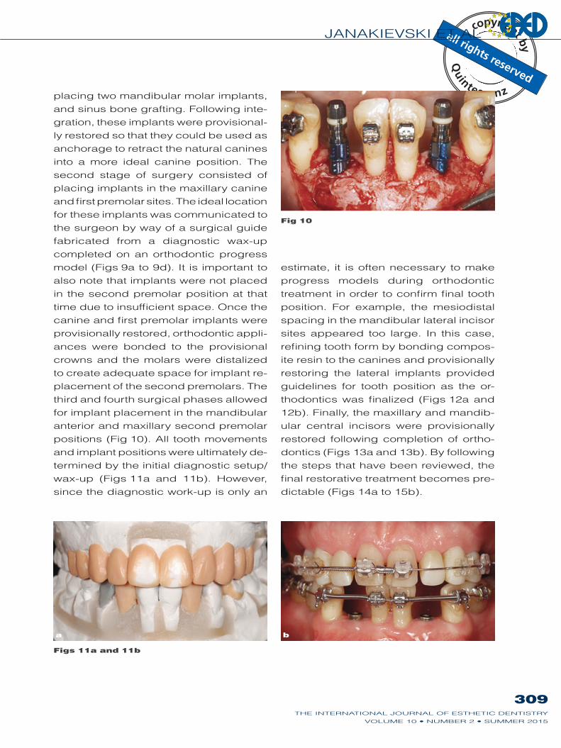
placing two mandibular molar implants,
and sinus bone grafting. Following inte-
gration, these implants were provisional-
ly restored so that they could be used as
anchorage to retract the natural canines
into a more ideal canine position. The
second stage of surgery consisted of
placing implants in the maxillary canine
and first premolar sites. The ideal location
for these implants was communicated to
the surgeon by way of a surgical guide
fabricated from a diagnostic wax-up
completed on an orthodontic progress
model (Figs 9a to 9d). It is important to
also note that implants were not placed
in the second premolar position at that
time due to insufficient space. Once the
canine and first premolar implants were
provisionally restored, orthodontic appli-
ances were bonded to the provisional
crowns and the molars were distalized
to create adequate space for implant re-
placement of the second premolars. The
third and fourth surgical phases allowed
for implant placement in the mandibular
anterior and maxillary second premolar
positions (Fig 10). All tooth movements
and implant positions were ultimately de-
termined by the initial diagnostic setup/
wax-up (Figs 11a and 11b). However,
since the diagnostic work-up is only an
estimate, it is often necessary to make
progress models during orthodontic
treatment in order to confirm final tooth
position. For example, the mesiodistal
spacing in the mandibular lateral incisor
sites appeared too large. In this case,
refining tooth form by bonding compos-
ite resin to the canines and provisionally
restoring the lateral implants provided
guidelines for tooth position as the or-
-
ular central incisors were provisionally
restored following completion of ortho-
the steps that have been reviewed, the
final restorative treatment becomes pre-
Fig 10
Figs 11a and 11b
a b

SCIENTIFIC SESSION
310THE INTERNATIONAL JOURNAL OF ESTHETIC DENTISTRY
Figs 12a and 12b
a b
Figs 13a and 13b
a b
Figs 14a to 14c
a b c
Figs 15a and 15b
a b

JANAKIEVSKI ET AL
311THE INTERNATIONAL JOURNAL OF ESTHETIC DENTISTRY
Ridge dimension (horizontal) –
restorative considerations
When an edentulous site is present, it is
important for the restorative dentist to un-
derstand the limitations of alveolar bone
regeneration and how they may influ-
ence the restorative treatment-planning
process. It is also critical to understand
the relationship between the underlying
bone and soft tissue. As has been shown
by Salama et al, there are limitations to
the amount of soft tissue height that
can predictably be maintained above
the crest of bone (Fig 16). The chart
in Figure 16, adapted from the Salama
et al article, shows the height of soft
tissue will vary, depending on the clin-
ical situation and the treatment option.
The most difficult clinical scenario for the
team to resolve is the presence of two
adjacent edentulous sites in the esthetic
zone. Achieving ideal papillae height in
these patients is not only unpredictable
but also directly impacted by the chal-
lenge the surgeon faces when attempt-
ing to augment the vertical bone height.
Therefore, placing adjacent implants in
the esthetic zone is generally avoided
in favor of treatment options that enable
the soft tissue height to be more predict-
ably maintained.
A deficiency in ridge form may arise as
a result of factors such as disease, miss-
ing teeth, and trauma. Prior to the restor-
ation or replacement of missing teeth, it
is necessary to evaluate the parameters
that will lead to an esthetic result.31 In
patients requiring multiple tooth replace-
ment, specific factors such as the span
of the edentulous space, periodontal
attachment levels on adjacent teeth, al-
veolar ridge form, and soft tissue profile
and thickness will influence the predict-
ability of the different treatment options
and ultimately the final esthetic outcome.
Therefore, creating an ideal recipient site
in both the horizontal and vertical dimen-
sions prior to the replacement of missing
teeth allows for ideal implant placement
and a predictable esthetic outcome.
Case example
A 17-year-old female patient presented
with a dental history that included trau-
ma to the maxillary anterior teeth at the
age of 10, which resulted in the avulsion
Fig 16
ClassRestorative environment
Proximity limitations (mm)
Vertical soft tissue limitations (mm)
1 Tooth-tooth 1.0
Tooth-pontic NA
3 Pontic-pontic NA 6.0
4 Tooth-implant
Implant-pontic NA
6 Implant-implant 3.0
Adapted from: Salama H, et al. The interproximal height of bone: a guidepost to predictable aesthetic strategies
and soft tissue contours in anterior tooth replacement. Pract Periodontics Aesthet Dent 1998;10:1131–1141.

SCIENTIFIC SESSION
312THE INTERNATIONAL JOURNAL OF ESTHETIC DENTISTRY
of the maxillary right central and lateral
incisors (Figs 17a and 17b). Although
the teeth were replanted, the lateral inci-
sor subsequently fractured and was ex-
the radiograph, the maxillary right cen-
tral incisor is ankylosed and undergo-
ing replacement resorption (Figs 18a to
18c). When this crown fractures, it will
create two adjacent edentulous sites.
When comparing the current papilla
height on the distal of the central inci-
sor to the contralateral papilla, an ap-
proximately 6 mm difference in height is
evident. Presently, the central incisor is
providing a supracrestal gingival attach-
ment. As a result, this discrepancy will
increase once the central incisor fails.
Therefore, it is important to develop a
treatment plan that will be predictable
and provide an excellent esthetic re-
sult. The three typical treatment plans
that would be considered for this clinical
situation are the following:
1. Augmentation of the ridge and the
placement of two implants – the max-
illary right central and lateral incisor
sites.
placement of one implant – the maxil-
lary right central incisor site and can-
tilever the lateral incisor.
Figs 17a and 17b
a b
Figs 18a to 18c a b c
JANAKIEVSKI ET AL
313THE INTERNATIONAL JOURNAL OF ESTHETIC DENTISTRY
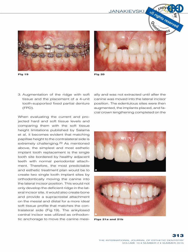
3. Augmentation of the ridge with soft
tissue and the placement of a 4-unit
tooth-supported fixed partial denture
(FPD).
When evaluating the current and pro-
jected hard and soft tissue levels and
comparing them with the soft tissue
height limitations published by Salama
et al, it becomes evident that matching
papillae height to the contralateral side is
extremely challenging. As mentioned
above, the simplest and most esthetic
implant tooth replacement is the single
tooth site bordered by healthy adjacent
teeth with normal periodontal attach-
ment. Therefore, the most predictable
and esthetic treatment plan would be to
create two single tooth implant sites by
orthodontically moving the canine into
the lateral incisor position. This would not
only develop the deficient ridge in the lat-
eral incisor site, it would also create bone
and provide a supracrestal attachment
on the mesial and distal for a more ideal
soft tissue profile that matches the con-
tralateral side (Fig 19). The ankylosed
central incisor was utilized as orthodon-
tic anchorage to move the canine mesi-
ally and was not extracted until after the
canine was moved into the lateral incisor
position. The edentulous sites were then
augmented, the implants placed, and fa-
cial crown lengthening completed on the
Fig 19 Fig 20
Figs 21a and 21b
a
b

SCIENTIFIC SESSION
314THE INTERNATIONAL JOURNAL OF ESTHETIC DENTISTRY
maxillary left central incisor to reposition
integration of the maxillary right canine
and central incisor implants, provisional
crown restorations were placed. Direct
composite was bonded to the right lat-
-
provement in a vertical soft tissue pos-
ition was achieved through horizontal
tooth movement, one that would have
been extremely difficult to accomplish
with adjacent edentulous sites.
Ridge dimension (horizontal) –
surgical considerations
The clinical scenario that yields the most
predictable and esthetic implant tooth
replacement is the single tooth site bor-
dered by healthy adjacent teeth with
normal periodontal attachment. Wheth-
er it is an orthodontic patient with con-
genitally missing teeth or a restorative
patient with a history of trauma or failed
treatment, the approach is similar.
In order to achieve an esthetic out-
come, there are certain biologic param-
eters that need to be understood. Dental
implant therapy has been criticized as
being unesthetic.33,34 Outcomes have
been noted showing facial tissue reces-
sion and interproximal papillae loss. This
was certainly more common in the early
years when implant therapy was transi-
tioning from the fully edentulous patient
to the partially edentulous patient. Fortu-
nately, the biologic limitations of implant
placement are better understood to-
day.
platform of an implant is a normal pro-
cess after abutment connection. This
remodeling occurs 360 degrees around
the implant in both the vertical and hori-
zontal directions. Therefore, the dimen-
sions of the alveolar ridge and recipient
site are of particular importance.
It has been shown that a dental im-
to a natural tooth.38 The resultant circum-
ferential bone remodeling can cause
periodontal attachment loss on an adja-
cent tooth with subsequent loss of papil-
lary height. Since there is no connective
tissue fiber insertion to an implant, the
form and height of the papillae are de-
pendent on a healthy attachment to the
tooth. Therefore, the orthodontist must
be aware of this limitation when creating
space to allow for implant replacement
of missing teeth.
Similarly, on the facial, a lack of bone
or bone remodeling can lead to gingi-
val margin recession and flattening of
the tissue form. Although modern re-
storative materials can help to over-
come some of the “shadowing” of the
tissues that often results with soft tissue
changes, the long-term esthetic result
may not be predictable. Recent esthetic
assessments have noted that esthetic
implant-supported crowns have a con-
vex facial–gingival contour, along with a
similar tissue color and texture as that of
the adjacent teeth.40,41 To achieve this
contour, it is important to plan for appro-
priate augmentation. In fact, a narrow al-
veolar ridge often requires augmentation
for both implant and soft tissue support.
Various methods have been developed
and studied, including the use of autog-
enous bone block grafts, guided bone
regeneration, and ridge split techniques.
Recently, a systematic review of studies
reporting lateral ridge augmentation out-
comes was completed,43 which showed
that all methods appear to be successful

315THE INTERNATIONAL JOURNAL OF ESTHETIC DENTISTRY
JANAKIEVSKI ET AL
when it comes to the survival of implants.
The guided bone regeneration technique
has the most long-term documentation
and appears to be the most common
procedure performed for lateral ridge
augmentation.43,44 Other important de-
tails, such as the differences in bone re-
modeling that each technique will have
on the esthetic outcome, are unknown
because this type of comparative study
has not been done.
Subsequently, it is necessary for al-
veolar ridge augmentation procedures
to regenerate bone not only for implant
support but also for “contour augmen-
tation” in order to provide a stable soft
tissue framework for the implant restor-
ation. Investigations into the stability
of these techniques continue, along with
the benefits of adding soft tissue grafting
together with implant placement.
Ridge dimension (horizontal) –
orthodontic considerations
As explained above, the most esthetic
implant restoration is one that has two
healthy teeth with normal periodontal
attachment on either side. In addition,
smaller edentulous sites provide adja-
cent teeth with a normal interproximal
periodontal attachment that can be
used as a guide and scaffold for ridge
augmentation procedures. When con-
fronted with multiple missing teeth, it is
often beneficial for the interdisciplinary
team to recommend orthodontic treat-
ment in order to move teeth in a horizon-
tal direction. Redistributing the space
and creating single tooth implant sites
can overcome the esthetic limitations of
consecutively placed implants. Using
orthodontic tooth movement to reduce
larger partially edentulous sites has sev-
eral advantages. It improves periodontal
attachment levels, helps to avoid con-
secutive implant placement, develops
better pontic sites, and helps the sur-
geon with the bone augmentation pro-
cedure. When considering this type of
lateral tooth movement, it is also impor-
tant to understand what happens biolog-
ically to the alveolar ridge. Although the
following research has focused on the
alveolar response to movement of pre-
molars and molars into narrower eden-
tulous ridges, the principles discussed
can also be applied to the alveolus in the
anterior maxilla.
A common question faced by every
orthodontist is whether or not to extract
an ankylosed primary molar that has no
permanent successor. If the decision to
extract the tooth is made before signifi-
cant eruption of the adjacent teeth has
occurred, then the periosteal pull be-
tween the erupting adjacent teeth will
stimulate bone deposition.49 However,
occasionally, the decision to extract the
ankylosed and submerged deciduous
second molar is made too late, resulting
in a narrow alveolar ridge with a vertical
defect. If nothing is done at the time of
surgery to try to maintain the buccolin-
gual volume of bone, it is typical to see
years following extraction. One option
to gain adequate ridge width and height
for implant placement in this site is to
perform a bone graft. Another possibility
exists if the patient is willing to undergo
orthodontic therapy. In this latter sce-
nario, it may be advantageous to move
the first premolar laterally into the sec-
ond premolar position, thereby creating
space for the single-tooth implant in the
first premolar location. When faced with

SCIENTIFIC SESSION
316THE INTERNATIONAL JOURNAL OF ESTHETIC DENTISTRY
this decision, clinicians are often fear-
ful that there is insufficient alveolar ridge
width in which to move the permanent
first premolar. However, previous stud-
ies have shown that a wider tooth root
can be moved through a narrow alveolar
ridge without compromising the eventu-
al bone support around the repositioned
tooth root. In fact, Hom showed that
the average increase in ridge width as a
molar is moved into a narrower ridge is
49 More recently, in a case series
of six patients, it was reported that ridge
width increased an average of 1.6 mm
when larger second premolars were
moved into narrower, edentulous first
premolar sites.
Another question often associated
with this treatment option is stability of the
and colleagues conducted a study
that evaluated 71 congenitally miss-
ing mandibular second premolar sites
following orthodontic movement of the
mandibular first premolar into the sec-
ond premolar site. Their objective was to
determine the stability of this orthodonti-
the completion of orthodontic treatment.
They found that the increased ridge
width was relatively stable, decreasing
A
-
the stability of alveolar bone developed
through orthodontic distalization of the
permanent maxillary canine in patients
with congenitally missing lateral inci-
sors. They also found excellent stabil-
ity in both the vertical and horizontal di-
after orthodontic treatment.
Ridge dimension (vertical) –
restorative considerations
Several treatment options exist for re-
placing multiple missing anterior teeth,
including implants, FPDs with pontics,
implants with pontics, and implants or FP-
Ds with pink ceramic/composite. In
order to determine the most appropriate
Fig 22
Figs 23a and 23b
a
b

JANAKIEVSKI ET AL
317THE INTERNATIONAL JOURNAL OF ESTHETIC DENTISTRY
alternative, it is helpful to know the soft
tissue limitations for the various treatment
options. According to the measured
values from Salama et al, it is more pre-
dictable to develop greater soft tissue
height above the bone by restoring ad-
jacent edentulous sites with pontics, as
compared to restoring with adjacent im-
plants (see Fig 16). The only location
where adjacent implants may be used
without significantly impacting the es-
thetic result is when two central incisors
are missing. Papillary height will still be
deficient between the two implants com-
pared to the natural teeth; however, it
will be more acceptable because it is in
the midline. Another option that is often
extremely esthetic is prosthetic replace-
ment of gingival tissue. For example, the
implant-supported FPD with pink porce-
lain. The result is esthetic since the lip
position during smile does not reveal the
transition between the prosthesis and
the patient’s mucosal tissue. The result
would not have been as esthetic if an
alveolar ridge augmentation procedure
had resulted in a ridge level that was still
short of ideal. This would have created
a situation where the junction between
prosthesis and soft tissue would have
been visible. Ultimately, understanding
the requirements of the restorative re-
cipient sites together with the surgical
limitations allows the team to develop
the best treatment plan for esthetic tooth
Ridge dimension (vertical) –
surgical considerations
The patient with multiple missing teeth
and a horizontally deficient alveolar
ridge can usually be treated predictably
in several ways. Lateral ridge augmen-
tation (as reviewed above) can be per-
formed successfully using various tech-
niques. Additional contour with either
tissue grafting can also benefit the re-
storative recipient sites, creating a con-
vexity of tissue form for either pontics or
implants.
Figs 24a to 24c
a
b
c

SCIENTIFIC SESSION
318THE INTERNATIONAL JOURNAL OF ESTHETIC DENTISTRY
The challenges increase as the ridge
becomes deficient in the vertical dimen-
sion. A recent review of vertical ridge
augmentation techniques and outcomes
illustrated the limitations with these pro-
cedures.61 Although there have been
multiple approaches to this problem, the
most commonly used techniques are
Vertically guided bone regeneration
requires the use of titanium-reinforced
membranes or titanium mesh to maintain
space for bone regeneration, with the
placement of a bone graft material to act
as a scaffold. Its application has been
shown to be effective in bone regener-
ation, with varying amounts of vertical
height and histologic evidence of bone
formation.63-66
for vertical ridge augmentation can be
attributed to the use of osteoconductive
graft products. An exciting and devel-
oping usage is that of growth factors to
aid with bone formation. Platelet-derived
growth factor (rhPDGF) enhanced matri-
ces have demonstrated the potential to
increase bone turnover and bone regen-
eration in both periodontal and dental
implant sites.67,68 In addition, bone mor-
-
oped and used in orthopedic medicine
and in dentistry as the first osteoinduc-
tive bone graft substitute, allowing sur-
geons to avoid complications associated
with harvesting autogenous bone.69 In
initially investigated in patients requiring
sinus augmentation and single tooth ex-
traction sockets.70,71 Since these initial
studies, clinicians have started applying
this approach to more challenging lat-
eral and vertical augmentation proced-
Vertical alveolar bone augmentation
using distraction osteogenesis was first
reported in the 1990s. Since then, mul-
tiple studies have evaluated the potential
to develop vertical bone growth, dem-
onstrating that distraction osteogenesis
can develop greater amounts of bone
vertically than other techniques.73,74
When the clinician plans treatment for
a patient with a severe vertical defect
Figs 25a to 25c
a
b
c

JANAKIEVSKI ET AL
319THE INTERNATIONAL JOURNAL OF ESTHETIC DENTISTRY
be given to techniques that will provide
the most predictable and stable regen-
erated bone. However, the complexity
of this technique certainly has the po-
tential for subsequent complications.
One study reported that complications
ranging from minor to more severe oc-
76 In addition,
this technique is effective in growing
bone in a vertical direction only.77,78
As discussed above, esthetic implant
restorations require facial convexity.
Therefore, it is common to plan for ad-
ditional horizontal ridge augmentation
and soft tissue grafting to achieve this
The three-dimensional reconstruction
of the alveolar ridge is a prerequisite to
achieving an optimal result at a site with
a severe ridge deficiency that is visible
in the esthetic zone (Figs 30a and 30b).
Ridge dimension (vertical) –
orthodontic considerations
Due to the challenges of vertical ridge
augmentation procedures, the dental
team needs to look at additional options
when confronted with patients who are
missing multiple teeth. Guided ortho-
dontic tooth eruption is one such option.
The primary goal of this procedure is to
develop bone in a vertical direction. This
is done by orthodontically applying ten-
sion to the periodontal ligament (PDL).
This stretching of the PDL fibers increas-
es osteoblastic activity and ultimately
Figs 26a and 26b
Figs 27a and 27b
a b
a b

SCIENTIFIC SESSION
320THE INTERNATIONAL JOURNAL OF ESTHETIC DENTISTRY
generates new bone formation.79–81 As
teeth are erupted, the soft tissue also
moves coronally. The interproximal bone
peaks can be developed to help support
the soft tissue and/or provide scaffold-
ing for bone augmentation procedures.
Therefore, in a patient with interproximal
bone loss and a resultant loss of papil-
lae, extrusion can predictably correct
the bone level and ultimately improve
the papillae height. In fact, it is believed
that lighter forces will result in a more
predictable soft tissue change for this
type of tooth movement.
There are four additional factors that
need to be considered when a tooth
is undergoing orthodontic extrusion.
The first is periodic reduction in length
to prevent occlusal interferences from
developing, unless there is no oppos-
ing tooth present. The second is that,
following any extrusive movement, it
is important to stabilize the tooth in its
new position for a period of 6 months
to allow time for the transseptal fibers
to reorganize horizontally.83 The third is
the importance of reviewing with the pa-
tient the esthetic change that generally
follows significant forced orthodontic
eruption. As the gingival margin moves
coronally with the extruded tooth, the es-
thetic change that accompanies it may
be an unexpected surprise for patients
if they are not made aware of it before
it happens. The fourth factor is that if a
tooth is ultimately planned for extraction
following extrusion, it is usually neces-
sary to perform prophylactic endodon-
tic therapy so that pulp exposure is not
a concern as the tooth is reduced in
length. Multiple authors have described
the extrusion of hopeless teeth prior to
extraction as a method of developing
implant sites.79,84 However, another way
to orthodontically augment an alveolar
ridge is to change the buccolingual incli-
nation of the teeth as they are extruded.
a hopeless maxillary incisor is extruded,
a larger volume of buccal bone will be
developed as the root moves coronally
and labially. The teeth that are consid-
ered for this type of tooth movement and
ridge development typically have signifi-
cant labial bone loss and a lack of PDL
in this area. Therefore, it is the PDLs on
the interproximal and lingua-palatal root
surfaces that lay down bone as the tooth
is orthodontically moved away from the
periodontal defect.
Case example
was congenitally missing a lateral in-
cisor. After completion of orthodontic
treatment, a dental implant was placed
in the maxillary right lateral incisor site.
Unfortunately, there were complica-
tions with the placement of the implant.
Seven subsequent surgical procedures
were performed in an attempt to repair
and save the implant. This resulted in
Fig 28

321THE INTERNATIONAL JOURNAL OF ESTHETIC DENTISTRY
JANAKIEVSKI ET AL
the creation of a large hard and soft tis-
sue defect with compromised adjacent
teeth. Given the severity of the defect
and the surgical challenge of regenerat-
ing bone in the vertical dimension, the
dental team discussed numerous inter-
disciplinary treatment options. These
ranged from a FPD with or without soft
tissue augmentation, orthodontic erup-
tion for extraction of the adjacent teeth,
to extraction and placement of implants
in the canine and central incisor sites
with pink prosthetic tissue replacement.
Along with the clinical findings, consid-
eration was given to the patient’s esthet-
ic expectations and request that the use
of prosthetic soft tissue replacement be
avoided. Therefore, with these goals in
mind, it was decided that distraction os-
teogenesis would produce the best pos-
sible chance of vertical bone regenera-
tion to the level required for an esthetic
result with a more natural soft tissue pos-
ition.
The first step in the treatment process
was to extract the maxillary right canine
and central incisor and augment the
ridge. The purpose of this augmentation
was not to increase ridge height, but
rather to create sufficient bone at the al-
veolar base for subsequent distraction.
The bone augmentation procedure uti-
lized a calvarial graft and was performed
by craniofacial surgeon, Dr. Richard
healing and integration of the graft, the
Figs 29a and 29b
Figs 30a and 30b
a b
a b

SCIENTIFIC SESSION
322THE INTERNATIONAL JOURNAL OF ESTHETIC DENTISTRY
distractor was placed. The patient was
instructed to activate the distractor three
times per day. Each activation resulted in
0.3 mm of vertical movement of the mo-
active movement, the bone segment had
-
tion process, a clear retainer with a facial
window was fabricated to replace the
missing teeth and mitigate the esthetic
the distraction procedure achieved sig-
nificant ridge height, subsequent bone
grafting was still necessary prior to im-
plant placement in order to enhance the
horizontal ridge form. The implants were
then placed and allowed to integrate for
3 months, during which time orthodon-
tic treatment was completed to improve
the general tooth position. The implant
exposure was used as an additional op-
portunity to augment the soft tissue and
prosthetically guided for about 6 months
using an implant-supported provisional.
This was followed by the placement of
a definitive, screw-retained implant res-
toration (see Figs 30a and 30b). With-
out the use of distraction osteogenesis
for aiding in vertical bone regeneration,
this reconstruction would have likely re-
quired the use of pink ceramic on the
restoration.
Ridge management in children –
tooth autotransplantation
Multiple techniques are available for the
management of missing teeth in adults.
However, we do not have the same per-
manent options for tooth replacement
in patients who are still growing. It is
widely accepted that implants should
not be placed until skeletal growth is
complete. It is also generally believed
that there are significant differences be-
tween the genders regarding comple-
tion of growth. Historically, the method
of choice to determine the cessation
of growth was to superimpose tracings
from two cephalometric head films taken
1 year apart when growth is thought to
be complete. If, when comparing these
two tracings, no skeletal changes were
seen, growth was considered to be com-
pleted, and it was thought that implants
could be predictably placed. However,
as new research continues to evaluate
latent growth, even into adulthood, the
interdisciplinary team is faced with new
challenges that ultimately affect both
research, it is now being recommended
to wait far longer than was previously
thought before placing implants.86-89
Dental trauma most often occurs be-
tween the ages of 7 and 11.90 Therefore,
when tooth avulsion occurs there is often
a debate about whether tooth replantation
is the correct treatment. Concerns usu-
ally arise regarding the risk of ankylosis,
or endodontic complications. This is not
the best time to make a long-term treat-
ment decision due to emotions surround-
ing the traumatic event. Replanting the
tooth has several advantages.91 Firstly,
the replanted tooth will immediately serve
as an esthetic replacement. Secondly, it
will provide time for interdisciplinary col-
laboration to determine the most favora-
ble treatment plan. Thirdly, it will main-
tain the alveolar ridge form better than a
socket graft at that time. If the tooth de-
velops ankylosis, decoronation or tooth
autotransplantation can be considered.

323THE INTERNATIONAL JOURNAL OF ESTHETIC DENTISTRY
JANAKIEVSKI ET AL
These treatment options are essentially a
ridge-management strategy.
An ankylosed incisor can compro-
mise the alveolar ridge in the growing
patient. As the adjacent teeth erupt, the
ankylosed tooth will pull them towards it,
resulting in tipping. In addition, the ridge
will not develop at the site of the anky-
losed tooth. The interdisciplinary team
will need to make a decision at the cor-
rect time about whether to extract the
ankylosed tooth or to perform decorona-
tion. Decoronation is a procedure that
is not often considered. The process in-
volves crown resection of an ankylosed
tooth just below the level of the bone
crest and the removal of the gutta per-
cha or the contents of the pulp canal.
The intention is to allow bone to form
over the crest and to remove the interfer-
ence with adjacent tooth eruption. The
continued replacement resorption of the
root will form bone and act as a natu-
ral ridge-preservation procedure. The
management of avulsed teeth requires a
continuum of care.93 With this approach,
options include tooth autotransplanta-
tion or future tooth replacement with a
dental implant.
When considering tooth replacement
in a child, tooth autotransplantation is
a way of providing a natural, functional
tooth. This is a surgical technique that
can be used to replace traumatized
maxillary incisors with premolars. In
addition, other applications of this pro-
cedure are to treat teeth that are ectopi-
cally positioned or to manage patients
with unilateral agenesis of premolars or
geminated incisors.94 A large variation
in success rates has been published.
When looking specifically at single-
rooted tooth (premolar) transplantation,
success rates are the highest, at over
This is attributable to the re-
duced risk to the PDL when harvesting
these teeth.
The condition of the PDL is very im-
portant in the tooth transplantation pro-
cess. Healthy PDL cells have an oste-
oinductive ability, helping to maintain
the alveolar ridge.99 Furthermore, it has
been recognized that progenitor cells
are present in the PDL.100 The benefits
of such vital cells can be appreciated in
some of the more challenging treatments
that involve alveolar ridge augmentation
prior to tooth transplantation. Al-
though case reports of this sequenced
treatment have reported performing the
augmentations with autogenous bone,
a recent report has demonstrated suc-
cessful tooth autotransplantation into a
ridge augmented with allograft bone.103
For the patient with a compromised
ridge form, the osteoinductive ability of
the PDL can certainly benefit the recon-
structive process.
Questions have been raised about
the esthetic outcomes of autotransplant-
ed premolars to incisor positions. Up to
dissatisfaction with the esthetics of their
transplanted tooth.104 This report noted
color and gingival width compared to the
natural incisors. The authors stated that
the compromised esthetic outcome was
directly related to suboptimal tooth pos-
itioning and restorative techniques.
Guidelines have been suggested to
improve orthodontic tooth positioning to
allow for optimal restoration.93 This inter-
disciplinary approach requires contin-
ued communication throughout the pro-
cess in order to appropriate the correct

SCIENTIFIC SESSION
324THE INTERNATIONAL JOURNAL OF ESTHETIC DENTISTRY
spacing for the transplanted tooth. Res-
toration can then follow, transforming the
facial morphology of the premolar into
that of an incisor and resulting in an op-
timal esthetic tooth replacement with a
functional natural tooth.
The experience gained with tooth au-
totransplantation today is a precursor for
future biological tooth replacements. In-
terdisciplinary teams from dentistry, en-
gineering, and subspecialties in biology
have been formed to lead the develop-
ment of tooth regeneration.106 Whether
it will be tissue engineering using stem
cell technology or biodegradable scaf-
folds, the outcomes of this collaborative
effort will have influence in other areas of
regenerative medicine.107,108
Case example
A 10-year old male patient presented
with a history of traumatic avulsion of
the maxillary left central incisor that had
occurred approximately 6 months prior
to presentation. The avulsed tooth was
recovered at the time of the injury. How-
Therefore, the replanted tooth subse-
quently became ankylosed and began
to experience infraocclusion (Figs 31a
and 31b).
After considering all the potential
treatment options, the treatment plan
chosen was to extract the ankylosed
central incisor and provide immediate
replacement with the autotransplanta-
tion of the developing mandibular left
second premolar. The benefit of this
Figs 31a and 31b
a b
Figs 32a and 32b
a b
JANAKIEVSKI ET AL
325THE INTERNATIONAL JOURNAL OF ESTHETIC DENTISTRY
treatment option was twofold: to provide
a natural, esthetic, and functional tooth,
and to promote continued alveolar ridge
development.
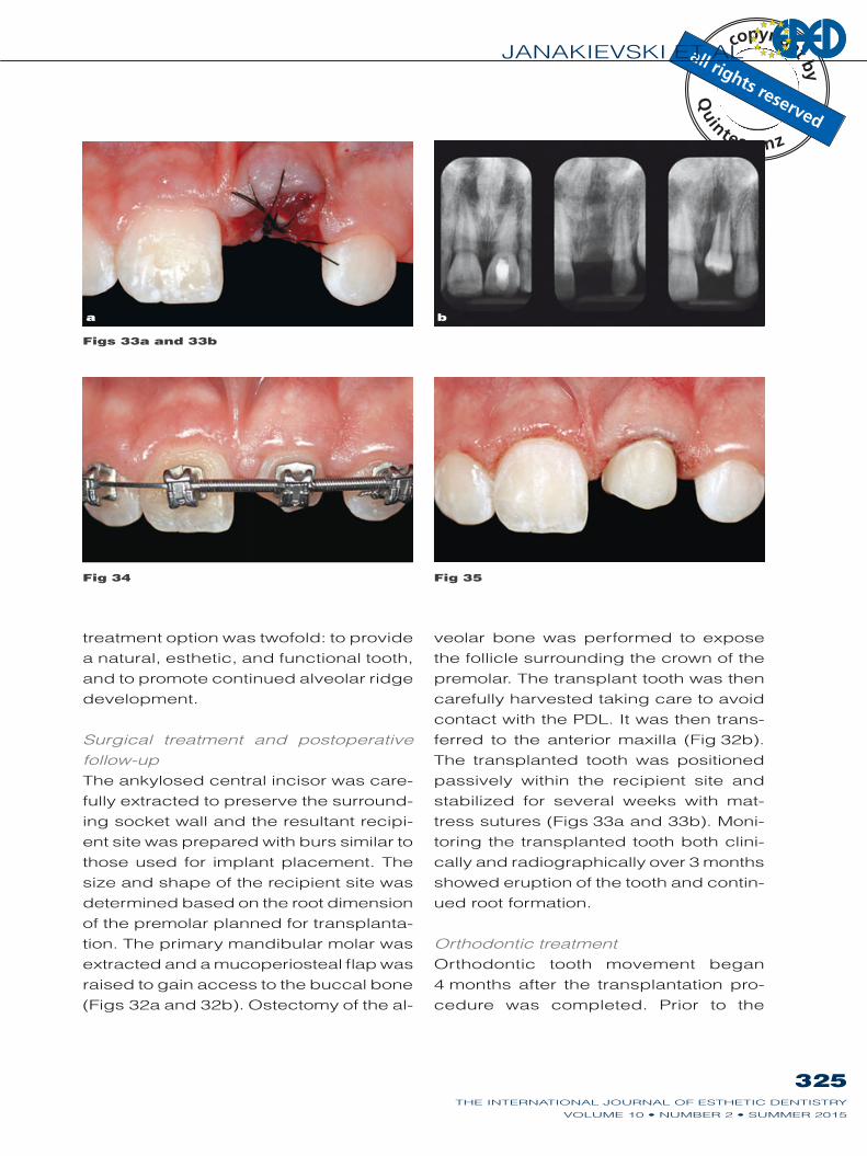
Surgical treatment and postoperative
follow-up
The ankylosed central incisor was care-
fully extracted to preserve the surround-
ing socket wall and the resultant recipi-
ent site was prepared with burs similar to
those used for implant placement. The
size and shape of the recipient site was
determined based on the root dimension
of the premolar planned for transplanta-
tion. The primary mandibular molar was
extracted and a mucoperiosteal flap was
raised to gain access to the buccal bone
-
veolar bone was performed to expose
the follicle surrounding the crown of the
premolar. The transplant tooth was then
carefully harvested taking care to avoid
contact with the PDL. It was then trans-
The transplanted tooth was positioned
passively within the recipient site and
stabilized for several weeks with mat-
tress sutures (Figs 33a and 33b). Moni-
toring the transplanted tooth both clini-
cally and radiographically over 3 months
showed eruption of the tooth and contin-
ued root formation.
Orthodontic treatment
Orthodontic tooth movement began
4 months after the transplantation pro-
cedure was completed. Prior to the
Fig 34 Fig 35
Figs 33a and 33b
a b

SCIENTIFIC SESSION
326THE INTERNATIONAL JOURNAL OF ESTHETIC DENTISTRY
restoration of the transplanted premolar
with composite, it is important for the or-
thodontist to position the tooth as ideally
as possible in order to achieve the most
natural and esthetic restorative result.93
This is based on the following planes of
space (Fig 34):
Incisogingivally: The cementoenamel
junction (CEJ) of the transplant tooth
should be aligned with the CEJ of the
adjacent central incisor.
Mesiodistally: The transplant tooth
should be positioned mesial of center,
leaving 1/3 of the remaining space on
the remaining space on the distal of
the transplant.
should be positioned slightly pala-
tal on the ridge in order to allow the
restorative material to be mainly ad-
ditive in nature, with minimal removal
of tooth structure.
Restorative phase
The restorative phase can be initiat-
ed after the preferred tooth position is
achieved. This restoration can be com-
pleted with composite resin using a
direct or indirect technique. Given the
differences in mesiodistal tooth dimen-
sions when comparing the transplant to
the natural central incisor, the composite
restoration should start subgingivally in
Figs 36a and 36b
a b
Figs 37a and 37b
a b

JANAKIEVSKI ET AL
327THE INTERNATIONAL JOURNAL OF ESTHETIC DENTISTRY
order to get a smooth emergence profile.
The difficulty in the direct application of
composite is applying and finishing the
material subgingivally. Following a slight
gingivectomy to improve the tissue sym-
metry, the transplant tooth was minimally
prepared to decrease the thickness at
the cervical height of contour and create
-
ite veneer was then fabricated directly
on the model, finished, and polished
(Figs 36a and 36b). The restoration was
adhesively bonded only after it was tried
in to verify fit, occlusion, and esthetics
(Figs 37a and 37b). Finally, the patient
was referred back to the orthodontist for
continuation and completion of ortho-
dontic treatment.
Conclusions
Successful collaborative treatment re-
quires shared knowledge and under-
standing among members of the dental
team. This results in more effective com-
munication and the clear assignment of
responsibilities to each clinician in order
to achieve the appropriate solution. This
article has discussed an interdiscipli-
nary approach for treating the estheti-
cally compromised patient; specifically,
patients who present with a deficiency
in spacing for tooth restoration/replace-
ment or a deficiency in alveolar ridge
form.
References
1. O’Connor RV. The excit-
ing world of interdiscipli-
nary dentistry. Int J Peri-
odontics Restorative Dent
multidisciplinary approach
to esthetic dentistry. Dent
3. Kokich VG, Spear FM.
Guidelines for managing
the orthodontic-restorative
patient. Semin Orthod
4. Keim RG. The art of interdis-
ciplinary teamwork. J Clin
Li TF, Garber DA, Adar P.
esthetically oriented revi-
sion of the original implant
protocol. J Esthet Dent
6. Genco RJ, Genco FD.
Common risk factors in the
management of periodontal
and associated systemic
diseases: the dental setting
and interprofessional collab-
7. Petri L. Concept analysis
of interdisciplinary col-
laboration. Nurs Forum
8. Irwin RS, Flaherty HM,
French CT, et al. Interdis-
ciplinary collaboration:
the slogan that must be
achieved for models of
delivering critical care
to be successful. Chest
9. Spear FM. The importance
of commitment and com-
munication in the general-
ist/specialist relationship.
Adv Esth Interdisc Dent
10. Klasser GD, Gremillion HA.
Past, present, and future of
predoctoral dental educa-
tion in orofacial pain and
TMDs: a call for interprofes-
sional education. J Dent
11. Wilder RS, O’Donnell JA,
at risk? A case for interpro-
fessional education. J Dent
C, Jepson N. Interac-
tions in the dental team:
understanding theoretical
complexities and practi-
13. Abrams H, Kopczyk RA,
Kaplan AL. Incidence of
anterior ridge deformi-
ties in partially edentulous
patients. J Prosthet Dent

SCIENTIFIC SESSION
328THE INTERNATIONAL JOURNAL OF ESTHETIC DENTISTRY
14. Kokich VG. Maxillary lateral
incisor implants: planning
with the aid of orthodon-
tics J Oral Maxillofac Surg
Anterior space manage-
ment: interdisciplinary con-
cepts. J Esthet Restor Dent
16. Feigenbaum NL. Aspects
of aesthetic smile design.
Pract Periodontics Aesthet
Dent 1991;3:9–13.
17. Cosyn J, Raes M, Packet
Disparity in embrasure fill
and papilla height between
tooth- and implant-borne
fixed restorations in the
anterior maxilla: a cross-
sectional study. J Clin Peri-
tooth size and its relation
to the analysis and treat-
ment of malocclusion. Angle
19. Kokich VG, Spear FM.
Guidelines for managing
the orthodontic-restorative
patient. Semin Orthod
-
thetic orthodontic interven-
tion for management of a
partially edentulous patient
with generalized wear and
malocclusion. J Esthet
100.
Mathews DP. Interdiscipli-
nary management of anter-
ior dental esthetics. J Am
169.
kinetics of anterior tooth
display. J Prosthet Dent
JG. Some esthetic factors
in a smile. J Prosthet Dent
Managing congenitally miss-
ing lateral incisors. Part I:
Canine substitution. J Esthet
Managing congenitally
missing lateral incisors. Part
II: tooth-supported restor-
ations. J Esthet Restor Dent
Managing congenitally
missing lateral incisors. Part
III: single-tooth implants.
J Esthet Restor Dent
A comparison of implant/
prosthesis loading with four
clinical variables. Int J Pros-
based decision making:
replacement of the single
missing tooth. Dent Clin
Garber D, Adar P. The
interproximal height of bone:
a guidepost to predictable
aesthetic strategies and soft
tissue contours in anterior
tooth replacement. Pract
Periodontics Aesthet Dent
1998;10:1131–1141.
30. Abrams H, Kopczyk RA,
Kaplan AL. Incidence of
anterior ridge deformi-
ties in partially edentulous
patients. J Prosthet Dent
31. Kois JC. Predictable single-
tooth peri-implant esthet-
ics: five diagnostic keys.
Compend Contin Educ Dent
Kokich VG. Interdisciplinary
management of single-tooth
implants. Semin Orthod
Lekholm U. Orthodontic
aspects of the use of oral
implants in adolescents: a
10-year follow-up study. Eur
implant-supported crowns in
the anterior maxilla: poten-
years) problems. World J
Wennström JL. Tissue alter-
ations at implant-supported
single-tooth replacements:
a 1-year prospective clinical
study. Clin Oral Implants
36. Grunder U, Gracis S, Capelli
M. Influence of the 3-D
bone-to-implant relationship
on esthetics. Int J Peri-
odontics Restorative Dent
37. Kokich VO Jr, Kinzer GA,
Janakievski J. Congenitally
missing maxillary lateral
incisors: restorative replace-
ment. Counterpoint. Am J
Orthod Dentofacial Orthop
38. Esposito M, Ekestubbe A,
Gröndahl K. Radiological
evaluation of marginal bone
loss at tooth surfaces facing
Clin Oral Implants Res
-
olds EC. A prospective clin-
ical study of non-submerged
immediate implants: clinic-
al outcomes and esthetic
results. Clin Oral Implants
40. Fürhauser R, Florescu D,
G, Watzek G. Evaluation of
soft tissue around single-
tooth implant crowns:
the pink esthetic score.
Clin Oral Implants Res
of early placed maxillary
anterior single tooth implants
using objective esthetic
criteria: a crosssectional,
follow-up using pink and
white esthetic scores. J Peri-
329THE INTERNATIONAL JOURNAL OF ESTHETIC DENTISTRY
JANAKIEVSKI ET AL
C, et al. Early implant
placement with simultane-
ous guided bone regenera-
tion following single-tooth
extraction in the esthetic
consecutive patients. J Peri-
43. Donos N, Mardas N, Chad-
ha V. Clinical outcomes of
implants following lateral
bone augmentation: system-
atic assessment of available
options (barrier membranes,
bone grafts, split oste-
otomy). J Clin Periodontol
44. Aghaloo TL, Moy PK. Which
hard tissue augmenta-
tion techniques are the
most successful in fur-
nishing bony support for
implant placement? Int J
Oral Maxillofac Implants
term stability of contour
augmentation in the esthetic
zone: histologic and histo-
morphometric evaluation of
months after augmentation.
outcomes following immedi-
ate and early implant place-
ment in the anterior maxilla
– a systematic review. Int
J Oral Maxillofac Implants
47. Grunder U. Crestal ridge
width changes when plac-
ing implants at the time of
tooth extraction with and
without soft tissue augmen-
tation after a healing period
consecutive cases. Int J
Periodontics Restorative
48. Rungcharassaeng K, Kan
JY, Yoshino S, Morimoto T,
Zimmerman G. Immedi-
ate implant placement and
provisionalization with and
without a connective tissue
graft: an analysis of facial
gingival tissue thickness. Int
J Periodontics Restorative
49. Donnelly MW, Swoope CC.
Periosteal tension in the
stimulation of bone growth
in the mandible [Masters
thesis]. Seattle: University of
Washington, 1973.
Alveolar ridge changes in
patients congenitally miss-
ing mandibular second
premolars. J Prosthet Dent
1994;71:144–149.
study on closing edentulous
spaces in the mandible.
effects of space closure of
the mandibular first molar
area in adults. Am J Orthod
Hansen K, Ekestubbe A,
Wennström J. Orthodon-
tic tooth movement into
edentulous ridge areas – a
case series. Eur J Orthod
M. Implant site development
in the distal region of the
mandible: bone formation
and its stability over time.
Am J Orthod Dentofacial
tooth movement: bone for-
mation and its stability over
time. Am J Orthod Dentofa-
-
spective study of 4-unit fixed
dental prostheses for the
adjacent teeth. Int J Prostho-
C, Garber D, Calamita
M, Salama H, Cabral G.
Prosthetic gingival recon-
struction in the fixed partial
and treatment planning. Int
J Periodontics Restorative
Zurek DJ. Selective use of
gingival-toned ceramics:
case reports. Quintessence
Weinländer M, Wegscheider
W, Piehslinger E. Implant-
prosthodontic rehabilitation
of anterior partial edentu-
lism: a clinical review. Int
J Oral Maxillofac Implants
60. Ishikawa T, Salama M,
Funato A, et al. Three-
dimensional bone and soft
tissue requirements for
optimizing esthetic results
in compromised cases with
multiple implants. Int J Peri-
odontics Restorative Dent
61. Rocchietta I, Fontana F,
Simion M. Clinical outcomes
of vertical bone augmenta-
tion to enable dental implant
placement: a systematic
review. J Clin Periodontol
Localized ridge augmenta-
tion/preservation. A system-
atic review. Ann Periodontol
S. Vertical ridge augmen-
tation: surgical protocol
and retrospective evalu-
ation of 48 consecutively
inserted implants. Int J Peri-
odontics Restorative Dent
1998;18:434–443.
Albrektsson T, Johansson
C. Histologic evaluation of
guided vertical ridge aug-
mentation around implants
in humans. Int J Peri-
odontics Restorative Dent

SCIENTIFIC SESSION
330THE INTERNATIONAL JOURNAL OF ESTHETIC DENTISTRY
Rasperini G, Maiorana C.
Vertical ridge augmentation
by expanded-polytetrafluor-
oethylene membrane and
a combination of intraoral
autogenous bone graft and
deproteinized anorganic
Clin Oral Implants Res
66. Urban IA, Lozada JL,
Jovanovic SA, Nagursky
H, Nagy K. Vertical ridge
augmentation with titan-
ium-reinforced, dense-
PTFE membranes and a
combination of particulated
autogenous bone and anor-
ganic bovine bone-derived
mineral: a prospective case
series in 19 patients. Int J
Oral Maxillofac Implants
67. Kaigler D, Avila G, Wisner-
Lynch L, et al. Platelet-
derived growth factor appli-
cations in periodontal and
peri-implant bone regenera-
68. Geurs N, Ntounis A, Vassilo-
poulos P, Van der Velden U,
growth factors in human
extraction sockets: a histo-
logic and histomorphometric
evaluation of short-term
healing. Int J Oral Maxillofac
69. McKay WF, Peckham SM,
-
hensive clinical review of
recombinant human bone
RE, et al. De novo bone
induction by recombinant
human bone morphogenetic
-
lary sinus floor augmenta-
tion. J Oral Maxillofac Surg
71. Fiorellini JP, Howell TH,
Cochran D, et al. Rand-
omized study evaluating
recombinant human bone
for extraction socket aug-
mentation. J Periodontol
Karp N, Thorne CH, Gray-
human mandible by gradual
distraction. Plast Reconstr
73. Esposito M, Grusovin MG,
Felice P, Karatzopoulos G,
Worthington HV, Coulthard
P. Interventions for replac-
ing missing teeth: horizontal
and vertical bone aug-
mentation techniques for
dental implant treatment.
Cochrane Database Syst
74. Chiapasco M, Romeo E,
Casentini P, Rimondini L.
Alveolar distraction osteo-
genesis vs. vertical guided
bone regeneration for the
correction of vertically
deficient edentulous ridges:
a 1-3-year prospective
study on humans. Clin Oral
-
lar distraction osteogenesis
for the correction of verti-
cally deficient edentulous
ridges: a multicenter pro-
spective study on humans.
Int J Oral Maxillofac
76. Enislidis G, Fock N, Millesi-
Schobel G, et al. Analysis
of complications following
alveolar distraction osteo-
genesis and implant place-
ment in the partially eden-
tulous mandible. Oral Surg
Oral Med Oral Pathol Oral
30.
77. Jensen OT, Cockrell R,
Kuhike L, Reed C. Anterior
maxillary alveolar distraction
osteogenesis: a prospec-
J Oral Maxillofac Implants
78. Froum SJ, Rosenberg ES,
Elian N, Tarnow D, Cho SC.
Distraction osteogenesis for
ridge augmentation: preven-
tion and treatment of compli-
cations. thirty case reports.
Int J Periodontics Restora-
79. Salama H, Salama M. The
role of orthodontic extru-
sive remodeling in the
enhancement of soft and
hard tissue profiles prior
to implant placement: a
systematic approach to the
management of extraction
site defects. Int J Peri-
odontics Restorative Dent
80. Mantzikos T, Shamus
I. Forced eruption and
implant site development:
soft tissue response. Am J
Orthod Dentofacial Orthop
81. Mantzikos T, Shamus I.
Forced eruption and implant
site development: an osteo-
physiologic response. Am J
Orthod Dentofacial Orthop
Nassar U, Olfert K. Implant
site development by ortho-
dontic extrusion. A system-
atic review. Angle Orthod
83. Reitan K. Clinical and histo-
logic observations on tooth
movement during and after
orthodontic treatment. Am J
Aalam A, Chee W. Ortho-
dontically assisted verti-
cal augmentation in the
esthetic zone. J Prosthodont

JANAKIEVSKI ET AL
331THE INTERNATIONAL JOURNAL OF ESTHETIC DENTISTRY
and coronal bone augmen-
tation using forced eruption
and buccal root torque:
a case report. Int J Peri-
odontics Restorative Dent
-
ridis S. Long-term vertical
changes of the anterior
maxillary teeth adjacent to
single implants in young and
mature adults. A retrospec-
tive study. J Clin Periodontol
87. Heij DG, Opdebeeck H, van
Steenberghe D, Kokich VG,
development, continuous
tooth eruption, and mesial
drift as compromising
factors for implant place-
ment. Int J Oral Maxillofac
term function of single-
implant restorations: A 17- to
19-year follow-up study on
implant infraposition related
to the shape of the face
and patients’ satisfaction.
Clin Implant Dent Relat Res
89. Jemt T, Ahlberg G, Henriks-
movements adjacent to
single-implant restorations
follow-up. Int J Prosthodont
90. Andreasen JO, Ravn JJ.
Epidemiology of traumatic
dental injuries to primary
and permanent teeth in a
Danish population sample.
91. Steiner DR. Avulsed maxil-
lary central incisors: the
case for replantation. Am J
Orthod Dentofacial Orthop
-
berg M, Frykholm A. Surgi-
cal treatment of ankylosed
and infrapositioned reim-
planted incisors in adoles-
cents. Scand J Dent Res
93. Janakievski J. Avulsed
maxillary central incisors:
the case for autotransplan-
tation. Counterpoint. Am J
Orthod Dentofacial Orthop
94. Tsukiboshi M. Autotrans-
plantation of teeth. Chicago:
-
tion of human teeth. A life-
table analysis of prognostic
factors. Int J Oral Surg
96. Kallu R, Vinckier F, Politis C,
Mwalili S, Willems G. Tooth
transplantations: a descrip-
tive retrospective study.
Int J Oral Maxillofac Surg
97. Andreasen JO, Paulsen HU,
A long-term study of 370
autotransplanted premolars.
Part II. Tooth survival and
pulp healing subsequent
to transplantation. Eur J
98. Andreasen JO, Paulsen HU,
Yu Z, Schwartz O. A long-
term study of 370 autotrans-
planted premolars. Part III.
Periodontal healing subse-
quent to transplantation. Eur
Induction of jaw bone forma-
tion by tooth autotransplan-
tation. Nor Tannlaegeforen
100. McCulloch CA. Progenitor
cell populations in the peri-
odontal ligament of mice.
101. Mensink G, Karagozoglu
KH, Strackee SD, van Tee-
-
ing AG. Autotransplantation
of two maxillary premolars
in a free vascularized fibula
reconstructed mandible.
Int J Oral Maxillofac Surg
O, Hjørting-Hansen E. Tooth
transplantation to bone graft
in cleft alveolus. Cleft Palate
103. Janakievski J. Tooth
autotransplantation to an
alveolar ridge augmented
with allograft bone. (manu-
script in press).
104. Czochrowska EM, Stenvik A,
outcome of autotransplanted
premolars replacing maxil-
lary incisors. Dent Traumatol
Haanaes HR. Management
of missing maxillary anter-
ior teeth with emphasis on
autotransplantation. Am J
Orthod Dentofacial Orthop
106. Snead M. Whole-tooth
regeneration: it takes a vil-
lage of scientists, clinicians,
and patients. J Dent Educ
107. Modino SA, Sharpe PT.
Tissue engineering of teeth
using adult stem cells. Arch
Mao JJ. Anatomically
shaped tooth and peri-
odontal regeneration by
cell homing. J Dent Res