INTERACTION AND IMPACT OF CASSAVA MOSAIC …
117
INTERACTION AND IMPACT OF CASSAVA MOSAIC BEGOMOVIRUSES AND THEIR ASSOCIATED SATELLITES By: HAPPYNESS GABRIEL MOLLEL Dissertation submitted in partial fulfillment for the degree of Master of Science The University of Witwatersrand, Johannesburg Republic of South Africa
Transcript of INTERACTION AND IMPACT OF CASSAVA MOSAIC …
INTERACTION AND IMPACT OF CASSAVA MOSAIC BEGOMOVIRUSES AND
THEIR ASSOCIATED SATELLITES
By:
HAPPYNESS GABRIEL MOLLEL
Dissertation submitted in partial fulfillment for the degree of Master of Science
The University of Witwatersrand, Johannesburg
Republic of South Africa

i
Declaration
This thesis is my work and has not been presented for a degree in any other University
Signed…………………….Date: 13th
January 2014
H. G. Mollel
.

ii
Dedication
This work is dedicated to my husband Juma. W. Yabeja, my mother Nay Zakayo and my father
Gabriel Jacob for their support and encouragement in the period of my studies. I would like also
to dedicate to my lovely son William. J. Yabeja for his patience during this period of studies.

iii
Acknowledgment
I wish to sincerely thank “International Centre for Genetic Engineering and Biotechnology”
(ICGEB) for funding this work. I am highly grateful to my supervisors Prof. M.E.C. Rey of the
University of Witwatersrand, South Africa, Dr. Joseph Ndunguru, Dr. Fred Tairo, Dr. Peter
Sseruwagi of Mikocheni Agricultural Research Institute, Tanzania and Dr. Herve Vanderschuren
of ETH, Zurich Switzerland for their timely advice and support in the course of my MSc. work. I
am also indebted to Dr. Deusdedith Mbanzibwa and my colleagues Cyprian Rajabu, Doreen
Mgonja and Margareth Lupembe of Mikocheni Agricultural Research Institute for their support
in laboratory work. Also Ms. Debbie Carmichael a student at the school of Molecular and Cell
Biology (MCB), university of the Witwatersrand, South Africa for her commendable assistance
in primer designing. Furthermore, I would like to thank Dr. Trino Ascencio- Ibanez, of North
Carolina State University, United State for his assistance in optimizing the gene gun. Finally,
but not least I thank Ms. Maabo Moralo for her support in the laboratory work in the University
of Witwatersrand, Johannesburg, South Africa.
iv
Research outputs
Publications
H.G. Mollel., O.A. Ndomba., F. Mwatui., E. Ateka., P. Sseruwagi., F.Tairo., H. Vanderschuren.,
M.E.C. Rey and J. Ndunguru., 2013. Genetic diversity and geographic distribution of DNA-II
and III molecules associated with cassava and Cassava mosaic disease in Uganda, Kenya,
Rwanda and Tanzania. Manuscript submitted to Plant Pathology.
Mollel, G.H., Sseruwagi, P., Tairo, F., Vanderschuren, H., Rey, M.E.C., and Ndunguru, J., 2013.
Molecular characterization of integrated DNA molecules associated with cassava mosaic
begomovirus. Proceedings of the 12th
International Plant Virus Epidemiology Symposium,
Arusha, Tanzania, 27th
-31st January, 2013.

v
Table of Contents
Declaration ................................................................................................................................... i Dedication ................................................................................................................................... ii Acknowledgment ....................................................................................................................... iii Research outputs ........................................................................................................................ iv List of tables .............................................................................................................................. vii
List of acronyms ........................................................................................................................ xi Abstract ....................................................................................................................................... 1
Background and Justification for study ...................................................................................... 5 Overall objectives ....................................................................................................................... 7 The specific aims of the study are: ............................................................................................. 8 CHAPTER ONE ......................................................................................................................... 9
LITERATURE REVIEW ........................................................................................................... 9 1.1 Cassava (Manihot esculenta Crantz) ................................................................................ 9
1.1.1 Introduction ............................................................................................................... 9 1.1.2 Description of cassava and its importance .............................................................. 11 1.1.3 Production constrains .............................................................................................. 13
1.1.4 Geminiviruses ......................................................................................................... 14
1.1.5 Cassava mosaic disease........................................................................................... 16 1.1.6 Cassava mosaic begomovirus transmission ............................................................ 19
1.2 Satellites ......................................................................................................................... 21
1.2.1 Discovery of sub viral agents associated with begomovirus diseases .................... 21 1.2.2 Alphasatellite .......................................................................................................... 22
1.2.3 Betasatellites ........................................................................................................... 23 1.2.4 Replication and compatibility of begomovirus subviral agents .............................. 24 1.2.5 Functions of begomovirus subviral agents ............................................................. 26
1.2.6 Common features between begomovirus components and satellites ...................... 26 1.2.7 Cassava begomovirus-associated satellites ............................................................. 28
CHAPTER TWO ...................................................................................................................... 31
A STUDY OF THE IMPACT OF DNA-II AND DNA-III ON CASSAVA MOSAIC
DISEASE, AND INVESTIGATION INTO TRANSCRIPTION OF PUTATIVE EPISOMAL
ORFs ......................................................................................................................................... 31
2.1 Abstract ............................................................................................................................... 31 2.2 Introduction ......................................................................................................................... 32 2.3 Materials and Methods ........................................................................................................ 34
2.3.1 Micropropagation and acclimatization of cassava cultivars TME3 and cv. 60444 ..... 34 2.3.2 Biolistic inoculation of virus and DNA-II and III clones ............................................ 36
2.3.2.1 Preparation of gold particles ..................................................................................... 36 2.3.2.2 Coating of virus and DNA-II and III infectious clones ............................................ 36 2.3.2.3 Transcript determination in the geminivirus – DNA-II and DNA-III - cassava host
systems .................................................................................................................................. 38 2.3.2.3.1 RNA extraction ...................................................................................................... 38

vi
2.3.3.2 Primer design and RT- PCR analysis of ORF C4 for DNA-II and ORF V1 for DNA-
III........................................................................................................................................... 40 2.3.3.4. Sequencing ............................................................................................................... 44 2.4 Results ............................................................................................................................. 44
2.4.1 Symptoms observation ................................................................................................. 44 2.4.2 Symptoms severity and disease severity progress curves ............................................ 47 2.4.3. RT-PCR for ORF C4 of DNA-II, ORF V1 and ORF C2 of DNA-III ........................ 50 2. 4. 4 Sequence analysis ...................................................................................................... 53 2.4.4.1 Transcript sequences for DNA-II.............................................................................. 53
2.4.4.2 Transcript sequences for ORF V1 of DNA-III ......................................................... 55 2.5 Discussion ....................................................................................................................... 57
CHAPTER THREE .................................................................................................................. 63 GENETIC DIVERSITY AND GEOGRAPHIC DISTRIBUTION OF DNA-II AND III
MOLECULES ASSOCIATED WITH CASSAVA AND CASSAVA MOSAIC DISEASE IN
UGANDA, KENYA, RWANDA AND TANZANIA .............................................................. 63
3.1 Abstract ........................................................................................................................... 63 3.2 Introduction ..................................................................................................................... 65
3.3 Materials and Methods .................................................................................................... 67 3.3.1 Sample collection ......................................................................................................... 67 3.3.2 DNA extraction ............................................................................................................ 69
3.3.3 Polymerase chain reaction (PCR) ................................................................................ 70
3.3.4 Molecular cloning ........................................................................................................ 71 3.3.5 Sequences analysis ....................................................................................................... 71 3.3.6 Statistical data analysis and presentation ..................................................................... 72
3.4 Results ............................................................................................................................. 72 3.4.1 Cassava mosaic disease symptom expression in the field ........................................... 72
3.4.2 PCR analysis of integrated DNA-II and DNA-III molecules ...................................... 73 3.4.3 Occurrence and geographical distribution of integrated DNA molecules ................... 75 3.4.4 Phylogenetic analysis and sequence comparisons ....................................................... 77
CHAPTER FOUR ..................................................................................................................... 83 SUMMARY AND RECOMMENDATIONS....................................................................... 83
Recommendations ..................................................................................................................... 85
Referrences ............................................................................................................................... 86

vii
List of tables
Table 1: Word cassava production in 2010……………………………………………………....10
Table 2: Production of staple tuber crops 2003 to 2008 (million tons)………………………......11
Table 3: Number of treatments used in studying putative transcription of DNA-II and
DNA-III...........................................................................................................................37
Table 4: Reaction of cassava plants to biolistic inoculation with EACMV-UG2 and ACMV,
and in combination with DNA-II, DNA-III and DNA-II + DNA-III infectious
clones...........................................................................................................................39
Table 5: List of primer pairs used for transcript detection of ORF C4 of episomal DNA-II in
antisense strand and ORF VI of DNA-III of sense strand..............................................43
Table 6: Samples collected from screen house for transcript analysis of DNA-II and
DNA-III..........................................................................................................................52
Table7: Description of amplified RT-PCR samples for DNA-II sent for sequencing .................54
Table 8: Description of amplified RT-PCR samples for DNA-III sent for sequencing.................56
Table 9: Cassava major growing areas where survey was conducted Uganda, Kenya, Rwanda
and Tanzania in 2009/2010.........................................................................................68
Table 10: Analysis of Variance for symptom severity on cassava samples collected from
Uganda, Kenya, Rwanda and Tanzania in 2009-2010................................................73
viii
List of figures
Figure 1: Cassava mosaic begomovirus genome structure composed of twinned particles; DNA-
A (left) and DNA-B (right). Functional ORFs on the DNA-A virion-sense (AV1 and
AV2) and the complementary-sense strand (C1 to C4). DNA-B has two ORFs: BV1
and BC1...................................................................................................................16
Figure 2: Cassava mosaic disease symptoms in cassava field......................................................19
Figure 3: Bipartite DNA-A (left) and DNA-B (right) begomovirus genome and satellite DNA-β
which is often associated with DNA-A component of monopartite
genome........................................................................................................................ 28
Figure 4: Genomic structure of cassava mosaic begomovirus associated satellite-like DNA
molecules. (A) satDNA-II with eight putative ORFs (V1-V3) on sense strand, and
(C1-C5) on antisense strand, and (B) satDNA-III with four putative OFRs (V1-V2)
on sense strand and (C1-C2) on antisense strand ........................................................30
Figure 5: Stages for acclimatization process: A) TME3 tissue culture cassava plantlets B) cv.
60444 tissue culture cassava plantlets C) Four- week old cassava plants in small pots
D) 8 week-old cassava plants in big pots ready for inoculation..................................35
Figure 6: Genomic structure of cassava mosaic begomovirus associated satellite-like DNA
molecules showing the positions for ORFs.................................................................41
Figure 7: Genome maps of circular DNA-II and III molecules showing primer positions..........42
ix
Figure 8: Cassava mosaic disease symptom development on cv. 60444 inoculated with A)
EACMV-UG2 alone, B) EACMV-UG2 + DNA-II, C) EACMV-UG2 + DNA-III, D)
EACMV-UG2 + DNA-II & III. CMD symptom development on TME 3 inoculated
with E) EACMV-UG2 alone, F) EACMV-UG2 + DNA-II, G) EACMV-UG2 +
DNA-III, H) EACMV-UG2 + DNA-II & III...............................................................46
Figure 9: Cassava mosaic disease symptom development on cv. 60444 inoculated with A)
ACMV alone, B) ACMV + DNA-II, C) ACMV+ DNA-III, D) ACMV + DNA-II &
III. CMD symptom development on TME 3 inoculated with E) ACMV alone, F)
ACMV+ DNA-II, G) ACMV + DNA-III, H) ACMV + DNA-II &
III..............................................................................................................................47
Figure 10: Disease severity progress curves for cassava plants inoculated with EACMV and in
combination with DNA-II, DNA-III and DNA-II + DNA-III in A) cv.60444 and B)
TME3...........................................................................................................................49
Figure 11: Disease severity progress curves for cassava plants inoculated with ACMV and in
combination with DNA-II, DNA-III and DNA-II + DNA-III in A) cv. 60444 and B)
TME3...........................................................................................................................50
Figure 12: Gel picture showing amplification of ORF C4 and ORF V1 transcripts on 1% agarose
by RT-PCR from cassava plants inoculated with either EACMV-UG2, ACMV and in
combination with DNA-II, DNA-III and DNA-II + DNA-III clones A) DNA-II; and
B) DNA-III..................................................................................................................51
Figure 13: Cassava plants showing cassava mosaic symptoms in the fields A: CMD symptoms;
B: mosaic severe; C: leaf curling and distortion D: leaf narrows in cassava
field...........................................................................................................................69

x
Figure 14: PCR amplification of integrated DNA-II molecules (16A) and DNA-III (16B) on 1%
agarose gel. M=Molecular weight marker of 1 kb plus; +C= positive control and -
C = negative control. Lanes 1-20 (16A & 16B) represent samples collected from
farmers.........................................................................................................................74
Figure 15: Occurrence of integrated DNA-II and DNA-III molecules in Uganda, Kenya,
Rwanda and Tanzania in 2009/2010............................................................................75
Figure 16: Geographical distribution of DNA-II and DNA-III molecules in Uganda, Kenya,
Rwanda and Tanzania in 2009/2010...........................................................................76
Figure 17: Phylogenetic trees obtained from alignment of the nucleotide sequences of DNA-II
and III with available sequence of satDNA-II and satDNA-III. 17A: DNA-II
sequences; 17B: DNA-III sequences obtained from Kenya, Uganda, Rwanda and
Tanzania in 2009/2010.................................................................................................79

xi
List of acronyms
ACMV Africa cassava mosaic virus
AYVD Ageratum yellow vein disease
ANOVA Analysis of variance
bp Base pairs
°C Degrees Celcius
CMD Cassava mosaic disease
CP Coat protein
CMBs Cassava begomoviruses
CBSD Cassava brown streak disease
cDNA Complementary DNA
DNA Deoxy-ribonucleic acid
DNA-1 Alphasatellites
DNA-β Betasatellites
DNA Deoxyribonucleic acid
dNTP Deoxynucleotide triphosphate
EACMV-UG East Africa cassava mosaic Ugandan variant
EACMCV East Africa cassava mosaic Cameroon virus
EACMV East African cassava mosaic virus
EACMKV East African cassava Kenya mosaic virus
EACMMV East African cassava Malawi virus
EACMZV East African cassava mosaic Zanzibar virus
EACMZV East Africa cassava mosaic Zanzibar virus
EDTA Ethylene-diaminetetraacetate

xii
EST Expressed sequence tag
FAO Food and Agricultural Organisation
HCl Hydrochloric acid
ICMV Indian cassava mosaic virus
MYMV Mungbean yellow mosaic virus
MARI Mikocheni Agricultural Research Institute
MgCl2 Magnesium chloride
MP Movement protein
MS Murashige and Skoog media
MED-NA-ME North African-Mediterranean-Middle East
μl Microlitre
µg Microgram
NW New world
NCBI National Centre for Biotechnology Information
NSP Nuclear shuttle protein
OW Old world
ORF Open reading frame
PCR Polymerase chain reaction
RT - PCR Reverse Transcriptase polymerase chain reaction
RNA Ribose nucleic acid
Rep Replication activator protein
Ren Replication enhancer protein
ssDNA Single stranded deoxyribonucleic acid
SDS Sodium dodecyl sulphate
xiii
SCR Satellites conserved region
Trap Transcription protein
TRV Tobacco ringspot virus
ToLCV Tomato leaf curl virus
TYLCD Tomato yellow leaf curl disease
TYLCCV Tomato yellow leaf curl China virus
TGMV Tomato golden mosaic virus
TAE Tris-acetate and EDTA
Tris-HCl Tris (hydroxymethyl) aminomethane hydrochloride
xiv
List of Appendices
Appendix 2.1: Alignment of DNA-II ORF C4 transcripts isolates collected from mock-
inoculated cassava plants and cassava plants inoculated with either EACMV-Ug2,
ACMV and in combination with DNA-II, DNA-III and DNA-II + DNA-III
clones. Highlighted colours are conserved nucleotides in all sequenced samples.
Yellow colour is G, Blue colour is C, Green colour is T and Red colour A.
Appendix 2.2: Percentage nucleotide sequence similarity matrix table of the DNA-II ORF C4
transcript sequences.
Appendix 2.3: Alignment of DNA-III ORF V1 and ORFC2 transcripts isolates collected from
mock-inoculated cassava plants and cassava plants inoculated with either
EACMV-Ug2, ACMV and in combination with DNA-II, DNA-II and DNA-II +
DNA-III clones. Highlighted colours are conserved nucleotides in all the
sequenced samples. Yellow colour is G, Blue colour is C, Green colour is T
however A nucleotide was not conserved.
Appendix 2.4: Percentage nucleotide sequence similarity matrix table of the DNA-III ORF VI
transcripts sequence.
1
Abstract
Cassava (Manihot esculenta Crantz) is affected by two major viral diseases, namely Cassava
brown streak disease (CBSD) and Cassava mosaic disease (CMD). Two of the most widely
distributed begomoviruses in East Africa associated with CMD, are East African cassava virus-
Uganda2 (EACMV-UG2) and African cassava mosaic virus (ACMV). Despite efforts of
generating improved Tropical Manihot Series (TMS) by traditional breeding and using highly
resistant geminivirus cassava landraces such as Tropical Manihot Esculenta1 (TME1) and
Tropical Manihot Esculenta3 (TME3), more recently two circular single stranded (ss) satellite-
like DNA molecules (episomal DNA-II and DNA-III) have been found to be associated with
CMD and are able to break resistance to EACMV-UG2 and enhance virus symptoms.
The nature of these satellite-like DNA molecules is unknown, and furthermore, the discovery of
integration of partial copies of DNA molecules (DNA-II and III fragments), and evidence for
transcription from cassava Expressed Sequence Tag (EST) database screening, has led to an even
more perplexing disease complex. In the present study, we attempted to further explore the
interaction between the satellite-like DNAs and their associated cassava-infecting begomoviruses
by investigating the impact of these DNA molecules on disease development in TME3 (tolerant)
and cv. 60444 (susceptible) cassava cultivars, and to also gather biological evidence for
transcription of integrated genomic and episomal (putative predicted ORFs) sequences in the
ACMV and EACMV-UG2-associated DNA-II and DNA-III.
Biolistic inoculation of EACMV-UG2, ACMV, and in co-bombardment with DNA-II, DNA-III,
DNA-II + DNA-III was successfully performed. CMD symptoms were developed earlier on

2
cassava plants inoculated with ACMV + DNA-II, ACMV + DNA-III, ACMV + DNA-II + DNA-
III and EACMV-UG2 + DNA-II, EACMV-UG2 + DNA-III, EACMV-UG2 + DNA-II + DNA-
III molecules compared with cassava plants inoculated with begomoviruses alone. Additionally,
CMD symptoms were more severe in cv.60444 compared to TME3 when inoculated with
begomoviruses alone, or in combination with DNA-II, DNA-III and DNA-II + DNA-III
molecules. DNA-II and III were able to break resistance to the highly CMD-tolerant cassava
landrace, TME3, and enhance virus symptoms.
In order to confirm EST-generated evidence for transcription of DNA-II and III fragments,
cDNA was subjected to RT-PCR. RT-PCR of transcripts was successful for only three putative
ORFs: ORF C4 of the antisense DNA-II strand, ORF V1 on sense DNA-II strand, and ORF C2
on antisense strand for DNA-III. Primers for transcripts amplified 250 bp and 220 bp for ORF
C4 of DNA-II and ORF V1 of DNA-III, respectively. Transcribed ORFs were confirmed by
sequencing, and the sequences were similar to the published sequences of Begomovirus
associated DNA-II satellite and Begomovirus associated DNA-III satellite, respectively. These
results showed that at least two putative ORFs for DNA-II and one (the largest ORF VI) DNA-
III can be transcribed.
Furthermore, surveys were undertaken in order to ascertain the distribution of episomal and
integrated DNA-II and III in cassava germplasm from several countries, namely Tanzania,
Uganda, Kenya and Rwanda. Results from this research successfully established genetic
diversity and wide geographical distribution of integrated DNA-II and DNA-III molecules. Two
primer pairs were designed from a central conserved sequence found in all the integrated DNA-II
3
or III fragments identified from the cDNA libraries (EST database). These primers also amplified
integrated sequences of expected size in cassava accessions and wild Manihot species which
were similar to satellite-like sequence occurrences in the ESTs.
Using designed primers, PCR amplification yielded integrated DNA-II and DNA-III products of
~895 bp and ~306 bp, respectively. Analysis of 363 field leaf samples detected the presence of
DNA-II or DNA-III from Kenya (3.3% or 8.3%), Uganda (18% or 2.5%), Rwanda (6.5% or
19.6%) and Tanzania (5.7% or 11.9%) , results which were confirmed by analysis of the
sequenced PCR amplicons. Detection of both DNA-II and DNA-III molecules on the samples
collected was also found from Kenya (73%), Uganda (69.1%), Rwanda (50%) and Tanzania
(69.3%). Interestingly integrated DNA-II and II copies were amplified from healthy,
symptomless and infected cassava samples. DNA-II sequences did not vary significantly (93.3%
- 99.8%) and were highly similar to the sequences of Begomovirus associated sat DNA-II
(AY836366) and 99% with mentha leaf deformity disease associated satellite DNA-II, while
DNA-III sequences and Begomovirus associated DNA-II satellite (AY833667).
In conclusion, this study has provided useful information that contributes to a further
understanding of the biological function of integrated and episomal DNA-II and III molecules in
begomoviruses infected cassava plant. However the relationship, if any between episomal and
integrated forms needs to be established in future, and investigation into whether the transcribed
ORFs can produce functional proteins, needs to be undertaken. How DNA-II and III interact
with EACMV-UG2 and ACMV in disease modulation remains to be explored, and the
replication of episomal DNA-II and III by these associated begomoviruses needs to be confirmed
4
if these DNA molecules are to truly show a satellite-like relationship. Furthermore, the findings
in this study that partial and varied-sized integrated DNA-II and III fragments occur widely in
healthy and infected cassava germplasm will enable researchers (plant virologists and breeders)
working on cassava in Sub Saharan Africa (SSA) to explore this complex more deeply in order
to develop durable management strategies for CMD.

5
Background and Justification for study
Cassava (Manihot esculenta Crantz) is one of the leading crops in Sub-Saharan Africa in terms
of production and has become an important source of income to households and small-scale
farmers (FAOSTAT, 2010). Cassava is considered as a staple root crop for more than 800
million people living in developing countries (Burns et al., 2010). About 70% of world cassava
root production is used for human consumption either directly after cooking or in processed
forms; the remaining 30% is used for animal feed and industrial products, such as starch, glucose
and alcohol (El-Sharkawy, 2004). However, the production across the region is greatly affected
by cassava mosaic disease (CMD) and cassava brown streak disease (CBSD). Several studies
(Gibson., 1996; Ogbe et al., 1996; Legg et al., 1999; Fondong et al., 2000; Berry and Rey, 2001;
Ndunguru et al., 2005; Bisimwa et al., 2012) have reported the occurrence of CMD in different
countries in Sub-Saharan Africa. A comprehensive cassava mosaic begomovirus (CMB)
characterization study by Ndunguru et al. (2005) in East Africa showed that seven cassava
mosaic geminivirus species occur in Tanzania. Mbanzibwa et al. (2009a) reported the prevalence
of two Ipomovirus species causing CBSD in the Lake Victoria basin and along the coastal belt of
Indian Ocean.
Plant viruses are occasionally associated with satellite molecules which contribute to increase
virus symptoms on infected plants. Satellites are either viruses or nucleic acids that depend on a
helper virus for their replication but lack general nucleotide sequence homology to the helper
virus (Mayo et al., 2005). Satellites associated with RNA plant viruses were the first satellites to
be discovered and described, and are divided into satellite viruses and satellite RNAs (Schneider,
1969). The majority of satellite RNAs do not encode functional proteins but can have a
6
remarkable effect on the symptoms induced by their helper viruses, ranging from mild symptoms
to severe symptom (Roossink et al., 1992). Two satellites associated with monopartite
begomovirus diseases; alphasatellites and betasatellites were isolated from different infected
crops of Old World (OW) begomovirus diseases such as in Tomato yellow leaf curl disease in
China, Cotton leaf curl disease in Pakistan and India, and Ageratum yellow vein in South East
Asia (Dry et al 1997; Briddon et al, 2004). These satellites depend on helper virus for the protein
encapsidation and movement within and between plants (Mayo et al., 2005).
More recently, two satellite-like molecules, DNA-II and DNA-III associated with CMBs
(African cassava mosaic virus (ACMV) and East African cassava mosaic virus (EACMV), were
described from infected cassava plants in Tanzania and were able to enhance CMD symptoms
and break resistance to geminiviruses in the highly tolerant landrace TME 3 (Ndunguru et al.,
2005; 2008; Ingelbrecht et al., 2008). Primers to amplify DNA-II (895bp) and DNA-III (306 bp)
were previously designed based on the sequences from conserved regions across different
matching ESTs. Surprisingly, these primers amplified integrated sequences in cassava accessions
and wild Manihot species which were similar to satellite-like sequence occurrences in the ESTs
when additional sequences from the cassava genome were examined.
The diversity and geographic distribution of DNA-II and III in Uganda, Kenya, Rwanda and
Tanzania is not well documented. Further, it is not known if the open reading frames (ORFs) of
the DNA-II and III are transcribed; this information would be helpful to establish if the
transcribed products potentially produce functional proteins involved during viral infection in the
plant host.

7
This study aimed to generate information on the genetic diversity and geographic distribution of
the integrated DNA-II and III molecules in Kenya, Uganda, Tanzania and Rwanda. Furthermore,
an investigation of the putative transcripts of genomic and episomal DNA-II and III molecules
was also conducted. Findings from this study will add to the body of knowledge on these unusual
DNA molecules and will help researchers (plant virologists and breeders) working on cassava in
Sub Saharan Africa (SSA) to develop durable management strategies for CMD in the future.
Overall objectives
The aim of this study is to further uncover the nature of these unusual episomal satellite-like
DNA molecules associated with putative begomoviruses in East African countries. Furthermore,
this study aimed to;
i. Confirm that these putative episomal DNA-II and III molecules influence the symptom
phenotype and severity of CMD in order to further understand the nature of resistance,
and investigate biological evidence for transcription of putative ORFs on DNA-II and
DNA-III.
ii. Screening of cassava germplasm to establish the geographic distribution and diversity
of integrated forms in cassava germplasm.
8
The specific aims of the study are:
i. To screen cassava genome germplasm in major cassava-producing areas in Uganda,
Kenya, Rwanda and Tanzania in order to establish genetic diversity and geographic
distribution of integrated DNA-II and DNA-III sequences.
ii. To investigate mechanisms of interaction between the satellite-like DNAs and their
associated cassava – infecting begomoviruses by investigating the impact of these DNA
molecules on disease development in TME3 (tolerant) and cv. 60444 (susceptible)
cassava cultivars.
iii. To determine if any putative regions or ORFs on episomal or integrated DNA-II or III are
transcribed.

9
CHAPTER ONE
LITERATURE REVIEW
1.1 Cassava (Manihot esculenta Crantz)
1.1.1 Introduction
Cassava (Manihot esculenta Crantz) is extensively cultivated as an annual crop in tropical and
subtropical regions of Africa, Asia and Latin America between 30°N and 30°S (El-Sharkawy,
1993). Cassava is considered as a staple root crop for more than 800 million people living in
developing tropical countries (Burns et al., 2010). The total worldwide cassava production in
2009 was about 241 million tonnes (Bull et al., 2011) with Africa being the leading producer
(FAOSTAT, 2010). About 70% of world cassava root production is used for human consumption
either directly after cooking or in processed forms; the remaining 30% is used for animal feed
and other industrial products, such as starch, glucose and alcohol (El-Sharkawy, 2004). Cassava
is being increasingly used for bio-ethanol production in Brazil and China (Narina and Odeny,
2011) while in Nigeria it is a major cash crop earning the country about US$ 3 billion annually
through export of the crop and related products (FAO, 2004). Of the estimated 18 million ha of
cassava cultivated worldwide, approximately two thirds are in Africa, producing 110 million
tonnes (mt) of tuberous roots annually (FAO, 2006). Worldwide average yield of cassava
production in 2010 was estimated to be 12.40 tonnes/ha (FAOstat, 2012) (Table 1).

10
Table 1: World cassava production in 2010
Location Area harvested (ha) Yield
(tons/ha)
Production (tons)
World 18,568,788 12.4 230,265,639
Africa 11,969,784 10.16 121,661,234
S. America 2,400,720 13.2 31,686,404
Asia 3,901,877 19.26 75,148,313
Tanzania 798,000 5.5 4,392,170
Kenya 61,573 5.25 323,389
Uganda 415,000 12.73 5,282,000
Malawi 195,828 20.43 4,000,990
Mozambique 950,000 6 5,700,000
Rwanda 197,394 12.04 2,377,210
Zambia 198,000 5.82 1,151,700
Source: FAOstat, 2012
Although cassava requires optimal conditions to reach high growth rates, it performs well in
drought areas and on poor soils and thus is considered one of the most productive tropical crops
on marginal lands (Zhang et al., 2010). Under prolonged water shortages in seasonally dry and
semi-arid environments with less than 700 mm of annual rain, improved cultivars can give dry
root yields of over 3 t ha-1 (El-Sharkawy, 2006). Five well known tuber crops (potato, cassava,
sweet potato, yams and taro) together account for 90% of world’s total staple tuber production

11
(Shewry, 2003). Among these tuber crops, FAO (2010) statistics show that cassava ranks second
after potato, accounting for 30% of total tuber production (Table 2).
Table 2: Production of staple tuber crops 2003 to 2008 (million tons)
Tuber
Crop
2003 2004 2005 2006 2007 2008 Ave.
Potatoes 314.2 336.3 325.1 305.6 323.5 314.1 319.8
Cassava 191.3 203.1 207.1 222.3 224.1 232.9 213.5
S/potato 129.8 129.6 126.7 105.7 99.7 110.1 116.9
Yams 44.2 46.9 49.0 52.2 46.6 51.7 48.5
Taro 10.7 10.9 11.2 11.7 11.3 11.8 11.3
1.1.2 Description of cassava and its importance
Cassava is a shrubby perennial plant that typically grows from one to three meters (3-10 feet) in
height. It is grown mainly for its carbohydrate rich tuberous roots (Katz and Weaver, 2003). It
belongs to the family Euphorbiceae that also includes other commercially important plants like
castor bean (Ricinus communis L.) and rubber (Havea bransiliensis L.). Cassava is believed to
have originated in South and Central America and was introduced into West Coast Africa in 16th
century and later to East Africa through Madagascar and Zanzibar, and later into Asia in the late
17th
century by Portuguese traders. Africa has become the largest producer of cassava,
constituting 54% of world production. Most of the spread of cassava in Africa away from the

12
coastal areas and riverside trading posts took place during the 20th
century due to colonial powers
encouraging its cultivation as a reserve against famine and the ability of the crop to withstand
locust attack (Hillocks, 2002). However cultivation of cassava started to decline in the 1960s due
to post colonial governments turning their attention to maize in terms of funding and research
efforts, as well as taste preference for maize (Haggblade and Zulu, 2003). Today, cassava is
cultivated in more than 80 countries mainly between 30º South and 30º North of the equator
(Fauquet and Fargette, 1990).
Cassava is suited to warm humid lowland tropics and can be grown in areas where the mean
annual temperature exceeds 20ºC with annual rainfall that varies between 5000 mm and 8000
mm (Pounti-Kearlas, 1998). Cassava is a very hardy plant, tolerating drought better than most
other crops, and growing well in very poor and acidic soils (Katz and Weaver, 2003). Cassava
crop can yield up to 13 millions kcal/acre (Bender and Bender, 2005).
Cassava naturally is grown by small-scale farmers using traditional methods, and often on land
not suitable for other crops (Katz and Weaver, 2003). Cassava is propagated by cutting a mature
stem into sections of approximately 15 cm and planting prior to the wet season. Most of the
harvest from cassava is used for human consumption, either fresh or in processed forms. Cassava
can be processed into a wide variety of products for food and industrial uses, such as starch,
flour, alcohol, glucose and others. The leaves, which are rich in proteins, vitamin C and other
nutrients, are consumed in some communities to supplement the low protein content of the roots.
Cassava is consumed by an estimated 600 million people (FAO, 2006). Cassava flour is used in
the production of biscuits, sausage rolls, meat pies and bread (Ogbe, 2001). The non-food
13
industrial uses of cassava include production of starch which is used in a wide range of products:
paper, textiles and pharmaceuticals (Tonukari, 2004). In most east African countries, though
maize is the dominant staple food, cassava is very important in Mozambique, Tanzania, Uganda,
and Burundi as a reserve against famine.
1.1.3 Production constraints
Cassava production in East and Southern Africa is particularly exposed to numerous biotic
stresses. Common constrains include pests and diseases, poor agronomic practices, low yielding
varieties, high cyanide levels, lack of clean planting materials and long maturity periods (Thresh
et al. 1994). Pests and diseases are the most economically important constrains (Hillocks, 1994)
to the cassava production. Pests infesting cassava include cassava green mite (Mononychellus
tanajoa), green spider mite (Mononychellus tanajoa), mealy bugs (Phenacoccus manihot),
cassava hornworm (Erinnyis ello), scales, thrips and whitely (Bemisia tabaci) (Montero, 2003).
Diseases of cassava include cassava mosaic disease (CMD), cassava brown streak disease
(CBSD), cassava bacterial blight (CBB), cassava anthracnose disease (CAD), cassava bud
necrosis, and root rots (Calvert and Thresh, 2002). Virus diseases, particularly CMD and CBSD,
are the most important economical constraints of cassava production in east and southern Africa
(Taylor and Fauquet 1997; Thresh et al. 1994; Thresh et al. 1997). The two diseases are thought
to have a risen from infection of cassava by viruses already present in the indigenous African
flora (Legg and Hillocks, 2003).

14
The perpetuation of these diseases in cassava production is influenced by the abundance of
efficient insect vectors for transmission and continuous use of unclean planting materials
normally selected from the previous seasons. Despite success with control of cassava mealy bug
and cassava green mite using biological control programs, CMD and CBSD have remained a
challenge to control using sustainable approaches though there are adequate information on the
pathogen and efficient diagnostic tools such as PCR and RT-PCR tools being well established.
Thus, the two diseases are now key research priorities of many root and tuber programs in many
African countries (Legg and Thresh, 2003).
1.1.4 Geminiviruses
Geminiviruses are viruses which infect plants and are characterized by circular single-stranded
DNA genomes encapsidated into small twinned icosahedra virions. The family Geminiviridae is
divided into four genera (Mastrevirus, Curtovirus, Topocuvirus and Begomovirus), which are
differentiated based on their genome structure; the host plants they infect and the type of insect
vector (Fauquet et al., 2008).
The genus Begomovirus consists of either two DNA molecules, DNA-A and DNA-B, each of
about 2.8 kb which are responsible for different functions in the infection process (Stanley et al.,
2005) (Fig. 1), or a single DNA component (monopartite). Begomoviruses are the largest group
of plant viruses and cause economically important diseases of many vegetable and fibre crops
(Rojas et al., 2005; Varma and Malathi, 2003). DNA-A contains six partially overlapping open
reading frames (ORFs) organized in two opposite transcriptional directions separated by an

15
intergenic region (IR). On the virion-sense strand, DNA-A component contains AV1 and AV2
ORFs, and AC1–AC4 are on the complementary-sense strand. The DNA-A encoded gene
products are replication-associated protein AC1 (Rep); AV1 coat protein (CP); and proteins that
participate in the control of replication (AC3) and gene expression (AC2) (TrAP). DNA-B
encodes proteins required for nuclear trafficking (BV1) and cell-to-cell movement (BC1) of the
viral DNA (Hamilton et al., 1984; Hanley-Bowdoin et al., 1999). Both DNA components (DNA-
A and DNA-B) share a high nucleotide identity in the IR (c. 200 nt) called the common region
(CR), which contains promoter and sequence elements required for DNA replication and
transcription (Lazarowitz, 1992; Eagle et al., 1994; Chatterji et al., 1999). Begomoviruses are
transmitted by the whitefly Bemisia tabaci (Bemisia tabaci Genn.), and are also spread through
infected cuttings, which are the usual mode of cassava propagation. CMBs have been reported
from all cassava-growing countries in Africa and CMD constitutes a threat to cassava
production.
In addition to sub-viral components, geminiviruses have also been shown to be accompanied by
smaller-sized DNA molecules, called defective-DNA molecules, that are derived from the helper
virus genomes, but are not satellite molecules (Patil and Dasgupta, 2006; Simon et al., 2004).
Monopartite begomoviruses are accompanied by circular single stranded DNAs (ssDNA) which
are alphasatellites and betasatellites. Most Old World monopartite begomoviruses are associated
with one or more betasatellite DNA(s), which are required for induction of typical disease
symptoms and depend on the helper begomovirus for replication and movement (reviewed by
Mansoor et al., 2006; Briddon and Stanley, 2006).

16
Figure 1: Cassava mosaic begomovirus genome structure composed of twinned particles; DNA-
A (left) and DNA-B (right). Functional ORFs on the DNA-A virion-sense (AV1 and AV2) and
the complementary-sense strand (C1 to C4). DNA-B has two ORFs: BV1 and BC1. In both
DNA-A and DNA-B component there is a non-coding intergenic region referred to as the
common region (CR) (Adapted from Alabi, et al., 2011).
1.1.5 Cassava mosaic disease
Cassava mosaic disease (CMD) occurs in all cassava- producing regions in Africa and Indian
subcontinents (Legg and Fauquet, 2004; Legg et al., 2006). Cassava mosaic disease is the major
constraint to cassava production in Africa (Thresh and Cooter, 2005). CMD symptoms are
variable according to season and variety, but always include a chlorotic mosaic on infected

17
leaves and the colour of chlorosis varies from pale green to whitish yellow. In moderate to severe
infections, leaves also show crumpling, the laminae are distorted, and the size is reduced (Fig. 2)
leading to the stunting of the growth of the plant. There are no symptoms on the stem or roots
(Legg and Thresh, 2000).
Cassava mosaic disease in Africa is caused by nine species of begomoviruses (Fauquet et al.,
2008). Cassava mosaic begomoviruses (CMBs) were originally placed into three groups based
on their reaction to monoclonal antibodies (Swanson and Harrison, 1994). Group A was limited
to West Africa, Burundi, Chad, Uganda and Western part of Kenya. Group B occurred in
Malawi, Madagascar, Zimbabwe and Eastern part of Kenya and Tanzania; and Group C was
restricted to India and Sri Lanka. However due to the movement of infected stakes, these
geographic boundaries are no longer defined. Due to the differences in their nucleotide
sequences, these viruses were later identified as different virus species (Hong et al., 1993) and
named African cassava mosaic virus (ACMV), East Africa cassava mosaic virus (EACMV) and
Indian cassava mosaic virus (ICMV) respectively.
More recently, nine species of CMBs have been classified (Fauquet et al., 2008): These viruses
are African cassava mosaic virus (ACMV); East African cassava mosaic virus (EACMV), East
African cassava mosaic Cameroon virus (EACMCV) (Fondong et al., 2000), East African
cassava mosaic Kenya virus (EACMKV) (Bull et al., 2006), East African cassava mosaic
Malawi virus (EACMMV) (Zhou et al., 1998), East African cassava mosaic Zanzibar virus
(EACMZV) (Maruthi et al., 2004)), African cassava mosaic Bukina Faso virus (ACMBFV)
18
(Tiendrébéogo et al., 2012), South African cassava mosaic (SACMV) (Berrie et al.,1998) and
Cassava mosaic Madagascar virus (CMMGV) (Harimalala et al., 2012).
Furthermore, cassava is often infected with more than one begomoviruses that can enhance
symptom severity due to synergistic effect and because of recombination (Pita et al., 2001). For
instance, ACMV-EACMV recombinant component A, previously designated EACMV-UG2, and
a pseudo-recombinant component B, designated EACMV-UG3 (Pita et al., 2001), caused the
pandemic of severe CMD currently devastating cassava much of East and Central Africa (Legg
et al., 2004). In 1997, only ACMV and EACMV were known to occur in Tanzania with the
former occurring only in the western part of the country (Ogbe et al., 1997). The discovery of
EACMZV on the island of Zanzibar (Maruthi et al., 2002) together with the recent spread into
Tanzania of the EACMV-UG2 associated pandemic of severe CMD (Legg et al., 2004, Ogbe et
al., 1997) has exacerbated the CMD situation.
Cassava mosaic disease has been reported only in Africa and Indian subcontinent despite the
large scale cultivation of cassava in Latin America and South East Asian countries (Fargette et
al., 2006). This is due to the inability of the polyphagous Bemisia tabaci B biotype to colonize
cassava effectively (Carabali et al., 2005), and recent studies have demonstrated a Southern
African clade composed of cassava whitefly haplotypes that are not part of the B group (Berry et
al., 2004; Esterhuizen et al., 2012).
In the 1920s, the search for virus-resistant genotypes started, and breeding for resistance has
been the major control strategy for CMD (Otim-Nape et al., 1998). Since that time, trials of

19
cultivars and selections have been done in several countries including Kenya, Tanzania, Uganda,
Madagascar, Democratic Republic of Congo and Nigeria (Jennings, 1994). The use of resistant
cassava landraces such as TME3 as has remained the most economical and ecologically
sustainable control measure.
Figure 2: Cassava mosaic disease symptoms in cassava field.
1.1.6 Cassava mosaic begomovirus transmission
Cassava mosaic begomoviruses are transmitted by the whitefly vector, Bemisia. tabacci (B.
tabaci). CMBs were first thought to be transmitted by the B biotype in Africa, but more studies
have shown that the whitefly associated with cassava in sub-Saharan Africa is a different
haplotype belonging to a unique clade and recently the Q-biotype has been reported in South

20
Africa (Berry et al., 2004; Esterhuizen et al., 2012; Mugerwa et al., 2012). The coat protein (CP)
of begomoviruses is adapted for transmission by the local whitefly population, which explains
the antigenic similarity of the CPs of the begomoviruses from the same area (Harrison and
Robson, 1999) and the proposed theory of co- adaptation between the CMBs and their local
whitefly population (Maruthi et al., 2002). The whitefly B biotype is more productive and has an
extreme broad host range (Colvin et al., 2004) which might have contributed to the transmission
of the new viruses from the weed hosts to cultivated crop plants hence the emergence of a
number of begomoviruses disease in the world.
Close relatives (haplotype variants) of both biotypes are endemic to adjacent locales, and in
some habitats B-like and Q-like haplotypes overlap with respect to geographic distribution and
host association. The prototype B and Q biotypes are highly productive, polyphagous, have a
propensity to develop insecticide resistance, and cause damage to plants directly through feeding
and honeydew excretion, and indirectly, by virus transmission (Dennehy et al., 2005; Horowitz
et al., 2005; Prabhaker et al., 2005; Chu et al., 2006; Dennehy et al., 2006; Brown, 2010;
Dennehy et al., 2010). The unrestricted movement of whitefly-infested and virus-infected plants
has resulted in the widespread distribution of these two aggressive biotypes beyond their
respective geographical range, and nearly worldwide (Chu et al., 2006; De La Rua et al., 2006;
Dennehy et al., 2006; Brown, 2007; Bethke et al., 2009; Mckenzie et al., 2009; Dennehy et al.,
2010). The onset of CMD epidemic in Uganda has led to an increase of whitefly populations
comprising two distinct clusters of B. tabaci (Ug1 and Ug2) (Legg et al., 2002, Sseruwagi et al.,
2005).

21
1.2 Satellites
Satellites are defined as viruses or nucleic acids that depend on a helper virus for their
replication but lack general nucleotide sequence homology to the helper virus (Mayo et al.,
2005). Satellites associated with RNA plant viruses were the first satellites to be discovered and
described, and are divided into satellite viruses and satellite RNAs (Schneider, 1969). Satellite
viruses encode a structural protein that encapsidates its own nucleic acid while satellite RNAs
rely on the helper virus structural protein for encapsidation and do not necessarily encode
additional non-structural proteins. Satellite RNAs also depends on the helper virus for their
replication, but provides a function that is necessary for the biological success of the helper
virus. The first satellite RNA was identified in 1969 in association with the nepovirus Tobacco
ringspot virus (Schneider, 1969), and since that time, a large number of satellite RNAs,
associated with several groups of plant viruses, have been reported (Mayo et al., 2005). The
majority of these satellite RNAs do not encode functional proteins but can have a dramatic effect
on the symptoms induced by their helper viruses, ranging from less severe symptoms to an
increase in symptom severity (Roossinck et al., 1992).
1.2.1 Discovery of sub viral agents associated with begomovirus diseases
The first begomovirus satellite to be discovered, was ToLCV-sat isolated from tomato plants
infected with the monopartite begomovirus Tomato leaf curl virus (ToLCV) in Australia (Dry et
al., 1997). The circular satellite is small (682 nucleotides), has no extensive open reading frames
and has little sequence similarity to its helper virus with the exception of sequences within the
22
apex of two stem-loop structures, one containing the ubiquitous geminivirus TAATATTAC
motif and the other containing a putative ToLCV Rep binding motif (Behjatnia et al., 1998).
ToLCV-sat is not required for ToLCV infectivity and has no effect on the symptoms induced by
the helper virus but is dependent on the helper begomovirus for its replication and encapsidation
and hence has a typical feature of satellite DNA.
1.2.2 Alphasatellite
Alphasatellites (DNA-1) are self- replicating satellite-like molecules, depend on the helper virus
for movement, encapsidation and vector transmission. The alphasatellites do not have known
specific functions that can be attributed to the begomovirus though they are associated with
monopartite Old World begomovirus diseases such as Cotton leaf curl disease in Pakistan and
India, Tomato yellow leaf curl disease in China and Ageratum yellow vein disease from South
East Asia (Briddon et al., 2004). DNA-1 components cannot strictly be defined as satellite DNAs
according to current guidelines because they do not rely on helper virus for replication (Mayo et
al., 2005).
Alphasatellites have a highly conserved genome organization, encompassing a replication-
associated protein 36kDa, adenine- rich region of 200 nts and origin of replication including a
conserved nanonucteotide TAGTATT/AC, similar to nanoviruses (Briddon et al., 2004).
According to the study conducted on the evolution of geminiviruses and satellites, it was
reported that there were no alphasatellites associated with New World begomovirus (Briddon
and Stanley 2006, Nawaz-ul-rehman and Fauquet 2009). However, recently, in a study by
23
Paprotka et al. (2010), the first two alphasatellites were discovered in the New World - one
associated with a Brazilian strain of Euphorbia mosaic virus (EuMV) and another with a newly
discovered Begomovirus species, Cleome leaf crumple virus (ClLCrV). Sequence analysis
indicates that these alphasatellites are different from all other known OW satellites.
1.2.3 Betasatellites
A novel ssDNA component of approximately half the size of the helper begomovirus was
isolated and shown to induce the yellow vein phenotype when re-introduced with AYVV into
Ageratum (Saunders et al., 2000). The component was named DNA-β because, in many respects,
it functionally resembled the DNA-B component of bipartite begomoviruses. Betasatellites
(DNA-β) are completely depending on the helper virus for replication, encapsidation and
movement within and between plants (Mayo et al., 2005). Betasatellites are associated with their
specific helper component, irrespective of host and geographical distribution. Betasatellites
encode a single gene, betaC1 (13kDa protein) in the complementary strand, and contain an
adenine-rich region 240 nt, and a 220 nt satellite-conserved region (SCR) which is highly
conserved among all betasatellites known (Briddon et al., 2004). While DNA-1 component was
derived from nanoviruses, the evolutionary origin of DNA-β remains unclear. DNA-β
components contain an A-rich region suggesting that, like DNA-1, they may have originated as a
bona fide component of another pathogenic agent prior to being captured by the begomovirus,
necessitating a slight increase in size.

24
1.2.4 Replication and compatibility of begomovirus subviral agents
Comparison of the growing number of DNA-β components has indicated that they have a highly
conserved structure. In addition to an A-rich region, DNA-β components encode a single gene
(βC1) and contain a highly conserved sequence of approximately 80 nucleotides, referred to as
the satellite-conserved region (SCR). PCR primers based on this sequence have provided a
simple and robust method for the detection and isolation of DNA-β components (Briddon et al.,
2002). The SCR is located adjacent to the putative stem-loop structure, which contains the
TAA/GTATTAC motif, which, by analogy to geminiviruses, is the site where Rep introduces a
nick during the initiation of virion-sense DNA replication. The fact that naturally occurring
mutants lacking the βC1 coding region (Briddon et al., 2003) and mutants in which the βC1
coding region and A-rich region have been deleted in vitro (Tao et al., 2004, Qian and Zhou,
2005) are maintained by the helper begomovirus is consistent with the involvement of the SCR
and stem-loop in replication, although the identification of precise DNA-β sequences that
contribute to replication awaits fine mapping studies. Two-dimensional gel electrophoresis
analysis of ToLCV and CLCuD DNA-β replication intermediates suggests that the satellite
replicates use similar rolling circle and recombination-dependent replication mechanisms
(Alberter et al., 2005). The observation that ToLCV-sat proliferated in the presence of ACMV
and BCTV (Dry et al., 1997) was unexpected because neither virus has the precise ToLCV and
ToLCV-sat high-affinity Rep binding motifs that should be necessary for trans-replication
according to the generally accepted model for the initiation of replication. The ToLCV high-
affinity Rep binding site is located adjacent to the stem-loop containing the nick site, as has been
found for other begomoviruses.

25
Comparison with DNA-β components shows that ToLCV-sat contains a slightly modified SCR
at this position, but the binding site is located outside of this region, within stem-loop II. In
contrast to all other reports for begomoviruses, it has been demonstrated that the binding sites are
not essential for ToLCV and ToLCV-sat proliferation in tomato (Lin et al., 2003), indicating that
high-affinity binding is not a prerequisite for replication for this monopartite begomovirus and its
satellite. However, it is possible that a more transient interaction involving the binding site, a
cryptic version of the motif or an entirely unrelated sequence, is necessary to correctly position
Rep within the origin. The apparently indispensable nature of the high-affinity binding site for
replication of bipartite begomoviruses (Fontes et al., 1994a and Fontes et al., 1994b) suggests
that it may have evolved to ensure that both genomic components are stably maintained.
DNA-1 components have been found in association with several distinct monopartite
begomoviruses that have been isolated from a range of plant species growing in different regions
throughout Africa and Asia (Mansoor et al., 1999, Mansoor et al., 2000a, Mansoor et al., 2000b,
Mansoor et al., 2001, Saunders and Stanley, 1999, Briddon et al., 2004 and Wu and Zhou, 2005).
Although they are frequently found in association with monopartite begomovirus/DNA-β
complexes, there have been no reports to suggest that DNA-1 components occur naturally in
association with bipartite begomoviruses.

26
1.2.5 Functions of begomovirus subviral agents
Begomovirus DNA-β complexes induce a variety of host-specific symptoms ranging from a vein
yellowing phenotype, in Ageratum, Eupatorium and Honeysuckle, that has no obvious adverse
effect on plant survival (Wong et al., 1993, Saunders et al., 2003 and Were et al., 2005a), to
severe leaf curl, chlorosis and stunting in crops such as cotton, tobacco and mungbean, that can
have a significant effect on productivity (Harrison et al., 1997, Briddon and Markham, 2000, Cui
et al., 2004b and Rouhibakhsh and Malathi, 2005). Initial studies to resolve the aetiology of
AYVD and CLCuD demonstrated that the DNA-β components made an important contribution
to the disease phenotype (Saunders et al., 2000 and Briddon et al., 2001). More recently, βC1
has been shown to be a repressor of host immunity i.e. RNA silencing (Cui, et al., 2005). AYVD
DNA-β does not significantly affect AYVV replication in an N. benthamiana leaf disk assay
although it does enhance the systemic accumulation of the helper begomovirus in its natural host
ageratum (Saunders et al., 2000).
1.2.6 Common features between begomovirus components and satellites
The unique common feature for all begomoviruses and their satellites (Fig. 3) is the rolling circle
replication (RCR) mechanism and the presence of a stem loop with the nanonucleotide origin of
replication (TAATATT/AC for betasatellites or TAGTATT/AC for alphasatellites). RCR is a
basic replication mechanism, for the replication of bacterial and archaeal plasmids as well as
circular human, animal and plant viruses, which depends on a replication-associated protein,
possessing nicking and ligation functions of double stranded DNA replicative forms (Ilyina and

27
Koonin, 1992). The begomovirus-encoded Rep protein performs the same functions as
prokaryotic plasmid replication associated proteins during the RCR (Jeske et al., 2001).
Although begomoviruses and their satellites are highly diverse in nucleotide sequence of their
genomes and ORFs (Briddon et al., 2008), the genetic architecture, localization, length of
individual genes at specific locus and the function of individual proteins are highly conserved
among the members of each genus (Fig. 3). For example, members of the genus Mastrevirus
have four ORFs in their genomes, LIR and SIR and a Rep protein interrupted by an intron,
irrespective of the species considered, monocot or dicot hosts and geographical distribution
(Stanley et al., 2005). The TrAP proteins of the OW Tomato yellow leaf curl China virus
(TYLCCNV-C2), African cassava mosaic virus (ACMV-AC2), Mungbean yellow mosaic virus
(MYMV-AC2), and the NW Tomato golden mosaic virus, (TGMV-AC2) have all been
associated with a transcription activator function of late viral genes ( Cui et al., 2005). Although
small differences are present in their mode of action and regulation of genes, all TrAPs of these
diverse viruses share the trans-activation of viral genes and nuclear localization. Similar
conclusions can be drawn for all geminivirus and satellite genes.

28
Figure 3: Cassava mosaic begomovirus genome structure composed of twinned particles; DNA-
A (left) and DNA-B (right) represent bipartite genome and satellite DNA-β is often associated
with DNA-A component of monopartite genome. ΒC1 protein of DNA β performs the role of
RNAi suppression in monopartite begomoviruses. The location of the conserved region of all
geminiviruses, namely, TAATATT/AC has been indicated (Briddon and Stanley, 2006).
1.2.7 Cassava begomovirus-associated satellites
Two novel subviral DNAs (DNA-II and DNA-III) have recently been isolated from cassava
infected with bipartite begomoviruses (Ndunguru et al., 2005) in Tanzania. DNA-II and DNA-III
(AY836366 and AY836367, respectively) were discovered in cassava-growing areas in the
coastal region and Lake Victoria basin in north-western of Tanzania respectively, and due to
their ability to modify symptoms in cassava when co-inoculated with their cognate begomovirus,
were thought to be satellite-like. Cassava plants containing DNA-II displayed very severe

29
symptoms, characterised mainly by leaf distortion, narrowing, yellowing, and mosaic. Cassava
plants from which DNA-III was detected expressed very unique symptoms, characterized by a
severe leaf narrowing due to rapid loss of leaf blades (filiform) and very prominent leaf
yellowing (Ndunguru et al., 2005). These circular single stranded DNA-II and III molecules are
relatively small, approximately 1032 and 1209 nucleotides, respectively. They contain a GC-rich
region, and yet are distinct from each other (23% nucleotide identity) and from all
begomoviruses and other subviral components. As they have been isolated only from
symptomatic plants, they may represent a novel type of satellite DNA that is adapted to bipartite
begomoviruses.
The genome structure of cassava mosaic begomovirus-associated satellite-like DNA molecules
consist of eight putative ORFs and four ORFs for DNA-II and DNA-III, respectively (Fig. 4).
Whether these ORFs of DNA-II and DNA-III are actually expressed is still unknown. Although
little is yet known about their replication and gene expression strategies, infectivity studies have
demonstrated that DNA-II and DNA-III enhance symptoms in cassava caused by African
cassava mosaic virus, East African cassava mosaic virus and East African cassava mosaic
Cameroon virus, and allow these begomoviruses to produce symptomatic infections in resistant
cassava landrace (TME3) (Ndunguru et al., 2008 unpublished data). These novel satellites need a
more detailed analysis and assessment of their contribution to the cassava mosaic disease
pandemic caused by begomoviruses that is currently affecting many central and east African
countries (reviewed by Legg and Fauquet, 2004).

30
An earlier study in Tanzania also showed a high occurrence of DNA-II and III in a large
proportion of cassava leaf samples tested (Ndomba, 2012). The aim of this study was to establish
genetic diversity and geographic distribution of DNA-II and DNA-III in Kenya, Uganda,
Rwanda and Tanzania. Moreover the study aimed to explore the interaction of these unusual sub-
genomic DNA-II and DNA-III with their associated begomovirus such as viruses EACMV-UG2
and ACMV in a screen-house experiment.
Figure 4: Genomic structure of cassava mosaic begomovirus associated satellite-like DNA
molecules. (A) satDNA-II with eight putative ORFs (V1toV3) on sense strand and (C1toC5) on
antisense strand, and (B) satDNA-III with four putative OFRs (V1toV2) on sense strand and
(C1to C2) on antisense strand (Adapted from Ndunguru et al., unpublished)
31
CHAPTER TWO
A STUDY OF THE IMPACT OF DNA-II AND DNA-III ON CASSAVA
MOSAIC DISEASE, AND INVESTIGATION INTO TRANSCRIPTION OF
PUTATIVE EPISOMAL ORFs
2.1 Abstract
A study was conducted to investigate the transcription of putative ORFs of DNA-II and DNA-III
molecules from cassava plants inoculated with either EACMV-Ug2, or ACMV, and co-
inoculated with DNA-II, DNA-III or both DNA-II and III infectious clones. The experiment was
conducted at Mikocheni Agricultural Research Institute greenhouse in Tanzania in 2012. Young
cassava mosaic disease symptomatic and symptomless cassava leaves from inoculated cassava
plants were collected for the analysis. Using primers to amplify selected regions (putative ORFs)
of the DNA-II and III genome, RT-PCR yielded transcripts of 250 bp of ORF C4 for DNA-II and
220 bp of ORF V1 for DNA-III, and these were confirmed by sequencing. Transcripts of ORF
C4 for DNA-II had >94% nt sequence similarity to cassava begomovirus associated satellite
DNA-II deposited in the Genebank (AY836366), while ORF V1 of DNA-III had >94% nt
similarity to cassava begomovirus associated satellite DNA-III (AY836367). This study reports
two ORFs, ORFs C4 for DNA-II in antisense strand and ORF V1 of DNA-III in sense strand
which could be transcribed in all experimental cassava treatments (healthy non-infected and
infected) of cv.60444 and TME3 cassava cultivars, confirming that these ORFs are also part of
expressed cassava genome-derived transcripts.
32
2.2 Introduction
Plant viruses are often associated with satellite DNA/RNA molecules, which modulate
replication and enhance symptom expression of their cognate helper virus (Roossinck et al.,
1992; Simon et al., 2004). These satellite molecules completely lack sequence identity to their
helper viruses and depend on the helper virus for replication, encapsidation, movement, and
transmission (Mayo et al., 2005). Satellites were initially reported to be associated with RNA
viruses and are very well characterized (Simon et al., 2004). In the last decade, more than 500
satellite sequences associated with DNA begomoviruses (family Geminiviridae) have been
isolated from a diverse range of cultivated crops and weeds (Briddon and Stanley, 2006; Briddon
et al.,2008). Two classes of DNA satellites are associated with several monopartite
begomoviruses, namely alphasatellites (formerly called DNA-1) and betasatellites (formerly
called DNA-) (Briddon et al., 2008). In addition to these sub-viral components, geminiviruses
are accompanied by smaller-sized DNA molecules, called defective DNA molecules, that are
derived from the helper virus genomes, but are not satellite molecules (Patil and Dasgupta, 2006;
Simon et al., 2004). Alphasatellites are nanovirus-like components approximately 1.3 kb that, in
some cases, suppress viral disease symptoms. Although alphasatellites encode a replication-
associated protein, they depend on the helper virus encoded proteins for movement and
encapsidation (Briddon and Stanley, 2006; Nawaz-ul-Rehman and Fauquet, 2009). The first
betastellite components identified were associated with cotton leaf curl disease (CLCuD) and
Ageratum yellow vein disease (AYVD) complexes. Since then, many betasatellites have been
reported to be associated with a diverse range of begomovirus disease complexes (Briddon et al.,
2004; Mansoor et al., 2001; Saunders and Stanley, 1999).

33
Two novel satellite-like DNA molecules (episomal DNA-II and DNA-III) originating in
Tanzania have recently been amplified, using universal betasatellite primers (Briddon et al.,
2001), from cassava infected with bipartite begomoviruses in cassava cultivation areas in the
Coastal and Lake Victoria regions (Ndunguru, 2005). Cassava plants containing episomal DNA-
II displayed very severe symptoms, characterized mainly by leaf distortion, yellowing, and
mosaic. Cassava plants from which episomal DNA-III was detected expressed very unique
symptoms, characterized by a severe leaf narrowing due to rapid loss of leaf blades (filiform) and
very prominent leaf yellowing (Ndunguru et al., 2005). These DNA molecules are relatively
small (~1032 bp for episomal DNA-II and ~1209 bp for episomal DNA-III) and contain a GC-
rich region, yet are distinct from each other (23% nucleotide identity), from all known
geminiviruses and other sub-viral components.
A previous study conducted in 2005 showed that these DNA molecules were isolated only from
cassava plants showing symptoms for cassava mosaic disease (CMD). A more recent study
(unpublished) showed that even in healthy cassava plants, these DNA-II and III molecules could
be detected by PCR in a partial form which resembled the episomal sequences deposited in
Genbank (AY836366 and AY836367), suggesting that fragments of DNA-II and DNA-III are
integrated into the cassava genome. Although little is yet known about their function, putative
mode of replication and gene expression strategies, infectivity studies have demonstrated that
episomal DNA-II and DNA-III enhance symptoms in cassava caused by African cassava mosaic
virus, East Africa cassava mosaic virus-UG2 (ECMV-UG2) and East African cassava mosaic
Cameroon virus (EACMCV), and allow these begomoviruses to produce symptomatic infections
in resistant cassava landrace (TME3) (Ndunguru et al., 2008 unpublished data). More recently,
34
portions (nearly full-length to smaller fragments) of DNA-II and DNA-III have been confirmed
to be integrated in genes at different locations in the cassava genome. Although both DNA-II and
DNA-III have several putative ORFs, it is not known if they are transcribed or not.
In this study we have established that transcripts of ORF C4 in the antisense strand of DNA-II,
ORF V1 in the sense strand, ORF C2 in the antisense strand of DNA-III molecule and part of
GC-rich region were expressed from mock- inoculated cassava cultivars TME3 and cv. 60444
(cassava plants inoculated with gold particles via biolistics) and diseased cassava plants (cassava
plants inoculated with EACMV-Ug2 alone, ACMV alone and co-inoculated with episomal
DNA-II or III or both DNA-II + DNA-III clones). Using RT-PCR, transcription from ORF C4 of
DNA-II and ORF VI for DNA-III was demonstrated using designed primers (Table 5).
2.3 Materials and Methods
2.3.1 Micropropagation and acclimatization of cassava cultivars TME3 and
cv. 60444
The experiment was conducted at Mikocheni Agricultural Research Institute (MARI), in
Tanzania in which two cassava cultivars, TME3 resistant to cassava mosaic disease (CMD) and
cv. 60444 a susceptible (control) were used. Plantlets of cassava cultivars were obtained from
tissue culture using nodal cuttings. Prior to biolistic inoculation, TME3 and cv. 60444 were
screened for the presence of CMD, associated episomal DNA-II and DNA-III and integrated
DNA molecules (DNA-II and DNA-III) using specific primers. CMD- free (but not integrated

35
DNA-II or DNA-III molecules) cassava plants were micro propagated in Murashige and Skoog
media (MS + Vitamin, sucrose and phytogel) in tissue culture laboratory. Cassava nodes were
cut from the base of plants and subculutred in a sterile bottle containing MS media asceptically
in lamina floor hood. Cassava plantlets were kept in growth room of 28°C for 16 hours light and
8 hours dark for shoot and root development until ready for acclimatization. Then cassava
plantlets were transferred into pots and kept in the greenhouse free from insects for 8 weeks until
use (Fig. 5).
Figure 5: Stages for acclimatization process; A) TME3 tissue culture cassava plantlets B) cv.
60444 tissue culture cassava plantlets C) Four- week old cassava plants in small pots D) 8 week-
old cassava plants in big pots ready for inoculation.
36
2.3.2 Biolistic inoculation of virus and DNA-II and III clones
2.3.2.1 Preparation of gold particles
Sixty micrograms of gold particles were sterilized in 800 µl absolute ethanol, vortexed
vigorously for 5 min and spun down at 13,000 rpm for 2 min. The supernatant was removed, 500
µl of absolute ethanol was added, vortexed for 2 min and centrifuged at 13,000 rpm for 2min.
The supernatant was removed; the pellet was washed with 500 µl of sterile double distilled
water, vortexed for 1 min and centrifuged at 13,000 rpm for 1 min. After washing gold particles,
the supernatant was removed and then 500 µl of sterile double distilled water was added,
vortexed and split into 10 tubes each with 50 µl.
2.3.2.2 Coating of virus and DNA-II and III infectious clones
Infectious clones of helper viruses, EACMV-UG2 and ACMV DNA components A and B were
obtained from MARI in pBlueScript vector while DNA-II and DNA-III infectious clones were in
pGEM-TR Easy vector. One microgram of each DNA component, DNA-II and DNA-III clones
was coated with 50 µl of gold particles as indicated in Table 3. The gold particles were vortexed,
and while vortexing, 1µg of DNA A, DNA B of EACMV-UG2 and ACMV components, DNA-
II and DNA-III clones were added. This was followed by the addition of 20µl of 0.1M
spermidine and 50 µl of 2.5M calcium chloride. The mixture was left on the bench for 10 min.
The mixture was then spun at 13,000 rpm for 2 min and the resultant supernatant was removed

37
and 500 µl of absolute ethanol was added. The mixture was vortexed for 2 min, centrifuged at
13,000 rpm for 1 min and the supernatant was removed. Lastly, 50 µl of absolute ethanol was
added to the coated DNA, vortexed and used to shoot 8-week old cassava plants in pots at a
pressure of 90 psi using a handheld DNA gene gun (Biorad). Coated DNA was inoculated
biolistically into TME3 or cv. 60444 cassava plants (Table 3). Mock plants were used as
negative controls. After bombardment, cassava plants were monitored visually in the greenhouse
for symptom development over 50 days and symptoms severity was scored using a scale of 1-5
(1 represents symptomless cassava plants and 5 represents most severe symptoms in cassava
plants).
Table 3: Number of treatments used in studying putative transcription of DNA-II and DNA-III
Treatments Virus specie/strain DNA molecules No. of plants
1 EACMV-UG2 - 4
2 EACMV-UG2 II 4
3 EACMV-UG2 III 4
4 EACMV-UG2 II + III 4
5 No virus II 4
6 No virus III 4
7 No virus II + III 4
8 Mock - 4
9 ACMV - 4
10 ACMV II 4
11 ACMV III 4
12 ACMV II + III 4
13 No virus II 4
14 No virus III 4
15 No virus II + III 4
16 Mock - 4

38
2.3.2.3 Transcript determination in the geminivirus – DNA-II and DNA-III -
cassava host systems
2.3.2.3.1 RNA extraction
Young apical symptomatic and symptomless cassava leaves were collected from biolistic-
inoculated cassava plants in greenhouse (Table 4). Total RNA was extracted using modified
CTAB protocol (Allen et al., 2006). Leaf samples were ground with pestle in a mortar containing
warmed 700 µl of 2% CTAB extraction buffer with 98% β-mercaptoethanol (10% CTAB, 1M
Tris-HCL, 0.5M EDTA, 5M NaCL), and the homogeneous mixture was transferred to clean 1.5
ml eppendorf tube. The mixture was incubated at 65°C in a water bath for 30 minutes, mixed by
inverting the tubes every 10 minutes and left at room temperature for 10 minutes. An equal
volume (700 µl) of chloroform: isoamyl (24:1) was added to the mixture and inverted for 10
minutes and centrifuged at 13,000 rpm for 15 min. The supernatant (500 µl) was transferred to a
new 1.5 ml tube; an equal volume of cold isopropanol was added to each tube and centrifuged at
13,000 rpm for 10 min to precipitate RNA. The RNA pellet was washed with 500 µl of 70%
ethanol treated with DEPC (diethylpyrocarbonate), air dried for 40-60 minutes and re-suspended
in 50 µl of DEPC treated water followed by further DNAse1 treatment.

39
Table 4: Reaction of cassava plants to biolistic inoculation with EACMV-UG2, ACMV,
and in combination with DNA-II, DNA-III and DNA-II + DNA-III infectious clones
Cassava variety Treatments
No. of plants
inoculated Symptomatic plants
TMS 60444
EACMV-UG2 4 3
EACMV-UG2 + II 4 4
EACMV-UG2 + III 4 4
EACMV-UG2 + II + III 4 4
II 4 0
III 4 0
II + III 4 0
Mock 4 0
ACMV 4 4
ACMV + II 4 4
ACMV + III 4 4
ACMV + II + III 4 4
II 4 0
III 4 0
II + III 4 0
Mock 4 0
TME3
EACMV-UG2 4 0
EACMV-UG2 + II 4 1
EACMV-UG2 + III 4 0
EACMV-UG2 + II +III 4 2
II 4 0
III 4 0
II + III 4 0
Mock 4 0
ACMV 4 3
ACMV + II 4 4
ACMV + III 4 4
ACMV + II + III 4 4
II 4 0
III 4 0
II + III 4 0
Mock 4 0

40
2.3.3.2 Primer design and RT- PCR analysis of ORF C4 for DNA-II and ORF
V1 for DNA-III
Primers to amplify putative ORFs for DNA-II and DNA-III, were designed from the genome
map of DNA-II (AY836366) and DNA-III (AY836367) respectively (Fig. 6). Primer pairs SatII-
2F/3R and SatIII-7F/9R were used to detect transcripts for DNA-II ORF C4 in the antisense
strand and ORF V1 in the sense strand, ORF C2 in the antisense strand and part of GC-rich
region for DNA-III, respectively (Table 5). A two step Reverse Transcriptase-Polymerase chain
reaction (RT- PCR) procedure was performed. Total genomic RNA was first transcribed into
cDNA using oligo dTs primer according to manufactures instructions. cDNA synthesis involved
2 steps; Master mix I involved sterile double distilled water (sdd H2O) (9 µl), oligo dT (1 µl) and
RNA as a template (2 µl). Then the mixture was incubated for 10min at 70°C, cooled on ice for 5
min and master mix II was added. Master II contained sdd H20 (6.5 µl), 5 X RT buffer (4.0 µl),
dNTPs (1 µl), RNAsin (0.5 µl) and RT enzyme (1 µl). The mixture was heated at 42°C for 55
min, 70°C for 10 min and then stored at 4°C for further use. The resulting cDNA was used as a
template in PCR of 30 cycles each consisting of 94°C, 1 min; 94°C, 1 min; 56°C, 1 min; 72°C, 2
min; 72°C; 10 min using designed specific primers from ORF C4 and ORF V1 for DNA-II and
DNA-III respectively (Table 5). PCR was performed in a 50 µl reaction volume, which
contained sterile distilled water (41.25 µl) 10x Dream Taq buffers (3.5 µl), 2.5 mM dNTPs (1.0
µl), 10 mM each amplification primer (1.0 µl), Dream Taq (0.25 µl) and cDNA template (2.0
µl). After completion of PCR, samples (25.0µl) were loaded onto a 1% agarose gel in 1xTAE
(Tris-acetate- ethylenediaminetetraacetic acid) buffer and the ethidium stained DNA-bands
visualized using ultraviolet light.

41
Figure 6: Genomic structure of cassava mosaic begomovirus associated satellite-like DNA
molecules. (A) satDNA-II with eight putative ORFs (V1-V3) on sense strand, and (C1-C5) on
antisense strand, and (B) satDNA-III with four putative OFRs (V1-V2) on sense strand and (C1-
C2) on antisense strand (Adapted from Ndunguru et al., unpublished)

42
Figure 7; Genome maps of circular DNA-II and III molecules showing primer positions

43
Table 5: List of primer pairs used for transcript detection of ORF C4 of DNA-II in
antisense strand, ORF C2 of the antisense strand and ORF VI of the sense strand
of DNA-III.
S/N Primer
name
Primer sequence 5’ - 3’ Remark
1 IIIV1-F TACGAAAGATGAGGAGAAG Detect transcript for ORF V1 for
DNA-III in sense strand
2 IIIV1-R AAAAGCACACATATGAAACC
A
Detect transcript for ORF V1 of
DNA-III in sense strand
3 SatII-2F GCAGTTCAGCAGTTCAACTG Detect transcript for ORF C4 of
DNA-II in antisense strand
4 SatII-3R TCGAGCTCCAAAGGTCCT Detect transcript for ORF C4 of
DNA-II in antisense strand
5 SatIII-7F CATGCTGTCAACGCCATTGCT
G
Detect transcript for ORF V1 in
sense strand, ORF C2 in the
antisense strand and part of GC-rich
region of DNA-III
6 SatIII-9R AACCACAGTGATTGACGTAGC Detect transcript for ORF VI in
sense strand, ORF C2 in the
antisense strand and part of GC-rich
region of DNA-III

44
2.3.3.4. Sequencing
RT-PCR products were precipitated with the addition of 10 µl of 3M sodium acetate and 300 µl
absolute ethanol, and then centrifuged at 13,000 rpm for 15 min. The pellet was washed with 400
µl 70% ethanol and air dried at room temperature for 1 hour. Precipitated RT-PCR products were
sent to North Carolina State University for sequencing using primers used for RT-PCR.
Sequences were aligned with MUSCLE alignment in Geneious Computer Software. All
sequence editing was performed using BioEdit computer software. Three sequences for each
primer set were done and the consensus sequence was used for analysis.
2.4 Results
2.4.1 Symptoms observation
Cassava mosaic disease (CMD) symptoms were observed visually on cv. 60444 and TME3
cassava plants inoculated with EACMV-UG2 alone, EACMV-UG2 + DNA-II, EACMV-UG2 +
DNA-III, EACMV-UG2 + DNA-II +DNA-III, ACMV alone, ACMV + DNA-II, ACMV +
DNA-III and ACMV + II + III 7days post inoculation (dpi) except for TME3 cassava plants
inoculated with EACMV-UG2 alone (Table 4). Cassava plants of cv. 60444 and TME3
inoculated with DNA-II alone, DNA-III alone, both DNA-II + DNA-III and mock plants
(cassava plants inoculated with gold particles) did not develop CMD symptoms as expected
(Table 4).

45
Cassava plants inoculated with either EACMV-UG2, ACMV, and in combination with DNA-II,
DNA-III and both DNA-II + DNA-III clones developed several CMD symptom phenotypes (Fig.
8 & 9) except for Figure 7E which did not show symptoms. CMD symptoms observed were
mosaic, yellowing and leaf deformation and symptoms started as mild symptoms and then
developed into severe symptoms over time. Fifteen out of sixteen plants of cv. 60444 inoculated
with EACMV-UG2 alone and in combination with DNA-II, DNA-III and DNA-II + DNA-III
clones developed cassava mosaic symptoms. Symptoms were observed on young emerging
leaves on plants inoculated with EACMV-UG2 alone and in combination with DNA-II or / and
III. TME3 (tolerant cassava cultivar) showed cassava mosaic symptoms on plants inoculated
with EACMV-UG2 in combination with DNA-II, DNA-III and both DNA-II + DNA-III clones.
Three plants out of sixteen TME3plants inoculated with EACMV-UG2 alone, and in
combination with DNA-II, DNA-III and both DNA-II + DNA-III clones developed CMD
symptoms (Table. 4). In addition, unusual malformation of cassava leaves (Fig. 8 F& G) was
observed in TME3 plants inoculated with EACMV-UG2 in combination with DNA-II and DNA-
III clones.
Cassava mosaic disease symptoms were also observed on TME3 and cv. 60444 plants
biolistically inoculated with another different virus ACMV alone and in combination with DNA-
II, DNA- III and both DNA-II + DNA-III clones (Fig. 9). CMD symptoms observation differed
between inoculated cassava cultivars cv. 60444 and TME3 infected with ACMV alone or co-
inoculated with DNA-II, DNA-III and both DNA-II + DNA-III clones. Symptoms observed in
the susceptible cassava cultivar, cv. 60444, were much more severe (3 scale) compared with
resistant cassava cultivar, TME3 (2 scale), when inoculated with ACMV in combination with

46
DNA-II and III clones (Fig. 9 D& H). All 16 plants of cv. 60444 inoculated with ACMV alone
and in combination with DNA-II, DNA-III and DNA-II + DNA-III clones developed CMD
symptoms while 15 plants out of 16 TME3 plants inoculated with ACMV alone, and in
combination with DNA-II, DNA-III and both DNA-II + DNA-III clones developed CMD
symptoms as shown previously (Table. 4).
Figure 8: Cassava mosaic disease symptom development on cv. 60444 plants inoculated with A)
EACMV-UG2 alone, B) EACMV-UG2 + DNA-II, C) EACMV-UG2 + DNA-III, D) EACMV +
DNA-II & III. CMD symptom development on TME3 plants inoculated with E) EACMV-UG2
alone, F) EACMV-UG2 + DNA-II, G) EACMV-UG2 + DNA-III, H) EACMV-UG2 + DNA-II
& III.

47
Figure 9: Cassava mosaic disease symptom development on cv. 60444 plants inoculated with A)
ACMV alone, B) ACMV + DNA-II, C) ACMV+ DNA-III, D) ACMV + DNA-II & III.
CMD symptom development on TME3 plants inoculated with E) ACMV alone, F) ACMV+
DNA-II, G) ACMV + DNA-III, H) ACMV + DNA-II & III.
2.4.2 Symptoms severity and disease severity progress curves
Cassava mosaic disease symptom severity on inoculated cassava plants was recorded every 7
days over 7 week (49 days) period. From the disease progress curves (Fig. 10), generally CMD
symptom severity score ranged from 1-3 with either of the four treatments in cv.60444 and
TME3. The highest score was achieved after 14 days post inoculation in cv.60444 when
inoculated with EACMV-UG2 + DNA-II, 21 day post inoculation in cv. 60444 and TME3

48
inoculated with EACMV-UG2 alone and EACMV-UG2 + DNA-II + DNA-III respectively, 28
day post inoculation in cv. 60444 inoculated with EACMV-UG2 + DNA-II + DNA-III and lastly
35 day post inoculation in cv. 60444 inoculated with EACMV-UG2 + DNA-III. However
cassava plants inoculated with EACMV-UG2 alone and in combination with DNA-III had a
severity score of 1 in TME3 throughout the 7 weeks monitoring period. Infection rates for
EACMV-UG2-inoculated cassava plants were observed to be 93% and 12.5% for cv. 60444 and
TME3, respectively and cassava plants inoculated with DNA-II, DNA-III , both DNA-II + DNA-
III and gold particle only did not show symptoms up to 49 days monitoring, as expected.
From the disease progress curve (Fig. 11), generally CMD symptom severity scores ranged from
2-3 with either of the four treatments in cv.60444 and TME3. The highest score was achieved
after 14 days post inoculation in cv.60444 and TME3 when inoculated with ACMV + DNA-II,
ACMV-DNA-II + DNA-III and ACMV + DNA-III, ACMV + DNA-II + DNA-III respectively.
In cv. 60444 inoculated with ACMV, ACMV + DNA-III, the highest severity score was
achieved after 21 day post inoculation. After 28 and 35 day post inoculation in TME3, the
highest severity score was achieved in cassava plants inoculated with ACMV + DNA-II and
ACMV alone respectively.
The observation made in this experiment is that, cassava plants cv. 60444 and TME3 inoculated
with either of EACMV-UG2 or ACMV and in combination with DNA-II, DNA-III or both
DNA-II+ DNA-III developed CMD symptoms earlier that cassava plants inoculation with
EACMV-UG2 or ACMV alone. However CMD symptoms never got severe with DNA-II, DNA-
III and both DNA-II + DNA-III when compared to cv.60444 or TME3 inoculated with EACMV-
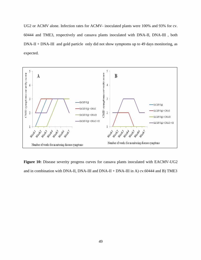
49
UG2 or ACMV alone. Infection rates for ACMV- inoculated plants were 100% and 93% for cv.
60444 and TME3, respectively and cassava plants inoculated with DNA-II, DNA-III , both
DNA-II + DNA-III and gold particle only did not show symptoms up to 49 days monitoring, as
expected.
Figure 10: Disease severity progress curves for cassava plants inoculated with EACMV-UG2
and in combination with DNA-II, DNA-III and DNA-II + DNA-III in A) cv.60444 and B) TME3

50
Figure 11: Disease severity progress curves for cassava plants inoculated with ACMV and in
combination with DNA-II, DNA-III and DNA-II + DNA-III in A) cv. 60444 and B) TME3.
2.4.3. RT-PCR for ORF C4 of DNA-II, ORF V1 and ORF C2 of DNA-III
The amplification of transcripts for ORF C4 of DNA-II and ORF V1, ORF C2 of DNA-III were
successful from cv.60444 and TME3 inoculated with EACMV-UG2 alone, ACMV alone,
begomovirus co-inoculated with DNA-II, DNA-III, DNA-II + DNA-III and mock -inoculated
cassava plants with gold particles. Primer pairs SatII-2F/SatII-3R and SatIII-7F/SatIII-9R
amplified a DNA fragment band size of ~250bp for DNA-II and ~220bp for DNA-III,
respectively (Fig. 12A&B) and primer pair IIIV1F/R yielded a band size of ~228 bp for DNA-
III.

51
Figure 12: Gel picture showing amplification of ORF C4 and ORF V1 transcripts on 1% agarose
by RT-PCR from cassava plants inoculated with either EACMV-UG2, ACMV and in
combination with DNA-II and DNA-III clones A) DNA-II; and B) DNA-III. L= Molecular
marker of 1kb plus, C= Negative control and, Lanes 1-20 are described in table 6 below.

52
Table 6: Samples collected from screen house for transcript analysis of DNA-II and
DNA-III
Sample No Sample description (Treatment)
1 EACMV-UG2 alone in cv. 60444
2 EACMV-UG2 + DNA-II in cv. 60444
3 EACMV-UG2 + DNA-III in cv. 60444
4 EACMV-UG2 + DNA-II +DNA-III in cv. 60444
5 Mock in cv. 60444
6 EACMV-UG2 alone in TME3
7 EACM EACMV-UG2 + DNA-II in TME3
8 EACMV-UG2 + DNA-III in TME3
9 EACMV-UG2 + DNA-II + DNA-III in TME3
10 Mock in TME3
11 ACMV alone in cv. 60444
12 ACMV + DNA-II in cv. 60444
13 ACMV + DNA-III in cv. 60444
14 ACMV + DNA-II + DNA-III in cv. 60444
15 Mock in cv. 60444
16 ACMV alone in TME3
17 ACMV+ DNA-II in TME3
18 ACMV+ DNA-III in TME3
19 ACMV +DNA-II +DNA-III
20 Mock in TME3

53
2. 4. 4 Sequence analysis
2.4.4.1 Transcript sequences for DNA-II
From NCBI BLAST search, it showed that transcript sequences for DNA-II ORF C4 had highly
(>94%) nt sequence similarity to the cassava begomovirus-associated satellite-like DNA-II
(AY836366) in Genbank. Alignment results of ORF C4 transcript sequences from 19 DNA-II
isolates illustrates that several sequences were highly conserved; for example nucleotide
sequences AGGCATC at position 76-82, GGTAA at position 37-41 and GAGAA at position
117-121 (Appendix 2.1). Phylogenetic tree and similarity distance for transcripts from different
samples are highly related up to 100% nucleotide sequence similarity (Appendix 2.2).

54
Table 7: Description of amplified RT- PCR samples for transcripts DNA-II sent for
sequencing.
Code number Sample number Sample Description (Treatments)
1.12 12 ACMV + DNA-II in cv. 60444
2.14 14 ACMV + DNA-II + DNA-III in cv.60444
3. 2 2 EACMV-UG2 + DNA-II in cv.60444
4.11 11 ACMV alone in cv.60444
5.20 20 Mock in TME3
6.8 8 EACMV-UG2 + DNA-III in TME3
7.3 3 EACMV-UG2 + DNA-III in cv.60444
8.1 1 EACMV-UG2 alone in cv.60444
9.5 5 Mock in cv.60444
10.15 15 Mock in cv.60444
11.18 18 ACMV+ DNA-III in TME3
12.4 4 EACMV-UG2 + DNA-II + DNA-III in cv.60444
13.7 7 EACMV-UG2 + DNA-II in TME3
14.16 16 ACMV alone in TME3
15.17 17 ACMV+ DNA-II in TME3
16.13 13 ACMV + DNA-III in cv.60444
17.9 9 EACMV-UG2 + DNA-II + DNA-III in TME3
18.6 6 EACMV-UG2 alone in TME3
19.10 10 Mock in TME3
55
2.4.4.2 Transcript sequences for ORF V1 of DNA-III
BLAST search shows that transcript sequences for DNA-III had high (>94%) nt sequence
similarity to the cassava begomovirus-associated satellite-like DNA-III (AY836367). Alignment
results for transcript DNA-III illustrated that several nucleotide sequences from several plant
sample isolates are highly conserved; for example nucleotide sequences
TCGTCGCCTTTGGTCCTCAATCACA at position 94-124, nucleotide C at position 70, 73, 89,
124 and similarly nucleotide G is conserved at several positions for example at position 40
(Appendix 2.3). Phylogenetic tree and nucleotide similarity distances for transcripts from
different isolates are highly related (up to 100% nt sequence similarity) (Appendix 2.4).
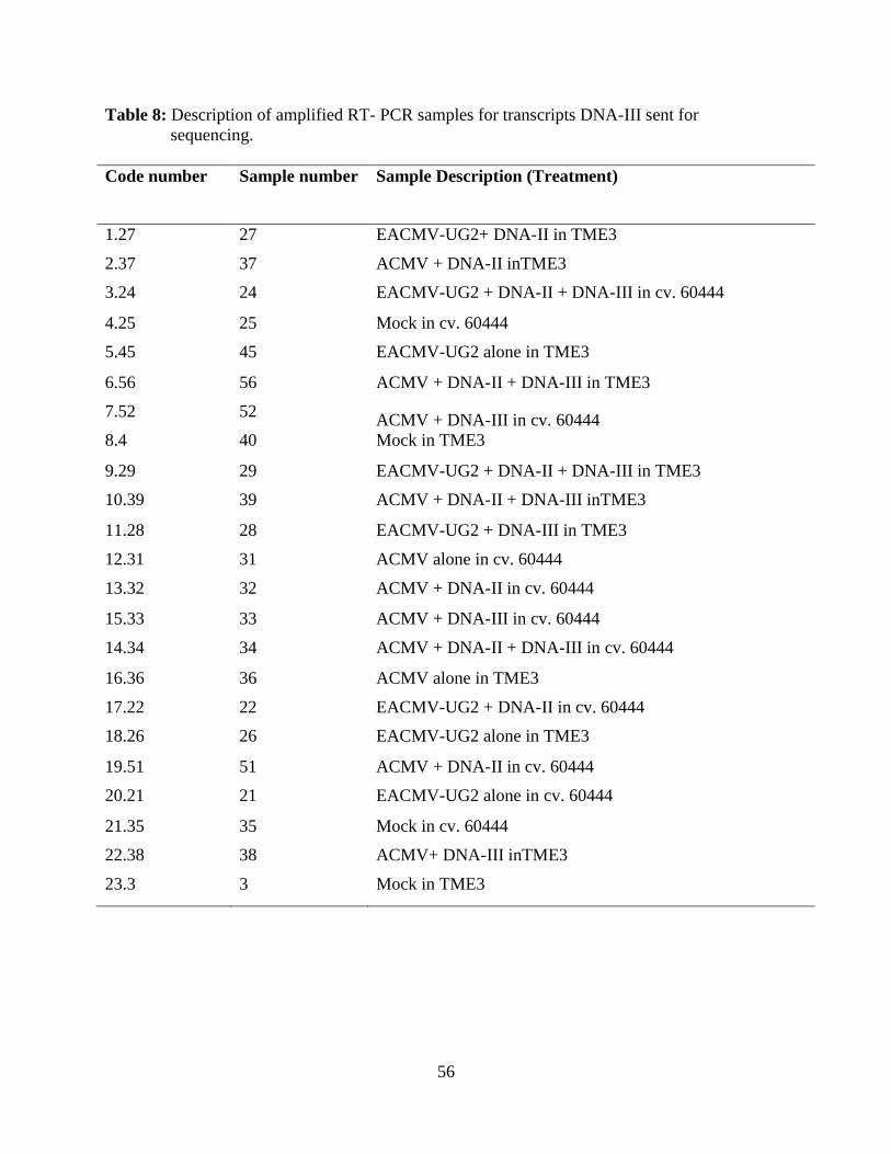
56
Table 8: Description of amplified RT- PCR samples for transcripts DNA-III sent for
sequencing.
Code number Sample number Sample Description (Treatment)
1.27 27 EACMV-UG2+ DNA-II in TME3
2.37 37 ACMV + DNA-II inTME3
3.24 24 EACMV-UG2 + DNA-II + DNA-III in cv. 60444
4.25 25 Mock in cv. 60444
5.45 45 EACMV-UG2 alone in TME3
6.56 56 ACMV + DNA-II + DNA-III in TME3
7.52 52 ACMV + DNA-III in cv. 60444
8.4 40 Mock in TME3
9.29 29 EACMV-UG2 + DNA-II + DNA-III in TME3
10.39 39 ACMV + DNA-II + DNA-III inTME3
11.28 28 EACMV-UG2 + DNA-III in TME3
12.31 31 ACMV alone in cv. 60444
13.32 32 ACMV + DNA-II in cv. 60444
15.33 33 ACMV + DNA-III in cv. 60444
14.34 34 ACMV + DNA-II + DNA-III in cv. 60444
16.36 36 ACMV alone in TME3
17.22 22 EACMV-UG2 + DNA-II in cv. 60444
18.26 26 EACMV-UG2 alone in TME3
19.51 51 ACMV + DNA-II in cv. 60444
20.21 21 EACMV-UG2 alone in cv. 60444
21.35 35 Mock in cv. 60444
22.38 38 ACMV+ DNA-III inTME3
23.3 3 Mock in TME3
57
2.5 Discussion
From the biolistic co-inoculation experiment conducted (Fig. 8), it was observed that cassava
mosaic disease (CMD) symptoms developed earlier on cv. 60444 compared to TME3 plants
when inoculated with EACMV-UG2, or EACMV-UG2 co-inoculated with DNA-II or DNA-III,
and both DNA-II + DNA-III infectious clones, and this may be due to the susceptible nature of
cv. 60444 compared with the tolerant TME3 cultivar. It was difficult to differentiate between
CMD symptom severity on cv. 60444 when plants were inoculated with EACMV-UG2 alone
and in combination with DNA-II, DNA-III or both DNA-II + DNA-III, as the highest severity
scores were the same. Probably the effect of DNA-II and DNA-III molecules on symptom
severity could have been discerned if the experiment had been extended. TME3 plants tolerant to
CMD developed cassava mosaic symptoms when EACMV-UG2 was co-inoculated with either
DNA-II or both DNA-II + DNA-III clones.
Failure of EACMV-UG2 alone to infect TME3 but in the presence of either of the satellites or
both indicated that EACMV-UG2 requires the episomal DNA-II and III molecules to induce
infection. In addition, DNA-II and DNA-III molecules appear to work with EACMV-UG2 to
break CMD tolerance in TME3, and enhance symptoms as previously reported (Ndunguru, 2008
unpublished data). It is postulated that these DNA-II and III sequences may participate in
assisting EACMV-UG2 to overcome antiviral defense mechanisms (either post transcriptional
gene silencing (PTGS) of virus derived transcripts, or transcriptional gene silencing (TGS)
through methylation of viral genome). Several studies support a possible role of methylation as
an important plant defense strategy against geminivirus. For example, in vitro methylation of

58
geminiviral DNA drastically reduced its infectivity (Brough., 1992; Ermak., et al 1993; Bisaro,
2006).
Another biolistic infection experiment (Fig. 9) was performed with a different begomovirus
species, ACMV, and DNA-II, DNA-III and both DNA-II + DNA-III in cv. 60444 and TME3.
Cassava mosaic disease symptoms severity ranged from 2-3 based on the CMD severity scale of
1-5 (Sseruwagi et al., 2004). The highest severity score was 3 in both cv. 60444 and TME3
however cv. 60444 plants developed symptoms earlier than TME3 plants. CMD symptoms were
developed earlier on cv.60444 than TME3 inoculated with ACMV alone and in combination
with DNA-II, DNA-III or both DNA-II + DNA-III. In cv. 60444, in contrast to EACMV-UG2,
cassava mosaic symptoms was more severe in plants inoculated with ACMV in combination
with DNA-II and III compared with those inoculated with ACMV alone, suggesting the
influence of DNA-II and DNA-III on symptom enhancement and may also be due to
susceptibility nature between ACMV and EACMV-UG2 in the cv. 60444. A research study
conducted by (Ayeh and Ramsell., 2008) showed differences in susceptibility between
begomoviruses in the same cassava cultivar. For example, a similar pattern of
resistance/susceptibility has also been observed in some of the cultivars on cassava fields in
Ghana. A study conducted in Uganda showed that different cassava varieties had different
responses of susceptibility to begomovirus infection in the field as well as in the laboratory
(Sserubombwe et al., 2008).
Tolerant TME3 cassava plants showed cassava mosaic symptoms when inoculated with ACMV
alone on young emerging leaves and recovered 42 days post inoculation. The same phenomenon

59
was observed in the field trials when cassava plants inoculated with ACMV did not develop
CMD symptoms. TME3 was reported to be tolerant to CMD because it has a dominant gene
(CMD2) important to cassava production (Akano et al., 2002) and which was able to tolerate
viral infection but recent studies showed that it is no longer tolerant (Ndunguru et al., 2008
unpublished ). Recovery is a well-known phenomenon in many plants in addition to cassava
(Hagen, et al., 2008; Chellappan et al., 2005). Symptom recovery in the infected plants over time
was shown to correlate with the accumulation of short interfering RNAs (siRNA) targeted
against specific genes of the virus (Rodriguez-Negrete et al., 2008).
In the two cassava cultivars used in this study (i.e. cv. 60444 and TME3) differences in symptom
expression were observed. In general CMD symptoms were observed earlier, and were more
severe, in cv.60444 than TME3 plants, and this is due to the susceptibility of cv. 60444 to
ACMV infection. Differences in symptom severity between the same cassava mosaic
begomovirus in the different cultivars have been also reported by Ndomba (2012). For example a
study conducted in Ghana revealed that there is a difference in symptom severity of cassava
breeding lines infected by the same infectious clones. Variation in cassava cultivars genetic
ability to resist CMD has also been reported by other studies (Lapidot and Friedmann, 2002;
Ogbe et al., 2002).
Recovery in TME3 could also be due to the fact that some leaves exclude the spread of virus
which may be related to expression of certain host defense mechanisms later on, leading to virus-
free leaves. Viruses may not be evenly distributed between leaves from inoculated plants and
some leaves may have lower virus titer due to patterns in virus movement.
60
Symptoms recovery from begomoviral infection of inoculated TME3 cassava plants in this
study may be due to presence of major dominant gene (CMD2) that controls resistance to CMBs
reported by Akano et al. (2002). But recent evidence showed that recovery to plant viruses in
many cases, may be due to innate basal immunity known as RNA silencing (Fregene et al.,
2001a; Dixon et al., 2001; Fraser, 2006, Patil and Fauquet, 2009). This occurs either in PTGS or
TGS silencing leading to a decrease in virus replication. RNA silencing involves the production
of small interfering RNAs (siRNAs) by the enzyme DICER, which splices viral mRNAs
(Rodriguez-Negrete et al., 2008). The production of siRNAs in plants challenged or containing
various geminiviruses can however be affected by temperature (Nelson and Citovsky, 2005).
RNA silencing was reported to increase as the temperature was raised from 25°C to 30°C, but in
this study this was not the case. Changes in symptom phenotype in this study may be related to
the presence of partially characterized DNA-II and DNA-III molecules. This observation was
also made from another study conducted by Ndunguru et al., (2013) (manuscript in preparation)
who observed enhanced CMD symptom on TME3 cassava land race due to the presence of
DNA-II and DNA-III in combination with the wild type EACMV-Ug2.
Putative ORFs were predicted for episomal DNA-II and DNA-III forms (Ndunguru et al., 2005).
RT-PCR transcripts of ORF C4 for DNA-II on the antisense strand and ORF V1 on the sense
strand, ORF C2 on the antisense strand and part of GC-rich region for DNA-III were transcribed
in all the experimental cassava plants. ORF C4 for DNA-II out of eight ORFs, ORF V1 and ORF
C2 for DNA-III out of four ORFs present in the genome structure of episomal DNA-II and
DNA-III respectively were amplified. Transcripts obtained were from inoculated cassava plants
as indicated in Table 6. Amplification of transcripts from mock-inoculated and begomovirus-

61
only inoculated cassava plants demonstrates that episomal DNA-II and DNA-III are integrated
into the cassava genome, at least in partial form, and additionally that transcription of DNA-II
and DNA-III molecules are independent from their associated virus. The transcriptions of the
remaining ORFs were not studied. In addition, RT-PCR was performed using cassava germplasm
samples from farmer’s field and transcripts of the same ORFs were obtained (result not shown
here). The results from this study provide evidence that transcription of ORFs is independent of
helper virus and environmental conditions.
In order to confirm transcription of episomal ORFs C4 and C2 from DNA-II, and VI from DNA-
III, the same experiments need to be performed in another host, such as N. benthamiana where
no integrated sequences occur. Furthermore, the roles/functions of the transcripts are yet to be
determined. However it is hypothesised, based on the ability of episomal DNA-II and III to
overcome tolerance/resistance in TME3 when co-inoculated with EACMV-UG2 (which does not
infect TME3 alone), and transcribed integrated DNA-II and III sequences, that silencing of
homologous cognate transcripts leads to silencing of critical genes involved in begomovirus
defence. Transcript sequencing results showed that nucleotide sequences are highly conserved
amongst DNA-II and III in inoculated and non-inoculated cassava plants, suggesting that these
sequences of DNA-II and DNA-III may have a genomic function in cassava, or begomovirus
infection in cassava plants.
In conclusion, this experiment has shown that ORF C4 of DNA-II on the antisense strand and
ORF V1 on the sense strand and ORF C2 on the antisense strand of DNA-III, as well as part of

62
GC-rich region, could be transcribed, and there is a need to conduct more research to understand
the biological functions of these conserved sequences.
63
CHAPTER THREE
GENETIC DIVERSITY AND GEOGRAPHIC DISTRIBUTION OF DNA-II
AND III MOLECULES ASSOCIATED WITH CASSAVA AND CASSAVA
MOSAIC DISEASE IN UGANDA, KENYA, RWANDA AND TANZANIA
Manuscript submitted to Plant Pathology (modified with additional data from Dr. Ndomba’s
PhD).
3.1 Abstract
DNA-II and III are small fragments, first detected as episomal ssDNA form associated with
cassava mosaic begomoviruses (East African cassava mosaic virus-Uganda2 and African
cassava mosaic virus), and later found during screening of cassava EST databases to be
integrated into the cassava genome. Co-inoculation of episomal ssDNA-II and DNA-III with
ACMV and EACMV-UG2 led to enhanced symptom severity in cassava cultivar TME3, leading
to speculation of the function of these DNA molecules. A study was conducted to determine the
genetic diversity and geographic distribution of integrated DNA-II and III molecules in Kenya,
Rwanda, Uganda and Tanzania in 2009 and 2010. Young CMD-symptomatic and symptomless
cassava plants (3-6 months) were collected in cassava fields across major cassava growing areas
in each country. The samples were subjected to DNA extraction and specific PCR primer
amplification in order to determine the frequency and genetic diversity of DNA-II/III in different
cassava germplasm. Sequence analysis showed that DNA-II and III molecules were integrated in
75.7% and 77.7% of the cassava genotypes, respectively. The distribution of both DNA-II and
DNA-III molecules in collected samples was 69.1 % in Uganda, 73.3% in Kenya, 50% in

64
Rwanda and 69.3% in Tanzania. DNA-II occurred at a frequency of 18.5% in Uganda, 3.3% in
Kenya, 6.5% in Rwanda and 5.7% in Tanzania, while DNA-III was 2.5% in Uganda, 8.3% in
Kenya, 19.6% in Rwanda and 11.9% in Tanzania. CMD symptoms were more severe (3.2 mean
symptom severity score based on 1-5 scoring scale), in Uganda followed by Rwanda (3.0) and
Tanzania (2.9) and least severe in Kenya (2.5). DNA-II and DNA-III were detected from cassava
plants displaying CMD symptoms of narrow and deformed leaves, and also non-symptomatic
cassava plants, and is suspected to play a role in disease phenotype modulation during viral
infection. There was little nucleotide sequence divergence (88% - 100%) between the DNA-II
and DNA-III sequences: DNA-II sequences had 95-99% nt sequence similarity to the sequences
of EACMV-associated episomal satDNA-II (AY836366) and 99% with mentha leaf deformity
disease associated satellite DNA II (EU862815), while DNA-III shared sequence similarity of
92-99% with EACMV-associated episomal satDNA-III (AY836367). This study revealed that
both DNA-II and DNA-III are widely distributed in the cassava genotypes of almost all the
cassava genotypes collected from surveyed areas in Uganda, Kenya, Rwanda and Tanzania.

65
3.2 Introduction
Cassava (Manihot esculenta Crantz) is an important staple food for over 700 million people in
Sub Saharan Africa (FAOSTAT, 2011). However, the production of the crop is hugely limited
by a number of pests and diseases, among which cassava mosaic disease (CMD) and cassava
brown streak disease (CBSD) are currently the most important (Legg et al., 2011).
Cassava mosaic disease is caused by cassava mosaic begomoviruses (CMBs) which belong to
the genus Begomovirus of the family Geminiviridae. The viruses are transmitted by whitefly
Bemisia tabaci Genn and spread by humans through planting of infected cuttings, which is the
usual mode of cassava propagation (Katz et al., 2003; Thresh et al., 1994). African cassava
mosaic virus (ACMV) and East African cassava mosaic virus (EACMV) are the most commonly
occurring CMBs on the sub-Saharan African continent (Pita et al., 2001). Cassava mosaic
begomovirus (CMB) genome consists of two circular molecules of single-stranded DNA (DNA-
A and DNA-B), each c. 2.8 kbp (Lazarowitz, 1992). Currently, there are nine reported species of
cassava begomoviruses (CBVs) infecting cassava in sub-Saharan Africa countries. These viruses
are African cassava mosaic virus (ACMV); East African cassava mosaic virus (EACMV), East
African cassava mosaic Cameroon virus (EACMCV) (Fondong et al., 2000), East African
cassava mosaic Kenya virus (EACMKV) (Bull et al., 2006), East African cassava mosaic
Malawi virus (EACMMV) (Zhou et al., 1998), East African cassava mosaic Zanzibar virus
(EACMZV) (Maruthi et al., 2004)), African cassava mosaic Bukina Faso virus (ACMBFV)
(Tiendrébéogo et al., 2012), South African cassava mosaic (SACMV) (Berrie et al.,1998) and
Cassava mosaic Madagascar virus (CMMGV) (Harimalala et al., 2012 ). The control of CMD

66
in East Africa has involved the introduction of improved Tropical Manihot Series (TMS) from
the International Institute of Tropical Agriculture (IITA), Ibadan, Nigeria, initially into Uganda
and later in Tanzania and other countries.
Despite the achievements of developing CMD resistant cassava varieties through traditional
breeding (Hahn et al., 1980) and using of several tolerant landraces, such as TME1 and TME3
(Akano et al., 2002), these cassava varieties are no longer resistant/tolerant to CMBs. More
recently, two episomal circular satellite-like molecules, DNA-II and DNA-III associated with
ACMV and EACMV-UG2, were detected in infected cassava plants using universal betasatellite
primers (Briddon et al., 2002), and were able to break resistance to EACMV-UG2 in the highly
resistant landrace TME3 (Ndunguru et al., 2005; 2008; Ingelbrecht et al., 2008). Additionally,
integrated DNA-II and III molecules were identified during screening of cassava EST databases
(unpublished). Plant virus sequences (19,831) were downloaded from the DPV web plant virus
database (Adams and Antoniw, 2006) and aligned to the set of 10577 transcripts in the CIAT-
RIKEN cassava (MATI16) full-length cDNA library using BLAST. Two viral sequences sharing
sequence similarity with cassava transcripts were detected, namely ACMV-associated DNA-II
and DNA-III (unpublished). The range of alignments for DNA-II and DNA-III was 112-346 bp
and 65-226 bp, respectively.
It is not known if or how these genomic DNA-II and III molecules in cassava interact with
cassava CMBs and cassava brown streak viruses (CBSVs), or whether the episomal forms are
transmitted by the whitefly (Bemisia tabaci) vector. The exact impact of DNA-II and III on
cassava production and their putative role in the current pandemic of cassava mosaic disease are

67
key priorities, and require further research. This study aimed to establish the genetic diversity
and geographic distribution of DNA molecules in Kenya, Rwanda, Uganda and Tanzania.
3.3 Materials and Methods
3.3.1 Sample collection
Samples were collected from the major cassava growing areas in Uganda, Kenya, Rwanda and
Tanzania (Table 9). Leaf samples were collected from CMD-symptomatic and symptomless
cassava plants. In particular, leaves that displayed an unusual narrow (filform) phenotype were
collected (Fig.13A). The samples were placed in sampling bags, and then stored in cool boxes
containing ice cubes until they were delivered to the laboratory for molecular analysis. CMD
symptom severity was assessed using a 1 to 5 scoring scale as described by Sseruwagi et al.
(2004); where 1= symptomless and 5= very severely distorted leaf symptoms. In each field, the
geographical location and geo-coordinates (latitude, longitude and altitude) were recorded using
a GPS.

68
Table 9: Cassava major growing areas where survey was conducted in Uganda, Kenya, Rwanda
and Tanzania in 2009/2010
Country
District/
Latitude Longitude Country
District/
Latitude Longitude Region Region
Uganda Wakiso 00°10'00''N 32°30'00''E Rwanda Kamonye 02°00'00''S 29°54'00''E
Luwero 01°00'00''N 32°20'00''E
Muhanga 02°05'00''S 29°45'00''E
Mpigi 00°50'00''N 32°00'00''E
Ruhango 02°13'57''S 29°46'49''E
Mukono 00°15'00''N 32°55'00''E
Nyanza 02°35'00''S 29°43'00''E
Kayunga 01°00'00''N 32°52'00''E
Huye 02°35'00''S 29°41'20''E
Jinja 00°26'20''N 33°12'11''E
Gakenke 01°42'00''S 29°47'00''E
Kenya Kisumu 00°15'00''S 34°55'00''E Tanzania Morogoro 08°00'00''N 37°00'00''E
Busia 00°25'00''N 34°15'00''E
Dodoma 06°00'00''N 36°00'00''E
Siaya 00°50'00''S 34°15'00''E
Tabora 05°30'00''N 32°49'00''E
Nyondo 00°45'00''S 38°12'11''E
Shinyanga 03°45'00''N 33°00'00''E
Mwanza 02°45'00''N 32°45'00''E
Kagera 02°00'00''N 31°30'00''E
Tanga 05°00'00''N 38°15'00''E
Coastal 07°00'00''S 39°00'00''E
Lindi 09°30'00''N 38°30'00''E
Mtwara 10°40'00''N 39°00'00''E
Songea 11°00'00''N 36°00'00''E
Iringa 09°30'00''N 35°00'00''E

69
Figure 13: Cassava plants showing cassava mosaic symptoms in cassava fields A; CMD
symptoms; B; mosaic severe; C; leaf curling and distortion D; leaf narrowing.
3.3.2 DNA extraction
Total genomic DNA was extracted from young CMD-symptomatic and asymptomatic cassava
leaves using a modified Dellaporta et al (1983) method. For each leaf sample, 700 mg were
ground with a pestle in a mortar containing 500 µl extraction buffer (1M Tris-HCl, 0.5M EDTA
and 5M NaCl) with 98% β-mercaptoethanol and the homogeneous mixture was transferred to
70
clean 1.5 ml eppendorf tubes. In each tube, 40 µl of sodium dodecyl sulphate (SDS) solution was
added, mixed and the contents incubated in a 65°C water bath for 30 min. The content was
mixed by inverting the tubes after every 10 min. Then 200 µl of 3M sodium acetate solution was
added to each tube, mixed, incubated in ice for 20 min and centrifuged at 14,000 rpm for 10 min
at room temperature. The supernatant (500 µl) was transferred to a new 1.5 ml tube to which 2 µl
of 10 mg/ml RNase A enzyme was added and incubated in water bath at 37°C for 30 min. To
precipitate the DNA, 500 µl of cold isopropanol was added to the contents, stored at -20°C for 30
min, followed by a 10 min centrifugation to precipitate the DNA. After washing with 70%
ethanol, the DNA were air-dried for 30 min, re-suspended in 100 µl of sterile double distilled
water and stored at-20°C.
3.3.3 Polymerase chain reaction (PCR)
PCR amplification for integrated DNA sequences was performed using specific primers: satIIF
5’-GCCGCACCACTGGATCTC-3’ forward primer, satIIR 5’-
CAGCAGCCAGTCAGGAAGTT-3’ reverse primer for DNA-II and satIIIF 5’-
AGGCCTCGTTACTAAAAGTGC-3’ forward primer, satIIIR 5’
ACCTGACGGCAGAAGGAAT-3’ reverse primer for DNA-III (German Plata CIAT,
Colombia). PCR was performed using a Thermorcycler (GeneAmpR PCR System 9700) with the
first cycle at 94°C for 1 min followed by 30 cycles at 94°C for 1 min, 55°C for 1 min, 72°C for
2 min and a final extension at 72°C for 10 min. A total reaction mixture of 50 µl made up of
41.75 µl of sterile distilled water, 2.5 µl PCR buffer (10x), 1.5 µl MgCl2 (25 mM), 0.5 µl dNTPs
(2.5 mM), 1 µl each of the forward (10 mM) and reverse primer (10mM), 0.25 µl Taq
71
polymerase and 2.0 µl of the DNA template was used. To detect the PCR products,
electrophoresis was performed at 100 volts in a 1% agarose gel buffered with 1X TAE (Savant
HG330 electrophoretic gel system), and stained with 6x Orange DNA loading dye. The gels were
run for 1 hour, and visualized using the bench top UV transilluminator, and photographed with a
gel documentation system (BioDoc-ItTM
Imaging System).
3.3.4 Molecular cloning
Amplicons of integrated DNA-II and DNA-III were purified from the agarose gels using a
QIAgen Gel Extraction Kit (Invitro, Life Science, USA) as per the manufacturer’s protocol. In
each country, at least five positive amplicons each of DNA-II and DNA-III were ligated into the
pGEMR-T Easy Vector using a TA Cloning Kit (Promega, USA) following the manufacturer’s
guidelines, and transformed into chemically highly competent cell Escherichia coli strain JM109
(Promega, USA). Transformed bacteria colonies were cultured in Luria Bertani broth containing
appropriate antibiotics and incubated at 37°C overnight while shaking at 200 rpm. Plasmids were
purified by using QIAgen Minprep Purification Kit (Promega, USA) and the purified plasmids
were sequenced using universal primers (M13F/R) at BeCA-ILRI- Nairobi, Kenya.
3.3.5 Sequences analysis
DNA sequences of integrated DNA-II and DNA-III molecules were assembled from contiguous
sequences with SEQMAN program (DNASTAR). Putative Open Reading Frames (ORFs) were
identified with a FramePlot software program. To determine whether the molecules were similar
72
to published DNAs sequences, they were initially aligned, using BLASTN, to known sequences
in the National Center for Biotechnology Information (NCBI). ORFs were translated to protein
and BLASTed (http://www.ncbi.nlm.nih.gov/BLAST/) to sequences in Genbank to determine
whether the proteins were similar to known satellite proteins, for example CAA65841,
CBJ19308, CAJ85966 and YP-184756. Sequences were aligned with the MEGA software,
(ClustalW) (Thompson et al., 1997) and phylogenetic trees constructed using the neighbour-
joining algorithm.
3.3.6 Statistical data analysis and presentation
Geo-coordinates were entered into excel and used to generate disease distribution maps for the
data collected in the surveyed areas. Significance of the DNA-II and III occurrence and
distribution data was analysed using a one-way analysis of variance (ANOVA) and means
separated using the least significance differences (LSD) at 5%.
3.4 Results
3.4.1 Cassava mosaic disease symptom expression in the field
Cassava mosaic disease symptoms varied widely among the infected plants in cassava fields.
The symptoms observed were mosaic, severe mosaic, leaf narrowing, leaf curling, leaf distortion
and leaf deformation (Fig.13A, B, and C &D).
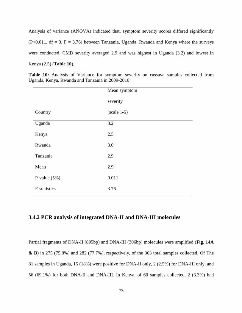
73
Analysis of variance (ANOVA) indicated that, symptom severity scores differed significantly
(P<0.011, df = 3, F = 3.76) between Tanzania, Uganda, Rwanda and Kenya where the surveys
were conducted. CMD severity averaged 2.9 and was highest in Uganda (3.2) and lowest in
Kenya (2.5) (Table 10).
Table 10: Analysis of Variance for symptom severity on cassava samples collected from
Uganda, Kenya, Rwanda and Tanzania in 2009-2010
3.4.2 PCR analysis of integrated DNA-II and DNA-III molecules
Partial fragments of DNA-II (895bp) and DNA-III (306bp) molecules were amplified (Fig. 14A
& B) in 275 (75.8%) and 282 (77.7%), respectively, of the 363 total samples collected. Of The
81 samples in Uganda, 15 (18%) were positive for DNA-II only, 2 (2.5%) for DNA-III only, and
56 (69.1%) for both DNA-II and DNA-III. In Kenya, of 60 samples collected, 2 (3.3%) had
Mean symptom
severity
Country (scale 1-5)
Uganda 3.2
Kenya 2.5
Rwanda 3.0
Tanzania 2.9
Mean 2.9
P-value (5%) 0.011
F-statistics 3.76

74
DNA-II only, 5 (8.3%) had DNA-III only, and 44 (73.3%) were positive for both DNA-II and
DNA-III. Of the 46 samples collected from Rwanda, 3 (6.5%) had DNA-II, 9 (19.6%) had DNA-
III, and 23 (50%) were positive for both DNA-II and DNA-III. Of the 176 samples from
Tanzania, 10 (5.7%) samples had DNA-II only, 21 (11.9%) had DNA-III only, and 122 (69.3%)
confirmed the presence of both DNA-II and DNA-III molecules. Combined PCR results for all
samples collected in the surveys showed that 67% (245/363) of the samples had with both
integrated DNA-II and DNA-III molecules. Only 8% (30/363) and 10% (37/363) had either
integrated DNA-II or DNA-III, respectively, and 14% (51/363) did not yield any amplification.
Figure 14: PCR amplification of integrated DNA-II molecules (16A) and DNA-III (16B) on 1%
agarose gel. M=Molecular weight marker of 1 kb plus; +C= positive control and -C = negative
control. Lanes 1-20 (16A &16B) represent samples collected from farmers’ field.
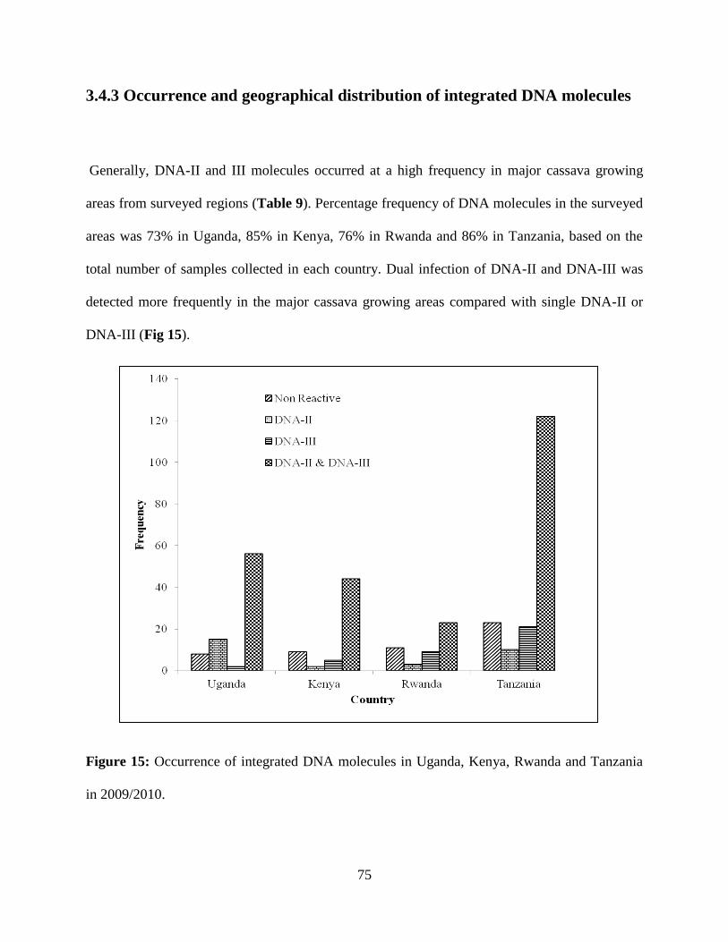
75
3.4.3 Occurrence and geographical distribution of integrated DNA molecules
Generally, DNA-II and III molecules occurred at a high frequency in major cassava growing
areas from surveyed regions (Table 9). Percentage frequency of DNA molecules in the surveyed
areas was 73% in Uganda, 85% in Kenya, 76% in Rwanda and 86% in Tanzania, based on the
total number of samples collected in each country. Dual infection of DNA-II and DNA-III was
detected more frequently in the major cassava growing areas compared with single DNA-II or
DNA-III (Fig 15).
Figure 15: Occurrence of integrated DNA molecules in Uganda, Kenya, Rwanda and Tanzania
in 2009/2010.

76
There was no apparent defined pattern in terms of geographical distribution of the integrated
DNA molecules among the countries and within the country, since the integrated DNA
molecules were frequently distributed in all the surveyed areas, either singly or as dual infection
(Fig. 16). The DNA molecules were distributed throughout the surveyed areas in major cassava
growing areas in Lake Victoria, Southern zone and Coastal zone in Tanzania, Central areas in
Uganda, Northern and Southern provinces in Rwanda and Western province in Kenya.
Figure 16: Geographical distribution of DNA-II and DNA-III molecules in Uganda, Kenya,
Rwanda and Tanzania in 2009/2010

77
3.4.4 Phylogenetic analysis and sequence comparisons
DNA-II and DNA-III sequences were aligned and gaps were removed. After alignment,
phylogenetic trees were generated in which DNA-II sequences grouped into a single cluster (Fig.
17A), while DNA-III sequences formed two clusters (Fig. 17 B). Grouping of the isolates was
random within the two clusters indicating that there was no specific definite geographical
distribution of genotypes. Analyzed sequences for DNA-II molecules obtained from Uganda,
Kenya, Rwanda and Tanzania revealed an average integrated sequence length of 841 nucleotides
(nt). The variation in length within the sequences obtained was due to deletion of 54 nt in
expected sequences of 895 bp within DNA-II molecules (unpublished). Pair-wise comparison
within the analysed sequences showed 88% - 100% sequence identity to each other. All DNA-II
sequences analysed had GC region typical of episomal DNA-II in the database (AY836366).
Nucleotide sequences derived from these sequences were compared with GeneBank sequences,
and were highly similar (95-99%) to the published episomal DNA sequence accession number
(AY836366) (Ndunguru et al, 2008) and 99% to Mentha leaf deformity associated DNA-II
(Accession No. EU862815) (Borah et al, 2010) associated with Menthal leaf deformity virus.
Analyzed sequences for DNA-III revealed a sequence length that ranged from 290-314 nt.
Within analysed sequences for DNA-III, sequences obtained from Rwanda had an average length
of 290 nt, while those of Kenya, Uganda and Tanzania ranged between 306 and 314 nt. The
variation in length within DNA-III sequences analysed was due to a deletion of 16 nt
(CTCAACTCTCCATGCC), in the Rwandan DNA-III isolates, in the expected sequence (306
bp) amplified in earlier studies (unpublished). In contrast the Kenyan, Ugandan and Tanzanian

78
DNA-III sequences had a longer length of 314 nt due to insertion of 8 nt. All analysed DNA-III
sequences had GC region typical of episomal DNA-III in the published sequence (AY836367).
The sequences were blasted against the GeneBank sequence and showed to be highly similar
(92-99%) to episomal DNA-III (Ndunguru et al, 2008).
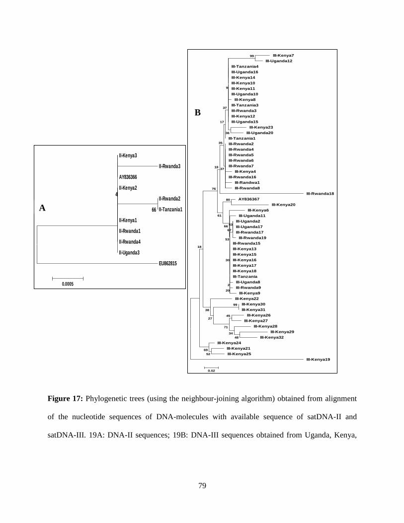
79
II-Kenya3
II-Rwanda3
AY836366
II-Kenya2
II-Rwanda2
II-Tanzania1
II-Kenya1
II-Rwanda1
II-Rwanda4
II-Uganda3
EU862815
66
4
0.0005
III-Kenya7
III-Uganda12
III-Tanzania4
III-Uganda16
III-Kenya14
III-Kenya10
III-Kenya11
III-Uganda10
III-Kenya8
III-Tanzania3
III-Rwanda3
III-Kenya12
III-Uganda15
III-Kenya23
III-Uganda20
III-Tanzania1
III-Rwanda2
III-Rwanda4
III-Rwanda5
III-Rwanda6
III-Rwanda7
III-Kenya4
III-Rwanda16
III-Randwa1
III-Rwanda8
III-Rwanda18
AY836367
III-Kenya20
III-Kenya6
III-Uganda11
III-Uganda2
III-Uganda17
III-Rwanda17
III-Rwanda19
III-Rwanda15
III-Kenya13
III-Kenya15
III-Kenya16
III-Kenya17
III-Kenya18
III-Tanzania
III-Uganda8
III-Rwanda9
III-Kenya9
III-Kenya22
III-Kenya30
III-Kenya31
III-Kenya26
III-Kenya27
III-Kenya28
III-Kenya29
III-Kenya32
III-Kenya24
III-Kenya21
III-Kenya25
III-Kenya19
99
48
34
45
71
52
99
69
27
38
18
80
43
67
20
2
30
53
66
61
76
18
36
37
35
17
37
9
0.02
Figure 17: Phylogenetic trees (using the neighbour-joining algorithm) obtained from alignment
of the nucleotide sequences of DNA-molecules with available sequence of satDNA-II and
satDNA-III. 19A: DNA-II sequences; 19B: DNA-III sequences obtained from Uganda, Kenya,
A
B
80
Rwanda and Tanzania in 2009/2010, obtained from Genbank with accession number AY836366,
AY836367 and EU862815.
3.5 Discussion
There is no prior published information from the literature on DNA-II or DNA-III-like
molecules (associated with ACMV or EACMV) integrated into the cassava, or any other plant
genome. Other DNA molecules associated with symptom modulation of begomovirus infections
have been reported in the literature. Amongst these are defective interfering molecules,
associated with helper begomoviruses in cassava in South Africa, and Tanzania, and which are
derived from helper virus DNA A or B genome (Choge et al., 2001; Paximadis et al, 2001;
Ndunguru et al., 2006; Abraham et al., 2012). Additionally, betasatellites have been reported in
several begomovirus diseases in the Old World, and are known to alter symptom phenotypes in
their respective hosts (Briddon and Stanley, 2006). Satellite DNAs or sub-viral agents are
dependent on their cognate helper begomovirus for replication, and can exacerbate or reduce
symptoms. Some examples include ToLCV-sat components associated with monopartite
begomovirus ToLCV in tomato plants (Dry et al., 1997), DNA-β associated with AYVV in
ageratum weed (Saunder et al., 2000), CLCuV in cotton plants (Briddon et al., 2000, 2001) and
DNA-1 components isolated from a range of plant species growing in different regions
throughout Africa and Asia, for example DNA-1 isolated from CLCuD (Briddon et al., 2004;
Mansoor et al., 2001; Mansoor et al., 200a; Mansoor et al., 200b; Wu and Zhou, 2005).
This study reports for the first time the presence of DNA-II and DNA-III molecules integrated
into the cassava genome of CMD-infected and non- infected cassava plants. While the nature of

81
these integrated DNA fragments are not known, they share high sequence identity with two
episomal forms amplified by betasatellite universal primers from ACMV/EACMV-infected
cassava in Tanzania (Ndunguru, 2005). These so- called episomal circular ssDNA satellite-like
DNA-II and III amplicons were found to exacerbate symptom severity in cassava, and to reduce
tolerance in TME3, when co-inoculated with EACMV-UG2 (Ndunguru et al., 2005, 2008).
Mentha leaf deformity is associated with a geminivirus-satellite complex of cotton leaf curl
Multan betasatellite (CLCuMB) (EU862816) and Tomato leaf curl Karnataka virus (ToLCKV)
infecting mint plants in the Punjab in India (Borah et al., 2012).
In addition to the betasatellite, the betasatellite primers amplified a 1019 nucleotide product
(EU862815). Surprisingly the Mentha leaf deformity associated DNA-II (MLDA-DNA-II)
showed 98% sequence identity with DNA-II (AY836366) reported from cassava associated with
EACMV-UG2 from cassava in Tanzania. Neither MLDA-DNA-II or cassava-associated DNA-II
appear to contain any known betasatellite ORFs, such as –C1, although putative ORFs have
been postulated. It is interesting to note that both cassava and mentha display severe leaf
deformity, and it would appear the episomal DNA-II plays a role in begomovirus symptom
phenotype severity. However the exact nature, origin and function of the integrated fragments is
not known. As with cassava, MLDA-DNA-II accumulated in the majority but not all
symptomatic mentha plants. DNA-III related sequences have not been reported in any other
plants in the Genebank database. MLDA-DNA-II could not be transmitted to Nicotiana
benthamiana by mechanical inoculation (Borah et al., 2012), but infectious clones of cassava-
associated DNA-II and III were able to infect cassava and N.benthamiana by agro-inoculation
and biolistic bombardment along with helper CMB (Ndunguru et al., 2005; 2008).
82
Further work is necessary to determine the role of these satellite-like and integrated DNA agents.
Since smaller fragments of cassava-associated DNA-II and III were first uncovered from EST
databases, and now have been mapped to scaffolds of the cassava genome (unpublished), it is
certain that there is some biological function. However what, if any, roles these DNAs may have
in gene function/regulation or begomovirus-host interaction, needs to be elucidated.
83
CHAPTER FOUR
SUMMARY AND RECOMMENDATIONS
Some plant viruses are associated with satellites that are composed of nucleic acid and depend on
co-infection of host cell with a helper virus for their multiplication. Many of them modulate
disease symptoms and increase viral accumulation (Roossinck et al., 1992; Simon et al., 2004).
The earliest satellites were associated with RNA viruses, but recently satellites associated with
DNA viruses, such as geminiviruses, have been discovered. These satellites include TLCV Sat-
DNA, betasatellites and alphasatellites which were isolated from monopartite begomoviruses and
satellite-like DNA molecules isolated from bipartite begomoviruses. Successful interaction
between betasatellites and the New Word (NW) bipartite begomovirus Cabbage leaf curl virus
(CbLCuV) has been reported by Nawaz-ul-Rehman et al. (2009). Also, the recent discovery of
satellite DNAs proves that the Old World (OW) bipartite begomoviruses can indeed be
associated with satellites. The association between satellites and viruses has caused massive
economic losses on crop yields through exacerbated disease symptoms.
Cassava mosaic disease (CMD) is the most important disease reducing cassava productivity in
sub-Saharan Africa. The disease is caused by at least nine species of begomovirus that occur
singly and in co-infection (Fauquet et al., 2008). Despite the efforts to develop CMD resistant
cassava varieties through convectional breeding (Hahn et al., 1980), and use of several tolerant
cassava landraces such as TME1 and TME3 (Akano et al., 2002), these cassava varieties are no
longer tolerant/resistant to CMBs. More recently, two circular single stranded DNA molecules,
84
DNA-II and DNA-III associated with cassava mosaic begomoviruses were detected in infected
cassava plants using universal betasatellite primers (Briddon et al., 2002), and were able to
enhance CMD symptoms and break resistance to EACMV-UG2 in the highly resistant landrace
TME3 (Ndunguru et al., 2005; 2008; Ingelbrecht et al., 2008). Additionally, DNA-II and III
molecules were found integrated in the cassava genome of both domesticated and wild Manihot
species during screening of cassava EST databases (unpublished). It has been demonstrated that
DNA-II and DNA-III molecules integrate into the host genome while the previously reported
satellites were not known to be integrated into genome.
In this study primer pairs satII-F/R and satIII-F/R were able to amplify the expected size for
DNA-II and DNA-III molecules integrated in the cassava genome respectively. DNA-II and
DNA-III molecules were identified from cassava leaf samples collected from healthy and CMD-
infected cassava plants that was confirmed by sequencing. A total of 363 cassava genotype were
collected from Kenya, Uganda, Rwanda and Tanzania countries and were screened for integrated
DNA-II and DNA-III and were found 86% of the samples .This showed that DNA-II and DNA-
III integrated in the cassava genome are widely distributed into the major cassava growing areas.
The other achievement made in this study was the successful biolistic inoculation of EACMV-
UG2, ACMV, DNA-II and DNA-III infectious clones to cv.60444 and TME3. Cassava leaf
samples collected from inoculated TME3 and cv. 60444 from screen house were used for
determination of transcription of ORFs for DNA-II and DNA-III. RT- PCR using primer pairs
designed from genome structure of satellite-like DNA-II and DNA-III were performed. SatII-
2F/3R and SatIII-7F/9R primers detected the expected sizes for transcript ORF C4 and ORFV1
85
for DNA-II in antisense strand and DNA-III in sense strand, respectively. This suggests that at
least part of DNA-II and DNA-III can be transcribed.
In conclusion this study has generated useful information on the genetic diversity and
geographical distribution of the DNA-II and DNA-III molecules from Kenya, Rwanda, Uganda
and Tanzania and transcripts analysis for ORF C4 and ORF V1 in antisense strand of DNA-II
and sense strand of DNA-III respectively. Information generated can be helpful in understanding
the biological function of integrated DNA molecules and if the transcribed ORF can produce
functional protein. Furthermore, the study findings will add to the body of knowledge on the
DNA II and III and help researchers (plant virologists and breeders) working on cassava in Sub
Saharan Africa (SSA) to develop durable management strategies for CMD.
Recommendations
The present study has opened up more areas for further research in order to understand the role
of DNA-II and DNA-III molecules in the etiology and pandemic of CMD and which contribute
to the loss of cassava productivity in Uganda, Kenya, Rwanda and Tanzania when associated
with cassava mosaic begomoviruses. The areas for further research include:
1. The exact impact of DNA-II and III on cassava production and roles in gene
function/regulation or begomovirus-host interaction needs to be elucidated.
2. How these genomic DNA-II and III molecules in cassava interact with cassava CMBs or
whether the episomal forms are transmitted by the whitefly (Bemisia tabaci) vector or co-
encapsidated.
86
3. Further study on the transcribed ORFs if they produce functional proteins, and design primers
for the remaining ORFs for DNA-II and DNA-III.
References
Alberter, M., Rezaian, A., Jeske, H., 2005. Replicative intermediates of Tomato leaf curl virus
and its satellite DNAs. Virol. 331, 441-448.
Alabi, O.J., Kumar, P.L., Naidu, R.A., 2011. Cassava mosaic disease: A curse to food security in
Sub-Saharan Africa. Online. APSnet Features. doi:10.1094/APSnetFeature-0701.
Bender, A., Bender, A.E., 2005. Dictionary of Food and Nutrition, Oxford University Press,
1995, second edition.
Behjatnia, S.A.A., Dry, I.B., Rezaian, M.A., 1998. Identification of the replication-associated
protein binding domain within the intergenic region of tomato leaf curl geminivirus.
Nucleic Acids Res. 26, 925-931.
Berrie, L.C., Palmer, K.E., Rybicki, E.P., Rey, M.E.C., 1998. Molecular characterisation of a
distinct South African cassava infecting geminivirus. Arch. Virol., 143, 2253-2260.
Bethke, J.A., Byrne, F.J., Hodges, G.S., McKenzie, C.L., Shatters, R.G., 2009. First record of the
Q biotype of the sweetpotato whitefly, Bemisia tabaci, in Guatemala Phytoparasitica 37,
61-64.
Bisimwa, E., Walangululu, J., Bragard, C., 2012. Occurrence and Distribution of Cassava
Mosaic Begomovirus Related to Agro-ecosystems in the Sud-kivu Province, Democratic
Republic of Congo. Asian J. Plant Pathol. 6, 1-12.

87
Bull, S.E., R.W. Briddon, W.S. Sserubombwe, K. Ngugi, P.G. Markham, Stanley, J., 2006.
Genetic diversity and phylogeography of cassava mosaic viruses in Kenya. J. Gen. Virol.
87, 3053-3065.
Burns, A., Gleadow, R., Cliff, J., Zacarias, A., Cavagnaro, T., 2010. Cassava: The drought, war
and famine crop in a changing world. Sustain. 2, 3572-3607.
Briddon, R.W., Stanley, J., 2006. Subviral agents associated with plant single-stranded DNA
viruses. Virol. 344, 198-210, Oxford University.
Briddon, R.W., Markham, P.G., 2000 .Cotton leaf curl virus disease. Virus Res. 71, 151-159.
Briddon, R.W., Mansoor, S., Bedford, I.D., Pinner. M.S., Saunders, K. Stanley, J.Y. Zafar, Y,
Malik, K. A., Markham, P.G., 2001. Identification of DNA components required for
induction of cotton leaf curl disease. Virol. 285, 234-243.
Briddon, R.W., Bull, S.E., Mansoor, S., Amin, I., Markham, P.G., 2002.Universal primers for the
PCR-mediated amplification of DNA-β: a molecule associated with some monopartite
begomoviruses. Mol. Bio-technol. 20, 315-318.
Briddon, R.W., Bull, S.E., Amin, I., Idris, A.M., Mansoor, S., Bedford, I.D., Dhawan, P., Rishi,
N., Siwatch, S.S., Abdel-Salam, A.M., Brown, J.K., Zafar, Y., Markham, P.G., 2003.
Diversity of DNA β: a satellite molecule associated with some monopartite
begomoviruses. Virol. 312, 106-121. Article
Briddon, R.W., Bull, S.E., Amin, I., Mansoor, S., Bedford, I.D., Rishi, N., Siwatch, S.S., Zafar,
Y., Abedel-Salam, A.M., Markham, P.G., 2004. Diversity of DNA1: a satellite-like
molecule associated with monopartite begomovirus-DNAB complexes. Virol. 324, 462-
474.

88
Briddon, R.W., Brown, J., Moriones, E., Stanley, J., Zerbini, M., Zhou X., Fauquet, C., 2008.
Recommendations for the classification and nomenclature of the DNA-β satellites of
begomoviruses. Arch. Virol.153, 763-781.
Briddon, R.W., Markham, P.G., 1995. Family Geminiviridae; In Virus Taxonomy. Sixth Report
of the International Committee on Taxonomy of Viruses, 158-165. Edited by Murphy,
F.A., Fauquet, C. M., Bishop, D.H.L., Ghabrial, S.A., Jarvis, A.W., Martelli, G.P., Mayo,
M.A., Summers., M. D. Vienna and New York: Springer-Verlag.
Brown, J.K., Frohlich, D.R., Rosell, R.C., 1995. The sweetpotato or silverleaf whiteflies:
biotypes of Bemisia tabaci or a species complex? Annu. Rev. Entomol. 40, 511-534.
Brown, J.K., 2010. Phylogenetic biology of the Bemisia tabaci sibling species group. 619, 31-67
in: Bionomics and Management of a Global Pest. P.A. Stansly and S.E. Naranjo (eds.),
Springer, Amsterdam. 350.
Bull, S.E., Tsai, W.S., Briddon, R.W., Markham, P.G., Stanley, J., Green, S.K., 2004. Diversity
of begomovirus DNA β satellites of Non-malvaceous plants in east and south east Asia,
Arch. Virol. 149, 1193-1200.
Carabali, A., Belloti, A.M., Mantoya-Lerma, J., Cuellar, M.E., 2005 Adaptation of Bemisia
tabaci biotype (Gennadius) to cassava, Manihot esculenta (Crantz).Crop Prot.24, 643-
649.
Colvin, J., Omongo, C.A., Maruthi, M.N., Otim-Nape, G.W., Thresh, J.M., 2004. Dual
begomovirus infections and high Bemisia tabaci populations: two factors driving the
spread of a cassava mosaic disease pandemic. Plant Pathol. 53, 577-584.
Cui, X., Xie, Y., Fauquet, C.M., Zhou, X., 2004b. A DNA β associated with Tomato yellow leaf
curl China virus is required for symptom induction, J. Virol. 78, 13966-13974.
89
Cui, X., Wang, G. Li., Hu, D., Zhou, X., 2005. A begomovirus DNAβ-encoded protein binds
DNA, functions as a suppressor of RNA silencing, and targets the cell nucleus, J.
Virol.79, 10764-10775.
Chu, D., Zhang, Y.J., Brown, J.K., Cong, B., Xu, B.Y., Wu, Q.J., Zhu, G.R., 2006. The
introduction of the exotic Q biotype of Bemisia tabaci from the Mediterranean region
into China on ornamental crops. Florida Ento. 89, 168f Virol. 73, 5481-9.
Czechowski, T., Bari, R.P., Stitt, M., Scheible, W.R., Udvardi, M.K., 2004. Real-time RT-PCR
profiling of over 1400 Arabidopsis transcription factors: Unprecedented sensitivity
reveals novel root- and shoot-specific genes. Plant J. 38, 366–379.
Czechowski, T., Stitt, M., Altmann, T., Udvardi, M.K., Scheible, W.R., 2005. Genome wide
identification and testing of superior reference genes for transcript normalization in
Arabidopsis. Plant Physiol. 139, 5–17.
De La Rua, P., Simon, B., Cifuentes, D., Martinez-Mora, C., Cenis, J.L., 2006. New insights into
the mitochondrial phylogeny of the whitefly Bemisia tabaci (Hemiptera Aleyrodidae) in
Mediterranean Basin. J. Zool. Syste. Evol. Res. 44, 25-33.
Dennehy, T.J., Degain, B.A, Harpold, V.S, Zaborac, M., Morin, S, Fabrick, J.A, Nichols, R.L.,
Brown, J.K., Byrne, F.J., Li, X., 2010. Extraordinary resistance to insecticides reveals
exotic Q biotype of Bemisia tabaci (Gennadius) in the New World. J. Eco Entomol. 103,
2174-2186.
Dennehy, T.J., Degain, B.A., Harpold, V.S., Brown, J.K., Byrne, F.J., Morin, S., Nichols, R.L.,
2006. First new world report of Q biotype of Bemisia tabaci (Gennadius) reveals high
levels of resistance to insecticides. Resistance Pest ManagementNewsletter. 5, 18-19.
90
Dennehy, T.J., Degain, B.A., Harpold, V.S., Brown, J.K., Morin, S., Fabrick, J.A., Byrne, F.J.,
Nichols, R.L., 2005. New challenges to management of whitefly resistance to insecticides
in Arizona. The University of Arizona-Cooperative Extension Report, Tucson, AZ.
Dry, L.R., Krake, J., Rigden, E., Rezaian, M.A., 1997. A novel subviral agent associated with a
geminivirus: the first report of a DNA satellite, Proc. Natl. Acad. Sci. U.S.A. 94, 7088-
7093.
Dubern, J., 1994. Transmission of African cassava mosaic geminivirus by the whitefly (Bemisia
tabaci) Trop. Sci. 34, 82-91.
Elsharkawy, M.A., 2004. Cassava biology and physiology. Plant mol. Biology: 56, 418-501.
Esterhuizen, L.L., Mabasa, K.G., Van Heerden, S.W., Czosnek, H., Brown, J.K., Van Heerden,
H., 2012. Genetic identification of members of the Bemisia tabaci cryptic species
complex from South Africa reveals native and introduced haplotypes. J. Appl.
Entomol.136:1-14.
FAO., 2006. Cassava production statistics http:/www.fao.org
Fauquet, C., Fargette, D., 1990. African cassava mosaic virus: Etiology, epidemiology, and
control. Plant Dis. 74, 404-11 Retrieved October 23, 2008.
Fauquet, C.M., Briddon, R.W., Brown, J.K., Moriones, E., Stanley, J., Zerbini, M., Zhou, X.,
2008. Geminivirus strain demarcation and nomenclature. Arch. Virol. 153, 783-821.
Fauquet, C.M., Sawyer, S., Idris, A.M., Brown, J.K., 2005. Sequence analysis and classification
of apparent recombinant begomoviruses infecting tomato in the Nile and Mediterranean
basins. Phytopathol. 95, 549-555.

91
Fontes, E.P.B., Eagle, P.A., Sipe, P.A., Luckow, V.A., Hanley-Bowdoin, L., 1994a. Interaction
between a geminivirus replication protein and origin DNA is essential for viral
replication, J. Biol. Chem. 269, 8459-8465.
Fontes, E.P.B., Gladfelter, H.J., Schaffer, R.L., Petty, I.T.D., Hanley-Bowdoin, L., 1994b.
Geminivirus replication origins have a modular organization, Plant Cell. 6, 405-416.
Haggblade, S., Zulu, B., 2003. The recent cassava surge in Zambia and Malawi. In InWent,
IFPRI, NEPAD, CTA Conference Success in African Agriculture. Pretoria, South Africa.
Hahn, S.K., Howland A.K., Terry, E.R., 1980a. Correlated resistance of cassava to mosaic and
bacterial blight diseases. Euphytic. 29, 305-311.
Harimalala. M, Lefeuvre, P.A., De Bruyn, F., Tiendrébéogo, M., Hoareau, J., Villemot, S.,
Ranomenjanahary, A., Andrianjaka, B., Reynaud, Lett, J.M., 2012. A novel cassava-
infecting begomovirus from Madagascar: Cassava mosaic Madagascar virus. Arch.
Virol. 157, 2027-2030.
Harrison, B.D., Robinson, D.J., 1999. Natural genomic and antigenic variation in whitefly-
transmitted geminiviruses (begomoviruses). Annu. Rev. Phytopathol. 37, 369-398.
Harrison, Y.L., Liu, S., Khalid, S., Hameed, G.W., Otim-Nape., Robinson, D.J., 1997. Detection
and relationships of cotton leaf curl virus and allied whitefly-transmitted geminiviruses
occurring in Pakistan. Ann. Appl. Biol. 130, 61-75.
Hillocks, R. J., 2002. Cassava in Africa, In: Hillocks, R.J, Thresh, J.M, Bellolli, A.C, eds.
Cassava: Biology, Production and Utilization, CAB International, Wallingford. 41-54.
Hong, Y.G., Robinson, D.J., Harrison, B.D., 1993. Nucleotide sequence evidence for the
occurrence of three distinct whitefly- CJG Synergism between ACMV and EACMV
transmitted geminiviruses in cassava. J. General. Virol. 74, 2437-2443.

92
Horowitz, A.R., Kontsedalov, S., Khasdan, V., Ishaaya, I., 2005. Biotypes B and Q of Bemisia
tabaci and their relevance to neonicotinoid and pyriproxyfen resistance. Arch. Insect
Bioch. Phy. 58, 216-225.
Ilyina T.V., Koonin, E.V., 1992. Conserved sequence motifs in the initiator proteins for rolling
circle DNA replication encoded by diverse replicons from eubacteria, eucaryotes and
archaebacteria, Nucleic Acids Res. 20, 3279-3285.
Jennings, D.L., 1994. Breeding for resistance to African cassava mosaic geminivirus in East
Africa. Trop. Sci. 34, 110-122.
Jeske, H., Lutgemeier, M., Preiss,W., 2001. DNA forms indicate rolling circle and
recombination-dependent replication of Abutilon mosaic virus, EMBO J. 20, 6158-6167.
Katz, S.H., Weaver, W.W., 2003. Encyclopedia of Food and Culture. New York: Schribner.
ISBN 0684805685
Lazarowitz, S.G., 1992. Geminiviruses: genome structure and gene function. Critical Reviews in
Plant Scie. 11, 327-49.
Legg, J.P., Fauquet, C.M., 2004. Cassava mosaic geminiviruses in Africa, Plant Mol. Biol. 56,
585-599.
Legg, J.P., French, R., Rogan, D., Okao-Okuja, G., Brown, J.K., 2002. A distinct Bemisia tabaci
(Gennadius) (Hemiptera: Sternorrhyncha: Aleyrodidae) genotype cluster is associated
with the epidemic of severe cassava mosaic virus disease in Uganda. Mol Ecol. 11, 1219-
1229.
Legg, J.P., Hillocks, R.J. Eds., 2003. Cassava brown streak virus disease: past, present and
future. Proceedings of an International Workshop, Mombasa, Kenya, October 27-30,
2002. Natural Resources International Limited. Aylesford, UK. 100pp.

93
Legg, J.P., Ndjelassili, F., Okao-Okuja, G., 2004. First report of cassava mosaic disease and
cassava mosaic geminiviruses in Gabon. Plant Pathol. 53, 232.
Legg, J.P., Owor, B., Sseruwagi, P., Ndunguru, J., 2006. Cassava mosaic virus disease in East
and Central Africa: epidemiology and management of a regional pandemic. Adv Virus
Res. 67, 355-418.
Legg, J.P., Thresh, J.M., 2003. Cassava virus diseases in Africa. In: Proceedings of the First
International Conference on Plant Virology in Sub-Saharan Africa (4-8 June 2001,
Ibadan, Nigeria), IITA, Ibadan, Nigeria, pp. 517-522.
Lin, A.A., Behjatnia, I.B. Dry, J.W., Randles, M.A. Rezaian., 2003. High-affinity Rep-binding
is not required for the replication of a geminivirus DNA and its satellite. Virol. 305, 353-
363.
Lisha, V.S., Antony, B., Palaniswami, M.S., Henneberry, T.J., 2003. Bemisia tabaci
(Homoptera: Aleyrodidae) biotype in India. J. Econo. Entomol. 96, 322-327.
Mansoor, S., Amin, I., Hussain, M., Zafar, Y., Bull, S., Briddon. R.W., Markham, P.G., 2001.
Association of a disease complex involving a begomovirus, DNA 1 and a distinct DNA
beta with leaf curl disease of okra in Pakistan. Plant Dis. 85, 922.
Mansoor, S., Khan, S.H, M. Hussain, M., Zafar, Y., Pinner, M.S., Briddon, R.W., Stanley. J.,
Markham, P.M., 2000a. Association of a begomovirus and nanovirus-like molecule with
ageratum yellow vein disease in Pakistan. Plant Dis. 84, 101.
Mansoor, S., Mukhtar, S., Hussain, S., Amin, M., Zafar, I., Malik, K., Markham, P.G.,
2000b.Widespread occurrence of Cotton leaf curl virus on radish in Pakistan. Plant Dis.
84, 809.
94
Mansoor, S.H., Khan, A., Bashir, M., Saeed, Y., Zafar, Y., Malik, K.A., Briddon, R.W., Stanley
J., Markham, P.G., 1999. Identification of a novel circular single-stranded DNA
associated with cotton leaf curl disease in Pakistan. Virol. 259, 190-199.
Mayo, M.A., Leibowitz, M.J., Palukaitis, P., Scholthof, K.B.G., Simon, A.E., Stanley, J.,
Taliansky, M., 2005. Satellites In: Fauquet, C.M., Mayo, M.A., Maniloff, J.,
Desselberger, U., Ball, L.A. (Eds.), 8th
Report of the International Committee on
Taxonomy of Viruses. Elsevier/Academic Press, London, 1163-1169.
McKenzie, C.L., Hodges, G., Osborne, L.S., Byrne, F.J., Shatters, Jr. R.G., 2009. Distribution of
Bemisia tabaci (Hemiptera: Aleyrodidae) biotypes in Florida investigating the Q
invasion. J. Economic Entomol. 102, 670-676.
Montero, W.R., 2003. Cassava; Biology, Production and Utilization. Crop. Sci 43,448
Mugerwa, H., Rey, M.E.C., Alicai, T., Ateka, E., Atuncha, H., Ndunguru, J., Sseruwagi, P.,
2012. Genetic diversity and geographic distribution of Bemisia tabaci (Gennadius)
(Hemiptera: Aleyrodidae) genotypes associated with cassava in East Africa. Ecol. Evol.
2, 2749-2762.
Ndomba, A. O, 2012. Influence of satellite DNA molecules severity of cassava begomoviruses
and the breakdown of resistance to cassava mosaic disease in Tanzania. A thesis
submitted in fulfillment of the requirements for the degree of Doctor of Philosophy,
Faculty of Science, University of the Witwatersrand, Johannesburg, South Africa.
Ndunguru, J., Fofana, B., Legg, J.P., Challappan, P., Taylor, N., Aveling, T., Thomson, G.,
Fauquet, C., 2008. Two novel satellite DNAs associated with bipartite cassava mosaic
begomoviruses enhancing symptoms and capable of breaking high virus resistance in

95
cassava landraces. In: Proceeding of the First Scientific Meeting of the Global Cassava
Partnership (GCP-I) Ghent, Belgium, July 21-25.
Ndunguru, J., Legg, J.P., Aveling, T.A.S., Thompson, G., Fauquet, C.M., 2005. Molecular
biodiversity of cassava begomoviruses in Tanzania: evolution of cassava geminiviruses in
Africa and evidence for East Africa being a center of diversity of cassava geminiviruses.
J. Virol. 2, 21.
Ogbe, F.O., 2001.Survey of cassava begomoviruses in Nigeria and the response of resistant
cassava genotypes to African cassava mosaic begomovirus infection PhD thesis,
University of Ibadan, Ibadan, Nigeria.
Ogbe, F.O., Legg, J.P., Raya, M.D., Muimba-Kankalongo, A., Theu, M.P., Kaitisha, G., Phiri,
N.A., Chalwe, A., 1997. Diagnostic survey of cassava mosaic viruses in Tanzania,
Malawi and Zambia. Roots 4, 12-15.
Otim-Nape, G.W., Thresh, J.M., Bua, A., Baguma, Y., Shaw, M.W., 1998.Temporal spread of
cassava mosaic virus disease in a range of cassava cultivars in different agro-ecological
regions of Uganda. Ann. Appl. Biol. 133, 415-430.
Patil, B.L., Dasgupta, I., 2006. Defective interfering DNAs of plant viruses. CRC Crit. Rev.
Plant Sci. 25, 47-64.
Patil, B.L., Dutt, N., Briddon, R.W., Bull, S.E., Rothenstein, D., Borah, B.K., Dasgupta, I.,
Stanley, J., Jeske, H., 2007. Deletion and recombination events between the DNA-A and
DNA-B components of Indian cassava-infecting geminiviruses generate defective
molecules in Nicotiana benthamiana. Virus Res. 124, 59-67.
Pita, J.S., Fondong, V.N., Sangare, A., Otim-Nape, G.W., Ogwal, S., Fauquet, C.M., 2001
Recombination, pseudorecombination and synergism of geminiviruses are determinant

96
keys to the epidemic of severe cassava mosaic disease in Uganda. J. Gen Virol. 82, 655-
665.
Prabhaker, N., Castle, S., Henneberry, T.J., Toscano, N.C., 2005. Assessment of cross-resistance
potential to neonicotinoid insecticides in Bemisia tabaci (Hemiptera: Aleyrodidae).
Bulletin of Entomol. Res. 95, 535-543.
Puonti-Kaerlas, J., 1998. Cassava Biotechnology, Biotechnology and Genetic Engineering
Reviews.15, 329-364.
Qian, Y., Zhou, X., 2005. Pathogenicity and stability of a truncated DNA β associated with
Tomato yellow leaf curl China virus, Virus Res. 109, 159-163.
Rojas, M.R., Hagen, C., Lucas, W.J., Gilbertson, R.L., 2005. Exploiting chinks in the plant’s
armor: evolution and emergence of geminiviruses. Annu Rev Phytopathol 43, 361–394.
Roossinck, M.J., Sleat, D., Palukaitis. P., 1992. Satellite RNAs of plant viruses: structures and
biological effects. Microbiol Rev. 56, 265-279.
Saunders, K., Bedford. I.D., Briddon, R.W., Markham, P.G., Wong. S.M., Stanley. J., 2000. A
novel virus complex causes Ageratum yellow vein disease, Proc. Natl. Acad. Sci. U.S.A.
97, 6890-6895.
Saunders, K., I.D. Bedford, T. Yahara, Stanley. J., 2003.The earliest recorded plant virus disease.
Nature, 422, 831.
Saunders, K., Stanley. P., 1999. A nanovirus-like DNA component associated with yellow vein
disease of Ageratum conyzoides: evidence for interfamilial recombination between plant
DNA viruses, Virol. 264, 142-152.
Schneider, L.R., 1969. Satellite-like particle of tobacco ringspot virus that resembles tobacco
ringspot virus. Sci. 166, 1627-1629.

97
Simon, A.E., Roossinck, M.J., Havelda. Z., 2004. Plant virus satellite and defective interfering
RNAs: new paradigms for a new century. Annu. Rev. Phytopathol. 42, 415-437.
Sseruwagi, P., Legg, J.P., Maruthi, M.N., Colvin, J., Rey, M.E.C., Brown, J.K., 2005. Genetic
diversity of Bemisia tabaci (Gennadius) Hemiptera: Aleyrodidae) populations and
presence of the B biotype and non B biotype that can induce silverleaf symptoms in
squach, in Uganda. Ann. Appl. Biol. 147, 253-265.
Sseruwagi, P., Sserumbombwe, W.S., Legg, J.P., Ndunguru, J., Thresh, J.M., 2004. Methods for
surveying the incidence and severity of cassava mosaic disease and whitefly vector
populations on cassava in Africa: A review. Virus Res. 100, 129-142.
Stanley, J., Bisaro, D. M., Briddon, R.W., Brown, J.K., Fauquet, C.M., Harrison, B.D., Rybicki
E.P., Stenger, D.C., 2005. Family Geminiviridae, Virus Taxonomy: Eighth Report of the
International Committee on Taxonomy of Viruses, Elsevier, London, 301-326.
Swanson, M.M., Harrison, B.D., 1994. Properties, relationships and distribution of cassava
mosaic geminiviruses. Trop. Sci. 34, 15-25.
Tao, X., Qing, L., Zhou. X., 2004. Function of A-rich region in DNA β associated with Tomato
yellow leaf curl China virus. Chin. Sci. Bull. 49, 1490-1493.
Thresh, J.M., Cooter, R.J., 2005. Strategies for controlling cassava mosaic virus disease in
Africa. Plant Pathol. 54, 587-614.
Thresh, J.M., Fargette, D., Otim-Nape, G.W., 1994. Effect of African cassava mosaic virus on
the yield of cassava. Trop. Sci. 28, 34-37.
Tiendrébéogo, F., Lefeuvre, P., Hoareau, M., Harimalala, M., De Bruyn, A., Villemot, A.,
Traoré, J., Konaté, G., Alfred S., Traoré, N., Barro, B., Reynaud, Traoré. O., Lett, J. M.,
98
2012. Evolution of African cassava mosaic virus by recombination between bipartite and
monopartite begomoviruses. Virol. J. 9, 67.
Tonukari, N.J, 2004. Cassava and future of starch. Electron. J. Biotechnol. Vol. 7 No 1 2004.
Varma, A., Malathi, V.G. 2003. Emerging geminivirus problems: a serious threat to crop
production. Ann. Appl. Biol. 142, 145-164.
Were, H.K., Takeshita, M., Furuya, N., Takanami, Y., 2005a. Molecular characterization of a
new begomovirus infecting honeysuckle in Kobe Japan. J. Fac. Agric., Kyushu Univ. 50,
61-71.
Wong, S.M., Swanson, M.M., Harrison, B.D., 1993. A geminivirus causing vein yellowing of
Ageratum conyzoides in Singapore. Plant Pathol. 42, 137-139.
Wu, X., Zhou, P., 2005. Interactions between a nanovirus-like component and the tobacco curly
shoot virus/satellite complex. Acta. Biochim. Biophys. Sinica 37, 25-31.
Zhou, X., Robinson, D.J., Harrison, B.D., 1998. Types of variation in DNA A among isolates of
East African cassava mosaic virus from Kenya, Malawi and Tanzania. J. Gen. Virol. 79,
2835-2840.

99
List of Appendices
Appendix 2.1: Alignment of DNA-II ORF C4 transcripts isolates collected from mock-inoculated cassava plants and cassava plants
inoculated with either EACMV-Ug2, ACMV and in combination with DNA-II, DNA-III and DNA-II + DNA-III clones. Highlighted
colours are conserved nucleotides in all sequenced samples. Yellow colour is G, Blue colour is C, Green colour is T and Red colour is
A

100
Appendix 2.2: Percentage nucleotide sequence similarity matrix table of the DNA-II ORF C4 transcript sequences
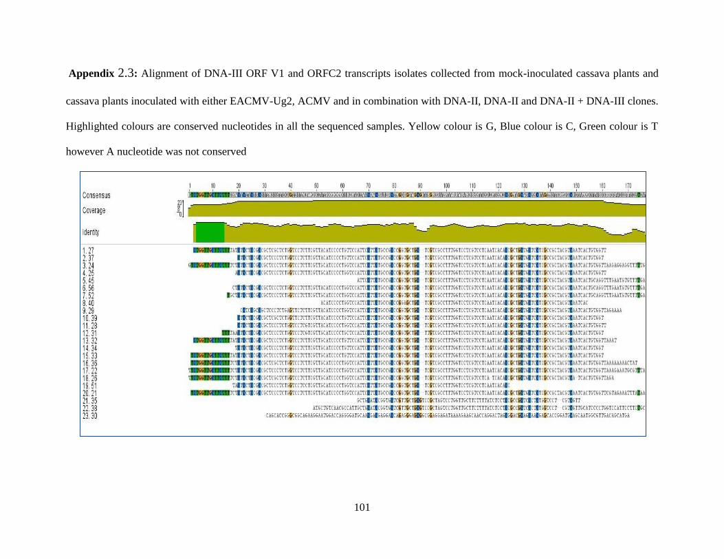
101
Appendix 2.3: Alignment of DNA-III ORF V1 and ORFC2 transcripts isolates collected from mock-inoculated cassava plants and
cassava plants inoculated with either EACMV-Ug2, ACMV and in combination with DNA-II, DNA-II and DNA-II + DNA-III clones.
Highlighted colours are conserved nucleotides in all the sequenced samples. Yellow colour is G, Blue colour is C, Green colour is T
however A nucleotide was not conserved
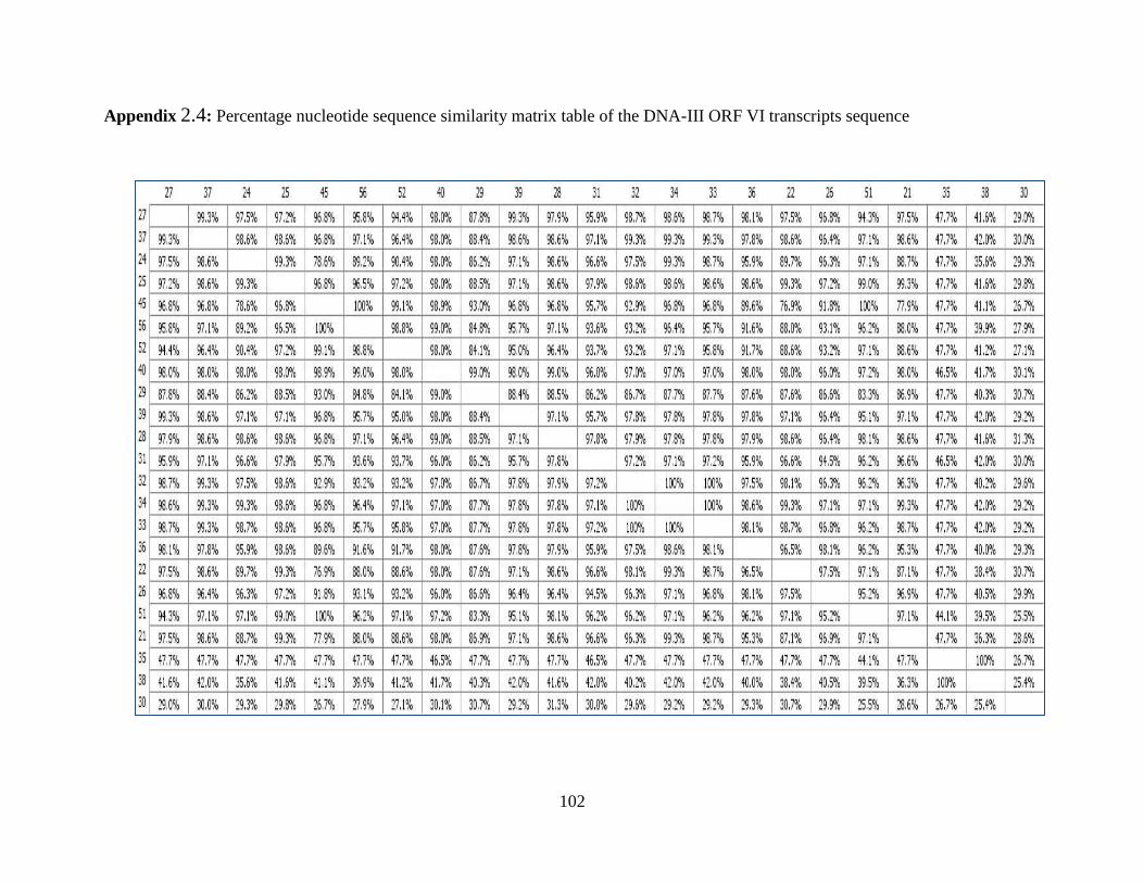
102
Appendix 2.4: Percentage nucleotide sequence similarity matrix table of the DNA-III ORF VI transcripts sequence