Inter-cellular Nanovesicle Mediated MicroRNA Transfer
19
Inter-cellular nanovesicle mediated microRNA transfer: a mechanism of environmental modulation of hepatocellular cancer cell growth Takayuki Kogure 1,2 , Wen-Lang Lin 1 , Irene K. Yan 1 , Chiara Braconi 2 , and Tushar Patel 1,2 Takayuki Kogure: [email protected]; Wen-Lang Lin: [email protected]; Irene K. Yan: [email protected]; Chiara Braconi: [email protected]; Tushar Patel: [email protected] 1 Mayo Clinic, Jacksonville, Florida 2 Ohio State University, Columbus, Ohio Abstract Hepatocellular carcinoma (HCC) is characterized by a propensity for multifocality, growth by local spread, and dysregulation of multiple signaling pathways. These features may be determined by the tumoral microenvironment. The potential of tumor cells to modulate HCC growth and behavior by secreted proteins has been extensively studied. In contrast the potential for genetic modulation is poorly understood. We investigated the role and involvement of tumor derived nanovesicles capable of altering gene expression, and characterized their ability to modulate cell signaling and biological effects in other cells. We show that HCC cells can produce nanovesicles, exosomes, that differ in both RNA and protein content from their cells of origin. These can be taken up and internalized by other cells, and can transmit a functional transgene. The microRNA content of these exosomes was examined, and a subset that is highly enriched within exosomes was identified. A combinatorial approach to identify potential targets identified transforming growth factor β activated kinase-1 (TAK1) as the most likely candidate pathway that could be modulated by these miRNA. Loss of TAK1 has been implicated in hepatocarcinogenesis and is a biologically plausible target for inter-cellular modulation. We showed that HCC cell derived exosomes can modulate TAK1 expression and associated signaling and enhance transformed cell growth in recipient cells. Conclusion: Exosome mediated miRNA transfer is an important mechanism of inter-cellular communication in HCC cells. These observations identify a unique inter-cellular mechanism that could potentially contribute to local spread, intrahepatic metastases or multifocal growth in HCC. Keywords Liver cancer; exosomes; gene expression; TAK1; vesicle Inter-cellular communications are an essential part of the relationship between cells that enable normal cellular function and maintain tissue homeostasis. Cells use a variety of approaches to communicate with each other such as direct membrane-to-membrane contact, or release of soluble mediators (1). Small vesicles shed from cells have also been shown to play important roles in cell-to-cell communication. These membrane bound vesicles contain membrane proteins similar to those of the donor cell and contain protein and RNA derived from their donor cell cytoplasm (2). They can be taken up and transfer their content to Address for correspondence: Tushar Patel, MBChB, AGAF, Mayo Clinic, 4500 San Pablo Road, Jacksonville, Florida 32224, Tel: 904 956 3257, Fax: 904 956 3359, [email protected]. NIH Public Access Author Manuscript Hepatology. Author manuscript; available in PMC 2012 October 1. Published in final edited form as: Hepatology. 2011 October ; 54(4): 1237–1248. doi:10.1002/hep.24504. NIH-PA Author Manuscript NIH-PA Author Manuscript NIH-PA Author Manuscript
-
Upload
anon579968442 -
Category
Documents
-
view
217 -
download
0
description
articol
Transcript of Inter-cellular Nanovesicle Mediated MicroRNA Transfer
Inter-cellular nanovesicle mediated microRNA transfer: amechanism of environmental modulation of hepatocellularcancer cell growth
Takayuki Kogure1,2, Wen-Lang Lin1, Irene K. Yan1, Chiara Braconi2, and Tushar Patel1,2
Takayuki Kogure: [email protected]; Wen-Lang Lin: [email protected]; Irene K. Yan:[email protected]; Chiara Braconi: [email protected]; Tushar Patel: [email protected] Mayo Clinic, Jacksonville, Florida2 Ohio State University, Columbus, Ohio
AbstractHepatocellular carcinoma (HCC) is characterized by a propensity for multifocality, growth bylocal spread, and dysregulation of multiple signaling pathways. These features may be determinedby the tumoral microenvironment. The potential of tumor cells to modulate HCC growth andbehavior by secreted proteins has been extensively studied. In contrast the potential for geneticmodulation is poorly understood. We investigated the role and involvement of tumor derivednanovesicles capable of altering gene expression, and characterized their ability to modulate cellsignaling and biological effects in other cells. We show that HCC cells can produce nanovesicles,exosomes, that differ in both RNA and protein content from their cells of origin. These can betaken up and internalized by other cells, and can transmit a functional transgene. The microRNAcontent of these exosomes was examined, and a subset that is highly enriched within exosomeswas identified. A combinatorial approach to identify potential targets identified transforminggrowth factor β activated kinase-1 (TAK1) as the most likely candidate pathway that could bemodulated by these miRNA. Loss of TAK1 has been implicated in hepatocarcinogenesis and is abiologically plausible target for inter-cellular modulation. We showed that HCC cell derivedexosomes can modulate TAK1 expression and associated signaling and enhance transformed cellgrowth in recipient cells. Conclusion: Exosome mediated miRNA transfer is an importantmechanism of inter-cellular communication in HCC cells. These observations identify a uniqueinter-cellular mechanism that could potentially contribute to local spread, intrahepatic metastasesor multifocal growth in HCC.
KeywordsLiver cancer; exosomes; gene expression; TAK1; vesicle
Inter-cellular communications are an essential part of the relationship between cells thatenable normal cellular function and maintain tissue homeostasis. Cells use a variety ofapproaches to communicate with each other such as direct membrane-to-membrane contact,or release of soluble mediators (1). Small vesicles shed from cells have also been shown toplay important roles in cell-to-cell communication. These membrane bound vesicles containmembrane proteins similar to those of the donor cell and contain protein and RNA derivedfrom their donor cell cytoplasm (2). They can be taken up and transfer their content to
Address for correspondence: Tushar Patel, MBChB, AGAF, Mayo Clinic, 4500 San Pablo Road, Jacksonville, Florida 32224, Tel: 904956 3257, Fax: 904 956 3359, [email protected].
NIH Public AccessAuthor ManuscriptHepatology. Author manuscript; available in PMC 2012 October 1.
Published in final edited form as:Hepatology. 2011 October ; 54(4): 1237–1248. doi:10.1002/hep.24504.
NIH
-PA Author Manuscript
NIH
-PA Author Manuscript
NIH
-PA Author Manuscript
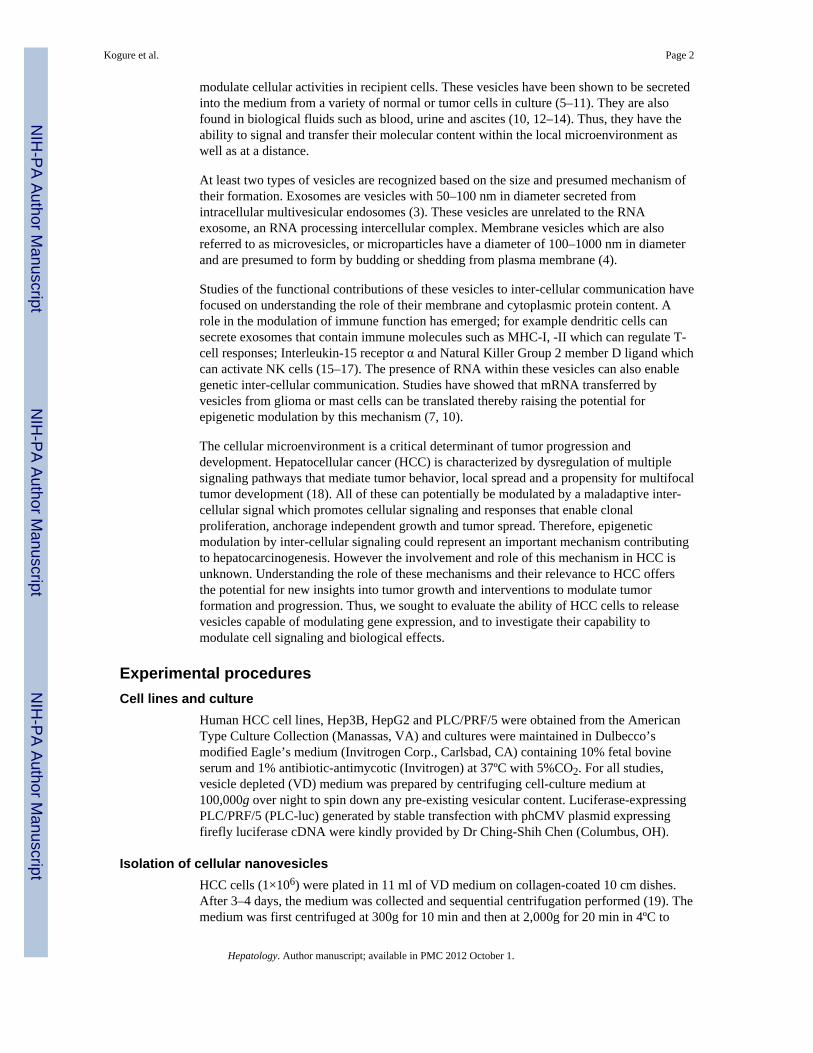
modulate cellular activities in recipient cells. These vesicles have been shown to be secretedinto the medium from a variety of normal or tumor cells in culture (5–11). They are alsofound in biological fluids such as blood, urine and ascites (10, 12–14). Thus, they have theability to signal and transfer their molecular content within the local microenvironment aswell as at a distance.
At least two types of vesicles are recognized based on the size and presumed mechanism oftheir formation. Exosomes are vesicles with 50–100 nm in diameter secreted fromintracellular multivesicular endosomes (3). These vesicles are unrelated to the RNAexosome, an RNA processing intercellular complex. Membrane vesicles which are alsoreferred to as microvesicles, or microparticles have a diameter of 100–1000 nm in diameterand are presumed to form by budding or shedding from plasma membrane (4).
Studies of the functional contributions of these vesicles to inter-cellular communication havefocused on understanding the role of their membrane and cytoplasmic protein content. Arole in the modulation of immune function has emerged; for example dendritic cells cansecrete exosomes that contain immune molecules such as MHC-I, -II which can regulate T-cell responses; Interleukin-15 receptor α and Natural Killer Group 2 member D ligand whichcan activate NK cells (15–17). The presence of RNA within these vesicles can also enablegenetic inter-cellular communication. Studies have showed that mRNA transferred byvesicles from glioma or mast cells can be translated thereby raising the potential forepigenetic modulation by this mechanism (7, 10).
The cellular microenvironment is a critical determinant of tumor progression anddevelopment. Hepatocellular cancer (HCC) is characterized by dysregulation of multiplesignaling pathways that mediate tumor behavior, local spread and a propensity for multifocaltumor development (18). All of these can potentially be modulated by a maladaptive inter-cellular signal which promotes cellular signaling and responses that enable clonalproliferation, anchorage independent growth and tumor spread. Therefore, epigeneticmodulation by inter-cellular signaling could represent an important mechanism contributingto hepatocarcinogenesis. However the involvement and role of this mechanism in HCC isunknown. Understanding the role of these mechanisms and their relevance to HCC offersthe potential for new insights into tumor growth and interventions to modulate tumorformation and progression. Thus, we sought to evaluate the ability of HCC cells to releasevesicles capable of modulating gene expression, and to investigate their capability tomodulate cell signaling and biological effects.
Experimental proceduresCell lines and culture
Human HCC cell lines, Hep3B, HepG2 and PLC/PRF/5 were obtained from the AmericanType Culture Collection (Manassas, VA) and cultures were maintained in Dulbecco’smodified Eagle’s medium (Invitrogen Corp., Carlsbad, CA) containing 10% fetal bovineserum and 1% antibiotic-antimycotic (Invitrogen) at 37ºC with 5%CO2. For all studies,vesicle depleted (VD) medium was prepared by centrifuging cell-culture medium at100,000g over night to spin down any pre-existing vesicular content. Luciferase-expressingPLC/PRF/5 (PLC-luc) generated by stable transfection with phCMV plasmid expressingfirefly luciferase cDNA were kindly provided by Dr Ching-Shih Chen (Columbus, OH).
Isolation of cellular nanovesiclesHCC cells (1×10⁶) were plated in 11 ml of VD medium on collagen-coated 10 cm dishes.After 3–4 days, the medium was collected and sequential centrifugation performed (19). Themedium was first centrifuged at 300g for 10 min and then at 2,000g for 20 min in 4ºC to
Kogure et al. Page 2
Hepatology. Author manuscript; available in PMC 2012 October 1.
NIH
-PA Author Manuscript
NIH
-PA Author Manuscript
NIH
-PA Author Manuscript
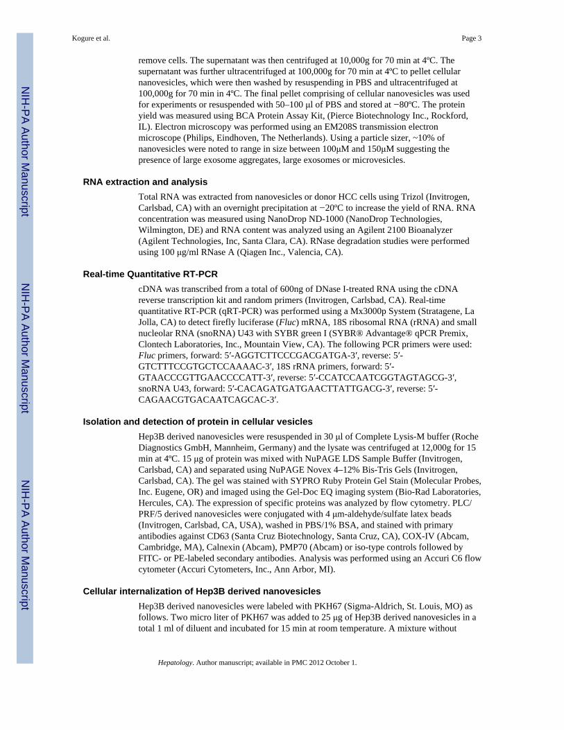
remove cells. The supernatant was then centrifuged at 10,000g for 70 min at 4ºC. Thesupernatant was further ultracentrifuged at 100,000g for 70 min at 4ºC to pellet cellularnanovesicles, which were then washed by resuspending in PBS and ultracentrifuged at100,000g for 70 min in 4ºC. The final pellet comprising of cellular nanovesicles was usedfor experiments or resuspended with 50–100 μl of PBS and stored at −80ºC. The proteinyield was measured using BCA Protein Assay Kit, (Pierce Biotechnology Inc., Rockford,IL). Electron microscopy was performed using an EM208S transmission electronmicroscope (Philips, Eindhoven, The Netherlands). Using a particle sizer, ~10% ofnanovesicles were noted to range in size between 100μM and 150μM suggesting thepresence of large exosome aggregates, large exosomes or microvesicles.
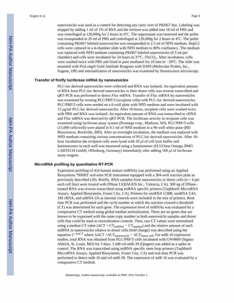
RNA extraction and analysisTotal RNA was extracted from nanovesicles or donor HCC cells using Trizol (Invitrogen,Carlsbad, CA) with an overnight precipitation at −20ºC to increase the yield of RNA. RNAconcentration was measured using NanoDrop ND-1000 (NanoDrop Technologies,Wilmington, DE) and RNA content was analyzed using an Agilent 2100 Bioanalyzer(Agilent Technologies, Inc, Santa Clara, CA). RNase degradation studies were performedusing 100 μg/ml RNase A (Qiagen Inc., Valencia, CA).
Real-time Quantitative RT-PCRcDNA was transcribed from a total of 600ng of DNase I-treated RNA using the cDNAreverse transcription kit and random primers (Invitrogen, Carlsbad, CA). Real-timequantitative RT-PCR (qRT-PCR) was performed using a Mx3000p System (Stratagene, LaJolla, CA) to detect firefly luciferase (Fluc) mRNA, 18S ribosomal RNA (rRNA) and smallnucleolar RNA (snoRNA) U43 with SYBR green I (SYBR® Advantage® qPCR Premix,Clontech Laboratories, Inc., Mountain View, CA). The following PCR primers were used:Fluc primers, forward: 5′-AGGTCTTCCCGACGATGA-3′, reverse: 5′-GTCTTTCCGTGCTCCAAAAC-3′, 18S rRNA primers, forward: 5′-GTAACCCGTTGAACCCCATT-3′, reverse: 5′-CCATCCAATCGGTAGTAGCG-3′,snoRNA U43, forward: 5′-CACAGATGATGAACTTATTGACG-3′, reverse: 5′-CAGAACGTGACAATCAGCAC-3′.
Isolation and detection of protein in cellular vesiclesHep3B derived nanovesicles were resuspended in 30 μl of Complete Lysis-M buffer (RocheDiagnostics GmbH, Mannheim, Germany) and the lysate was centrifuged at 12,000g for 15min at 4ºC. 15 μg of protein was mixed with NuPAGE LDS Sample Buffer (Invitrogen,Carlsbad, CA) and separated using NuPAGE Novex 4–12% Bis-Tris Gels (Invitrogen,Carlsbad, CA). The gel was stained with SYPRO Ruby Protein Gel Stain (Molecular Probes,Inc. Eugene, OR) and imaged using the Gel-Doc EQ imaging system (Bio-Rad Laboratories,Hercules, CA). The expression of specific proteins was analyzed by flow cytometry. PLC/PRF/5 derived nanovesicles were conjugated with 4 μm-aldehyde/sulfate latex beads(Invitrogen, Carlsbad, CA, USA), washed in PBS/1% BSA, and stained with primaryantibodies against CD63 (Santa Cruz Biotechnology, Santa Cruz, CA), COX-IV (Abcam,Cambridge, MA), Calnexin (Abcam), PMP70 (Abcam) or iso-type controls followed byFITC- or PE-labeled secondary antibodies. Analysis was performed using an Accuri C6 flowcytometer (Accuri Cytometers, Inc., Ann Arbor, MI).
Cellular internalization of Hep3B derived nanovesiclesHep3B derived nanovesicles were labeled with PKH67 (Sigma-Aldrich, St. Louis, MO) asfollows. Two micro liter of PKH67 was added to 25 μg of Hep3B derived nanovesicles in atotal 1 ml of diluent and incubated for 15 min at room temperature. A mixture without
Kogure et al. Page 3
Hepatology. Author manuscript; available in PMC 2012 October 1.
NIH
-PA Author Manuscript
NIH
-PA Author Manuscript
NIH
-PA Author Manuscript
nanovesicles was used as a control for detecting any carry over of PKH67 dye. Labeling wasstopped by adding 1 ml of 1% of BSA and the mixture was added into 18 ml of PBS andwas centrifuged at 120,000g for 2 hours in 4ºC. The supernatant was removed and the pelletwas resuspended in 20 ml of PBS and centrifuged at 120,000g for 2 hours in 4ºC. The pelletcontaining PKH67-labeled nanovesicles was resuspended in 2.5 ml of NPD medium. HepG2cells were cultured in a 4-chamber slide with NPD medium to 80% confluency. The mediumwas replaced with NPD medium containing PKH67-labeled nanovesicles (0.5 ml perchamber) and cells were incubated for 24 hours in 37ºC, 5%CO2. After incubation, cellswere washed twice with PBS and fixed in pure methanol for 10 min in −20ºC. The slide wasmounted with ProLong® Gold Antifade Reagents with DAPI (Molecular Probes, Inc.,Eugene, OR) and internalization of nanovesicles was examined by fluorescence microscopy.
Transfer of firefly luciferase mRNA by nanovesiclesPLC-luc derived nanovesicles were collected and RNA was isolated. An equivalent amountof RNA from PLC-luc derived nanovesicles or their donor cells was reverse transcribed andqRT-PCR was performed to detect Fluc mRNA. Transfer of Fluc mRNA by nanovesicleswas examined by treating PLC/PRF/5 (recipient cells) with PLC-luc derived nanovesicles.PLC/PRF/5 cells were seeded on a 6-well plate with NPD medium and were incubated with15 μg/ml PLC-luc derived nanovesicles. After 16-hours, recipient cells were washed twicewith PBS and RNA was isolated. An equivalent amount of RNA was transcribed to cDNAand Fluc mRNA was detected by qRT-PCR. The luciferase activity in recipient cells wasexamined using luciferase assay system (Promega corp., Madison, WI). PLC/PRF/5 cells(15,000 cells/well) were plated in 0.1 ml of NPD medium in a 96-well white plate (BDBiosciences, Rockville, MD). After an overnight incubation, the medium was replaced withNPD medium containing various concentrations of PLC-luc derived nanovesicles. After 16-hour incubation the recipient cells were lysed with 20 μl of cell lysis buffer andluminescence in each well was measured using a luminometer (FLUOstar Omega, BMGLABTECH GmbH, Offenburg, Germany) immediately after adding 100 μl of luciferaseassay reagent.
MicroRNA profiling by quantitative RT-PCRExpression profiling of 424 human mature miRNAs was performed using an AppliedBiosystems 7900HT real-time PCR instrument equipped with a 384-well reaction plate aspreviously described (20). Briefly, RNA samples from nanovesicles or donor cells (n = 4 pereach cell line) were treated with DNase I (QIAGEN Inc., Valencia, CA). 500 ng of DNase-treated RNA was reverse transcribed using miRNA specific primers (TaqMan® MicroRNAAssays, Applied Biosystems, Foster City, CA). Primers for snoRNA U38B, snoRNA U43,18S rRNA, and snRNA U6 as internal controls were included in the mix of primers. Real-time PCR was performed and the cycle number at which the reaction crossed a threshold(CT) was determined for each gene. The expression level of miRNAs was evaluated by acomparative CT method using global median normalization. There are no genes that areknown to be expressed with the same copy number in both nanovesicle samples and donorcells that could be used as normalization controls. Thus, raw CT values were normalizedusing a median CT value (ΔCT = CTmiRNA − CTmedian) and the relative amount of eachmiRNA in nanovesicles relative to donor cells (fold change) was described using theequation 2−ΔΔCT where ΔΔCT =ΔCTnanovesicle − ΔCTdonor cell. For miR-16 expressionstudies, total RNA was obtained from PLC/PRF/5 cells incubated with GW4869 (Sigma-Aldrich, St. Louis, MO) for 3 days. 5 nM cel-miR-39 (Qiagen) was added as a spike-incontrol. The RNA was transcribed using miRNA specific stem loop primers (TaqMan®MicroRNA Assays, Applied Biosystems, Foster City, CA) and real-time PCR wasperformed to detect miR-16 and cel-miR-39. The expression of miR-16 was evaluated by acomparative CT method.
Kogure et al. Page 4
Hepatology. Author manuscript; available in PMC 2012 October 1.
NIH
-PA Author Manuscript
NIH
-PA Author Manuscript
NIH
-PA Author Manuscript

Statistical analysisData were analyzed by ANOVA followed by Fisher’s PLSD test. Results were considered tobe statistically significant when p < 0.05. Data were expressed as the mean and standarderror.
Additional Experimental procedures are described in the supplementary material.
ResultsCan tumor cell derived nanovesicles be isolated?
In order to study the potential of tumor cell derived nanovesicles in tumor growth, we firstoptimized conditions for their isolation. The approach used was based on their differentialsedimentation properties and used sequential ultracentrifugation for their isolation fromculture supernatant from HCC cells in culture. Electron microscopy showing membranelimited particles that were homogeneous in appearance and ranging from 40–100 nm in size,(Figure 1). By flow cytometry, the isolated particles expressed markers associated withexosomes (CD63), but not those associated with mitochondria (COX IV), peroxisomes(PMP70), or endoplasmic reticulum (calnexin) (Supplementary Figure 1). The yield wasconfirmed by measuring protein content. The yield (mean ± SE of eight separate isolations)from Hep3B cells was 0.84 ± 0.05 μg/106 cells/day, whereas the yield from PLC/PRF/5 cellswas 0.88 ± 0.05 μg/106 cells/day. Thus, these nanovesicles have characteristics of exosomesand could be isolated in a consistent manner.
Is the cellular content of exosomes similar to the cells of origin?Next, we sought to determine whether the cellular constituents of exosomes were similar tothose of the cells of origin. First, we evaluated the profile of total RNA extracted fromexosomes by capillary electrophoresis (Figure 2A). Compared to the donor cells, RNAextracted from exosomes did not show clear bands of 18S and 28S ribosomal RNA.However, a distinguishable band was detected below 200 bases suggesting that the RNAcontent of exosomes is selectively enhanced for small RNAs such as microRNAs. Next, weexamined the expression of 18S rRNA and snoRNA U43 by qRT-PCR using equivalentamount of RNA from exosomes and donor cells. These are commonly used as an internalcontrol for small RNA quantification in mammalian cells (Figure 2B). Compared to theirexpression in either Hep3B or PLC/PRF/5 cells, the expression of these 2 genes was reducedin exosomes derived from these cells. RNA degradation and the yield of RNA obtained wasnot reduced by RNase treatment compared to controls indicating that the RNA was withinthe isolated particles (Supplementary figure 2). We next examined the protein expressionprofile in exosomes. Equivalent amount of proteins extracted from Hep3B-derivedexosomes or from their donor cells were separated by SDS-PAGE and stained with SYPRORuby (Figure 2C). The protein from exosomes had a different profile showing severaldistinct bands. Thus, both the RNA and protein content of exosomes is different from that oftheir cells of origin.
Can cellular exosomes be taken up and internalized by other cells?To examine the potential for uptake and internalization by other cells, we labeled exosomesderived from Hep3B with the fluorescent dye PKH67 as described in the Methods section.PKH67-labeled exosomes were incubated with HepG2 cells for 24 hours and localization ofexosomes was examined by fluorescent microscopy (Figure 3). We observed internalizationof PKH67-labeled exosomes as endosome-like vesicles in the cytoplasm of HepG2 cells.These studies indicate that tumor cell derived exosomes can be taken up by other cells.
Kogure et al. Page 5
Hepatology. Author manuscript; available in PMC 2012 October 1.
NIH
-PA Author Manuscript
NIH
-PA Author Manuscript
NIH
-PA Author Manuscript
Can exosomes transfer a functional transgene to other HCC cells?We examined whether exosomes can deliver functional mRNA of a transgene to other HCCcells. Exosomes were collected from PLC-luc cells, which are stably transfected to express afunctional luciferase expressing construct. The presence of Fluc mRNA in exosome wasverified by qRT-PCR; CT values (mean ± SE) of Fluc mRNA were 29.0 ± 0.1 in exosomesand 31.5 ± 0.2 in donor cells (Figure 4A). PLC/PRF/5 cells were then incubated withvarious concentrations of PLC-luc-derived exosomes for 16 hours. Fluc mRNA expressionand luciferase activity was then assessed in the recipient PLC/PRF/5 cells. We detected FlucmRNA in the recipient PLC/PRF/5 cells with ΔCT values (CTFluc mRNA − CT18S rRNA,mean ± SE) of 24.7 ± 0.2 compared to 18.0 ± 0.1 in donor PLC-luc cells (Figure 4B). Inaddition, a concentration-dependent increase in luciferase activity was detected in recipientcells incubated with PLC-luc derived exosomes consistent with a gene-dosing effect (Figure4C). A reduction of luciferase activity was noted with PLC-luc derived exosomes incubatedin recipient cells pre-treated with cycloheximide compared to controls, indicating arequirement for new protein translation for luciferase activity (Figure 4D). These data showthat exosomes can deliver a functionally active Fluc mRNA to other cells.
Do exosomes contain microRNAs?Since HCC cell-derived exosomes contain an enriched fraction of small RNAs (Figure 1),we hypothesized that exosomes contain selected miRNAs that could contribute to inter-cellular communication. To examine this possibility, we performed microRNA expressionprofiling in both Hep3B and PLC/PRF/5 HCC cells and exosomes derived from these cells.Four independent samples were used for each cell line/exosome pair. The expression of total424 miRNAs and the internal control genes (18S rRNA, snRNA U6, snoRNA U38B andsnoRNA U43) were measured by qRT-PCR. The expression level of individual miRNAs inexosomes and donor cells were expressed as the relative expression to global medianexpression of all miRNA since no internal control genes are available for exosomes. Theraw CT values of 18S rRNA and snoRNA U43 vary between donor cells and their exosomes(Figure 2B), and the ability for spiked controls to be expressed in exosomes is unknown.
Of the miRNAs examined, only 134 miRNAs were identified in exosomes isolated fromHep3B cells. Of these, 55 miRNAs were differentially expressed in exosomes more than 4-fold compared to their expression in their donor cells; 25 miRNAs of those were enriched(up to 166-fold) and 30 miRNAs were decreased (up to 113-fold). Notably, 11 miRNAswere detected exclusively in exosomes indicating a very high enrichment in exosomescompared to donor cells (Figure 5A). Similar observations were made in PLC/PRF/5-derived exosomes. 140 miRNAs were identified in PLC/PRF/5-derived exosomes of which74 miRNAs were differentially distributed in exosomes more than 4-fold compared to thedonor cells. Of these, 28 miRNAs were enriched (up to 71-fold), with 20 miRNA detectedexclusively in exosomes and 45 miRNAs were decreased (up to 255-fold). There was amoderate correlation in levels of miRNAs contained in exosomes isolated from Hep3B andPLC/PRF/5 (Figure 5B), indicating the existence of a common mechanism of selectiveenrichment of exosomes with specific miRNAs. The detailed lists of miRNAs andexpression levels are available in Supplemental table 1.
The release of miRNA into membrane vesicles can occur via a ceramide dependent manner(21). To evaluate the potential role of this pathway, we treated cells with an nSMaseinhibitor, GW4869, which is known to inhibit ceramide biosynthesis and examined theexpression of miR-16, a microRNA that is expressed in both donor cells and in exosomes.The cellular expression of miR-16 was unchanged whereas the extracellular expression ofmiR-16 in exosomes was reduced following incubation with GW4869 compared to controls.Thus, miRNA release into exosomes occurs via a ceramide dependent pathway.
Kogure et al. Page 6
Hepatology. Author manuscript; available in PMC 2012 October 1.
NIH
-PA Author Manuscript
NIH
-PA Author Manuscript
NIH
-PA Author Manuscript
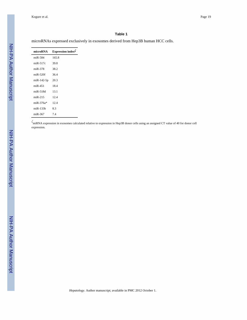
What are potential targets of miRNA enriched in exosomes?To identify potential roles of exosome-derived miRNA in cell to cell communication, wefocused on the 11 miRNAs that were exclusively detected, and hence highly enriched, inHep3B-derived exosomes (Table 1). We postulated that these miRNA would coordinateregulation of a set of genes, and used a combinatorial approach to analyze potential targetsof this set of miRNA in combination. Using the miRror algorithm which integrates datafrom a dozen miRNA target prediction programs (22)(http://www.proto.cs.huji.ac.il/mirror/), 108 genes were predicted as the targets of these 11miRNAs (Supplemental table 2). Network analysis of these 108 genes using the String 8.3program (23) indicated the central involvement of transforming growth factor-β activatedkinase-1 (TAK1) signaling (Figure 6). This analysis predicted the TAK1 pathway as themost likely candidate pathway that would be modulated by the selected group of miRNAacting in concert. TAK1 is an upstream member of the mitogen-activated protein kinasekinase kinase (MAP3K) family and has been implicated as an essential component ofcellular homeostasis and tumorigenesis in the liver (24, 25).
Can exosomes modulate TAK1 expression and signaling in recipient cells?The involvement of TAK1 in cell responses to environmental changes and a demonstratedrole in HCC formation and growth make this kinase a highly attractive and biologicallyplausible target for inter-cellular modulation (26). TAK1 can be activated by cytokines andstress stimuli such as transforming growth factor (TGF)-β, tumor necrosis factor (TNF)-α,IL-1β and lipopolysaccharides. TAK1 forms a complex with TNF receptor-associated factorand TAK-1 binding protein (TAB)1, -2, and -3, which is necessary for the recruitment andactivation of TAK1. TAK1 can subsequently activate c-Jun NH2-terminal kinase (JNK)/p38MAPK and nuclear factor (NF)-κB. To examine the potential of exosomes to modulate theTAK1 pathway, Hep3B cells were incubated with Hep3B-derived exosomes (10 μg/ml). Theexpression of TAK1 and TAB2 proteins and the activation by phosphorylation ofdownstream JNK and p38 MAP kinases was examined by immunoblot analysis (Figure 7).A reduction in TAK1 of upto 45% and of TAB2 of upto 42% was noted after 72 hours.Concomitantly, a decrease in constitutive active-site phosphorylation of JNK1, JNK2/3 andp38 MAPK were also noted after 72 hours. Thus, exosomes can modulate the constitutiveexpression of TAK1 and modulate downstream signaling associated with TAK1.
Can exosomes modulate transformed cell growth or cell death in target cells?We began by assessing the effect of exosomes on anchorage-independent growth, a hallmarkof transformed cell behavior. HCC cells were seeded in soft agar in the presence or absenceof Hep3B derived exosomes and allowed to grow with or without Hep3B-derived exosomesfor 7 days. Incubation with Hep3B-derived exosomes increased the number of colonies insoft agar 9.6-fold in Hep3B cells and 1.6-fold in HepG2 cells (Figure 8A) after 7 days.These studies indicate a cell-type dependent yet consistent effect of exosomes ontransformed cell growth in vitro. A loss of Tak1 has been associated with spontaneoushepatocyte death. Thus, we determined the effect of exosomes on cell death. Incubation ofHep3B cells with Hep3B-derived exosomes (10 μg/ml) for 24 hrs increased caspase-3/7activity by 6.1-fold after 24 hours (Figure 8B). This was associated with a 20.5% reductionin cell viability after 72 hours in Hep3B cells incubated with 10 μg/ml Hep3B derivedexosomes (Figure 8C). Similar effects were also noted in PLC/PRF/5 cells incubated withHep3B-derived exosomes. The enhancement in anchorage independent growth in the settingof this modest reduction in cell viability indicate that exosomes have a potent overall effecton transformed cell behavior.
Kogure et al. Page 7
Hepatology. Author manuscript; available in PMC 2012 October 1.
NIH
-PA Author Manuscript
NIH
-PA Author Manuscript
NIH
-PA Author Manuscript

DiscussionAlthough the role of genetic alterations in oncogenes and tumor suppressor genes has beenextensively studied, epigenetic mechanisms contributing to tumor development are less wellcharacterized. The influence of the cellular microenvironment on tumor development andgrowth is becoming increasingly recognized. In these studies, we show that HCC cells canepigenetically modulate gene expression and cell signaling related to transformed cellgrowth in vitro through the release of miRNA contained within exosomes. Moreover, weidentify selective enhancement of miRNA content within these cellular vesicles, and thepotential of exosomal miRNA transfer to effect cellular gene expression and cell behavior intarget cells. The significance of these studies is that they identify a mechanism of cell-to-cellcommunication that may have widespread effects on tissue behavior. The potential of thecellular microenvironment to modulate gene expression and to stimulate cell signalingcontributing to tumor behavior could be exploited for therapeutic targeting.
A selective subset of miRNA is present within exosomes which is markedly different fromthat in their cells of origin. Mature miRNA are released into the cytoplasmic space wherethey associate with the RISC complex to effect gene expression. The selective enrichment ofthese miRNA in cellular exosomes, the consistency in expression between differentisolations, and the cell-type specificity indicates the presence of a mechanism for their activeelaboration within these particles. This may arise from selective transport into a membranebound exosome, or association and sequestration with proteins that are selectively enrichedwithin the exosome. Alternatively, the possibility exists that these miRNA are rapidlydegraded within the cytoplasm but protected from this when they are sequestered in vesiclesprior to their elaboration from cells as cellular exosomes. Since the tumor cells studied varyin their cellular behavior, it is not unexpected that some differences were noted between thecell types in miRNA content. These observations regarding cell-type specificity of miRNAcontent are similar to those made with respect to protein content of exosomes. Theyemphasize the need to study and interpret data on an individual cell-type specific basisrather than generalizing across cell types.
It is noteworthy that our combinatorial analysis identified TAK1 as a central target ofexosome mediated miRNA. TAK1 is an essential inhibitor of hepatocarcinogenesis, and itsabsence in vivo is associated with the spontaneous development of hepatocellular cancerrelated to aberrant responses to inflammatory and stress signaling, and associated withhepatocyte injury and apoptosis. TAK1 can also have a direct effect on cancer progressionthrough repression of the telomerase reverse transcriptase gene (27). Thus, the modulationof TAK1 expression and signaling by exosome mediated inter-cellular signaling couldrepresent an important mechanism of tumor progression that may not be dependent on clonalexpansion or hyperproliferative effects.
Influences from cellular elements within the tumoral microenvironment such as tumor-associated fibroblasts or soluble mediators such as cytokines, hormones or neurotransmittersare the focus of intense study to understand the influence of this space on tumor behavior.While the autocrine or paracrine effects of proteins that are secreted into the extracellularspace has been extensively studied, the involvement of genetic modulation by miRNA addsanother layer of complexity and regulation that may be equally as important. We speculatethat environmentally mediated epigenetic modulation is likely to be an importantdeterminant of tumor cell growth, and cellular resistance to environmental perturbations.
Inter-cellular communication by exosomes can trigger signal transducing intra-cellularmechanisms that can modulate cell behavior. The potential role of these may be tocoordinate cellular responses that are adaptive and can serve to maintain tissue homeostasis.
Kogure et al. Page 8
Hepatology. Author manuscript; available in PMC 2012 October 1.
NIH
-PA Author Manuscript
NIH
-PA Author Manuscript
NIH
-PA Author Manuscript

Alternatively, in tumor cells, exosome-mediated intercellular signaling may serve amaladaptive role to promote tumor growth. The current studies demonstrate the potential fortumor cells themselves to exert paracrine or autocrine effects mediated by miRNA asextracellular effectors of cell-to-cell communication. The aberrant expression of selectedmiRNA in tumor cells and their ability to modulate multiple oncogenic or tumor suppressorgenes make them well-suited for such a role. Therefore further studies to understand themechanisms of cellular exosome production, selective enrichment of RNA genes and othergenetic content, and cell-type specificity of response are justified to establish the roles ofthis mechanism in the development and progression of HCC.
Supplementary MaterialRefer to Web version on PubMed Central for supplementary material.
AcknowledgmentsFinancial support
Supported in part by Grant DK069370 from the National Institutes of Health
Abbreviations
CNV cellular nanovesicles
HCC hepatocellular carcinoma
miRNA microRNA
VD vesicle-depleted
TAB TAK-1 binding protein
TAK1 transforming growth factor β activated kinase-1
References1. Hui EE, Bhatia SN. Micromechanical control of cell-cell interactions. Proc Natl Acad Sci U S A.
2007; 104:5722–5726. [PubMed: 17389399]2. Thery C, Ostrowski M, Segura E. Membrane vesicles as conveyors of immune responses. Nat Rev
Immunol. 2009; 9:581–593. [PubMed: 19498381]3. van Niel G, Porto-Carreiro I, Simoes S, Raposo G. Exosomes: a common pathway for a specialized
function. J Biochem. 2006; 140:13–21. [PubMed: 16877764]4. Cocucci E, Racchetti G, Meldolesi J. Shedding microvesicles: artefacts no more. Trends Cell Biol.
2009; 19:43–51. [PubMed: 19144520]5. Saunderson SC, Schuberth PC, Dunn AC, Miller L, Hock BD, MacKay PA, Koch N, et al. Induction
of exosome release in primary B cells stimulated via CD40 and the IL-4 receptor. J Immunol. 2008;180:8146–8152. [PubMed: 18523279]
6. Zitvogel L, Regnault A, Lozier A, Wolfers J, Flament C, Tenza D, Ricciardi-Castagnoli P, et al.Eradication of established murine tumors using a novel cell-free vaccine: dendritic cell-derivedexosomes. Nat Med. 1998; 4:594–600. [PubMed: 9585234]
7. Valadi H, Ekstrom K, Bossios A, Sjostrand M, Lee JJ, Lotvall JO. Exosome-mediated transfer ofmRNAs and microRNAs is a novel mechanism of genetic exchange between cells. Nat Cell Biol.2007; 9:654–659. [PubMed: 17486113]
8. Mallegol J, Van Niel G, Lebreton C, Lepelletier Y, Candalh C, Dugave C, Heath JK, et al. T84-intestinal epithelial exosomes bear MHC class II/peptide complexes potentiating antigenpresentation by dendritic cells. Gastroenterology. 2007; 132:1866–1876. [PubMed: 17484880]
Kogure et al. Page 9
Hepatology. Author manuscript; available in PMC 2012 October 1.
NIH
-PA Author Manuscript
NIH
-PA Author Manuscript
NIH
-PA Author Manuscript

9. Smalheiser NR. Exosomal transfer of proteins and RNAs at synapses in the nervous system. BiolDirect. 2007; 2:35. [PubMed: 18053135]
10. Skog J, Wurdinger T, van Rijn S, Meijer DH, Gainche L, Sena-Esteves M, Curry WT Jr, et al.Glioblastoma microvesicles transport RNA and proteins that promote tumour growth and providediagnostic biomarkers. Nat Cell Biol. 2008; 10:1470–1476. [PubMed: 19011622]
11. Gutwein P, Stoeck A, Riedle S, Gast D, Runz S, Condon TP, Marme A, et al. Cleavage of L1 inexosomes and apoptotic membrane vesicles released from ovarian carcinoma cells. Clin CancerRes. 2005; 11:2492–2501. [PubMed: 15814625]
12. Li J, Sherman-Baust CA, Tsai-Turton M, Bristow RE, Roden RB, Morin PJ. Claudin-containingexosomes in the peripheral circulation of women with ovarian cancer. BMC Cancer. 2009; 9:244.[PubMed: 19619303]
13. Zhou H, Cheruvanky A, Hu X, Matsumoto T, Hiramatsu N, Cho ME, Berger A, et al. Urinaryexosomal transcription factors, a new class of biomarkers for renal disease. Kidney Int. 2008;74:613–621. [PubMed: 18509321]
14. Andre F, Schartz NE, Movassagh M, Flament C, Pautier P, Morice P, Pomel C, et al. Malignanteffusions and immunogenic tumour-derived exosomes. Lancet. 2002; 360:295–305. [PubMed:12147373]
15. Utsugi-Kobukai S, Fujimaki H, Hotta C, Nakazawa M, Minami M. MHC class I-mediatedexogenous antigen presentation by exosomes secreted from immature and mature bone marrowderived dendritic cells. Immunol Lett. 2003; 89:125–131. [PubMed: 14556969]
16. Luketic L, Delanghe J, Sobol PT, Yang P, Frotten E, Mossman KL, Gauldie J, et al. Antigenpresentation by exosomes released from peptide-pulsed dendritic cells is not suppressed by thepresence of active CTL. J Immunol. 2007; 179:5024–5032. [PubMed: 17911587]
17. Viaud S, Terme M, Flament C, Taieb J, Andre F, Novault S, Escudier B, et al. Dendritic cell-derived exosomes promote natural killer cell activation and proliferation: a role for NKG2Dligands and IL-15Ralpha. PLoS One. 2009; 4:e4942. [PubMed: 19319200]
18. Whittaker S, Marais R, Zhu AX. The role of signaling pathways in the development and treatmentof hepatocellular carcinoma. Oncogene. 2010; 29:4989–5005. [PubMed: 20639898]
19. Thery C, Amigorena S, Raposo G, Clayton A. Isolation and characterization of exosomes from cellculture supernatants and biological fluids. Curr Protoc Cell Biol. 2006; Chapter 3(Unit 3):22.[PubMed: 18228490]
20. Kuhn DE, Nuovo GJ, Martin MM, Malana GE, Pleister AP, Jiang J, Schmittgen TD, et al. Humanchromosome 21-derived miRNAs are overexpressed in down syndrome brains and hearts.Biochem Biophys Res Commun. 2008; 370:473–477. [PubMed: 18387358]
21. Kosaka N, Iguchi H, Yoshioka Y, Takeshita F, Matsuki Y, Ochiya T. Secretory mechanisms andintercellular transfer of microRNAs in living cells. J Biol Chem. 2010 Jun 4; 285(23):17442–52.Epub 2010 Mar 30. [PubMed: 20353945]
22. Friedman Y, Naamati G, Linial M. MiRror: a combinatorial analysis web tool for ensembles ofmicroRNAs and their targets. Bioinformatics. 2010; 26:1920–1921. [PubMed: 20529892]
23. Jensen LJ, Kuhn M, Stark M, Chaffron S, Creevey C, Muller J, Doerks T, et al. STRING 8--aglobal view on proteins and their functional interactions in 630 organisms. Nucleic Acids Res.2009; 37:D412–416. [PubMed: 18940858]
24. Bettermann K, Vucur M, Haybaeck J, Koppe C, Janssen J, Heymann F, Weber A, et al. TAK1suppresses a NEMO-dependent but NF-kappaB-independent pathway to liver cancer. Cancer Cell.2010; 17:481–496. [PubMed: 20478530]
25. Inokuchi S, Aoyama T, Miura K, Osterreicher CH, Kodama Y, Miyai K, Akira S, et al. Disruptionof TAK1 in hepatocytes causes hepatic injury, inflammation, fibrosis, and carcinogenesis. ProcNatl Acad Sci U S A. 2010; 107:844–849. [PubMed: 20080763]
26. Malato Y, Willenbring H. The MAP3K TAK1: a chock block to liver cancer formation.Hepatology. 2010; 52:1506–1509. [PubMed: 20879030]
27. Fujiki T, Miura T, Maura M, Shiraishi H, Nishimura S, Imada Y, Uehara N, et al. TAK1 repressestranscription of the human telomerase reverse transcriptase gene. Oncogene. 2007; 26:5258–5266.[PubMed: 17325661]
Kogure et al. Page 10
Hepatology. Author manuscript; available in PMC 2012 October 1.
NIH
-PA Author Manuscript
NIH
-PA Author Manuscript
NIH
-PA Author Manuscript

Figure 1. Characterization of isolated vesicles(A) Electron microscopy was performed on a whole mount of particles isolated from PLC/PRF/5 cells following multistage differential centrifugation. A homogeneous population ofparticles was obtained. (B) Flow cytometry was performed on isolated particles conjugatedwith 4 μm-aldehyde/sulfate latex beads using primary antibodies to CD63 or isotypecontrols followed by a phycoerythrin labeled secondary antibodies.
Kogure et al. Page 11
Hepatology. Author manuscript; available in PMC 2012 October 1.
NIH
-PA Author Manuscript
NIH
-PA Author Manuscript
NIH
-PA Author Manuscript
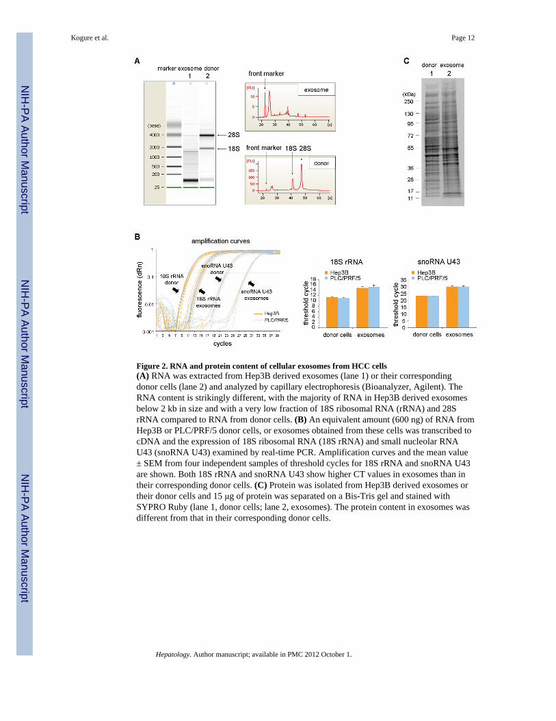
Figure 2. RNA and protein content of cellular exosomes from HCC cells(A) RNA was extracted from Hep3B derived exosomes (lane 1) or their correspondingdonor cells (lane 2) and analyzed by capillary electrophoresis (Bioanalyzer, Agilent). TheRNA content is strikingly different, with the majority of RNA in Hep3B derived exosomesbelow 2 kb in size and with a very low fraction of 18S ribosomal RNA (rRNA) and 28SrRNA compared to RNA from donor cells. (B) An equivalent amount (600 ng) of RNA fromHep3B or PLC/PRF/5 donor cells, or exosomes obtained from these cells was transcribed tocDNA and the expression of 18S ribosomal RNA (18S rRNA) and small nucleolar RNAU43 (snoRNA U43) examined by real-time PCR. Amplification curves and the mean value± SEM from four independent samples of threshold cycles for 18S rRNA and snoRNA U43are shown. Both 18S rRNA and snoRNA U43 show higher CT values in exosomes than intheir corresponding donor cells. (C) Protein was isolated from Hep3B derived exosomes ortheir donor cells and 15 μg of protein was separated on a Bis-Tris gel and stained withSYPRO Ruby (lane 1, donor cells; lane 2, exosomes). The protein content in exosomes wasdifferent from that in their corresponding donor cells.
Kogure et al. Page 12
Hepatology. Author manuscript; available in PMC 2012 October 1.
NIH
-PA Author Manuscript
NIH
-PA Author Manuscript
NIH
-PA Author Manuscript

Figure 3. Internalization of Hep3B-derived exosomes into HepG2 cellsHepG2 cells in culture were incubated with Hep3B-derived exosomes that were labeled withPKH67 (green). HepG2 cells were also incubated with PKH67 dye without exosomes as acontrol to detecting any carry-over of the dye (exosomes (−)). Cells are fixed with coldmethanol at −20ºC and mounted with ProLong® Gold Antifade Reagent with DAPI asdescribed in Experimental procedures. High magnification images of HepG2 cells incubatedwith exosomes (A–C), or low magnification images of HepG2 cells incubated withexosomes (D–F), or controls without PKH67 (G–I) are shown. Hep3B derived exosomesare shown to be internalized into the cytoplasm of HepG2 cells.
Kogure et al. Page 13
Hepatology. Author manuscript; available in PMC 2012 October 1.
NIH
-PA Author Manuscript
NIH
-PA Author Manuscript
NIH
-PA Author Manuscript

Figure 4. Cell-to-cell transfer of firefly luciferase by exosomes(A) Real-tine PCR was performed on an cDNA transcribed from an equivalent amount (600ng) of RNA from PLC-luc derived exosomes or their donor cells (n = 3, each in duplicate).PCR amplification curves for Fluc mRNA and 18S rRNA are shown. (B) PLC/PRF/5 cellswere incubated with 15 μg/ml of PLC-luc derived exosomes for 16 hours. RNA was isolatedand equivalent amount of RNA (300 ng) was transcribed to cDNA (n = 3). Amplificationcurves by quantitative real-time PCR for Fluc mRNA and 18S rRNA in PLC/PRF/5(recipient cells) and PLC-luc (donor cells) are shown. (C) PLC/PRF/5 cells in a 96-wellplate were incubated with various concentrations of PLC-luc derived exosomes, andluciferase activity was assessed in these cells after 16 hours. Bars express the mean value ofluminescence ± SEM of four separate determinations. *, p < 0.05. (D) PLC/PRF/5 cells werepretreated with 25 μg/ml of cycloheximide for 2 hours to inhibit de novo protein synthesis.Cells were washed with PBS and incubated with 100 μg/ml of PLC-luc derived exosomes.Luciferase activity was assessed in these cells after 6 hours. Bars express the mean value ofluminescence ± SEM of five separate determinations. *, p < 0.05.
Kogure et al. Page 14
Hepatology. Author manuscript; available in PMC 2012 October 1.
NIH
-PA Author Manuscript
NIH
-PA Author Manuscript
NIH
-PA Author Manuscript

Figure 5. Identification of microRNAs in HCC derived exosomesProfiling of miRNAs in exosomes or their donor cells was performed from four independentreplicates as described in Experimental procedures. (A) The mean values of fold change ofexpression of miRNA detected in exosomes relative to that in donor cells is shown (n = 4)and the numbers of miRNA that were exclusively detected in either donor cells or exosomesare depicted. 134 miRNAs were identified in Hep3B-derived exosomes, and 140 miRNAs inPLC/PRF/5 derived exosomes. Some miRNAs were predominantly expressed in exosomescompared to their donor cells. (B) Correlation of miRNA expression in Hep3B derivedexosomes and in PLC/PRF/5 derived exosomes are shown. A total of 97 miRNAs weredetected in exosomes from both Hep3B and PLC/PRF/5. (C) PLC/PRF/5 cells wereincubated for 3 days with GW4869, a neutral sphingomyelinase inhibitor which can inhibitceramide biosynthesis. The cellular expression of miR-16 in donor cells was unchangedwhereas expression of miR-16 in exosomes obtained from these cells was reduced followingincubation with GW4869 compared to controls. Thus, miRNA release into exosomes occursvia a ceramide dependent pathway. Bars express mean ± SEM from three independentexperiments. *, p < 0.05.
Kogure et al. Page 15
Hepatology. Author manuscript; available in PMC 2012 October 1.
NIH
-PA Author Manuscript
NIH
-PA Author Manuscript
NIH
-PA Author Manuscript

Figure 6. Bioinformatics analysis of miRNA target genesThe set of target genes that could be regulated by the 11 microRNAs predominantlyexpressed in Hep3B derived exosomes were analyzed using the miRror program. 108 geneswere predicted by combinatorial analysis of these microRNAs. Network analysis of thesegenes using String 8.3 software indicated the central involvement of TAK1 signaling.
Kogure et al. Page 16
Hepatology. Author manuscript; available in PMC 2012 October 1.
NIH
-PA Author Manuscript
NIH
-PA Author Manuscript
NIH
-PA Author Manuscript
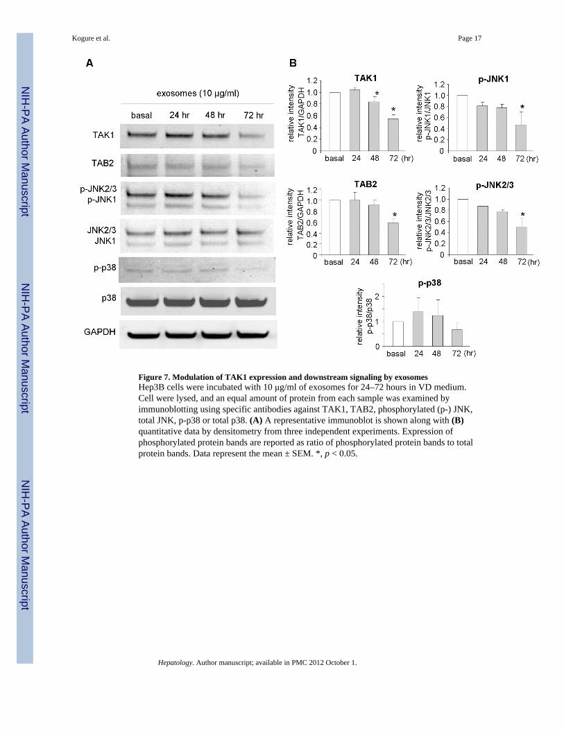
Figure 7. Modulation of TAK1 expression and downstream signaling by exosomesHep3B cells were incubated with 10 μg/ml of exosomes for 24–72 hours in VD medium.Cell were lysed, and an equal amount of protein from each sample was examined byimmunoblotting using specific antibodies against TAK1, TAB2, phosphorylated (p-) JNK,total JNK, p-p38 or total p38. (A) A representative immunoblot is shown along with (B)quantitative data by densitometry from three independent experiments. Expression ofphosphorylated protein bands are reported as ratio of phosphorylated protein bands to totalprotein bands. Data represent the mean ± SEM. *, p < 0.05.
Kogure et al. Page 17
Hepatology. Author manuscript; available in PMC 2012 October 1.
NIH
-PA Author Manuscript
NIH
-PA Author Manuscript
NIH
-PA Author Manuscript

Figure 8. Effect of exosomes on HCC cell behavior(A) Transformed cell growth was assessed by determining anchorage-independent growth insoft agar. Cells in culture medium containing 0.4% agarose with or without 15 μg of Hep3B-derived exosomes were plated in a 96-well plate (1000 cells per well) over a base agar layerconsisted of culture medium containing 0.6% agarose. After incubation for 7 days, thenumber of colonies was evaluated fluorometrically and was expressed as fluorescencerelative to that in controls without exosomes. Bars represent the mean ± SEM of six separatedeterminations. *, p < 0.05. (B) Induction of caspase-3/7 activation. Hep3B cells were platedin a 96-well plate (10,000 cells per well) and treated with various concentrations of Hep3Bderived exosomes for 24 hours. Caspase-3/7 activity was assessed using a commercialluminometric assay. The data was expressed as relative value of luminescence to controlwithout exosomes. Bars represent the mean ± SEM of three independent experiments. *, p <0.05. (C) Cell viability assay. Cells (5,000 cells per well) were plated in a 96-well plate withVD medium and incubated with varying concentrations of Hep3B-derived exosomes for 72hours. Cell viability was assessed using an MTS assay and was expressed as a percentage ofcontrol without exosomes. Bars represent the mean ± SEM of 6 separate determinations. *, p< 0.05.
Kogure et al. Page 18
Hepatology. Author manuscript; available in PMC 2012 October 1.
NIH
-PA Author Manuscript
NIH
-PA Author Manuscript
NIH
-PA Author Manuscript
NIH
-PA Author Manuscript
NIH
-PA Author Manuscript
NIH
-PA Author Manuscript
Kogure et al. Page 19
Table 1
microRNAs expressed exclusively in exosomes derived from Hep3B human HCC cells.
microRNA Expression indexξ
miR-584 165.8
miR-517c 39.8
miR-378 38.2
miR-520f 36.4
miR-142-5p 20.3
miR-451 18.4
miR-518d 13.1
miR-215 12.4
miR-376a* 12.4
miR-133b 8.3
miR-367 7.4
ξmiRNA expression in exosomes calculated relative to expression in Hep3B donor cells using an assigned CT value of 40 for donor cell
expression.
Hepatology. Author manuscript; available in PMC 2012 October 1.