INTENTIONAL MAPS IN POSTERIOR PARIETAL CORTEXauthors.library.caltech.edu/673/1/ANDarn02.pdf · 17...
38
Annu. Rev. Neurosci. 2002. 25:189–220 doi: 10.1146/annurev.neuro.25.112701.142922 Copyright c 2002 by Annual Reviews. All rights reserved INTENTIONAL MAPS IN POSTERIOR PARIETAL CORTEX Richard A. Andersen and Christopher A. Buneo Division of Biology, California Institute of Technology, Mail Code 216-76, Pasadena, California 91125; email: [email protected]; [email protected] Key Words eye movements, arm movements, optic flow, spatial representations, neural prosthetics ■ Abstract The posterior parietal cortex (PPC), historically believed to be a sensory structure, is now viewed as an area important for sensory-motor integration. Among its functions is the forming of intentions, that is, high-level cognitive plans for movement. There is a map of intentions within the PPC, with different subregions dedicated to the planning of eye movements, reaching movements, and grasping movements. These areas appear to be specialized for the multisensory integration and coordinate trans- formations required to convert sensory input to motor output. In several subregions of the PPC, these operations are facilitated by the use of a common distributed space representation that is independent of both sensory input and motor output. Attention and learning effects are also evident in the PPC. However, these effects may be general to cortex and operate in the PPC in the context of sensory-motor transformations. INTRODUCTION The posterior parietal cortex (PPC) has traditionally been viewed as a sensory “as- sociation” area, associating different modalities and having higher-level sensory functions such as spatial attention and spatial awareness. In this review, we high- light a new view of the PPC that is emerging. It is proposed that the PPC, rather than serving a purely sensory or motor role, subserves higher-level cognitive functions related to action. Among these higher cognitive functions is the formation of inten- tions, or early plans for movement. These intentions are anatomically segregated within the PPC, with regions specialized for the planning of saccades, reaches, and grasps. Moreover, these intentions are highly abstract and are evident in the discharge of single neurons even when a specific intention is not carried out. The different intention-related regions of the PPC appear to participate in opera- tions critical to the earliest stages of movement planning: multisensory integration and coordinate transformations. These functions are facilitated by employing a rather unique, distributed code. The response fields of neurons in at least two re- gions are in retinal coordinates, independent of both the sensory modality used to 0147-006X/02/0721-0189$14.00 189 Annu. Rev. Neurosci. 2002.25:189-220. Downloaded from arjournals.annualreviews.org by CALIFORNIA INSTITUTE OF TECHNOLOGY on 09/08/05. For personal use only.
Transcript of INTENTIONAL MAPS IN POSTERIOR PARIETAL CORTEXauthors.library.caltech.edu/673/1/ANDarn02.pdf · 17...
17 May 2002 14:25 AR AR160-06.tex AR160-06.sgm LaTeX2e(2002/01/18)P1: GJB10.1146/annurev.neuro.25.112701.142922
Annu. Rev. Neurosci. 2002. 25:189–220doi: 10.1146/annurev.neuro.25.112701.142922
Copyright c© 2002 by Annual Reviews. All rights reserved
INTENTIONAL MAPS IN POSTERIOR
PARIETAL CORTEX
Richard A. Andersen and Christopher A. BuneoDivision of Biology, California Institute of Technology, Mail Code 216-76, Pasadena,California 91125; email: [email protected]; [email protected]
Key Words eye movements, arm movements, optic flow, spatial representations,neural prosthetics
■ Abstract The posterior parietal cortex (PPC), historically believed to be a sensorystructure, is now viewed as an area important for sensory-motor integration. Among itsfunctions is the forming of intentions, that is, high-level cognitive plans for movement.There is a map of intentions within the PPC, with different subregions dedicated tothe planning of eye movements, reaching movements, and grasping movements. Theseareas appear to be specialized for the multisensory integration and coordinate trans-formations required to convert sensory input to motor output. In several subregionsof the PPC, these operations are facilitated by the use of a common distributed spacerepresentation that is independent of both sensory input and motor output. Attentionand learning effects are also evident in the PPC. However, these effects may be generalto cortex and operate in the PPC in the context of sensory-motor transformations.
INTRODUCTION
The posterior parietal cortex (PPC) has traditionally been viewed as a sensory “as-sociation” area, associating different modalities and having higher-level sensoryfunctions such as spatial attention and spatial awareness. In this review, we high-light a new view of the PPC that is emerging. It is proposed that the PPC, rather thanserving a purely sensory or motor role, subserves higher-level cognitive functionsrelated to action. Among these higher cognitive functions is the formation of inten-tions, or early plans for movement. These intentions are anatomically segregatedwithin the PPC, with regions specialized for the planning of saccades, reaches,and grasps. Moreover, these intentions are highly abstract and are evident in thedischarge of single neurons even when a specific intention is not carried out.
The different intention-related regions of the PPC appear to participate in opera-tions critical to the earliest stages of movement planning: multisensory integrationand coordinate transformations. These functions are facilitated by employing arather unique, distributed code. The response fields of neurons in at least two re-gions are in retinal coordinates, independent of both the sensory modality used to
0147-006X/02/0721-0189$14.00 189
Ann
u. R
ev. N
euro
sci.
2002
.25:
189-
220.
Dow
nloa
ded
from
arj
ourn
als.
annu
alre
view
s.or
gby
CA
LIF
OR
NIA
IN
STIT
UT
E O
F T
EC
HN
OL
OG
Y o
n 09
/08/
05. F
or p
erso
nal u
se o
nly.
17 May 2002 14:25 AR AR160-06.tex AR160-06.sgm LaTeX2e(2002/01/18)P1: GJB
190 ANDERSEN ¥ BUNEO
cue target locations (i.e., audition vs. vision) and the action that will ultimately beperformed (i.e., reaches vs. saccades). However, these retinal fields are also gainmodulated by eye, head, and limb positions. As a result, groups of parietal cells donot generally represent space in a single, defined spatial reference frame. Rather,they code locations in a distributed manner, which can be read out by other groupsof neurons in a variety of reference frames.
We describe a potential medical application that utilizes the finding that thePPC encodes movement intentions. The intention-related activity in the PPC can,in principle, be used to operate a neural prosthesis for paralyzed patients. Sucha neural prosthesis would consist of recording the activity of PPC neurons, in-terpreting the movement intentions of the subject with computer algorithms, andusing these predictions of the subject’s intentions to operate external devices suchas a robot limb or a computer. We describe preliminary investigations in healthymonkeys that estimate the number of parietal cells needed to operate such a pros-thesis (Meeker et al. 2001, Shenoy et al. 1999b). We also describe a recent findingthat monkeys can use this intended movement activity to position a cursor on acomputer screen just by thinking about a reach movement, without actually gen-erating a reach (D. Meeker, S. Cao, J. W. Burdick & R. A. Andersen, unpublishedobservations). This result was obtained without extensive training and stronglysuggests that we are in fact tapping into the highly abstract neural signals thatrepresent the earliest plans for movement.
THE PPC SUBSERVES COGNITIVE FUNCTIONSRELATED TO ACTION
Many of the deficits observed following lesions of the PPC are consistent withthe area playing a high-level, cognitive role in sensory-motor integration. Patientswith PPC lesions do not have primary sensory or motor deficits. However, whenthey attempt to connect these functions, for instance during sensory guided move-ments, then defects become apparent. Patients with PPC lesions often suffer fromoptic ataxia; that is, difficulty in estimating the location of stimuli in 3D space, asindicated by pronounced errors in reaching movements (Balint 1909, Rondot et al.1977). Patients with PPC lesions can also suffer from one or more of the apraxias,a class of deficits characterized by the inability to plan movements (Geshwind& Damasio 1985). These can range from a complete inability to follow verbalcommands for simple movements, to difficulty in performing sequences of move-ments. Patients with parietal lobe damage also have difficulty correctly shapingtheir hands as they prepare to grasp objects, which again points to a disconnectionbetween the visual sensory apparatus that registers the shape of objects and themotor systems that shape the configuration of the hand (Goodale & Milner 1992,Perenin & Vighetto 1988).
Neglect is another deficit commonly attributed to lesions of the PPC, althoughthere is currently some debate about whether it is damage to the PPC or to thenearby superior temporal gyrus that is the source of this defect (Critchley 1953,Karnath et al. 2001). The hallmark of neglect is the lack of awareness within the
Ann
u. R
ev. N
euro
sci.
2002
.25:
189-
220.
Dow
nloa
ded
from
arj
ourn
als.
annu
alre
view
s.or
gby
CA
LIF
OR
NIA
IN
STIT
UT
E O
F T
EC
HN
OL
OG
Y o
n 09
/08/
05. F
or p
erso
nal u
se o
nly.
17 May 2002 14:25 AR AR160-06.tex AR160-06.sgm LaTeX2e(2002/01/18)P1: GJB
INTENTIONAL MAPS IN THE PPC 191
personal and extrapersonal space contralateral to the lesioned hemisphere, with themost profound deficits seen with right hemisphere lesions in right-handed humans.
These clinical results are extremely informative and useful and have helpedguide much of the neurophysiological investigation of the PPC. However, to un-derstand the neural mechanisms and circuits within the PPC that are involved insensory-motor integration requires that the investigator, rather than relying on thehappenstance of medical defects, be able to control the parameters of the exper-iments. Moreover, refined techniques need to be applied. In the case of humans,this has generally taken the form of fMRI studies, and in the case of monkeys,electrophysiological recording and anatomical studies. The monkey has proven tobe a good model for the study of the PPC, since sophisticated motor behaviorssuch as hand-eye coordination are similar in the two species of primates, and thereis extensive evidence to suggest that the PPC in both species performs similarfunctions (Connolly et al. 2000, DeSouza et al. 2000, Rushworth et al. 2001b).Evidence from these studies provides additional support for the concept that thePPC is neither strictly sensory nor motor but rather is involved in high-level cogni-tive functions related to action (Mountcastle et al. 1975, Andersen 1987, Goodale& Milner 1992). These functions include early-movement planning, particularlythe coordinate transformations required for sensory-guided movement. The ac-tivity of PPC may also be influenced by spatial attention and learning. However,these functions are general to cortex, and in the PPC appear to operate in the morespecific context of sensory-motor operations.
INTENTION
Intention is an early plan for a movement. It specifies the goal of a movement and thetype of movement. For instance, “I wish to pick up the coffee cup” specifies both thegoal and type of movement. An intention is high level and abstract. For instance,we can have intentions without actually acting upon them. Moreover, a neuralcorrelate of intention does not necessarily contain information about the details ofa movement, for instance the joint angles, torques, and muscle activations requiredto make a movement. As discussed below, intentions are initially coded in visualcoordinates in at least some of the cortical areas within the PPC. This encodingis consistent with a more cognitive representation of intentions, specifying thegoals of movements rather than the exact muscle activations required to executethe movement.
An intention is also a broad category of cortical functions, which include de-cision making (Gold & Shadlen 2001) and “motor attention” (Rushworth et al.2001a). For instance, decision making can be considered a competition betweenpotential movement intentions (Platt & Glimcher 1999). It may also be the casethat the earliest intentions sit atop a sequence of increasingly more specificmovement plans. In the example above, the earliest intention may reflect the desireto grasp the cup, with further specifications including which limb (right or left),the trajectory of the movement to avoid obstacles, the coordination of eye andhand movements, the speed of the movement, etc. Only further research will be
Ann
u. R
ev. N
euro
sci.
2002
.25:
189-
220.
Dow
nloa
ded
from
arj
ourn
als.
annu
alre
view
s.or
gby
CA
LIF
OR
NIA
IN
STIT
UT
E O
F T
EC
HN
OL
OG
Y o
n 09
/08/
05. F
or p
erso
nal u
se o
nly.
17 May 2002 14:25 AR AR160-06.tex AR160-06.sgm LaTeX2e(2002/01/18)P1: GJB
192 ANDERSEN ¥ BUNEO
able to resolve which parameters of a movement are coded at which stages in thesensory-motor pathway.
Distinguishing Intention from Attention
The issue of intention versus attention has been most prominent in the study ofthe PPC, which is perhaps not surprising considering this area is at the interfacebetween sensory and motor systems. Mountcastle and colleagues (1975) first notedneural activity in the PPC related to the behaviors of monkeys. Robinson andcolleagues (1978) later argued that these effects could be due to sensory stimulationand attention during movement. In experiments designed to tease apart sensoryand movement components of activity, Andersen et al. (1987) found both, whichis consistent with a role for this area in sensory-motor transformations.
One common method of separating sensory from motor components is theso-called memory task (Hikosaka & Wurtz 1983) in which an animal is cued as tothe location for a movement by a briefly flashed stimulus but must withhold theresponse until a go signal. Typically, PPC neurons show bursts of activity to the cueand the movement, indicating both sensory- and motor-related activity. However,during the memory period the cells in many parietal areas have persistent activity,even in the dark (Gnadt & Andersen 1988, Snyder et al. 1997). This persistentactivity by and large does not represent the sensory memory of the target. Thiscan be demonstrated using tasks in which animals memorize the locations of twostimuli and subsequently make movements to both locations. For eye and armmovements the persistent activity in the delay period for nearly all neurons in thePPC is only present for the next planned movement (Batista & Andersen 2001,Mazzoni et al. 1996a), even though the animals must hold in memory two cuedlocations. This result indicates that the sensory memory of the target locationsis either contained in a very small subset of neurons within the PPC, or in areasoutside the PPC, perhaps in the frontal lobe (Tian et al. 2000).
The results of the double movement tasks rule out the coding of a sensorymemory in the delay period activity. However, this activity could reflect either thedirection of a movement plan or the direction of attention. Experimentally it hasbeen very difficult to distinguish movement planning or preparation from spatialattention. Most studies of attention in monkeys use experimental paradigms thatrequire animals to make eye or limb movements as part of the experimental design,or have the potential artifact of the animal covertly planning these movements. Thisfact is reason for concern in studies of the dorsal, sensory-motor pathway sincethere is extensive overlap of circuitry concerned with attention and eye movements,as demonstrated by fMRI experiments in humans (Corbetta et al. 1998). The findingthat the locus of spatial attention can affect the metrics of saccades electricallyevoked from the superior colliculus (SC) further argues for a very tight couplingof spatial attention and eye movements (Kustov & Robinson 1996). These andother results have led Rizzolatti and colleagues to argue for a motor theory ofspatial attention (1994). They propose that spatial attention is an early form ofmotor preparation, at least for eye movements.
Ann
u. R
ev. N
euro
sci.
2002
.25:
189-
220.
Dow
nloa
ded
from
arj
ourn
als.
annu
alre
view
s.or
gby
CA
LIF
OR
NIA
IN
STIT
UT
E O
F T
EC
HN
OL
OG
Y o
n 09
/08/
05. F
or p
erso
nal u
se o
nly.
28 May 2002 11:39 AR AR160-06.tex AR160-06.sgm LaTeX2e(2002/01/18)P1: GJB
INTENTIONAL MAPS IN THE PPC 193
Some investigators have used antisaccade and antireach tasks to separate sen-sory from movement processing. In these paradigms, animals are trained to makemovements in the opposite direction from flashed visual targets. For the caseof reaches, activity in the medial intraparietal area (MIP) has been reported tocode mostly the direction of the movement, and not the location of the stimulus(Eskandar & Assad 1999, Kalaska 1996). Gottlieb & Goldberg (1999) have re-ported that the reverse is true in the lateral intraparietal area (LIP) for eye move-ments, i.e., that cells respond to the stimulus and not the direction of plannedmovement. However, a recent report by Zhang & Barash (2000) indicates that,after a brief transient linked to the stimulus, most cells in LIP code the directionof the planned eye movement. Moreover, a smaller class of cells encode both thelocation of the stimulus and the movement plan, which suggests that LIP is in-volved in the intermediate stages of the sensory-motor transformations requiredfor the antisaccade task. Overall, these antisaccade and antireach results reinforcethe idea that PPC cells have both sensory- and movement-related responses, andoccupy an intermediate stage in the sensory-motor transformation process.
We recently conducted an experiment specifically designed to separate theeffects of spatial attention from those of intention (Snyder et al. 1997). In thisexperiment, animals attended to a flashed target and planned a movement to itduring a delay period, but in one case they were instructed to plan a saccadeand in the other a reach (see Figure 1a). The only difference in the task duringthe memory period was the movement the animals were planning to make. Wereasoned that if PPC activity reflected a sensory memory or attention, it shouldbe the same in the two conditions, but if it reflected the movement plan it shouldbe different.
Figure 1 shows two intention-specific neurons, one from area LIP (b) and onefrom an area we refer to as the parietal reach region (PRR) (c). In this task themonkey plans an eye or an arm movement to the same location in space. The acti-vity of the LIP neuron illustrated in panel (b) shows a transient response to theonset of the briefly flashed target. This is followed by activity during the delayperiod if the animal is planning an eye movement (left histogram), but not if heis planning an arm movement to the same location (right histogram). In contrast,the cell in panel (c) shows no activity above baseline in the delay period when theanimal is planning an eye movement, but strong activity when he is planning anarm movement. Such results were typical in the PPC: In general, we found thatduring eye movement planning area LIP was much more active, and during limbmovement planning PRR was more active. PRR included MIP, 7a, and the dorsalaspect of the parieto-occipital (PO) area, though MIP was found to have the highestconcentration of reach-related neurons. The results from both LIP and PRR arguestrongly for a role of the PPC in movement planning.
A subsequent experiment showed that activity in the PPC is also related to theshifting of movement plans, when spatial attention is held constant (Snyder et al.1998a). Cells with a particular movement preference (reach or saccade) showedgreater activity if a plan was changed from the nonpreferred to the preferred move-ment (for the same target location), compared to simply reaffirming the preferred
Ann
u. R
ev. N
euro
sci.
2002
.25:
189-
220.
Dow
nloa
ded
from
arj
ourn
als.
annu
alre
view
s.or
gby
CA
LIF
OR
NIA
IN
STIT
UT
E O
F T
EC
HN
OL
OG
Y o
n 09
/08/
05. F
or p
erso
nal u
se o
nly.
28 May 2002 11:39 AR AR160-06.tex AR160-06.sgm LaTeX2e(2002/01/18)P1: GJB
194 ANDERSEN ¥ BUNEO
plan. This result is reminiscent of proposals that the PPC plays a role in shiftingattention (Steinmetz & Constantinidis 1995), but in this case it is the intendedmovement that shifts, and not the spatial locus of attention.
Default Plans
The experiments by Snyder et al. (1997) were not the first to attempt to separateattention from intended movement activity. Bushnell and colleagues (1981) trainedanimals to either reach or saccade to a target while recording from PPC neurons.They reasoned that if the PPC was involved in attention then they should seethe same level of activity regardless of the motor output, and this was what theyreported. However, they recorded from only nine cells, and inspection of theirFigure 1 suggests that the animal may have looked to the stimulus after the reach.Thus the animal may have been planning an eye movement as well as an armmovement during the task.
This potential problem of covert planning of eye movements is a general prob-lem for experiments examining attention to targets placed away from the fixationpoint. While the formation of covert plans is unlikely to be critical for studies ofattention in the ventral visual pathway, which current evidence suggests is largelyinvolved in visual recognition, it is certainly a problem when studying the dorsalpathway, which is involved in movement planning. The issue of covert planningwas directly addressed in Snyder et al. (1997). In the population of cells fromwhich we recorded, 68% were significantly modulated in the delay period by onemovement plan (reach or saccade) but not the other. Interestingly, even duringthe cue period 44% showed this specificity. We reasoned that the remaining cellsshowing significant activity for both movement plans might reflect covert plansfor movement, since it is very natural to look to where you reach. To control forthis possibility, we had the animals also perform a “dissociation” task in whichthey simultaneously planned an eye and an arm movement in different directions,with one movement into the response field and the other outside.
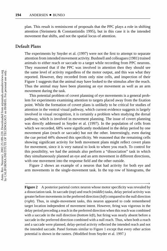
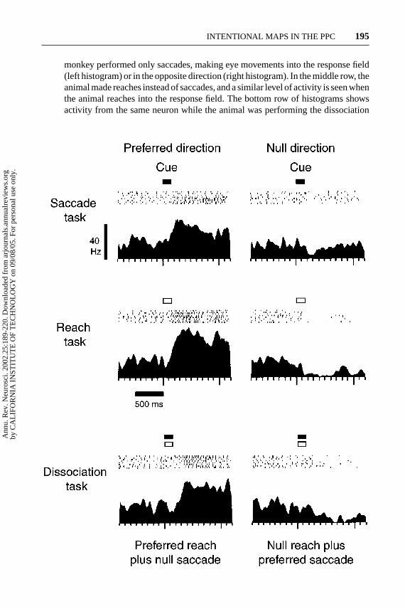
Figure 2 shows an example of a neuron that had activity for both eye andarm movements in the single-movement task. In the top row of histograms, the
−−−−−−−−−−−−−−−−−−−−−−−−−−−−−−−−−−−−−−−−−−−−−−−−−−−−−−−−−→Figure 2 A posterior parietal cortex neuron whose motor specificity was revealed bya dissociation task. In saccade (top) and reach (middle) tasks, delay period activity wasgreater before movements in the preferred direction (left) compared to the null direction(right). Thus, in single-movement tasks, this neuron appeared to code rememberedtarget location independent of movement intent. However, firing was vigorous in thedelay period preceding a reach in the preferred direction when this reach was combinedwith a saccade in the null direction (bottom left), but firing was nearly absent before asaccade in the preferred direction combined with a null reach. Thus, when both a reachand a saccade were planned, delay-period activity reflected the intended reach and notthe intended saccade. Panel formats similar to Figure 1 except that every other actionpotential is shown in the rasters. (Modified from Snyder et al. 1997.)
Ann
u. R
ev. N
euro
sci.
2002
.25:
189-
220.
Dow
nloa
ded
from
arj
ourn
als.
annu
alre
view
s.or
gby
CA
LIF
OR
NIA
IN
STIT
UT
E O
F T
EC
HN
OL
OG
Y o
n 09
/08/
05. F
or p
erso
nal u
se o
nly.
17 May 2002 14:25 AR AR160-06.tex AR160-06.sgm LaTeX2e(2002/01/18)P1: GJB
INTENTIONAL MAPS IN THE PPC 195
monkey performed only saccades, making eye movements into the response field(left histogram) or in the opposite direction (right histogram). In the middle row, theanimal made reaches instead of saccades, and a similar level of activity is seen whenthe animal reaches into the response field. The bottom row of histograms showsactivity from the same neuron while the animal was performing the dissociation
Ann
u. R
ev. N
euro
sci.
2002
.25:
189-
220.
Dow
nloa
ded
from
arj
ourn
als.
annu
alre
view
s.or
gby
CA
LIF
OR
NIA
IN
STIT
UT
E O
F T
EC
HN
OL
OG
Y o
n 09
/08/
05. F
or p
erso
nal u
se o
nly.
17 May 2002 14:25 AR AR160-06.tex AR160-06.sgm LaTeX2e(2002/01/18)P1: GJB
196 ANDERSEN ¥ BUNEO
task. In the histogram on the left, the animal was simultaneously planning an armmovement into the response field and an eye movement out of the response field.In this case the cell was very active. In the histogram on the right, the animal wasplanning an eye movement into the response field, but an arm movement out ofthe field. Although this is the same eye movement plan that evoked activity in thesingle-movement case, now there is no activity or even a slight suppression. Thepattern of activity of this neuron can be explained if the animal is forming a covertplan in the single-movement cases, in this example a covert arm movement plan.Of the neurons in the population, 62% that were not specific for single movementswere specific in the dissociation task, bringing to 84% the number of cells thatshowed movement planning specificity in the delay period. Interestingly, morecells also revealed specificity for the cue response in the dissociation task, with atotal of 45% being specific for reaches and 62% for saccades.
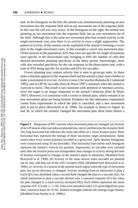
Covert planning may explain activity that is seen in go/no-go tasks. In thesetasks a stimulus appears in the response field and the animal is later cued whether tomake a movement to it or not. Activity in area 5 for reaches (Kalaska & Crammond1995), and in LIP for saccades (Pare & Wurtz 1997) continues when the animal iscued not to move. This result is not consistent with attention or intention activity,since the target is no longer important to the animal’s behavior (Pare & Wurtz1997). However, it is consistent with a covert or default plan, which remains if nonew movement plans are being formed. Evidence for this alternative explanationcomes from experiments in which the plan is cancelled, and a new movementplan is put in place (Bracewell et al. 1996). An example is shown in Figure 3aand 3b, in which the monkey changed the movement plan three times before a
−−−−−−−−−−−−−−−−−−−−−−−−−−−−−−−−−−−−−−−−−−−−−−−−−−−−−−−−−→Figure 3 Responses of PPC neurons when movement plans are changed. (a) Activityof an LIP neuron when saccades are planned into, out of, and then into the receptive field.The long horizontal bar indicates the onset and offset of a visual fixation point. Shorthorizontal bars represent the timings of three successive target presentations. Spikerasters show every action potential recorded in a given trial. Spike-density histogramswere constructed using 50 ms binwidths. Thin horizontal lines below each histogramrepresent the animal’s vertical eye position. Importantly, no saccades were initiateduntil after the fixation point was extinguished; thus changes in activity during the timeof fixation correspond to changes in the animal’s plans or intentions. (Modified fromBracewell et al. 1996). (b) Activity of the same neuron when saccades are plannedout of, into, and then out of the cell’s receptive field. (Modified from Bracewell et al.1996). (c) Activity of a neuron in the parietal reach region when the type of movementplan, but not its direction, is changed. Activity resulting from an instruction to plan areach (R1) was abolished when a second flash changed the plan to a saccade (S2). Aninitial instruction to plan a saccade elicited only a transient response (S1) but whenthe plan changed to a reach activity increased (R2). Each ribbon represents the meanresponse of 8–12 trials+/−1 SE. Data were smoothed with a 121-point digital low-passfilter, transition band 20–32 Hz. Dashed rectangles indicate the timings target flashes.(Modified from Snyder et al. 1998a.)
Ann
u. R
ev. N
euro
sci.
2002
.25:
189-
220.
Dow
nloa
ded
from
arj
ourn
als.
annu
alre
view
s.or
gby
CA
LIF
OR
NIA
IN
STIT
UT
E O
F T
EC
HN
OL
OG
Y o
n 09
/08/
05. F
or p
erso
nal u
se o
nly.
17 May 2002 14:25 AR AR160-06.tex AR160-06.sgm LaTeX2e(2002/01/18)P1: GJB
INTENTIONAL MAPS IN THE PPC 197
Ann
u. R
ev. N
euro
sci.
2002
.25:
189-
220.
Dow
nloa
ded
from
arj
ourn
als.
annu
alre
view
s.or
gby
CA
LIF
OR
NIA
IN
STIT
UT
E O
F T
EC
HN
OL
OG
Y o
n 09
/08/
05. F
or p
erso
nal u
se o
nly.
17 May 2002 14:25 AR AR160-06.tex AR160-06.sgm LaTeX2e(2002/01/18)P1: GJB
198 ANDERSEN ¥ BUNEO
saccade, alternating between planning into, out of, and into the response field (a),or out, in, and out (b). A similar result is found even when the type of movementplan is changed, but not the direction (Snyder et al. 1998a). In Figure 3c, the darkhistogram shows activity for a reach-specific neuron when the first cue presentedin the response field instructs a reach (R1), and the second stimulus appears at thesame location instructing a change in plan to a saccade (S2). Note that althoughno movement is made during the time shown in the histogram, the activity turnsoff when the animal changes to the nonpreferred plan for a movement to the samelocation. The lighter histogram shows activity for the same cell when the monkeyplans a saccade first (S1), and then changes the plan to a reach (R2). Again theactivity is consistent with the cell’s activity expressing the intent of the animal, withbaseline activity after the cue transient when the animal is planning a saccade, andhigh activity during the delay when he changes his plan to a reach. Taken together,the data from various labs suggest that default plans are formed in parietal areas tostimuli of behavioral significance in the case of no alternative plans, but are erasedif alternative plans are formed.
Dynamic Evolution of Intention-Related Activity
Several studies point to a dynamic evolution in the relation of PPC activity totask requirements, changing from sensory to cognitive to motor as the demands ofthe task change. For instance, we recently examined the activity of PRR neuronswhen monkeys plan reaches to auditory versus visual targets in a memory-reachtask. We found that at cue onset activity for visually cued trials carried moreinformation about spatial location than activity for auditory cued trials. However,as the trials progressed and the animal was preparing a movement, the amount ofspatial location information increased for the auditory cued trials so that by thetime of the reach movement, it was not significantly different from the visuallycued trials (Y. C. Cohen & R. A. Andersen, unpublished observations).
In another study, we trained animals to make saccades to a specific location cuedon an object, but after the cue and before the saccade the object was rotated. Earlyin the task area LIP cells carried information about the location of the cue and theorientation of the object, both pieces of information being important for solvingthe task. However, near the time of the eye movement many of these same neuronspredominately coded just the direction of the intended movement (Breznen et al.1999).
Platt & Glimcher (1999) showed in a delayed eye movement task that the earlyactivity of LIP neurons varied as a function of the expected probability that a stimu-lus was a target for a saccade, as well as the amount of reward previously associatedwith the target. However, during later periods of the trial the cells coded only thedirection of the planned eye movement. A similar evolution has been shown in LIPand dorsal prefrontal cortex in eye movement tasks instructed by motion signals.The strength of the motion signal is an important determinant of activity in thebeginning of the trial, but at the end of the trial the activity codes the decision or
Ann
u. R
ev. N
euro
sci.
2002
.25:
189-
220.
Dow
nloa
ded
from
arj
ourn
als.
annu
alre
view
s.or
gby
CA
LIF
OR
NIA
IN
STIT
UT
E O
F T
EC
HN
OL
OG
Y o
n 09
/08/
05. F
or p
erso
nal u
se o
nly.
17 May 2002 14:25 AR AR160-06.tex AR160-06.sgm LaTeX2e(2002/01/18)P1: GJB
INTENTIONAL MAPS IN THE PPC 199
movement plan of the animal (Leon & Shadlen 1999, Shadlen & Newsome 1996).These studies emphasize the fact that the circuits involved in sensory-motor trans-formations are distributed in nature, involving parietal, frontal, and prefrontal areas(Chaffee & Goldman-Rakic 1998). Moreover, activity in these circuits can evolvedynamically to reflect sensory, cognitive, and movement components of behavior.
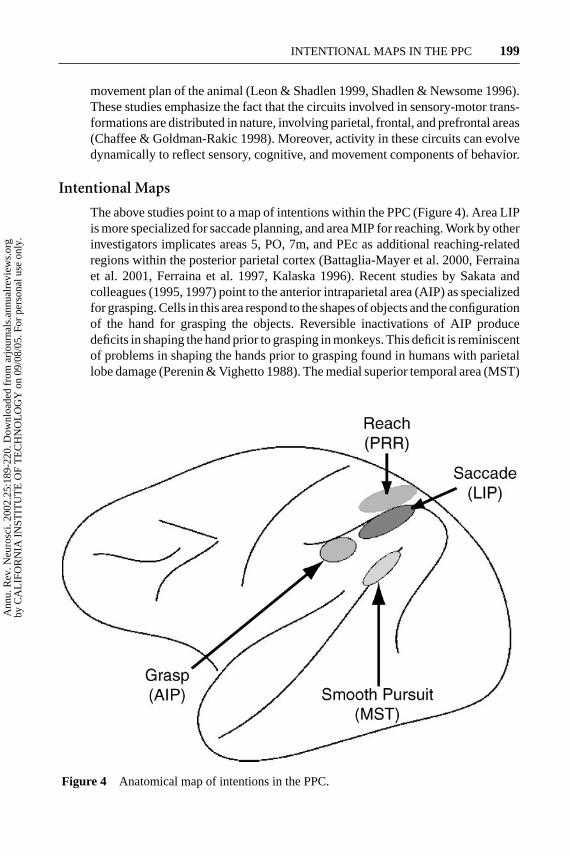
Intentional Maps
The above studies point to a map of intentions within the PPC (Figure 4). Area LIPis more specialized for saccade planning, and area MIP for reaching. Work by otherinvestigators implicates areas 5, PO, 7m, and PEc as additional reaching-relatedregions within the posterior parietal cortex (Battaglia-Mayer et al. 2000, Ferrainaet al. 2001, Ferraina et al. 1997, Kalaska 1996). Recent studies by Sakata andcolleagues (1995, 1997) point to the anterior intraparietal area (AIP) as specializedfor grasping. Cells in this area respond to the shapes of objects and the configurationof the hand for grasping the objects. Reversible inactivations of AIP producedeficits in shaping the hand prior to grasping in monkeys. This deficit is reminiscentof problems in shaping the hands prior to grasping found in humans with parietallobe damage (Perenin & Vighetto 1988). The medial superior temporal area (MST)
Figure 4 Anatomical map of intentions in the PPC.
Ann
u. R
ev. N
euro
sci.
2002
.25:
189-
220.
Dow
nloa
ded
from
arj
ourn
als.
annu
alre
view
s.or
gby
CA
LIF
OR
NIA
IN
STIT
UT
E O
F T
EC
HN
OL
OG
Y o
n 09
/08/
05. F
or p
erso
nal u
se o
nly.
17 May 2002 14:25 AR AR160-06.tex AR160-06.sgm LaTeX2e(2002/01/18)P1: GJB
200 ANDERSEN ¥ BUNEO
appears to play a specialized role in smooth-pursuit eye movements. Cells in thisarea are active for pursuit, even during brief periods when the pursuit target isextinguished (Newsome et al. 1988). Inactivations of this area produce pursuitdeficits that are not a result of sensory deficits (Dursteler & Wurtz 1988).
Experiments using fMRI in humans are consistent with the monkey results.Rushworth and colleagues (2001b) found that peripheral attention tasks activatedthe lateral bank of the intraparietal sulcus, whereas planning manual movementsactivated the medial bank. They concluded that their results were consistent withthe monkey studies, with the medial bank specialized for manual movements andthe lateral bank for attention and eye movements. A similar result has recentlybeen reported by Connolly et al. (2000) using event-related fMRI and an eyeand hand movement task similar to the one employed by Snyder et al. (1997).An area specialized for grasping has also been identified in the anterior aspectof the intraparietal sulcus in humans (Binkofski et al. 1998). This area may behomologous to monkey AIP.
Multisensory Integration and Coordinate Transformations
Producing a movement in response to a sensory stimulus requires that a host ofproblems be solved. From the sensory side, different sensory modalities are codedin different reference frames, vision in retinal or eye-centered coordinates, sound inhead-centered coordinates, and touch in body-centered coordinates. These differ-ent coordinate frames need to be resolved in some way, since a particular movementmight need to be directed to a visual, auditory, or somatosensory stimulus or anycombination of these. From the motor side, the locations of these stimuli mustultimately be transformed into the natural coordinates of the muscles in order tomake movements.
Lesions to the PPC can produce optic ataxia, where patients mislocalize stimulito which they are reaching. These mislocalizations are apparent in all three di-mensions. Since these effects are often present with no primary sensory or motordefects, they suggest that the PPC is important for multisensory integration as wellas for the coordinate transformations required for making sensory-guided move-ments. In this section, we discuss electrophysiological experiments supporting thisview. In particular, we provide evidence that spatial locations are represented ina common coordinate frame in at least some parts of the PPC, independent ofsensory input or motor output.
AREA LIP: SACCADE PLANNING IN EYE-CENTERED COORDINATES We can easilymake eye movements to visual or auditory targets. If area LIP is involved in makingeye movements, then cells in this area should respond when an animal is planningan eye movement, regardless of the sensory modality of the stimulus. Recently wehave found this to be the case (Grunewald et al. 1999, Linden et al. 1999, Mazzoniet al. 1996b). However, this observation raises the question, do these two modalitiesshare a common reference frame, and if so, what is it? Cells in the intermediate
Ann
u. R
ev. N
euro
sci.
2002
.25:
189-
220.
Dow
nloa
ded
from
arj
ourn
als.
annu
alre
view
s.or
gby
CA
LIF
OR
NIA
IN
STIT
UT
E O
F T
EC
HN
OL
OG
Y o
n 09
/08/
05. F
or p
erso
nal u
se o
nly.
28 May 2002 11:49 AR AR160-06.tex AR160-06.sgm LaTeX2e(2002/01/18)P1: GJB
INTENTIONAL MAPS IN THE PPC 201
layers of the SC use a common, eye-position-dependent reference frame for rep-resenting saccades to visual, auditory, or somatosensory stimuli (Groh & Sparks1996a, Groh & Sparks 1996b, Jay & Sparks 1987a, Jay & Sparks 1987b). This isnot surprising given that the SC is near the final motor output stage for saccadesand that motor error is expressed in eye-centered coordinates. However, area LIPis intermediate between sensory and motor areas; thus it is not immediately appar-ent what reference frame should be used to represent visual and auditory targetsin this region.
In experiments in which monkeys made saccades to auditory targets, we foundthat a majority of the neurons coded these targets in eye-centered coordinates,although some also coded auditory targets in head-centered coordinates, or in areference frame intermediate between the eye and head reference frames (Stricanneet al. 1996). Moreover, many of the response fields of LIP neurons were gainmodulated by eye position. These data suggest that area LIP may be one of thesites involved in the transformation of auditory signals from head- to eye-centeredcoordinates. Recent experiments examining cells in the temporo-parietal cortex(TpT) (Wu & Andersen 2001), an auditory association area that projects into thePPC (Pandya & Kuypers 1969), and the inferior colliculus (Groh et al. 2001),indicate that cells antecedent to LIP code auditory locations in head-centeredcoordinates, with many neurons also gain modulated by eye position. These resultssupport a model in which head-centered auditory signals are gain modulated byeye position and are then read out at subsequent levels in eye-centered coordinates(Xing & Andersen 2000b).
PRR: REACH PLANNING IN EYE-CENTERED COORDINATES The LIP results suggestthat this area encodes sensory stimuli as motor error for saccades. If this is the case,then one might predict that PRR would code sensory stimuli as motor error as well,i.e., in limb coordinates. We tested this prediction by training monkeys to reachto targets from two different initial arm positions while fixating their gaze in twodifferent directions. As illustrated for one PRR neuron in Figure 5, the responsefield did not vary with changes in limb position (a,b), but shifted with gaze direc-tion (c,d). This result indicates that PRR codes limb movements in eye-centeredcoordinates. This result, as well as those obtained in LIP, indicates that the PPCis capable of encoding intended movements in eye-centered coordinates indepen-dent of the type of movement to be made, i.e., saccades (LIP) and reaches (PRR)(see Figure 6).
The finding that area LIP encodes intended saccades in eye-centered coordinatesfor both visual and auditory stimuli, as well as the finding that area PRR encodesreaches in eye-centered coordinates, led us to an unusual prediction: that PRRwould code reaches to auditory stimuli in eye-centered coordinates. This predictionis based on the assumption that the PPC may use a common reference framefor movement planning, independent of sensory input or motor output. Such aresult would be quite surprising since sounds, which are initially coded in head-centered coordinates, could simply be converted to body- and then limb-centered
Ann
u. R
ev. N
euro
sci.
2002
.25:
189-
220.
Dow
nloa
ded
from
arj
ourn
als.
annu
alre
view
s.or
gby
CA
LIF
OR
NIA
IN
STIT
UT
E O
F T
EC
HN
OL
OG
Y o
n 09
/08/
05. F
or p
erso
nal u
se o
nly.
28 May 2002 11:49 AR AR160-06.tex AR160-06.sgm LaTeX2e(2002/01/18)P1: GJB
202 ANDERSEN ¥ BUNEO
coordinates—a transformation to eye-centered coordinates is not required. In astudy in which monkeys planned reaches to sounds in complete darkness, we foundthat this prediction was correct. Under these conditions, many cells in PRR encodedthe intended movement in eye-centered coordinates (Cohen & Andersen 2000).
EYE-CENTERED CODING IN OTHER AREAS Recent stimulation studies suggest thatthe SC, rather than coding desired gaze displacement or gaze direction in space,encodes the desired gaze direction in retinal coordinates (Klier et al. 2001). Electro-physiological studies of the SC have provided evidence for an eye-centered codingof limb movements in this structure as well (Stuphorn et al. 2000). The ventralpremotor cortex also appears to contain neurons that code the location of reachtargets in eye-centered coordinates (Mushiake et al. 1997), though these cells maycoexist with others having more arm-centered properties (Graziano et al. 1994,1997). These results, as well as those obtained in the PPC, support the existenceof a distributed network devoted to eye-hand coordination that uses a commoneye-centered reference frame for representing the spatial aspects of eye and armmovements (Figure 6).
COMPENSATION FOR EYE MOVEMENTS If saccade and reach plans are coded ineye-centered coordinates, then problems can arise in situations where a movementplan is formed and an intervening saccade is made before the movement is executed.The problem is particularly acute in cases of movements planned to rememberedlocations in the dark. Mays & Sparks (1980) found that under these circumstances,activity shifts within the eye movement map of the SC to compensate for theintervening saccade and to still code the correct motor vector. Gnadt & Andersen(1988) reported a similar result in area LIP. Duhamel et al. (1992) extended theseresults by showing that it was not necessary to make an eye movement for thisupdating to take place. They interpreted this updated activity as sensory and amechanism for maintaining perceptual stability across eye movements. The resultsof Snyder et al. (1997, 1998a) provide an alternative explanation: that this activityreflects a default plan for an eye movement. Whether this shift also accounts forperceptual stability remains a possibility and requires additional investigation.
Accounting for eye movements is also a problem when reach plans are codedin eye-centered coordinates. Imagine that an animal plans an arm movement ineye-centered coordinates to the remembered location of a stimulus in the darkand then makes an intervening eye movement before the arm movement takesplace. If the reach plan is not adjusted to take into account the retinal positionof the stimulus after the eye movement, then areas downstream of PRR will usethe previous retinal position of the stimulus to calculate the motor error. This willresult in an error corresponding to the size and direction of the intervening saccade.
We directly tested the effect of intervening saccades on intended reachactivity in PRR. Figure 5e–g shows the design of the experiment and resultsfrom one cell. When the flash occurred outside the response field there wasno response (e), and when it fell within the response field there was robust
Ann
u. R
ev. N
euro
sci.
2002
.25:
189-
220.
Dow
nloa
ded
from
arj
ourn
als.
annu
alre
view
s.or
gby
CA
LIF
OR
NIA
IN
STIT
UT
E O
F T
EC
HN
OL
OG
Y o
n 09
/08/
05. F
or p
erso
nal u
se o
nly.
17 May 2002 14:25 AR AR160-06.tex AR160-06.sgm LaTeX2e(2002/01/18)P1: GJB
INTENTIONAL MAPS IN THE PPC 203
planning activity (f ). Note that the histograms demonstrate planning activity; theactual arm movement occurs at a time later than that shown. In (g), the task beganwith the same configuration of eye, hand, and stimulus as in (e). However, afterthe stimulus was extinguished, the animal was instructed to make a saccade to anew location on the board, bringing the response field over the location on theboard where the animal was planning the reach. The activity shifted in PRR suchthat the cell was now active, coding the correct location of the planned reach ineye coordinates even though the reach cue never appeared in the response field.Thus the cell compensated for the saccade to maintain the correct coding of thereach target in eye coordinates. All 34 PRR cells tested with this paradigm showedsuch compensation for saccades. A remapping of reach plans in eye coordinateshas been demonstrated in psychophysical experiments in humans (Henriques et al.1998), consistent with this physiological finding.
Another possible example of this type of compensation for eye movements,which must still be experimentally verified, is the compensation for smooth-pursuiteye movements that must occur for self-motion perception. During forward loco-motion, self-motion perception is estimated from the focus of expansion of thevisual field. However, when subjects make smooth eye movements during forwardlocomotion, as would occur when tracking an object on the ground, these eyemovements introduce an additional, laminar motion on the retinas that disrupts thefocus of expansion, generally shifting it in the direction of the eye movement. Cellsin the dorsal subdivision of the medial superior temporal area (MSTd) are thoughtto play a role in self-motion perception because they are sensitive to optic flowstimuli and because they are tuned to the spatial location of the focus of expansion(Duffy & Wurtz 1995). In experiments from our laboratory, we found that thesefocus tuning curves shift to compensate for smooth-pursuit gaze movements. Thiscompensation appears to depend on both efference copies of commands to movethe eye or head and the visual information in the optic flow pattern (Bradley et al.1996, Shenoy et al. 1999a). To guide locomotion, this signal would eventuallyneed to be coded in body- or world-centered coordinates; however, it is currentlynot known in what reference frame MSTd neurons code focus-position signals. Itwould be consistent with the data from LIP and PRR if the MSTd cells compensatedfor the eye movements to maintain the correct heading direction in eye-centeredcoordinates.
GAIN FIELDS The common representation of space in the PPC is embodied notonly in eye-centered response fields, but also in the gain modulation of thesefields by body-position signals. These gain field effects are found throughout thePPC and include modulation of retinotopic fields by eye-, head-, body-, and limb-position signals (Andersen et al. 1993). Computational studies have shown thatthese gain effects can be the mechanism for transforming between coordinateframes (Salinas & Abbott 1995, Zipser & Andersen 1988). Moreover, groups ofneurons with retinal response fields, modulated by various body part–positionsignals, can conceivably be read out in multiple frames of reference (Pouget &
Ann
u. R
ev. N
euro
sci.
2002
.25:
189-
220.
Dow
nloa
ded
from
arj
ourn
als.
annu
alre
view
s.or
gby
CA
LIF
OR
NIA
IN
STIT
UT
E O
F T
EC
HN
OL
OG
Y o
n 09
/08/
05. F
or p
erso
nal u
se o
nly.
17 May 2002 14:25 AR AR160-06.tex AR160-06.sgm LaTeX2e(2002/01/18)P1: GJB
204 ANDERSEN ¥ BUNEO
Snyder 2000, Xing & Andersen 2000b) as would be needed to direct movements ofthe eyes, head, or hand. Thus the representation of space in the PPC is distributedand is comprised of eye-centered response fields with gain modulation.
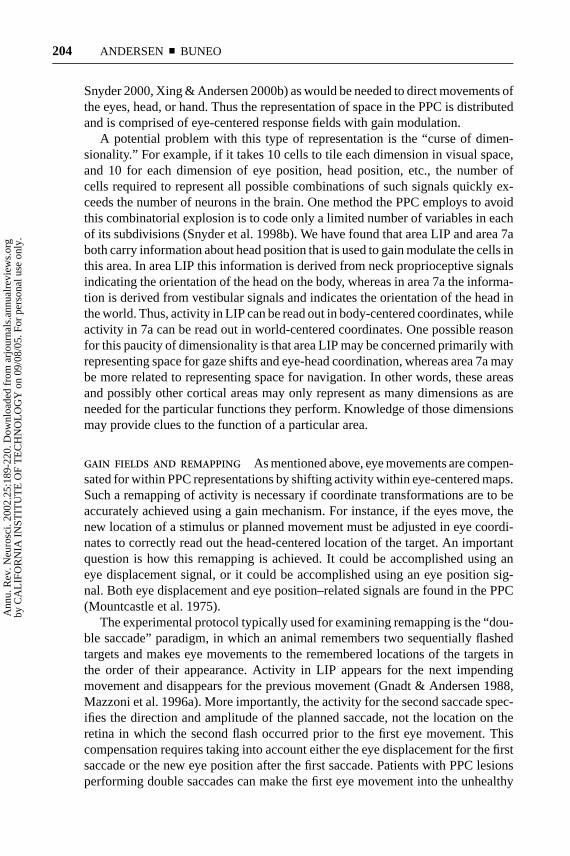
A potential problem with this type of representation is the “curse of dimen-sionality.” For example, if it takes 10 cells to tile each dimension in visual space,and 10 for each dimension of eye position, head position, etc., the number ofcells required to represent all possible combinations of such signals quickly ex-ceeds the number of neurons in the brain. One method the PPC employs to avoidthis combinatorial explosion is to code only a limited number of variables in eachof its subdivisions (Snyder et al. 1998b). We have found that area LIP and area 7aboth carry information about head position that is used to gain modulate the cells inthis area. In area LIP this information is derived from neck proprioceptive signalsindicating the orientation of the head on the body, whereas in area 7a the informa-tion is derived from vestibular signals and indicates the orientation of the head inthe world. Thus, activity in LIP can be read out in body-centered coordinates, whileactivity in 7a can be read out in world-centered coordinates. One possible reasonfor this paucity of dimensionality is that area LIP may be concerned primarily withrepresenting space for gaze shifts and eye-head coordination, whereas area 7a maybe more related to representing space for navigation. In other words, these areasand possibly other cortical areas may only represent as many dimensions as areneeded for the particular functions they perform. Knowledge of those dimensionsmay provide clues to the function of a particular area.
GAIN FIELDS AND REMAPPING As mentioned above, eye movements are compen-sated for within PPC representations by shifting activity within eye-centered maps.Such a remapping of activity is necessary if coordinate transformations are to beaccurately achieved using a gain mechanism. For instance, if the eyes move, thenew location of a stimulus or planned movement must be adjusted in eye coordi-nates to correctly read out the head-centered location of the target. An importantquestion is how this remapping is achieved. It could be accomplished using aneye displacement signal, or it could be accomplished using an eye position sig-nal. Both eye displacement and eye position–related signals are found in the PPC(Mountcastle et al. 1975).
The experimental protocol typically used for examining remapping is the “dou-ble saccade” paradigm, in which an animal remembers two sequentially flashedtargets and makes eye movements to the remembered locations of the targets inthe order of their appearance. Activity in LIP appears for the next impendingmovement and disappears for the previous movement (Gnadt & Andersen 1988,Mazzoni et al. 1996a). More importantly, the activity for the second saccade spec-ifies the direction and amplitude of the planned saccade, not the location on theretina in which the second flash occurred prior to the first eye movement. Thiscompensation requires taking into account either the eye displacement for the firstsaccade or the new eye position after the first saccade. Patients with PPC lesionsperforming double saccades can make the first eye movement into the unhealthy
Ann
u. R
ev. N
euro
sci.
2002
.25:
189-
220.
Dow
nloa
ded
from
arj
ourn
als.
annu
alre
view
s.or
gby
CA
LIF
OR
NIA
IN
STIT
UT
E O
F T
EC
HN
OL
OG
Y o
n 09
/08/
05. F
or p
erso
nal u
se o
nly.
17 May 2002 14:25 AR AR160-06.tex AR160-06.sgm LaTeX2e(2002/01/18)P1: GJB
INTENTIONAL MAPS IN THE PPC 205
visual field, but are not able to generate an accurate second saccade (Heide et al.1995). Although it has been argued that this proves that an eye-displacement signalmediates remapping, eye displacement and eye position are in fact confounded inthis task. The deficit was seen when the displacement of the eyes was in the direc-tion of the unhealthy field, but also when the eye position after the first movementwas in the unhealthy field.
In other experiments, area LIP was reversibly inactivated in monkeys in ex-periments designed to directly examine whether eye-displacement or eye-positionsignals are used for remapping in double-saccade experiments (Li & Andersen2001). Both initial eye position and the direction of eye movements were variedin individual trials in order to tease apart eye-position and eye-displacement con-tributions. It was found that the largest deficits were seen when the animal madethe first eye movement into the unhealthy visual field, largely independent of thedirection of the eye movement. This result suggests that eye-position signals playa large role in the compensation for intervening saccades.
A recent computational study illustrates that dynamic neural networks can betrained to perform the double saccade task using eye-position signals (Xing &Andersen 2000a). These networks show activity similar to that recorded fromLIP when monkeys perform the same task. These include eye-centered responsefields that are gain modulated by eye position, and activity that shifts within aneye-centered map of visual space to correct for intervening saccades. Thus, thegain field mechanism can account for dynamic compensation for intervening eyemovements in eye-centered coordinates.
GAIN FIELDS: OTHER USES Since their discovery in areas of the PPC, gain effectshave been identified throughout the brain. This suggests that multiplicative andadditive interactions between different inputs to neurons may reflect a generalmethod of neural computation. Although the role of gain fields in coordinatetransformations has been highlighted in this review, gain fields appear to play arole in many other functions, including attention, navigation, decision making, andobject recognition. Some examples are discussed briefly below (see also Salinas& Thier 2000).
The direction of attention can modulate the activity of V4 neurons (McAdams& Maunsell 2000, Reynolds et al. 2000), and this effect has been proposed toplay a role in the binding of features in objects (Salinas & Abbott 1997). In ad-dition, although smooth pursuit shifts the focus tuning of many MSTd neurons,as mentioned above, other MSTd neurons do not shift their focus tuning but aregain modulated by the pursuit signal (Bradley et al. 1996, Shenoy et al. 1999a).This gain modulation is consistent with an intermediate step toward the produc-tion of shifting focus-tuning curves, and thus may play a role in the perceptionof self-motion for navigation. Monkeys and humans have been shown to choosebetween two targets for a reach depending on eye position, essentially choos-ing targets that tend to center the reach with respect to the head (Scherbergeret al. 1999). Eye-position gain effects have been shown in PRR and may bias
Ann
u. R
ev. N
euro
sci.
2002
.25:
189-
220.
Dow
nloa
ded
from
arj
ourn
als.
annu
alre
view
s.or
gby
CA
LIF
OR
NIA
IN
STIT
UT
E O
F T
EC
HN
OL
OG
Y o
n 09
/08/
05. F
or p
erso
nal u
se o
nly.
17 May 2002 14:25 AR AR160-06.tex AR160-06.sgm LaTeX2e(2002/01/18)P1: GJB
206 ANDERSEN ¥ BUNEO
the decision of animals to choose targets based on eye position (Scherberger &Andersen 2001).
We have recently trained monkeys to make object-based saccades by cueinga location on an object, extinguishing the object, and then presenting the objectagain at a different orientation. In this task, the animals must saccade to the previ-ously cued location on the object to obtain their reward (Breznen et al. 1999). Wefind that area LIP does not code the cued location in an explicit object-centeredreference frame in this task, even though it requires the animal to code the tar-get in such a reference frame. Rather, cells in area LIP carry information aboutthe cued location, movement vector, and orientation of the object, all in retinalcoordinates. Some cells show a gain modulation of the cue or movement vectoractivity by object orientation. This result is surprising given the finding that le-sions to the PPC in humans often produce deficits in object-centered coordinates(Arguin & Bub 1993, Driver & Mattingley 1998). However, computational studiesshow that it is not necessary to use cells with object-centered response fields tosolve object-based tasks; rather, distributed coding using retinal response fields fortarget locations and object orientations, and gain modulations between the two, issufficient (Pouget & Sejnowski 1997). This distributed representation can be usedto form response fields in object-centered coordinates, as has been reported in thesupplementary eye fields (SEF) (Olson 2001). However, the SEF results may alsobe explained as a result of gain modulation of retinal response fields by objectposition (Pouget & Sejnowski 1997), and more thorough mapping of the responsefields will be required to distinguish between the two possibilities.
A COMMON DISTRIBUTED CODE FOR INTENDED MOVEMENTS IN AREAS LIP AND
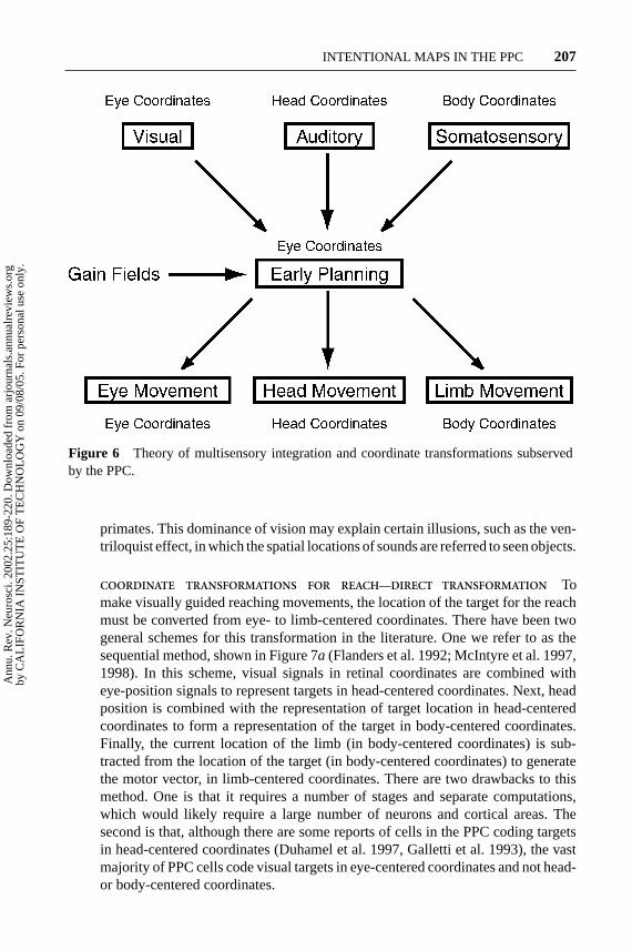
PRR The above results, summarized in Figure 6, suggest that LIP and PRR usea common space representation in which response fields are represented in eye-centered coordinates. This representation exists independent of whether the targetsare visual or auditory. Likewise, this representation is used regardless of whetherthe output is to move the limb or make an eye movement. This general schemegenerated a nonintuitive, but correct, prediction that auditory targets for reachwould be coded in eye-centered coordinates in PRR. Currently, we do not know ifsomatosensory stimuli, such as proprioceptive signals coding the position of thehand, are coded in eye coordinates in PRR and LIP. This would be an interestingquestion for future experimentation, and if true, would provide further evidence forthe generality of this model. In both LIP and PRR, the eye-centered response fieldsare gain modulated by eye-, head-, and limb-position signals. This gain modulationmay provide the mechanism for converting stimuli in various reference frames intoeye-centered coordinates. Likewise, these gain modulations may allow other areasto read out signals from LIP and PRR in different coordinate frames, includingeye-, head-, body-, and limb-centered coordinates.
Why use a common coordinate frame for PRR and LIP? One possibility is tofacilitate hand-eye coordination. Presumably the orchestration of these movementswould be facilitated if they used a common reference frame (Battaglia-Mayer et al.2000). A second reason may be that vision is the most accurate spatial sense in
Ann
u. R
ev. N
euro
sci.
2002
.25:
189-
220.
Dow
nloa
ded
from
arj
ourn
als.
annu
alre
view
s.or
gby
CA
LIF
OR
NIA
IN
STIT
UT
E O
F T
EC
HN
OL
OG
Y o
n 09
/08/
05. F
or p
erso
nal u
se o
nly.
28 May 2002 11:56 AR AR160-06.tex AR160-06.sgm LaTeX2e(2002/01/18)P1: GJB
INTENTIONAL MAPS IN THE PPC 207
Figure 6 Theory of multisensory integration and coordinate transformations subservedby the PPC.
primates. This dominance of vision may explain certain illusions, such as the ven-triloquist effect, in which the spatial locations of sounds are referred to seen objects.
COORDINATE TRANSFORMATIONS FOR REACH—DIRECT TRANSFORMATION Tomake visually guided reaching movements, the location of the target for the reachmust be converted from eye- to limb-centered coordinates. There have been twogeneral schemes for this transformation in the literature. One we refer to as thesequential method, shown in Figure 7a (Flanders et al. 1992; McIntyre et al. 1997,1998). In this scheme, visual signals in retinal coordinates are combined witheye-position signals to represent targets in head-centered coordinates. Next, headposition is combined with the representation of target location in head-centeredcoordinates to form a representation of the target in body-centered coordinates.Finally, the current location of the limb (in body-centered coordinates) is sub-tracted from the location of the target (in body-centered coordinates) to generatethe motor vector, in limb-centered coordinates. There are two drawbacks to thismethod. One is that it requires a number of stages and separate computations,which would likely require a large number of neurons and cortical areas. Thesecond is that, although there are some reports of cells in the PPC coding targetsin head-centered coordinates (Duhamel et al. 1997, Galletti et al. 1993), the vastmajority of PPC cells code visual targets in eye-centered coordinates and not head-or body-centered coordinates.
Ann
u. R
ev. N
euro
sci.
2002
.25:
189-
220.
Dow
nloa
ded
from
arj
ourn
als.
annu
alre
view
s.or
gby
CA
LIF
OR
NIA
IN
STIT
UT
E O
F T
EC
HN
OL
OG
Y o
n 09
/08/
05. F
or p
erso
nal u
se o
nly.
28 May 2002 11:56 AR AR160-06.tex AR160-06.sgm LaTeX2e(2002/01/18)P1: GJB
208 ANDERSEN ¥ BUNEO
A second scheme is referred to as the combinatorial method. As shown inFigure 7b, retinal target location, eye, head, and limb position are all combined atonce, and the target location in limb-centered coordinates is then read out from thisrepresentation (Battaglia-Mayer et al. 2000). As mentioned above, a drawback tothis approach is the “curse of dimensionality.”
A third scheme we refer to as the direct method. Figure 7c shows that thisapproach subtracts the current position of the hand (in eye coordinates) from theposition of the target (in eye coordinates) to directly generate the motor vector inlimb coordinates. An advantage of this approach over the sequential method is thatit requires fewer computational stages. In addition the computation is restricted toonly dimensions in eye coordinates and does not suffer the curse of dimensionalityof the combinatorial approach.
Recently, we have provided evidence for the direct transformation scheme.Single cells in area 5, a somatomotor cortical area within the PPC, have been foundto code target locations simultaneously in eye- and limb-centered coordinates(Buneo et al. 2002). This result suggests that the PPC transforms target locationsdirectly between these two frames of reference. Moreover, cells in PRR code thetarget location in eye-centered coordinates, but the initial hand position introducesa gain on this response that is also eye centered. These two findings, taken together,suggest that a simple gain field mechanism underlies the transformation from eye-to limb-centered coordinates. A convergence of input from cells in PRR onto area5 neurons can perform this transformation directly (see Figure 7d) without havingto resort to intermediate coordinate frames or a large combination of retina-, eye-,head-, and limb-position signals.
Psychophysical evidence supporting a sequential scheme has been provided byFlanders et al. (1992), Henriques et al. (1998), and McIntyre et al. (1997, 1998).These results, as well as our own physiological studies supporting an alternativedirect scheme, may reflect an underlying context dependence in the coordinatetransformations that subserves visually guided reaching (Carrozzo et al. 1999).For example, direct transformations may be the preferred scheme when both targetlocation and the current hand position are simultaneously visible, even for a briefinstant. In contrast, a sequential scheme may be used when visual informationabout the current position of the hand is unavailable.
MOVEMENT DECISIONS
Experiments in LIP by Platt & Glimcher (1999) and by Shadlen and colleagues(Kim & Shadlen 1999, Shadlen & Newsome 1996) have found activity related tothe decision of a monkey to make eye movements. Both the prior probability andamount of reward influence the effectiveness of visual stimuli in LIP, consistentwith a role for this area in decision making. As monkeys accumulate sensoryinformation to make a movement plan, activity increases for neurons in LIP andthe prefrontal cortex (Kim & Shadlen 1999, Leon & Shadlen 1999, Shadlen &Newsome 1996). These results are consistent with these areas weighting decisionvariables for the purpose of planning eye movements (Gold & Shadlen 2001). The
Ann
u. R
ev. N
euro
sci.
2002
.25:
189-
220.
Dow
nloa
ded
from
arj
ourn
als.
annu
alre
view
s.or
gby
CA
LIF
OR
NIA
IN
STIT
UT
E O
F T
EC
HN
OL
OG
Y o
n 09
/08/
05. F
or p
erso
nal u
se o
nly.
17 May 2002 14:25 AR AR160-06.tex AR160-06.sgm LaTeX2e(2002/01/18)P1: GJB
INTENTIONAL MAPS IN THE PPC 209
fact that these effects appear in multiple brain areas suggests that decision makingis a distributed function that includes the PPC.
ATTENTION
The PPC has been classically thought to play a central, perhaps controlling rolein attention. Strong evidence for this idea is the finding of neglect, an inabilityto attend to the contralateral visual field, after PPC lesions in humans (Critchley1953). However, many of the processes involved in visual-motor transformations,for example the shaping of the hand for grasping, appear to operate unconsciously(Goodale & Milner 1992). In fact, lesions to the PPC in monkeys produce visual-motor deficits and not neglect (Faugier-Grimaud et al. 1978, Lamotte & Acuna1978). Rather, it has been reported that lesions to the superior temporal gyrus pro-duce neglect similar to that found in humans (Watson et al. 1994). Interestingly, arecent report by Karnath and colleagues (2001) suggests that the superior temporalgyrus damage may also be the source of neglect seen in humans. Thus, the locusof cortical lesions that produce neglect is still an open question that will likely beresolved with further research.
Although there have been several studies reporting attentional effects on neuralactivity in the PPC, these experiments have been performed in conjunction witheye or limb movements, or in peripheral attention paradigms where animals arelikely to form covert plans for eye movements. As yet no experiments have beenperformed similar to those of Snyder et al. (1997, 1998a). In those experiments,intention was isolated from attentional effects; similar experimental designs areneeded to isolate attentional effects from intentional effects.
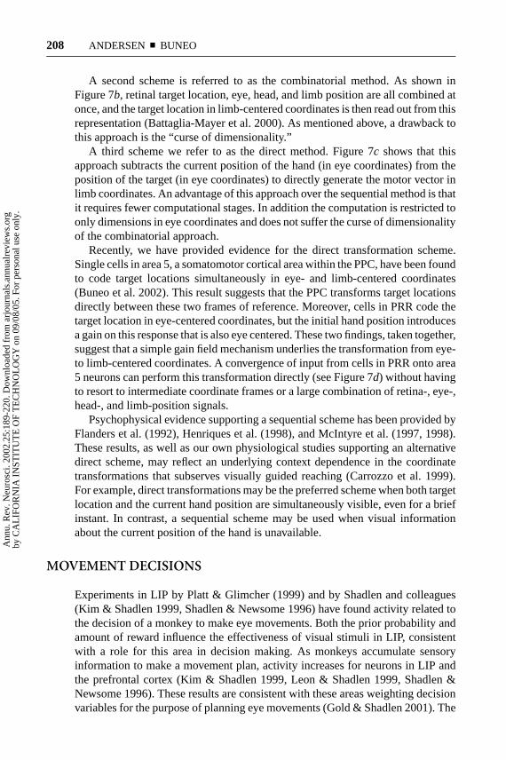
It has been argued that the lower degree of activation of LIP neurons whenmonkeys reach rather than saccade to targets is due to less attention being requiredfor reaching (Colby & Goldberg 1999). This reasoning would predict less activityin PRR as well, but in fact the reverse is true. Figure 8 shows the populationactivity from one monkey for recordings obtained in LIP and PRR. When thismonkey planned, a saccade activity was high in LIP and low in PRR. On the otherhand, when reaches were planned the reverse was true. This figure also shows thatwhen covert planning was controlled in dual-movement trials the separation forsaccades and reaches was even greater. This double dissociation between saccadesand reaches for LIP and PRR shows that the effects are due to planning, and not ageneral reduction in attention when the animal reaches.
In a recent study, Powell & Goldberg (2000) flashed stimuli around the timeof eye movements and found responses for LIP neurons even while the animalswere planning eye movements outside of the cells’ response fields. They arguedthat this demonstrated that LIP is more involved in registering the salience ofvisual stimuli than in planning eye movements. This interpretation is at odds withthat of Mazzoni et al. (1996a), who showed that, when monkeys were planningeye movements outside of the response field of LIP neurons, a flash in the centerof their response field produced only a very brief transient before the activitywas suppressed. A closer inspection of the data in Powell & Goldberg shows
Ann
u. R
ev. N
euro
sci.
2002
.25:
189-
220.
Dow
nloa
ded
from
arj
ourn
als.
annu
alre
view
s.or
gby
CA
LIF
OR
NIA
IN
STIT
UT
E O
F T
EC
HN
OL
OG
Y o
n 09
/08/
05. F
or p
erso
nal u
se o
nly.
28 May 2002 12:9 AR AR160-06.tex AR160-06.sgm LaTeX2e(2002/01/18)P1: GJB
210 ANDERSEN ¥ BUNEO
Figure 8 Population response from one monkey for areas LIP (left) and PRR (right). Cellshad significant activity during the delay period of either the reach or saccade task of Snyderet al. (1997). Solid gray traces represent the average activity of the population of cells for sac-cades into the response field. Solid black traces represent activity for reaches into the responsefield. Dashed lines represent activity in the dual-movement task, with gray traces represent-ing saccades into the response field and black traces representing reaches. Histograms weresmoothed with a 181-point digital low-pass filter with a –3 dB point at 9 Hz. (From Snyderet al. 2000.)
a similar effect, with activity dying out prior to and during the eye movement.A similar sensory transient can be seen when a reach target is flashed withinan LIP response field, which quickly dies away in this case hundreds of msecbefore the reach movement (see Figure 1b, right histogram). Interestingly, units insimulated networks programmed to hold a movement plan for one location whilea distractor stimulus is briefly flashed at another location [conditions similar tothe experiment of Powell & Goldberg (2000)] also exhibit input transients (Xing& Andersen 2000a). However, lateral inhibitory connections in these networksquickly suppressed the activity due to the distractor, similar to the suppressionseen in LIP experiments.
In conclusion, it is likely that pure attentional effects will be found in the PPC.However, because this area is specialized for sensory-motor integration, there is theadditional challenge of designing paradigms that rule out movement planning as asource of activity. This concern is less problematic in areas of the visual cortex thatare involved in recognition, where there is ample evidence of attentional effects.Attentional effects in PPC may be general, since attention-related activity has beenreported throughout the cerebral cortex. Thus attention effects in PPC would be re-lated to planning movements, much like attention effects in inferotemporal cortexwould be related to visual recognition. Interestingly, recent studies suggest PPC andprefrontal structures may regulate spatial aspects of attention in the ventral, recog-nition pathway (Kastner & Ungerleider 2000). Whether PPC is specialized for at-tention and is the controller for attention throughout cortex is an important question.
Ann
u. R
ev. N
euro
sci.
2002
.25:
189-
220.
Dow
nloa
ded
from
arj
ourn
als.
annu
alre
view
s.or
gby
CA
LIF
OR
NIA
IN
STIT
UT
E O
F T
EC
HN
OL
OG
Y o
n 09
/08/
05. F
or p
erso
nal u
se o
nly.
17 May 2002 14:25 AR AR160-06.tex AR160-06.sgm LaTeX2e(2002/01/18)P1: GJB
INTENTIONAL MAPS IN THE PPC 211
LEARNING AND ADAPTATION
Like attention, learning is a distributed function and like atttentional effects, learn-ing effects in the PPC tend to be most apparent in the context of sensory-motoroperations, for example prism adaptation. As first demonstrated by Held & Hein(1963), when human subjects reach to visual targets while wearing displacingprisms, they initially miss-reach in the direction of target displacement but gradu-ally recover and reach correctly if provided with appropriate feedback about theirerrors. Using positron emission tomography (PET) to monitor changes in cerebralblood flow, Clower and colleagues (1996) showed that the prism adaptation processresults in selective activation of the PPC contralateral to the reaching arm, whenconfounding sensory, motor, and cognitive effects are ruled out. Similarly, Rosettiand colleagues (1998) found that hemispatial neglect resulting from damage tothe right hemisphere can be at least partially ameliorated by first having affectedpatients make reaching movements in the presence of a prismatic shift, then re-moving the prisms. They interpreted these effects as resulting from the stimulationof neural structures responsible for sensorimotor transformations, including thePPC as well as the cerebellum. A recent electrophysiological study employing aprism adaptation paradigm suggests that the ventral premotor cortex plays a rolein this process as well (Kurata & Hoshi 1999).
An example of the effects of learning in the PPC was revealed in a recentelectrophysiological study of LIP (Grunewald et al. 1999). The responses of LIPneurons to auditory stimuli in a passive fixation task were examined before and afteranimals were trained to make saccades to auditory targets. Before such training, thenumber of cells responding to auditory stimuli in LIP was statistically insignificant.After training, however, 12% showed significant responses to auditory stimuli. Thisindicates that at least some LIP neurons become active for auditory stimuli onlyafter an animal has learned that these stimuli are important for oculomotor behavior.As with the learning effects discussed above, effects of this nature have beenreported in other areas of cortex, e.g., area 3a for tactile discrimination (Recanzoneet al. 1992), premotor cortex for arbitrary associations (Mitz et al. 1991), and thefrontal eye fields (FEF) for visual search training (Bichot et al. 1996), highlightingthe distributed nature of learning in cortex.
READING OUT INTENTIONS—THE PPCAND NEURAL PROSTHETICS
The experimental results reviewed above indicate that activity related to an an-imal’s intentions to make reaches or saccades is strong and robust in the PPC.These experiments are typical of neurophysiological experiments in demonstratingcorrelations between behaviors or perception and neural activity. It would bepowerful to be able to test these proposed linkages more directly. One methodto achieve a more direct demonstration of intention coding is to perform exper-iments that close the loop. This type of experiment is demonstrated in Figure 9.
Ann
u. R
ev. N
euro
sci.
2002
.25:
189-
220.
Dow
nloa
ded
from
arj
ourn
als.
annu
alre
view
s.or
gby
CA
LIF
OR
NIA
IN
STIT
UT
E O
F T
EC
HN
OL
OG
Y o
n 09
/08/
05. F
or p
erso
nal u
se o
nly.
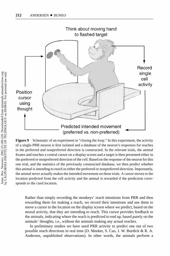
17 May 2002 14:25 AR AR160-06.tex AR160-06.sgm LaTeX2e(2002/01/18)P1: GJB
212 ANDERSEN ¥ BUNEO
Figure 9 Schematic of an experiment in “closing the loop.” In this experiment, the activityof a single-PRR neuron is first isolated and a database of the neuron’s responses for reachesin the preferred and nonpreferred direction is constructed. In the relevant trials, the animalfixates and touches a central cursor on a display screen and a target is then presented either inthe preferred or nonpreferred direction of the cell. Based on the response of the neuron for thisone trial, and the statistics of the previously constructed database, we then predict whetherthis animal is intending to reach in either the preferred or nonpreferred direction. Importantly,the animal never actually makes the intended movement on these trials. A cursor moves to thelocation predicted from the cell activity and the animal is rewarded if the prediction corre-sponds to the cued location.
Rather than simply recording the monkeys’ reach intentions from PRR and thenrewarding them for making a reach, we record their intentions and use them tomove a cursor to the location on the display screen where we predict, based on theneural activity, that they are intending to reach. This cursor provides feedback tothe animals, indicating where the reach is predicted to end up, based purely on theanimals’ thoughts, i.e., without the animals making any actual reaches.
In preliminary studies we have used PRR activity to predict one out of twopossible reach directions in real time (D. Meeker, S. Cao, J. W. Burdick & R. A.Andersen, unpublished observations). In other words, the animals perform a
Ann
u. R
ev. N
euro
sci.
2002
.25:
189-
220.
Dow
nloa
ded
from
arj
ourn
als.
annu
alre
view
s.or
gby
CA
LIF
OR
NIA
IN
STIT
UT
E O
F T
EC
HN
OL
OG
Y o
n 09
/08/
05. F
or p
erso
nal u
se o
nly.
17 May 2002 14:25 AR AR160-06.tex AR160-06.sgm LaTeX2e(2002/01/18)P1: GJB
INTENTIONAL MAPS IN THE PPC 213
task in which they locate a cursor on the computer screen by using intended move-ment activity without making an actual reach. In other studies, offline analysis ofsingle-cell recordings from PRR indicates that reliably predicting one out of eightdirections could require only a small number of simultaneously recorded neurons,perhaps in the range of 10 to 15 (Meeker et al. 2001, Shenoy et al. 1999b).
The high-level planning activity observed in PRR could be used in the control ofa neural prosthetic for paralyzed patients. Patients with paralysis due to peripheralneuropathies, trauma, and stroke can often still think about making movements butcannot execute them. The idea of a cortical prosthetic is to record these intentionsto move, interpret the intentions using real-time decode algorithms running oncomputers, and then convert these decoded intentions to control signals that canoperate external devices. These external devices could include stimulators imbed-ded in the patient’s muscles that would allow the patient to move his/her own body,a robot limb, or a computer interface for communication.
Research on neural prosthetics is a burgeoning and young field. Several groupsare working toward using motor cortex for such a prosthesis (Chapin et al. 1999,Isaacs et al. 2000, Wessberg et al. 2000, Kennedy et al. 2000, Maynard et al.1999), which makes sense because it is the area of cortex closest to the motoroutput. There may also be advantages for using higher, cognitive areas of thesensory-motor system such as PRR for the control of prosthetics. Because this isa field that is still at its infancy, it is not clear if one area is optimal for prostheticcontrol. Moreover, because different areas of the sensory-motor pathway no doubtprovide different, useful information, it may turn out to be most optimal to developmultiarea prosthetics that can read out and decode these different signals. Beloware listed some potential attributes of PRR for prosthetic control.
1.) Motor cortex is known to undergo degradation as a result of paralysis. Forinstance with spinal cord lesions, cortico-spinal neurons are destroyed andthe somatosensory reafferent signals to this area of the cortex are also lost.However, PRR may undergo less degradation after paralysis as it is moreclosely tied to the visual system.
2.) Learning is an important aspect of neural prosthetic success. For instance,cochlear prosthetics do not produce natural auditory sensations with elec-trical stimulation, but patients learn to interpret these stimulus-induced per-cepts, for instance in understanding speech. Implanted arrays of electrodesin humans will likely only sample a part of the workspace of the subject.Although other parts of space not well sampled can be inferred from theactivity of cells in sampled regions, the resolution cannot be as good aswould result from a more even sampling. Thus, neural plasticity would bean important advantage for a cortical prosthetic and, as mentioned above,there is evidence that the PPC does play a role in adjusting the registrationof sensory-motor representations for accurate behaviors.
3.) To successfully close the loop, patients require feedback to the cortex re-garding the success of the movement. Whereas these feedback signals arelost with paralysis in motor areas, they are largely intact in PRR, as the
Ann
u. R
ev. N
euro
sci.
2002
.25:
189-
220.
Dow
nloa
ded
from
arj
ourn
als.
annu
alre
view
s.or
gby
CA
LIF
OR
NIA
IN
STIT
UT
E O
F T
EC
HN
OL
OG
Y o
n 09
/08/
05. F
or p
erso
nal u
se o
nly.
17 May 2002 14:25 AR AR160-06.tex AR160-06.sgm LaTeX2e(2002/01/18)P1: GJB
214 ANDERSEN ¥ BUNEO
reafference to this region is largely visual. Moreover, the evidence presentedabove, that the PPC performs early coordinate transformations required forreaching in retinal coordinates, suggests that vision can be used by thepatients for correcting motor error computations.
4.) The fact that the intended movement signal is a high-level, cognitive signalmay have advantages. For instance, if PRR is coding intentions in abstractor general terms, it may require fewer neurons to control devices that aredissimilar to the human limb. In addition, the fact that this planning activitycan be sustained for long periods of time may help in the decoding ofthe movement plan by providing prolonged, stable signals related to theintentions of the patient.
5.) We have recently found that, during the delays when monkeys are planninga movement, there are broadband gamma oscillations in the local field po-tentials (LFPs) in both LIP and PRR. The LFPs in both areas are tuned to thedirection of planned movements and change their strength with behavioralstate, becoming much larger in amplitude when the animal plans a move-ment and rapidly decreasing during the movement execution. Although itis somewhat difficult to record single-cell activity over long periods of timewith chronically implanted electrodes, LFPs are stable and relatively easyto record with chronic electrodes, since they reflect the activity of corticalcolumns rather than single cells. Thus, using LFPs in LIP and PRR may bean important breakthrough for obtaining long-term recordings with existingtechnology.
CONCLUSION
The PPC is important for sensory-motor integration, particularly the forming ofintentions or high-level cognitive plans for movement. There is a map of inten-tions within the PPC, with subregions for saccades, reaching, and grasping. Theseregions appear to be specialized for multisensory integration and coordinate trans-formations. Within each subregion is a map of the working space. In PRR andLIP these maps are in eye-centered coordinates, regardless of sensory input ormovement plan. Within these maps, activity is gain modulated by eye-, head-, andlimb-position signals. This distributed, abstract representation is consistent withintentions in this area being high level and cognitive.
These ideas perhaps raise more questions than they answer. For instance, whatother features of intended movements are coded in the activity of PPC neurons?Are the trajectories, distance of reaching, or the dynamics of the planned movementcoded in PRR activity? Is the transition from a high-level, cognitive intention tothe motor output of the cortex one of multiple stages and continual refinementof the plan, or is the intention to move converted to an executable plan in onestep of convergence onto motor cortical areas? Are there additional cortical areasin PPC for intending other types of movement, such as leg movements and headmovements?
Ann
u. R
ev. N
euro
sci.
2002
.25:
189-
220.
Dow
nloa
ded
from
arj
ourn
als.
annu
alre
view
s.or
gby
CA
LIF
OR
NIA
IN
STIT
UT
E O
F T
EC
HN
OL
OG
Y o
n 09
/08/
05. F
or p
erso
nal u
se o
nly.
17 May 2002 14:25 AR AR160-06.tex AR160-06.sgm LaTeX2e(2002/01/18)P1: GJB
INTENTIONAL MAPS IN THE PPC 215
There are a number of outstanding questions regarding the finding of a commoncoordinate frame for spatial location in LIP and PRR. For instance, do other parietalareas, such as the grasp area, also code in retinal coordinates with gain modulationby eye- and other body part–position signals? Are somatosensory signals alsorepresented in eye coordinates in these areas? Does this common reference framealso support intended head movements? Are there tasks in which the intentions tomove do not use this common, distributed reference frame? Experiments designedto answer these questions will help to determine how generally the concepts ofcognitive intentions and common reference frames can be applied to the PPC.
ACKNOWLEDGMENTS
We wish to acknowledge the generous support of the James G. Boswell Foundation,the National Institutes of Health (NIH), the Defense Advanced Research ProjectsAgency (DARPA), Sloan-Swartz Center for Theoretical Neurobiology, the Officeof Naval Research (ONR), and the Christopher Reeves Foundation. We also thankDr. Paul Glimcher for helpful comments.
The Annual Review of Neuroscienceis online at http://neuro.annualreviews.org
LITERATURE CITED
Andersen RA. 1987.The Role of the InferiorParietal Lobule in Spatial Perception andVisual-Motor Integration. Bethesda, MD:Am. Physiol. Soc., pp. 483–518
Andersen RA, Essick GK, Siegel RM. 1987.Neurons of area 7a activated by both visualstimuli and oculomotor behavior.Exp. BrainRes.67:316–22
Andersen RA, Snyder LH, Li CS, StricanneB. 1993. Coordinate transformations in therepresentation of spatial information.Curr.Opin. Neurobiol.3:171–76
Arguin M, Bub DN. 1993. Evidence for an inde-pendent stimulus-centered spatial referenceframe from a case of visual hemineglect.Cor-tex29:349–57
Balint R. 1909. Seelenlahmung des “Schau-ens,” optische Ataxie, raumliche Storungder Aufmerksamkeit.Monatsschr. Psychiatr.Neurol.25:51–81
Batista AP, Buneo CA, Snyder LH, AndersenRA. 1999. Reach plans in eye-centered coor-dinates.Science285:257–60
Batista AP, Andersen RA. 2001. The parietalreach region codes the next planned move-ment in a sequential reach task.J. Neuro-physiol.85:539–44
Battaglia-Mayer A, Ferraina S, Mitsuda T,Marconi B, Genovesio A, et al. 2000.Early coding of reaching in the parietooc-cipital cortex. J. Neurophysiol.83:2374–91
Bichot NP, Schall JD, Thompson KG. 1996.Visual feature selectivity in frontal eye fieldsinduced by experience in mature macaques.Nature381:697–99
Binkofski F, Dohle C, Posse S, Stephan KM,Hefter H, et al. 1998. Human anterior intra-parietal area subserves prehension: a com-bined lesion and functional MRI activationstudy.Neurology50:1253–59
Bracewell RM, Mazzoni P, Barash S, Ander-sen RA. 1996. Motor intention activity inthe macaque’s lateral intraparietal area. II.Changes of motor plan.J. Neurophysiol.76:1457–64
Ann
u. R
ev. N
euro
sci.
2002
.25:
189-
220.
Dow
nloa
ded
from
arj
ourn
als.
annu
alre
view
s.or
gby
CA
LIF
OR
NIA
IN
STIT
UT
E O
F T
EC
HN
OL
OG
Y o
n 09
/08/
05. F
or p
erso
nal u
se o
nly.
17 May 2002 14:25 AR AR160-06.tex AR160-06.sgm LaTeX2e(2002/01/18)P1: GJB
216 ANDERSEN ¥ BUNEO
Bradley DC, Maxwell M, Andersen RA, BanksMS, Shenoy KV. 1996. (Neural) mechanismsof heading perception in primate visual cor-tex.Science273:1544–47
Breznen B, Sabes PN, Andersen RA. 1999.Parietal coding of object-based saccades: ref-erence frames.Soc. Neurosci.(Abstr.) 25:1547
Buneo CA, Jarvis MR, Batista AP, AndersenRA. 2002. Direct visuomotor transforma-tions for reaching.Nature.In press
Bushnell MC, Goldberg ME, Robinson DL.1981. Behavioral enhancement of visual re-sponses in monkey cerebral cortex. I. Modu-lation in posterior parietal cortex related toselective visual attention.J. Neurophysiol.46:755–74
Carrozzo M, McIntyre J, Zago M, LacquanitiF. 1999. Viewer-centered and body-centeredframes of reference in direct visuomotortransformations.Exp. Brain Res.129:201–10
Chaffee MV, Goldman-Rakic PS. 1998. Match-ing patterns of activity in primate prefrontalarea 8a and parietal area 7ip during a spa-tial working memory task.J. Neurophysiol.79:2919–40
Chapin JK, Moxon KA, Markowitz RS,Nicolelis MA. 1999. Real-time control ofa robot arm using simultaneously recordedneurons in the motor cortex.Nat. Neurosci.2:664–70
Clower DM, Hoffman JM, Votaw JR, Faber TL,Woods RP, Alexander GE. 1996. Role of pos-terior parietal cortex in the recalibration ofvisually guided reaching.Nature 383:618–21
Cohen YE, Andersen RA. 2000. Reaches tosounds encoded in an eye-centered referenceframe.Neuron27:647–52
Colby CL, Goldberg ME. 1999. Space and at-tention in parietal cortex.Annu. Rev. Neu-rosci.22:319–49
Connolly JD, Menon RS, Goodale MA. 2000.Human frontoparietal areas active during apointing but not a saccade delay.Soc. Neu-rosci. (Abstr.) 26:1329
Corbetta M, Akbudak E, Conturo TE, Snyder
AZ, Ollinger JM, et al. 1998. A common net-work of functional areas for attention and eyemovements.Neuron21:761–73
Critchley M. 1953.The Parietal Lobes.London:Arnold
DeSouza JF, Dukelow SP, Gati JS, Menon RS,Andersen RA, Vilis T. 2000. Eye positionsignal modulates a human parietal pointingregion during memory-guided movements.J. Neurosci.20:5835–40
Driver J, Mattingley JB. 1998. Parietal neglectand visual awareness.Nat. Neurosci.1:17–22
Duffy CJ, Wurtz RH. 1995. Response of mon-key MSTd neurons to optic flow stimuliwith shifted centers of motion.J. Neurosci.15:5192–208
Duhamel JR, Colby CL, Goldberg ME. 1992.The updating of the representation of vi-sual space in parietal cortex by intended eyemovements.Science255:90–92
Duhamel JR, Bremmer F, Ben Hamed S, GrofW. 1997. Spatial invariance of visual recep-tive fields in parietal cortex neurons.Nature389:845–48
Dursteler MR, Wurtz RH. 1988. Pursuit and op-tokinetic deficits following chemical lesionsof cortical areas Mt and Mst.J. Neurophysiol.60:940–65
Eskandar EN, Assad JA. 1999. Dissociation ofvisual, motor and predictive signals in pari-etal cortex during visual guidance.Nat. Neu-rosci.2:88–93
Faugier-Grimaud S, Frenois C, Stein DG. 1978.Effects of posterior parietal lesions on visu-ally guided behavior in monkeys.Neuropsy-chologia16:151–68
Ferraina S, Battaglia-Mayer A, Genovesio A,Marconi B, Onorati P, Caminiti R. 2001.Early coding of visuomanual coordinationduring reaching in parietal area PEc.J. Neu-rophysiol.85:462–67
Ferraina S, Garasto MR, Battaglia-Mayer A,Ferraresi P, Johnson PB, et al. 1997. Visualcontrol of hand-reaching movement: activityin parietal area 7m.Eur. J. Neurosci.9:1090–95
Flanders M, Helms-Tillery SI, Soechting JF.
Ann
u. R
ev. N
euro
sci.
2002
.25:
189-
220.
Dow
nloa
ded
from
arj
ourn
als.
annu
alre
view
s.or
gby
CA
LIF
OR
NIA
IN
STIT
UT
E O
F T
EC
HN
OL
OG
Y o
n 09
/08/
05. F
or p
erso
nal u
se o
nly.
17 May 2002 14:25 AR AR160-06.tex AR160-06.sgm LaTeX2e(2002/01/18)P1: GJB
INTENTIONAL MAPS IN THE PPC 217
1992. Early stages in a sensorimotor trans-formation.Behav. Brain Sci.15:309–62
Galletti C, Battaglini PP, Fattori P. 1993.Parietal neurons encoding spatial locationsin craniotopic coordinates.Exp. Brain Res.96: 221–29
Geshwind N, Damasio AR. 1985. Apraxia. InHandbook of Clinical Neurology, ed. PJVinken, GW Bruyn, HL Klawans, pp. 423–32. Amsterdam: Elsevier
Gnadt JW, Andersen RA. 1988. Memory relatedmotor planning activity in posterior parietalcortex of macaque.Exp. Brain Res.70:216–20
Gold JI, Shadlen MN. 2001. Neural computa-tions that underlie decisions about sensorystimuli. Trends Cogn. Sci.5:10–16
Goodale MA, Milner AD. 1992. Separate visualpathways for perception and action.TrendsNeurosci.15:20–25
Gottlieb J, Goldberg ME. 1999. Activity ofneurons in the lateral intraparietal area ofthe monkey during an antisaccade task.Nat.Neurosci.2:906–12
Graziano MS, Hu XT, Gross CG. 1997. Visu-ospatial properties of ventral premotor cor-tex.J. Neurophysiol.77:2268–92
Graziano MS, Yap GS, Gross CG. 1994. Codingof visual space by premotor neurons.Science266:1054–57
Groh JM, Sparks DL. 1996a. Saccades to so-matosensory targets. 2. Motor convergencein primate superior colliculus.J. Neurophys-iol. 75:428–38
Groh JM, Sparks DL. 1996b. Saccades to so-matosensory targets. 3. Eye-position-depen-dent somatosensory activity in primate supe-rior colliculus.J. Neurophysiol.75:439–53
Groh JM, Trause AS, Underhill AM, Clark KR,Inati S. 2001. Eye position influences audi-tory responses in primate inferior colliculus.Neuron29:509–18
Grunewald A, Linden JF, Andersen RA. 1999.Responses to auditory stimuli in macaque lat-eral intraparietal area. I. Effects of training.J. Neurophysiol.82:330–42
Heide W, Blankenburg M, Zimmermann E,Kompf D. 1995. Cortical control of double-
step saccades—implications for spatial ori-entation.Ann. Neurol.38:739–48
Held R, Hein A. 1963. Movement producedstimulation in the development of visuallyguided behavior.J. Comp. Physiol. Psychol.53:236–41
Henriques DY, Klier EM, Smith MA, Lowy D,Crawford JD. 1998. Gaze-centered remap-ping of remembered visual space in an open-loop pointing task.J. Neurosci.18:1583–94
Hikosaka O, Wurtz RH. 1983. Visual and ocu-lomotor functions of monkey substantia ni-gra pars reticulata. III. Memory-contingentvisual and saccade responses.J. Neurophys-iol. 49:1268–84
Isaacs RE, Weber DJ, Schwartz AB. 2000.Work toward real-time control of a corticalneural prothesis.IEEE Trans. Rehabil. Eng.8:196–98
Jay MF, Sparks DL. 1987a. Sensorimotor inte-gration in the primate superior colliculus. 1.Motor convergence.J. Neurophysiol.57:22–34
Jay MF, Sparks DL. 1987b. Sensorimotor in-tegration in the primate superior colliculus.2. Coordinates of auditory signals.J. Neuro-physiol.57:35–55
Kalaska JF. 1996. Parietal cortex area 5 and vi-suomotor behavior.Can. J. Physiol. Pharma-col. 74:483–98
Kalaska JF, Crammond DJ. 1995. Deciding notto GO: neural correlates of response selectionin a GO/NOGO task in primate premotor andparietal cortex.Cerebr. Cortex5:410–28
Karnath H, Ferber S, Himmelbach M. 2001.Spatial awareness is a function of the tem-poral not the posterior parietal lobe.Nature411:950–53
Kastner S, Ungerleider LG. 2000. Mechanismsof visual attention in the human cortex.Annu.Rev. Neurosci.23:315–41
Kennedy PR, Bakay RAE, Moore MM, AdamsK, Goldwaithe J. 2000. Direct control of acomputer from the human central nervoussystem.IEEE Trans. Rehabil. Eng.8:198–202
Kim JN, Shadlen MN. 1999. Neural correlatesof a decision in the dorsolateral prefrontal
Ann
u. R
ev. N
euro
sci.
2002
.25:
189-
220.
Dow
nloa
ded
from
arj
ourn
als.
annu
alre
view
s.or
gby
CA
LIF
OR
NIA
IN
STIT
UT
E O
F T
EC
HN
OL
OG
Y o
n 09
/08/
05. F
or p
erso
nal u
se o
nly.
17 May 2002 14:25 AR AR160-06.tex AR160-06.sgm LaTeX2e(2002/01/18)P1: GJB
218 ANDERSEN ¥ BUNEO
cortex of the macaque.Nat. Neurosci.2:176–85
Klier EM, Wang H, Crawford JD. 2001. Thesuperior colliculus encodes gaze commandsin retinal coordinates.Nat. Neurosci.4:627–32
Kurata K, Hoshi E. 1999. Reacquisition deficitsin prism adaptation after muscimol microin-jection into the ventral premotor cortex ofmonkeys.J. Neurophysiol.81:1927–38
Kustov AA, Robinson DL. 1996. Shared neuralcontrol of attentional shifts and eye move-ments.Nature384:74–77
Lamotte RH, Acuna C. 1978. Defects in ac-curacy of reaching after removal of poste-rior parietal cortex in monkeys.Brain Res.139:309–26
Leon MI, Shadlen MN. 1999. Effect of expectedreward magnitude on the response of neu-rons in the dorsolateral prefrontal cortex ofthe macaque.Neuron24:415–25
Li CS, Andersen RA. 2001. Inactivation ofmacaque lateral intraparietal area delays ini-tiation of the second saccade predominantlyfrom contralesional eye positions in a double-saccade task.Exp. Brain Res.137:45–57
Linden JF, Grunewald A, Andersen RA. 1999.Responses to auditory stimuli in macaque lat-eral intraparietal area. II. Behavioral modu-lation.J. Neurophysiol.82:343–58
Mays LE, Sparks DL. 1980. Saccades are spa-tially, not retinocentrically, coded.Science208:1163–65
Maynard EM, Hatsopoulos NG, Ojakangas CL,Acuna BD, Sanes JN, et al. 1999. Neuronalinteractions improve cortical population cod-ing of movement direction.J. Neurosci.19:8083–93
Mazzoni P, Bracewell RM, Barash S, AndersenRA. 1996a. Motor intention activity in themacaque’s lateral intraparietal area. I. Disso-ciation of motor plan from sensory memory.J. Neurophysiol.76:1439–56
Mazzoni P, Bracewell RM, Barash S, Ander-sen RA. 1996b. Spatially tuned auditory re-sponses in area LIP of macaques performingdelayed memory saccades to acoustic targets.J. Neurophysiol.75:1233–41
McAdams CJ, Maunsell JHR. 2000. Attentionto both space and feature modulates neuronalresponses in macaque area V4.J. Neurophys-iol. 83:1751–55
McIntyre J, Stratta F, Lacquaniti F. 1997.Viewer-centered frame of reference for poin-ting to memorized targets in three-di-mensional space.J. Neurophysiol.78:1601–18
McIntyre J, Stratta F, Lacquaniti F. 1998. Short-term memory for reaching to visual targets:psychophysical evidence for body-cente-red reference frames.J. Neurosci.18:8423–35
Meeker D, Shenoy KV, Cao S, Pesaran B,Scherberger H, et al. 2001. Cognitive controlsignals for prosthetic systems.Soc. Neurosci.(Abstr.) 27:63.6
Mitz AR, Godschalk M, Wise SP. 1991.Learning-dependent neuronal-activity in thepremotor cortex-activity during the acqui-sition of conditional motor associations.J.Neurosci.11:1855–72
Mountcastle VB, Lynch JC, Georgopoulos A,Sakata H, Acuna C. 1975. Posterior parietalassociation cortex of the monkey: commandfunctions for operations within extrapersonalspace.J. Neurophysiol.38:871–908
Mushiake H, Tanatsugu Y, Tanji J. 1997. Neu-ronal activity in the ventral part of premotorcortex during target-reach movement is mod-ulated by direction of gaze.J. Neurophysiol.78:567–71
Newsome WT, Wurtz RH, Komatsu H. 1988.Relation of cortical areas MT and MST topursuit eye movements. II. Differentiationof retinal from extraretinal inputs.J. Neuro-physiol.60:604–20
Olson CR. 2001. Object-based vision and at-tention in primates.Curr. Opin. Neurobiol.11:171–79
Pandya DN, Kuypers HG. 1969. Cortico-cortical connections in the rhesus monkey.Brain Res.13:13–36
Pare M, Wurtz RH. 1997. Monkey posteriorparietal cortex neurons antidromically acti-vated from superior colliculus.J. Neurophys-iol. 78:3493–97
Ann
u. R
ev. N
euro
sci.
2002
.25:
189-
220.
Dow
nloa
ded
from
arj
ourn
als.
annu
alre
view
s.or
gby
CA
LIF
OR
NIA
IN
STIT
UT
E O
F T
EC
HN
OL
OG
Y o
n 09
/08/
05. F
or p
erso
nal u
se o
nly.
17 May 2002 14:25 AR AR160-06.tex AR160-06.sgm LaTeX2e(2002/01/18)P1: GJB
INTENTIONAL MAPS IN THE PPC 219
Perenin MT, Vighetto A. 1988. Optic ataxia:a specific disruption in visuomotor mecha-nisms. I. Different aspects of the deficit inreaching for objects.Brain 111:643–74
Platt ML, Glimcher PW. 1999. Neural corre-lates of decision variables in parietal cortex.Nature400:233–38
Pouget A, Sejnowski TJ. 1997. A new view ofhemineglect based on the response propertiesof parietal neurones.Philos. Trans. R. Soc.Lond. B. Biol. Sci.352:1449–59
Pouget A, Snyder LH. 2000. Computationalapproaches to sensorimotor transformations.Nat. Neurosci.3:1193–98
Powell KD, Goldberg ME. 2000. Response ofneurons in the lateral intraparietal area to adistractor flashed during the delay period ofa memory-guided saccade.J. Neurophysiol.84:301–10
Recanzone GH, Merzenich MM, Jenkins WM.1992. Frequency discrimination training en-gaging a restricted skin surface results in anemergence of a cutaneous response zone incortical area 3a.J. Neurophysiol.67:1057–70
Reynolds JH, Pasternak T, Desimone R. 2000.Attention increases sensitivity of V4 neu-rons.Neuron26:703–14
Rizzolatti G, Riggio L, Sheliga B. 1994. Spaceand selective attention. InAttention and Per-formance, ed. C Umilta, M Moscovitch, pp.231–65. Cambridge, MA: MIT Press
Robinson DL, Goldberg ME, Stanton GB.1978. Parietal association cortex in the pri-mate: sensory mechanisms and behavioralmodulations.J. Neurophysiol.41:910–32
Rondot P, Recondo J, de Ribadeau Dumas J.1977. Visuomotor ataxia.Brain 100:355–76
Rossetti Y, Rode G, Pisella L, Farne A, Li L,et al. 1998. Prism adaptation to a rightwardoptical deviation rehabilitates left hemispa-tial neglect.Nature395:166–69
Rushworth MFS, Ellison A, Walsh V. 2001a.Complementary localization and lateraliza-tion or orienting and motor attention.Nat.Neurosci.4:656–61
Rushworth MFS, Paus T, Sipila PK. 2001b. At-tention systems and the organization of the
human parietal cortex.J. Neurosci.21:5262–71
Sakata H, Taira M, Murata A, Mine S. 1995.Neural mechanisms of visual guidance ofhand action in the parietal cortex of the mon-key.Cereb. Cortex5:429–38
Sakata H, Taira M, Kusunoki M, Murata A,Tanaka Y. 1997. The TINS lecture. The pari-etal association cortex in depth perceptionand visual control of hand action.TrendsNeurosci.20:350–57
Salinas E, Abbott LF. 1995. Transfer of codedinformation from sensory to motor networks.J. Neurosci.15:6461–74
Salinas E, Abbott LF. 1997. Invariant visual re-sponses from attentional gain fields.J. Neu-rophysiol.77:3267–72
Salinas E, Thier P. 2000. Gain modulation: amajor computational principle of the centralnervous system.Neuron27:15–21
Scherberger H, Andersen RA. 2001. Neural ac-tivity in the posterior parietal cortex duringdecision processes for generating visually-guided eye and arm movements in the mon-key.Soc. Neurosci. (Abstr.) 27:237.7
Scherberger H, Goodale MA, Andersen RA.1999. Reaching and saccadic target selectionboth follow a head- rather than trunk-centered reference frame during visual dou-ble simultaneous stimulation in the monkey.Soc. Neurosci. (Abstr.) 25:2189
Shadlen MN, Newsome WT. 1996. Motion per-ception: seeing and deciding.Proc. Natl.Acad. Sci. USA93:628–33
Shenoy KV, Bradley DC, Andersen RA. 1999a.Influence of gaze rotation on the visual re-sponse of primate MSTd neurons.J. Neuro-physiol.81:2764–86
Shenoy KV, Kureshi SA, Meeker D, GillikanBL, Dubowitz DJ, et al. 1999b. Toward pros-thetic systems controlled by parietal cortex.Soc. Neurosci. (Abstr.) 25:383
Snyder LH, Batista AP, Andersen RA. 1997.Coding of intention in the posterior parietalcortex.Nature386:167–70
Snyder LH, Batista AP, Andersen RA. 1998a.Change in motor plan, without a change in thespatial locus of attention, modulates activity
Ann
u. R
ev. N
euro
sci.
2002
.25:
189-
220.
Dow
nloa
ded
from
arj
ourn
als.
annu
alre
view
s.or
gby
CA
LIF
OR
NIA
IN
STIT
UT
E O
F T
EC
HN
OL
OG
Y o
n 09
/08/
05. F
or p
erso
nal u
se o
nly.
17 May 2002 14:25 AR AR160-06.tex AR160-06.sgm LaTeX2e(2002/01/18)P1: GJB
220 ANDERSEN ¥ BUNEO
in posterior parietal cortex.J. Neurophysiol.79:2814–19
Snyder LH, Grieve KL, Brotchie P, Ander-sen RA. 1998b. Separate body- and world-referenced representations of visual space inparietal cortex.Nature394:887–91
Snyder LH, Batista AP, Andersen RA. 2002.Intention-related activity in the posteriorparietal cortex: a review.Vision Res.40:1433–41
Steinmetz MA, Constantinidis C. 1995. Neuro-physiological evidence for a role of posteriorparietal cortex in redirecting visual attention.Cereb. Cortex5:448–56
Stricanne B, Andersen RA, Mazzoni P. 1996.Eye-centered, head-centered, and intermedi-ate coding of remembered sound locations inarea LIP.J. Neurophysiol.76:2071–76
Stuphorn V, Bauswein E, Hoffman K-P. 2000.Neurons in the primate superior colliculuscoding for arm movements in gaze-relatedcoordinates. J. Neurophysiol. 83:1283–99
Tian J, Schlag J, Schlag-Rey M. 2000. Testingquasi-visual neurons in the monkey’s frontaleye field with the triple-step paradigm.Exp.Brain Res.130:433–40
Watson RT, Valenstein E, Day A, Heilman KM.
1994. Posterior neocortical systems subserv-ing awareness and neglect—neglect associ-ated with superior temporal sulcus but notarea-7 lesions.Arch. Neurol.51:1014–21
Wessberg J, Stambaugh CR, Kralik JD, BeckPD, Laubach M, et al. 2000. Real-timeprediction of hand trajectory by ensem-bles of cortical neurons in primates.Nature408:361–65
Wu S, Andersen RA. 2001. The representationof auditory space in temporo-parietal cortex.Soc. Neurosci. (Abstr.) 27:166.15
Xing J, Andersen RA. 2000a. Memory activityof LIP neurons for sequential eye movementssimulated with neural networks.J. Neuro-physiol.84:651–65
Xing J, Andersen RA. 2000b. Models of theposterior parietal cortex which perform mul-timodal integration and represent space inseveral coordinate frames.J. Cogn. Neurosci.12:601–14
Zhang M, Barash S. 2000. Neuronal switchingof sensorimotor transformations for antisac-cades.Nature408:971–75
Zipser D, Andersen RA. 1988. A back-pro-pagation programmed network that simulatesresponse properties of a subset of posteriorparietal neurons.Nature331:679–84
Ann
u. R
ev. N
euro
sci.
2002
.25:
189-
220.
Dow
nloa
ded
from
arj
ourn
als.
annu
alre
view
s.or
gby
CA
LIF
OR
NIA
IN
STIT
UT
E O
F T
EC
HN
OL
OG
Y o
n 09
/08/
05. F
or p
erso
nal u
se o
nly.
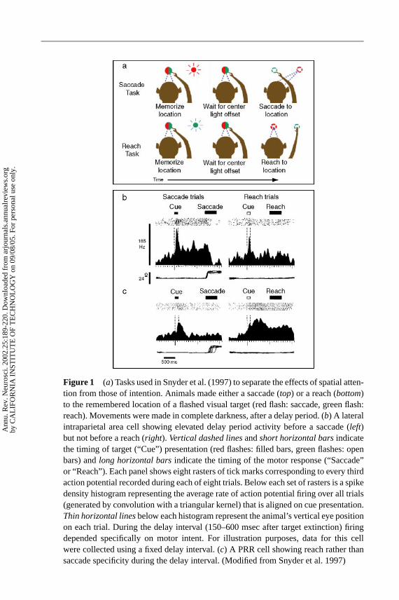
25 May 2002 12:31 AR AR160-06-COLOR.tex AR160-06-COLOR.SGM LaTeX2e(2002/01/18)P1: GDL
Figure 1 (a) Tasks used in Snyder et al. (1997) to separate the effects of spatial atten-tion from those of intention. Animals made either a saccade (top) or a reach (bottom)to the remembered location of a flashed visual target (red flash: saccade, green flash:reach). Movements were made in complete darkness, after a delay period. (b) A lateralintraparietal area cell showing elevated delay period activity before a saccade (left)but not before a reach (right). Vertical dashed linesandshort horizontal barsindicatethe timing of target (“Cue”) presentation (red flashes: filled bars, green flashes: openbars) andlong horizontal barsindicate the timing of the motor response (“Saccade”or “Reach”). Each panel shows eight rasters of tick marks corresponding to every thirdaction potential recorded during each of eight trials. Below each set of rasters is a spikedensity histogram representing the average rate of action potential firing over all trials(generated by convolution with a triangular kernel) that is aligned on cue presentation.Thin horizontal linesbelow each histogram represent the animal’s vertical eye positionon each trial. During the delay interval (150–600 msec after target extinction) firingdepended specifically on motor intent. For illustration purposes, data for this cellwere collected using a fixed delay interval. (c) A PRR cell showing reach rather thansaccade specificity during the delay interval. (Modified from Snyder et al. 1997)
Ann
u. R
ev. N
euro
sci.
2002
.25:
189-
220.
Dow
nloa
ded
from
arj
ourn
als.
annu
alre
view
s.or
gby
CA
LIF
OR
NIA
IN
STIT
UT
E O
F T
EC
HN
OL
OG
Y o
n 09
/08/
05. F
or p
erso
nal u
se o
nly.
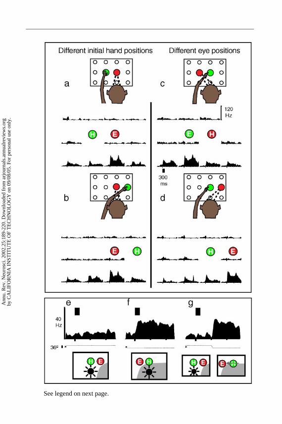
25 May 2002 12:31 AR AR160-06-COLOR.tex AR160-06-COLOR.SGM LaTeX2e(2002/01/18)P1: GDL
See legend on next page.
Ann
u. R
ev. N
euro
sci.
2002
.25:
189-
220.
Dow
nloa
ded
from
arj
ourn
als.
annu
alre
view
s.or
gby
CA
LIF
OR
NIA
IN
STIT
UT
E O
F T
EC
HN
OL
OG
Y o
n 09
/08/
05. F
or p
erso
nal u
se o
nly.
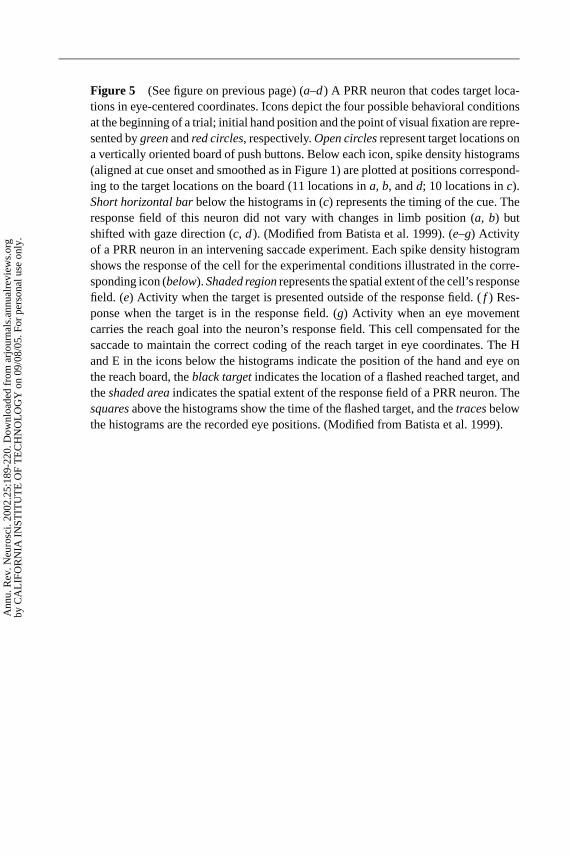
25 May 2002 12:31 AR AR160-06-COLOR.tex AR160-06-COLOR.SGM LaTeX2e(2002/01/18)P1: GDL
Figure 5 (See figure on previous page) (a–d) A PRR neuron that codes target loca-tions in eye-centered coordinates. Icons depict the four possible behavioral conditionsat the beginning of a trial; initial hand position and the point of visual fixation are repre-sented bygreenandred circles, respectively.Open circlesrepresent target locations ona vertically oriented board of push buttons. Below each icon, spike density histograms(aligned at cue onset and smoothed as in Figure 1) are plotted at positions correspond-ing to the target locations on the board (11 locations ina, b, andd; 10 locations inc).Short horizontal barbelow the histograms in (c) represents the timing of the cue. Theresponse field of this neuron did not vary with changes in limb position (a, b) butshifted with gaze direction (c, d). (Modified from Batista et al. 1999). (e–g) Activityof a PRR neuron in an intervening saccade experiment. Each spike density histogramshows the response of the cell for the experimental conditions illustrated in the corre-sponding icon (below).Shaded regionrepresents the spatial extent of the cell’s responsefield. (e) Activity when the target is presented outside of the response field. (f ) Res-ponse when the target is in the response field. (g) Activity when an eye movementcarries the reach goal into the neuron’s response field. This cell compensated for thesaccade to maintain the correct coding of the reach target in eye coordinates. The Hand E in the icons below the histograms indicate the position of the hand and eye onthe reach board, theblack targetindicates the location of a flashed reached target, andtheshaded areaindicates the spatial extent of the response field of a PRR neuron. Thesquaresabove the histograms show the time of the flashed target, and thetracesbelowthe histograms are the recorded eye positions. (Modified from Batista et al. 1999).
Ann
u. R
ev. N
euro
sci.
2002
.25:
189-
220.
Dow
nloa
ded
from
arj
ourn
als.
annu
alre
view
s.or
gby
CA
LIF
OR
NIA
IN
STIT
UT
E O
F T
EC
HN
OL
OG
Y o
n 09
/08/
05. F
or p
erso
nal u
se o
nly.
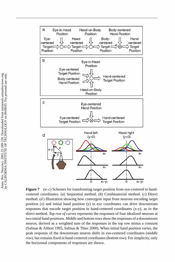
25 May 2002 12:31 AR AR160-06-COLOR.tex AR160-06-COLOR.SGM LaTeX2e(2002/01/18)P1: GDL
Figure 7 (a–c) Schemes for transforming target position from eye-centered to hand-centered coordinates. (a) Sequential method. (b) Combinatorial method. (c) Directmethod. (d ) Illustration showing how convergent input from neurons encoding targetposition (x) and initial hand position (y) in eye coordinates can drive downstreamresponses that encode target position in hand-centered coordinates (x-y), as in thedirect method.Top row of curvesrepresents the responses of four idealized neurons attwo initial hand positions.Middleandbottom rowsshow the responses of a downstreamneuron, derived as a weighted sum of the responses in the top row minus a constant(Salinas & Abbott 1995, Salinas & Thier 2000). When initial hand position varies, thepeak response of the downstream neuron shifts in eye-centered coordinates (middlerow), but remains fixed in hand-centered coordinates (bottom row). For simplicity, onlythe horizontal components of responses are shown.
Ann
u. R
ev. N
euro
sci.
2002
.25:
189-
220.
Dow
nloa
ded
from
arj
ourn
als.
annu
alre
view
s.or
gby
CA
LIF
OR
NIA
IN
STIT
UT
E O
F T
EC
HN
OL
OG
Y o
n 09
/08/
05. F
or p
erso
nal u
se o
nly.
P1: FDS
May 17, 2002 12:24 Annual Reviews AR160-FM
Annual Review of NeuroscienceVolume 25, 2002
CONTENTS
THE HUMAN GENOME PROJECT AND ITS IMPACT ON PSYCHIATRY,W. Maxwell Cowan, Kathy L. Kopnisky, and Steven E. Hyman 1
AUDITORY SYSTEM DEVELOPMENT: PRIMARY AUDITORY NEURONSAND THEIR TARGETS, Edwin W. Rubel and Bernd Fritzsch 51
AMPA RECEPTOR TRAFFICKING AND SYNAPTIC PLASTICITY,Roberto Malinow and Robert C. Malenka 103
MOLECULAR CONTROL OF CORTICAL DENDRITE DEVELOPMENT,Kristin L. Whitford, Paul Dijkhuizen, Franck Polleux, and Anirvan Ghosh 127
FUNCTIONAL MRI OF LANGUAGE: NEW APPROACHES TOUNDERSTANDING THE CORTICAL ORGANIZATION OFSEMANTIC PROCESSING, Susan Bookheimer 151
INTENTIONAL MAPS IN POSTERIOR PARIETAL CORTEX,Richard A. Andersen and Christopher A. Buneo 189
BEYOND PHRENOLOGY: WHAT CAN NEUROIMAGING TELL US ABOUTDISTRIBUTED CIRCUITRY? Karl Friston 221
TRANSCRIPTIONAL CODES AND THE CONTROL OF NEURONALIDENTITY, Ryuichi Shirasaki and Samuel L. Pfaff 251
THE ROLE OF HYPOCRETINS (OREXINS) IN SLEEP REGULATION ANDNARCOLEPSY, Shahrad Taheri, Jamie M. Zeitzer, and Emmanuel Mignot 283
A DECADE OF MOLECULAR STUDIES OF FRAGILE X SYNDROME,William T. O’Donnell and Stephen T. Warren 315
CONTEXTUAL INFLUENCES ON VISUAL PROCESSING, Thomas D. Albrightand Gene R. Stoner 339
LARGE-SCALE SOURCES OF NEURAL STEM CELLS, David I. Gottlieb 381
SCHIZOPHRENIA AS A DISORDER OF NEURODEVELOPMENT,David A. Lewis and Pat Levitt 409
THE CENTRAL AUTONOMIC NERVOUS SYSTEM: CONSCIOUS VISCERALPERCEPTION AND AUTONOMIC PATTERN GENERATION,Clifford B. Saper 433
THE ROLE OF NOTCH IN PROMOTING GLIAL AND NEURAL STEM CELLFATES, Nicholas Gaiano and Gord Fishell 471
v
Ann
u. R
ev. N
euro
sci.
2002
.25:
189-
220.
Dow
nloa
ded
from
arj
ourn
als.
annu
alre
view
s.or
gby
CA
LIF
OR
NIA
IN
STIT
UT
E O
F T
EC
HN
OL
OG
Y o
n 09
/08/
05. F
or p
erso
nal u
se o
nly.
P1: FDS
May 17, 2002 12:24 Annual Reviews AR160-FM
vi CONTENTS
MULTIPLE SCLEROSIS: DEEPER UNDERSTANDING OF ITSPATHOGENESIS REVEALS NEW TARGETS FOR THERAPY,Lawrence Steinman, Roland Martin, Claude Bernard, Paul Conlon,and Jorge R. Oksenberg 491
WIRED FOR REPRODUCTION: ORGANIZATION AND DEVELOPMENT OFSEXUALLY DIMORPHIC CIRCUITS IN THE MAMMALIAN FOREBRAIN,Richard B. Simerly 507
CENTRAL NERVOUS SYSTEM DAMAGE, MONOCYTES ANDMACROPHAGES, AND NEUROLOGICAL DISORDERS IN AIDS,Kenneth C. Williams and William F. Hickey 537
LEARNING AND MEMORY FUNCTIONS OF THE BASAL GANGLIA,Mark G. Packard and Barbara J. Knowlton 563
INDEXESSubject Index 595Cumulative Index of Contributing Authors, Volumes 16–25 603Cumulative Index of Chapter Titles, Volumes 16–25 607
ERRATAAn online log of corrections to Annual Review of Neuroscience chapters(if any, 1997 to the present) may be found at http://neuro.annualreviews.org/
Ann
u. R
ev. N
euro
sci.
2002
.25:
189-
220.
Dow
nloa
ded
from
arj
ourn
als.
annu
alre
view
s.or
gby
CA
LIF
OR
NIA
IN
STIT
UT
E O
F T
EC
HN
OL
OG
Y o
n 09
/08/
05. F
or p
erso
nal u
se o
nly.