Integrative Physiology · Sobirin et al Natural Killer T Cells in Heart Failure 1039...
19
1037 Integrative Physiology M yocardial infarction (MI) leads to the development of heart failure (HF), which is the major cause of death in post-MI patients. The changes in left ventricular (LV) ge- ometry, such as cavity dilatation associated with myocyte hy- pertrophy and interstitial fibrosis, referred to as remodeling, contribute to the development of depressed cardiac function in HF after MI. 1 It has been reported that monocytes and lympho- cytes are infiltrated in noninfarcted area as well as infarcted area of LV after MI. 2,3 Chemokines, monocyte chemoattrac- tant protein-1 (MCP-1), and RANTES (regulated on activation normally T-cell expressed and secreted), are essential factors in the recruitment and activation of monocyte and lymphocyte. These chemokines are also increased in noninfarcted LV after MI and contribute to local inflammation through the release of inflammatory cytokines including tumor necrosis factor-α (TNF-α). 2,4 Targeted deletion of CC chemokine receptor 2 or anti–MCP-1 gene therapy has been shown to attenuate LV re- modeling after MI. 2,5 Thus, chronic tissue inflammation plays an important role in LV remodeling process. Invariant natural killer T (iNKT) cells are innate-like T-lymphocyte population coexpressing NK markers and an αβ T-cell receptor that recognize glycolipid antigens. They can rapidly and robustly produce a mixture of T-helper type 1 (T H 1) and T H 2 cytokines, such as TNF-α, interferon-γ (IFN-γ), interleukin (IL)-10, and IL-4, and also a vast array of chemokines in shaping subsequent adaptive immune re- sponse. 6 Thus, iNKT cells can function as a bridge between the innate and adaptive immune systems, and orchestrate Original received March 25, 2012; revision received August 8, 2012; accepted August 10, 2012. In July 2012, the average time from submission to first decision for all original research papers submitted to Circulation Research was 11.2 days. From the Department of Cardiovascular Medicine, Hokkaido University Graduate School of Medicine, Sapporo Faculty of Medicine, Sapporo, Japan (M.A.S., S.K., M.T., A.F., T.H., T.O., K.H., T.S., P.A., S.T., N.I., H.T.); Diponegoro University, Semarang, Indonesia (M.A.S.); RIKEN Research Center for Allergy and Immunology, Kanagawa, Japan (M.T.); the Department of Immunology, Graduate School of Medicine, Chiba University, Chiba, Japan (T.N.); and the Division of Immunobiology, Kitasato University School of Medicine, Kanagawa, Japan (K.I.). The online-only Data Supplement is available with this article at http://circres.ahajournals.org/lookup/suppl/doi:10.1161/CIRCRESAHA.112. 270132/-/DC1. Correspondence to Shintaro Kinugawa, MD, PhD, Department of Cardiovascular Medicine, Hokkaido University Graduate School of Medicine, Kita-15, Nishi-7, Kita-ku, Sapporo 060–8638, Japan. E-mail [email protected] © 2012 American Heart Association, Inc. Circulation Research is available at http://circres.ahajournals.org DOI: 10.1161/CIRCRESAHA.112.270132 Activation of Natural Killer T Cells Ameliorates Postinfarct Cardiac Remodeling and Failure in Mice Mochamad Ali Sobirin, Shintaro Kinugawa, Masashige Takahashi, Arata Fukushima, Tsuneaki Homma, Taisuke Ono, Kagami Hirabayashi, Tadashi Suga, Putri Azalia, Shingo Takada, Masaru Taniguchi, Toshinori Nakayama, Naoki Ishimori, Kazuya Iwabuchi, Hiroyuki Tsutsui Rationale: Chronic inflammation in the myocardium is involved in the development of left ventricular (LV) remodeling and failure after myocardial infarction (MI). Invariant natural killer T (iNKT) cells have been shown to produce inflammatory cytokines and orchestrate tissue inflammation. However, no previous studies have determined the pathophysiological role of iNKT cells in post-MI LV remodeling. Objective: The purpose of this study was to examine whether the activation of iNKT cells might affect the development of LV remodeling and failure. Methods and Results: After creation of MI, mice received the injection of either α-galactosylceramide (αGC; n=27), the activator of iNKT cells, or phosphate-buffered saline (n=31) 1 and 4 days after surgery, and were followed during 28 days. Survival rate was significantly higher in MI+αGC than MI+PBS (59% versus 32%, P<0.05). LV cavity dilatation and dysfunction were significantly attenuated in MI+αGC, despite comparable infarct size, accompanied by a decrease in myocyte hypertrophy, interstitial fibrosis, and apoptosis. The infiltration of iNKT cells were increased during early phase in noninfarcted LV from MI and αGC further enhanced them. It also enhanced LV interleukin (IL)-10 gene expression at 7 days, which persisted until 28 days. AntienIL-10 receptor antibody abrogated these protective effects of αGC on MI remodeling. The administration of αGC into iNKT cell-deficient Jα18 −/− mice had no such effects, suggesting that αGC was a specific activator of iNKT cells. Conclusions: iNKT cells play a protective role against post-MI LV remodeling and failure through the enhanced expression of cardioprotective cytokines such as IL-10. (Circ Res. 2012; 111:1037-1047.) Key Words: natural killer T cells ◼ myocardial infarction ◼ inflammation ◼ heart failure ◼ cytokines by guest on April 3, 2017 http://circres.ahajournals.org/ Downloaded from by guest on April 3, 2017 http://circres.ahajournals.org/ Downloaded from by guest on April 3, 2017 http://circres.ahajournals.org/ Downloaded from by guest on April 3, 2017 http://circres.ahajournals.org/ Downloaded from by guest on April 3, 2017 http://circres.ahajournals.org/ Downloaded from by guest on April 3, 2017 http://circres.ahajournals.org/ Downloaded from by guest on April 3, 2017 http://circres.ahajournals.org/ Downloaded from by guest on April 3, 2017 http://circres.ahajournals.org/ Downloaded from by guest on April 3, 2017 http://circres.ahajournals.org/ Downloaded from
Transcript of Integrative Physiology · Sobirin et al Natural Killer T Cells in Heart Failure 1039...
1037
Integrative Physiology
Myocardial infarction (MI) leads to the development of heart failure (HF), which is the major cause of death
in post- MI patients. The changes in left ventricular (LV) ge-ometry, such as cavity dilatation associated with myocyte hy-pertrophy and interstitial fibrosis, referred to as remodeling, contribute to the development of depressed cardiac function in HF after MI.1 It has been reported that monocytes and lympho-cytes are infiltrated in noninfarcted area as well as infarcted area of LV after MI.2,3 Chemokines, monocyte chemoattrac-tant protein-1 (MCP-1), and RANTES (regulated on activation normally T- cell expressed and secreted), are essential factors in the recruitment and activation of monocyte and lymphocyte. These chemokines are also increased in noninfarcted LV after MI and contribute to local inflammation through the release
of inflammatory cytokines including tumor necrosis factor-α (TNF-α).2,4 Targeted deletion of CC chemokine receptor 2 or anti–MCP-1 gene therapy has been shown to attenuate LV re-modeling after MI.2,5 Thus, chronic tissue inflammation plays an important role in LV remodeling process.
Invariant natural killer T (iNKT) cells are innate- like T- lymphocyte population coexpressing NK markers and an αβ T- cell receptor that recognize glycolipid antigens. They can rapidly and robustly produce a mixture of T- helper type 1 (T
H1) and T
H2 cytokines, such as TNF-α, interferon-γ
(IFN-γ), interleukin (IL)-10, and IL-4, and also a vast array of chemokines in shaping subsequent adaptive immune re-sponse.6 Thus, iNKT cells can function as a bridge between the innate and adaptive immune systems, and orchestrate
Original received March 25, 2012; revision received August 8, 2012; accepted August 10, 2012. In July 2012, the average time from submission to first decision for all original research papers submitted to Circulation Research was 11.2 days.
From the Department of Cardiovascular Medicine, Hokkaido University Graduate School of Medicine, Sapporo Faculty of Medicine, Sapporo, Japan (M.A.S., S.K., M.T., A.F., T.H., T.O., K.H., T.S., P.A., S.T., N.I., H.T.); Diponegoro University, Semarang, Indonesia (M.A.S.); RIKEN Research Center for Allergy and Immunology, Kanagawa, Japan (M.T.); the Department of Immunology, Graduate School of Medicine, Chiba University, Chiba, Japan (T.N.); and the Division of Immunobiology, Kitasato University School of Medicine, Kanagawa, Japan (K.I.).
The online- only Data Supplement is available with this article at http://circres.ahajournals.org/lookup/suppl/doi:10.1161/CIRCRESAHA.112. 270132/-/DC1.
Correspondence to Shintaro Kinugawa, MD, PhD, Department of Cardiovascular Medicine, Hokkaido University Graduate School of Medicine, Kita-15, Nishi-7, Kita- ku, Sapporo 060–8638, Japan. E- mail [email protected]
© 2012 American Heart Association, Inc.
Circulation Research is available at http://circres.ahajournals.org DOI: 10.1161/CIRCRESAHA.112.270132
Activation of Natural Killer T Cells Ameliorates Postinfarct Cardiac Remodeling and Failure in Mice
Mochamad Ali Sobirin, Shintaro Kinugawa, Masashige Takahashi, Arata Fukushima, Tsuneaki Homma, Taisuke Ono, Kagami Hirabayashi, Tadashi Suga, Putri Azalia, Shingo Takada, Masaru Taniguchi,
Toshinori Nakayama, Naoki Ishimori, Kazuya Iwabuchi, Hiroyuki Tsutsui
Rationale: Chronic inflammation in the myocardium is involved in the development of left ventricular (LV) remodeling and failure after myocardial infarction (MI). Invariant natural killer T (iNKT) cells have been shown to produce inflammatory cytokines and orchestrate tissue inflammation. However, no previous studies have determined the pathophysiological role of iNKT cells in post- MI LV remodeling.
Objective: The purpose of this study was to examine whether the activation of iNKT cells might affect the development of LV remodeling and failure.
Methods and Results: After creation of MI, mice received the injection of either α-galactosylceramide (αGC; n=27), the activator of iNKT cells, or phosphate- buffered saline (n=31) 1 and 4 days after surgery, and were followed during 28 days. Survival rate was significantly higher in MI+αGC than MI+PBS (59% versus 32%, P<0.05). LV cavity dilatation and dysfunction were significantly attenuated in MI+αGC, despite comparable infarct size, accompanied by a decrease in myocyte hypertrophy, interstitial fibrosis, and apoptosis. The infiltration of iNKT cells were increased during early phase in noninfarcted LV from MI and αGC further enhanced them. It also enhanced LV interleukin (IL)-10 gene expression at 7 days, which persisted until 28 days. AntienIL-10 receptor antibody abrogated these protective effects of αGC on MI remodeling. The administration of αGC into iNKT cell- deficient Jα18−/− mice had no such effects, suggesting that αGC was a specific activator of iNKT cells.
Conclusions: iNKT cells play a protective role against post- MI LV remodeling and failure through the enhanced expression of cardioprotective cytokines such as IL-10. (Circ Res. 2012; 111:1037-1047.)
Key Words: natural killer T cells ◼ myocardial infarction ◼ inflammation ◼ heart failure ◼ cytokines
by guest on April 3, 2017
http://circres.ahajournals.org/D
ownloaded from
by guest on A
pril 3, 2017http://circres.ahajournals.org/
Dow
nloaded from
by guest on April 3, 2017
http://circres.ahajournals.org/D
ownloaded from
by guest on A
pril 3, 2017http://circres.ahajournals.org/
Dow
nloaded from
by guest on April 3, 2017
http://circres.ahajournals.org/D
ownloaded from
by guest on A
pril 3, 2017http://circres.ahajournals.org/
Dow
nloaded from
by guest on April 3, 2017
http://circres.ahajournals.org/D
ownloaded from
by guest on A
pril 3, 2017http://circres.ahajournals.org/
Dow
nloaded from
by guest on April 3, 2017
http://circres.ahajournals.org/D
ownloaded from
1038 Circulation Research September 28, 2012
tissue inflammation. Indeed, we have shown that iNKT cells activate vascular wall inflammation in atherogenesis and adi-pose tissue inflammation in obesity- induced glucose intoler-ance.7,8 On the other hand, iNKT cells play a protective role against autoimmune and inflammatory diseases such as type 1 diabetes,9,10 allergic encephalomyelitis,9,11 and rheumatoid arthritis.12 These findings suggest that iNKT cells may have bidirectional effects on tissue inflammation. However, no pre-vious studies have examined the changes of iNKT cells and their pathophysiological role in LV remodeling and failure after MI.
Therefore, the purpose of the present study was to de-termine whether iNKT cells might affect the development of LV remodeling and failure after MI. We demonstrated that the activation of iNKT cells by α-galactosylceramide (αGC), a specific activator for iNKT cells,13 attenuated the development of LV remodeling and failure after MI in mice. The enhanced gene expression of IL-10 might be involved in these beneficial effects of iNKT cells on this disease process.
MethodsAll procedures and animal care were approved by our institutional an-imal research committee and conformed to the animal care guideline for the Care and Use of Laboratory Animals in Hokkaido University Graduate School of Medicine.
Experiment 1: Time- Dependent Changes of iNKT Cell Receptors in Post- MI Hearts
Animal ModelsMI was created in male C57BL/6J mice, 6 to 8 weeks old and 20 to 25 g body weight, by ligating the left coronary artery as de-scribed previously.14 Sham operation without ligating the coronary artery was also performed as control. MI mice were euthanized and the hearts were excised at days 3, 7, 14, and 28 for quantita-tive reverse transcriptase–polymerase chain reaction (qRT-PCR) measurements.
Quantitative RT-PCRQuantitative PCR for Vα14Jα18 (a specific marker of iNKT cells) was performed, as described previously.8
Experiment 2: Effects of iNKT Cell Activation on Post- MI Heart Animal ModelsSham and MI mice were created in male C57BL/6J as described in experiment 1. Each group of mice was randomly divided into 2 groups; either αGC (0.1 μg/g body weight; Funakoshi Company, Ltd, Tokyo, Japan), the activator of iNKT cells, or phosphate- buffered saline (PBS) was administered via intraperitoneal injection 1 and 4 days after surgery. The concentration of αGC was chosen based on the previous study of its efficacy.8 Thus, the experiment was performed in the following 4 groups of mice; sham+PBS (n=10), sham+αGC (n=10), MI+PBS (n=31), and MI+αGC (n=27).
SurvivalThe survival analysis was performed in all 4 groups of mice. During the study period, the cages were inspected daily for dead animals. All dead mice were examined for the presence of MI as well as pleural effusion and cardiac rupture.
Echocardiographic and Hemodynamic MeasurementsEchocardiographic and hemodynamic measurements were per-formed under light anesthesia with tribromoethanol/amylene hydrate (avertin; 2.5% wt/vol, 8 μL/g ip), as described previously.14
Myocardial Histopathology, Infarct Size, Myocardial Apoptosis, and Matrix Metalloproteinase ZymographyMyocyte cross- sectional area, collagen volume fraction, infarct size, myocardial apoptosis, and zymographic matrix metalloproteinase (MMP) levels were determined as described previously.14,15
Isolation of Cardiac Mononuclear Cell and Flow CytometryCardiac mononuclear cells from 3 mice were isolated, pooled, and subjected to flow cytometric analysis as previously described.7,16
Quantitative RT-PCRQuantitative PCR for Vα14Jα18, CD11c (a marker of M1 mac-rophages), arginase-1 (a marker of M2 macrophages), MCP-1, RANTES, IFN-γ, IL-4, IL-6, TNF-α, and IL-10 was performed, as described previously.8
ImmunohistochemistryLV sections were immunostained with antibody against mouse MAC3 (a macrophage marker), mouse CD3 (a T- cell marker), or mouse my-eloperoxidase (a leukocyte marker), followed by counterstaining with hematoxylin.
Plasma Cytokine ConcentrationPlasma IL-10, TNF-α, IFN-γ, IL-6, and IL-4 levels were measured by commercially available ELISA kit (R&D systems, Inc) in all groups.
Experiment 3: Effects of IL-10 Neutralization on α GC- Treated Post- MI HeartsMI mice were divided into the following 3 groups; MI+αGC (n=18), MI+anti–IL-10 receptor antibody (n=12), and MI+αGC+anti–IL-10 receptor antibody (n=19). αGC was administered identi-cally as in experiment 2. Anti–IL-10 receptor antibody (500 μg/mouse, BD Pharmingen, San Diego, CA) was administered via
Non-standard Abbreviations and Acronyms
αGC α-galactosylceramide
HF heart failure
IFN-γ interferon-γ
IL interleukin
iNKT invariant natural killer T
LV left ventricle
MCP-1 monocyte chemoattractant protein-1
MI myocardial infarction
MMP matrix metalloproteinase
NK natural killer
PBS phosphate- buffered saline
qRT- PCR quantitative reverse transcriptase–polymerase chain reaction
RANTES regulated on activation normally T cell expressed and secreted
TH1 T- helper type 1
TH2 T- helper type 2
TNF-α tumor necrosis factor-α
by guest on April 3, 2017
http://circres.ahajournals.org/D
ownloaded from
Sobirin et al Natural Killer T Cells in Heart Failure 1039
intraperitoneal injection 1, 4, and 14 days after surgery. The con-centration of anti–IL-10 receptor antibody was chosen based on the previous study of its efficacy.12 Four weeks after surgery, echo-cardiographic and hemodynamic measurements were performed. Separate groups of mice were used in the MI+αGC group in experiment 2.
Experiment 4: Specificity of αGC for NKT CellsVα14+ NKT cell- deficient Jα18−/− (Jα18 KO) mice were provided by Dr M. Taniguchi (RIKEN, Yokohama, Japan) and back- crossed 10 times to C57BL/6J.17 Sham and MI mice were created in male Jα18 KO mice as described in experiment 1. Each group of mice was treated identically to experiment 2. Thus, the experi-ment was performed in the following 4 groups; KO+sham+PBS, KO+sham+αGC, KO+MI+PBS, and KO+MI+αGC. One week after surgery, all mice (n=9 for each group) were euthanized and used for immunohistochemistry (n=3 for each group) and for qRT- PCR (n=6 for each group). These analyses were performed as described in experiment 2.
Statistical AnalysisData are expressed as mean±SEM. Survival analysis was performed by the Kaplan- Meier method, and between- group differences in sur-vival were tested by the log- rank test. A between- group compari-son of means was performed by 1-way ANOVA, followed by t test. The Bonferroni correction was applied for multiple comparisons of means. P<0.05 was considered statistically significant.
The authors had full access to and take full responsibility for the integrity of the data. All authors had read and agreed to the manu-script as written.
Results
Experiment 1: Time- Dependent Changes of iNKT Cell Receptors in Post- MI HeartsThe quantification of iNKT cells by Vα14/Jα18 gene expres-sion demonstrated that iNKT cell infiltration into the nonin-farcted LV was significantly enhanced at 7 days (1.7±0.2-fold changes from baseline, P<0.05 versus baseline) after MI and returned to baseline at 14 and 28 days after MI (1.0±0.2- and 1.1±0.1-fold changes from baseline, respectively). In the infarcted LV, its gene expression was significantly elevated 7 days and remained elevated 28 days after MI (data not shown).
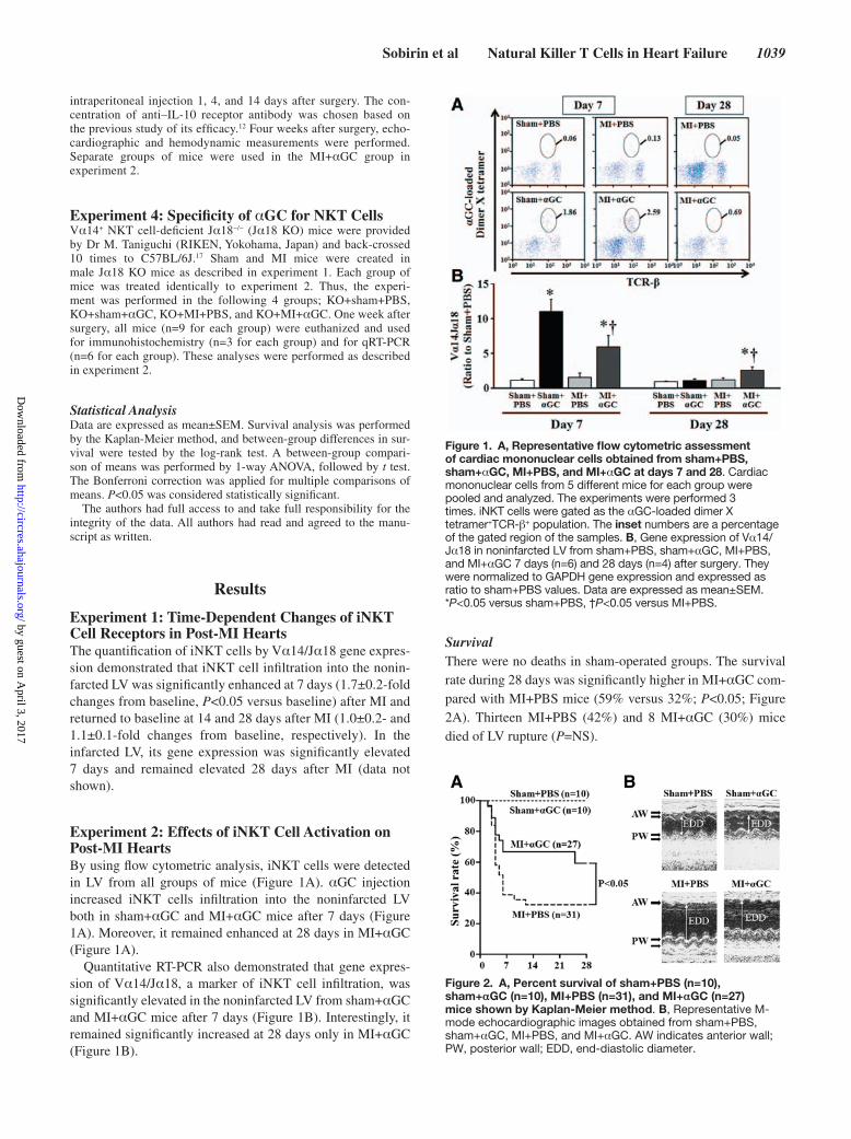
Experiment 2: Effects of iNKT Cell Activation on Post- MI HeartsBy using flow cytometric analysis, iNKT cells were detected in LV from all groups of mice (Figure 1A). αGC injection increased iNKT cells infiltration into the noninfarcted LV both in sham+αGC and MI+αGC mice after 7 days (Figure 1A). Moreover, it remained enhanced at 28 days in MI+αGC (Figure 1A).
Quantitative RT- PCR also demonstrated that gene expres-sion of Vα14/Jα18, a marker of iNKT cell infiltration, was significantly elevated in the noninfarcted LV from sham+αGC and MI+αGC mice after 7 days (Figure 1B). Interestingly, it remained significantly increased at 28 days only in MI+αGC (Figure 1B).
SurvivalThere were no deaths in sham- operated groups. The survival rate during 28 days was significantly higher in MI+αGC com-pared with MI+PBS mice (59% versus 32%; P<0.05; Figure 2A). Thirteen MI+PBS (42%) and 8 MI+αGC (30%) mice died of LV rupture (P=NS).
Figure 1. A, Representative flow cytometric assessment of cardiac mononuclear cells obtained from sham+PBS, sham+αGC, MI+PBS, and MI+αGC at days 7 and 28. Cardiac mononuclear cells from 5 different mice for each group were pooled and analyzed. The experiments were performed 3 times. iNKT cells were gated as the α GC- loaded dimer X tetramer+TCR-β+ population. The inset numbers are a percentage of the gated region of the samples. B, Gene expression of Vα14/Jα18 in non infarcted LV from sham+PBS, sham+αGC, MI+PBS, and MI+αGC 7 days (n=6) and 28 days (n=4) after surgery. They were normalized to GAPDH gene expression and expressed as ratio to sham+PBS values. Data are expressed as mean±SEM. *P<0.05 versus sham+PBS, †P<0.05 versus MI+PBS.
Figure 2. A, Percent survival of sham+PBS (n=10), sham+αGC (n=10), MI+PBS (n=31), and MI+αGC (n=27) mice shown by Kaplan- Meier method. B, Representative M- mode echocardiographic images obtained from sham+PBS, sham+αGC, MI+PBS, and MI+αGC. AW indicates anterior wall; PW, posterior wall; EDD, end- diastolic diameter.
by guest on April 3, 2017
http://circres.ahajournals.org/D
ownloaded from

1040 Circulation Research September 28, 2012
Echocardiography and HemodynamicsThe echocardiographic and hemodynamic data from 4 groups of survived mice at 28 days are shown in Figure 2B and Table 1.
There were no significant differences in either echocardio-graphic or hemodynamic parameters between sham+PBS and sham+αGC mice. LV diameters were significantly greater and LV fractional shortening was significantly lower in MI mice than sham mice. These changes were ameliorated by the treat-ment of MI mice with αGC. There were no significant differ-ences in heart rate or aortic blood pressure among groups. LV end- diastolic pressure (LVEDP) was significantly increased, and LV +dP/dt and LV −dP/dt were significantly decreased in MI compared with sham, which was ameliorated by the treat-ment of MI mice with αGC.
Organ Weights, Infarct Size, and HistologyThere were no significant differences in heart weight/body weight and lung weight/body weight between sham+PBS and sham+αGC mice (Table 1). In agreement with LVEDP, heart weight/body weight and lung weight/body weight were increased in MI mice, and these increases were significantly attenuated in MI+αGC (Table 1).
Infarct size measured by the morphometric analysis was comparable (56±2% versus 55±1%; P=NS) between MI+PBS (n=6) and MI+αGC (n=6) groups (Table 1).
Histomorphometric analysis of noninfarcted LV sections showed that myocyte cross- sectional area was increased in MI+PBS compared with sham mice and was significantly at-tenuated in MI+αGC (Figure 3A). Collagen volume fraction
was also increased in MI+PBS compared with sham mice and was significantly attenuated in MI+αGC (Figure 3A).
There were rare TUNEL- positive nuclei in both sham and sham+αGC mice. The number of TUNEL- positive myocytes in the noninfarcted LV was increased in MI+PBS and was sig-nificantly decreased in MI+αGC (Figure 3B).
Myocardial MMP ActivityRepresentative gelatin zymography of the noninfarcted LV tissue at day 7 from 4 groups of mice was shown in Figure 4A. There were no zymographic MMP-2 and 9 levels in the sham+PBS and sham+αGC. Zymographic MMP-2 level was significantly increased in MI+PBS mice compared with sham mice at day 7. αGC injection significantly decreased this after MI (Figure 4B). Zymographic MMP-9 level was also increased in MI+PBS mice compared with sham mice at day 7, which, however, was not affected by αGC (Figure 4C).
Zymographic MMP-2 level was increased in MI+PBS mice also at day 28, and αGC injection tended to decrease it (3.7±1.1 versus 2.1±0.8 in ratio to sham, P=0.08).
Inflammatory and Cytokine Gene ExpressionImmunohistochemical stainings for MAC3 and CD3 were in-creased in MI+PBS compared with sham+PBS and were fur-ther increased by αGC at day 7 (Figure 5). MPO- positive cells were not detected in the LV tissue from either group of mice (data not shown).
Table 1. Echocardiography, Hemodynamics, and Organ Weights in Experiment 2
Sham+PBS (n=10)
Sham+αGC (n=10)
MI+PBS (n=10)
MI+αGC (n=16)
Echocardiography
Heart rate, bpm 522±10 522±12 531±16 520±13
LVEDD, mm 3.4±0.1 3.4±0.04 5.4±0.1* 5.0±0.1*†
LVESD, mm 2.1±0.03 2.1±0.04 4.5±0.1* 4.1±0.1*†
FS, % 38.2±0.7 38.3±0.6 16.5±0.6* 18.8±0.6*†
AWT, mm 0.63±0.01 0.62±0.01 0.31±0.01* 0.30±0.01*
PWT, mm 0.68±0.02 0.68±0.01 0.97±0.01* 0.96±0.02*
Hemodynamics
Heart rate, min 507±9 499±9 485±23 495±11
Mean AoP, mm Hg 78.1±2 77.7±2 75.0±3 79.3±1
LVEDP, mm Hg 1.7±0.3 2.3±0.1 10.7±1.1* 6.6±0.6*†
LV +dP/dt, mm Hg/s 15 625±623 14 972±398 7352±697* 9386±476*†
LV –dP/dt, mm Hg/s 9983±697 9130±691 5045±482* 5861±286*
Organ weights
Body wt, g 25.1±0.3 24.9±0.2 24.5±0.4 24.8±0.3
Heart wt/body wt, mg/g 4.6±0.1 4.5±0.1 6.8±0.2* 6.1±0.1*†
Lung wt/body wt, mg/g 5.2±0.03 5.2±0.1 7.2±0.7* 5.9±0.2†
Infarct size, % … … 56±2 55±1
LVEDD indicates left ventricular end-diastolic diameter; LVESD, left ventricular end-systolic diameter; FS, fractional shortening; AWT, anterior wall thickness; PWT, posterior wall thickness; AoP, aortic pressure; LVEDP, left ventricular end-diastolic pressure; wt, weight. Data are mean±SEM.
*P<0.05 versus sham+PBS.†P<0.05 versus MI+PBS.
by guest on April 3, 2017
http://circres.ahajournals.org/D
ownloaded from

Sobirin et al Natural Killer T Cells in Heart Failure 1041
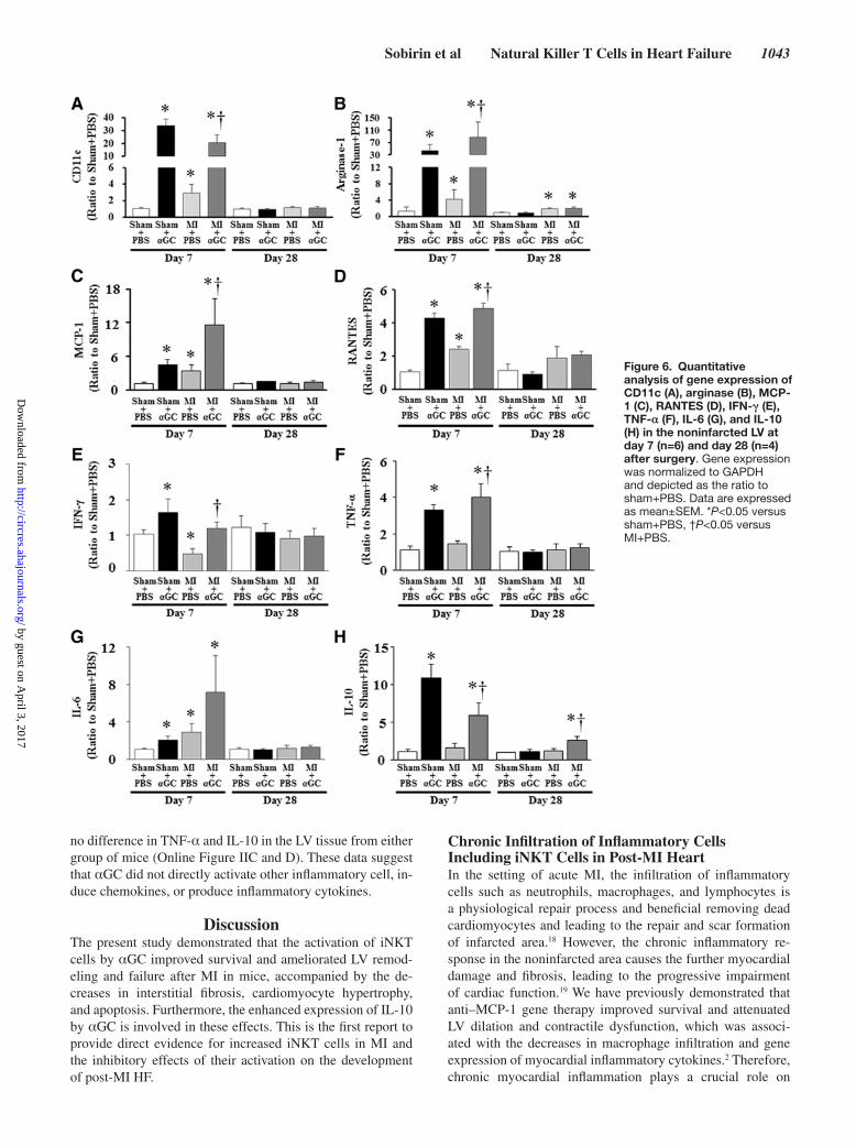
CD11c (a marker of M1 macrophage) and arginase 1 (a marker of M2 macrophage) gene expressions were significa-ntly increased in noninfarcted LV from MI+PBS compared with sham+PBS at day 7 (Figure 6A and 6B). αGC signifi-cantly increased their expressions in both sham and MI ani-mals at day 7. Arginase 1 but not CD11c was increased in noninfarcted LV from MI+PBS and MI+αGC at day 28. There was no significant difference in arginase 1 between
these 2 groups. MCP-1 and RANTES gene expressions were increased in noninfarcted LV from MI+PBS compared with sham+PBS at day 7 (Figure 6C and 6D). αGC significantly increased their expressions in both sham and MI animals at day 7. In contrast, there was no significant difference in their expressions among all groups at day 28.
IFN-γ, TNF-α, IL-6, and IL-10 gene expression levels were significantly increased in sham and MI mice by αGC at
Figure 3. A, Representative high- power photomicrographs of LV cross sections stained with Masson trichrome from sham+PBS (a), sham+αGC (b), MI+PBS (c), and MI+αGC (d) and summary data of myocyte cross- sectional area and collagen volume fraction in 4 groups of mice (n=6). Scale bar, 20 μm. B, Representative photomicrographs TUNEL staining of LV sections from MI+PBS (a) and MI+αGC (b) and summary data for the number of TUNEL- positive cells in the noninfarcted LV (n=6). Scale bar, 20 μm. Data are expressed as mean±SEM. *P<0.05 versus sham+PBS, †P<0.05 versus MI+PBS.
Figure 4. Representative LV zymographic MMP-2 and MMP-9 activities in noninfarcted LV at 7 days after surgery (A) and their densitometric analysis (B and C; n=5 for each). P indicates positive control. Data are expressed as mean±SEM. *P<0.05 versus sham+PBS, †P<0.05 versus MI+PBS.
by guest on April 3, 2017
http://circres.ahajournals.org/D
ownloaded from

1042 Circulation Research September 28, 2012
day 7 (Figure 6E through 6H). IL-10 gene expression alone significantly elevated up to 2.6-fold in the noninfarcted LV from MI+αGC mice at day 28 (Figure 6H). These time- dependent and α GC- mediated changes in IL-10 gene expression (Figure 6H) in the LV were matched with those in NKT cell infiltration (Figure 1B). IL-4 was not detected in either group.
Plasma Cytokine ConcentrationPlasma IL-10 level was similar among sham+PBS, sham+αGC, and MI+PBS groups (9.0±0.5 versus 9.8±2.3 versus 10.6±2.3 pg/mL). However, in parallel to IL-10 gene expression in the LV, it significantly increased up to 2-fold in MI+αGC (21.1±2.3 pg/mL) compared with sham and MI+PBS mice (P<0.05). Plasma IFN-γ level was similar among 4 groups of mice (1.4±0.3 versus 1.7±0.3 versus 0.9±0.2 versus 1.0±0.2 pg/mL, P=NS). Plasma TNF-α, IL-6, and IL-4 levels were not detected in either group.
Experiment 3: Effects of IL-10 Neutralization on α GC- Treated Post- MI Heart SurvivalThe survival rate during 28 days tended to be higher in MI+αGC than in MI+anti–IL-10 receptor antibody and MI+αGC+anti–IL-10 receptor antibody (66.7% versus 44.4% and 42.1%, P=0.4).
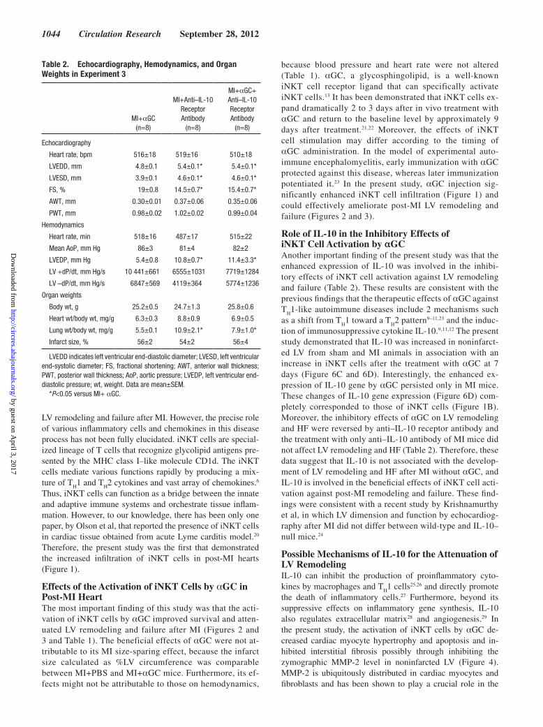
Echocardiography and HemodynamicsThe echocardiographic and hemodynamic data from 3 groups of surviving mice are shown in Table 2. IL-10 receptor
antibody injection significantly increased LV diameters, LVEDP, and decreased LV fractional shortening in α GC- treated MI mice. In contrast, there were no differences in these parameters between MI+anti–IL-10 receptor antibody and MI+αGC+anti–IL-10 receptor antibody. There was no significant difference in heart rate and aortic blood pressure among 3 groups.
Organ Weights and Infarct SizeIn agreement with LVEDP, lung weight/body weight ratio was significantly increased in MI+αGC+anti–IL-10 recep-tor antibody compared with MI+αGC (Table 2). There were also no differences in these parameters between MI+anti–IL-10 receptor antibody and MI+αGC+anti–IL-10 receptor antibody.
Infarct size was comparable (56±2%, 54±2%, and 56±4%; P=NS) among MI+αGC (n=8), MI+anti–IL-10 antibody (n=8), and MI+αGC+anti–IL-10 receptor antibody (n=8) groups.
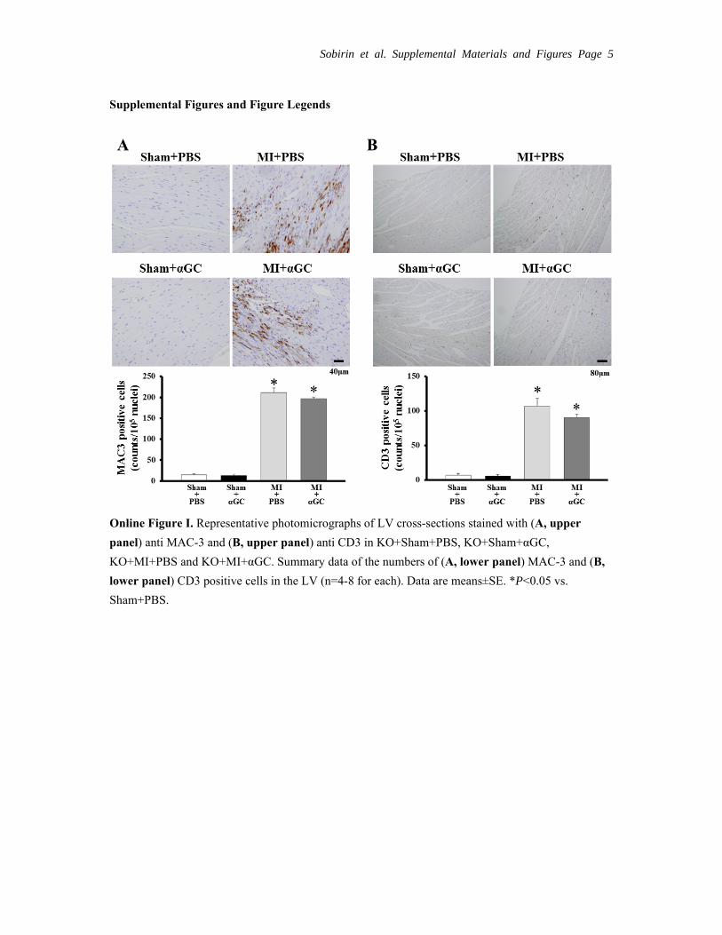
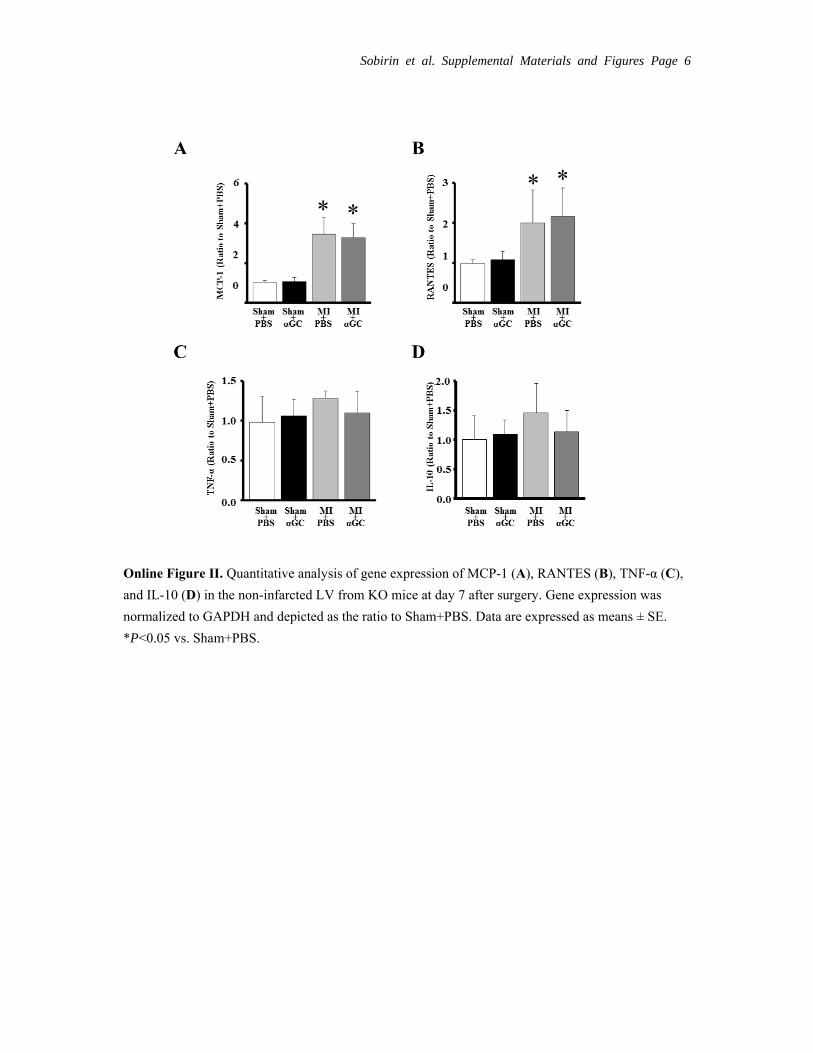
Experiment 4: Specificity of αGC for iNKT CellsImmunohistochemical stainings for MAC3 and CD3 were increased in KO+MI+PBS compared with KO+sham+PBS. In contrast to the results from wild- type (Figure 5), αGC did not alter them (Online Figure I). MPO- positive cells were not detected in the LV tissue from either group of mice (data not shown). MCP-1 and RANTES were increased in KO+MI+PBS compared with KO+sham+PBS and were not affected by αGC (Online Figure IIA and B). There was
Figure 5. Representative photomicrographs of LV cross sections stained with (A, upper panel) anti-MAC3 and (B, upper panel), anti-CD3 in sham+PBS, sham+αGC, MI+PBS, and MI+αGC. Summary data of the numbers of (A, lower panel) MAC3 and (B, lower panel) CD3-positive cells in the LV (n=4–8 for each). Data are mean±SEM. *P<0.05 versus sham+PBS, †P<0.05 versus MI+PBS.
by guest on April 3, 2017
http://circres.ahajournals.org/D
ownloaded from
Sobirin et al Natural Killer T Cells in Heart Failure 1043
no difference in TNF-α and IL-10 in the LV tissue from either group of mice (Online Figure IIC and D). These data suggest that αGC did not directly activate other inflammatory cell, in-duce chemokines, or produce inflammatory cytokines.
DiscussionThe present study demonstrated that the activation of iNKT cells by αGC improved survival and ameliorated LV remod-eling and failure after MI in mice, accompanied by the de-creases in interstitial fibrosis, cardiomyocyte hypertrophy, and apoptosis. Furthermore, the enhanced expression of IL-10 by αGC is involved in these effects. This is the first report to provide direct evidence for increased iNKT cells in MI and the inhibitory effects of their activation on the development of post- MI HF.
Chronic Infiltration of Inflammatory Cells Including iNKT Cells in Post- MI HeartIn the setting of acute MI, the infiltration of inflammatory cells such as neutrophils, macrophages, and lymphocytes is a physiological repair process and beneficial removing dead cardiomyocytes and leading to the repair and scar formation of infarcted area.18 However, the chronic inflammatory re-sponse in the noninfarcted area causes the further myocardial damage and fibrosis, leading to the progressive impairment of cardiac function.19 We have previously demonstrated that anti–MCP-1 gene therapy improved survival and attenuated LV dilation and contractile dysfunction, which was associ-ated with the decreases in macrophage infiltration and gene expression of myocardial inflammatory cytokines.2 Therefore, chronic myocardial inflammation plays a crucial role on
Figure 6. Quantitative analysis of gene expression of CD11c (A), arginase (B), MCP-1 (C), RANTES (D), IFN-γ (E), TNF-α (F), IL-6 (G), and IL-10 (H) in the noninfarcted LV at day 7 (n=6) and day 28 (n=4) after surgery. Gene expression was normalized to GAPDH and depicted as the ratio to sham+PBS. Data are expressed as mean±SEM. *P<0.05 versus sham+PBS, †P<0.05 versus MI+PBS.
by guest on April 3, 2017
http://circres.ahajournals.org/D
ownloaded from
1044 Circulation Research September 28, 2012
LV remodeling and failure after MI. However, the precise role of various inflammatory cells and chemokines in this disease process has not been fully elucidated. iNKT cells are special-ized lineage of T cells that recognize glycolipid antigens pre-sented by the MHC class I–like molecule CD1d. The iNKT cells mediate various functions rapidly by producing a mix-ture of T
H1 and T
H2 cytokines and vast array of chemokines.6
Thus, iNKT cells can function as a bridge between the innate and adaptive immune systems and orchestrate tissue inflam-mation. However, to our knowledge, there has been only one paper, by Olson et al, that reported the presence of iNKT cells in cardiac tissue obtained from acute Lyme carditis model.20 Therefore, the present study was the first that demonstrated the increased infiltration of iNKT cells in post- MI hearts (Figure 1).
Effects of the Activation of iNKT Cells by αGC in Post- MI HeartThe most important finding of this study was that the acti-vation of iNKT cells by αGC improved survival and atten-uated LV remodeling and failure after MI (Figures 2 and 3 and Table 1). The beneficial effects of αGC were not at-tributable to its MI size- sparing effect, because the infarct size calculated as %LV circumference was comparable between MI+PBS and MI+αGC mice. Furthermore, its ef-fects might not be attributable to those on hemodynamics,
because blood pressure and heart rate were not altered (Table 1). αGC, a glycosphingolipid, is a well- known iNKT cell receptor ligand that can specifically activate iNKT cells.13 It has been demonstrated that iNKT cells ex-pand dramatically 2 to 3 days after in vivo treatment with αGC and return to the baseline level by approximately 9 days after treatment.21,22 Moreover, the effects of iNKT cell stimulation may differ according to the timing of αGC administration. In the model of experimental auto-immune encephalomyelitis, early immunization with αGC protected against this disease, whereas later immunization potentiated it.23 In the present study, αGC injection sig-nificantly enhanced iNKT cell infiltration (Figure 1) and could effectively ameliorate post- MI LV remodeling and failure (Figures 2 and 3).
Role of IL-10 in the Inhibitory Effects of iNKT Cell Activation by αGCAnother important finding of the present study was that the enhanced expression of IL-10 was involved in the inhibi-tory effects of iNKT cell activation against LV remodeling and failure (Table 2). These results are consistent with the previous findings that the therapeutic effects of αGC against T
H1-like autoimmune diseases include 2 mechanisms such
as a shift from TH1 toward a T
H2 pattern9–11,23 and the induc-
tion of immunosuppressive cytokine IL-10.9,11,12 The present study demonstrated that IL-10 was increased in noninfarct-ed LV from sham and MI animals in association with an increase in iNKT cells after the treatment with αGC at 7 days (Figure 6C and 6D). Interestingly, the enhanced ex-pression of IL-10 gene by αGC persisted only in MI mice. These changes of IL-10 gene expression (Figure 6D) com-pletely corresponded to those of iNKT cells (Figure 1B). Moreover, the inhibitory effects of αGC on LV remodeling and HF were reversed by anti–IL-10 receptor antibody and the treatment with only anti–IL-10 antibody of MI mice did not affect LV remodeling and HF (Table 2). Therefore, these data suggest that IL-10 is not associated with the develop-ment of LV remodeling and HF after MI without αGC, and IL-10 is involved in the beneficial effects of iNKT cell acti-vation against post- MI remodeling and failure. These find-ings were consistent with a recent study by Krishnamurthy et al, in which LV dimension and function by echocardiog-raphy after MI did not differ between wild- type and IL-10–null mice.24
Possible Mechanisms of IL-10 for the Attenuation of LV RemodelingIL-10 can inhibit the production of proinflammatory cyto-kines by macrophages and T
H1 cells25,26 and directly promote
the death of inflammatory cells.27 Furthermore, beyond its suppressive effects on inflammatory gene synthesis, IL-10 also regulates extracellular matrix28 and angiogenesis.29 In the present study, the activation of iNKT cells by αGC de-creased cardiac myocyte hypertrophy and apoptosis and in-hibited interstitial fibrosis possibly through inhibiting the zymographic MMP-2 level in noninfarcted LV (Figure 4). MMP-2 is ubiquitously distributed in cardiac myocytes and fibroblasts and has been shown to play a crucial role in the
Table 2. Echocardiography, Hemodynamics, and Organ Weights in Experiment 3
MI+αGC (n=8)
MI+Anti–IL-10 Receptor Antibody
(n=8)
MI+αGC+ Anti–IL-10 Receptor Antibody
(n=8)
Echocardiography
Heart rate, bpm 516±18 519±16 510±18
LVEDD, mm 4.8±0.1 5.4±0.1* 5.4±0.1*
LVESD, mm 3.9±0.1 4.6±0.1* 4.6±0.1*
FS, % 19±0.8 14.5±0.7* 15.4±0.7*
AWT, mm 0.30±0.01 0.37±0.06 0.35±0.06
PWT, mm 0.98±0.02 1.02±0.02 0.99±0.04
Hemodynamics
Heart rate, min 518±16 487±17 515±22
Mean AoP, mm Hg 86±3 81±4 82±2
LVEDP, mm Hg 5.4±0.8 10.8±0.7* 11.4±3.3*
LV +dP/dt, mm Hg/s 10 441±661 6555±1031 7719±1284
LV –dP/dt, mm Hg/s 6847±569 4119±364 5774±1236
Organ weights
Body wt, g 25.2±0.5 24.7±1.3 25.8±0.6
Heart wt/body wt, mg/g 6.3±0.3 8.8±0.9 6.9±0.5
Lung wt/body wt, mg/g 5.5±0.1 10.9±2.1* 7.9±1.0*
Infarct size, % 56±2 54±2 56±4
LVEDD indicates left ventricular end-diastolic diameter; LVESD, left ventricular end-systolic diameter; FS, fractional shortening; AWT, anterior wall thickness; PWT, posterior wall thickness; AoP, aortic pressure; LVEDP, left ventricular end-diastolic pressure; wt, weight. Data are mean±SEM.
*P<0.05 versus MI+ αGC. by guest on April 3, 2017
http://circres.ahajournals.org/D
ownloaded from
Sobirin et al Natural Killer T Cells in Heart Failure 1045
development of cardiac remodeling after MI.30 Theoretically, an increase in MMP activity would result in a decrease in the MMP substrate, collagens, whereas an inhibition of MMP would result in an increase in collagens. However, our previ-ous study showed that the selective disruption of the MMP-2 gene attenuated interstitial fibrosis after MI.30 Therefore, the decrease in zymographic MMP-2 level by αGC might be in-volved in the attenuation of interstitial fibrosis in our model. On the other hand, MMP-9 is mainly expressed in infiltrating inflammatory cells such as neutrophils and T lymphocytes. A previous report showed that subcutaneous injection of re-combinant IL-10 suppressed inflammation and attenuated LV remodeling after MI in mice by inhibiting fibrosis via sup-pression of HuR/MMP-9 and by enhancing capillary density through the activation of STAT3.31 Moreover, the previous study by Burchfield et al showed that IL-10 from transplanted bone marrow mononuclear cells contributed to cardiac protec-tion after MI in association with a decrease in T lymphocyte accumulation, reactive hypertrophy, and myocardial collagen deposition.32 However, in the present study, zymographic MMP-9 level was not affected by αGC, which was consistent with the infiltration of lymphocyte observed by immunohisto-chemical staining for CD3 (Figure 5). We also measured the protein levels of HuR/MMP-9 or STAT3 in the noninfarcted LV. However, these protein levels were not affected by αGC (data not shown).
Role of Other Inflammatory Cells and CytokinesIn agreement with the increase in macrophage infiltration by αGC, MCP-1 gene expression was increased. αGC increased not only M1 macrophages but also M2 macrophages, which tune inflammatory responses and promote tissue repair.33 Therefore, the increase in M2 macrophage might neutralize the effect of the increased M1 macrophage and MCP-1. The present study also showed that TNF-α was increased in non-infarcted LV from MI+αGC (Figure 6). TNF-α is a proinflam-matory cytokine considered to be cardiotoxic and induce LV dysfunction.34 However, in contrast, TNF-α has also protec-tive effects during the maladaptive transition to HF.35 Indeed, the treatment of patients with HF with either soluble TNF receptor (RENEWAL) or an anti- TNF antibody (ATTACH) could not show clinical benefits.36,37 Therefore, the increase in TNF-α by αGC would not necessarily lead to the aggravation of LV remodeling.
LimitationsThere are several limitations to be acknowledged in the present study. First, we could not directly demonstrate the location of iNKT cells by the immunohistochemical analysis using biotinylated CD1d dimer (BD Bioscience) with loading of αGC according to the previous report by Kamijyuku et al.38 We tried the double immunohistochemi-cal staining, using antibodies for anti–Armenian hamster TCR-β-PE (BD Bioscience) and anti- mouse NK 1.1-APC (BD Bioscience) according to the newly published paper.39 Furthermore, we also performed in situ hybridization us-ing digoxigenin- labeled DNA probes for mouse Vα14Jα18. Unfortunately, however, we could not detect iNKT cells in
situ in the heart. Even though we defined iNKT cells within the heart by using the gene expression as well as the flow cytometric analysis, further studies are needed to overcome some technical difficulties of in situ detection and clarify this important issue. Second, the underlying mechanisms re-sponsible for the activation of iNKT cells after MI remain to be established. To date, the endogenous ligand for iNKT cells has not been known. Based on our results using αGC, a glycosphingolipid, sphingolipid ceramide may be a crucial intermediate, since ceramide has been shown to be synthe-sized by long- chain fatty acids and actually increased in the heart after coronary microembolization.40 Third, the source of IL-10 production after the stimulation of αGC remains to be determined. IL-10 has been shown to be produced by iNKT cells themselves on exogenous stimulation.41 In addi-tion, IL-10 can be expressed and secreted from macrophages activated by iNKT cells.42,43 In the present study, the activa-tion of iNKT cells by αGC injection increased the infiltra-tion of macrophage in sham and MI mice at 7 days; however, there was no difference in it between MI+PBS and MI+αGC at 28 days (Figure 6). Therefore, the main source of IL-10 production at later phase of αGC injection would be the cells other than macrophages.
In conclusion, iNKT cells have a protective effect on LV remodeling and failure after MI via enhanced IL-10 expres-sion. Therefore, therapies designed to activate iNKT cells may be beneficial against the development of post- MI heart failure.
AcknowledgmentsWe thank Kaoruko Kawai, Akiko Aita, and Miwako Fujii for excel-lent technical assistance.
Sources of FundingThis study was supported by grants from the Ministry of Education, Science, and Culture (17390223, 20590854, 20117004, 21390236) and Hokkaido Heart Association Grant for Research.
DisclosuresNone.
References 1. Pfeffer MA, Braunwald E. Ventricular remodeling after myocardial in-
farction: experimental observations and clinical implications. Circulation. 1990;81:1161–1172.
2. Hayashidani S, Tsutsui H, Shiomi T, Ikeuchi M, Matsusaka H, Suematsu N, Wen J, Egashira K, Takeshita A. Anti- monocyte chemoattractant protein-1 gene therapy attenuates left ventricular remodeling and fail-ure after experimental myocardial infarction. Circulation. 2003;108: 2134–2140.
3. Varda- Bloom N, Leor J, Ohad DG, Hasin Y, Amar M, Fixler R, Battler A, Eldar M, Hasin D. Cytotoxic T lymphocytes are activated following myo-cardial infarction and can recognize and kill healthy myocytes in vitro. J Mol Cell Cardiol. 2000;32:2141–2149.
4. Shiomi T, Tsutsui H, Hayashidani S, Suematsu N, Ikeuchi M, Wen J, Ishibashi M, Kubota T, Egashira K, Takeshita A. Pioglitazone, a peroxi-some proliferator- activated receptor- gamma agonist, attenuates left ven-tricular remodeling and failure after experimental myocardial infarction. Circulation. 2002;106:3126–3132.
5. Kaikita K, Hayasaki T, Okuma T, Kuziel WA, Ogawa H, Takeya M. Targeted deletion of CC chemokine receptor 2 attenuates left ventricu-lar remodeling after experimental myocardial infarction. Am J Pathol. 2004;165:439–447.
by guest on April 3, 2017
http://circres.ahajournals.org/D
ownloaded from

1046 Circulation Research September 28, 2012
6. Matsuda JL, Mallevaey T, Scott- Browne J, Gapin L. CD1 d- restricted iNKT cells, the ‘ Swiss- Army knife’ of the immune system. Curr Opin Immunol. 2008;20:358–368.
7. Nakai Y, Iwabuchi K, Fujii S, et al. Natural killer T cells accelerate athero-genesis in mice. Blood. 2004;104:2051–2059.
8. Ohmura K, Ishimori N, Ohmura Y, Tokuhara S, Nozawa A, Horii S, Andoh Y, Fujii S, Iwabuchi K, Onoe K, Tsutsui H. Natural killer T cells are involved in adipose tissues inflammation and glucose intolerance in diet- induced obese mice. Arterioscler Thromb Vasc Biol. 2010;30:193–199.
9. Hong S, Wilson MT, Serizawa I, Wu L, Singh N, Naidenko OV, Miura T, Haba T, Scherer DC, Wei J, Kronenberg M, Koezuka Y, Van Kaer L. The natural killer T- cell ligand alpha- galactosylceramide prevents autoimmune diabetes in non- obese diabetic mice. Nat Med. 2001;7:1052–1056.
10. Sharif S, Arreaza GA, Zucker P, et al. Activation of natural killer T cells by alpha- galactosylceramide treatment prevents the onset and recurrence of autoimmune Type 1 diabetes. Nat Med. 2001;7:1057–1062.
11. Furlan R, Bergami A, Cantarella D, Brambilla E, Taniguchi M, Dellabona P, Casorati G, Martino G. Activation of invariant NKT cells by alphaGalCer ad-ministration protects mice from MOG35–55-induced EAE: critical roles for administration route and IFN- gamma. Eur J Immunol. 2003;33:1830–1838.
12. Miellot A, Zhu R, Diem S, Boissier MC, Herbelin A, Bessis N. Activation of invariant NK T cells protects against experimental rheumatoid arthritis by an IL-10-dependent pathway. Eur J Immunol. 2005;35:3704–3713.
13. Van Kaer L. Alpha- galactosylceramide therapy for autoimmune diseases: prospects and obstacles. Nat Rev Immunol. 2005;5:31–42.
14. Kinugawa S, Tsutsui H, Hayashidani S, Ide T, Suematsu N, Satoh S, Utsumi H, Takeshita A. Treatment with dimethylthiourea prevents left ventricular remodeling and failure after experimental myocardial infarc-tion in mice: role of oxidative stress. Circ Res. 2000;87:392–398.
15. Namba T, Tsutsui H, Tagawa H, Takahashi M, Saito K, Kozai T, Usui M, Imanaka- Yoshida K, Imaizumi T, Takeshita A. Regulation of fibrillar collagen gene expression and protein accumulation in volume- overloaded cardiac hypertrophy. Circulation. 1997;95:2448–2454.
16. Leuschner F, Panizzi P, Chico- Calero I, Lee WW, Ueno T, Cortez- Retamozo V, Waterman P, Gorbatov R, Marinelli B, Iwamoto Y, Chudnovskiy A, Figueiredo JL, Sosnovik DE, Pittet MJ, Swirski FK, Weissleder R, Nahrendorf M. Angiotensin- converting enzyme inhibition prevents the release of monocytes from their splenic reservoir in mice with myocardial infarction. Circ Res. 2010;107:1364–1373.
17. Kawano T, Cui J, Koezuka Y, Toura I, Kaneko Y, Motoki K, Ueno H, Nakagawa R, Sato H, Kondo E, Koseki H, Taniguchi M. CD1 d- restricted and TCR- mediated activation of valpha14 NKT cells by glycosylcerami-des. Science. 1997;278:1626–1629.
18. Blankesteijn WM, Creemers E, Lutgens E, Cleutjens JP, Daemen MJ, Smits JF. Dynamics of cardiac wound healing following myocardial in-farction: observations in genetically altered mice. Acta Physiol Scand. 2001;173:75–82.
19. Frangogiannis NG, Smith CW, Entman ML. The inflammatory response in myocardial infarction. Cardiovasc Res. 2002;53:31–47.
20. Olson CM Jr, Bates TC, Izadi H, Radolf JD, Huber SA, Boyson JE, Anguita J. Local production of IFN- gamma by invariant NKT cells modu-lates acute Lyme carditis. J Immunol. 2009;182:3728–3734.
21. Crowe NY, Uldrich AP, Kyparissoudis K, Hammond KJ, Hayakawa Y, Sidobre S, Keating R, Kronenberg M, Smyth MJ, Godfrey DI. Glycolipid antigen drives rapid expansion and sustained cytokine production by NK T cells. J Immunol. 2003;171:4020–4027.
22. Wilson MT, Johansson C, Olivares- Villagomez D, Singh AK, Stanic AK, Wang CR, Joyce S, Wick MJ, Van Kaer L. The response of natu-ral killer T cells to glycolipid antigens is characterized by surface re-ceptor down- modulation and expansion. Proc Natl Acad Sci U S A. 2003;100:10913–10918.
23. Jahng AW, Maricic I, Pedersen B, Burdin N, Naidenko O, Kronenberg M, Koezuka Y, Kumar V. Activation of natural killer T cells potentiates or prevents experimental autoimmune encephalomyelitis. J Exp Med. 2001;194:1789–1799.
24. Krishnamurthy P, Lambers E, Verma S, Thorne T, Qin G, Losordo DW, Kishore R. Myocardial knockdown of mRNA- stabilizing protein HuR at-tenuates post- MI inflammatory response and left ventricular dysfunction in IL-10-null mice. FASEB J.24:2484–2494.
25. Fiorentino DF, Zlotnik A, Vieira P, Mosmann TR, Howard M, Moore KW, O’Garra A. IL-10 acts on the antigen- presenting cell to inhibit cytokine production by Th1 cells. J Immunol. 1991;146:3444–3451.
26. Frangogiannis NG, Mendoza LH, Lindsey ML, Ballantyne CM, Michael LH, Smith CW, Entman ML. IL-10 is induced in the reper-fused myocardium and may modulate the reaction to injury. J Immunol. 2000;165:2798–2808.
27. Wang P, Wu P, Siegel MI, Egan RW, Billah MM. Interleukin (IL)-10 inhib-its nuclear factor kappa B (NF kappa B) activation in human monocytes. IL-10 and IL-4 suppress cytokine synthesis by different mechanisms. J Biol Chem. 1995;270:9558–9563.
28. Lacraz S, Nicod LP, Chicheportiche R, Welgus HG, Dayer JM. IL-10 inhibits metalloproteinase and stimulates TIMP-1 production in human mononuclear phagocytes. J Clin Invest. 1995;96:2304–2310.
29. Silvestre JS, Mallat Z, Duriez M, Tamarat R, Bureau MF, Scherman D, Duverger N, Branellec D, Tedgui A, Levy BI. Antiangiogenic effect of interleukin-10 in ischemia- induced angiogenesis in mice hindlimb. Circ Res. 2000;87:448–452.
30. Hayashidani S, Tsutsui H, Ikeuchi M, Shiomi T, Matsusaka H, Kubota T, Imanaka- Yoshida K, Itoh T, Takeshita A. Targeted deletion of MMP-2 attenuates early LV rupture and late remodeling after experimental myocardial infarction. Am J Physiol Heart Circ Physiol. 2003;285: H1229–H1235.
31. Krishnamurthy P, Rajasingh J, Lambers E, Qin G, Losordo DW, Kishore R. IL-10 inhibits inflammation and attenuates left ventricular remodeling after myocardial infarction via activation of STAT3 and suppression of HuR. Circ Res. 2009;104:e9–e18.
32. Burchfield JS, Iwasaki M, Koyanagi M, Urbich C, Rosenthal N, Zeiher AM, Dimmeler S. Interleukin-10 from transplanted bone marrow mono-nuclear cells contributes to cardiac protection after myocardial infarction. Circ Res. 2008;103:203–211.
33. Mantovani A, Sica A, Locati M. Macrophage polarization comes of age. Immunity. 2005;23:344–346.
34. Kubota T, McTiernan CF, Frye CS, Slawson SE, Lemster BH, Koretsky AP, Demetris AJ, Feldman AM. Dilated cardiomyopathy in transgenic mice with cardiac- specific overexpression of tumor necrosis factor- alpha. Circ Res. 1997;81:627–635.
35. Wang X, Oka T, Chow FL, Cooper SB, Odenbach J, Lopaschuk GD, Kassiri Z, Fernandez- Patron C. Tumor necrosis factor- alpha- converting enzyme is a key regulator of agonist- induced cardiac hypertrophy and fi-brosis. Hypertension. 2009;54:575–582.
36. Mann DL, McMurray JJ, Packer M, et al. Targeted anticytokine thera-py in patients with chronic heart failure: results of the Randomized Etanercept Worldwide Evaluation (RENEWAL). Circulation. 2004;109: 1594–1602.
37. Chung ES, Packer M, Lo KH, Fasanmade AA, Willerson JT. Randomized, double- blind, placebo- controlled, pilot trial of infliximab, a chime-ric monoclonal antibody to tumor necrosis factor- alpha, in patients with moderate- to- severe heart failure: results of the anti- TNF Therapy Against Congestive Heart Failure (ATTACH) trial. Circulation. 2003;107: 3133–3140.
38. Kamijuku H, Nagata Y, Jiang X, et al. Mechanism of NKT cell activa-tion by intranasal coadministration of alpha- galactosylceramide, which can induce cross- protection against influenza viruses. Mucosal Immunol. 2008;1:208–218.
39. Barral P, Sanchez- Nino MD, van Rooijen N, Cerundolo V, Batista FD. The location of splenic NKT cells favours their rapid activation by blood- borne antigen. EMBO J. 2012;31:2378–2390.
40. Thielmann M, Dorge H, Martin C, Belosjorow S, Schwanke U, van De Sand A, Konietzka I, Buchert A, Kruger A, Schulz R, Heusch G. Myocardial dysfunction with coronary microembolization: signal trans-duction through a sequence of nitric oxide, tumor necrosis factor- alpha, and sphingosine. Circ Res. 2002;90:807–813.
41. Sonoda KH, Faunce DE, Taniguchi M, Exley M, Balk S, Stein- Streilein J. NK T cell- derived IL-10 is essential for the differentiation of antigen- specific T regulatory cells in systemic tolerance. J Immunol. 2001;166:42–50.
by guest on April 3, 2017
http://circres.ahajournals.org/D
ownloaded from
Sobirin et al Natural Killer T Cells in Heart Failure 1047
What Is Known?
• Chronic tissue inflammation plays an important role in the develop-ment of left ventricular (LV) dysfunction and LV remodeling.
• Invariant natural killer T (iNKT) cells are a specialized lineage of T cells with NK marker. These cells produce a mixture of TH1 and TH2 cytokines and a vast array of chemokines to orchestrate tissue inflammation.
• iNKT cells play a protective role in experimental autoimmune and inflammatory diseases.
What New Information Does This Article Contribute?
• iNKT cells could be detected in normal heart, and their infiltration was increased in noninfarcted LV after myocardial infarction (MI).
• The activation of iNKT cells by α-galactosylceramide (αGC) improved survival and ameliorated LV remodeling and failure after MI in mice, accompanied by decreases in interstitial fibrosis, cardiomyocyte hypertrophy, and apoptosis.
• An increase in the expression of interleukin (IL)-10 by αGC was involved in the favorable effects for LV remodeling after MI.
iNKT cells regulate tissue inflammation by producing a mixture of TH1 and TH2 cytokines. Although chronic tissue inflammation is involved in the development of LV remodeling and failure, the pathophysiological role of iNKT cells in these processes have not been elucidated. Our study shows that infiltration of iNKT cells was increased in noninfarcted LV and their activation by αGC improved survival and ameliorated LV remodeling and fail-ure after MI via enhanced expression of IL-10. These findings indicate a previously undescribed protective effect of iNKT cells on LV remodeling and failure after MI. Given that iNKT cells can bridge innate and adaptive immune systems, they could act as an upstream regulator of cytokine networks in the heart. Therapies designed to regulate iNKT cells and to modulate cytokine network may be beneficial in ameliorating LV remodeling and failure.
Novelty and Significance
42. Platzer C, Docke W, Volk H, Prosch S. Catecholamines trigger IL-10 release in acute systemic stress reaction by direct stimulation of its promoter/enhancer activity in monocytic cells. J Neuroimmunol. 2000;105:31–38.
43. Troidl C, Mollmann H, Nef H, Masseli F, Voss S, Szardien S, Willmer M, Rolf A, Rixe J, Troidl K, Kostin S, Hamm C, Elsasser A. Classically and alternatively activated macrophages contribute to tissue remodelling after myocardial infarction. J Cell Mol Med. 2009;13:3485–3496.
by guest on April 3, 2017
http://circres.ahajournals.org/D
ownloaded from
TsutsuiMasaru Taniguchi, Toshinori Nakayama, Naoki Ishimori, Kazuya Iwabuchi and HiroyukiHomma, Taisuke Ono, Kagami Hirabayashi, Tadashi Suga, Putri Azalia, Shingo Takada,
Mochamad Ali Sobirin, Shintaro Kinugawa, Masashige Takahashi, Arata Fukushima, TsuneakiFailure in Mice
Activation of Natural Killer T Cells Ameliorates Postinfarct Cardiac Remodeling and
Print ISSN: 0009-7330. Online ISSN: 1524-4571 Copyright © 2012 American Heart Association, Inc. All rights reserved.is published by the American Heart Association, 7272 Greenville Avenue, Dallas, TX 75231Circulation Research
doi: 10.1161/CIRCRESAHA.112.2701322012;111:1037-1047; originally published online August 10, 2012;Circ Res.
http://circres.ahajournals.org/content/111/8/1037World Wide Web at:
The online version of this article, along with updated information and services, is located on the
http://circres.ahajournals.org/content/suppl/2012/08/10/CIRCRESAHA.112.270132.DC1Data Supplement (unedited) at:
http://circres.ahajournals.org//subscriptions/
is online at: Circulation Research Information about subscribing to Subscriptions:
http://www.lww.com/reprints Information about reprints can be found online at: Reprints:
document. Permissions and Rights Question and Answer about this process is available in the
located, click Request Permissions in the middle column of the Web page under Services. Further informationEditorial Office. Once the online version of the published article for which permission is being requested is
can be obtained via RightsLink, a service of the Copyright Clearance Center, not theCirculation Researchin Requests for permissions to reproduce figures, tables, or portions of articles originally publishedPermissions:
by guest on April 3, 2017
http://circres.ahajournals.org/D
ownloaded from
Sobirin et al. Supplemental Materials and Figures Page 1
Supplemental Material
Detailed Methods An expanded Methods section is available in the online Data Supplement at http://circres.ahajournals.org.
All procedures and animal care were approved by our institutional animal research committee and
conformed to the animal care guideline for the Care and Use of Laboratory Animals in Hokkaido
University Graduate School of Medicine.
Experiment 1: Time-dependent Changes of iNKT Cell Receptors in Post-MI Hearts
Animal Models
MI was created in male C57BL/6J mice, 6-8 weeks old and 20 to 25 g body weight, by
ligating the left coronary artery as described previously.1 Sham operation without ligating the coronary
artery was also performed as control. MI mice were sacrificed and the hearts were excised at day 3, 7,
14 and 28 for quantitative reverse transcriptase (qRT)-PCR measurements.
Quantitative Reverse Transcriptase PCR
Total RNA was extracted from LV in sham mice and non-infarcted and infarcted LV from
MI mice by using QuickGene-810 (FujiFilm, Tokyo, Japan) according to the manufacturer’s
instructions. cDNA was synthesized with the high capacity cDNA reverse transcription kit (Applied
Biosystems, Foster City, CA). TaqMan quantitative PCR was performed with the 7300 real-time PCR
system (Applied Biosystems) to amplify samples for Vα14Jα18 (a specific marker of iNKT cells).2
This transcript was normalized to GAPDH. The primer was purchased from Applied Biosystems.
Experiment 2: Effects of iNKT Cell Activation on Post-MI Hearts
Animal Models
Sham and MI mice were created in male C57BL/6J as described in Experiment 1. Each
group of mice was randomly divided into 2 groups; either α-galactosylceramide (αGC; 0.1g/g body
weight; Funakoshi Company, Ltd., Tokyo, Japan), the activator of iNKT cells, or phosphate-buffered
saline (PBS) was administered via intraperitoneal injection 1 and 4 days after surgery. The
concentration of αGC was chosen based on the previous study of its efficacy.2 Thus, the experiment
was performed in the following 4 groups of mice; sham+PBS (n=10), sham+αGC (n=10), MI+PBS
(n=31), and MI+αGC (n=27).
Four weeks after surgery, echocardiographic studies and the hemodynamics measurement
were performed. After collecting blood samples, mice were sacrificed and organ weight was measured.
These measurements were performed in all survived mice (n=10 for sham+PBS, n=10 for sham+αGC,
n=10 for MI+PBS, and n=16 for MI+αGC). The mice were further divided into 2 groups; for the
histological analysis, including infarct size, myocyte cross-sectional area, collagen volume fraction,
TUNEL staining (n=6 for each group), and for the quantitative reverse transcriptase PCR (n=4 for
Sobirin et al. Supplemental Materials and Figures Page 2
each group). Additional mice were also created for MMP zymography (n=5 for each group) and for
flow cytometry analysis (n=9 for each group).
A separate group of additional mice treated identically was created. One week after surgery,
all mice (n=15 for each group) were sacrificed. These mice were used for immunohistochemistry (n=3
for each group), for the quantitative reverse transcriptase PCR (n=6 for each group), and for flow
cytometry (n=9 for each group).
Survival
The survival analysis was performed in all 4 groups of mice. During the study period, the
cages were inspected daily for deceased animals. All deceased mice were examined for the presence of
MI as well as pleural effusion and cardiac rupture.
Echocardiographic and Hemodynamic Measurements
Echocardiographic and hemodynamic measurements were performed under light anesthesia
with tribromoethanol/amylene hydrate (avertin; 2.5% wt/vol, 8 L/g ip) with known short duration of
action and modest cardiodepressive effects. A two-dimensional parasternal short-axis view was
obtained at the levels of the papillary muscles. In general, the best views obtained with the transducer
lightly applied to the mid upper left anterior chest wall. The transducer was then gently moved
cephalad or caudad and angulated until desirable images were obtained. After it had been ensured that
the imaging was on the axis, two-dimensional targeted M-mode tracings were recorded at a paper
speed of 50mm/s. A 1.4-Fr micromanometer-tipped catheter (Millar Instruments, Houston, Texas) was
inserted into the right carotid artery and then advanced into the left ventricle (LV) to measure LV
pressures.
Myocardial Histopathology and Infarct Size
After mice were sacrificed, the heart was excised and dissected into right ventricle and LV
including septum. LV was cut into three transverse sections; apex, middle ring, and base. From the
middle ring, 5-m sections were cut and stained with Masson’s trichrome. Myocyte cross-sectional
area and collagen volume fraction were determined by quantitative morphometry of tissue sections
from the mid-LV as described previously.3
Infarct length was measured along the endocardial and epicardial surfaces in each of the
cardiac sections, and the values from all specimens were summed. Infarct size (as a percentage) was
calculated as total infarct circumference divided by total cardiac circumference.1
Myocardial Apoptosis
To detect apoptosis, tissue sections from the mid-LV were stained with the terminal
deoxynucleotidyl transferase-mediated dUTP nick end-labeling (TUNEL) staining (TaKaRa Shuzo Co.
Ltd., Ohtsu, Japan). The number of TUNEL positive cardiac myocyte nuclei was counted, and the data
were normalized per 105 total nuclei identified by hematoxylin-positive staining in the same sections.
The proportion of apoptotic cells was counted in the non-infarcted LV.
Sobirin et al. Supplemental Materials and Figures Page 3
MMP Zymography
Zymographic MMP 2 and 9 levels in LV non-infarcted tissue was determined using gelatin
zymography kit (Primary Cell Co., Ltd, Sapporo, Japan). The zymograms were digitized, and the
size-fractionated bands, which indicated proteolytic levels, were measured by the integrated optical
density in a rectangular region of interest.1
Isolation of Cardiac Mononuclear Cell and Flow Cytometry
LV tissue was harvested, minced with a fine scissors, placed in 10 ml RPMI-1640 with 5%
FBS, 1 mg/ml collagenase type IV and 100 U/ml DNase I, and shaken at 37 ºC for 45 min. Tissue was
then triturated through nylon mesh and centrifuged (1400 rpm, 5min, 4 ºC). Red blood cells were lysed
with Tris-NH4Cl solution. Cardiac mononuclear cells were isolated by density-gradient centrifugation
with 33% PercollTM, as previously described.4 Cardiac mononuclear cells from 3 mice were pooled,
and subjected to flow cytometric analysis. All reagents were purchased from Sigma-Aldrich (St Louis,
MO). Cardiac cell numbers were determined with Trypan blue (Wako Pure Chemical Industries, Ltd.,
Osaka, Japan).
The cells were incubated with 2.4G2 monoclonal antibody (mAb) to block non-specific
binding of primary mAb and then reacted with Dimer X (CD1d:Ig recombinant fusion protein; BD
Biosciences Pharmingen, San Diego, CA) loaded with αGC, followed by detection with phycoerythrin
(PE)-conjugated anti-mouse IgG1 mAb (BD) according to the manufacturer’s protocol.5 After washing,
cells were stained with a combination of fluorescein isothiocyanate (FITC)-anti-TCRβ and
PE-anti-mouse IgG1 (all from BD Biosciences). Stained cells were acquired with FACS Canto II flow
cytometer (BD Biosciences Immunocytometry Systems, San Jose, CA) and analyzed with FlowJo
(Tommy Digital Biology, Tokyo, Japan). Propidium iodide (Sigma-Aldrich, St Louis, MO) positive
cells were electronically gated as dead cells from the analysis.
RT-PCR
RNA was extracted and cDNA was synthesized were described in Experiment 1. TaqMan
quantitative PCR was performed with the 7300 real-time PCR system (Applied Biosystems) to
amplify samples for Vα14Jα18, CD11c (a marker of M1 macrophages), arginase-1 (a marker of M2
macrophages), MCP-1, RANTES, interferon-γ (IFN-γ), IL-4, IL-6, TNF-α, and IL-10 cDNA. These
transcripts were normalized to GAPDH.
Immunohistochemistry
LV sections were immunostained with antibody against mouse MAC3 (a macrophage
marker), mouse CD3 (a T cell marker), or mouse myeloperoxidase (a leucocyte marker), followed by
counter-staining with hematoxylin.
Plasma Cytokines Concentration
Plasma IL-10, TNF-α, IFN-γ, IL-6, and IL-4 levels were measured by commercially
Sobirin et al. Supplemental Materials and Figures Page 4
available ELISA kit (R&D systems, Inc.) in all groups.
Experiment 3: Effects of IL-10 Neutralization on αGC-Treated Post-MI Hearts
MI mice were divided into the following 3 groups of mice; MI+αGC (n=18), MI+anti-IL-10
receptor antibody (n=12), and MI+αGC+anti-IL-10 receptor antibody (n=19). αGC was administered
identically as in Experiment 2. Anti-IL-10 receptor antibody (500µg/mouse, BD Pharmingen, San
Diego, CA) was administered via intraperitoneal injection 1, 4, and 14 days after surgery. The
concentration of anti-IL-10 receptor antibody was chosen based on the previous study of its efficacy.6
Four weeks after surgery, echocardiographic and hemodynamics measurement were performed as
described in Experiment 2. Separate set of mice from Experiment 2 was used in MI+αGC group.
Experiment 4: Specificity of αGC for NKT Cells
Vα14+ NKT cell-deficient Jα18-/- (Jα18 KO) mice were provided from Dr. M. Taniguchi
(RIKEN, Yokohama, Japan) and backcrossed 10 times to C57BL/6J.7 Sham and MI mice were created
in male Jα18 KO mice as described in Experiment 1. Each group of mice was treated identically to
Experiment 2. Thus, the experiment was performed in the following 4 groups of mice; KO+sham+PBS,
KO+sham+αGC, KO+MI+PBS, and KO+MI+αGC. One week after surgery, all mice (n=9 for each
group) were sacrificed, and used for immunohistochemistry (n=3 for each group), and for the
quantitative reverse transcriptase PCR (n=6 for each group). These analyses were performed as
described in Experiment 2.
Statistical Analysis
Data were expressed as means ± SE. Survival analysis was performed by the Kaplan-Meier
method, and between-group differences in survival were tested by the log-rank test. A between-group
comparison of means was performed by 1-way ANOVA, followed by t test. The Bonferroni correction
was applied for multiple comparisons of means. P<0.05 was considered statistically significant.
The authors had full access to and take full responsibility for the integrity of the data. All authors
had read and agreed to the manuscript as written.
Sobirin et al. Supplemental Materials and Figures Page 5
Supplemental Figures and Figure Legends
Online Figure I. Representative photomicrographs of LV cross-sections stained with (A, upper
panel) anti MAC-3 and (B, upper panel) anti CD3 in KO+Sham+PBS, KO+Sham+αGC,
KO+MI+PBS and KO+MI+αGC. Summary data of the numbers of (A, lower panel) MAC-3 and (B,
lower panel) CD3 positive cells in the LV (n=4-8 for each). Data are means±SE. *P<0.05 vs.
Sham+PBS.
Sobirin et al. Supplemental Materials and Figures Page 6
Online Figure II. Quantitative analysis of gene expression of MCP-1 (A), RANTES (B), TNF-α (C),
and IL-10 (D) in the non-infarcted LV from KO mice at day 7 after surgery. Gene expression was
normalized to GAPDH and depicted as the ratio to Sham+PBS. Data are expressed as means ± SE.
*P<0.05 vs. Sham+PBS.

Sobirin et al. Supplemental Materials and Figures Page 7
Supplemental References
1. Kinugawa S, Tsutsui H, Hayashidani S, Ide T, Suematsu N, Satoh S, Utsumi H, Takeshita A.
Treatment with dimethylthiourea prevents left ventricular remodeling and failure after
experimental myocardial infarction in mice: role of oxidative stress. Circ Res.
2000;87:392-398.
2. Ohmura K, Ishimori N, Ohmura Y, Tokuhara S, Nozawa A, Horii S, Andoh Y, Fujii S,
Iwabuchi K, Onoe K, Tsutsui H. Natural killer T cells are involved in adipose tissues
inflammation and glucose intolerance in diet-induced obese mice. Arterioscler Thromb Vasc
Biol. 2010;30:193-199.
3. Namba T, Tsutsui H, Tagawa H, Takahashi M, Saito K, Kozai T, Usui M, Imanaka-Yoshida K,
Imaizumi T, Takeshita A. Regulation of fibrillar collagen gene expression and protein
accumulation in volume-overloaded cardiac hypertrophy. Circulation. 1997;95:2448-2454.
4. Leuschner F, Panizzi P, Chico-Calero I, Lee WW, Ueno T, Cortez-Retamozo V, Waterman P,
Gorbatov R, Marinelli B, Iwamoto Y, Chudnovskiy A, Figueiredo JL, Sosnovik DE, Pittet MJ,
Swirski FK, Weissleder R, Nahrendorf M. Angiotensin-converting enzyme inhibition prevents
the release of monocytes from their splenic reservoir in mice with myocardial infarction. Circ
Res. 2010;107:1364-1373.
5. Nakai Y, Iwabuchi K, Fujii S, Ishimori N, Dashtsoodol N, Watano K, Mishima T, Iwabuchi C,
Tanaka S, Bezbradica JS, Nakayama T, Taniguchi M, Miyake S, Yamamura T, Kitabatake A,
Joyce S, Van Kaer L, Onoe K. Natural killer T cells accelerate atherogenesis in mice. Blood.
2004;104:2051-2059.
6. Miellot A, Zhu R, Diem S, Boissier MC, Herbelin A, Bessis N. Activation of invariant NK T
cells protects against experimental rheumatoid arthritis by an IL-10-dependent pathway. Eur J
Immunol. 2005;35:3704-3713.
7. Kawano T, Cui J, Koezuka Y, Toura I, Kaneko Y, Motoki K, Ueno H, Nakagawa R, Sato H,
Kondo E, Koseki H, Taniguchi M. CD1d-restricted and TCR-mediated activation of valpha14
NKT cells by glycosylceramides. Science. 1997;278:1626-1629.