Integrating Aptamers and Microfluidics for Biological...
4
Integrating Aptamers and Microfluidics for Biological Manipulation and Sensing Qiao Lin 1 *, Jinho Kim 1 , Jing Zhu 1 , Jaeyoung Yang 1 , John Hilton 1 , ThaiHuu Nguyen 1 , Renjun Pei 2 , Kyung-Ae Yang 2 , and Milan Stojanovic 2 1 Departments of Mechanical Engineering and 2 Medicine, Columbia University, USA *Corresponding Author, [email protected] Abstract—We present an overview of our efforts to integrate aptamers and microfluidic devices, including manipulation of biomolecules and cells using aptamers, and isolation of target- binding nucleic acids. The aptamer-based devices for target manipulation are capable of specific analyte extraction and enrichment as well as isocratic elution, and can be coupled to biodetection systems for highly sensitive analyte detection. The microfluidic devices for isolation of target-binding nucleic acids demonstrate the potential for integrated selection of aptamers having predefined binding characteristics against a broad spectrum of practically important biological analytes. Keywords-aptamer; microfluidic; extraction; enrichment; detection I. INTRODUCTION Aptamers are oligonucleotides that specifically bind to biological analytes such as small molecules, proteins, cells, and organisms. Synthetically developed with high batch-to- batch consistency, aptamers are attractive for basic biology research and clinical applications. Aptamer-target binding is generally reversible while the binding strength can be manipulated using environmental parameters such as pH and temperature. When integrated with microfluidic platforms, aptamers hold the potential to address the needs of biological applications for detection of low abundance analytes. Here we present an overview of our efforts to integrate aptamers and microfluidics, including using aptamers for manipulation of biomolecules and cells, and isolation of target-binding nucleic acids. The aptamer-based manipulation devices allow selective analyte extraction and enrichment as well as isocratic elution, and can be coupled to biodetection systems to achieve highly sensitive detection of the analytes. The microfluidic devices for target-binding nucleic acid isolation demonstrate the potential for integrated selection of aptamers that exhibit predefined binding characteristics against practically important biological analytes. II. MANIPULATION AND SENSING OF BIOMOLECULES AND CELLS USING APTAMERS A. Specific Extraction, Enrichment, Isocratic Elution, and Label-Free Detection of Biolecules Extraction and isolation of analytes from complex samples are in general necessary for sensitive analyte detection. Existing devices, however, often lack specificity in that they extract the analytes as well as impurities in solutions. We have developed microfluidic chips that are based on the affinity interaction between analytes and aptamer molecules to realize specific extraction and enrichment. We have also utilized the temperature-dependent reversibility of aptamer-analyte complexes to isocratically elute the captured analytes for detection. We envision that this approach of aptamer-based specific enrichment and detection of biomolecules can be extended to create microfluidic platforms for high-throughput analysis of biomolecules in complex samples [1]. Our devices typically consist of a microchamber packed with microbeads functionalized with aptamer molecules. The beads are retained in the chamber using a weir, while samples and reagents are introduced into the chamber. Micro temperature sensors and heaters are integrated on-chip for closed-loop temperature control, enabling thermally induced disruption of affinity binding for release of the captured analyte, which can then be transferred for subsequent analysis (Fig. 1). In one experiment to demonstrate the practical utility of aptamer-based microfluidic chips, we performed analyte extraction and enrichment by continuous infusion of dilute adenosine monophosphate labeled with the fluorescent dye thiazole orange (TO-AMP) solutions (4, 10, and 25 nM). Fluorescence signal was observed to consistently increase with the sample infusion time, eventually approaching a constant value. For the 4, 10 and 25 nM samples, these saturation fluorescence values corresponded to effective TO-AMP concentrations of 2.27, 5.88, and 15.3 μM, or enrichment factors of 566, 588, and 612, respectively [2] (Fig. 2). (a) (b) (c) Fig. 1. (a) Schematic of the microfluidic device. (b) Cross-section view along line A-A as shown in panel (a). (c) Photograph of a packaged device. 978-1-4673-6352-5/13/$31.00 ©2013 IEEE NEMS2013,Suzhou,China,April 7-10,2013 1245
Transcript of Integrating Aptamers and Microfluidics for Biological...

Integrating Aptamers and Microfluidics for Biological Manipulation and Sensing
Qiao Lin1*, Jinho Kim1, Jing Zhu1, Jaeyoung Yang1, John Hilton1,
ThaiHuu Nguyen1, Renjun Pei2, Kyung-Ae Yang2, and Milan Stojanovic2 1Departments of Mechanical Engineering and 2Medicine, Columbia University, USA
*Corresponding Author, [email protected]
Abstract—We present an overview of our efforts to integrate aptamers and microfluidic devices, including manipulation of biomolecules and cells using aptamers, and isolation of target-binding nucleic acids. The aptamer-based devices for target manipulation are capable of specific analyte extraction and enrichment as well as isocratic elution, and can be coupled to biodetection systems for highly sensitive analyte detection. The microfluidic devices for isolation of target-binding nucleic acids demonstrate the potential for integrated selection of aptamers having predefined binding characteristics against a broad spectrum of practically important biological analytes.
Keywords-aptamer; microfluidic; extraction; enrichment; detection
I. INTRODUCTION
Aptamers are oligonucleotides that specifically bind to biological analytes such as small molecules, proteins, cells, and organisms. Synthetically developed with high batch-to-batch consistency, aptamers are attractive for basic biology research and clinical applications. Aptamer-target binding is generally reversible while the binding strength can be manipulated using environmental parameters such as pH and temperature. When integrated with microfluidic platforms, aptamers hold the potential to address the needs of biological applications for detection of low abundance analytes.
Here we present an overview of our efforts to integrate aptamers and microfluidics, including using aptamers for manipulation of biomolecules and cells, and isolation of target-binding nucleic acids. The aptamer-based manipulation devices allow selective analyte extraction and enrichment as well as isocratic elution, and can be coupled to biodetection systems to achieve highly sensitive detection of the analytes.
The microfluidic devices for target-binding nucleic acid isolation demonstrate the potential for integrated selection of aptamers that exhibit predefined binding characteristics against practically important biological analytes.
II. MANIPULATION AND SENSING OF BIOMOLECULES
AND CELLS USING APTAMERS
A. Specific Extraction, Enrichment, Isocratic Elution, and Label-Free Detection of Biolecules
Extraction and isolation of analytes from complex samples are in general necessary for sensitive analyte detection. Existing devices, however, often lack specificity in that they extract the analytes as well as impurities in solutions. We have developed microfluidic chips that are based on the affinity interaction between analytes and aptamer molecules to realize specific extraction and enrichment. We have also utilized the temperature-dependent reversibility of aptamer-analyte complexes to isocratically elute the captured analytes for detection. We envision that this approach of aptamer-based specific enrichment and detection of biomolecules can be extended to create microfluidic platforms for high-throughput analysis of biomolecules in complex samples [1].
Our devices typically consist of a microchamber packed with microbeads functionalized with aptamer molecules. The beads are retained in the chamber using a weir, while samples and reagents are introduced into the chamber. Micro temperature sensors and heaters are integrated on-chip for closed-loop temperature control, enabling thermally induced disruption of affinity binding for release of the captured analyte, which can then be transferred for subsequent analysis (Fig. 1).
In one experiment to demonstrate the practical utility of aptamer-based microfluidic chips, we performed analyte extraction and enrichment by continuous infusion of dilute adenosine monophosphate labeled with the fluorescent dye thiazole orange (TO-AMP) solutions (4, 10, and 25 nM). Fluorescence signal was observed to consistently increase with the sample infusion time, eventually approaching a constant value. For the 4, 10 and 25 nM samples, these saturation fluorescence values corresponded to effective TO-AMP concentrations of 2.27, 5.88, and 15.3 µM, or enrichment factors of 566, 588, and 612, respectively [2] (Fig. 2).
(a)(b)
(c)
Fig. 1. (a) Schematic of the microfluidic device. (b) Cross-section view along line A-A as shown in panel (a). (c) Photograph of a packaged device.
978-1-4673-6352-5/13/$31.00 ©2013 IEEE NEMS2013,Suzhou,China,April 7-10,20131245

We have examined the temperature-dependent affinity binding of argine vasopressin (AVP) with its specific RNA aptamer (called a spiegelmer). In the investigation, angiotensin (AGT), a nonspecific polypeptide, was included as a standard in AVP solution. The molecules were detected via matrix assisted laser desorption/ionization mass spectrometry (MALDI-MS). The normalized AVP mass peak (tightest binding: 0, non-binding: 1) as a function of temperature was obtained, for example, at an AVP concentration of 100 nM. At 45 °C, strong binding between AVP and spiegelmer was observed with a normalized peak value of 0.34. Beyond 45 °C and up to about 65 °C, very weak binding was detected as the normalized peak approached 1. Similarly weak binding between AVP and the spiegelmer was also observed in the temperatures range 20-30 °C, as reflected by normalized AVP peaks greater than 0.8 [3] (Fig. 3.). This shows that temperature can be used as a versatile parameter to control the binding affinity between aptamers and analytes.
We have demonstrated sensitive analyte detection as allowed by our devices. For example, AVP was detected at physiologically significant low levels (~ 1 pM). A dilute sample of 1 pM AVP was continuously infused into the aptamer chamber, followed by thermally induced release of the captured AVP for MALDI-MS measurements. A mass spectrum obtained from the resulting sample showed that there appeared to be an enhanced detection of the molecular ion peak for the enriched 1 pM AVP sample [4] (Fig. 4a-b). We also performed quantification of various concentrations (1-1000 pM) of AVP (tagged with the fluorophore TAMRA) by
surface enhanced Raman spectroscopy (SERS) via a nanopillar array on substrates functionalized with a AVP-specific aptamer. Following incubation with the sample solutions, the substrates were scanned to collect intensities of a characteristic Raman peak of TAMRA. The Raman signal was seen to monotonically increase with AVP concentration (Fig. 4c). This approach is being extended to detection without labeling the molecules.
B. Specific Capture, Enrichment, and Temperature Controlled Release of Cells
Isolation of cells from heterogeneous mixtures is critically important in both basic cell biology studies and clinical diagnostics. Although microfluidic devices have recently been used for affinity-based isolation of cells, they in general do not allow easy release and collection of live cells from affinity surfaces for subsequent analysis. We have developed a microfluidic device whose surface is functionalized with aptamers for specific affinity capture and enrichment, and temperature-mediated nondestructive release and retrieval of cells.
The aptamer-functionalized device has been applied to the specific capture of CCRF-CEM cells. A mixture of CCRF-CEM cells and Toledo cells (as non-specific cells for control) was introduced into an aptamer-modified microchamber and incubated at room temperature. After washing, most of non-specifically adsorbed Toledo cells were removed, leaving only specifically captured CCRF-CEM cells on the surface as the target cells dominated the chamber surface (Fig. 5a). We
0 20 40 60 800
100
200
300
400
4 nM 10 nM 25 nM
Time [hrs]
Flu
ore
sc
en
ce
[a
.u]
Fig. 2. Demonstration of aptamer-based microfluidic enrichment of low-concentration analyte samples by continuous flow of dilute TO-AMP solutions.
0
0.2
0.4
0.6
0.8
1
1.2
15 25 35 45 55 65 75
Temperature (°C)
No
rmal
ized
Pea
k
Fig. 3. Temperature dependence of the equilibrium binding between AVP and an affinity spiegelmer.
Fig. 4. MALDI-MS detection of 1 pM AVP solution (a) without and (b) with analyte enrichment using continuous infusion. (c) SERS-based detection of various concentrations (1 pM, 10 pM, 100 pM, and 1 nM) of TAMRA-vasopressin in a buffer solution.
1246
observed an exponential increase in cell density with incubation duration on the chamber surface. Approximately 92% of introduced cells exposed to the aptamer-functionalized surface were captured after incubation for 1 min (Fig. 5b). In addition, we tested the thermally induced release of the captured cells from the surfaces. After the chamber with cells captured was rinsed with buffer for 2 min at 48°C, approximately 80 % of cells were released from the surfaces. Negligible cell release was observed when rinsing at room temperature [5] (Fig. 5c).
III. MICROFLUIDIC ISOLATION AND ENRICHMENT OF
TARGET-BINDING NUCLEIC ACIDS
Conventional aptamer generation methods involve labor-intensive and time-consuming protocols; as such, aptamers for many practically important analytes are not yet readily available. While several microfluidic devices demonstrated improved selection efficiencies with reduced assay times for various targets, they still require additional off-chip processes and complicated flow control components for sample handling [6, 7]. Aiming to ultimately enable efficient and automated selection of aptamers, we have been developing microfluidic chips that integrate all individual steps required for isolation and enrichment of target-binding nucleic acids onto a single chip. DNA strands are hydrodynamically and electrokinetically manipulated in our chips to enable generation of aptamer candidates for a broad spectrum of analytes.
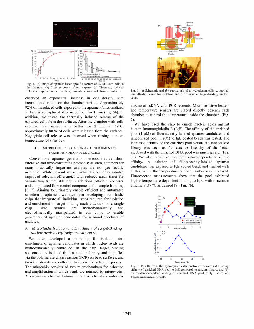
A. Microfluidic Isolation and Enrichment of Target-Binding Nucleic Acids by Hydrodynamical Control
We have developed a microchip for isolation and enrichment of aptamer candidates in which nucleic acids are hydrodynamically controlled. In the chip, target binding sequences are isolated from a random library and amplified via the polymerase chain reaction (PCR) on bead surfaces, and then the strands are collected to repeat the selection process. The microchip consists of two microchambers for selection and amplification in which beads are retained by microweirs. A serpentine channel between the two chambers enhances
mixing of ssDNA with PCR reagents. Micro resistive heaters and temperature sensors are placed directly beneath each chamber to control the temperature inside the chambers (Fig. 6).
We have used the chip to enrich nucleic acids against human Immunoglobulin E (IgE). The affinity of the enriched pool (1 μM) of fluorescently labeled aptamer candidates and randomized pool (1 μM) to IgE-coated beads was tested. The increased affinity of the enriched pool versus the randomized library was seen as fluorescence intensity of the beads incubated with the enriched DNA pool was much greater (Fig. 7a). We also measured the temperature-dependence of the affinity. A solution of fluorescently-labeled aptamer candidates was exposed to IgE-coated beads and washed with buffer, while the temperature of the chamber was increased. Fluorescence measurements show that the pool exhibited highly temperature dependent binding to IgE, with maximum binding at 37 °C as desired [8] (Fig. 7b).
(a)
(b) (c)
Fig. 5. (a) Image of aptamer-based specific capture of CCRF-CEM cells in the chamber. (b) Time response of cell capture. (c) Thermally induced release of captured cells from the aptamer-functionalized chamber surfaces.
(b)
(a)
Fig. 6. (a) Schematic and (b) photograph of a hydrodynamically controlled microfluidic device for isolation and enrichment of target-binding nucleic acids.
(b)
(a)
Fig. 7. Results from the hydrodynamically controlled device: (a) Binding affinity of enriched DNA pool to IgE compared to random library, and (b) temperature-dependent binding of enriched DNA pool to IgE based on fluorescence measurements.
1247

B. Microfluidic Isolation and Enrichment of Target-Binding Nucleic Acids by Electrokinetical Control
We have also been investigating an alternative approach to simplify chip design and operation for generation of target-binding nucleic acids by manipulating the transport of DNA strands using on-chip electrophoresis. Similar to the hydrodynamically controlled device, this chip uses surface-functionalized microbeads for isolation and amplification of aptamer candidates in two chambers. Nucleic acids, however, are electrophoretically manipulated between the two chambers connected by a microchannel filled with agarose gel. The electrophoretic transport of strands on-chip is realized by an electric field generated between the two chambers (Fig. 8).
Using this device, we have demonstrated isolation and enrichment of target-binding nucleic acids against steroid deoxycholic acid (DCA), human IgE, and MCF-7 cells. Experimental results show that weakly binding nucleic acids were hydrodynamically removed during selection, while strongly binding aptamer nucleic acids were isolated on the
target-functionalized beads (Fig. 9a). The binding affinity of the randomized nucleic acid pool to IgE significantly improved by 4 times after 4 rounds of isolation and enrichment (Fig. 9b). Thus, our microfluidic approach has the potential to offer a simple yet efficient tool for selection of aptamer candidates against a variety of target molecules [9].
IV. CONCLUSION
Aptamers can strongly bind to specific biological targets. When integrated with microfluidic platforms, aptamers hold the potential to address the needs of biological applications for detection of low abundance analytes. We presented an overview of our effort to develop microfluidic devices that are functionalized with aptamers for manipulation of biomolecules and cells, and that are capable of isolating target-binding nucleic acids from a randomized library. The aptamer-functionalized microfluidic devices allow selective extraction and enrichment, isocratic elution, and highly sensitive detection of analytes. The microfluidic devices for isolation of target-binding nucleic acids demonstrate the potential for integrated selection of aptamers having predefined binding characteristics against a broad spectrum of practically important biological analytes.
ACKNOWLEDGMENTS
We gratefully acknowledge financial support from the National Science Foundation (Award No. CBET-0854030) and the National Institutes of Health (Award Nos. RR025816-02 and CA147925-01).
REFERENCES
[1] T. Nguyen, R. Pei, C. Qiu, J. Ju, M. Stojanovic, and Q. Lin, “An aptameric microfluidic system for specific purification, enrichment, and mass spectrometric detection of biomolecules,” J. Microelectromech. Sys., 18, pp. 1198-1207
[2] T. Nguyen, R. Pei, M. Stojanovic, and Q. Lin, “Demonstration and characterization of biomolecular enrichment on microfluidic aptamer-functionalized surfaces,” Sens. Actuat. B: Chem., 155, pp. 58-66.
[3] T. Nguyen, R. Pei, D. Landry, M. Stojanovic, and Q. Lin, “Label-free microfluidic characterization of temperature-dependent biomolecular interactions,” Biomicrofluidics, 5: 034118.
[4] T. Nguyen, R. Pei, D. Landry, M. Stojanovic, and Q. Lin, “Microfluidic aptameric affinity sensing of vasopressin for clinical diagnostic and therapeutic applications,” Sens. Actuat. B: Chem., 154, pp. 59-66.
[5] J. Zhu, T. Nguyen, R. Pei, M. Stojanovic, Q. Lin, “Specific capture and temperature-mediated release of cells in an aptameric microchip,” Lab Chip, 12, pp. 3504-3513, 2012.
[6] X. Lou, J. Qian, Y. Xiao, L. Viel, A. Gerdon, E. Lagally, P. Atzberger, T. Tarasow, A. Heeger, and T. Soh, “Micromagnetic selection of aptamers in microfluidic channels,” Proc. Natl. Acad. Sci., 106, pp. 2989-2994, 2009.
[7] C. Huang, H. Lin, S. Shiesh, and G. Lee, “An integrated microfluidic system for rapid screening of alpha-fetoprotein-specific aptamers,” Biosens. Bioelectron., 35, pp. 50-55, 2012.
[8] J. Hilton, J. Kim, T. Nguyen, M. Barbu, R. Pei, M. Stojanovic, and Q. Lin, “Isolation of thermally sensitive aptamers on a microchip,” Proc. Int. Conf. Micro Electro Mechanical Systems, pp. 100-103, Paris, France, 2012.
[9] J. Kim, J. Hilton, K. Yang, R. Pei, M. Stojanovic, and Q. Lin, “Nucleic acid isolation and enrichment on a microchip,” Sens. Actuat. B: Phys., doi. 10.1016/j.sna.2012.07.022, in press.
(a)
(b)
1 cm
Fig. 8. (a) Schematic and (b) photograph of an electrokinetically controlled microfluidic device for isolation and enrichment of target-binding nucleic acids.
(a)
(b)
Fig. 9. Results from the electrokinetically controlled device: (a) isolation of target-binding nucleic acids against steroid DCA, IgE, and MCF-7 cells, and(b) improved binding affinity of the nucleic acid pool to IgE after 4 rounds isolation and enrichment processes.
1248