InSituExpressionofRegulatoryCytokinesby ...downloads.hindawi.com/journals/jir/2012/361730.pdf ·...
8
Hindawi Publishing Corporation Clinical and Developmental Immunology Volume 2012, Article ID 361730, 7 pages doi:10.1155/2012/361730 Clinical Study In Situ Expression of Regulatory Cytokines by Heart Inflammatory Cells in Chagas’ Disease Patients with Heart Failure Denise Bertulucci Rocha Rodrigues, 1, 2 Marlene Antonia dos Reis, 1 Audrey Romano, 3 San´ ıvia Aparecida de Lima Pereira, 1, 2 Vicente de Paula Antunes Teixeira, 1 Sebasti˜ ao Tostes Junior, 1, 2 and Virmondes Rodrigues Jr. 1, 4 1 Laboratory of Immunology, Institute of Biological Sciences, Federal University of Triˆ angulo Mineiro, 38125-180 Uberaba, MG, Brazil 2 Departamento de Imunologia e Patologia, University of Uberaba, 38055-500 Uberaba, MG, Brazil 3 INSERM-U906, 13385 Marseille, France 4 Laboratory of Immunology, Federal University of Triˆ angulo Mineiro, 38125-180 Uberaba, MG, Brazil Correspondence should be addressed to Virmondes Rodrigues Jr., [email protected] Received 23 February 2012; Accepted 15 May 2012 Academic Editor: Anderson S´ a-Nunes Copyright © 2012 Denise Bertulucci Rocha Rodrigues et al. This is an open access article distributed under the Creative Commons Attribution License, which permits unrestricted use, distribution, and reproduction in any medium, provided the original work is properly cited. Chagas’ disease is caused by the protozoan parasite Trypanosoma cruzi. The immune system plays an important role in the reduction of parasite load, but may also contribute to the development of lesions observed during the chronic phase of the disease. We analyzed cytokines produced by inflammatory heart cells in 21 autopsy samples obtained from patients with Chagas’ disease divided according to the presence or absence of heart failure (HF). Left ventricular sections were analyzed by immunohistochemistry using antibodies against human IL-4, IFN-γ, TGF-β, TNF-α, and NOS2. In situ mRNA expression was quantified by a Low Density Array. The number of IFN-γ-positive cells was significantly higher than IL-4 positive cells. TNF-α, TGF-β and NOS2 were detected in 65%, 62% and 94% of samples respectively. There was an association between TNF-α-producing cells and the presence of HF. Subjects with HF presented higher levels of STAT4 mRNA, whereas FoxP3 and STAT6 levels were similar in the two groups. A Th1 cytokine pattern predominated in the cardiac inflammatory cell infiltrate of Chagas’ disease patients associated with HF. High degree of fibrosis was associated with low NOS2 expression. These results support the idea that Th1 immune responses are involved in heart lesions of Chagas’ disease patients. 1. Introduction Chagas’ disease is caused by the protozoan parasite Try- panosoma cruzi, which is transmitted to humans by contact with the feces of blood-sucking triatomine insects, transfu- sion of blood or blood derivatives, and organ transplanta- tion. According to the WHO estimates, 16–18 million people were infected in the Americas in the 1980s [1]. Although transmission is controlled in some countries, the infection persists and each year a large number of patients in the chronic phase develop symptoms [2]. During the chronic phase, a long period of latency, called the indeterminate phase which lasts several years or throughout life, is observed in approximately 60% of patients with Chagas’ disease. Clinical manifestations that result in heart and/or digestive organ damage occur in the remaining 40% of patients at different intensities, alone or in association [2, 3]. During this phase, parasites are rarely detected in peripheral blood and the diagnosis is based on the presence of parasite-specific antibodies [4]. Heart complications are the major cause of morbidity and mortality in Chagas’ disease patients. Differences in the degree of damage to the conduction system are observed, and heart failure occurs among more severe cases [2]. Myo- carditis can affect patients during the chronic phase, irre- spective of clinical manifestations, although the degree of
Transcript of InSituExpressionofRegulatoryCytokinesby ...downloads.hindawi.com/journals/jir/2012/361730.pdf ·...
Hindawi Publishing CorporationClinical and Developmental ImmunologyVolume 2012, Article ID 361730, 7 pagesdoi:10.1155/2012/361730
Clinical Study
In Situ Expression of Regulatory Cytokines byHeart Inflammatory Cells in Chagas’ Disease Patients withHeart Failure
Denise Bertulucci Rocha Rodrigues,1, 2 Marlene Antonia dos Reis,1
Audrey Romano,3 Sanıvia Aparecida de Lima Pereira,1, 2 Vicente de Paula Antunes Teixeira,1
Sebastiao Tostes Junior,1, 2 and Virmondes Rodrigues Jr.1, 4
1 Laboratory of Immunology, Institute of Biological Sciences, Federal University of Triangulo Mineiro, 38125-180 Uberaba, MG, Brazil2 Departamento de Imunologia e Patologia, University of Uberaba, 38055-500 Uberaba, MG, Brazil3 INSERM-U906, 13385 Marseille, France4 Laboratory of Immunology, Federal University of Triangulo Mineiro, 38125-180 Uberaba, MG, Brazil
Correspondence should be addressed to Virmondes Rodrigues Jr., [email protected]
Received 23 February 2012; Accepted 15 May 2012
Academic Editor: Anderson Sa-Nunes
Copyright © 2012 Denise Bertulucci Rocha Rodrigues et al. This is an open access article distributed under the Creative CommonsAttribution License, which permits unrestricted use, distribution, and reproduction in any medium, provided the original work isproperly cited.
Chagas’ disease is caused by the protozoan parasite Trypanosoma cruzi. The immune system plays an important role in thereduction of parasite load, but may also contribute to the development of lesions observed during the chronic phase of thedisease. We analyzed cytokines produced by inflammatory heart cells in 21 autopsy samples obtained from patients withChagas’ disease divided according to the presence or absence of heart failure (HF). Left ventricular sections were analyzed byimmunohistochemistry using antibodies against human IL-4, IFN-γ, TGF-β, TNF-α, and NOS2. In situ mRNA expression wasquantified by a Low Density Array. The number of IFN-γ-positive cells was significantly higher than IL-4 positive cells. TNF-α,TGF-β and NOS2 were detected in 65%, 62% and 94% of samples respectively. There was an association between TNF-α-producingcells and the presence of HF. Subjects with HF presented higher levels of STAT4 mRNA, whereas FoxP3 and STAT6 levels weresimilar in the two groups. A Th1 cytokine pattern predominated in the cardiac inflammatory cell infiltrate of Chagas’ diseasepatients associated with HF. High degree of fibrosis was associated with low NOS2 expression. These results support the idea thatTh1 immune responses are involved in heart lesions of Chagas’ disease patients.
1. Introduction
Chagas’ disease is caused by the protozoan parasite Try-panosoma cruzi, which is transmitted to humans by contactwith the feces of blood-sucking triatomine insects, transfu-sion of blood or blood derivatives, and organ transplanta-tion. According to the WHO estimates, 16–18 million peoplewere infected in the Americas in the 1980s [1]. Althoughtransmission is controlled in some countries, the infectionpersists and each year a large number of patients in thechronic phase develop symptoms [2].
During the chronic phase, a long period of latency,called the indeterminate phase which lasts several years or
throughout life, is observed in approximately 60% of patientswith Chagas’ disease. Clinical manifestations that result inheart and/or digestive organ damage occur in the remaining40% of patients at different intensities, alone or in association[2, 3]. During this phase, parasites are rarely detected inperipheral blood and the diagnosis is based on the presenceof parasite-specific antibodies [4].
Heart complications are the major cause of morbidityand mortality in Chagas’ disease patients. Differences in thedegree of damage to the conduction system are observed,and heart failure occurs among more severe cases [2]. Myo-carditis can affect patients during the chronic phase, irre-spective of clinical manifestations, although the degree of

2 Clinical and Developmental Immunology
myocarditis and fibrosis that follows the inflammatory pro-cess is more intense in patients with heart failure [5, 6]. Theinflammatory process observed in Chagas’ disease is main-ly characterized by the presence of CD8 and CD4 T lym-phocytes and macrophages [7]. The presence of activatedCD8 T lymphocytes has been demonstrated [8]. The extentand nature of the inflammatory reaction contribute to tissuedamage and reduce cardiac function. These events lead toheart failure which is observed in severe cases of chronicChagas’ disease.
The balance of the T helper cell subpopulation has beenstudied in experimental models of T. cruzi infection and theresults show that the Th1 subpopulation plays a role in themechanism of parasite control [9, 10]. In this respect, IFN-γand TNF-α synergistically act on NOS2 transcription and onthe production of high levels of nitric oxide, which exhibitsstrong antiparasitic effects [9, 11, 12]. These cytokines havebeen shown to play a role in parasite control and also con-tribute to tissue damage [13, 14]. On the other hand, IL-4,a prototype Th2 cytokine, is associated with an increase ofparasitemia. In humans, most data regarding the T helper cellbalance is limited to the analysis of PMBC immune responses[15] and CD4 and CD8 cell infiltration [16, 17].
To address the nature of inflammatory cells in hearttissue and possible implications in the pathogenesis of heartfailure in Chagas’ disease, we investigated the number of cellsexpressing IFN-γ, TNF-α, IL-4, TGF-β, and NOS2 and thelevels of FoxP3, STAT4, and STAT6 mRNA in heart tissuesof subjects who had died during the chronic phase of thedisease. To our knowledge, this is the first study investigat-ing T helper cell subpopulation in heart tissue. The resultsdemonstrated that the production of all mediators men-tioned above and that the number of cells producing IFN-γwere higher than that producing IL-4. Moreover, large num-bers of TNF-α-producing cells and high levels of STAT4mRNA were associated with the occurrence of heart failure.
2. Materials and Methods
2.1. Patients. Twenty-one specimens of the left ventricularwall were obtained at autopsy of subjects with Chagas’disease who had died at the General Hospital of TrianguloMineiro Federal University, Uberaba, MG, Brazil. The autop-sies were performed within 2 to 6 hours after death, and twosamples were collected from the midportion of the lateralwall of the left ventricle. One specimen was immediatelyfixed in buffered formalin, and the other was frozen in liquidnitrogen. After embedding in paraffin, serial 5 μm sectionswere transferred to glass slides. Frozen samples were used inthe low density array (LDA). All specimens were obtainedfrom patients who had a positive reaction to anti-T. cruziantibodies and who were considered to be in the chroniccardiac phase. Heart involvement affected only the electricalconduction system in 10 patients. In the remaining 11subjects, the clinical records indicated heart failure, such asleg edema, and chest radiographs demonstrated global heartenlargement [2, 6]. Samples obtained from three healthysubjects who died in car accidents were used as controls inthe LDA assays. The procedures were approved by the Ethics
Committee of the Federal University of Triangulo Minei-ro.
2.2. Histopathology and Quantification of Fibrosis. Fixedtissues were dehydrated and embedded in paraffin. Sections(5 μm) were stained with hematoxylin-eosin (HE) and ana-lyzed by light microscopy.
Morphometric analysis of fibrosis was performed usingthe KS300 automatic image-analyzing system (Kotron Elec-tronic, Munich, Germany). Fibrous connective tissues wereevaluated by picrosirius red staining, and the results areexpressed as percentage of fibrosis area [18]. For further ana-lysis, patients were empirically divided according to fibrosisintensity into low-grade fibrosis (up to 4%) and high-gradefibrosis (>4%).
2.3. Immunohistochemistry. For immunohistochemistry, de-paraffinized sections were treated with 3% hydrogen per-oxide in methanol for 10 min and incubated for 30 min at90◦C for antigen detection. The sections were incubated in2% bovine serum albumin for 30 min at room temperat-ure to reduce nonspecific binding. Next, the sections wereindividually incubated with anti-cytokine monoclonal anti-bodies specific for human IL-4 (1 : 100) (R&D, Minneapolis,MN, USA), TNF-α (1:200) (R&D); TGF-β (1 : 100) (R&D),NOS2 (1 : 100) (Santa Cruz Biotech, Santa Cruz, CA, USA),and IFN-γ (1 : 200) (Genzyme, Cambridge, MA, USA). Allantibodies were diluted in 2% bovine serum albumin priorto use and incubated with the samples for 2 hours at 37◦C.For the secondary antibody, the sections were incubated withbiotinylated anti-mouse Ig, anti-rabbit Ig, and anti-goat Igfrom Link System 002488 (Dako, Carpinteria, CA, USA) for30 min at 37◦C. After washing, the sections were incubatedwith streptavidin-peroxidase conjugate (Dako) for 30 min.The reaction was developed with diaminobenzidine (Sigma,St. Louis, MO, USA). The sections were counterstained withhematoxylin.
For histopathological analysis, the number of cells pos-itive for each cytokine was counted in 20 fields at 400Xmagnification. The number of cells in each field and thearea of each field (0.091575 mm2) were determined. Thedensity of positive cells is expressed as the number of cells permm2.
2.4. Low Density Array. After cell harvest, RNA was extractedwith the TRIzol reagent (Invitrogen, Grand Island, NY, USA)according to manufacturer instructions. RNA purity andintegrity were assessed with the Agilent 2100 bioanalyzerusing the RNA 6000 Nano LabChip reagent set (Agilent Tech-nologies, Santa Clara, CA, USA). RNA was quantified spec-trophotometrically and then stored at −80◦C. cDNA wassynthesized using the High-Capacity cDNA Archive Kit(Applied Biosystem, Carlsbad, CA, USA). The master mix-ture contained 1X reverse transcription buffer, 1X deoxynu-cleotide triphosphate mixture, 1 unit/μL RNase inhibitor,1 unit/μL MultiScribe Reverse Transcriptase, and 1X randomprimers. One μg of total RNA was diluted in sterile water toa final volume of 100 μL. The reaction mixture was incubat-ed at 25◦C for 10 min, followed by heat inactivation of the
Clinical and Developmental Immunology 3
enzyme at 37◦C for 120 min cDNA was stored at −20◦C.Next, 2 μL single-stranded cDNA (corresponding to 100 ngtotal RNA) was diluted in 98 μL nuclease-free water and100 μL TaqMan Universal PCR Master Mix, and 100 μL ofthe sample-specific PCR mixture was loaded into the sampleport of Micro Fluidic Cards. The cards were then centri-fuged twice for 1 min at 1200 g and sealed to prevent well-to-well contamination. The cards were placed in the MicroFluidic Card Sample Block of an ABI Prism 7900 HTSequence Detection System (SDS Software 2.1, Applied Bio-systems). The thermal cycling conditions were 2 min at 50◦Cand 10 min at 94.5◦C, followed by 40 cycles at 97◦C for30 s and at 59.7◦C for 1 min. Each Micro Fluidic Card hasa unique barcode, and Sequence Detection System platedocuments store information about plate type, detector,sample/target gene configurations, thermal cycling condi-tions, data collection, and raw fluorescence data during eachcycle. Micro Fluidic Cards were analyzed with RQ docu-ments and the RQ Manager Software for automated dataanalysis. Experiments for three different donor cells andone healthy control, carried out in duplicate, were analyzedtogether as 1 relative quantity (RQ) study. Expression valuesof the target genes were normalized to the concentration of18S rRNA. Gene expression values were calculated by thecomparative threshold cycle (Ct) method, in which RNAsamples from the control subject were designated as calibrat-ors. In short, the Ct data for all human genes tested and18S rRNA in each sample were used to create ΔCt values(Ctexperimental − Ct18S rRNA). Thereafter, ΔΔCt values werecalculated by subtracting the ΔCt of the calibrator fromthe Ct value of each target. RQ was calculated using theequation: RQ = 2−ΔΔCt. The Micro Fluidic Cards detect atwo-fold difference in gene expression at a confidence level of99.7%.
2.5. Statistical Analysis. Statistical analysis (unpaired t-testand Mann-Whitney test) was performed using the StatViewsoftware. Correlations between the numbers of cells positivefor each cytokine were analyzed using the Spearman (rS) andthe Pearson (r) correlation coefficients. Differences foundwere considered to be significant at P < 0.05.
3. Results
3.1. Histopathology, Fibrosis Quantification, and CytokineExpression. Myocardial samples from 21 chronic chagasicsubjects were examined. Eleven patients had chronic heartfailure, and 10 subjects did not present any previous clinicalor anatomopathological signs of heart failure due to Chagas’disease. These samples were also classified according to fib-rosis intensity.
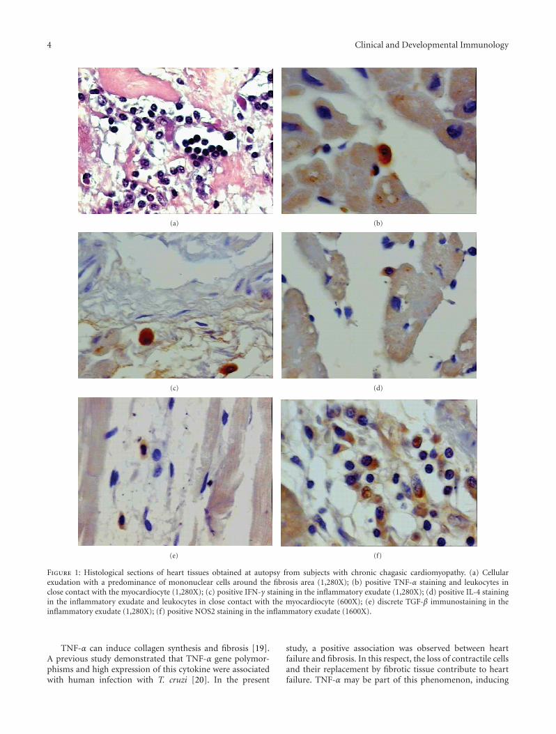
An inflammatory mononuclear cell infiltrate was ob-served in most cases inside and around the fibrosis area.The intensity of the inflammatory reactions was higher insubjects with heart failure. The panels in Figure 1 illustratethe inflammatory reaction and the immunohistochemicalresults. A significant association was observed between TNF-α-positive cells and the presence of heart failure (P = 0.040).TNF-α-positive cells were observed in 10 of the 11 cases with
heart failure, with a median of 4 immunostained cells (range:0 to 22 cells/mm2), and in 4 of the 10 cases without heart fail-ure (Figure 2(a)).
There was a positive association between IFN-γ-pro-ducing cells and heart failure (P = 0.032). IFN-γ-positivecells were present in 8 of the 11 cases with heart failure,with a median of 24 immunostained cells (range: 0 to85 cells), and in 7 of the 10 cases without heart failure(Figure 2(a)). TNF-α and IFN-γ are cytokines involved in theinduction of NOS2 and have been implicated in the controlof intracellular parasite growth and tissue damage. AlthoughTNF-α and IFN-γ were associated with heart failure, nosignificant correlation was observed between NOS2-positivecells and heart failure. Cells positive for NOS2 were presentin 10 of the 11 cases with heart failure and in 6 of the 10 caseswithout heart failure (Figure 2(a)). Moreover, there was nosignificant association between the presence of cells positivefor the anti-inflammatory cytokines, IL-4 and TGFβ, and theoccurrence of heart failure (Figure 2(a)). On the other hand,a positive correlation was observed between the number ofIFN-γ-positive and NOS2-positive cells, irrespective of theoccurrence of heart failure (P = 0.02) (Figure 3(a)). Therewas also a positive correlation between the number of cellspositive for IFN-γ and TNF-α (P = 0.03) (Figure 3(b)).
The intensity of fibrosis was also analyzed and wasclassified into low (up to 4% of total area) or high (>4%).Interestingly, the occurrence of intense fibrosis was associ-ated with small numbers of NOS2-positive cells, suggestingthat nitric oxide may protect against the development offibrosis (Figure 2(b)). Fibrosis was not associated with anyof the other cytokines tested (Figure 2(b)).
3.2. Th1/Th2 Cytokine Balance. The numbers of IFN-γ-and IL-4-positive cells were compared for the evaluation ofthe balance between Th1 and Th2 cytokine patterns. Thenumber of IFN-γ-positive cells was significantly higher thanthat of IL-4-positive cells (P = 0.02), irrespective of theoccurrence of heart failure, indicating that T. cruzi inducedmyocarditis by eliciting a Th1-like response (Figure 4).Furthermore, LDA analysis revealed significantly higherSTAT4 levels in patients with heart failure compared tothose without heart failure. Moreover, STAT4 levels weresignificantly higher than STAT6 levels in the heart failuregroup (Figure 5). These data suggest that heart failure inChagas’ disease is associated with a Th1 immune response.No significant differences between groups were observed forthe other cytokine genes tested.
4. Discussion
In this study, we report the results of the expression ofcytokines in heart tissue obtained from the left ventricularwall of subjects with Chagas’ disease. The autopsies wereperformed within 2 to 6 hours after death, and the specimenswere immediately frozen in liquid nitrogen or fixed inparaformaldehyde in phosphate buffer. Clinical involvementof the heart was analyzed considering previous clinical dataand anatomopathological features observed during auto-psy.
4 Clinical and Developmental Immunology
(a) (b)
(c) (d)
(e) (f)
Figure 1: Histological sections of heart tissues obtained at autopsy from subjects with chronic chagasic cardiomyopathy. (a) Cellularexudation with a predominance of mononuclear cells around the fibrosis area (1,280X); (b) positive TNF-α staining and leukocytes inclose contact with the myocardiocyte (1,280X); (c) positive IFN-γ staining in the inflammatory exudate (1,280X); (d) positive IL-4 stainingin the inflammatory exudate and leukocytes in close contact with the myocardiocyte (600X); (e) discrete TGF-β immunostaining in theinflammatory exudate (1,280X); (f) positive NOS2 staining in the inflammatory exudate (1600X).
TNF-α can induce collagen synthesis and fibrosis [19].A previous study demonstrated that TNF-α gene polymor-phisms and high expression of this cytokine were associatedwith human infection with T. cruzi [20]. In the present
study, a positive association was observed between heartfailure and fibrosis. In this respect, the loss of contractile cellsand their replacement by fibrotic tissue contribute to heartfailure. TNF-α may be part of this phenomenon, inducing

Clinical and Developmental Immunology 5
0
10
20
30
40
50
60
70N
um
ber
of c
ells
Heart failureNo heart failure
TNF-α IFN-γ IL-4 TGF-β NOS2
∗
∗
(a)
Moderate fibrosisHigh fibrosis
0
10
20
30
40
50
60
70
Nu
mbe
r of
cel
ls
TNF-α IFN-γ IL-4 TGF-β NOS2
∗
(b)
Figure 2: Number of inflammatory cells expressing cytokines and NOS2 in heart tissue from patients in the chronic phase of Chagas’ disease.(a) Subjects were divided according to the presence (gray bar) or absence of heart failure (open bar). (b) Subjects were divided according tothe presence of a high degree (hatched bar) or low degree of fibrosis (open bar). Horizontal lines represent the median, boxes represent the25th to 75th percentiles, and vertical lines indicate the 10th to 90th percentiles. ∗P < 0.05 (Mann-Whitney test).
0
10
20
30
40
50
60
70
80
90
0 5 10 15 20 25 30 35 40
Nu
mbe
r of
IFN
-γ+
cells
Number of NOS2+ cells
(a)
0 10 20 30 40 50 60 70 80 900
2.5
5
7.5
10
12.5
15
17.5
20
22.5
Nu
mbe
r of
IFN
-α+
cells
Number of IFN-γ+ cells
(b)
Figure 3: Correlation between the number of IFN-γ- and NOS2-immunostained inflammatory cells (a) and the number of TNF-α- andIFN-γ-immunostained inflammatory cells in 21 subjects with chronic chagasic cardiopathy (b). ∗P < 0.05 (Spearman’s correlation).
tissue damage and fibrosis development. Other studies haveshown TNF-producing cells in association with areas oftissue damage in acute models of T. cruzi infection [14]. Inaddition, the presence of this cytokine has been constantlyobserved in histopathological studies of hearts from subjectswho had died of Chagas’ disease [8]. TNF-α may alsocontribute to the development of heart failure throughapoptosis and the induction of NOS2, producing nitric oxidewhich exerts strong negative inotropic effects [21–25]. TNF-α and IFN-γ act synergistically on NOS2 expression andthe subsequent induction of death of the parasite [9]. Inthe present study, IFN-γ was positively associated with heartfailure. Considering that IFN-γ has antifibrotic propertiesand TNF-α is involved in fibrosis, the synergistic effect of the
two cytokines on NOS2 expression may have contributed tothe development of heart failure. These results suggest thatcytokines involved in parasite control, such as TNF-α andIFN-γ and the major mediator of parasite death, nitric oxide,may persist and induce mechanisms of tissue damage thatcontribute to the development of heart failure.
This study clearly demonstrated the predominance of aTh1 immune response in the inflammatory reaction seen inthe heart of subjects with severe forms of Chagas’ disease.The local production of IFN-γ is functional, since STAT4mRNA was overexpressed in subjects with heart failure.Furthermore, IFN-γ-induced genes are upregulated in heartsamples from patients with Chagas’ disease [26]. Studies haveshown a protective role of CD4+ T lymphocytes and IFN-γ

6 Clinical and Developmental Immunology
0
5
10
15
20
25
30
35
40
45
50
Nu
mbe
r of
cel
ls
IL-4IFN-γ
∗
Figure 4: Number of IFN-γ- and IL-4-immunostained inflam-matory cells in 21 subjects with chronic chagasic cardiopathy.Horizontal lines represent the median, boxes represent the 25thto 75th percentiles, and vertical lines indicate the 10th to 90thpercentiles. ∗P < 0.05 (Mann-Whitney test).
Heart failure
No heart failure
1
2
4
8
16
32
64
128
Rel
ativ
e R
NA
cop
ies
FoxP3STAT6 STAT4
∗
Figure 5: Relative number of mRNA copies in heart tissue ofpatients in the chronic phase of Chagas’ disease according tothe presence (gray bar) or absence of heart failure (open bar).Horizontal lines represent the median, boxes represent the 25thto 75th percentiles, and vertical lines indicate the 10th to 90thpercentiles. ∗P < 0.05 (Mann-Whitney test).
responses in anti-T. cruzi immunity in murine models ofChagas’ disease [27–30]. Conversely, studying chronic hu-man infection with T. cruzi, other investigators demonstratedthe production of higher levels of IFN-γ by PBMC fromcardiac patients when compared to asymptomatic subjectsand associated this production with pathogenesis [31, 32].
In conclusion, the present results show that the heartinfiltrating T cells in Chagas’ disease mainly have a Th1phenotype. Severe heart involvement leading to heart failureseems to be multifactorial and associated with the presenceof IFN-γ- and TNF-α-producing cells. Continuous antigenstimulation may help sustain the inflammatory response,with the intensity and pattern of this response promoting tis-sue damage and heart cell dysfunction that lead to heart fail-ure.
Conflict of Interests
The authors declare no conflict of interests.
Acknowledgments
This study was supported by grants from FAPEMIG andCNPq.
References
[1] World Health Organization, “Chagas’ disease,” in TechnicalReport Series Geneve, Organization WH, Ed., vol. 1, pp. 50–55,1984.
[2] A. Prata, “Clinical and epidemiological aspects of Chagasdisease,” The Lancet Infectious Diseases, vol. 1, no. 2, pp. 92–100, 2001.
[3] J. R. Coura, L. L. de Abreu, J. B. Pereira, and H. P. Willcox,“Morbidity in Chagas’ disease. IV. Longitudinal study of 10years in Pains and Iguatama, Minas Gerais, Brazil,” Memoriasdo Instituto Oswaldo Cruz, vol. 80, no. 1, pp. 73–80, 1985.
[4] M. A. Magnani, F. Ferriolli, and A. F. de Siqueira, “Specificimmunoglobulins (IgA, IgG, and IgM) in serum of patientswith chronic Chagas’ disease analyzed by indirect immunoflu-orescence reactions,” Revista do Instituto de Medicina Tropicalde Sao Paulo, vol. 15, no. 2, pp. 72–75, 1973.
[5] E. R. Lopes, E. Chapadeiro, Z. A. Andrade, H. O. Almeida, andA. Rocha, “Pathological anatomy of hearts from asymptomaticChagas disease patients dying in a violent manner,” Memoriasdo Instituto Oswaldo Cruz, vol. 76, no. 2, pp. 189–197, 1981.
[6] H. Almeida, L. Miziara, and V. Teixeira, “Contribuicao aoestudo da insuficiencia cardıaca na doenca de chagas: relacaoentre dilatacao do ventrıculo e hipertrofia do miocardio,”Revista Goiana de Medicina, vol. 28, no. 2, pp. 33–38, 1982.
[7] M. D. L. Higuchi, M. M. Reis, V. D. Aiello et al., “Association ofan increase in CD8+ T cells with the presence of Trypanosomacruzi antigens in chronic, human, chagasic myocarditis,” TheAmerican Journal of Tropical Medicine and Hygiene, vol. 56, no.5, pp. 485–489, 1997.
[8] D. D. Reis, E. M. Jones, S. Tostes Jr. et al., “Characterizationof inflammatory infiltrates in chronic chagasic myocardiallesions: presence of tumor necrosis factor-α+ cells and dom-inance of granzyme A+, CD8+ lymphocytes,” The AmericanJournal of Tropical Medicine and Hygiene, vol. 48, no. 5, pp.637–644, 1993.
[9] J. S. Silva, G. N. R. Vespa, M. A. G. Cardoso, J. C. S.Aliberti, and F. Q. Cunha, “Tumor necrosis factor alpha medi-ates resistance to Trypanosoma cruzi infection in mice byinducing nitric oxide production in infected gamma inter-feron-activated macrophages,” Infection and Immunity, vol.63, no. 12, pp. 4862–4867, 1995.
[10] R. T. Gazzinelli, A. Talvani, M. M. Camargo et al., “Inductionof cell-mediated immunity during early stages of infectionwith intracellular protozoa,” Brazilian Journal of Medical andBiological Research, vol. 31, no. 1, pp. 89–104, 1998.
[11] G. N. R. Vespa, F. Q. Cunha, and J. S. Silva, “Nitric oxide isinvolved in control of Trypanosoma cruzi-induced parasitemiaand directly kills the parasite in vitro,” Infection and Immunity,vol. 62, no. 11, pp. 5177–5182, 1994.
[12] J. C. Aliberti, J. T. Souto, A. P. Marino et al., “Modulationof chemokine production and inflammatory responses ininterferon-γ- and tumor necrosis factor-R1-deficient miceduring Trypanosoma cruzi infection,” The American Journal ofPathology, vol. 158, no. 4, pp. 1433–1440, 2001.

Clinical and Developmental Immunology 7
[13] R. M. E. Arantes, H. H. F. Marche, M. T. Bahia, F. Q. Cunha,M. A. Rossi, and J. S. Silva, “Interferon-γ-induced nitric oxidecauses intrinsic intestinal denervation in Trypanosoma cruzi-infected mice,” The American Journal of Pathology, vol. 164, no.4, pp. 1361–1368, 2004.
[14] E. S. Lima, Z. A. Andrade, and S. G. Andrade, “TNF-α isexpressed at sites of parasite and tissue destruction in thespleen of mice acutely infected with Trypanosoma cruzi,” Inter-national Journal of Experimental Pathology, vol. 82, no. 6, pp.327–336, 2001.
[15] L. M. G. Bahia-Oliveira, J. A. S. Gomes, J. R. Cancado et al.,“Immunological and clinical evaluation of chagasic patientssubjected to chemotherapy during the acute phase of Trypan-osoma cruzi infection 14-30 years ago,” The Journal of InfectiousDiseases, vol. 182, no. 2, pp. 634–638, 2000.
[16] I. Higuchi and M. Osame, “Recent progress of molecularimmunology on inflammatory myopathy,” Nippon Rinsho, vol.55, no. 12, pp. 3331–3335, 1997.
[17] D. D. Reis, E. M. Jones, S. Tostes et al., “Expression of majorhistocompatibility complex antigens and adhesion moleculesin hearts of patients with chronic Chagas’ disease,” The Ameri-can Journal of Tropical Medicine and Hygiene, vol. 49, no. 2, pp.192–200, 1993.
[18] G. S. Montes and L. C. Junqueira, “The use of the Picrosirius-polarization method for the study of the biopathology ofcollagen,” Memorias do Instituto Oswaldo Cruz, vol. 86, sup-plement 3, pp. 1–11, 1991.
[19] M. K. Connolly, A. S. Bedrosian, J. Mallen-St. Clair et al., “Inliver fibrosis, dendritic cells govern hepatic inflammation inmice via TNF-α,” The Journal of Clinical Investigation, vol. 119,no. 11, pp. 3213–3225, 2009.
[20] C. W. Pissetti, D. Correia, R. F. de Oliveira et al., “Genetic andfunctional role of TNF-alpha in the development Trypano-soma cruzi infection,” PLoS Neglected Tropical Diseases, vol. 5,no. 3, article e976, 2011.
[21] M. S. Finkel, C. V. Oddis, T. D. Jacob, S. C. Watkins, B. G.Hattler, and R. L. Simmons, “Negative inotropic effects ofcytokines on the heart mediated by nitric oxide,” Science, vol.257, no. 5068, pp. 387–389, 1992.
[22] V. Rodrigues Jr., G. S. Agrelli, S. C. Leon, D. N. Silva Teixeira,S. Tostes, and D. B. Rocha-Rodrigues, “Fas/Fas-L expres-sion, apoptosis and low proliferative response are associatedwith heart failure in patients with chronic Chagas’ disease,”Microbes and Infection, vol. 10, no. 1, pp. 29–37, 2008.
[23] O. H. L. Bing, “Hypothesis: apoptosis may be a mechanism forthe transition to heart failure with chronic pressure overload,”Journal of Molecular and Cellular Cardiology, vol. 26, no. 8, pp.943–948, 1994.
[24] G. Torre-Amione, S. Kapadia, J. Lee, R. D. Bies, R. Lebovitz,and D. L. Mann, “Expression and functional significance oftumor necrosis factor receptors in human myocardium,” Cir-culation, vol. 92, no. 6, pp. 1487–1493, 1995.
[25] S. Tostes Jr., D. B. Rocha-Rodrigues, G. de Araujo Pereira, andV. Rodrigues Jr., “Myocardiocyte apoptosis in heart failure inchronic Chagas’ disease,” International Journal of Cardiology,vol. 99, no. 2, pp. 233–237, 2005.
[26] E. Cunha-Neto, V. J. Dzau, P. D. Allen et al., “Cardiac geneexpression profiling provides evidence for cytokinopathy asa molecular mechanism in Chagas’ disease cardiomyopathy,”The American Journal of Pathology, vol. 167, no. 2, pp. 305–313, 2005.
[27] F. G. Araujo, “Development of resistance to Trypanosoma cruziin mice depends on a viable population of L3T4+ (CD4+) Tlymphocytes,” Infection and Immunity, vol. 57, no. 7, pp. 2246–2248, 1989.
[28] R. E. McCabe, S. G. Meagher, and B. T. Mullins, “Endogenousinterferon-γ, macrophage activation, and murine host defenseagainst acute infection with Trypanosoma cruzi,” The Journalof Infectious Diseases, vol. 163, no. 4, pp. 912–915, 1991.
[29] M. E. Rottenberg, L. Sporrong, I. Persson, H. Wigzell, andA. Orn, “Cytokine gene expression during infection of micelacking CD4 and/or CD8 with Trypanosoma cruzi,” Scandi-navian Journal of Immunology, vol. 41, no. 2, pp. 164–170,1995.
[30] S. G. Reed, “Immunology of Trypanosoma cruzi infections,”Chemical Immunology, vol. 70, no. 1, pp. 124–143, 1998.
[31] L. M. G. Bahia-Oliveira, J. A. S. Gomes, M. O. C. Rocha et al.,“IFN-γ in human Chagas’ disease: protection or pathology?”Brazilian Journal of Medical and Biological Research, vol. 31,no. 1, pp. 127–131, 1998.
[32] E. Crema, I. D. O. Monteiro, M. G. Z. Gomes, A. A. Silva, andV. Rodrigues Junior, “Evaluation of cytokines (MIG, IFN-γ,TNF-α, IL-4, IL-5, and IL-10) during the different evolutivephases of chagasic esophagopathy,” Clinical Immunology, vol.119, no. 2, pp. 213–218, 2006.

Submit your manuscripts athttp://www.hindawi.com
Stem CellsInternational
Hindawi Publishing Corporationhttp://www.hindawi.com Volume 2014
Hindawi Publishing Corporationhttp://www.hindawi.com Volume 2014
MEDIATORSINFLAMMATION
of
Hindawi Publishing Corporationhttp://www.hindawi.com Volume 2014
Behavioural Neurology
EndocrinologyInternational Journal of
Hindawi Publishing Corporationhttp://www.hindawi.com Volume 2014
Hindawi Publishing Corporationhttp://www.hindawi.com Volume 2014
Disease Markers
Hindawi Publishing Corporationhttp://www.hindawi.com Volume 2014
BioMed Research International
OncologyJournal of
Hindawi Publishing Corporationhttp://www.hindawi.com Volume 2014
Hindawi Publishing Corporationhttp://www.hindawi.com Volume 2014
Oxidative Medicine and Cellular Longevity
Hindawi Publishing Corporationhttp://www.hindawi.com Volume 2014
PPAR Research
The Scientific World JournalHindawi Publishing Corporation http://www.hindawi.com Volume 2014
Immunology ResearchHindawi Publishing Corporationhttp://www.hindawi.com Volume 2014
Journal of
ObesityJournal of
Hindawi Publishing Corporationhttp://www.hindawi.com Volume 2014
Hindawi Publishing Corporationhttp://www.hindawi.com Volume 2014
Computational and Mathematical Methods in Medicine
OphthalmologyJournal of
Hindawi Publishing Corporationhttp://www.hindawi.com Volume 2014
Diabetes ResearchJournal of
Hindawi Publishing Corporationhttp://www.hindawi.com Volume 2014
Hindawi Publishing Corporationhttp://www.hindawi.com Volume 2014
Research and TreatmentAIDS
Hindawi Publishing Corporationhttp://www.hindawi.com Volume 2014
Gastroenterology Research and Practice
Hindawi Publishing Corporationhttp://www.hindawi.com Volume 2014
Parkinson’s Disease
Evidence-Based Complementary and Alternative Medicine
Volume 2014Hindawi Publishing Corporationhttp://www.hindawi.com