Insights into the red algae and eukaryotic evolution from … into the red algae and eukaryotic...
10
Insights into the red algae and eukaryotic evolution from the genome of Porphyra umbilicalis (Bangiophyceae, Rhodophyta) Susan H. Brawley a,1 , Nicolas A. Blouin a,b , Elizabeth Ficko-Blean c , Glen L. Wheeler d , Martin Lohr e , Holly V. Goodson f , Jerry W. Jenkins g,h , Crysten E. Blaby-Haas i , Katherine E. Helliwell d,j , Cheong Xin Chan k,l , Tara N. Marriage m , Debashish Bhattacharya n , Anita S. Klein o , Yacine Badis p , Juliet Brodie q , Yuanyu Cao o,2 , Jonas Collén c , Simon M. Dittami c , Claire M. M. Gachon p , Beverley R. Green r , Steven J. Karpowicz s , Jay W. Kim t , Ulrich Johan Kudahl j , Senjie Lin u , Gurvan Michel c , Maria Mittag v , Bradley J. S. C. Olson m , Jasmyn L. Pangilinan h , Yi Peng h , Huan Qiu n , Shengqiang Shu h , John T. Singer w , Alison G. Smith j , Brittany N. Sprecher u , Volker Wagner v , Wenfei Wang x , Zhi-Yong Wang y , Juying Yan h , Charles Yarish z , Simone Zäuner-Riek aa , Yunyun Zhuang u,3 , Yong Zou v , Erika A. Lindquist h , Jane Grimwood g,h , Kerrie W. Barry h , Daniel S. Rokhsar h , Jeremy Schmutz g,h , John W. Stiller bb , Arthur R. Grossman y , and Simon E. Prochnik h a School of Marine Sciences, University of Maine, Orono, ME 04469; b Department of Molecular Biology, University of Wyoming, Laramie, WY 82071; c Sorbonne Universités, Université Pierre and Marie Curie Paris 06, CNRS, UMR 8227, Integrative Biology of Marine Models, Station Biologique de Roscoff, CS 90074, 29688 Roscoff, France; d Marine Biological Association of the United Kingdom, Plymouth, PL1 2PB, United Kingdom; e Institut für Molekulare Physiologie, Pflanzenbiochemie, Johannes Gutenberg-Universität Mainz, 55128 Mainz, Germany; f Department of Chemistry & Biochemistry, University of Notre Dame, South Bend, IN 46556; g HudsonAlpha Institute for Biotechnology, Huntsville, AL 35806; h Department of Energy, Joint Genome Institute, Walnut Creek, CA 94598; i Biology Department, Brookhaven National Laboratory, Upton, NY 11973; j Department of Plant Sciences, University of Cambridge, Cambridge CB2 3EA, United Kingdom; k Institute for Molecular Bioscience, University of Queensland, Brisbane, QLD 4072, Australia; l School of Chemistry and Molecular Biosciences, University of Queensland, Brisbane, QLD 4072, Australia; m Division of Biology, Kansas State University, Manhattan, KS 66506; n Department of Ecology, Evolution & Natural Resources, Rutgers University, New Brunswick, NJ 08901; o Department of Biological Sciences, University of New Hampshire, Durham, NH 03824; p Scottish Association for Marine Sciences, Scottish Marine Institute, Oban PA37 1QA, United Kingdom; q Natural History Museum, Department of Life Sciences, London SW7 5BD, United Kingdom; r Department of Botany, University of British Columbia, Vancouver BC, Canada V6T 1Z4; s Tau Biosciences LLC, Edmond, OK 73003; t Department of Biomolecular Engineering, University of California, Santa Cruz, CA 95064; u Department of Marine Sciences, University of Connecticut, Groton, CT 06340; v Institut für Allgemeine Botanik und Pflanzenphysiologie, Friedrich-Schiller- Universität Jena, 07743 Jena, Germany; w Department of Molecular & Biomedical Science, University of Maine, Orono, ME 04469; x Basic Forestry and Proteomics Research Center, HIST, Fujian Agriculture and Forestry University, Fuzhou 350002, China; y Department of Plant Science, Carnegie Institution for Science, Stanford, CA 94305; z Department of Ecology & Evolutionary Biology, University of Connecticut, Stamford, CT 06901; aa Institute of Molecular Physiology and Biotechnology, University of Bonn, 53115 Bonn, Germany; and bb Department of Biology, East Carolina University, Greenville, NC 27858 Edited by Stephen R. Palumbi, Stanford University, Pacific Grove, CA, and approved June 6, 2017 (received for review February 22, 2017) Porphyra umbilicalis (laver) belongs to an ancient group of red algae (Bangiophyceae), is harvested for human food, and thrives in the harsh conditions of the upper intertidal zone. Here we pre- sent the 87.7-Mbp haploid Porphyra genome (65.8% G + C con- tent, 13,125 gene loci) and elucidate traits that inform our understanding of the biology of red algae as one of the few mul- ticellular eukaryotic lineages. Novel features of the Porphyra ge- nome shared by other red algae relate to the cytoskeleton, calcium signaling, the cell cycle, and stress-tolerance mechanisms including photoprotection. Cytoskeletal motor proteins in Porphyra are re- stricted to a small set of kinesins that appear to be the only uni- versal cytoskeletal motors within the red algae. Dynein motors are absent, and most red algae, including Porphyra, lack myosin. This surprisingly minimal cytoskeleton offers a potential explanation for why red algal cells and multicellular structures are more limited in size than in most multicellular lineages. Additional discoveries further relating to the stress tolerance of bangiophytes include ancestral enzymes for sulfation of the hydrophilic galactan-rich cell wall, evidence for mannan synthesis that originated before the divergence of green and red algae, and a high capacity for nutrient uptake. Our analyses provide a comprehensive under- standing of the red algae, which are both commercially important and have played a major role in the evolution of other algal groups through secondary endosymbioses. cytoskeleton | calcium-signaling | carbohydrate-active enzymes | stress tolerance | vitamin B 12 T he red algae are one of the founding groups of photosyn- thetic eukaryotes (Archaeplastida) and among the few mul- ticellular lineages within Eukarya. A red algal plastid, acquired through secondary endosymbiosis, supports carbon fixation, fatty acid synthesis, and other metabolic needs in many other algal groups in ways that are consequential. For example, diatoms and haptophytes have strong biogeochemical effects; apicomplexans cause human disease (e.g., malaria); and dinoflagellates include both coral symbionts and toxin-producing “red tides” (1). The evolutionary processes that produced the Archaeplastida and secondary algal lineages remain under investigation (2–5), but it is clear that both nuclear and plastid genes from the ancestral red algae have contributed dramatically to broader eukaryotic evolution and diversity. Consequently, the imprint of red algal metabolism on the Earth’s climate system, aquatic foodwebs, and Author contributions: S.H.B., N.A.B., D.B., S.L., C.Y., K.W.B., D.S.R., J.S., J.W.S., A.R.G., and S.E.P. designed research; S.H.B., N.A.B., E.F.-B., G.L.W., M.L., H.V.G., J.W.J., C.E.B.-H., K.E.H., C.X.C., T.N.M., D.B., A.S.K., Y.B., J.B., Y.C., J.C., S.M.D., C.M.M.G., B.R.G., S.J.K., J.W.K., U.J.K., S.L., G.M., M.M., B.J.S.C.O., J.L.P., Y.P., H.Q., S.S., A.G.S., B.N.S., V.W., W.W., Z.-Y.W., J.Y., S.Z.-R., Y. Zou, E.A.L., J.G., K.W.B., D.S.R., J.S., J.W.S., A.R.G., and S.E.P. performed research; S.H.B., N.A.B., E.F.-B., G.L.W., M.L., H.V.G., J.W.J., C.E.B.-H., K.E.H., C.X.C., D.B., S.M.D., C.M.M.G., S.J.K., G.M., H.Q., A.G.S., J.S., J.W.S., and S.E.P. contributed new reagents/analytic tools; S.H.B., N.A.B., E.F.-B., G.L.W., M.L., H.V.G., J.W.J., C.E.B.-H., K.E.H., C.X.C., T.N.M., D.B., A.S.K., Y.B., J.B., Y.C., J.C., S.M.D., C.M.M.G., B.R.G., S.J.K., J.W.K., U.J.K., S.L., G.M., M.M., B.J.S.C.O., H.Q., A.G.S., B.N.S., V.W., W.W., Z.-Y.W., S.Z.-R., Y. Zou, J.S., J.W.S., A.R.G., and S.E.P. analyzed data; S.H.B., N.A.B., S.L., J.T.S., C.Y., Y. Zhuang, and A.R.G. prepared samples; and S.H.B., N.A.B., E.F.-B., G.L.W., M.L., H.V.G., J.W.J., C.E.B.-H., K.E.H., C.X.C., T.N.M., D.B., A.S.K., Y.B., J.B., Y.C., J.C., S.M.D., C.M.M.G., B.R.G., S.J.K., J.W.K., U.J.K., S.L., G.M., M.M., B.J.S.C.O., H.Q., J.T.S., A.G.S., B.N.S., V.W., W.W., Z.-Y.W., C.Y., S.Z.-R., Y. Zhuang, Y. Zou, J.W.S., A.R.G., and S.E.P. wrote the paper. The authors declare no conflict of interest. This article is a PNAS Direct Submission. Freely available online through the PNAS open access option. Data deposition: The Whole Genome Shotgun project has been deposited at the DNA Data Bank of Japan/European Nucleotide Archive/GenBank (accession no. MXAK00000000). The accession no. for the chloroplast genome is MF385003. 1 To whom correspondence should be addressed. Email: [email protected]. 2 Present address: Genetics Program, University of New Hampshire, Durham, NH 03824. 3 Present address: College of Environmental Science and Engineering, Ocean University of China, Qingdao 266100, China. This article contains supporting information online at www.pnas.org/lookup/suppl/doi:10. 1073/pnas.1703088114/-/DCSupplemental. www.pnas.org/cgi/doi/10.1073/pnas.1703088114 PNAS | Published online July 17, 2017 | E6361–E6370 EVOLUTION PNAS PLUS
Transcript of Insights into the red algae and eukaryotic evolution from … into the red algae and eukaryotic...
Insights into the red algae and eukaryotic evolutionfrom the genome of Porphyra umbilicalis(Bangiophyceae, Rhodophyta)Susan H. Brawleya,1, Nicolas A. Blouina,b, Elizabeth Ficko-Bleanc, Glen L. Wheelerd, Martin Lohre, Holly V. Goodsonf,Jerry W. Jenkinsg,h, Crysten E. Blaby-Haasi, Katherine E. Helliwelld,j, Cheong Xin Chank,l, Tara N. Marriagem,Debashish Bhattacharyan, Anita S. Kleino, Yacine Badisp, Juliet Brodieq, Yuanyu Caoo,2, Jonas Collénc, Simon M. Dittamic,Claire M. M. Gachonp, Beverley R. Greenr, Steven J. Karpowiczs, Jay W. Kimt, Ulrich Johan Kudahlj, Senjie Linu,Gurvan Michelc, Maria Mittagv, Bradley J. S. C. Olsonm, Jasmyn L. Pangilinanh, Yi Pengh, Huan Qiun, Shengqiang Shuh,John T. Singerw, Alison G. Smithj, Brittany N. Sprecheru, Volker Wagnerv, Wenfei Wangx, Zhi-Yong Wangy, Juying Yanh,Charles Yarishz, Simone Zäuner-Riekaa, Yunyun Zhuangu,3, Yong Zouv, Erika A. Lindquisth, Jane Grimwoodg,h,Kerrie W. Barryh, Daniel S. Rokhsarh, Jeremy Schmutzg,h, John W. Stillerbb, Arthur R. Grossmany, and Simon E. Prochnikh
aSchool of Marine Sciences, University of Maine, Orono, ME 04469; bDepartment of Molecular Biology, University of Wyoming, Laramie, WY 82071;cSorbonne Universités, Université Pierre and Marie Curie Paris 06, CNRS, UMR 8227, Integrative Biology of Marine Models, Station Biologique de Roscoff, CS90074, 29688 Roscoff, France; dMarine Biological Association of the United Kingdom, Plymouth, PL1 2PB, United Kingdom; eInstitut für MolekularePhysiologie, Pflanzenbiochemie, Johannes Gutenberg-Universität Mainz, 55128 Mainz, Germany; fDepartment of Chemistry & Biochemistry, University ofNotre Dame, South Bend, IN 46556; gHudsonAlpha Institute for Biotechnology, Huntsville, AL 35806; hDepartment of Energy, Joint Genome Institute,Walnut Creek, CA 94598; iBiology Department, Brookhaven National Laboratory, Upton, NY 11973; jDepartment of Plant Sciences, University of Cambridge,Cambridge CB2 3EA, United Kingdom; kInstitute for Molecular Bioscience, University of Queensland, Brisbane, QLD 4072, Australia; lSchool of Chemistryand Molecular Biosciences, University of Queensland, Brisbane, QLD 4072, Australia; mDivision of Biology, Kansas State University, Manhattan, KS 66506;nDepartment of Ecology, Evolution & Natural Resources, Rutgers University, New Brunswick, NJ 08901; oDepartment of Biological Sciences, University ofNew Hampshire, Durham, NH 03824; pScottish Association for Marine Sciences, Scottish Marine Institute, Oban PA37 1QA, United Kingdom; qNaturalHistory Museum, Department of Life Sciences, London SW7 5BD, United Kingdom; rDepartment of Botany, University of British Columbia, Vancouver BC,Canada V6T 1Z4; sTau Biosciences LLC, Edmond, OK 73003; tDepartment of Biomolecular Engineering, University of California, Santa Cruz, CA 95064;uDepartment of Marine Sciences, University of Connecticut, Groton, CT 06340; vInstitut für Allgemeine Botanik und Pflanzenphysiologie, Friedrich-Schiller-Universität Jena, 07743 Jena, Germany; wDepartment of Molecular & Biomedical Science, University of Maine, Orono, ME 04469; xBasic Forestry andProteomics Research Center, HIST, Fujian Agriculture and Forestry University, Fuzhou 350002, China; yDepartment of Plant Science, Carnegie Institution forScience, Stanford, CA 94305; zDepartment of Ecology & Evolutionary Biology, University of Connecticut, Stamford, CT 06901; aaInstitute of MolecularPhysiology and Biotechnology, University of Bonn, 53115 Bonn, Germany; and bbDepartment of Biology, East Carolina University, Greenville, NC 27858
Edited by Stephen R. Palumbi, Stanford University, Pacific Grove, CA, and approved June 6, 2017 (received for review February 22, 2017)
Porphyra umbilicalis (laver) belongs to an ancient group of redalgae (Bangiophyceae), is harvested for human food, and thrivesin the harsh conditions of the upper intertidal zone. Here we pre-sent the 87.7-Mbp haploid Porphyra genome (65.8% G + C con-tent, 13,125 gene loci) and elucidate traits that inform ourunderstanding of the biology of red algae as one of the few mul-ticellular eukaryotic lineages. Novel features of the Porphyra ge-nome shared by other red algae relate to the cytoskeleton, calciumsignaling, the cell cycle, and stress-tolerance mechanisms includingphotoprotection. Cytoskeletal motor proteins in Porphyra are re-stricted to a small set of kinesins that appear to be the only uni-versal cytoskeletal motors within the red algae. Dynein motors areabsent, and most red algae, including Porphyra, lack myosin. Thissurprisingly minimal cytoskeleton offers a potential explanationfor why red algal cells and multicellular structures are more limitedin size than in most multicellular lineages. Additional discoveriesfurther relating to the stress tolerance of bangiophytes includeancestral enzymes for sulfation of the hydrophilic galactan-richcell wall, evidence for mannan synthesis that originated beforethe divergence of green and red algae, and a high capacity fornutrient uptake. Our analyses provide a comprehensive under-standing of the red algae, which are both commercially importantand have played a major role in the evolution of other algalgroups through secondary endosymbioses.
cytoskeleton | calcium-signaling | carbohydrate-active enzymes |stress tolerance | vitamin B12
The red algae are one of the founding groups of photosyn-thetic eukaryotes (Archaeplastida) and among the few mul-
ticellular lineages within Eukarya. A red algal plastid, acquiredthrough secondary endosymbiosis, supports carbon fixation, fattyacid synthesis, and other metabolic needs in many other algalgroups in ways that are consequential. For example, diatoms and
haptophytes have strong biogeochemical effects; apicomplexanscause human disease (e.g., malaria); and dinoflagellates includeboth coral symbionts and toxin-producing “red tides” (1). Theevolutionary processes that produced the Archaeplastida andsecondary algal lineages remain under investigation (2–5), but itis clear that both nuclear and plastid genes from the ancestralred algae have contributed dramatically to broader eukaryoticevolution and diversity. Consequently, the imprint of red algalmetabolism on the Earth’s climate system, aquatic foodwebs, and
Author contributions: S.H.B., N.A.B., D.B., S.L., C.Y., K.W.B., D.S.R., J.S., J.W.S., A.R.G., andS.E.P. designed research; S.H.B., N.A.B., E.F.-B., G.L.W., M.L., H.V.G., J.W.J., C.E.B.-H.,K.E.H., C.X.C., T.N.M., D.B., A.S.K., Y.B., J.B., Y.C., J.C., S.M.D., C.M.M.G., B.R.G., S.J.K.,J.W.K., U.J.K., S.L., G.M., M.M., B.J.S.C.O., J.L.P., Y.P., H.Q., S.S., A.G.S., B.N.S., V.W.,W.W., Z.-Y.W., J.Y., S.Z.-R., Y. Zou, E.A.L., J.G., K.W.B., D.S.R., J.S., J.W.S., A.R.G., andS.E.P. performed research; S.H.B., N.A.B., E.F.-B., G.L.W., M.L., H.V.G., J.W.J., C.E.B.-H.,K.E.H., C.X.C., D.B., S.M.D., C.M.M.G., S.J.K., G.M., H.Q., A.G.S., J.S., J.W.S., and S.E.P.contributed new reagents/analytic tools; S.H.B., N.A.B., E.F.-B., G.L.W., M.L., H.V.G.,J.W.J., C.E.B.-H., K.E.H., C.X.C., T.N.M., D.B., A.S.K., Y.B., J.B., Y.C., J.C., S.M.D., C.M.M.G.,B.R.G., S.J.K., J.W.K., U.J.K., S.L., G.M., M.M., B.J.S.C.O., H.Q., A.G.S., B.N.S., V.W., W.W.,Z.-Y.W., S.Z.-R., Y. Zou, J.S., J.W.S., A.R.G., and S.E.P. analyzed data; S.H.B., N.A.B., S.L., J.T.S.,C.Y., Y. Zhuang, and A.R.G. prepared samples; and S.H.B., N.A.B., E.F.-B., G.L.W., M.L., H.V.G.,J.W.J., C.E.B.-H., K.E.H., C.X.C., T.N.M., D.B., A.S.K., Y.B., J.B., Y.C., J.C., S.M.D., C.M.M.G., B.R.G.,S.J.K., J.W.K., U.J.K., S.L., G.M., M.M., B.J.S.C.O., H.Q., J.T.S., A.G.S., B.N.S., V.W., W.W.,Z.-Y.W., C.Y., S.Z.-R., Y. Zhuang, Y. Zou, J.W.S., A.R.G., and S.E.P. wrote the paper.
The authors declare no conflict of interest.
This article is a PNAS Direct Submission.
Freely available online through the PNAS open access option.
Data deposition: The Whole Genome Shotgun project has been deposited at theDNA Data Bank of Japan/European Nucleotide Archive/GenBank (accession no.MXAK00000000). The accession no. for the chloroplast genome is MF385003.1To whom correspondence should be addressed. Email: [email protected] address: Genetics Program, University of New Hampshire, Durham, NH 03824.3Present address: College of Environmental Science and Engineering, Ocean University ofChina, Qingdao 266100, China.
This article contains supporting information online at www.pnas.org/lookup/suppl/doi:10.1073/pnas.1703088114/-/DCSupplemental.
www.pnas.org/cgi/doi/10.1073/pnas.1703088114 PNAS | Published online July 17, 2017 | E6361–E6370
EVOLU
TION
PNASPL
US
human health is immense. Moreover, the oldest taxonomicallyresolved multicellular eukaryote in the fossil record (1.2 Ga) isthe bangiophyte red alga Bangiomorpha, which closely resemblesthe extant marine alga Bangia (6). As is typical of most bangio-phytes, Porphyra grows in one of Earth’s most physically stressfulhabitats, the intertidal zone, where organisms are exposed todaily and seasonally fluctuating temperatures, high levels of ir-radiance (including UV), and severe osmotic stress and desic-cation. Porphyra and its ancestors have competed successfully inthis dynamic and severe environment for over a billion years,through numerous changes in climate and mass extinctions.Here we describe the genome of Porphyra umbilicalis. Exam-
ination of the Porphyra genome and complete genomes of otherred algae [Chondrus crispus (7), Cyanidioschyzon merolae (8),Galdieria sulphuraria (9), Porphyridium purpureum (10), Pyropiayezoensis (11)] revealed numerous additional differences be-tween the red algae and other eukaryotic lineages, including areduced complement of motor proteins, unique signaling mole-cules, and augmented stress tolerance mechanisms, especiallyin Porphyra.
Results and DiscussionGenomic Analysis. An 87.7-Mbp assembly of the P. umbilicalis(hereafter, Porphyra) nuclear genome was generated fromPacBio whole-genome shotgun sequencing, with insertions anddeletions corrected using Illumina whole-genome shotgun reads(SI Appendix, Methods). The Porphyra genome has a substan-tial repeat component (43.9%) for a compact genome, with themost common repeat classes being DNA (15.5 Mbp) and LTR(14.9 Mbp) elements (SI Appendix, Table S5). Gene models werepredicted at 13,125 loci using de novo gene prediction algorithmssupported by evidence from protein homology and expressiondata (SI Appendix, Table S6). A typical gene has ∼two exons,implying abundant splicing for a red alga; however, only 235 al-ternative splice-forms were identified from expressed sequencetag coverage of genes (SI Appendix, Table S6). Overall, the ge-nome is 65.8% G+C, but protein-coding regions average 72.9%and reach up to 94% G+C (SI Appendix, Fig. S7). Nearly 98% ofthe sequenced transcripts (expressed sequence tags) can bemapped to the genome assembly, and we identified completecomplements of genes encoding RNA polymerase subunits andall other conserved proteins involved in transcription, trans-lation, and DNA synthesis (SI Appendix, Table S10), suggestingthat the genome is nearly complete.Phylogenomic analysis (12) of the red algae (Rhodophyta)
distinguishes a class (Cyanidiophyceae) of extremophilic unicel-lular species and two sister clades of mesophilic species, whichwe refer to here as the SCRP (Stylonematophyceae, Comp-sopogonophyceae, Rhodellophyceae, Porphyridiophyceae) andthe BF (Bangiophyceae, Florideophyceae) (SI Appendix, Fig.S11). The SCRP clade contains unicells, microscopic filaments,and microscopic blades, whereas the BF clade holds macro-
phytes (“seaweeds”) that comprise the majority of describedspecies (13). Phylogenomic comparisons of Bangiophyceae(e.g., P. umbilicalis, P. yezoensis) and Florideophyceae (e.g.,C. crispus, Calliarthron tuberculosum) suggest that these two redalgal classes are highly diverged (SI Appendix, Fig. S12). The ab-sence of some pathways and genes from red algae is likely be-cause of genomic reduction in the red algal ancestor (4), and weconfirmed that Porphyra lacks genes described previously as lostin other red algae, including those encoding enzymes of theglycosyl-phosphatidylinositol (GPI) anchor biosynthesis pathway(Kyoto Encyclopedia of Genes and Genomes map00563,22 genes), autophagy proteins (KO pathway ko04140, 17 genes),and most flagellar proteins (4).
Cytoskeleton. The cytoskeleton of red algae is poorly character-ized, despite the long-recognized absence of flagella from the redalgae (14, 15). Nuclear-associated organelles that lack centriolesappear to organize the mitotic spindle (1), and freeze-substitutionreveals cytoplasmic microtubules and bundles of actin microfila-ments (16). Cytoskeletal inhibitors and fluorescent probes (e.g.,FITC-phalloidin for microfilaments) demonstrate that actin mi-crofilaments form cortical rings during cytokinesis and duringsperm/egg fusion, ensheath migrating secretory vesicles and or-ganelles, and are prominently labeled in amoeboid red algalspores (17–21). Certainly the composition of the red algal cyto-skeleton must determine many of the capabilities and limitationsof red algae because of the fundamental roles the cytoskeletonplays in intracellular transport, secretion of cell wall materials,regulation of cell size and shape, and responses to developmentaland environmental signals that influence cell polarity and com-plex tissue development in many eukaryotes (22, 23). Here wereport that Porphyra and other red algae have significantly re-duced cytoskeletons and consider the consequences for sizeand complexity.We identified four closely related actin genes in Porphyra, as
well as the chromatin remodeling, actin-related protein Arp4,but no other Arp proteins (SI Appendix, Table S15). The lack ofthe dynactin complex Arp1 is consistent with loss of a dyneinmotor, but the general absence of Arp2/Arp3 from red algae issurprising. In eukaryotes, Arp2/3 nucleates the formation ofbranched microfilaments that mediate amoeboid motion (22),which is observed in many types of red algal spores, includingPorphyra neutral spores (24) and Pyropia pulchella archeospores(21). How these spores move without Arp2/3 is an intriguingquestion; perhaps they rapidly polymerize microfilaments with theaid of other nucleating machinery, such as formins (22), which arepresent. However, although formins in many animals, fungi, andplants are members of expanded gene families that have beendifferentiated to support processes required to build complexmorphologies (e.g., polarized tip growth and cell plate orientation)(25, 26), Porphyra has only two formins (SI Appendix, Table S15).In addition to formins, Porphyra and other red algae (SI Appendix,Tables S15 and S16) contain profilin, which interacts with formins;cofilin, a key depolymerizing factor; and severin, which cuts mi-crofilaments to promote remodeling. However, we did not findother well-conserved, widely distributed actin-modifying proteins(e.g., WASP/WAVE, CapZ, fimbrin) in Porphyra, and few if anyconvincing homologs in other red algae (SI Appendix, Table S16).The most striking limitation to microfilament-mediated phenom-ena in Porphyra and most other red algae is the absence of myosin.Myosin genes were not detected in any of the available genomes ofthe BF clade (Fig. 1), and only nonspecific (27) myosin inhibitors(2,3-Butanedione monoxime) were used previously (e.g., ref. 21)to infer myosin activity. We do find that the single myosin anno-tated previously (9) in the extremophileGaldieria, but absent fromCyanidioschyzon (8), is also found in three classes of the SCRPclade, but not in Porphyridiophyceae (Fig. 1 and SI Appendix,Fig. S17).
Significance
Fossil evidence shows that red algae (Rhodophyta) are one ofthe most ancient multicellular lineages. Their ecological, evolu-tionary, and commercial importance notwithstanding, few redalgal nuclear genomes have been sequenced. Our analyses ofthe Porphyra umbilicalis genome provide insights into how thismacrophyte thrives in the stressful intertidal zone and into thebasis for its nutritional value as human food. Many of the noveltraits (e.g., cytoskeletal organization, calcium signaling path-ways) we find encoded in the Porphyra genome are extended toother red algal genomes, and our unexpected findings offer apotential explanation for why the red algae are constrained tosmall stature relative to other multicellular lineages.
E6362 | www.pnas.org/cgi/doi/10.1073/pnas.1703088114 Brawley et al.
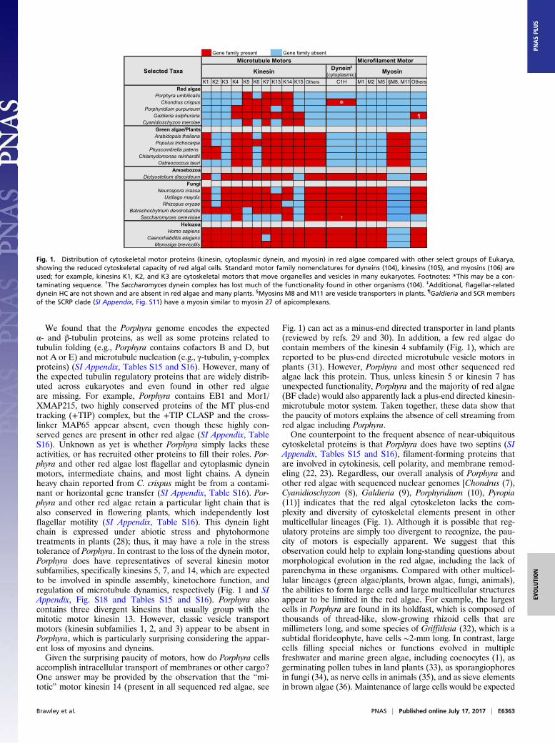
We found that the Porphyra genome encodes the expectedα- and β-tubulin proteins, as well as some proteins related totubulin folding (e.g., Porphyra contains cofactors B and D, butnot A or E) and microtubule nucleation (e.g., γ-tubulin, γ-complexproteins) (SI Appendix, Tables S15 and S16). However, many ofthe expected tubulin regulatory proteins that are widely distrib-uted across eukaryotes and even found in other red algaeare missing. For example, Porphyra contains EB1 and Mor1/XMAP215, two highly conserved proteins of the MT plus-endtracking (+TIP) complex, but the +TIP CLASP and the cross-linker MAP65 appear absent, even though these highly con-served genes are present in other red algae (SI Appendix, TableS16). Unknown as yet is whether Porphyra simply lacks theseactivities, or has recruited other proteins to fill their roles. Por-phyra and other red algae lost flagellar and cytoplasmic dyneinmotors, intermediate chains, and most light chains. A dyneinheavy chain reported from C. crispus might be from a contami-nant or horizontal gene transfer (SI Appendix, Table S16). Por-phyra and other red algae retain a particular light chain that isalso conserved in flowering plants, which independently lostflagellar motility (SI Appendix, Table S16). This dynein lightchain is expressed under abiotic stress and phytohormonetreatments in plants (28); thus, it may have a role in the stresstolerance of Porphyra. In contrast to the loss of the dynein motor,Porphyra does have representatives of several kinesin motorsubfamilies, specifically kinesins 5, 7, and 14, which are expectedto be involved in spindle assembly, kinetochore function, andregulation of microtubule dynamics, respectively (Fig. 1 and SIAppendix, Fig. S18 and Tables S15 and S16). Porphyra alsocontains three divergent kinesins that usually group with themitotic motor kinesin 13. However, classic vesicle transportmotors (kinesin subfamilies 1, 2, and 3) appear to be absent inPorphyra, which is particularly surprising considering the appar-ent loss of myosins and dyneins.Given the surprising paucity of motors, how do Porphyra cells
accomplish intracellular transport of membranes or other cargo?One answer may be provided by the observation that the “mi-totic” motor kinesin 14 (present in all sequenced red algae, see
Fig. 1) can act as a minus-end directed transporter in land plants(reviewed by refs. 29 and 30). In addition, a few red algae docontain members of the kinesin 4 subfamily (Fig. 1), which arereported to be plus-end directed microtubule vesicle motors inplants (31). However, Porphyra and most other sequenced redalgae lack this protein. Thus, unless kinesin 5 or kinesin 7 hasunexpected functionality, Porphyra and the majority of red algae(BF clade) would also apparently lack a plus-end directed kinesin-microtubule motor system. Taken together, these data show thatthe paucity of motors explains the absence of cell streaming fromred algae including Porphyra.One counterpoint to the frequent absence of near-ubiquitous
cytoskeletal proteins is that Porphyra does have two septins (SIAppendix, Tables S15 and S16), filament-forming proteins thatare involved in cytokinesis, cell polarity, and membrane remod-eling (22, 23). Regardless, our overall analysis of Porphyra andother red algae with sequenced nuclear genomes [Chondrus (7),Cyanidioschyzon (8), Galdieria (9), Porphyridium (10), Pyropia(11)] indicates that the red algal cytoskeleton lacks the com-plexity and diversity of cytoskeletal elements present in othermulticellular lineages (Fig. 1). Although it is possible that reg-ulatory proteins are simply too divergent to recognize, the pau-city of motors is especially apparent. We suggest that thisobservation could help to explain long-standing questions aboutmorphological evolution in the red algae, including the lack ofparenchyma in these organisms. Compared with other multicel-lular lineages (green algae/plants, brown algae, fungi, animals),the abilities to form large cells and large multicellular structuresappear to be limited in the red algae. For example, the largestcells in Porphyra are found in its holdfast, which is composed ofthousands of thread-like, slow-growing rhizoid cells that aremillimeters long, and some species of Griffithsia (32), which is asubtidal florideophyte, have cells ∼2-mm long. In contrast, largecells filling special niches or functions evolved in multiplefreshwater and marine green algae, including coenocytes (1), asgerminating pollen tubes in land plants (33), as sporangiophoresin fungi (34), as nerve cells in animals (35), and as sieve elementsin brown algae (36). Maintenance of large cells would be expected
Fig. 1. Distribution of cytoskeletal motor proteins (kinesin, cytoplasmic dynein, and myosin) in red algae compared with other select groups of Eukarya,showing the reduced cytoskeletal capacity of red algal cells. Standard motor family nomenclatures for dyneins (104), kinesins (105), and myosins (106) areused; for example, kinesins K1, K2, and K3 are cytoskeletal motors that move organelles and vesicles in many eukaryotes. Footnotes: *This may be a con-taminating sequence. †The Saccharomyces dynein complex has lost much of the functionality found in other organisms (104). ‡Additional, flagellar-relateddynein HC are not shown and are absent in red algae and many plants. §Myosins M8 and M11 are vesicle transporters in plants. ¶Galdieria and SCR membersof the SCRP clade (SI Appendix, Fig. S11) have a myosin similar to myosin 27 of apicomplexans.
Brawley et al. PNAS | Published online July 17, 2017 | E6363
EVOLU
TION
PNASPL
US
to require vigorous multidirectional intracellular transport, whichseems unlikely with a motor repertoire as limited as that seen inPorphyra and the other red algae with sequenced genomes (Fig. 1).Similarly, brown algae (46-m kelps) (36), animals, and plants
assemble large, complex 3D body plans with true parenchyma,but multicellular forms of red algae mostly consist of simplefilaments or filaments interwoven and tacked together by sec-ondary pit plugs (1) (i.e., pseudoparenchyma). Red algae areusually ≤50-cm long and only a few species reach 2 m in length(36, 37). Fungi cannot make parenchyma, but saprophytic my-celia grow to ≥50-m length (38). Failure of the red algae to formlarge multicellular structures is not straightforwardly attributedto cytoskeletal limitations; however, plants, which retain twomotors (myosin, kinesin) despite their independent loss of dy-nein, suffer serious stunting and other developmental abnor-malities following gene knockouts of myosins (23, 39). Analysisof major transcriptional and developmental regulators does notoffer a compelling explanation for why red algae have failed toevolve tissues comparable to those in brown algae, plants, andanimals (40). Taken together, our comparative analysis of thegenomes of Porphyra and other red algae leads us to speculatethat the small number of cytoskeletal elements in red algaecompared with those in other multicellular lineages (Fig. 1) hasconstrained the ability of red algae to develop larger, morecomplex cells and multicellular structures.
Stress. The ecological success of Porphyra and many of the closelyrelated bangiophyceans (37, 41) in the intertidal zone suggeststhat these species developed cellular mechanisms to cope withthis harsh environment. In particular, Porphyra grows from themid-to-high intertidal zone, where it is routinely exposed duringdaily low tides to high light, desiccation, and extreme fluctuationsin temperature and salinity. Blades can lose up to 95% of theirwater on some days, but are metabolically active as soon as theyare rehydrated by the rising tide (24). Here we infer noveladaptations to cope with these stresses.
Photoprotection. Light is required for photosynthesis, but severecellular damage can result from exposure of photosynthetic or-ganisms to the high levels of light (visible and UV) that arepresent in the mid-to-high intertidal zone where most bangio-phycean algae, including Porphyra, grow. Porphyra has the samecomplement of genes (SI Appendix, Table S19) to carry outphotosynthesis as other red algae, but exhibits a notable set ofphotoprotection strategies. Among the 13 genes encoding chlo-rophyll a-binding light-harvesting complex proteins are two“RedCap” genes (42), which may be involved in reorganizationof the photosynthetic antenna during the shift from darkness tolight (43). Porphyra also has 11 genes encoding “high light-in-duced” or “one-helix” proteins (here OHPs) that have essentialroles for photoacclimation and cell viability under stressful en-vironmental conditions (43, 44). Mechanistically, OHPs mayregulate chlorophyll and tetrapyrrole biosynthesis, stabilizephotosystem I (PSI), bind free chlorophyll or chlorophyll-breakdown products from damaged PSII complexes during thedamage/repair cycle, or bind carotenoids that dissipate excessabsorbed light energy. In contrast to the 11 Porphyra OHPs (SIAppendix, Table S19), we found 4 OHPs in Chondrus, whichexperiences less drying and light stress because of its low in-tertidal/subtidal habitat, 6 in P. yezoensis, 7 in Porphyridium, andonly 1 OHP in Cyanidioschyzon, which inhabits a stable hotspringenvironment (SI Appendix, Photosynthesis, Photoprotection, StressGenes). More analysis is needed, but the putative gene familyexpansion in Porphyra suggests positive selection for increasedgene dosage. Porphyra was one of the first organisms wherequenching of excess excitation energy in response to desiccationstress was observed (44), but the molecular mechanisms re-sponsible for this quenching in red algae remain unclear.
Porphyra encodes genes for catalases and peroxidases, as wellas the biosynthesis of numerous antioxidants, such as ascorbicacid (vitamin C) and tocopherol (vitamin E) (SI Appendix, Ta-bles S20–S22). When overexcitation of photosynthetic electrontransport occurs and reactive oxygen is generated, catalase de-toxifies hydrogen peroxide in red algae, and the expansion of thecatalase gene family in Porphyra and Pyropia (five genes) (SIAppendix, Table S21) compared with other red algae (one to twogenes) could reflect the demand for detoxification of reactiveoxygen species that cannot diffuse away from blades exposed bythe falling tide to high light and air while they are still hydratedand photosynthetically active. Tocopherols prevent photooxidativedamage of polyunsaturated fatty acids (45), and the Porphyraγ-tocopherol methyl transferase (SI Appendix, Table S22) cata-lyzes synthesis of α- and β-tocopherol. The isomer composition isunknown, but α-tocopherol has been identified in Porphyra bladesand is considered the more potent antioxidant (46, 47). The32 heat shock proteins (Hsp) in Porphyra indicate a possible ex-pansion of the Hsp40 family (SI Appendix, Table S20), whichare cochaperones of Hsp70 and play an important role inprotein maturation and repair under normal and stressedconditions (48, 49).Porphyra is frequently exposed to elevated intensities of
UV radiation in the intertidal zone and shows remarkabletolerance to both UV-A and UV-B (50, 51). Porphyra has atleast two strategies to protect photosynthesis and other keycellular processes from UV damage: mycosporine-like aminoacids (MAAs) and circadian control over the timing of UV-sensitive processes.MAAs act as “sunscreens” and comprise up to 1% of the dry
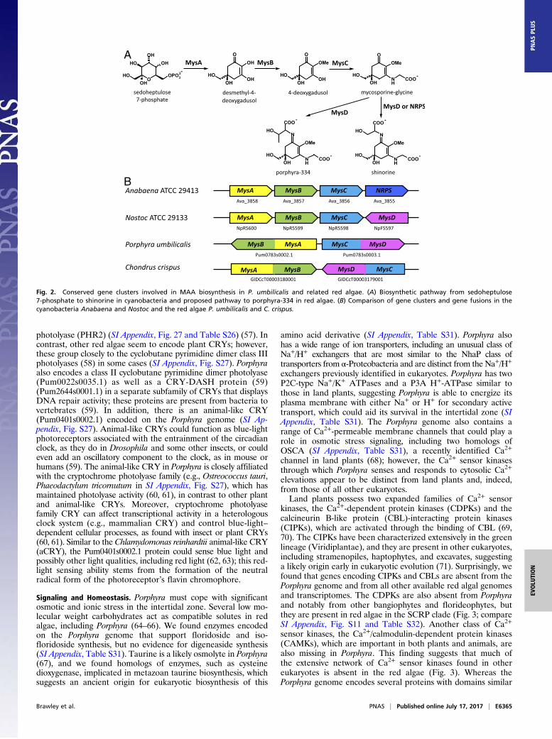
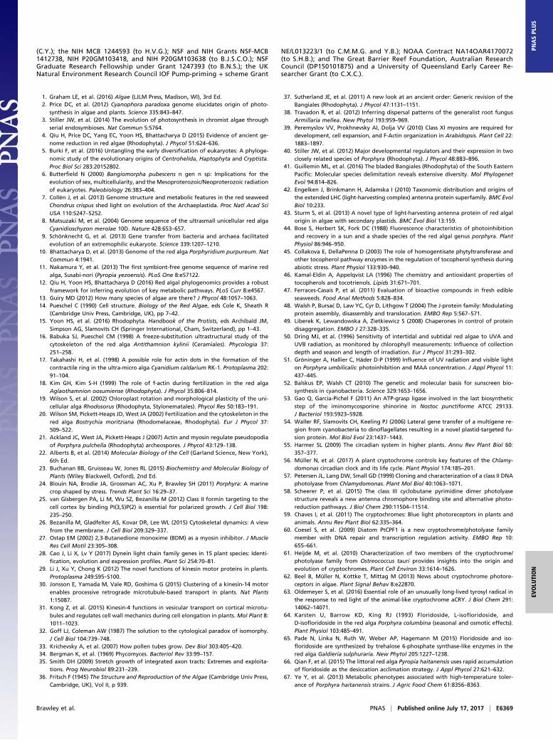
weight of Porphyra, with the compound porphyra-334 being themajor MAA (51). Four proteins—MysA, MysB, MysC, andMysD—support the biosynthesis of the MAA shinorine in cya-nobacteria, such as Nostoc (52, 53), whereas MysD is replaced bya nonribosomal peptide synthase in Anabaena (Fig. 2). Cyano-bacterial MysD shows a relaxed substrate specificity, with con-densation of threonine instead of serine onto mycosporine-glycine to yield porphyra-334 (53). The Porphyra genomecontains a gene encoding a MysA and MysB protein fusion thatis also found in several other red algae that synthesize MAAs, aswell as in some dinoflagellates, which were proposed to haveacquired the still separate but neighboring genes from a cyano-bacterium (54) (Fig. 2 and SI Appendix, Figs. S23 and S24). Thepresence of the MysA–MysB fusion in red algae suggests thatdinoflagellates could have acquired these genes from red algaethrough secondary or serial endosymbiosis (54) (SI Appendix,Fig. S23). Moreover, Porphyra and related species contain a geneencoding a MysC–MysD fusion protein, and in the Porphyragenome the MysA–MysB and MysC–MysD fusion genes are nextto each other but transcribed on opposite DNA strands withadjacent 5′-ends (Fig. 2). Although the Chondrus genome alsocontains clustered MysA–MysB and MysC–MysD, the fusionproteins are encoded on opposite strands with the 3′-ends ofthe genes adjacent. Conservation of the MAA gene cluster inPorphyra and Chondrus and the two gene-fusion events suggestthat this arrangement provides a selective advantage and effi-cient MAA biosynthesis for red algae that experience highUV irradiance.Developmental and abiotic stress responses are often associ-
ated with photoreceptors in eukaryotes. The plant circadianclock contains blue- and red-light photoreceptors, includingcryptochromes (CRY) and phytochromes (PHY), to entrain thecircadian clock (55, 56); these photoreceptors are also involvedin other fundamental processes in plants, including growth anddevelopment. Porphyra does not appear to encode a PHY pho-toreceptor or a typical plant CRY photoreceptor, although it hasmaintained four genes of the CRY/photolyase family. The Por-phyra CRY that is most like plant CRYs is similar to a DNA
E6364 | www.pnas.org/cgi/doi/10.1073/pnas.1703088114 Brawley et al.
photolyase (PHR2) (SI Appendix, Fig. 27 and Table S26) (57). Incontrast, other red algae seem to encode plant CRYs; however,these group closely to the cyclobutane pyrimidine dimer class IIIphotolyases (58) in some cases (SI Appendix, Fig. S27). Porphyraalso encodes a class II cyclobutane pyrimidine dimer photolyase(Pum0022s0035.1) as well as a CRY-DASH protein (59)(Pum2644s0001.1) in a separate subfamily of CRYs that displaysDNA repair activity; these proteins are present from bacteria tovertebrates (59). In addition, there is an animal-like CRY(Pum0401s0002.1) encoded on the Porphyra genome (SI Ap-pendix, Fig. S27). Animal-like CRYs could function as blue-lightphotoreceptors associated with the entrainment of the circadianclock, as they do in Drosophila and some other insects, or couldeven add an oscillatory component to the clock, as in mouse orhumans (59). The animal-like CRY in Porphyra is closely affiliatedwith the cryptochrome photolyase family (e.g., Ostreococcus tauri,Phaeodactylum tricornutum in SI Appendix, Fig. S27), which hasmaintained photolyase activity (60, 61), in contrast to other plantand animal-like CRYs. Moreover, cryptochrome photolyasefamily CRY can affect transcriptional activity in a heterologousclock system (e.g., mammalian CRY) and control blue-light–dependent cellular processes, as found with insect or plant CRYs(60, 61). Similar to the Chlamydomonas reinhardtii animal-like CRY(aCRY), the Pum0401s0002.1 protein could sense blue light andpossibly other light qualities, including red light (62, 63); this red-light sensing ability stems from the formation of the neutralradical form of the photoreceptor’s flavin chromophore.
Signaling and Homeostasis. Porphyra must cope with significantosmotic and ionic stress in the intertidal zone. Several low mo-lecular weight carbohydrates act as compatible solutes in redalgae, including Porphyra (64–66). We found enzymes encodedon the Porphyra genome that support floridoside and iso-floridoside synthesis, but no evidence for digeneaside synthesis(SI Appendix, Table S31). Taurine is a likely osmolyte in Porphyra(67), and we found homologs of enzymes, such as cysteinedioxygenase, implicated in metazoan taurine biosynthesis, whichsuggests an ancient origin for eukaryotic biosynthesis of this
amino acid derivative (SI Appendix, Table S31). Porphyra alsohas a wide range of ion transporters, including an unusual class ofNa+/H+ exchangers that are most similar to the NhaP class oftransporters from α-Proteobacteria and are distinct from the Na+/H+
exchangers previously identified in eukaryotes. Porphyra has twoP2C-type Na+/K+ ATPases and a P3A H+-ATPase similar tothose in land plants, suggesting Porphyra is able to energize itsplasma membrane with either Na+ or H+ for secondary activetransport, which could aid its survival in the intertidal zone (SIAppendix, Table S31). The Porphyra genome also contains arange of Ca2+-permeable membrane channels that could play arole in osmotic stress signaling, including two homologs ofOSCA (SI Appendix, Table S31), a recently identified Ca2+
channel in land plants (68); however, the Ca2+ sensor kinasesthrough which Porphyra senses and responds to cytosolic Ca2+
elevations appear to be distinct from land plants and, indeed,from those of all other eukaryotes.Land plants possess two expanded families of Ca2+ sensor
kinases, the Ca2+-dependent protein kinases (CDPKs) and thecalcineurin B-like protein (CBL)-interacting protein kinases(CIPKs), which are activated through the binding of CBL (69,70). The CIPKs have been characterized extensively in the greenlineage (Viridiplantae), and they are present in other eukaryotes,including stramenopiles, haptophytes, and excavates, suggestinga likely origin early in eukaryotic evolution (71). Surprisingly, wefound that genes encoding CIPKs and CBLs are absent from thePorphyra genome and from all other available red algal genomesand transcriptomes. The CDPKs are also absent from Porphyraand notably from other bangiophytes and florideophytes, butthey are present in red algae in the SCRP clade (Fig. 3; compareSI Appendix, Fig. S11 and Table S32). Another class of Ca2+
sensor kinases, the Ca2+/calmodulin-dependent protein kinases(CAMKs), which are important in both plants and animals, arealso missing in Porphyra. This finding suggests that much ofthe extensive network of Ca2+ sensor kinases found in othereukaryotes is absent in the red algae (Fig. 3). Whereas thePorphyra genome encodes several proteins with domains similar
A
B
Fig. 2. Conserved gene clusters involved in MAA biosynthesis in P. umbilicalis and related red algae. (A) Biosynthetic pathway from sedoheptulose7-phosphate to shinorine in cyanobacteria and proposed pathway to porphyra-334 in red algae. (B) Comparison of gene clusters and gene fusions in thecyanobacteria Anabaena and Nostoc and the red algae P. umbilicalis and C. crispus.
Brawley et al. PNAS | Published online July 17, 2017 | E6365
EVOLU
TION
PNASPL
US
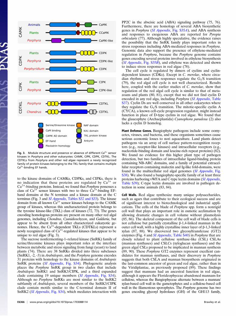
to the kinase domains of CAMKs, CDPKs, and CIPKs, there isno indication that these proteins are regulated by Ca2+ orCa2+-binding proteins. Instead, we found that Porphyra possesses aclass of Ca2+ sensor kinases with two to three Ca2+-binding EF-hand domains at the N terminus and a kinase domain at the Cterminus (Fig. 3 and SI Appendix, Tables S32 and S33). The kinasedomain from all known Ca2+ sensor kinases belongs to the CAMKgroup of kinases, whereas this uncharacterized protein belongs tothe tyrosine kinase-like (TKL) class of kinases (72, 73). The genesencoding homologous proteins are present on many other red algalgenomes, including Chondrus, Cyanidioschyzon, and Galdieria, butappear to be absent from all other characterized eukaryotic ge-nomes. Hence, the Ca2+-dependent TKLs (CDTKLs) represent anewly recognized class of Ca2+-regulated kinases that appear to beunique to red algae (Fig. 3).The sucrose nonfermenting-1–related kinase (SnRK) family of
serine/threonine kinases plays important roles at the interfacebetween metabolic and stress signaling from fungi (yeast) to landplants (74). There are 38 SnRKs divided into three subclasses(SnRK1, -2, -3) in Arabidopsis, and the Porphyra genome encodes31 proteins with homology to the kinase domains of ArabidopsisSnRK proteins (SI Appendix, Fig. S34). Phylogenetic analysisplaces the Porphyra SnRK group in two clades that includeArabidopsis SnRK1 and SnRK3/CIPK, and a third expandedclade containing 19 unique members (SI Appendix, Fig. S34).Although no Porphyra SnRKs are most similar to the SnRK2subfamily of Arabidopsis, several members of the SnRK3/CIPKclade contain motifs similar to the C-terminal domain II ofSnRK2 (SI Appendix, Fig. S34), which mediates interaction with
PP2C in the abscisic acid (ABA) signaling pathway (75, 76).Furthermore, there are homologs of several ABA biosyntheticgenes in Porphyra (SI Appendix, Fig. S35A), and ABA synthesisand responses to exogenous ABA are reported for Pyropiaorbicularis (77). Although highly speculative, the evidence raisesthe possibility that the SnRK family plays important roles instress responses including ABA-mediated responses in Porphyra.Genomic data also support the presence of ethylene-mediatedregulation in Porphyra, because the Porphyra genome containsgenes encoding several proteins involved in ethylene biosynthesis(SI Appendix, Fig. S35B), and ethylene was detected and shownto induce stress responses in red algae (78).The cell cycle is regulated by dimers of cyclins and cyclin-
dependent kinases (CDKs). Except in C. merolae, where circa-dian rhythms and stress responses regulate the G1/S transition(79), the red algal cell cycle is not well characterized. Resultshere, coupled with the earlier studies of C. merolae, show thatregulation of the red algal cell cycle is similar to that of meta-zoans and plants (80, 81), except that we did not find cyclin Dencoded in any red alga, including Porphyra (SI Appendix, TableS37). Cyclin Ds are well conserved in all other eukaryotes wherethey regulate the G1/S transition. The mitotic-specific cyclin A(CYCA), a known cell-cycle progression regulator, might insteadfunction in place of D-type cyclins in red algae. We found thatthe glaucophyte (Archaeplastida) Cyanophora paradoxa (2) alsolacks a cyclin D homolog.
Plant Defense Genes. Bangiophyte pathogens include some oomy-cetes, viruses, and bacteria, and these organisms sometimes causeserious economic losses to nori aquaculture. Land plants detectpathogens via an array of cell surface pattern-recognition recep-tors (e.g., receptor-like kinases) and intracellular receptors (e.g.,nucleotide-binding domain and leucine-rich repeat proteins) (82).We found no evidence for this higher plant type of pathogendetection, but two families of intracellular ligand-binding proteincontaining NB-ARC domains, and a family of potential extracel-lular receptors containing malectin and Ig-like fold-domains, werefound in the multicellular red algal genomes (SI Appendix, Fig.S38). We also found a bangiophyte-specific family of at least threeproteins harboring vWFA and C-type lectin domains (SI Appendix,Table S39); C-type lectin domains are involved in pathogen de-tection in some animals (83, 84).
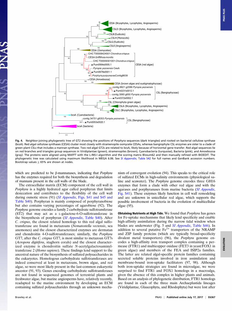
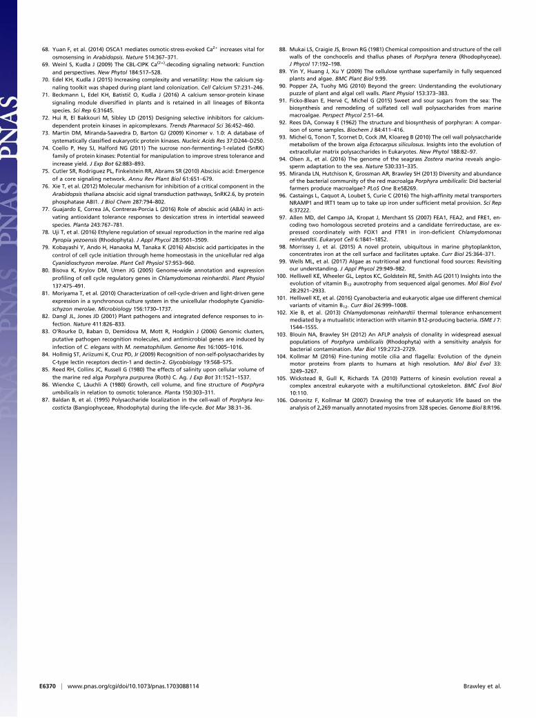
Cell Walls. Red algae synthesize many unique polysaccharides,such as agars that contribute to their ecological success and areof significant interest to biotechnological and industrial appli-cations. The cells of the blade of Porphyra spp. form a nonrigidcell wall that plays an important role in osmotic acclimation byallowing dramatic changes in cell volume without plasmolysis(85, 86). The skeletal component of the cell wall of blade cells isnot cellulose but partially crystalline β-1,4-linked mannan in theouter cell wall, with a highly crystalline inner layer of β-1,3-linkedxylan (87, 88). We discovered two glycosyltransferase (GT2)enzymes (Fig. 4 and SI Appendix, Table S40) in Porphyra that areclosely related to plant cellulose synthase-like (CSL) CSLAs(mannan synthases) and CSLCs (xyloglucan synthases) and thegreen algal CSLs proposed to be implicated in mannan synthesis(89, 90). These Porphyra GT2 enzymes represent excellent can-didates for mannan synthases, and their discovery in Porphyrasuggests that both CSLA and mannan biosynthesis originated inthe last-common ancestor of green and red algae, rather than inthe Viridiplantae, as previously proposed (89). These findingssuggest that mannans had an ancestral function in red algae,although it appears the Florideophyceae abandoned mannans forcellulose, whereas the Bangiophyceae alternate between a mannan/xylan-based cell wall in the gametophytes and a cellulose-based cellwall in the filamentous sporophytes. The Porphyra genome has twogenes encoding glycosyl hydrolases (GH) of the GH113 family,
CaMK
CDPK
CDPK
CCaMK
CaMK-like
CIPK
CIPK-like
CDTKL
CDTKL
CDPK-like
Animals
Plants
Porphyra
Porphyridium
Plants
Porphyra
Plants
Porphyra
Porphyra
Chondrus
Serine/threonine kinase
CaM binding
EF-hand
NAF domain
Ankyrin
Pleckstrin-homology
CAMK AD domain TKL protein kinase
KA1 domain
CAM
KKPI
CKP
DC
CD
TKL
Fig. 3. Module structure and presence or absence of different Ca2+ sensorkinases in Porphyra and other eukaryotes: CAMK, CIPK, CDPK, CDTKL. TheCDTKLs from Porphyra and other red algae represent a newly recognizedfamily of protein kinases belonging to the TKL family that contains multipleCa2+-binding EF hands.
E6366 | www.pnas.org/cgi/doi/10.1073/pnas.1703088114 Brawley et al.
which are predicted to be β-mannanases, indicating that Porphyrahas the enzymes required for both the biosynthesis and degradationof mannans present in the cell walls of the blade.The extracellular matrix (ECM) component of the cell wall in
Porphyra is a highly hydrated agar called porphyran that limitsdesiccation and contributes to the flexibility of the cell wallduring osmotic stress (91) (SI Appendix, Figs. S41 and S45 andTable S40). Porphyran is mainly composed of porphyranobiosebut also contains varying percentages of agarobiose (92). ThePorphyra genome encodes a family 2 carbohydrate sulfotransferase(ST2) that may act as a L-galactose-6-O-sulfotransferase inthe biosynthesis of porphyran (SI Appendix, Table S40). AfterC. crispus, the closest related homologs to this red algal sulfo-transferase are found in metazoans (Nematostella vectensis, seaanemones) and the closest characterized enzymes are dermatanand chondroitin 4-O-sulfotransferases; similarly, the PorphyraGT7, after the C. crispus GT7, is most similar to metazoan GT7s(Acropora digitifera, staghorn corals) and the closest character-ized enzyme is chondroitin sulfate N-acetylgalactosaminyl-transferase 2 (Homo sapiens). These findings lend support to theancestral nature of the biosynthesis of sulfated polysaccharides inthe eukaryotes. Homologous carbohydrate sulfotransferases areindeed conserved at least in metazoans, brown algae, and redalgae, so were most likely present in the last-common eukaryoticancestor (91, 93). Genes encoding carbohydrate sulfotransferasesare not found in sequenced genomes of terrestrial plants andfreshwater algae, but marine angiosperms have, relatively recently,readapted to the marine environment by developing an ECMcontaining sulfated polysaccharides through an unknown mecha-
nism of convergent evolution (94). This speaks to the critical roleof sulfated ECMs in high-salinity environments (physiological sa-line and seawater). The Porphyra genome encodes three GH16enzymes that form a clade with other red algae and with theagarases and porphyranases from marine bacteria (SI Appendix,Fig. S41). These enzymes likely function in cell wall remodelingand are unknown in unicellular red algae, which supports thepossible involvement of bacteria in the evolution of multicellularalgae (95).
Obtaining Nutrients at High Tide.We found that Porphyra has genesfor Fe-uptake mechanisms that likely lend specificity and enablehigh-affinity assimilation during the narrow tidal window whenblades are underwater (Fig. 5 and SI Appendix, Table S46). Inaddition to several putative Fe2+ transporters of the NRAMPand ZIP family proteins (which are typically broad-specificitydivalent metal transporters) (96), the Porphyra genome en-codes a high-affinity iron transport complex containing a per-mease (FTR1) and multicopper oxidase (FET3 in yeast/FOX1 ingreen algae) and members of the FEA and ISIP2a families.The latter are related algal-specific protein families containingsecreted soluble proteins involved in iron assimilation andmembrane-bound iron-uptake facilitators (97, 98). Althoughboth iron-uptake strategies are found in microalgae, we weresurprised to find FTR1 and FOX1 homologs in a macroalga,given the absence of this complex in higher plants and animals.Based on an analysis of phylogenetic distribution, FTR1 homologsare found in each of the three main Archaeplastida lineages(Viridiplantae, Glaucophyta, and Rhodophyta) but were lost after
CESA (Bryophytes, Lycophytes, Angiosperms)
CSLD (Bryophytes, Lycophytes, Angiosperms)
CSLB (Eudicots)CSLH (Monocots)
CSLG (Eudicots)
CSLE (Angiosperms)
CESA (Oomycetes)CHC T00008424001 Chondrus crispusCESA Griffithsia monilis
CHC T00009301001 Chondrus crispusPum0564s0005.1Pyropia yezoensis CesAPum0678s0001.1
Porphyra purpurea esContig8639
CESA (red algae)
CESA (Amoebozoa)
CESA (brown algae and eustigmatophyceae)contig 8821 g2086 Pyropia yezoensis
Pum0231s0012.1contig 3060 g650 Pyropia yezoensis
Pum0372s0002.1
CSL (Bangiophyceae)
CSL (Chlorophyta green algae)
CSLA (Bryophytes, Lycophytes, Angiosperms)
CSLC (Bryophytes, Lycophytes, Angiosperms)
bcsA (Cyanobacteria)contig 34751 g8353 Pyropia yezoensis
Pum0053s0020.1CSL (Bangiophyceae)
bcsA (bacteria)
100
100
85
98
100
100
100
100
82
94
100
100
100
85
100
100
100
100
100
100
100
100
65
93
82
100
95
81
100
99
80
0.5
Fig. 4. Neighbor-joining phylogenetic tree of GT2 showing the positions of Porphyra sequences (dark triangles) and rooted on bacterial cellulose synthase(bcsA). Red algal cellulose synthases (CESA) cluster most closely with stramenopile oomycete CESAs, whereas bangiophyte CSL enzymes are sister to a clade ofgreen plant CSLs that includes a mannan synthase. Two red algal GT2s are related to bcsA, likely because of horizontal gene transfer. Red algal sequences lieon red branches and triangles group sequences in Viridiplantae (green), stramenopiles (brown), Cyanobacteria (turquoise), Bacteria (pink), and Amoebozoa(gray). The proteins were aligned using MAFFT with the L-INS-I algorithm and the scoring matrix Blosum62 and then manually refined with BIOEDIT. Thephylogenetic tree was calculated using maximum likelihood in MEGA 6.06. See SI Appendix, Table S42 for full names and GenBank accession numbers.Bootstrap values ≥ 65% are shown at nodes.
Brawley et al. PNAS | Published online July 17, 2017 | E6367
EVOLU
TION
PNASPL
US
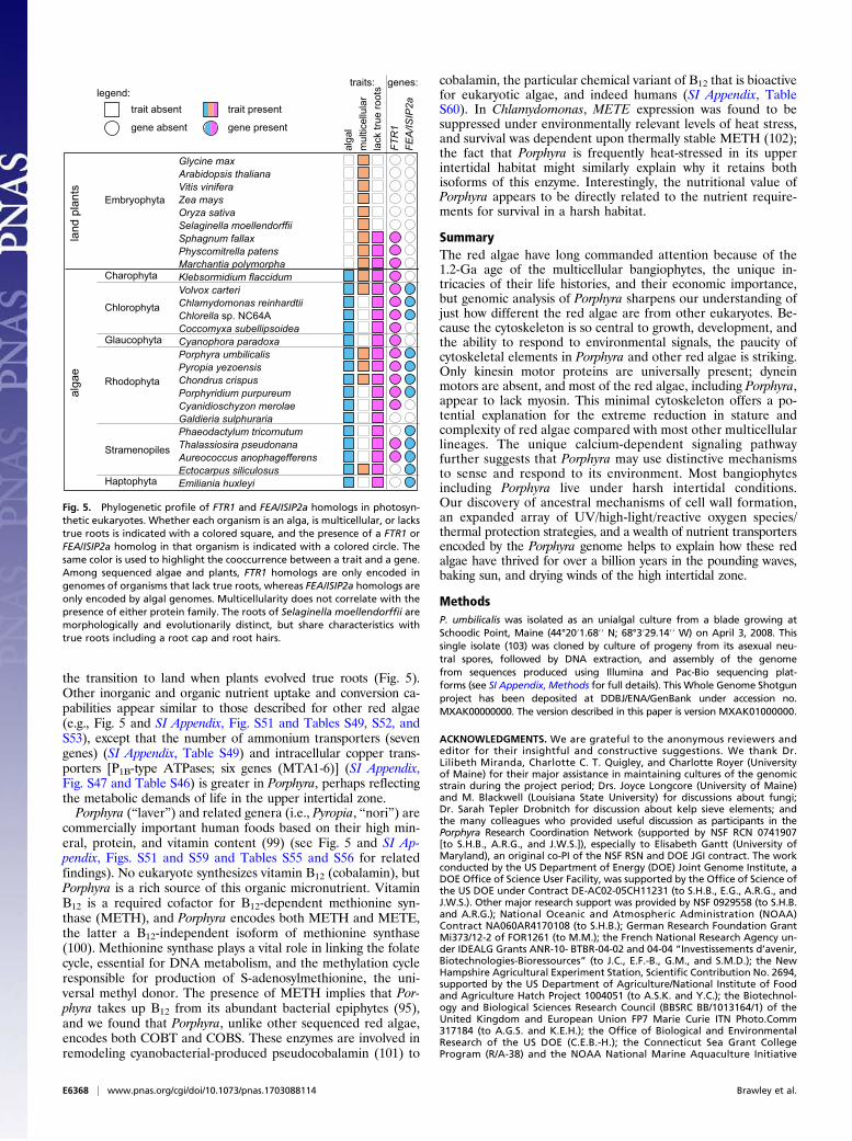
the transition to land when plants evolved true roots (Fig. 5).Other inorganic and organic nutrient uptake and conversion ca-pabilities appear similar to those described for other red algae(e.g., Fig. 5 and SI Appendix, Fig. S51 and Tables S49, S52, andS53), except that the number of ammonium transporters (sevengenes) (SI Appendix, Table S49) and intracellular copper trans-porters [P1B-type ATPases; six genes (MTA1-6)] (SI Appendix,Fig. S47 and Table S46) is greater in Porphyra, perhaps reflectingthe metabolic demands of life in the upper intertidal zone.Porphyra (“laver”) and related genera (i.e., Pyropia, “nori”) are
commercially important human foods based on their high min-eral, protein, and vitamin content (99) (see Fig. 5 and SI Ap-pendix, Figs. S51 and S59 and Tables S55 and S56 for relatedfindings). No eukaryote synthesizes vitamin B12 (cobalamin), butPorphyra is a rich source of this organic micronutrient. VitaminB12 is a required cofactor for B12-dependent methionine syn-thase (METH), and Porphyra encodes both METH and METE,the latter a B12-independent isoform of methionine synthase(100). Methionine synthase plays a vital role in linking the folatecycle, essential for DNA metabolism, and the methylation cycleresponsible for production of S-adenosylmethionine, the uni-versal methyl donor. The presence of METH implies that Por-phyra takes up B12 from its abundant bacterial epiphytes (95),and we found that Porphyra, unlike other sequenced red algae,encodes both COBT and COBS. These enzymes are involved inremodeling cyanobacterial-produced pseudocobalamin (101) to
cobalamin, the particular chemical variant of B12 that is bioactivefor eukaryotic algae, and indeed humans (SI Appendix, TableS60). In Chlamydomonas, METE expression was found to besuppressed under environmentally relevant levels of heat stress,and survival was dependent upon thermally stable METH (102);the fact that Porphyra is frequently heat-stressed in its upperintertidal habitat might similarly explain why it retains bothisoforms of this enzyme. Interestingly, the nutritional value ofPorphyra appears to be directly related to the nutrient require-ments for survival in a harsh habitat.
SummaryThe red algae have long commanded attention because of the1.2-Ga age of the multicellular bangiophytes, the unique in-tricacies of their life histories, and their economic importance,but genomic analysis of Porphyra sharpens our understanding ofjust how different the red algae are from other eukaryotes. Be-cause the cytoskeleton is so central to growth, development, andthe ability to respond to environmental signals, the paucity ofcytoskeletal elements in Porphyra and other red algae is striking.Only kinesin motor proteins are universally present; dyneinmotors are absent, and most of the red algae, including Porphyra,appear to lack myosin. This minimal cytoskeleton offers a po-tential explanation for the extreme reduction in stature andcomplexity of red algae compared with most other multicellularlineages. The unique calcium-dependent signaling pathwayfurther suggests that Porphyra may use distinctive mechanismsto sense and respond to its environment. Most bangiophytesincluding Porphyra live under harsh intertidal conditions.Our discovery of ancestral mechanisms of cell wall formation,an expanded array of UV/high-light/reactive oxygen species/thermal protection strategies, and a wealth of nutrient transportersencoded by the Porphyra genome helps to explain how these redalgae have thrived for over a billion years in the pounding waves,baking sun, and drying winds of the high intertidal zone.
MethodsP. umbilicalis was isolated as an unialgal culture from a blade growing atSchoodic Point, Maine (44°20′1.68′′ N; 68°3′29.14′′ W) on April 3, 2008. Thissingle isolate (103) was cloned by culture of progeny from its asexual neu-tral spores, followed by DNA extraction, and assembly of the genomefrom sequences produced using Illumina and Pac-Bio sequencing plat-forms (see SI Appendix,Methods for full details). This Whole Genome Shotgunproject has been deposited at DDBJ/ENA/GenBank under accession no.MXAK00000000. The version described in this paper is version MXAK01000000.
ACKNOWLEDGMENTS. We are grateful to the anonymous reviewers andeditor for their insightful and constructive suggestions. We thank Dr.Lilibeth Miranda, Charlotte C. T. Quigley, and Charlotte Royer (Universityof Maine) for their major assistance in maintaining cultures of the genomicstrain during the project period; Drs. Joyce Longcore (University of Maine)and M. Blackwell (Louisiana State University) for discussions about fungi;Dr. Sarah Tepler Drobnitch for discussion about kelp sieve elements; andthe many colleagues who provided useful discussion as participants in thePorphyra Research Coordination Network (supported by NSF RCN 0741907[to S.H.B., A.R.G., and J.W.S.]), especially to Elisabeth Gantt (University ofMaryland), an original co-PI of the NSF RSN and DOE JGI contract. The workconducted by the US Department of Energy (DOE) Joint Genome Institute, aDOE Office of Science User Facility, was supported by the Office of Science ofthe US DOE under Contract DE-AC02-05CH11231 (to S.H.B., E.G., A.R.G., andJ.W.S.). Other major research support was provided by NSF 0929558 (to S.H.B.and A.R.G.); National Oceanic and Atmospheric Administration (NOAA)Contract NA060AR4170108 (to S.H.B.); German Research Foundation GrantMi373/12-2 of FOR1261 (to M.M.); the French National Research Agency un-der IDEALG Grants ANR-10- BTBR-04-02 and 04-04 “Investissements d’avenir,Biotechnologies-Bioressources” (to J.C., E.F.-B., G.M., and S.M.D.); the NewHampshire Agricultural Experiment Station, Scientific Contribution No. 2694,supported by the US Department of Agriculture/National Institute of Foodand Agriculture Hatch Project 1004051 (to A.S.K. and Y.C.); the Biotechnol-ogy and Biological Sciences Research Council (BBSRC BB/1013164/1) of theUnited Kingdom and European Union FP7 Marie Curie ITN Photo.Comm317184 (to A.G.S. and K.E.H.); the Office of Biological and EnvironmentalResearch of the US DOE (C.E.B.-H.); the Connecticut Sea Grant CollegeProgram (R/A-38) and the NOAA National Marine Aquaculture Initiative
Chlorophyta
Haptophyta
Cyanophora paradoxa
Selaginella moellendorffiiOryza sativa
Physcomitrella patens
Glycine max Arabidopsis thaliana
Klebsormidium flaccidumCharophyta
Embryophyta
Cyanidioschyzon merolaeGaldieria sulphuraria
Pyropia yezoensisPorphyra umbilicalis
Glaucophyta
Rhodophyta
Thalassiosira pseudonanaPhaeodactylum tricornutum
Aureococcus anophagefferens
Emiliania huxleyi Ectocarpus siliculosus
Stramenopiles
Sphagnum fallax
Zea maysVitis vinifera
Coccomyxa subellipsoideaChlorella sp. NC64A
Volvox carteriChlamydomonas reinhardtii
Porphyridium purpureumChondrus crispus
FTR
1
mul
ticel
lula
rla
ck tr
ue ro
ots
FEA
/ISIP
2a
genes:traits:
Marchantia polymorpha
land
pla
nts
alga
e
alga
l
legend:trait absent
gene absent
trait present
gene present
Fig. 5. Phylogenetic profile of FTR1 and FEA/ISIP2a homologs in photosyn-thetic eukaryotes. Whether each organism is an alga, is multicellular, or lackstrue roots is indicated with a colored square, and the presence of a FTR1 orFEA/ISIP2a homolog in that organism is indicated with a colored circle. Thesame color is used to highlight the cooccurrence between a trait and a gene.Among sequenced algae and plants, FTR1 homologs are only encoded ingenomes of organisms that lack true roots, whereas FEA/ISIP2a homologs areonly encoded by algal genomes. Multicellularity does not correlate with thepresence of either protein family. The roots of Selaginella moellendorffii aremorphologically and evolutionarily distinct, but share characteristics withtrue roots including a root cap and root hairs.
E6368 | www.pnas.org/cgi/doi/10.1073/pnas.1703088114 Brawley et al.
(C.Y.); the NIH MCB 1244593 (to H.V.G.); NSF and NIH Grants NSF-MCB1412738, NIH P20GM103418, and NIH P20GM103638 (to B.J.S.C.O.); NSFGraduate Research Fellowship under Grant 1247393 (to B.N.S.); the UKNatural Environment Research Council IOF Pump-priming + scheme Grant
NE/L013223/1 (to C.M.M.G. and Y.B.); NOAA Contract NA14OAR4170072(to S.H.B.); and The Great Barrier Reef Foundation, Australian ResearchCouncil (DP150101875) and a University of Queensland Early Career Re-searcher Grant (to C.X.C.).
1. Graham LE, et al. (2016) Algae (LJLM Press, Madison, WI), 3rd Ed.2. Price DC, et al. (2012) Cyanophora paradoxa genome elucidates origin of photo-
synthesis in algae and plants. Science 335:843–847.3. Stiller JW, et al. (2014) The evolution of photosynthesis in chromist algae through
serial endosymbioses. Nat Commun 5:5764.4. Qiu H, Price DC, Yang EC, Yoon HS, Bhattacharya D (2015) Evidence of ancient ge-
nome reduction in red algae (Rhodophyta). J Phycol 51:624–636.5. Burki F, et al. (2016) Untangling the early diversification of eukaryotes: A phyloge-
nomic study of the evolutionary origins of Centrohelida, Haptophyta and Cryptista.Proc Biol Sci 283:20152802.
6. Butterfield N (2000) Bangiomorpha pubescens n gen n sp: Implications for theevolution of sex, multicellularity, and the Mesoproterozoic/Neoproterozoic radiationof eukaryotes. Paleobiology 26:383–404.
7. Collén J, et al. (2013) Genome structure and metabolic features in the red seaweedChondrus crispus shed light on evolution of the Archaeplastida. Proc Natl Acad SciUSA 110:5247–5252.
8. Matsuzaki M, et al. (2004) Genome sequence of the ultrasmall unicellular red algaCyanidioschyzon merolae 10D. Nature 428:653–657.
9. Schönknecht G, et al. (2013) Gene transfer from bacteria and archaea facilitatedevolution of an extremophilic eukaryote. Science 339:1207–1210.
10. Bhattacharya D, et al. (2013) Genome of the red alga Porphyridium purpureum. NatCommun 4:1941.
11. Nakamura Y, et al. (2013) The first symbiont-free genome sequence of marine redalga, Susabi-nori (Pyropia yezoensis). PLoS One 8:e57122.
12. Qiu H, Yoon HS, Bhattacharya D (2016) Red algal phylogenomics provides a robustframework for inferring evolution of key metabolic pathways. PLoS Curr 8:e4567.
13. Guiry MD (2012) How many species of algae are there? J Phycol 48:1057–1063.14. Pueschel C (1990) Cell structure. Biology of the Red Algae, eds Cole K, Sheath R
(Cambridge Univ Press, Cambridge, UK), pp 7–42.15. Yoon HS, et al. (2016) Rhodophyta. Handbook of the Protists, eds Archibald JM,
Simpson AG, Slamovits CH (Springer International, Cham, Switzerland), pp 1–43.16. Babuka SJ, Pueschel CM (1998) A freeze-substitution ultrastructural study of the
cytoskeleton of the red alga Antithamnion kylinii (Ceramiales). Phycologia 37:251–258.
17. Takahashi H, et al. (1998) A possible role for actin dots in the formation of thecontractile ring in the ultra-micro alga Cyanidium caldarium RK-1. Protoplasma 202:91–104.
18. Kim GH, Kim S-H (1999) The role of f-actin during fertilization in the red algaAglaothamnion oosumiense (Rhodophyta). J Phycol 35:806–814.
19. Wilson S, et al. (2002) Chloroplast rotation and morphological plasticity of the uni-cellular alga Rhodosorus (Rhodophyta, Stylonematales). Phycol Res 50:183–191.
20. Wilson SM, Pickett-Heaps JD, West JA (2002) Fertilization and the cytoskeleton in thered alga Bostrychia moritziana (Rhodomelaceae, Rhodophyta). Eur J Phycol 37:509–522.
21. Ackland JC, West JA, Pickett-Heaps J (2007) Actin and myosin regulate pseudopodiaof Porphyra pulchella (Rhodophyta) archeospores. J Phycol 43:129–138.
22. Alberts B, et al. (2014) Molecular Biology of the Cell (Garland Science, New York),6th Ed.
23. Buchanan BB, Gruisseau W, Jones RL (2015) Biochemistry and Molecular Biology ofPlants (Wiley Blackwell, Oxford), 2nd Ed.
24. Blouin NA, Brodie JA, Grossman AC, Xu P, Brawley SH (2011) Porphyra: A marinecrop shaped by stress. Trends Plant Sci 16:29–37.
25. van Gisbergen PA, Li M, Wu SZ, Bezanilla M (2012) Class II formin targeting to thecell cortex by binding PI(3,5)P(2) is essential for polarized growth. J Cell Biol 198:235–250.
26. Bezanilla M, Gladfelter AS, Kovar DR, Lee WL (2015) Cytoskeletal dynamics: A viewfrom the membrane. J Cell Biol 209:329–337.
27. Ostap EM (2002) 2,3-Butanedione monoxime (BDM) as a myosin inhibitor. J MuscleRes Cell Motil 23:305–308.
28. Cao J, Li X, Lv Y (2017) Dynein light chain family genes in 15 plant species: Identi-fication, evolution and expression profiles. Plant Sci 254:70–81.
29. Li J, Xu Y, Chong K (2012) The novel functions of kinesin motor proteins in plants.Protoplasma 249:S95–S100.
30. Jonsson E, Yamada M, Vale RD, Goshima G (2015) Clustering of a kinesin-14 motorenables processive retrograde microtubule-based transport in plants. Nat Plants1:15087.
31. Kong Z, et al. (2015) Kinesin-4 functions in vesicular transport on cortical microtu-bules and regulates cell wall mechanics during cell elongation in plants. Mol Plant 8:1011–1023.
32. Goff LJ, Coleman AW (1987) The solution to the cytological paradox of isomorphy.J Cell Biol 104:739–748.
33. Krichevsky A, et al. (2007) How pollen tubes grow. Dev Biol 303:405–420.34. Bergman K, et al. (1969) Phycomyces. Bacteriol Rev 33:99–157.35. Smith DH (2009) Stretch growth of integrated axon tracts: Extremes and exploita-
tions. Prog Neurobiol 89:231–239.36. Fritsch F (1945) The Structure and Reproduction of the Algae (Cambridge Univ Press,
Cambridge, UK), Vol II, p 939.
37. Sutherland JE, et al. (2011) A new look at an ancient order: Generic revision of theBangiales (Rhodophyta). J Phycol 47:1131–1151.
38. Travadon R, et al. (2012) Inferring dispersal patterns of the generalist root fungusArmillaria mellea. New Phytol 193:959–969.
39. Peremyslov VV, Prokhnevsky AI, Dolja VV (2010) Class XI myosins are required fordevelopment, cell expansion, and F-Actin organization in Arabidopsis. Plant Cell 22:1883–1897.
40. Stiller JW, et al. (2012) Major developmental regulators and their expression in twoclosely related species of Porphyra (Rhodophyta). J Phycol 48:883–896.
41. Guillemin ML, et al. (2016) The bladed Bangiales (Rhodophyta) of the South EasternPacific: Molecular species delimitation reveals extensive diversity. Mol PhylogenetEvol 94:814–826.
42. Engelken J, Brinkmann H, Adamska I (2010) Taxonomic distribution and origins ofthe extended LHC (light-harvesting complex) antenna protein superfamily. BMC EvolBiol 10:233.
43. Sturm S, et al. (2013) A novel type of light-harvesting antenna protein of red algalorigin in algae with secondary plastids. BMC Evol Biol 13:159.
44. Bose S, Herbert SK, Fork DC (1988) Fluorescence characteristics of photoinhibitionand recovery in a sun and a shade species of the red algal genus porphyra. PlantPhysiol 86:946–950.
45. Collakova E, DellaPenna D (2003) The role of homogentisate phytyltransferase andother tocopherol pathway enzymes in the regulation of tocopherol synthesis duringabiotic stress. Plant Physiol 133:930–940.
46. Kamal-Eldin A, Appelqvist LA (1996) The chemistry and antioxidant properties oftocopherols and tocotrienols. Lipids 31:671–701.
47. Ferraces-Casais P, et al. (2011) Evaluation of bioactive compounds in fresh edibleseaweeds. Food Anal Methods 5:828–834.
48. Walsh P, Bursa�c D, Law YC, Cyr D, Lithgow T (2004) The J-protein family: Modulatingprotein assembly, disassembly and translocation. EMBO Rep 5:567–571.
49. Liberek K, Lewandowska A, Zietkiewicz S (2008) Chaperones in control of proteindisaggregation. EMBO J 27:328–335.
50. Dring MJ, et al. (1996) Sensitivity of intertidal and subtidal red algae to UVA andUVB radiation, as monitored by chlorophyll measurements: Influence of collectiondepth and season and length of irradiation. Eur J Phycol 31:293–302.
51. Gröninger A, Hallier C, Häder D-P (1999) Influence of UV radiation and visible lighton Porphyra umbilicalis: photoinhibition and MAA concentration. J Appl Phycol 11:437–445.
52. Balskus EP, Walsh CT (2010) The genetic and molecular basis for sunscreen bio-synthesis in cyanobacteria. Science 329:1653–1656.
53. Gao Q, Garcia-Pichel F (2011) An ATP-grasp ligase involved in the last biosyntheticstep of the iminomycosporine shinorine in Nostoc punctiforme ATCC 29133.J Bacteriol 193:5923–5928.
54. Waller RF, Slamovits CH, Keeling PJ (2006) Lateral gene transfer of a multigene re-gion from cyanobacteria to dinoflagellates resulting in a novel plastid-targeted fu-sion protein. Mol Biol Evol 23:1437–1443.
55. Harmer SL (2009) The circadian system in higher plants. Annu Rev Plant Biol 60:357–377.
56. Müller N, et al. (2017) A plant cryptochrome controls key features of the Chlamy-domonas circadian clock and its life cycle. Plant Physiol 174:185–201.
57. Petersen JL, Lang DW, Small GD (1999) Cloning and characterization of a class II DNAphotolyase from Chlamydomonas. Plant Mol Biol 40:1063–1071.
58. Scheerer P, et al. (2015) The class III cyclobutane pyrimidine dimer photolyasestructure reveals a new antenna chromophore binding site and alternative photo-reduction pathways. J Biol Chem 290:11504–11514.
59. Chaves I, et al. (2011) The cryptochromes: Blue light photoreceptors in plants andanimals. Annu Rev Plant Biol 62:335–364.
60. Coesel S, et al. (2009) Diatom PtCPF1 is a new cryptochrome/photolyase familymember with DNA repair and transcription regulation activity. EMBO Rep 10:655–661.
61. Heijde M, et al. (2010) Characterization of two members of the cryptochrome/photolyase family from Ostreococcus tauri provides insights into the origin andevolution of cryptochromes. Plant Cell Environ 33:1614–1626.
62. Beel B, Müller N, Kottke T, Mittag M (2013) News about cryptochrome photore-ceptors in algae. Plant Signal Behav 8:e22870.
63. Oldemeyer S, et al. (2016) Essential role of an unusually long-lived tyrosyl radical inthe response to red light of the animal-like cryptochrome aCRY. J Biol Chem 291:14062–14071.
64. Karsten U, Barrow KD, King RJ (1993) Floridoside, L-isofloridoside, andD-isofloridoside in the red alga Porphyra columbina (seasonal and osmotic effects).Plant Physiol 103:485–491.
65. Pade N, Linka N, Ruth W, Weber AP, Hagemann M (2015) Floridoside and iso-floridoside are synthesized by trehalose 6-phosphate synthase-like enzymes in thered alga Galdieria sulphuraria. New Phytol 205:1227–1238.
66. Qian F, et al. (2015) The littoral red alga Pyropia haitanensis uses rapid accumulationof floridoside as the desiccation acclimation strategy. J Appl Phycol 27:621–632.
67. Ye Y, et al. (2013) Metabolic phenotypes associated with high-temperature toler-ance of Porphyra haitanensis strains. J Agric Food Chem 61:8356–8363.
Brawley et al. PNAS | Published online July 17, 2017 | E6369
EVOLU
TION
PNASPL
US
68. Yuan F, et al. (2014) OSCA1 mediates osmotic-stress-evoked Ca2+ increases vital for
osmosensing in Arabidopsis. Nature 514:367–371.69. Weinl S, Kudla J (2009) The CBL-CIPK Ca(2+)-decoding signaling network: Function
and perspectives. New Phytol 184:517–528.70. Edel KH, Kudla J (2015) Increasing complexity and versatility: How the calcium sig-
naling toolkit was shaped during plant land colonization. Cell Calcium 57:231–246.71. Beckmann L, Edel KH, Batistic O, Kudla J (2016) A calcium sensor-protein kinase
signaling module diversified in plants and is retained in all lineages of Bikonta
species. Sci Rep 6:31645.72. Hui R, El Bakkouri M, Sibley LD (2015) Designing selective inhibitors for calcium-
dependent protein kinases in apicomplexans. Trends Pharmacol Sci 36:452–460.73. Martin DM, Miranda-Saavedra D, Barton GJ (2009) Kinomer v. 1.0: A database of
systematically classified eukaryotic protein kinases. Nucleic Acids Res 37:D244–D250.74. Coello P, Hey SJ, Halford NG (2011) The sucrose non-fermenting-1-related (SnRK)
family of protein kinases: Potential for manipulation to improve stress tolerance andincrease yield. J Exp Bot 62:883–893.
75. Cutler SR, Rodriguez PL, Finkelstein RR, Abrams SR (2010) Abscisic acid: Emergenceof a core signaling network. Annu Rev Plant Biol 61:651–679.
76. Xie T, et al. (2012) Molecular mechanism for inhibition of a critical component in the
Arabidopsis thaliana abscisic acid signal transduction pathways, SnRK2.6, by proteinphosphatase ABI1. J Biol Chem 287:794–802.
77. Guajardo E, Correa JA, Contreras-Porcia L (2016) Role of abscisic acid (ABA) in acti-vating antioxidant tolerance responses to desiccation stress in intertidal seaweed
species. Planta 243:767–781.78. Uji T, et al. (2016) Ethylene regulation of sexual reproduction in the marine red alga
Pyropia yezoensis (Rhodophyta). J Appl Phycol 28:3501–3509.79. Kobayashi Y, Ando H, Hanaoka M, Tanaka K (2016) Abscisic acid participates in the
control of cell cycle initiation through heme homeostasis in the unicellular red algaCyanidioschyzon merolae. Plant Cell Physiol 57:953–960.
80. Bisova K, Krylov DM, Umen JG (2005) Genome-wide annotation and expressionprofiling of cell cycle regulatory genes in Chlamydomonas reinhardtii. Plant Physiol
137:475–491.81. Moriyama T, et al. (2010) Characterization of cell-cycle-driven and light-driven gene
expression in a synchronous culture system in the unicellular rhodophyte Cyanidio-
schyzon merolae. Microbiology 156:1730–1737.82. Dangl JL, Jones JD (2001) Plant pathogens and integrated defence responses to in-
fection. Nature 411:826–833.83. O’Rourke D, Baban D, Demidova M, Mott R, Hodgkin J (2006) Genomic clusters,
putative pathogen recognition molecules, and antimicrobial genes are induced by
infection of C. elegans with M. nematophilum. Genome Res 16:1005–1016.84. Hollmig ST, Ariizumi K, Cruz PD, Jr (2009) Recognition of non-self-polysaccharides by
C-type lectin receptors dectin-1 and dectin-2. Glycobiology 19:568–575.85. Reed RH, Collins JC, Russell G (1980) The effects of salinity upon cellular volume of
the marine red alga Porphyra purpurea (Roth) C. Ag. J Exp Bot 31:1521–1537.86. Wiencke C, Läuchli A (1980) Growth, cell volume, and fine structure of Porphyra
umbilicalis in relation to osmotic tolerance. Planta 150:303–311.87. Baldan B, et al. (1995) Polysaccharide localization in the cell-wall of Porphyra leu-
costicta (Bangiophyceae, Rhodophyta) during the life-cycle. Bot Mar 38:31–36.
88. Mukai LS, Craigie JS, Brown RG (1981) Chemical composition and structure of the cellwalls of the conchocelis and thallus phases of Porphyra tenera (Rhodophyceae).J Phycol 17:192–198.
89. Yin Y, Huang J, Xu Y (2009) The cellulose synthase superfamily in fully sequencedplants and algae. BMC Plant Biol 9:99.
90. Popper ZA, Tuohy MG (2010) Beyond the green: Understanding the evolutionarypuzzle of plant and algal cell walls. Plant Physiol 153:373–383.
91. Ficko-Blean E, Hervé C, Michel G (2015) Sweet and sour sugars from the sea: Thebiosynthesis and remodeling of sulfated cell wall polysaccharides from marinemacroalgae. Perspect Phycol 2:51–64.
92. Rees DA, Conway E (1962) The structure and biosynthesis of porphyran: A compar-ison of some samples. Biochem J 84:411–416.
93. Michel G, Tonon T, Scornet D, Cock JM, Kloareg B (2010) The cell wall polysaccharidemetabolism of the brown alga Ectocarpus siliculosus. Insights into the evolution ofextracellular matrix polysaccharides in Eukaryotes. New Phytol 188:82–97.
94. Olsen JL, et al. (2016) The genome of the seagrass Zostera marina reveals angio-sperm adaptation to the sea. Nature 530:331–335.
95. Miranda LN, Hutchison K, Grossman AR, Brawley SH (2013) Diversity and abundanceof the bacterial community of the red macroalga Porphyra umbilicalis: Did bacterialfarmers produce macroalgae? PLoS One 8:e58269.
96. Castaings L, Caquot A, Loubet S, Curie C (2016) The high-affinity metal transportersNRAMP1 and IRT1 team up to take up iron under sufficient metal provision. Sci Rep6:37222.
97. Allen MD, del Campo JA, Kropat J, Merchant SS (2007) FEA1, FEA2, and FRE1, en-coding two homologous secreted proteins and a candidate ferrireductase, are ex-pressed coordinately with FOX1 and FTR1 in iron-deficient Chlamydomonasreinhardtii. Eukaryot Cell 6:1841–1852.
98. Morrissey J, et al. (2015) A novel protein, ubiquitous in marine phytoplankton,concentrates iron at the cell surface and facilitates uptake. Curr Biol 25:364–371.
99. Wells ML, et al. (2017) Algae as nutritional and functional food sources: Revisitingour understanding. J Appl Phycol 29:949–982.
100. Helliwell KE, Wheeler GL, Leptos KC, Goldstein RE, Smith AG (2011) Insights into theevolution of vitamin B12 auxotrophy from sequenced algal genomes. Mol Biol Evol28:2921–2933.
101. Helliwell KE, et al. (2016) Cyanobacteria and eukaryotic algae use different chemicalvariants of vitamin B12. Curr Biol 26:999–1008.
102. Xie B, et al. (2013) Chlamydomonas reinhardtii thermal tolerance enhancementmediated by a mutualistic interaction with vitamin B12-producing bacteria. ISME J 7:1544–1555.
103. Blouin NA, Brawley SH (2012) An AFLP analysis of clonality in widespread asexualpopulations of Porphyra umbilicalis (Rhodophyta) with a sensitivity analysis forbacterial contamination. Mar Biol 159:2723–2729.
104. Kollmar M (2016) Fine-tuning motile cilia and flagella: Evolution of the dyneinmotor proteins from plants to humans at high resolution. Mol Biol Evol 33:3249–3267.
105. Wickstead B, Gull K, Richards TA (2010) Patterns of kinesin evolution reveal acomplex ancestral eukaryote with a multifunctional cytoskeleton. BMC Evol Biol10:110.
106. Odronitz F, Kollmar M (2007) Drawing the tree of eukaryotic life based on theanalysis of 2,269 manually annotated myosins from 328 species. Genome Biol 8:R196.
E6370 | www.pnas.org/cgi/doi/10.1073/pnas.1703088114 Brawley et al.