INSIGHTS INTO THE FUNCTIONS OF MUNC18-1 IN ...
178
INSIGHTS INTO THE FUNCTIONS OF MUNC18-1 IN NEUROTRANSMITTER RELEASE APPROVED BY SUPERVISORY COMMITTEE _______________________________________ Jose Rizo-Rey, Ph.D. _______________________________________ Yi Liu, Ph.D. _______________________________________ Ege T. Kavalali, Ph.D. _______________________________________ Xuelian Luo, Ph.D.
Transcript of INSIGHTS INTO THE FUNCTIONS OF MUNC18-1 IN ...
INSIGHTS INTO THE FUNCTIONS OF MUNC18-1 IN
NEUROTRANSMITTER RELEASE
APPROVED BY SUPERVISORY COMMITTEE
_______________________________________
Jose Rizo-Rey, Ph.D.
_______________________________________
Yi Liu, Ph.D.
_______________________________________
Ege T. Kavalali, Ph.D.
_______________________________________
Xuelian Luo, Ph.D.

Dedication
This work is dedicated to my family for their support and love.
INSIGHTS INTO THE FUNCTIONS OF MUNC18-1 IN
NEUROTRANSMITTER RELEASE
by
LIJING SU
DISSERTATION
Presented to the Faculty of the Graduate School of Biomedical Sciences
The University of Texas Southwestern Medical Center at Dallas
In Partial Fulfillment of the Requirements
For the Degree of
DOCTOR OF PHILOSOPHY
The University of Texas Southwestern Medical Center at Dallas
Dallas, TX
May, 2013

Copyright
By
LIJING SU, 2013
All Rights Reserved
Acknowledgements
I would like to acknowledge my mentor Dr. Jose Rizo-Rey in the first place. He is the
person who helped me to transfer from Oklahoma state university to UT Southwestern Medical
Center at Dallas. My life changed completely from the moment I joined his laboratory. His lab is
full of fantastic and talented people where I am happy and learn a lot of scientific knowledge. Dr.
Jose Rizo-Rey is not only a great scientist, but also a great teacher and a warmhearted friend. As
a scientist, he guided me with patience and enthusiasm in all my projects to think, to experiment
and to pursue new ideas. As a teacher, he provided me tremendous help in learning NMR
spectroscopy and other biophysical techniques. As a friend, he offers as much help as possible
whenever I have difficulties in life. Overall, I can say that I couldn’t have been able to achieve
what I did at UT Southwestern Medical Center without him.
I also would like to acknowledge two previous lab members who are important for my
dissertation work, Dr. Cong Ma and Dr. Yi Xu. Dr. Cong Ma was a very talented postdoc in the
lab, who provided me an opportunity to join his project. It was our collaborative work that made
it possible for me to complete my dissertation. He is also a good friend to me. He encourages and
guides me to think deeper and more critically in scientific questions. Dr. Yi Xu was a former
graduate student working on Munc18-1. She helped me to learn skills in protein purification, and
gave me advices in many of my experiments.
I want to thank my dissertation committee members, Dr. Yi Liu, Dr. Ege Kavalali and Dr.
Xuelian Luo, for their constructive suggestions and support during my graduate study. I also
thank Dr. Diana Tomchick for her help and guidance in crystallography. I want to thank many
current and previous lab members in the Rizo-Rey lab. Alpay B. Seven, who likes to read and

think, gave many suggestions to my experiments whenever I asked him for help. Yilun Sun, a
wonderful technician in the lab, provided excellent technical assistance. I thank Amy Zhou, Kyle
Brewer, Dr. Tim Craig, Dr. Junjie Xu, Dr. Yinbin Xu, Dr. Wei Li, Dr. Mengru Ho, Yi Zhang,
and Wenhao Li for their support, discussions and friendship. I also thank my friend Tingting
Weng for her help and encouragement.
Finally, I would like to thank my family with all my heart, my parents Yongchao Su and
Jinzhi Ye, my sister Bisang Su and her family, my brother Ningke Su and his family, for their
love, support and encouragement during my Ph.D. journey.

vii
INSIGHTS INTO THE FUNCTIONS OF MUNC18-1 IN
NEUROTRANSMITTER RELEASE
LIJING SU, Ph.D.
The University of Texas Southwestern Medical Center at Dallas, 2013
JOSE RIZO-REY, Ph.D
Neurotransmitter release is an exquisitely regulated process that transmits signals
between neurons. The release process includes: docking of synaptic vesicles at the active zone of
the pre-synaptic plasma membrane, priming to a release ready state, and then membrane fusion
and release of neurotransmitters triggered by Ca2+
. Several conserved proteins are involved in
regulating the entire process.
The central membrane fusion machinery in neurons includes Munc18-1 and the SNARE
proteins syntaxin-1, SNAP-25 and synaptobrevin. The SNAREs form tight SNARE complexes
that bring the vesicle membrane and plasma membrane into close proximity and provide forces

viii
to induce membrane fusion. Munc18-1 is essential because deletion of Munc18-1 in mice leads
to a complete loss of neurotransmitter secretion. However, its molecular mechanism of action is
still unclear. This work is aimed to unravel the critical roles of Munc18-1 in regulating
neurotransmitter release.
The functions of Munc18-1 discovered so far are related to the SNAREs. Recently we
found that Munc18-1 interacts with synaptobrevin and the SNARE four-helix bundle with week
affinity, which have been shown to stimulate in vitro SNARE-dependent liposome fusion.
Biophysical characterization of these two interactions could provide important information to
uncover the roles of Munc18-1 in membrane fusion. Cross-linking and NMR spectroscopy
experiments showed that Munc18-1 interacts with the C-terminus of the synaptobrevin SNARE
motif through some positively charged residues located in its domain 3a. NMR spectroscopy and
ITC experiments revealed that the Munc18-1 N-terminal domain interacts with the C-terminal
part of synaptobrevin and syntaxin-1 on the SNARE four-helix bundle, and that the affinity is
higher than full length Munc18-1.
In our in vitro reconstitution experiments that try to establish the vital functions of
Munc18-1 and Munc13 in neurotransmitter release, I found that Munc18-1 displaces SNAP-25
from syntaxin-1/SNAP-25 complex to form Munc18-1/syntaxin-1 complex on liposomes in the
presence of NSF/α-SNAP/ATP. When NSF/α-SNAP were incorporated in the lipid mixing
assays between synaptobrevin-liposmes and syntaxin-1/SNAP-25-liposomes, Munc18-1 together
with Munc13 activate lipid mixing that is inhibited by NSF/α-SNAP. These results suggest that
Munc18-1 functions with Munc13 to promote SNARE complex formation in an NSF/α-SNAP
resistant manner and to guide the synaptic vesicle exocytosis through a tightly regulated pathway.
ix
TABLE OF CONTENTS
Committee signatures……………………………………………..…………………....………….і
Dedication…………………………………………………………………………………………ii
Title Page……………………………………………………...………………..……….………..iii
Acknowledgements……………………………………………..………………….……….……..v
Abstract……………………………………………………….…………………………..….…..vii
Table of Contents……………………………………………..………………………………......ix
Prior Publications………………………………………..…………………..…………………...xii
List of Figures………………………………………………………………………………..….xiii
List of Tables……………………………………………………………………………………xvi
List of Abbreviations……………………………………………………..…………………….xvii
Chapter 1 General Introduction ...................................................................................................... 1
1.1 Neurons and Signal Transduction ......................................................................................... 1
1.2 The Synaptic Vesicle Cycle .................................................................................................. 5
1.3 Membrane Fusion .................................................................................................................. 7
1.4 Proteins involved in Neurotransmitter Release ................................................................... 10
1.4.1 SNARE proteins ........................................................................................................... 10
1.4.2 Sec1/Munc18 (SM) protein Munc18-1......................................................................... 20
1.4.3 NSF/SNAPs .................................................................................................................. 25
1.4.4 Munc13s ....................................................................................................................... 26
1.4.5 Complexins ................................................................................................................... 30
1.4.6 Synaptotagmins ............................................................................................................ 33
Chapter 2 Characterization of interaction between Munc18-1 and synaptobrevin ....................... 36
2.1 Introduction ......................................................................................................................... 36
x
2.2 Materials and methods ........................................................................................................ 38
2.2.1 Recombinant DNA constructs ...................................................................................... 38
2.2.2 Expression and purification of recombinant proteins ................................................... 38
2.2.3 Chemical cross-linking ................................................................................................. 41
2.2.4 1H-15N Heteronuclear Single Quantum Coherence (HSQC) Experiments ................. 41
2.2.5 Isothermal Titration Calorimetry (ITC) Experiments .................................................. 42
2.2.6 Gel filtration binding assay........................................................................................... 42
2.2.7 Carr Purcell Meiboom Gill (CPMG) experiments ....................................................... 42
2.2.8 Crystallization of squid Munc18-1 and squid Munc18-1 with synaptobrevin (77-96) 43
2.3 Results ................................................................................................................................. 43
2.3.1 Munc18-1 domain 3a binds to the C-terminus of the SNARE motif of synaptobrevin
(29-96) ................................................................................................................................... 43
2.3.2 Squid Munc18-1 binds to the C-terminus of synaptobrevin (29-96) with similar affinity
............................................................................................................................................... 46
2.3.3 Crystallization of synaptobrevin (77-96) with sMunc18-1 .......................................... 52
2.4 Discussion ........................................................................................................................... 56
Chapter 3 Towards the structure of Munc18-1/SNARE four-helix bundle complex ................... 64
3.1 Introduction ......................................................................................................................... 64
3.2 Materials and method .......................................................................................................... 65
3.2.1 Recombinant DNA constructs ...................................................................................... 65
3.2.2 Expression and purification of recombinant proteins ................................................... 65
3.2.3 NMR spectroscopy ....................................................................................................... 65
3.2.4 Backbone and methyl group assignments of Munc18NL ............................................ 66
3.2.5 Isothermal Titration Calorimetry (ITC) Experiments .................................................. 66
3.2.6 SAXS experiments ....................................................................................................... 67
3.3 Results ................................................................................................................................. 67
3.3.1 Binding of Munc18-1 to the individual SNARE motifs. .............................................. 67
3.3.2 To investigate the residues of Munc18-1 involved in the SNARE four-helix bundle
binding. .................................................................................................................................. 70
3.3.3 To identify the residues of the SNARE four-helix bundle involved in Munc18-
1binding. ................................................................................................................................ 75
xi
3.3.4 To define the binding sites in the complex of Munc18N/SNARE four-helix bundle. . 80
3.3.5 Munc18N and complexin I binding to the SNARE four-helix bundle are compatible. 89
3.3.6 To define the structure of the complex of Munc18-1/SNARE four-helix bundle by X-
ray crystallography and SAXS. ............................................................................................. 91
3.4 Discussion ........................................................................................................................... 95
Chapter 4 Reconstitution of the vital functions of Munc18-1 and Munc13 in neurotransmitter
release ........................................................................................................................................... 98
4.1 Introduction ......................................................................................................................... 98
4.2 Materials and methods ...................................................................................................... 101
4.2.1 Recombinant DNA constructs .................................................................................... 101
4.2.2 Expression and purification of recombinant proteins ................................................. 102
4.2.3 NMR spectroscopy ..................................................................................................... 105
4.2.4 Lipid mixing assay using syntaxin-1/Munc18-1 liposomes ....................................... 105
4.2.5 Lipid mixing assay using syntaxin-1/SNAP-25 liposomes ........................................ 106
4.2.6 Content mixing assay .................................................................................................. 107
4.2.7 Liposome co-floatation assays .................................................................................... 107
4.3 Results ............................................................................................................................... 108
4.3.1 Munc18-1 displaces SNAP-25 from syntaxin-1 in solution ....................................... 108
4.3.2 NSF/α-SNAP facilitate displacement of SNAP-25 from syntaxin-1 by Munc18-1 on
membranes ........................................................................................................................... 113
4.3.3 Reconstitution of a strict Munc13 requirement for lipid mixing ................................ 117
4.3.4 Lipid mixing is accompanied by content mixing ....................................................... 124
4.3.5 Munc18-1- and Munc13-1-dependent lipid mixing in the presence of NSF/α-SNAP 128
4.4 Discussion ......................................................................................................................... 135
Chapter 5 Summary and Future Directions ................................................................................ 143
BIBLIOGRAPHY ....................................................................................................................... 146
xii
PRIOR PUBLICATIONS
Cong Ma*, Lijing Su*, Alpay B. Seven, Yibin Xu and Josep Rizo. Reconstitution of the
Vital Functions of Munc18 and Munc13 in Neurotransmitter Release. Science. 2013,
339(6118):421-5 (* co-first authors)
Weng T, Mishra A, Guo Y, Wang Y, Su L, Huang C, Zhao C, Xiao X, Liu L. Regulation of
lung surfactant secretion by microRNA-150. Biochem Biophys Res Commun. 2012,
422(4):586-9.
Xu Y, Seven AB, Su L, Jiang QX, Rizo J. Membrane bridging and hemifusion by
denaturated Munc18. PLoS One. 2011, 6(7): e22012.
Mishra A, Chintagari NR, Guo Y, Weng T, Su L, Liu L. Purinergic P2X7 receptor regulates
lung surfactant secretion in a paracrine manner. J Cell Sci. 2011, 124(Pt4): 657-68.
Xu Y, Su L, Rizo J. Binding of Munc18-1 to synaptobrevin and to the SNARE four-helix
bundle. Biochemistry. 2010, 49(8): 1568-76.
Chintagari NR, Mishra A, Su L, Wang Y, Ayalew S, Hartson SD, Liu L. Vacuolar ATPase
regulates surfactant secretion in rat alveolar type II cells by modulating lamellar body
calcium. PLoS One. 2010, 5(2):e9228.
Yang C, Su L, Wang Y, Liu L. UTP regulation of ion transport in alveolar epithelial cells
involves distinct mechanisms. Am J Physiol Lung Cell Mol Physiol. 2009, 297(3):L439-54.
Gou D, Mishra A, Weng T, Su L, Chintagari NR, Wang Z, Zhang H, Gao L, Wang P,
Stricker HM, Liu L. Annexin A2 interactions with Rab14 in alveolar type II cells. J Biol
Chem. 2008, 283(19):13156-64.
Wang P, Chintagari NR, Gou D, Su L, Liu L. Physical and functional interactions of SNAP-
23 with annexin A2. Am J Respir Cell Mol Biol. 2007, 37(4):467-76.

xiii
LIST OF FIGURES
Figure 1.1 Structure of a typical neuron ......................................................................................... 3
Figure 1.2 Structure of a typical chemical synapse ........................................................................ 4
Figure 1.3 The synaptic vesicle cycle. ............................................................................................ 6
Figure 1.4 Transition states in membrane fusion. ........................................................................... 9
Figure 1.5 Structure of the SNARE complex and its components. .............................................. 16
Figure 1.6 Models of the closed and open conformations of syntaxin-1. ..................................... 17
Figure 1.7 Model of the assembled SNARE core complex in two opposing membranes. ........... 18
Figure 1.8 Model of the synaptic SNARE complex inserted into a membrane. ........................... 19
Figure 1.9 Structure of Munc18-1 bound to syntaxin-1. .............................................................. 22
Figure 1.10 Domain diagram and functional model of Munc13-1. .............................................. 29
Figure 1.11 Structure of the complexin-I-SNARE complex. ....................................................... 32
Figure 1.12 Structure of synaptotagmin-I. .................................................................................... 35
Figure 2.1 Cross-linking of synaptobrevin and Munc18-1 ........................................................... 45
Figure 2.2 sMunc18-1 binds to mamalian syntaxin-1 and SNARE complex. .............................. 48
Figure 2.3 sMunc18-1 binds to the C-terminus of the synaptobrevin SNARE motif................... 50
Figure 2.4 1D NMR spectra of sMunc18-1 binding to synaptobrevin 77-96. .............................. 51

xiv
Figure 2.5 Crystallization of sMunc18-1 and sMunc18-1 with synaptobrevin peptide. .............. 54
Figure 2.6 Crystallization of sMunc18-1 expressed with the new construct and sMunc18-1 with
synaptobrevin peptide. .................................................................................................................. 55
Figure 2.7 Proposed model of neurotransmitter release involving three types of Munc18-1-
SNARE interactions ...................................................................................................................... 63
Figure 3.1 Munc18-1 binding to the individual SNAREs. ........................................................... 69
Figure 3.2 TROSY-HSQC and HMQC spectra of Munc18-1. ..................................................... 73
Figure 3.3 Binding of the SNARE four-helix bundle to Munc18-1 and chemical structures of
MTSL and DOTA-M8-Dy3+
. ........................................................................................................ 74
Figure 3.4 Binding of the SNARE four-helix bundle to Munc18N. ............................................. 77
Figure 3.5 Titration of Munc18N to the 2H-
15N labeled SNARE four-helix bundle. ................... 79
Figure 3.6 Potential residues of Munc18N involved in binding and paramagnetic probe labeling
places of Munc18N and the SNARE four-helix bundle. .............................................................. 84
Figure 3.7 HMQC experiments of 2H-
13CH3-Iso/Leu/Val-Munc18NL in the presence of DOTA-
M8-Dy3+
labeled SNARE four-helix bundle ................................................................................ 86
Figure 3.8 PRE of Munc18NL caused by DOTA-Mn2+/Gd3+ labeling. .................................... 87
Figure 3.9 HMQC experiments of 2H-
13CH3-Iso/Leu/Val-syntaxin-1-SNARE four-helix bundle
in the presence of DOTA-Mn2+
or DOTA-Gd3+
labeled Munc18NL. .......................................... 88
Figure 3.10 Binding of Munc18N and complexin I to the SNARE four-helix bundle. ................ 90
xv
Figure 3.11 SAXS profiles of Munc18-1, the SNARE four-helix bundle, and the complex of
Munc18-1/SNARE four-helix bundle. .......................................................................................... 93
Figure 4.1 Munc18-1 displaces SNAP-25 from syntaxin-1 in solution. ..................................... 112
Figure 4.2 NSF/α-SNAP facilitate displacement of SNAP-25 from syntaxin-1 by Munc18-1, and
syntaxin-1/SNAP-25 complexes readily aggregate. ................................................................... 116
Figure 4.3 Characterization of the Munc13-1 C1C2BMUN fragment and reconstituted
proteoliposomes. ......................................................................................................................... 121
Figure 4.4 Requirement of Munc13 for lipid mixing of syntaxin-1/Munc18-1-liposomes with
synaptobrevin-liposomes. ........................................................................................................... 123
Figure 4.5 Analysis of donor liposome leakiness. ...................................................................... 126
Figure 4.6 Reconstitution of membrane fusion with syntaxin-1/Munc18-1-liposomes and
synaptobrevin-liposomes. ........................................................................................................... 127
Figure 4.7 NSF/α-SNAP inhibit lipid mixing between syntaxin-1/SNAP-25-liposomes and
synaptobrevin-liposomes, and Munc18-1/Munc13-1 activate lipid mixing. .............................. 131
Figure 4.8 NSF/α-SNAP inhibit lipid mixing between co-expressed syntaxin-1/SNAP-25-
liposomes and synaptobrevin-liposomes, and Munc18-1/Munc13-1 activate lipid mixing. ...... 132
Figure 4.9 NSF-α-SNAP inhibit lipid mixing between syntaxin-1-SNAP-25-liposomes and
synaptobrevin-liposomes, and Munc18-1-Munc13-1 activate lipid mixing in the absence of
synaptotagmin-1. ......................................................................................................................... 134
xvi
Figure 4.10 Model of synaptic vesicle fusion intergrating the function of the eight major
components of the release machinery ......................................................................................... 142

xvii
LIST OF TABLES
Table 2.1 The specific information for expression and purification of the SNARE proteins and
Munc18-1. ..................................................................................................................................... 41
Table 3.1 Different amounts of Munc18-1, the SNARE four-helix bundle, and the complex of
Munc18-1/SNARE four-helix bundle in the mixtures of Munc18-1 and the SNARE four-helix
bundle. ........................................................................................................................................... 94
xviii
LIST OF ABBREVIATIONS
1D, 2D, 3D one dimensional, two dimensional, three dimensional
C.elegans caenorhabditis elegans
CMC critical micellar concentration
DLS dynamic light scattering
DOPS 1,2-dioleoyl-sn-glycero-3-phospho-L-serine
DTT dithiothreitol
E.coli Eschericheria coli
EDTA ethylene diamine tetraacedic acid
EGTA ethylene glycol-bis (β-aminoethyl ether)-tetraacetic acid
FPLC fast performance liquid chromatography
FRET fluorescence resonance energy transfer
GST glutathione S-transferase
HEPES N-(2-hydroxyethyl) piperizine-N’2-ethanesulphonic acid
HOPS homotypic fusion and vacuole protein sorting
hr hour
HSQC heteronuclear single quantum coherence spectroscopy
IPTG isopropyl β-D-thiogalactopyranoside
Kd dissociation constant
KDa kilodalton
LB luria broth
NBD PE 1,2-dipalmitoyl-sn-glycero-3-phosphoethanolamine-N-
(7-nitro-2-1,3-benzoxadiazol-4-yl)
Ni-NTA nickel-nitrilotriacetic acid
NMR nuclear magnetic resonance
NSF N-ethylmaleimide-sensitive factor
xix
OD optical density
PBS phosphate buffered saline
PCS pseudocontact chemical shift
PCR polymerase chain reaction
POPC 1-palmitoyl, 2-oleoyl-sn-glycero-3-phosphocholine
POPE 1-palmitoyl-2-oleoyl-sn-glycero-3-phosphoethanolamine
ppm part per million
PRE Paramagnetic resonance enhancement
Rho PE 1,2-dioleoyl-sn-glycero-3-phosphoethanolamine-N-
(lissamine rhodamine B sulfonyl)
RT room temperature
SDS-PAGE sodium dodecyl sulfate- polyacrylamid gel
electrophoresis
SM Sec1/Munc18
sMunc18-1 squid Munc18-1
SNAP-25 rat synaptosome associated protein- 25KDa
SNAPs soluble NSF attachment proteins
α-SNAP soluble NSF attachment protein isoform α
SNARE SNAP receptor
SNC the C-terminal SNARE motif of rat SNAP25
SNN the N-terminal SNARE motif of rat SNAP25
Syb synaptobrevin
TRIS tris (hydroxymethyl) aminomethane
t-SNARE target membrane SNARE
UV ultraviolet
VAMP vesicle associated membrane protein
v-SNARE vesicle SNARE

xx
WT wild type
β-OG octyl-β-D-glucopyranoside

1
Chapter 1 General Introduction
1.1 Neurons and Signal Transduction
The brain is one of the most important organs of a body. It is the center of the nervous
system and it controls all actions of an animal through a complex neural network. The human
neural network is composed of approximately 100 billion neurons (Figure 1.1) (Williams et al.
1988) which receive, process and transmit information through electrical and chemical signaling.
Neurons are the functional units of the brain. They are specialized cells that have special
structures in order to conduct signals from one to another. A typical neuron includes three
different parts: a cell body or the soma, dendrites, and an axon. The cell body is the heart of a
neuron. It contains the nucleus and machineries to synthesize almost all the proteins for the cell
activities, and gives rise to multiple dendrites and an axon. Dendrites are thin and branching
extensions from the cell body. They are hundreds of micrometers long and normally branch
several times to provide an enlarged surface area to receive signals from axons of other neurons.
The cell body and dendrites can both receive signals. The axon is a long structure that arises
from the cell body and extends from less than 1 mm to more than 1 m. It transmits signals away
from the cell body to distant targets through its multiple terminals. The axon terminals form
synapses where signal transduction occurs.
Neurons communicate with each other through synapses. Synapses transmit electrical or
chemical signals from one neuron cell to another. Normally, a synapse is a structure formed by
one axon terminal (presynaptic neuron) and one part of a dendrite or cell body (postsynaptic
neuron), which are in close apposition, with a synaptic cleft in between. There are extensive
arrays of molecular machinery in the presynaptic and postsynaptic sites of the synapse, which
exquisitely regulates signal transduction.

2
Signals pass through two different types of synapses, electrical synapse and chemical
synapse. In an electrical synapse, the cell membranes of presynapse and postsynapse are
connected by gap junctions that transfer the voltage changes from the presynaptic cell to the
postsynaptic cell and induce a series of molecular events in the postsynaptic cell. In a chemical
synapse (Figure 1.2), signals are transmitted through neurotransmitters released from the
presynaptic cell to the postsynaptic cell. Neurotransmitters bind to receptors in the postsynaptic
cell membrane and initiate some molecular pathways or electrical responses that regulate the
activities of the postsynaptic neuron. Signal transductions through chemical synapses are indirect
and tightly regulated by a protein machinery. This work focused on neurotransmitter release in
chemical synapses that provide the majority of nerve cell connections.

3
Figure 1.1 Structure of a typical neuron
A typical neuron includes a cell body, an axon and axon terminals, and dendrites. Impulses are
accepted from the cell body, and then carried away through the axon to the axon terminals.
(http://www.wpclipart.com/medical/anatomy/cells/neuron/neuron.png.html)

4
Figure 1.2 Structure of a typical chemical synapse
The structure of a typical chemical synapse is composed of an axon terminal, one part of a
dentrite and the synaptic cleft between them. Neurotransmitters are released from the axon
terminal, and then act on the receptors in the dendrite to transmit signals (From Wikipedia).
5
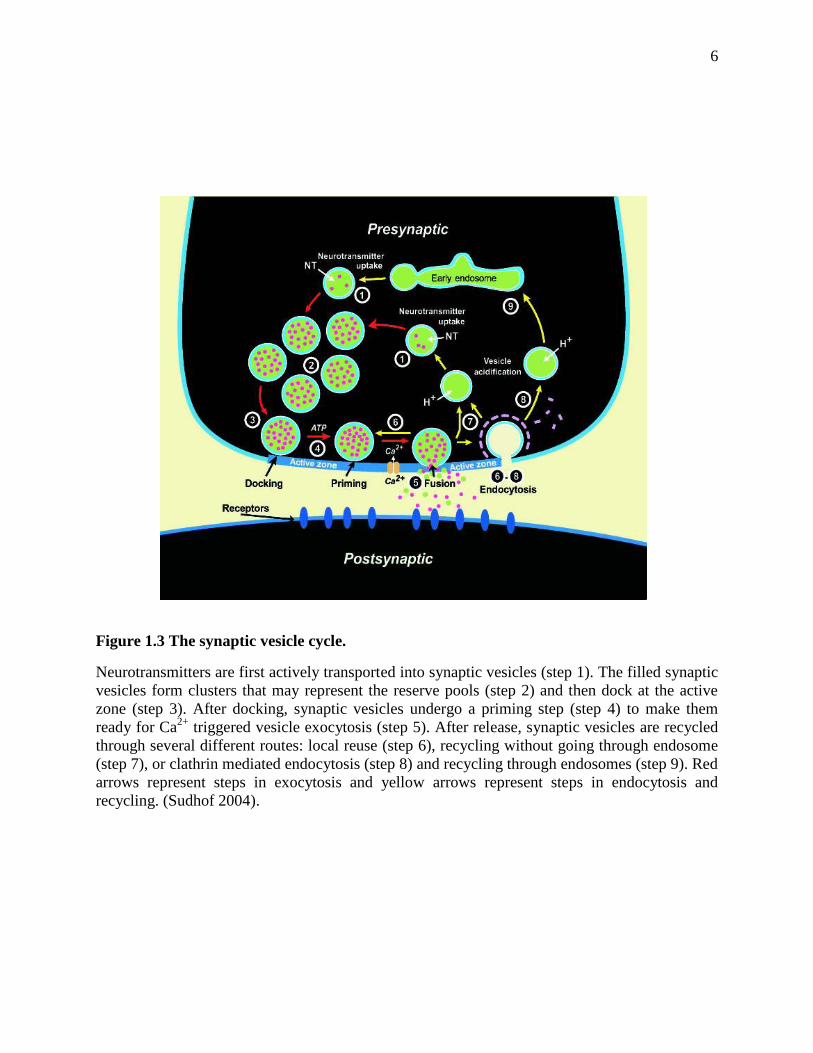
1.2 The Synaptic Vesicle Cycle
The release of neurotransmitters from the presynaptic cell is mediated by synaptic vesicle
exocytosis at the active zone of the axon terminal. Synaptic vesicles undergo a trafficking cycle
(Sudhof 2004) (Figure 1.3) to support frequent rounds of exocytosis and neurotransmitter release.
Synaptic vesicles first actively uptake neurotransmitters in the cytoplasm (step 1), and then
cluster in a place close to the active zone (step 2). After that, synaptic vesicles translocate and
dock at the active zone (step 3), and are primed to a release-ready state (step 4). Upon
stimulation by an action potential, the Ca2+
channels in the active zone open and induce a sudden
influx of Ca2+
, which triggers fast membrane fusion and neurotransmitter release (step 5). After
fusion, synaptic vesicles are proposed to be recycled through three different pathways: (1)
Vesicles close the fusion pore and remain docked at the active zone, and then reacidify and
reload neurotransmitters to start a new cycle (step 6); (2) Empty vesicles leave the active zone
but reacidify and reload neurotransmitters locally (step 7); (3) Vesicles undergo clathrin
mediated endocytosis (step 8) and go through endosome (step 9) to reacidify and reload
neurotransmitters (Sudhof 2004). Of the three pathways, (1) and (2) are fast, and (3) is slower.
The synaptic vesicle cycle is regulated by a number of molecular machineries to rapidly respond
to stimulation and release neurotransmitters to communicate with other neuron cells. Of all the
steps, Ca2+
triggered membrane fusion is the most important one.
6
Figure 1.3 The synaptic vesicle cycle.
Neurotransmitters are first actively transported into synaptic vesicles (step 1). The filled synaptic
vesicles form clusters that may represent the reserve pools (step 2) and then dock at the active
zone (step 3). After docking, synaptic vesicles undergo a priming step (step 4) to make them
ready for Ca2+
triggered vesicle exocytosis (step 5). After release, synaptic vesicles are recycled
through several different routes: local reuse (step 6), recycling without going through endosome
(step 7), or clathrin mediated endocytosis (step 8) and recycling through endosomes (step 9). Red
arrows represent steps in exocytosis and yellow arrows represent steps in endocytosis and
recycling. (Sudhof 2004).

7
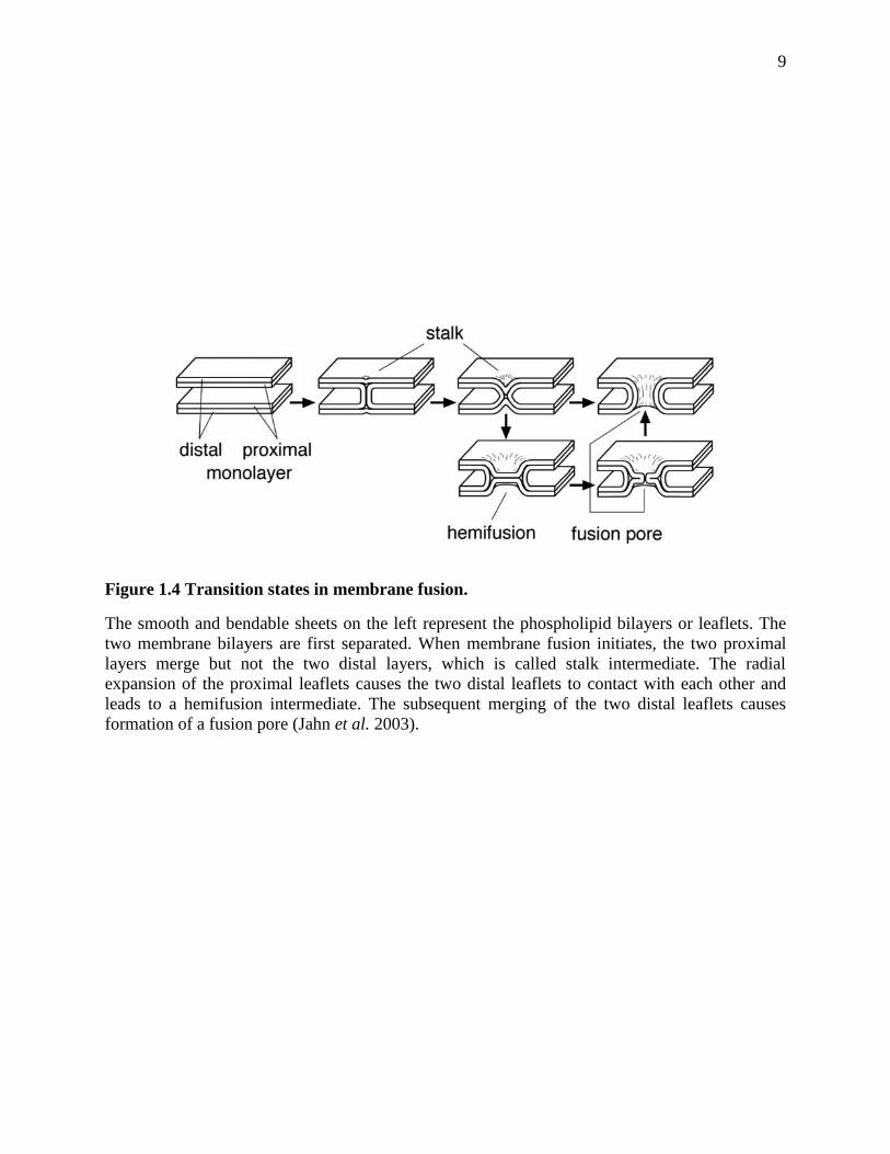
1.3 Membrane Fusion
Membrane fusion is fundamental for many biological processes, such as exocytosis,
protein sorting, fertilization and enveloped virus infection. Neurotransmitter release is a
specialized type of exocytosis. Despite the differences in the biological processes, all kinds of
fusion reactions proceed through the same elementary processes: membrane contact, membrane
merger, and the opening of a fusion pore (Jahn et al. 2003). Membrane fusion doesn’t occur
spontaneously. It is highly regulated by conserved protein families.
Biological membranes are covered by a layer of water molecules that adhere to the
hydrophilic surface of the membranes and hinder approximation of two opposing membranes
through hydration repulsion. Besides, phospholipid bilayers create other forces, such as
electrostatic repulsion of the negatively charged phospholipid headgroups and van der Walls
forces, to keep the opposing membranes away from each other. In order to fuse, membranes first
need to be brought into proximity, which is called docking and it is regulated by some tethering
factors.
However, actual fusion requires a much closer apposition. A small area of the two
opposing phospholipid bilayer needs to approach closer than 3 nm in order for membrane merger
to occur (Helm et al. 1993). When two phospholipid bilayers are within 3 nm, the repulsion
between each other becomes huge, which makes a very high energy barrier for membrane fusion
to occur. Specialized proteins can lower the energy barrier and make the fusion favorable.
Membrane merger also requires lipid reorganization, which needs some local packing defects of
the phospholipid bilayers. The packing defects can be created by changes of membrane curvature
or lipid composition, insertion of membrane proteins or some regulating amphipathic proteins.

8
Once the energy barrier is overcome and the packing defects are achieved, membranes
start to fuse, and eventually a fusion pore forms to connect the contents of the two compartments.
There are transition states during the process of fusion pore formation to minimize the exposure
of hydrophobic surfaces of phospholipids to the aqueous environment. Right now, the most
favored model of describing the transition states of membrane fusion is the stalk hypothesis
(Kozlov et al. 1983; Chernomordik et al. 1987). The stalk hypothesis proposes some distinct
steps in the formation of a fusion pore: merging of the two opposing proximal leaflets, stalk
formation, generation of hemifusion intermediates, and opening of a fusion pore (Figure 1.4)
(Jahn et al. 2003). Membrane proximity is facilitated by a combination of thermal fluctuations of
phospholipid, application of mechanical energy, and shielding of charges, which temporarily
reduce the repulsive forces of the two membranes. The proximal leaflets can merge and form
metastable intermediates because of the membrane temporary relaxation. The first step of
merging is stalk formation, in which the two proximal leaflets are merged while the two distal
leaflets are still separated. After stalk formation, the radial expansion of the two proximal leaflets
pulls the two distal leaflets toward each other, and eventually causes the merging of the distal
leaflets, which is the hemifusion intermediate. Then the relaxation of energetically unfavorable
void interstices will cause the opening of a fusion pore. The stalk hypothesis is supported by both
theoretical calculations and experimental evidence in several model systems (Chernomordik
1996; Lee et al. 1997; Basanez et al. 1998).
9
Figure 1.4 Transition states in membrane fusion.
The smooth and bendable sheets on the left represent the phospholipid bilayers or leaflets. The
two membrane bilayers are first separated. When membrane fusion initiates, the two proximal
layers merge but not the two distal layers, which is called stalk intermediate. The radial
expansion of the proximal leaflets causes the two distal leaflets to contact with each other and
leads to a hemifusion intermediate. The subsequent merging of the two distal leaflets causes
formation of a fusion pore (Jahn et al. 2003).

10
1.4 Proteins involved in Neurotransmitter Release
The release of neurotransmitters from presynaptic terminals is a process that is mediated
by synaptic vesicle exocytosis and incudes several steps. Ca2+
triggered neurotransmitter release
is a very fast process. It is strictly regulated by intracellular Ca2+
concentration, and can happen
within a few hundred microseconds. To achieve this highly precise spatial and temporal
regulation, a series of conserved proteins are involved in each step of neurotransmitter release.
The central protein machinery consists of N-ethylmaleimide sensitive factor (NSF), soluble NSF
adaptor proteins (SNAPs), the neuronal SNAP receptors (SNAREs) syntaxin-1, SNAP-25 and
synaptobrevin, and the Sec1/Munc18 (SM) protein Munc18-1 (Brunger 2005; Jahn et al. 2006;
Rizo et al. 2008; Sudhof et al. 2009). These proteins have homologs in most types of
intracellular membrane traffic, suggesting that this central protein machinery comprises a
conversed mechanism of membrane fusion in synaptic vesicle exocytosis and other membrane
traffic events. Besides these universally required proteins, there are other neuronal specific
proteins involved in regulating Ca2+
triggered fast release, such as the Ca2+
sensor
synaptotagmin-1 (Fernandez-Chacon et al. 2001), Rab3, the priming factors RIMs (Rab3-
interating molecules) and the large active zone proteins Munc13s, and the small SNARE
complex associating protein complexins.
1.4.1 SNARE proteins
SNARE proteins are a conserved protein superfamily required for most intrancellular
membrane trafficking processes from yeast to neurons (Clary et al. 1990; Bennett et al. 1993).
SNAREs include a v-SNARE from the vesicle membrane and t-SNAREs from the target
membrane. The most known and best characterized SNAREs are the neuronal SNAREs, which
include the synaptic vesicle membrane protein synaptobrevin, known as v-SNARE, and the
11
plasma membrane proteins syntaxin-1 and SNAP-25, known as t-SNAREs. All SNAREs contain
one or two SNARE motifs, which are 60-70 residues long and have a tendency to form coil-coils.
Synaptobrevin 2 is a member of the vesicle associated membrane protein (VAMP) family.
It is the v-SNARE protein responsible for synaptic vesicle exocytosis in forebrain synapses. It
consists of a short NH2-terminal sequence, a SNARE motif, and a COOH-terminal
transmembrane region (Figure 1.5). The first indication of synaptobrevin being involved in
neurotransmitter release is that tetanus toxin and botulinum-B toxin block neurotransmitter
release by specific proteolytic cleavage of synaptobrevin (Link et al. 1992; Schiavo et al. 1992).
Synaptobrevin 2 knockout mice died immediately after birth probably due to impaired exocytosis
in the brown fat cells which accumulate in the upper back and cause an abnormal body shape
(Schoch et al. 2001). Analysis of the knockout hippocampal neurons from embryos revealed that
spontaneous synaptic vesicle fusion and fusion induced by hypertonic sucrose were decreased
around 10 fold, while Ca2+
-triggered fast fusion was decreased more than 100 fold (Schoch et al.
2001). The strong impairment but not complete abrogation of neurotransmitter release by
knocking out synaptobrevin 2 suggests that synaptobrevin 2 is not strictly required for synaptic
vesicle fusion, but it is essential for keeping a normal rate of fusion without or with stimulation
(Schoch et al. 2001).
Syntaxin-1 is a plasma membrane protein with a short NH2-terminal sequence (N-
peptide), an NH2-terminal 3 helix bundle Habc domain, a short flexible linker, a SNARE motif
and a COOH-terminal transmembrane region (Figure 1.5) (Fernandez et al. 1998). In human
neurons, syntaxin-1 has two isoforms, syntaxin-1A and syntaxin-1B, which are highly
homologous and might perform similar functions (Fujiwara et al. 2006). Syntaxin-1 can adopt
two different conformations: a closed conformation and an open conformation (Figure 1.6). In
12
closed conformation, syntaxin-1 Habc domain can fold back and bind to the syntaxin-1 SNARE
motif to form a four helix bundle, which is stabilized through binding to Munc18-1 and is
incompatible for SNARE complex formation (Dulubova et al. 1999; Misura et al. 2000). The
SNARE motif of syntaxin-1 together with the SNARE motifs of synaptobrevin and SNAP-25
form the SNARE complex, in which syntaxin-1 is in the open conformation. The closed
conformation of syntaxin-1 is important for its trafficking to the plasma membrane, but it has to
undergo a conformational change in order to allow SNARE complex formation, which is central
for neurotransmitter release. This conformation switch has been shown to be regulated by
Munc13-1 (Ma et al. 2011) and syntaxin-1 N-peptide (Burkhardt et al. 2008). The syntaxin-1 N-
peptide binds to a hydrophobic pocket on the surface of Munc18-1 according to the refined
crystal structure of Munc18-1/syntaxin-1 (Figure 1.9) (Burkhardt et al. 2008). N-peptide also
plays a role in Munc18-1 stimulation of SNARE-dependent liposome fusion (Shen et al. 2007).
SNAP-25 is a plasma membrane associated protein which consists of an NH2-terminal
SNARE motif (SNN), a flexible linker region that has four cysteines, and a COOH-terminal
SNARE motif (SNC) (Figure 1.5) (Chapman et al. 1994). SNAP-25 contributes two SNARE
motifs to the SNARE complex. It is associated to the plasma membrane through palmitoylation
of the four cysteines in the flexible linker. In vitro, SNAP-25 can cause oligomerization of the
SNARE complexes probably because the two SNARE motifs can be assembled into different
SNARE complexes. To simplify experiments, the two SNAP-25 SNARE motifs SNN and SNC
are often used. Experiments from SNAP-25 knockout mice revealed that SNAP-25 is not
required for normal nervous system development, normal axonal outgrowth and stimulus-
independent spontaneous neurotransmitter release. However, it is essential for Ca2+-triggered

13
release at the neuromuscular junctions and central synapses (Washbourne et al. 2002),
suggesting that regulated exocytosis requires the specialized SNAREs and SNARE complexes.
The SNARE motifs of synaptobrevin, syntaxin-1 and SNAP-25 form a highly stable four-
helix bundle complex called the SNARE core complex (Figure 1.5c), which is SDS and
proteolysis resistant and stable at temperatures below 90 °C (Hayashi et al. 1994; Fasshauer et al.
1998). Early electron microscopy (EM), electron paramagnetic resonance (EPR) spectroscopy
and fluorescence resonance energy transfer (FRET) revealed that the SNARE complex is a
parallel four-stranded coiled-coil structure with the membrane anchors of syntaxin-1 and
synaptobrevin located at the same end (Hanson et al. 1997; Lin et al. 1997; Poirier et al. 1998).
Later, the crystal structure of the neuronal SNARE complex showed that it is a parallel four-helix
bundle structure with a length of 120 Å (Figure 1.7) (Sutton et al. 1998). This complex is mainly
stabilized by hydrophobic interactions at the core. The surface of this four-helix bundle consists
of distinct hydrophilic, hydrophobic and charged regions, with negative charges at the middle
regions and positive charges at the ends. These characteristics of the complex might be important
for inducing membrane fusion and for the regulatory factors that are involved in
neurotransmission (Sutton et al. 1998).
The SNARE complex is proposed to zipper from the membrane distal N-termini to the
membrane proximal C-termini to form trans-SNARE complex (two transmembrane regions
located in two different membranes) (Fig 1.7) and release energy from assembly (Hanson et al.
1997; Lin et al. 1997). Thus the SNARE complex formation might be able to overcome the
repulsion between two opposing membranes and bring them into close proximity, facilitating or
driving membrane fusion (Figure 1.7) (Hanson et al. 1997). However, if there is any flexibility in
the linker region between the SNARE core complex and the transmembrane region, energy
14
released from assembly may not be able to transfer to the membrane and SNARE complex may
not be able to directly execute fusion (Rizo et al. 2006). Recently, a group reported the X-ray
structure of the SNARE complex with the C-terminal linkers and transmembrane regions of
syntaxin-1 and synaptobrevin (Stein et al. 2009). The structure shows that the assembly proceeds
from the known SNARE core complex all the way to the linker and transmembrane region
(Figure 1.8), suggesting that the final phase of the SNARE complex assembly is directly coupled
to membrane merger (Stein et al. 2009). However, this structure is about the cis-SNARE
complex (two transmembrane regions located in the same membrane), thus it is unclear that
whether the trans-SNARE complex assemble in the same way.
Previous studies showed that SNAP-25 binds to syntaxin-1 with high affinity (EC50 of
about 0.4uM) (Pevsner et al. 1994). Later work revealed that the two target-SNAREs, SNAP-25
and syntaxin-1, can form stable coiled coil complex (Fasshauer et al. 1997). This finding led to
the hypothesis that SNAP-25 and syntaxin-1 form the acceptor t-SNARE heterodimers at the
plasma membrane, when synaptic vesicles dock at the active zones, synaptobrevin sequentially
associates with the t-SNARE heterodimer and form the SNARE complex (Chen et al. 2001;
Fasshauer et al. 2004; Rickman et al. 2004). In vitro reconstitution experiments with
synaptobrevin and syntaxin-1/SNAP-25 incorporated into different liposomes showed efficient
lipid mixing upon the SNARE complex formation, suggesting that the SNAREs constitute the
minimal membrane fusion machinery (Weber et al. 1998). However, the efficiency of these
experiments depends strongly on the protein to lipid ratios and homogeneity of the liposomes
(Kweon et al. 2003; Chen et al. 2006). Moreover, these experiments provide no explanation of
the physiologically strict requirements of Munc18-1 and Munc13. Knock out Munc18-1 or
Munc13-1/2 mice resulted in the total abrogation of neurotransmitter release (Verhage et al.

15
2000; Varoqueaux et al. 2002). Hence, the model that SNAREs alone cause membrane fusion is
still under debate.
16
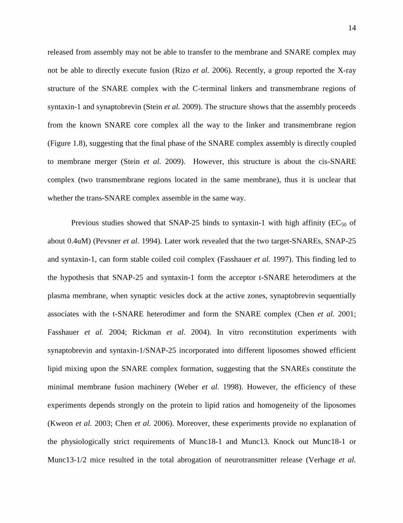
Figure 1.5 Structure of the SNARE complex and its components.
(A)Ribbon diagram of the structure of the Syntaxin Habc domain (Fernandez et al. 1998); (B)
Domain diagram of syntaxin-1. Syntaxin-1 is composed of a SNARE motif (yellow), a
transmembrane region (TM), and a self folded (A) three α-helix bundle Habc domain (orange). (C)
Ribbon diagram of the crystal structure of the SNARE complex (Sutton et al. 1998) with each of the
4 SNARE motifs: Syntaxin-1 (Yellow), Synaptobrevin (Red), and the N (Blue) and C (Green)-
terminal SNARE motifs of SNAP25 (SNN and SNC). (D) Domain diagram of synaptobrevin with
one SNARE motif (Red) and one transmembrane region (TM). (E) Domain diagram of SNAP 25
with SNN in blue and SNC in green.
17
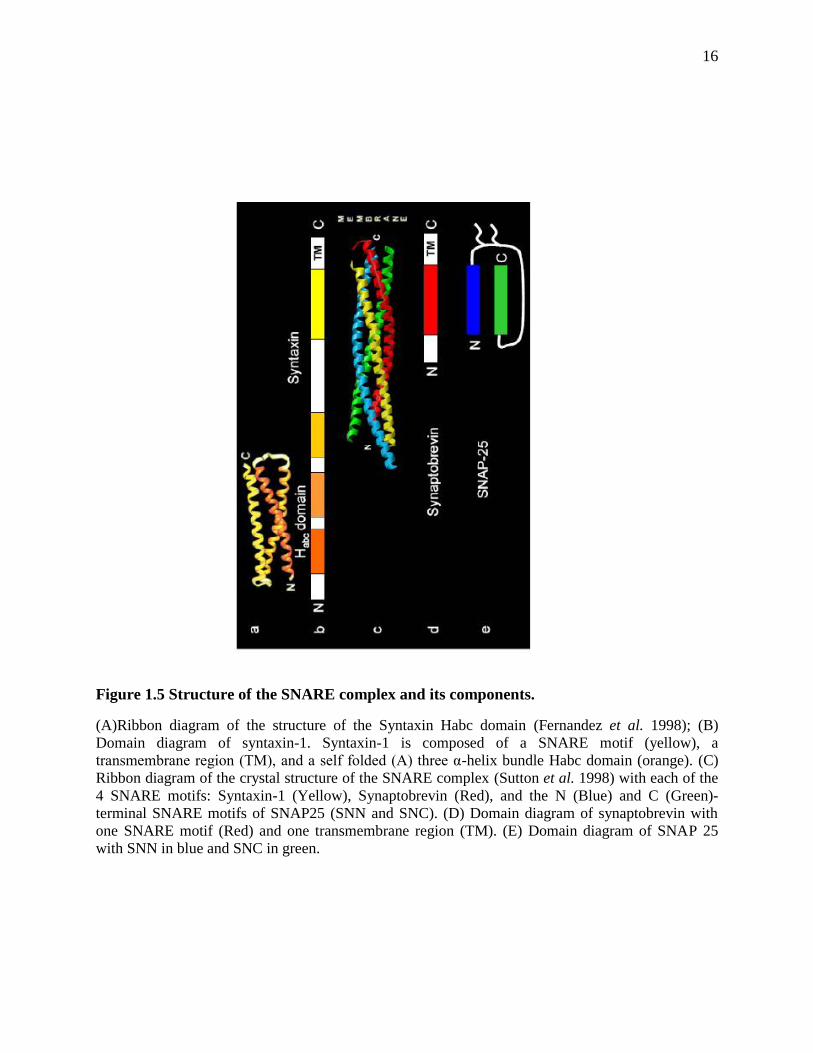
Figure 1.6 Models of the closed and open conformations of syntaxin-1.
Left panel represents the closed conformation of syntaxin-1 with the SNARE motif (yellow)
bound to the Habc domain (orange), forming a four-helix bundle. The closed conformation is
stabilized through binding to Munc18-1 (purple). Right panel represents the open conformation
of syntaxin-1 in the SNARE complex with synaptobrevin in red, SNN in green, and SNC in blue.
18
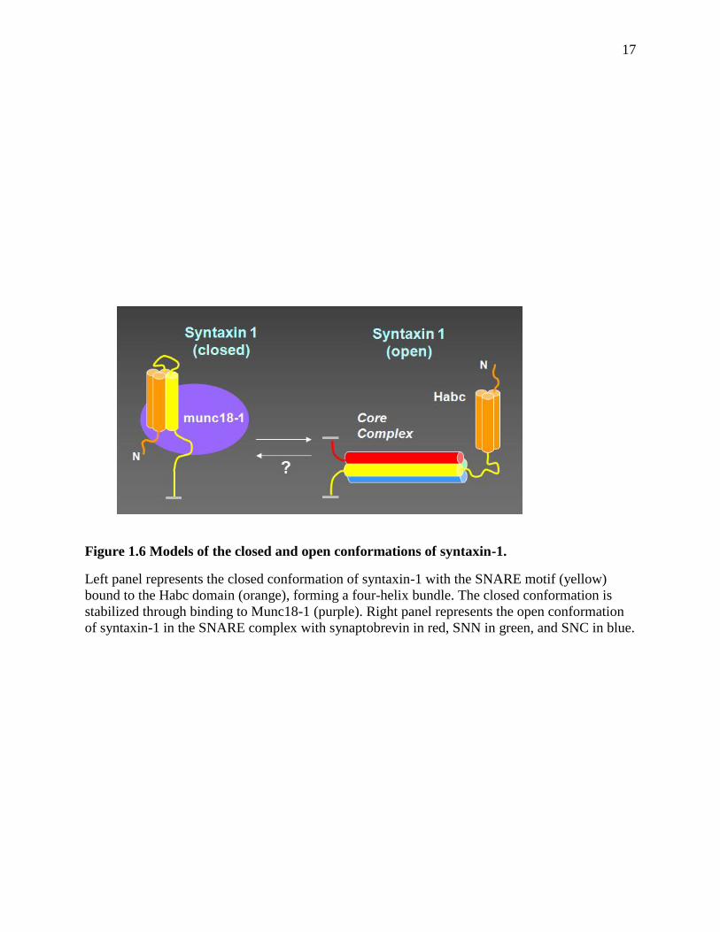
Figure 1.7 Model of the assembled SNARE core complex in two opposing membranes.
The ribbon diagram represents the crystal structure of the core complex with the SNARE motifs
of SNC in greem, SNN in navy blue, synaptobrevin in red, and syntaxin-1 in yellow. The
cylinders represent the transmembrane regions of syntaxin-1 and synaptobrevin, which are
inserted into the plasma and synaptic vesicle membranes, respectively. The curved lines
represent short sequences that connect the SNARE motifs and the transmembrane
regions.(Sutton et al. 1998; Rizo et al. 2002)

19
Figure 1.8 Model of the synaptic SNARE complex inserted into a membrane.
The SNARE complex here includes two SNARE motifs of SNAP-25 (green), syntaxin-1 with
SNARE motif in red, transmembrane region in yellow, and the linker in grey, and synaptobrevin
with SNARE motif in navy blue, transmembrane region in yellow, and linker in grey. The
aromatic residues (black sticks) within the linker regions of synaptobrevin (grey) are shown. The
hydrophilic head groups of the phospholipids are shown as balls, their aliphatic chains as sticks.
They stand for the membrane.
(Stein et al. 2009)
20
1.4.2 Sec1/Munc18 (SM) protein Munc18-1
Sec1/Munc18 (SM) proteins are highly conserved 60-70 KDa proteins that are
universally required for intracellular membrane fusion (Rizo et al. 2002; Toonen et al. 2007;
Sudhof et al. 2009). Structure studies of SM proteins revealed that these proteins are highly
conserved in the overall fold in different organisms and trafficking steps (Misura et al. 2000;
Bracher et al. 2001; Bracher et al. 2002). UNC-18 is the first identified SM protein through
genetic screens for uncoordinated phenotypes in C. elegans (Brenner 1974). Deletion of UNC-
18 results in deficient locomotions in C. elegans (Brenner 1974). Mutation studies revealed that
UNC-18 plays important roles in axon transport system, the acetylcholine flow in motor neurons,
and synaptic vesicle docking (Gengyo-Ando et al. 1993; Weimer et al. 2003). Later, Sec1 was
identified to be involved in the yeast secretory pathway in genetic screens (Novick et al. 1980).
The mammalian homolog of UNC-18, Munc18-1, was first connected to synaptic vesicle fusion
by the identification that Munc18-1 interacts tightly to the t-SNARE syntaxin-1 (Hata et al.
1993).
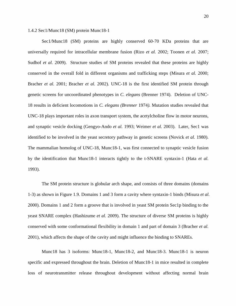
The SM protein structure is globular arch shape, and consists of three domains (domains
1-3) as shown in Figure 1.9. Domains 1 and 3 form a cavity where syntaxin-1 binds (Misura et al.
2000). Domains 1 and 2 form a groove that is involved in yeast SM protein Sec1p binding to the
yeast SNARE complex (Hashizume et al. 2009). The structure of diverse SM proteins is highly
conserved with some conformational flexibility in domain 1 and part of domain 3 (Bracher et al.
2001), which affects the shape of the cavity and might influence the binding to SNAREs.
Munc18 has 3 isoforms: Munc18-1, Munc18-2, and Munc18-3. Munc18-1 is neuron
specific and expressed throughout the brain. Deletion of Munc18-1 in mice resulted in complete
loss of neurotransmitter release throughout development without affecting normal brain
21
assembly, which suggests that Munc18-1 is essential for synaptic vesicle exocytosis (Verhage et
al. 2000). Munc18-2 is 62% sequence identical to Munc18-1 (Tellam et al. 1995). It is
predominantly expressed in epithelial cells and forms a complex with syntaxin 3, which is
involved in the regulation of apical membrane transport (Riento et al. 1998). Munc18-3 is 51%
sequence identical to Munc18-1 and it is ubiquitously expressed (Tellam et al. 1995). It interacts
with syntaxin 4, which is required for the integration of GLUT4 storage vesicles into the plasma
membrane in adipocytes (Thurmond et al. 2000).
Analysis of neurons from Munc18-1 knockout mice revealed that Ca2+
-triggered release,
minis or exocytosis evoked by hypertonic sucrose or by α-latrotoxin were completely abrogated
(Verhage et al. 2000), suggesting that Munc18-1 is absolutely required for neurotransmitter
secretion. However, the functions of Munc18-1 are still not well understood. Munc18-1 has been
shown to play an important role in secretory vesicle docking to the plasma membrane in
chromaffin cells from Munc18-1 knockout mice (Voets et al. 2001), but docking was not
affected in brain synapses, which suggests that the most important function is downstream of
docking. The functions of Munc18-1 have been tightly linked to the SNAREs since Munc18-1
interacts with the SNAREs in different modes. So far, two different binding modes have been
discovered, and they play different roles in synaptic vesicle exocytosis: (1) Munc18-1-syntaxin-1
interaction; (2) Munc18-1-SNARE complex interaction.

22
Figure 1.9 Structure of Munc18-1 bound to syntaxin-1.
(A) A ribbon diagram of Munc18-1 bound to the helical bundle of synaxin-1 in the closed
conformation. Munc18-1 domain 1 (blue) binds the syntaxin N-peptide (red) and forms an arch
with domains 2 (light blue) and 3 (teal). The cavity of the arch binds the syntaxin-1 helical
bundle formed by the SNARE motif helix (light pink) bound to the Habc domain (hot pink) in
the closed conformation. The location of the groove between domains 1 and 2 and the syntaxin-
binding cavity are indicated with arrows. (B) Surface presentation of a rotated view of Munc18-1
bound to syntaxin-1 [domains colored as in (A)]. The view reveals two syntaxin-binding sites
and a groove implicated in SNARE complex binding. The syntaxin N-peptide is bound to
domain 1 of Munc18-1, and is connected to the syntaxin-1 Habc helical bundle by an unresolved
linker (aa 10–26; hatched line). (Carr et al. 2010)
23
Munc18-1-syntaxin-1 interaction. The observation that Munc18-1 interacts tightly with
syntaxin-1, and that other SM protein homologs also bind to their cognate syntaxins (Grabowski
et al. 1997; Nichols et al. 1998), led to the assumption that Munc18-1 functions by interacting
with syntaxin-1. The crystal structure of Munc18-1-syntaxin-1 showed that Munc18-1 cradles
syntaxin-1 in its central cavity and that syntaxin-1is in a closed conformation (Figure 1.9)
(Misura et al. 2000). In the structure, Munc18-1 domain 1 and domain 3a are involved in the
binding. Domain 1 contributes a large contact surface to the interaction. The syntaxin Habc
domain and SNARE motif form a four-helix bundle, which is called ‘closed’ conformation. The
very N-terminus of syntaxin-1, called syntaxin-1 N-peptide, binds to a hydrophobic pocket of
domain 1. The biological relevance of Munc18-1/syntaxin-1 interaction was supported by several
findings. First, the expression level of syntaxin-1 was 70% decreased in Munc18-1 knockout
neurons (Verhage et al. 2000). In Munc18-1 knock down PC12 cells, the expression level of
syntaxin-1 was reduced and the plasma membrane localization of syntaxin-1 was also severely
perturbed. The localization of syntaxin-1 and the impaired secretion capability were restored by
reintroduction of Munc18-1 (Arunachalam et al. 2008). These results suggest that Munc18-1
plays a role in stabilizing syntaxin-1 and syntaxin-1 trafficking to the plasma membrane. Second,
the Munc18-1-syntaxin-1 interaction is important in stimulating vesicle docking in chromaffin
cells. Munc18-1 null chromaffin cells showed a vesicle docking defect, and this defect is
associated with binding to syntaxin-1 and can be rescued by Munc18 (Gulyas-Kovacs et al.
2007). Third, when binds to Munc18-1, syntaxin-1 is in the closed conformation, which
competes with the SNARE core-complex (syntaxin-1 is in an open conformation) formation
(Yang et al. 2000). The syntaxin-1 N-peptide has been shown to connect the two binding modes
of syntaxin-1 to each other and is essential for synaptic vesicle exocytosis (Khvotchev et al.
24
2007). Moreover, syntaxin-1 N-peptide binding to Munc18-1 domain 1 was reported to play a
role in controlling SNARE core-complex assembly (Burkhardt et al. 2008; Rathore et al. 2010).
Overall, the Munc18-1-syntaxin-1 interaction is important in synaptic vesicle exocytosis, but has
never been shown to be critical for membrane fusion so far.
Munc18-1-SNARE complex interaction. After many years of characterizing Munc18-1-
syntaxin-1 interaction, Munc18-1 was shown to bind directly to the SNARE complex by our
group and Rothman’s group (Dulubova et al. 2007; Shen et al. 2007). Yeast SM protein Sec1p
binds specifically to the ternary SNARE complex rather than the closed conformation of the
cognate syntaxin Sso1p (Togneri et al. 2006). Thus, Munc18-/SNARE complex interaction
might represent the most general role of Munc18-1, and might underlie its crucial function in
fusion. Mutations on Munc18-1 domain 1 that disrupt Munc18-1-SNARE complex interaction
result in impaired synaptic vesicle priming (Deak et al. 2009). In vitro reconstitution studies
revealed that Munc18-1 enhances SNARE-dependent lipid mixing in a manner that associates
with SNARE complex binding (Shen et al. 2007; Rodkey et al. 2008). Later, further
characterization of Munc18-1-SNARE complex interaction showed that Munc18-1 directly binds
to the SNARE four-helix bundle with a much weaker affinity (6-13 μM) than the SNARE core
complex that includes syntaxin Habc domain, and that syntaxin Habc domain competes with the
SNARE four-helix bundle for binding to Munc18-1 (Xu et al. 2010). The Munc18-1/SNARE
four-helix bundle binding mode might be crucial for membrane fusion and the idea is supported
by in vitro reconstitution data showing that Munc18-1 binding to SNARE four-helix bundle is
sufficient for its stimulation of lipid mixing (Diao et al. 2010; Shen et al. 2010). However, the
physiological relevance of this weak interaction is not known yet. In vitro characterization of the
25
binding sites involved in Munc18-1/SNARE four-helix bundle interaction would certainly
provide key information for in vivo study.
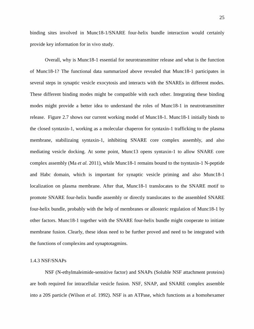
Overall, why is Munc18-1 essential for neurotransmitter release and what is the function
of Munc18-1? The functional data summarized above revealed that Munc18-1 participates in
several steps in synaptic vesicle exocytosis and interacts with the SNAREs in different modes.
These different binding modes might be compatible with each other. Integrating these binding
modes might provide a better idea to understand the roles of Munc18-1 in neutrotransmitter
release. Figure 2.7 shows our current working model of Munc18-1. Munc18-1 initially binds to
the closed syntaxin-1, working as a molecular chaperon for syntaxin-1 trafficking to the plasma
membrane, stabilizaing syntaxin-1, inhibiting SNARE core complex assembly, and also
mediating vesicle docking. At some point, Munc13 opens syntaxin-1 to allow SNARE core
complex assembly (Ma et al. 2011), while Munc18-1 remains bound to the tsyntaxin-1 N-peptide
and Habc domain, which is important for synaptic vesicle priming and also Munc18-1
localization on plasma membrane. After that, Munc18-1 translocates to the SNARE motif to
promote SNARE four-helix bundle assembly or directly translocates to the assembled SNARE
four-helix bundle, probably with the help of membranes or allosteric regulation of Munc18-1 by
other factors. Munc18-1 together with the SNARE four-helix bundle might cooperate to initiate
membrane fusion. Clearly, these ideas need to be further proved and need to be integrated with
the functions of complexins and synaptotagmins.
1.4.3 NSF/SNAPs
NSF (N-ethylmaleimide-sensitive factor) and SNAPs (Soluble NSF attachment proteins)
are both required for intracellular vesicle fusion. NSF, SNAP, and SNARE complex assemble
into a 20S particle (Wilson et al. 1992). NSF is an ATPase, which functions as a homohexamer
26
(Fleming et al. 1998). Each subunit of NSF hexamer consists of three domains: the N-terminal
domain and two nucleotide binding domains (D1 and D2) (Tagaya et al. 1993; Zhao et al. 2012).
The N domain is essential for interaction with the SNAP-SNARE complex, while the D1 domain
provides the main ATPase activity required for NSF function, and the D2 domain mediates
nucleotide –dependent hexamerization (Nagiec et al. 1995). SNAPs contain three isoforms: α-,
β-, and γ-SNAP. β-SNAP is specifically expressed in the brain. α- and γ-SNAPs are ubiquitously
expressed in all cell types. α- and β-SNAPs share 83% identity to each other. γ-SNAP is less
related to α- and β-SNAP, but has similar overall structure (Whiteheart et al. 1993). NSF and α-
SNAP are homologs to yeast Sec18p and Sec17p, which are required for all types of intracellular
fusion in yeast (Mayer et al. 1996). α-SNAP binds to the SNARE complex and serves as an
adaptor protein for NSF binding to form the 20S particle. The ATPase activity of NSF couples
the energy released from ATP hydrolysis to disrupt the cis-SNARE complexes and recycle the
SNAREs for another round of fusion, while the trans-SNARE complexes are resistant according
to Rothman’s group (Sollner et al. 1993; Weber et al. 2000). Meanwhile, NSF and α-SNAP
were reported to be capable of disrupting the t-SNARE complex in in vitro reconstitution assays
(Weber et al. 2000).
1.4.4 Munc13s
Munc13s are mammalian homologs of C. elegans Unc13, which plays a role in regulating
neurotransmitter release and is essential for normal worm movements (Brose et al. 1995).
Munc13s are critical for synaptic vesicle exocytosis because their absence leads to strong
impairment of neurotransmitter release, which is largely caused by a defect in synaptic vesicle
priming (Aravamudan et al. 1999; Augustin et al. 1999; Varoqueaux et al. 2002). Munc13s are
27
also reported to play a role in synaptic vesicle docking (Augustin et al. 1999; Weimer et al. 2006;
Hammarlund et al. 2007; Siksou et al. 2009).
There are three isoforms of Munc13 expressed in the brain, Munc13-1, Munc13-2, and
Munc13-3. Munc13-1 is enriched in synaptosomes and localized to plasma membranes, and is
not expressed in synaptic vesicles (Brose et al. 1995). Munc13s are large and brain specific
proteins with multiple domains, including three C2 domains (C2A, C2B, and C2C), one C1 domain,
one calmodulin binding site and a large central executive domain called MUN domain (Fig 1.10
A). The C2B domain functions as a Ca2+
-phospholipid binding module, while the other two C2
domains do not bind Ca2+
(Lu et al. 2006; Shin et al. 2010). The MUN domain is the major
domain responsible for the priming activity of Munc13s, although the C2C domain may also be
involved in the activity (Basu et al. 2005; Madison et al. 2005; Stevens et al. 2005). Munc13-1
and Munc13-2 double knockout mice revealed no primed vesicles, and also total abrogation of
spontaneous and evoked release (Varoqueaux et al. 2002), while the MUN domain was sufficient
to rescue release in hipocampal neurons lacking Munc13-1/2 (Basu et al. 2005). Previous studies
suggested that the C-terminal part of Munc13-1, which includes the MUN domain and C2C,
directly interacts with N-terminal part of syntaxin-1 (Betz et al. 1997). Another study showed
that a constitutively open form of syntaxin-1 (LE mutant) partially rescued synaptic vesicle
priming and neurotransmitter release in unc13 null C. elegans (Richmond et al. 2001). These
data together suggest that Munc13 functions in priming by directly interacting to the syntaxin-1
N-terminus and thus opening the ‘closed’ conformation syntaxin-1, therefore modulating or
regulation the SNARE core complex formation. However, no binding of Munc13-1 MUN
domain to syntaxin-1 was observed at low micromolar concentrations (Basu et al. 2005). Instead,
the MUN domain binds weakly to the membrane anchored SNARE four-helix bundle, the
28
syntaxin-1 SNARE motif, and the SNARE complex in solution through cooperation with
Munc18-1 (Guan et al. 2008; Ma et al. 2011). In vitro biochemistry experiments demonstrated
that the MUN domain catalyzes the opening of syntaxin-1 from the closed syntaxin-1/Munc18-1
complex to form the SNARE core complex (Ma et al. 2011). This transition might be mediated
by MUN domain through extracting the syntaxin-1 SNARE motif from the closed conformation
and providing a template for SNARE complex formation (Figure 1.10 B). However, the
syntaxin-1 LE mutant can only partially rescue the release in Unc13 null C. elegans, and the LE
mutant does not rescue the release in Munc13-1/2 double knockout mice (Gerber et al. 2008).
These data suggest that MUN domain probably also plays important roles in other processes of
neurotransmitter release.
29
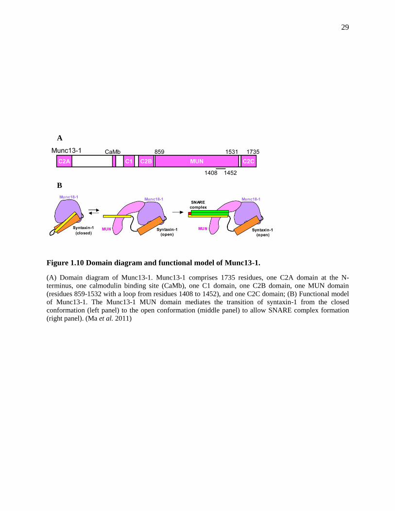
Figure 1.10 Domain diagram and functional model of Munc13-1.
(A) Domain diagram of Munc13-1. Munc13-1 comprises 1735 residues, one C2A domain at the N-
terminus, one calmodulin binding site (CaMb), one C1 domain, one C2B domain, one MUN domain
(residues 859-1532 with a loop from residues 1408 to 1452), and one C2C domain; (B) Functional model
of Munc13-1. The Munc13-1 MUN domain mediates the transition of syntaxin-1 from the closed
conformation (left panel) to the open conformation (middle panel) to allow SNARE complex formation
(right panel). (Ma et al. 2011)
A
B

30
1.4.5 Complexins
Complexins are neuron specific small cytoplasmic proteins that play important roles in
late stage of synaptic transmission. There are four isoforms of complexins in mammals
(McMahon et al. 1995; Reim et al. 2005), and they bind tightly to the SNARE complex (Chen et
al. 2002; Pabst et al. 2002; Bowen et al. 2005; Liu et al. 2006; Li et al. 2007). Complexin is
unstructured in isolation, but it forms a central α-helix when it binds to the SNARE complex
(Pabst et al. 2000; Chen et al. 2002). The crystal structure of the complexin central helix
region/SNARE four-helix bundle showed complexin-I binds in an antiparallel α-helical
conformation to a groove on the SNARE four-helix bundle, which is formed by syntaxin-1 and
synaptobrevin helices (Figure 1.11B) (Chen et al. 2002). This interaction is proposed to stabilize
the fully assembled SNARE complex (Chen et al. 2002). Later studies revealed that the N
terminus of complexin-I interacts with the C terminus of the SNARE complex, which increases
synaptic vesicle fusogenicty at a step after Ca2+
-triggered release. Disrupting this interaction
abrogate the facilitatory function of complexin-I (Xue et al. 2010).
The function of complexins in synaptic vesicle exocytosis is complicated. Complexin null
mice died at birth and showed a dramatically decreased neurotransmitter release in neurons
(Reim et al. 2001). However, overexpression of complexin in PC12 cells inhibits exocytosis
probably by preventing SNARE complex recycling (Liu et al. 2007). These biological data
suggest that complexins play both stimulatory and inhibitory roles in synaptic transmission. The
central helix of complexin is not sufficient for its function, and the N-terminus is required for its
facilitation of fusion, while the regions flanking the central helix likely have an inhibitory role.
So far two alternative models have been proposed to accommodate the stimulatory and inhibitory
functions of complexin (Brose 2008; Sorensen 2009; Stein et al. 2009; Sudhof et al. 2009; Neher
31
2010). First, the complexin central helix and N-terminus binding to the SNARE four-helix
bundle might stablilize the partially assembled SNARE complex, then promotes the progression
of SNARE complex formation, therefore sensitizes them to activation of synaptotagmin (Jahn et
al. 2012). Second, complexin directly competes with synaptobrevin binding to the C-terminal
part of the SNARE complex (Xue et al. 2007; Yang et al. 2010; Kummel et al. 2011), then
blocks the progression of SNARE complex formatiom and acts as a clamp that inhibits
neurotransmitter release. The clamp is released by synaptotagmin upon Ca2+
triggering (Jahn et
al. 2012).

32
Figure 1.11 Structure of the complexin-I-SNARE complex.
(A) Domain diagram of Complexin-I. Complexin-I contains an N-terminal sequence (residues 1-29), an
accessory α-helix (residues 30-48), a central α-helix (residues 49-70), and a C-terminal sequence
(residues 71-134). (B) Ribbon diagram of the complexin/SNARE complex with complexin in pink,
syntaxin-1 in yellow, synaptobrevin in red, SNAP-25 N-terminal SNARE motif (SNN) in blue, SNAP-25
C-terminal SNARE motif (SNC) in green. Only residues 32-72 of complexin were observable in the
crystal structure. (Chen et al. 2002)
33
1.4.6 Synaptotagmins
Synaptotagmins constitute a large family of membrane trafficking proteins that consist of
a short N-terminal intravesicular sequence, a transmembrane region, a short linker sequence, and
two C2-domains (C2A and C2B) in their cytoplasmic region (Figure 1.12) (Bai et al. 2004;
Sudhof 2004). There are 15 isoforms of synaptotagmins discovered so far in vertebrates (Sudhof
2002), of which synaptotagmin 1 and 2 are abundantly expressed in synaptic vesicles but
differentially distributed in brain (Geppert et al. 1991; Ullrich et al. 1994). The C2 domains of
eight synaptotagmins bind to Ca2+
, which is crucial for their functions in neurotransmitter release.
Synaptotagmin binds phospholipids in a Ca2+
dependent manner, which suggest that
synaptotagmin might be a Ca2+
receptor in exocytosis (Brose et al. 1992). Synaptotagmin 1
knockout hippocampal neurons showed severely impaired synaptic transmission, abrogation of
Ca2+
-triggered synchronous release, but unaffected asynchronous release (Geppert et al. 1994).
The in vivo results indicate that synaptotagmin 1 acts as a Ca2+
sensor for fast synchronous
release, but not for asynchronous release.
Structural and biophysical studies showed that both C2A and C2B domains adopt a β
sandwich structure (Figure 1.12). The C2A domain binds three Ca2+
ions through five conserved
aspartates from the top loops of the β sandwich, while the C2B domainm binds two Ca2+
ions
through the same conserved aspartates residues (Figure 1.12) (Ubach et al. 1998; Fernandez et al.
2001). The two C2 domains bind to Ca2+
ions with low affinities in solution (0.5-5mM), but the
affinities for Ca2+
increase dramatically (up to 1000 fold) when the C2 domains bind to
phospholipid membranes because of the additional coordination sites provided by the negative
charged headgroups of the phospholipids (Fernandez-Chacon et al. 2001). However, mutations
that disrupt the C2A domain binding to Ca2+
-phospholipids have little effect on release

34
(Fernandez-Chacon et al. 2002; Robinson et al. 2002; Stevens et al. 2003). Similar mutations on
the C2B domain that disrupt its Ca2+
-phospholipids binding decrease evoked transmitter release
by >95%, which suggests that Ca2+
binding to the C2B domain is essential for synaptic
transmission but Ca2+
binding to the C2A domain is not (Mackler et al. 2002; Nishiki et al.
2004). Why the C2B domain is essential but not the C2A domain? Later in vitro experiments
showed that synaptotagmin is able to bring two membranes into close proximity (~ 4 nm) upon
Ca2+
binding, and that the C2B domain is sufficient for this activity because of the abundance of
basic residues around its surface (Arac et al. 2006). In addition of binding to the Ca2+
-
phospholipids, synaptotagmin also binds to the SNARE complexes in a partially Ca2+
dependent
manner (Bennett et al. 1992; Chapman et al. 1995; Li et al. 1995; Shin et al. 2003). Together, a
model for synaptotagmin function in neurotransmitter release was proposed by our group: after
Ca2+
influx into the presynapse, synaptotagmin binds to membranes, and cooperates with the
SNAREs to bring the synaptic vesicle and plasma membrane together and stimulate membrane
fusion mainly through the highly positive charged C2B domain (Arac et al. 2006). There are
other models proposed by other groups. McMahon’s group proposed that: the C2 domains of
synaptotagmin insert into the target membranes in response to Ca2+
binding, induce high positive
curvature in the membranes, which lowers the energy barrier for bilayer-bilayer membrane
fusion, and then trigger fusion in a SNARE-dependent manner (Martens et al. 2007).

35
Figure 1.12 Structure of synaptotagmin-I.
(A) The domain diagram of Synaptotagmin-I. The C2A domain is colored in orange, the C2B domain is
colored in blue, and the transmembrane domain (TM) is in black; (B) Ribbon diagrams of the individual
synaptotagmine-I C2A and C2B domains. Ca2+
ions are represented by blue spheres (Rizo et al. 2006).
A
B

36
Chapter 2 Characterization of interaction between Munc18-1 and synaptobrevin
2.1 Introduction
Intracelluar membrane fusion is regulated by a conserved machinery composed of several
protein families (Brunger 2005; Jahn et al. 2006). The most important components are soluble N-
ethylmaleimide sensitive factor attachment protein receptors (SNAREs) and Sec1/Munc18 (SM)
proteins, which form the core of the fusion machinery (Sudhof 2004; Verhage et al. 2007; Rizo
et al. 2008). The neuronal SNAREs, synaptobrevin from synaptic vesicles, syntaxin-1 and
SNAP-25 from the plasma membranes, form tight SNARE complexes that bring the two
opposing membranes into close proximity and provide forces to induce membrane fusion. The
neuronal SM protein Munc18-1 is essential for synaptic transmission (Verhage et al. 2000). A lot
of research has been carried out to investigate the functions of Munc18-1, but its key role is still
highly unclear.
Munc18-1 was first found to bind tightly to ‘closed’ syntaxin-1 (Hata et al. 1993), which
plays a role in syntaxin-1 membrane trafficking and synaptic vesicle docking (Pabst et al. 2000;
Arunachalam et al. 2008), and gates entry of syntaxin-1 into SNARE complexes (Gerber et al.
2008). Later, like other SM proteins, Munc18-1 was reported to bind directly to SNARE
complexes and the syntaxin N-terminal sequences often contribute to this binding (Carr et al.
1999; Peng et al. 2002; Carpp et al. 2006; Latham et al. 2006; Stroupe et al. 2006; Dulubova et
al. 2007; Shen et al. 2007). This interaction enhances SNARE-dependent lipid mixing in in vitro
reconstitution assays (Shen et al. 2007; Rodkey et al. 2008) and suggests a role of Munc18-1 in
assisting SNARE complex assembly after the opening of syntaxin-1, and the interaction is
important for neurotransmitter release (Khvotchev et al. 2007). Moreover, the interactions of
Munc18-1 with the syntaxin-1 Habc domain within the SNARE complex are crucial for the
37
synaptic vesicle priming step that makes the synaptic vesicles into a release ready state (Deak et
al. 2009). However, none of these interactions are important for the downstream membrane
fusion step. Munc18-1 and SM proteins are strictly required for general membrane fusion (Rizo
et al. 2002), and some evidence supported a role of yeast SM protein Sec1p in a step after
SNARE complex formation (Grote et al. 2000). These data together lead to a hypothesis that
Munc18-1 might function directly in fusion besides regulates SNARE complex formation.
We proposed a model that Munc18-1 binding to the SNARE complex is more efficient in
exerting mechanical force on the membranes to induce fusion than the SNARE complex alone,
and this model assigns a direct role for Munc18-1 in the fusion step, which might explain the
physiological requirement of Munc18-1 (Rizo et al. 2006). Binding of Munc18-1 to the SNARE
four-helix bundle formed by the motifs of synaptobrevin, syntaxin-1 and SNAP-25 is the central
aspect of this model. Some data showed that Munc18-1 indeed binds to the SNARE four-helix
bundle (Dulubova et al. 2007; Shen et al. 2007), and Munc18-1 binding to synaptobrevin in the
SNARE four-helix bundle is important for Munc18-1 stimulation of lipid mixing in
reconstitution assays (Shen et al. 2007). However, no directly interaction of Munc18-1 to
synaptobrevin was ever detected, and some isothermal titration calorimetry (ITC) data from
another group showed that Munc18-1 interacts with the SNARE complex and syntaxin N-
terminal region with similar affinities, which suggested that Munc18-1 binding to syntaxin N-
terminal region is solely responsible for the SNARE complex binding (Burkhardt et al. 2008).
Hence, it is still unclear whether Munc18-1 directly binds to synaptobrevin and the SNARE four-
helix bundle. Dr. Yi Xu from the lab demonstrated that Munc18-1 binds directly to the
synaptobrevin SNARE motif and the SNARE four-helix bundle at low micromolar affinities, and
that the syntaxin-1 N-terminal region competes with the SNARE four-helix bundle for binding to

38
Munc18-1. I characterized the interaction of Munc18-1 with synaptobrevin, and made great
efforts to crystallize the Munc18-1/synaptobrevin complex and solve the complex structure.
However, the complex structure was not obtained at the end.
2.2 Materials and methods
2.2.1 Recombinant DNA constructs
Bacterial expression vectors to express rat Munc18-1, and fragments corresponding to the
cytoplasmic regions of rat syntaxin-1A (residues 2-253) or the SNARE motifs of rat
synaptobrevin-2 (29-93, 29-96, and 49-96) and human SNAP-25B (11-82 as SNN and 141-203
as SNC) as GST-fusion proteins were available in the lab (Dulubova et al. 2007; Xue et al. 2008).
A vector to express squid Munc18-1 as a His-tagged protein was a kind gift from W.
Weissenhorn (Bracher et al. 2000). Later, squid Munc18-1 was cloned into pGEX-KG vector as
GST-fusion protein.
2.2.2 Expression and purification of recombinant proteins
The DNA constructs were transformed into Escherichia coli BL21 (DE3) cells for
expression. The expression and purification of four SNARE proteins, Munc18-1 and squid
Munc18-1 are similar except minor differences in the induction temperature and purification
chromatography (table 2.1). First, one colony of bacteria in the transformation plate was picked
into 100 ml LB with 100 µg/ml ampicillin, and was incubated in a 37 °C shaker at 250 rpm
overnight. The next day, 15 ml of the overnight culture cells was transferred to 1 L LB media
with 100 µg/ml ampicillin. The big flasks were incubated in a 37 °C shaker for 2.5-3 hr till
OD600 reached 0.8-0.9. Then the temperature was lowered to 25 °C (for SNN, SNC, Syb, squid
Munc18-1) or 20 °C (for Syx and Munc18-1), and 0.4 mM IPTG (isopropyl β-D-

39
thiogalactopyranoside, from Sigma) was added into each 1 L culture to induce protein expression.
Cells were then incubated at 25 °C or 20 °C for 18-20 h to allow protein expression.
For isotopic labeled synaptobrevin-2 29-96, a similar growth and expression strategy was
used except using 15
N M9 minimal media instead of LB. 1L 15
N M9 minimal media is composed
of 6.8 g Na2HPO4, 3.0 g KH2PO4, 0.5 g NaCl, 2.0 mM MgSO4, 100 µM CaCl2, 1.0 g 15
NH4Cl
and 4.0 g D-(+)-glucose.
After expression, the cells were harvested by centrifugation at 4000 rpm for 30 minutes
in swing buckets rotor (Beckman Instruments) and the cell pellets were re-suspended in 15 ml
PBS buffer containing 2 mM EDTA, 2 mM EGTA, 1 mM AEBSF, and 10 µl/ml sigma inhibitor
cocktail (Sigma). The cell suspension was then frozen in liquid nitrogen and then can be stored
in -80 °C freezer for further purification.
For purification, the frozen cell suspension was thawed in a room-temperature water bath
and then passed through a high pressure homogenizer (Emulsiflex C-5, Avestin Inc.) at 10,000
psi for 3 to 4 times. The lysate was then clarified by centrifugation at 20,000 rpm in a JA-20
rotor (Beckman Instruments) for 30 minutes. The supernatant was incubalted with 1.5 ml (each
liter cell culture) prewashed glutathione sepharose beads (Amersham Pharmacia Biotech.) at
4 °C overnight or 3 hr at room temperature to allow the GST-fusion protein binding to the
glutathione beads. Sepharose 4B beads were prewashed with at least 10 bed volumes of H2O and
then PBS. After incubation, the supernatant and beads mixture was then run through a gravity
flow chromatography column (Bio-Rad) to remove most of non-binding proteins. The beads
were then successively washed with 100 ml PBS containing 1% triton X-100, 1 M NaCl, and
PBS to remove non-specific bound proteins. Then benzonase was used to remove protein-bound

40
DNA. Beads were washed with Benzonase buffer (50 mM Tris pH 8.0, 2 mM MgCl2) and then
incubated with 10 units per liter culture Benzonase at room temperature for 1 hr to digest bound
DNA. After Benzonase treatment, the beads were washed with PBS, equilibrated with thrombin
cleavage buffer (50 mM Tris pH 8.0, 150 mM NaCl, 2.5 mM CaCl2), and then incubated with 3-
5 units of thrombin at room temperature for two hours or at 4 °C overnight to cleave proteins
from GST tag. After thrombin cleavage, the flow through was collected and purified through a
Superdex 75 HiLoad 16/60 size exclusion chromatography column (Amersham Pharmacia
Biotech.). For syntaxin-1 (2-253) and synaptobrevin-2 (29-96), proteins were first purified by ion
exchange chromatography (Source Q and Source S). Briefly, a linear gradient of salt (0-1 M
NaCl) was used to separate the protein from other proteins. Then size exclusion chromatography
was performed. The purity of the fractions of proteins was assessed by SDS-PAGE and
Coomassie blue staining. The right protein fractions were collected and concentrated to a desired
concentration, and protein inhibitors containing 1 mM 1:100 Sigma Inhibitor cocktail, 1 mM
AEBSF, 2 mM EDTA, 2 mM EGTA, and 2 mM TCEP were added. Small aliquots of proteins
were made, and were frozen in liquid nitrogen and stored at -80 °C. A typical yield was 3-5 mgs
per liter of culture.
SNARE complexes were assembled and purified as described (Chen et al. 2002;
Dulubova et al. 2007; Deak et al. 2009). A C-terminal fragment of synatobrevin-2 (77-96) was
synthesized as a peptide in UTSouthwestern Medical Center protein chemistry facility.
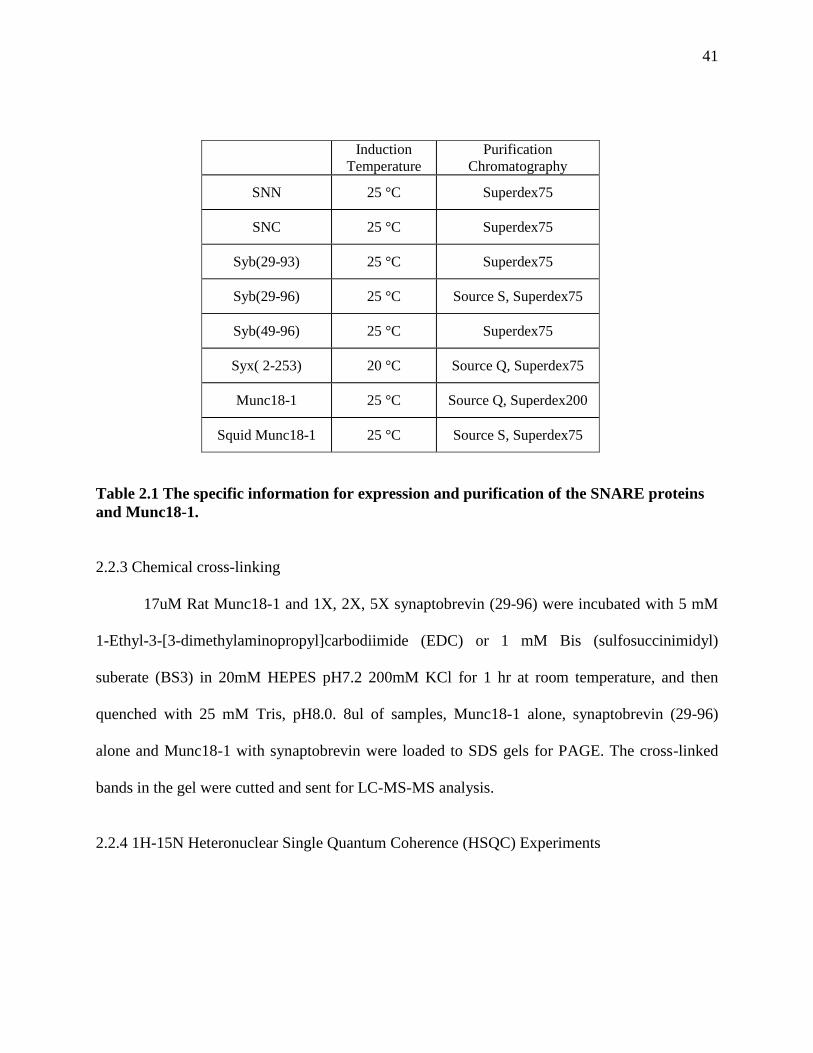
41
Induction
Temperature Purification
Chromatography
SNN 25 °C Superdex75
SNC 25 °C Superdex75
Syb(29-93) 25 °C Superdex75
Syb(29-96) 25 °C Source S, Superdex75
Syb(49-96) 25 °C Superdex75
Syx( 2-253) 20 °C Source Q, Superdex75
Munc18-1 25 °C Source Q, Superdex200
Squid Munc18-1 25 °C Source S, Superdex75
Table 2.1 The specific information for expression and purification of the SNARE proteins
and Munc18-1.
2.2.3 Chemical cross-linking
17uM Rat Munc18-1 and 1X, 2X, 5X synaptobrevin (29-96) were incubated with 5 mM
1-Ethyl-3-[3-dimethylaminopropyl]carbodiimide (EDC) or 1 mM Bis (sulfosuccinimidyl)
suberate (BS3) in 20mM HEPES pH7.2 200mM KCl for 1 hr at room temperature, and then
quenched with 25 mM Tris, pH8.0. 8ul of samples, Munc18-1 alone, synaptobrevin (29-96)
alone and Munc18-1 with synaptobrevin were loaded to SDS gels for PAGE. The cross-linked
bands in the gel were cutted and sent for LC-MS-MS analysis.
2.2.4 1H-15N Heteronuclear Single Quantum Coherence (HSQC) Experiments

42
1H-
15N HSQC spectra of
15N Synaptobrevin (29-96) were recorded on Varian INOVA600
spectrometers at 25 °C. 40 µM 15
N synaptobrevin(29-96) without and with 40 µM squid
Munc18-1 in 20mM HEPES (pH 7.2) and 120 mM KCl were used in the HSQC experiments.
2.2.5 Isothermal Titration Calorimetry (ITC) Experiments
ITC experiments were performed using a VP-ITC (MicroCal, Northhampton, MA)
calorimeter at 20 °C. Squid Munc18-1 and synaptobrevin(49-96) or synaptobrevin(77-96) were
prepared in a buffer containing 20 mM HEPES (PH7.4) and 100 mM KCl. 0.4 mM
synaptobrevin(49-96) or 0.5 mM synaptobrevin(77-96) was successively injected 20 μM squid
Munc18. The data were fitted with a nonlinear least squares routine using a single-site binding
model with MicroCal OriginTM
software for ITC version 5.0.
2.2.6 Gel filtration binding assay
Purified 5 µM 400 µl Munc18-1, 6 µM 400 µl syntaxin-1 (2-253), 6 µM 400 µl SNARE
complex composed of Syx(2-253), Syb(29-93), SNN, and SNC in a buffer containing 20 mM
Tris, pH7.4, and 120mM NaCl were injected to a Superdex S200 (10/300GL) column to get gel
filtration profiles of these proteins alone. 400 μl complex samples containing 5 µM Munc18-1
and 6 µM SNARE complexes or 6 µM syntaxin-1 (2-253) were incubated on ice for 30 minutes,
and then injected into the column to detect complex formation. Data were analyzed by
UNICORN 5.01 (Amersham Biosciences).
2.2.7 Carr Purcell Meiboom Gill (CPMG) experiments
Unlabeled 50 µM squid Munc18-1, 50 µM synaptobrevin (77-96), and 50 µM squid
Munc18-1 with 50 µM synaptobrevin (77-96) were used to record methyl regions of 1D 1H
NMR spectra without (1H-NMR) and with application of a Carr Purcell Meiboom Gill (CPMG)

43
sequence for 100 ms (CPMG 100 ms). Data were used to analyze the binding of synaptobrevin
(77-96) to squid Munc18-1.
2.2.8 Crystallization of squid Munc18-1 and squid Munc18-1 with synaptobrevin (77-96)
Purified squid Munc18-1 was concentrated to 10 mg/ml in 20 mM HEPES, pH 7.4, 100
mM KCl, 10 mM DTT, and then was used to set up crystallography trays. Initial screening
conditions were adapted and modified from squid Munc18-1 crystallization paper (Bracher et al.
2000) using hanging drop trays. Later, optimization of conditions was performed to get nice and
single crystals (6%-8% PEG 1,000, 0.4 M ammonium sulfate, 0.1 M sodium citrate pH 6.6-7.6,
10 mM CaCl2, and 10 mM DTT at 20 °C and 20 mM HEPES, pH 7.4, 100 mM KCl, 10 mM
DTT at 4 °C). 1mM synaptobrevin peptide (77-96) was added to nice and single crystals in cryo
buffer overnight to soak synaptobrevin peptide into sMunc18-1 crystals. For sMunc18-1 and
synaptobrevin (77-96) co-crystallization, 9.3 mg/ml sMunc18-1 was mixed with 1 mM peptide,
and the mixture was incubated at room temperature for 1 hour. Crystallization screening was
performed with the mixture using screens from Hampton Research. Some crystals were
optimized using additive screen from Hampton Research.
2.3 Results
2.3.1 Munc18-1 domain 3a binds to the C-terminus of the SNARE motif of synaptobrevin (29-96)
Dr. Yi Xu found that Munc18-1 interacts with the SNARE motif of synaptobrevin (29-96)
with an affinity of 6uM. However, we didn’t know which part of Munc18-1 and synaptobrevin
(29-96) bind to each other. To find out the residues involved in binding, I first performed
chemical cross-linking experiments between WT Munc18-1 and synaptobrevin (29-96). Two
kinds of cross-linkers were used: EDC, which links close carboxyl groups with primary amines,
and BS3, which cross-links close primary amines with primary amines. No cross-linking was

44
observed in EDC reactions even when 5 times excess of synaptobrevin (29-96) was added (Fiure
2.1A). However, BS3 was able to cross-linking Munc18-1 and synaptobrevin (29-96) in all
concentrations of synaptobrevin (Figure 2.1A). LC-MS-MS analysis of one of the cross-linked
products from the gel revealed that one peptide of synaptobrevin (KYWWK, residues 87-91) was
cross-linked to a sequence of Munc18-1 (KMPQYQK, residues 333-339). These sequences were
mapped to a helix in domain 3a of Munc18-1 located in the cavity where syntaxin-1 binds and to
the very C-terminus of synaptobrevin SNARE motif (Figure 2.1B), which is adjacent to the
synaptic vesicle membrane.
To confirm the binding site on synaptobrevin with a more accurate method, Dr.Yi Xu
performed 1H-
15N heteronuclear single quantum coherence (HSQC) experiments of 5 µM
15N-
labeled synaptobrevin(29-96) in the presence and absence of 6 µM rat Munc18-1. The HSQC
spectra suggested that Munc18-1 indeed binds to the C-terminus of synaptobrevin, however, the
quality of the data was not very good because of insolubility of rat Munc18-1 in 120 mM salt.
Hence, I turned to use squid Munc18-1 (sMunc18-1) which behaves much better than rat
Munc18-1 in vitro. Moreover, squid Munc18-1 shares high identify both sequentially (66.4%)
and structurally to rat Munc18-1 (Bracher et al. 2000).

45
Figure 2.1 Cross-linking of synaptobrevin and Munc18-1
(A) SDS−PAGE of samples containing synaptobrevin(29−96), Munc18-1, or both after cross-linking
with EDC or BS3. The relative concentrations of both proteins are indicated above the lanes. The
positions of molecular mass markers are indicated at the left. The red arrow at the right indicates the
position of the cross-linked product.
(B) Ribbon diagrams of the crystal structure of the SNARE complex (Sutton 1998) and the Munc18-
1−syntaxin-1 complex (Misura et al., 2000) with synaptobrevin colored red and Munc18-1 colored purple
(other proteins colored gray). The sequences of synaptobrevin and Munc18-1 that were cross-linked are
colored yellow.
46
2.3.2 Squid Munc18-1 binds to the C-terminus of synaptobrevin (29-96) with similar affinity
Squid Munc18-1 co-elutes with the cytoplamic domain of rat syntaxin-1 (2-253) and with
mammalian SNARE complex (Figure 2.2), which suggests that sMunc18-1 binds tightly to the
mammalian SNAREs and functions like rat Munc18-1 in vitro. Hence, I used this protein in the
following experiments. To find out exactly which residues in synaptobrevin (29-96) are involved
in Munc18-1 binding, we acquired HSQC spectra of 40 µM 15
N synaptobrevin (29-96) in the
absence and presence of 40 µM sMunc18-1. Synaptobrevin (29-96) is unstructured in solution
(Hazzard et al. 1999), so the cross-peaks of the residues are very sharp by itself. The residues
involved in interaction would be broadened due to binding to a large protein like sMunc18-1 (70
KDa), behaving like sMunc18-1, while other residues would remain flexible and would have no
change in their cross-peaks. The results indeed showed selective broadening of some specific
cross-peaks of synaptobrevin (29-96) upon sMunc18-1 addition (Figure 2.3A). Those cross-
peaks correspond to the C-terminal residues (75-95) of synaptobrevin (29-96) (Figure 2.3B),
which correlates well with the cross-linking results obtained with rat Munc18-1. The results also
showed that synaptobrevin residues 60-70, which were previously suggested to mediate the
SNARE complex dependent Munc18-1 enhancement of liposomes lipid mixing (Shen et al.
2007), are not involved in sMunc18-1 binding by itself.
We also wanted to find out which residues of Munc18-1 interact with synaptobervin (29-
96), but NMR experiments were hindered by the large molecular weight of Munc18-1. We
turned to use crystallography, intending to get the X-ray structure of the sMunc18-
1/synaptobrevin complex. Since synaptobrevin C-terminus is responsible for Munc18-1 binding,
we obtained a synthetic peptide including residues 77-96 of synaptobrevin for crystallography.
To assure that the peptide synaptobrevin (77-96) still binds to sMunc18-1, I recorded 1D 1H
47
NMR spectra of sMunc18-1 alone, peptide alone, and a mixture of the peptide and sMunc18-1
without and with application of a Carr−Purcell−Meiboom−Gill (CPMG) pulse sequence for 100
ms. Large proteins like sMunc18-1 relax much faster than small peptides like synaptobrevin (77-
76) upon CPMG application. If the peptide binds to sMunc18-1, the mixture behaves like
sMunc18-1; otherwise the mixture behaves like the peptide. Within 100 ms, the CPMG sequence
leads to almost complete relaxation of the sMunc18-1 methyl signals, and also the mixture of
sMunc18-1 and synaptobrevin peptide, but not the synaptobrevin peptide alone (Figure 2.4),
which indicates that the synaptobrevin C-terminus alone indeed binds to sMunc18-1. In addition,
I also measured the affinity of this peptide (77-96) and another fragment of synaptobrevin (49-96)
to sMunc18-1 using ITC. The results showed Kd values of 13.5 and 13.3 µM for binding of the
peptide and synaptobrevin (49-96) to sMunc18-1 (Figure 2.3C,D). These values are comparable
to the Kd values (obtained by Dr. Yi Xu) obtained by FRET for binding of rat Munc18-1 to
synaptobrevin(1-96), and their similarity confirms that the C-terminus of the SNARE motif is
responsible for synaptobrevin binding to Munc18-1. Hence, the synaptobrevin peptide (77-96)
was used for following crystallography experiments.
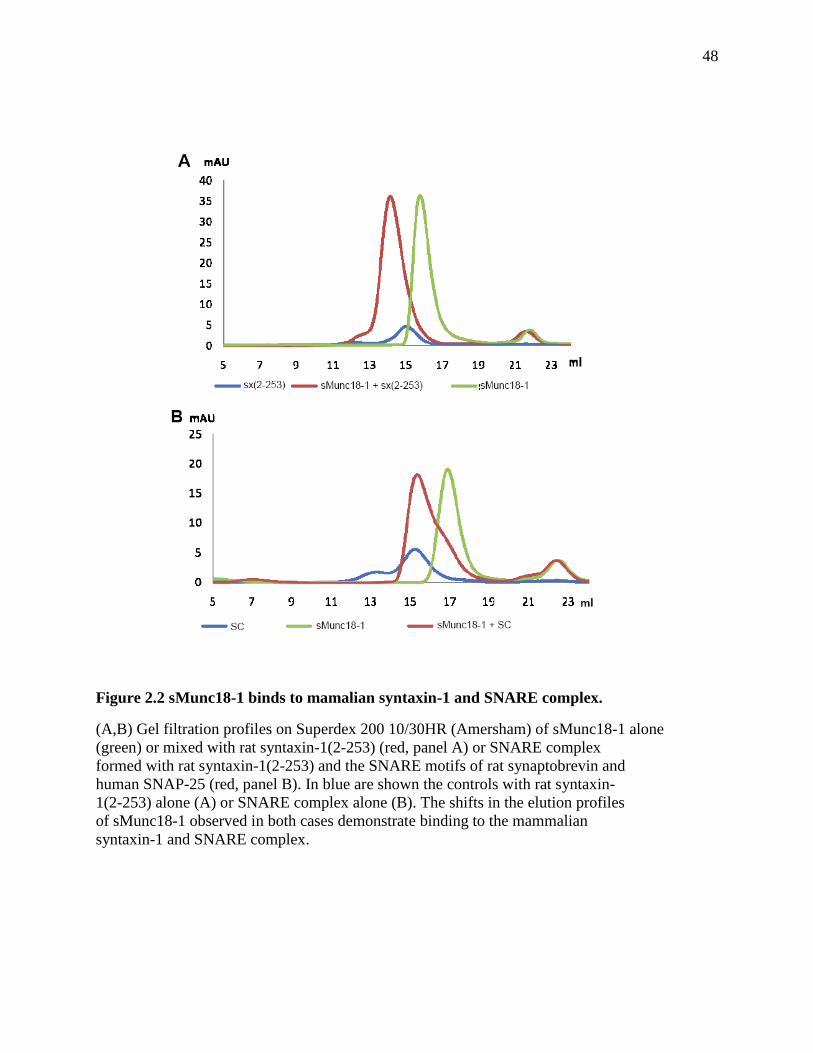
48
Figure 2.2 sMunc18-1 binds to mamalian syntaxin-1 and SNARE complex.
(A,B) Gel filtration profiles on Superdex 200 10/30HR (Amersham) of sMunc18-1 alone
(green) or mixed with rat syntaxin-1(2-253) (red, panel A) or SNARE complex
formed with rat syntaxin-1(2-253) and the SNARE motifs of rat synaptobrevin and
human SNAP-25 (red, panel B). In blue are shown the controls with rat syntaxin-
1(2-253) alone (A) or SNARE complex alone (B). The shifts in the elution profiles
of sMunc18-1 observed in both cases demonstrate binding to the mammalian
syntaxin-1 and SNARE complex.

49
Figure 2.3

50
Figure 2.3 sMunc18-1 binds to the C-terminus of the synaptobrevin SNARE motif.
(A) 1H-15NHSQC spectra of 40 μMsynaptobrevin(29-96) in the absence (black) and presence
(red) of 40 μMsMunc18-1.Well-resolved cross-peaks that were strongly broadened and
correspond to residues at the C-terminus of the synaptobrevin SNARE motif are labeled in blue.
Three well-resolved cross-peaks that do not exhibit such strong broadening and correspond to the
region spanning residues 60-70 are labeled in black. (B) Ribbon diagram of the SNARE complex
with synaptobrevin colored red; residues corresponding to the cross-peaks labeled in blue in
panel A are colored yellow. (C and D) ITC analysis of binding of synaptobrevin(49-96) (C) or
synaptobrevin(77-96) (D) to sMunc18-1.
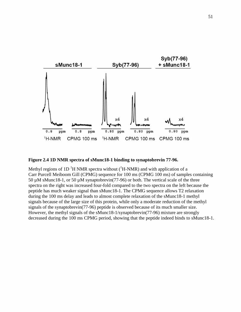
51
Figure 2.4 1D NMR spectra of sMunc18-1 binding to synaptobrevin 77-96.
Methyl regions of 1D 1H NMR spectra without (
1H-NMR) and with application of a
Carr Purcell Meiboom Gill (CPMG) sequence for 100 ms (CPMG 100 ms) of samples containing
50 µM sMunc18-1, or 50 µM synaptobrevin(77-96) or both. The vertical scale of the three
spectra on the right was increased four-fold compared to the two spectra on the left because the
peptide has much weaker signal than sMunc18-1. The CPMG sequence allows T2 relaxation
during the 100 ms delay and leads to almost complete relaxation of the sMunc18-1 methyl
signals because of the large size of this protein, while only a moderate reduction of the methyl
signals of the synaptobrevin(77-96) peptide is observed because of its much smaller size.
However, the methyl signals of the sMunc18-1/synaptobrevin(77-96) mixture are strongly
decreased during the 100 ms CPMG period, showing that the peptide indeed binds to sMunc18-1.
52
2.3.3 Crystallization of synaptobrevin (77-96) with sMunc18-1
Since sMunc18-1 can be crystallized, I used two different methods to get the complex
structure of sMunc18-1 and the synaptobrevin peptide (77-96): crystallize sMunc18-1 first and
soak the peptide into the sMunc18-1 alone crystals; or mix sMunc18-1 and the peptide, then co-
crystallize the mixture; and then solve the structure of the crystals from the two methods to look
for peptide densities in the structure. We got the sMunc18-1 plasmid from W. Weissenhorn
(Bracher et al. 2000). It is a His-tagged protein in the pQE vector 30, and has a maximum yield
of 2 mg/L LB culture. At first, I got some thin crystals of sMunc18-1 alone (Figure 2.5A,B) in
conditions including 10% PEG 1000, 0.4 M ammonium sulfate, 0.1 M sodium citrate pH 6.6 and
7.6, 20 mM DTT, 10 mM CaCl2, and 6 mg/ml sMunc18-1. I tried to soak the crystals with 100
µM and 1mM peptide at 20 °C for 24 hours. Some crystals sustained after soaking and some did
not. Those crystals that survived (Figure 2.5C,D) were sent to synchrotron to collect diffraction
data. They initially diffracted to 3.2 Å, but failed to give diffraction after a while because they
were too small and too thin. Later, sMunc18-1 was cloned into a pGEX-KT vector without His-
tag, and it had a much higher yield (10 mg/ml). Crystallization of the proteins expressed with this
new construct yielded bigger and thicker single crystals (Figure 2.6A,B) in similar conditions
containing 6%-8% PEG 1000, 0.4 M ammonium sulfate, 0.1 M sodium citrate pH 6.6 and 7.6, 20
mM DTT, 10 mM CaCl2, and 10 mg/ml sMunc18-1, and they diffracted to 3.5 Å in house. The
nice and single crystals were soaked with 100 µM and 1mM peptide at 20 °C for 24 hours
(Figure 2.6C,D). The best crystal diffracted to 3.4 Å in house, and a complete data set was
collected. Isomorphous replacement was used to solve the structure. However, no synaptobrevin
peptide densities were found. To co-crystallize sMunc18-1 and the peptide, 9.3 mg/ml sMunc18-
1 and 1mM synaptobrevin peptide mixture was used for screening. Some crystals were obtained,

53
but they were not very nice (Figure 2.6E). After additive screen, nicer crystals were obtained
(Figure 2.6F), and they diffracted to 4 Å in synchrotron. However, no peptide densities were
found after solving the structure.
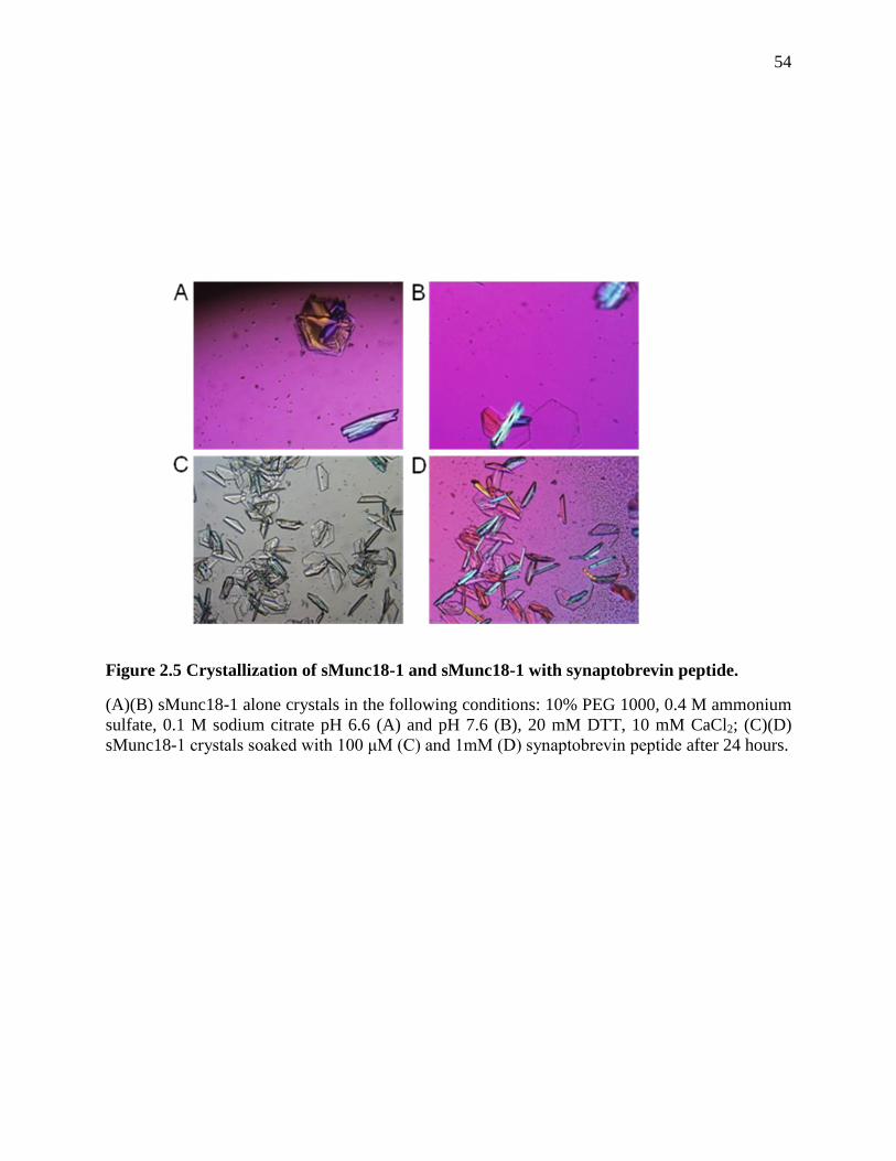
54
Figure 2.5 Crystallization of sMunc18-1 and sMunc18-1 with synaptobrevin peptide.
(A)(B) sMunc18-1 alone crystals in the following conditions: 10% PEG 1000, 0.4 M ammonium
sulfate, 0.1 M sodium citrate pH 6.6 (A) and pH 7.6 (B), 20 mM DTT, 10 mM CaCl2; (C)(D)
sMunc18-1 crystals soaked with 100 μM (C) and 1mM (D) synaptobrevin peptide after 24 hours.
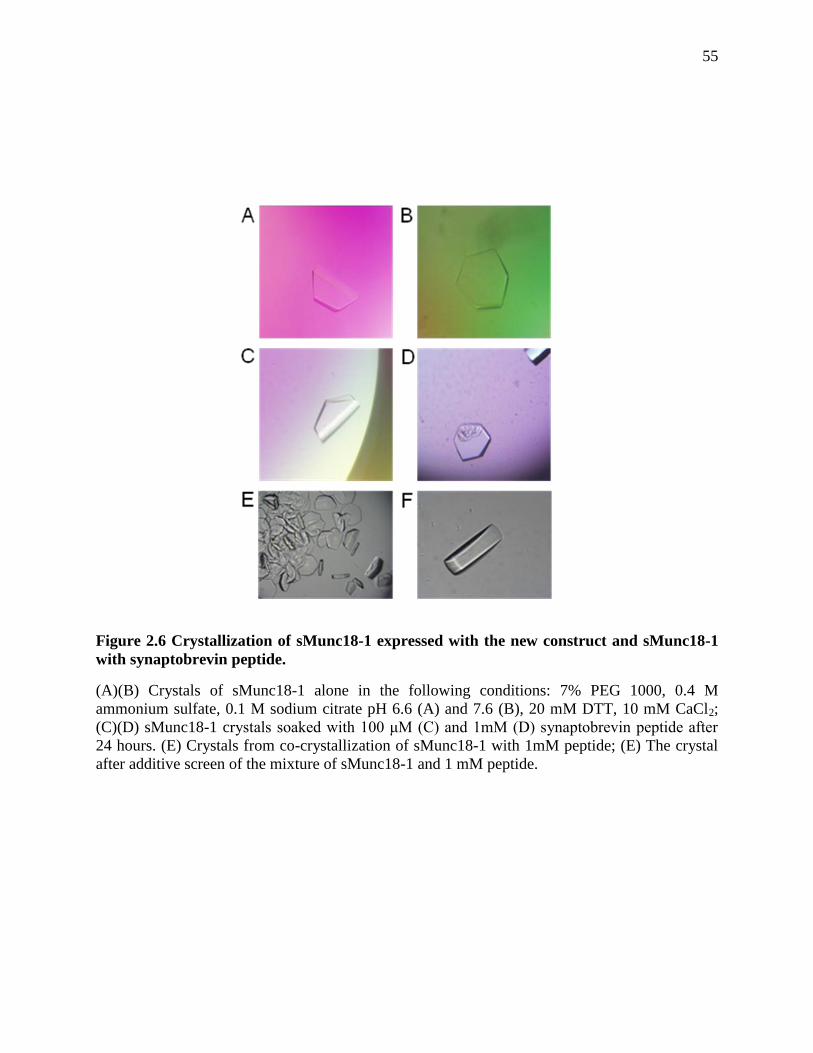
55
Figure 2.6 Crystallization of sMunc18-1 expressed with the new construct and sMunc18-1
with synaptobrevin peptide.
(A)(B) Crystals of sMunc18-1 alone in the following conditions: 7% PEG 1000, 0.4 M
ammonium sulfate, 0.1 M sodium citrate pH 6.6 (A) and 7.6 (B), 20 mM DTT, 10 mM CaCl2;
(C)(D) sMunc18-1 crystals soaked with 100 μM (C) and 1mM (D) synaptobrevin peptide after
24 hours. (E) Crystals from co-crystallization of sMunc18-1 with 1mM peptide; (E) The crystal
after additive screen of the mixture of sMunc18-1 and 1 mM peptide.

56
2.4 Discussion
SM proteins and SNAREs are the two central components of the intracellular membrane
fusion machinery. The roles of SNAREs are well defined, but the main role of SM proteins is
still highly unclear. This uncertainty arises in part because of the diversity of SM
protein/SNARE interactions that have been identified, and in part because no definitive evidence
has been presented for the various models of SM protein function that have been proposed. To
explain the very strong inhibition in membrane fusion observed in the absence of SM proteins
(Rizo et al. 2002; Verhage et al. 2007) , one of these models predicted that SM proteins bind to
the SNARE four-helix bundle, enabling application of force to the membranes by the SNAREs
(Rizo et al. 2006). Previous reconstitution experiments showed that Munc18-1 can strongly
enhance SNARE dependent lipid mixing and suggested that this ability depends on Munc18-
1/synaptobrevin interactions, but no biochemical evidence for such interactions was described
(Shen et al. 2007). Dr. Yi Xu from the lab showed that Munc18-1 binds directly to both
synaptobrevin and the SNARE four-helix bundle with an affinity in the low micromolar range.
Her data suggest that these interactions involve the same cavity of Munc18-1 where the closed
syntaxin-1 binds. Furthermore, Dr. Yi Xu and I together find that the C-terminus of
synaptobrevin is responsible for Munc18-1 binding, placing Munc18-1 right at the site where
membrane fusion occurs. These results reinforce the notion that Munc18-1, and SM proteins in
general, play a direct role in membrane fusion, and suggest a model whereby a sequence of
distinct Munc18-1/SNARE interactions occurs during synaptic vesicle exocytosis (Figure3.7)
Several of the models proposed for SM protein function may be at least partially correct,
since SM proteins may play several roles in distinct steps, but the key unanswered question is:
why are SM proteins so critical for membrane fusion? The initial finding that Munc18-1 binds to
57
syntaxin-1 led to the proposal that Munc18-1 forms part of the synaptic vesicle fusion machinery
(Hata et al. 1993). This overall notion is now supported by overwhelming evidence, but the
binary Munc18-1/syntaxin-1 interaction stabilizes the closed conformation of syntaxin-1 and
gates entry of syntaxin-1 into SNARE complexes (Dulubova et al. 1999; Yang et al. 2000; Chen
et al. 2008; Gerber et al. 2008). Nevertheless, this binary interaction might play positive roles by
stabilizing both proteins in vivo (Verhage et al. 2000; Gerber et al. 2008), and by assisting in
vesicle docking, since syntaxin-1 and Munc18-1 play a role in docking in chromaffin cells, while
synaptobrevin does not (Gerber et al. 2008). Importantly, overexpression of SNAP-25 rescues
the docking defect in chromaffin cells from Munc18-1 KO mice, likely by promoting formation
of syntaxin-1/SNAP-25 heterodimers that bind to synaptotagmin-1, but does not rescue the
secretion defect (de Wit et al. 2009). Hence, Munc18-1 must play an additional function
downstream of docking and syntaxin-1/SNAP-25 heterodimer assembly. There is in fact
evidence for participation of Munc18-1 in more than one of the steps that lead to exocytosis
(Gerber et al. 2008; Guan et al. 2008; Deák et al. 2009).
Although Munc18-1 binding to closed syntaxin-1 hinders SNARE complex formation
from an energetic point of view (Chen et al. 2008), Munc18-1 could still play a role in assisting
SNARE complex assembly in downstream events (Dulubova et al. 1999; Misura et al. 2000;
Rizo et al. 2008). Indeed, Munc18-1 enhances assembly of SNARE complexes between co-
expressed syntaxin-1/SNAP-25 heterodimers and synaptobrevin, which might underlie the
stimulation of SNARE-dependent lipid mixing in reconstitution assays and depends on binding
of Munc18-1 to the syntaxin-1 N-terminus (Shen et al. 2007). Since the co-expressed
heterodimers appear to be in an inhibited state where the Habc domain hinders full interactions
between the SNARE motifs (Starai et al. 2008), it seems likely that Munc18-1 helps to disinhibit
58
this state by binding to the syntaxin-1 N-terminal region. It is unclear whether these events are
related to the mechanism of neurotransmitter release in vivo, but in any case there is little doubt
that interactions of Munc18-1 with the syntaxin-1 N-terminal region are important for release
(Khvotchev et al. 2007; Deák et al. 2009). However, these interactions do not appear to play a
role after vesicle priming (Deák et al. 2009). In addition, it seems unlikely that a mere role for
Munc18-1 in assisting SNARE complex assembly can explain its essential nature for
neurotransmitter release (Verhage et al. 2000), since SNARE complexes can be readily
assembled in vitro. In addition, stimulation of SNARE-dependent lipid mixing by Munc18-1
appears to depend on interactions with synaptobrevin (Shen et al. 2007), and some evidence
indicates that the SM protein Sec1p plays a key role in exocytosis downstream of SNARE
complex formation (Grote et al. 2000).
These observations suggest that the key function of Munc18-1 and SM proteins in
membrane fusion involves interactions with the SNARE four-helix bundle, as initially inferred
for Sec1p (Carr et al. 1999) and as predicted by our previous model of how SM proteins and
SNAREs form the core fusion machinery (Rizo et al. 2006). Some results suggested the
existence of such interactions (Togneri et al. 2006; Dai et al. 2007; Dulubova et al. 2007;
Maximov et al. 2009), but ITC data indicated that Munc18-1 binding to the SNARE complex
involves binding to only the syntaxin-1 N-terminal region (Burkhardt et al. 2008), and direct
binding of SM proteins to the isolated SNARE four-helix bundle has not been reported. Our
results now show unambiguously that Munc18-1 indeed binds to the SNARE four-helix bundle
and to the very C-terminus of the synaptobrevin SNARE motif. It seems very likely that both
interactions involve the same residues, since they have similar affinities and synaptobrevin
competes with the SNARE four-helix bundle for Munc18-1 binding. Dr. Yi Xu’s data also
59
provide strong evidence that these interactions involve the cavity of Munc18-1 where closed
syntaxin-1 binds, which explains the ITC data obtained with Munc18-1 and the SNARE complex
(Burkhardt et al. 2008). These results need to be interpreted with caution, since the interactions
we report have moderate affinity and their physiological relevance remains to be established.
However, the finding that Munc18-1 binds to the very C-terminus of the synaptobrevin SNARE
motif is very intriguing, since it places Munc18-1 right at the site of fusion and hence suggests a
fundamental new view whereby Munc18-1 is intimately and directly involved in fusion. Such a
role could certainly explain the critical nature of Munc18-1 and SM proteins for membrane
fusion in vivo.
The original idea of how Munc18-1 could help to induce fusion (Rizo et al. 2006;
Rodkey et al. 2008) was intended to provide such an explanation and was based on a simple
mechanical principle: flexibility in the linker region between the SNARE motifs and TM regions
could hinder transduction of the energy of SNARE complex assembly onto the membranes and
allow assembled SNARE complexes to diffuse to the middle of the interrmembrane space;
however, binding of a bulky protein such as Munc18-1 to assembling SNARE complexes would
prevent such diffusion and would help applying torque on the two membranes to induce fusion,
because Munc18-1 would push the membranes away while SNARE complexes bring them
together. Note that the torque would be weakest if Munc18-1 binds to the N-terminus of the
SNARE four-helix bundle and strongest if the binding site is at the C-terminus, as suggested by
our data. Indeed, it seems impossible that SNARE complexes could form without fusion if
Munc18-1 is bound to their C-termini (even if there is some flexibility in the linker regions).
Moreover, Munc18-1 contains two highly positive surfaces surrounding the SNARE-binding
cavity. These surfaces could bind to the two apposed membranes and actively help to bend them

60
to induce fusion (Figure 3.7), as proposed for synaptotagmin-1 (Arac et al. 2006). In this context,
only weak binding of Munc18-1 to liposomes has been reported (Starai et al. 2008), but such
interactions could be greatly strengthened by the intrinsic cooperativity of the system.
We proposed a model that involves three different types of interactions of Munc18-1 with
the SNAREs (Figure 3.7): first the binary interaction with closed syntaxin-1; second the
interaction with the N-terminal region of syntaxin-1 as syntaxin-1 opens and SNARE complexes
start to form via by the help of Munc13 (Guan et al. 2008; Ma et al. 2011); and third the binding
to the C-terminus of synaptobrevin. Although the affinity of Munc18-1 for synaptobrevin is
moderate, and weaker than its affinity for the syntaxin-1 N-terminal regions in binding assays
with isolated proteins, the transition from syntaxin-1 to synaptobrevin binding could be favored
by strong cooperativity with the proposed interactions of Munc18-1 with both membranes, which
would bring the membranes together and could therefore cooperate, in addition, with the pulling
forces of the assembling SNARE complexes. Moreover, membranes appear to enhance binding
of Munc18-1 to the SNARE four-helix bundle (Shen et al. 2007) and the functional importance
of the synaptobrevin region that binds to Munc18-1 has been shown by the impairment in
neurotransmitter release caused by mutation of two tryptophane residues in this region
(Maximov et al. 2009), although the molecular target of this region in vivo remains to be
determined. Candidates for such targets are also Munc13s, complexins and syntaptotagmin-1,
factors that play key roles in neurotransmitter release and also bind to the SNARE four-helix
bundle (Chen et al. 2002; Dai et al. 2007; Guan et al. 2008). These factors could provide
additional cooperativity to the system, but they might also compete with Munc18-1 for binding
to the four-helix bundle.
61
Clearly, our model remains highly speculative, and to better understand the mechanisms
of membrane fusion and neurotransmitter release, it will be particularly important to study the
interplay between the interactions among all these proteins and the lipids in the context of trans-
SNARE complexes formed between two apposed membranes. The data generated by Dr. Yi Xu
and the ones presented here have now yielded a hypothesis that can help guiding these very
challenging studies and proposes a fundamentally new view of how Munc18-1 might cooperate
with the SNAREs to induce membrane fusion.

62
Figure 2.7

63
Figure 2.7 Proposed model of neurotransmitter release involving three types of Munc18-1-
SNARE interactions
The model assumes that Munc18-1 (purple) is initially bound to closed syntaxin-1 (Habc domain,
orange; SNARE motif, yellow) (top left panel). Partial assembly of SNARE complexes of
syntaxin-1 with synaptobrevin (red) and SNAP-25 (green) occurs by an unknown mechanism
that likely involves Munc13 (not shown); here we propose that Munc18-1 is bound to only the
N-terminal region of syntaxin-1 at this stage (top right panel). The next step is proposed to
involve the transition of Munc18-1 from the syntaxin-1 N-terminal region to the C-terminus of
the synaptobrevin SNARE motif, which could be favored by cooperativity with other
interactions such as Munc18-1 binding to the vesicle membrane (bottom right panel). The central
aspect of this model is that membrane fusion results from the cooperative action of Munc18-1
and the SNAREs, which would be favored by the binding of Munc18-1 to synaptobrevin and
could involve interactions of basic residues of Munc18-1 (indicated by the + signs) with both
membranes (bottom left panel; see the text for further details). Other proteins involved in
triggering release and conferring its Ca2+
sensitivity are not represented for the sake of simplicity,
but they are expected to cooperate with Munc18-1 and the SNAREs to trigger release.

64
Chapter 3 Towards the structure of Munc18-1/SNARE four-helix bundle complex
3.1 Introduction
The SNAREs and Sec1/Munc18 (SM) proteins constitute the core of the intracellular
membrane fusion machinery (Sudhof 2004; Verhage et al. 2007; Rizo et al. 2008). The roles of
the neuronal SNAREs are well characterized. The neuronal SM protein Munc18-1 is strictly
required for synaptic transmission (Verhage et al. 2000). As explained in the introduction of
Chapter 2, research that has been carried out to investigate the functions of Munc18-1 revealed
that Munc18-1 plays important roles in synaptic vesicle docking and priming. However, none of
them can explain the fundamental requirement of Munc18-1 in neurotransmitter release.
Moreover, the molecular mechanism of action of Munc18-1 is still unclear, especially in the
Ca2+
-triggered membrane fusion step. Dr. Yi Xu from the lab found that Munc18-1 binds to the
SNARE four-helix bundle with weak affinity (Xu et al. 2010). This weak interaction was
reported to be responsible for Munc18-1 stimulation of SNARE-dependent in vitro liposome
fusion (Shen et al. 2007; Shen et al. 2010). Hence, this weak interaction might be crucial for the
key role of Munc18-1 in synaptic vesicle fusion.
Exploring the structure of Munc18-1/SNARE four-helix bundle will certainly provide
important information for further characterization of the significance of this weak interaction in
vivo. The structural information of the complex of Munc18-1/SNARE four-helix bundle will
also help determine how Munc18-1, synaptotagmin, complexin, the SNARE complex, and other
proteins cooperate with each other in Ca2+
-triggered synaptic vesicle fusion.
To obtain a high resolution structure of Munc18-1/SNARE four-helix bundle complex, I
have been applying the methods of NMR spectroscopy, X-ray crystallography, and SAXS (small
angle X-ray scattering). I did not solve the structure of this complex yet, but I have some data

65
showing that Munc18-1 binds to the C-terminal part of the SNARE four-helix bundle, which is
adjacent to the place where membrane fusion occurs, and that Munc18-1 binding to the SNARE
four-helix bundle is compatible with binding of the SNARE four-helix bundle to complexin-I,
which functions in a late stage of synaptic vesicle exocytosis. These results are interesting and
intriguing because it places Munc18-1 right at the site where membrane fusion occurs,
suggesting that Munc18-1 might be directly involved in executing membrane fusion.
3.2 Materials and method
3.2.1 Recombinant DNA constructs
Bacteria expression vectors to express rat Munc18-1, fragments corresponding to the
SNARE motifs of rat synaptobrevin-2 (29-93), synatxin-1a (191-253) and human SNAP-25B
(11-82 as SNN and 141-203 as SNC), and the SNARE motifs mutants syntaxin-1a (H239C),
synaptobrevin (S61C and A72C) as GST-fusion proteins were available in the lab (Dulubova et
al. 2007; Xu et al. 2010). A vector to express squid Munc18-1 as a His-tagged protein was a kind
gift from W. Weissenhorn (Bracher et al. 2000). Later, squid Munc18-1 was cloned into pGEX-
KG vector as GST-fusion protein. Munc18N (1-136) and Munc18NL (1-139) were cloned into
pGEX-KG vector from full length Munc18-1. Point mutants of Munc18NL (K46C and R64C)
and SNC (E183C) were generated from the WT constructs using the QuickChange site-directed
mutagenesis kit (Stratagene) and custom-designed primers, and were verified by sequencing.
3.2.2 Expression and purification of recombinant proteins
The expression and purification for recombinant proteins were similar to those described
in Chapter 2.
3.2.3 NMR spectroscopy
66
1H-
15N HSQC spectra of
15N syntaxin-1a (191-253),
15N SNN and
15N SNC were recorded on
Varian INOVA600 spectrometers at 15 °C. 20 µM 15
N syntaxin-1a (191-253), 15
N SNN and 15
N
SNC without and with 40 µM squid Munc18-1 in 20mM HEPES (pH 7.4) and 120 mM KCl
were used in the HSQC experiments. 1H-
15N TROSY-HSQC spectra of 85 µM
15N sMunc18-1
and 1H-
13C HMQC spectra of 11 µM
2H-
13C
1H3-Iso/Leu/Val labeled sMunc18-1and Munc18-1
without and with the SNARE four-helix bundle, and 30 µM 1
H-13
C1H3-Iso labeled sMunc18-1
without and with the SNARE four-helix bundle were recorded on Varian INOVA800
spectrometers at 32 °C and 25 °C. 1H-
15N TROSY-HSQC spectra of 40 µM
2H-
15N labeled
syntaxin-1 (191-253), synaptobrevin (29-93), SNN, and SNC assembled the SNARE four-helix
bundle without and with 40 µM Munc18N, 100 µM 15
N labeled SNCE183C w/o DOTA-M8
assembled the SNARE four-helix bundle, and 40 µM 15
N Munc18NL without and with 10 µM,
20 µM, and 40 µM the SNARE four-helix bundle were recorded on Varian INOVA600
spectrometers at 25 °C. 1H-
13C HMQC spectra of 20 µM
2H-
13C
1H3-Iso/Leu/Val labeled
syntaxin-1 (191-253) assembled the SNARE four-helix bundle without and with 20 µM
Munc18NLK46C/R64C-DOTA-Mn2+
/DOTA-Gd3+
were recorded on Varian INOVA600
spectrometers at 25 °C.
3.2.4 Backbone and methyl group assignments of Munc18NL
The backbone and methyl groups were assigned as described in (Chen et al. 2002; Chen
et al. 2008).
3.2.5 Isothermal Titration Calorimetry (ITC) Experiments
ITC experiments were performed using a VP-ITC (MicroCal, Northhampton, MA) calorimeter at
20 °C. The SNARE four-helix bundle or Munc18N were prepared in a buffer containing 20 mM

67
HEPES (PH7.4), 120 mM KCl and 1mM TCEP. 0.5 mM Munc18N was successively injected to
20 μM the SNARE four-helix bundle. The data were fitted with a nonlinear least squares routine
using a single-site binding model with MicroCal OriginTM
software for ITC version 5.0.
3.2.6 SAXS experiments
Samples of 39 μM Munc18-1 in a buffer containing 20 mM HEPES (PH7.4), 300 mM
KCl, 10% glycerol and 1mM TCEP; 120 μM the SNARE four-helix bundle in a buffer
containing 20 mM HEPES (PH7.4), 120 mM KCl and 1mM TCEP; and 40 μM, 62 μM, 100 μM
mixtures of Munc18-1 and the SNARE four-helix bundle in a buffer containing 20 mM HEPES
(PH7.4), 120 mM KCl, 10% glycerol and 1mM TCEP were used for SAXS experiments. Data
were collected in a BioSAXS-1000 instrument in Rigaku Americas Corporation, the Woodlands,
TX. ATSAS2.4 was used to process all the data.
3.3 Results
3.3.1 Binding of Munc18-1 to the individual SNARE motifs.
In order to get a high resolution structure of Munc18-1/SNARE four-helix bundle
complex, NMR spectroscopy and X-ray crystallography were simultaneously applied. To define
the complex structure by NMR spectroscopy, binding sites in the two proteins and the relative
orientation between them need to be determined. To find the binding sites in Munc18-1 and the
SNARE four-helix bundle, the first thing and easiest thing to do is to investigate the bindings of
Munc18-1 to the individual four SNAREs and to see whether these interactions could provide
some hints for the mode of binding to the SNARE four-helix bundle.
The binding site on the synaptobrevin SNARE motif is described in Chapter 2. For the
other three SNARE motifs, I performed 1H-
15N HSQC experiments of 20 µM
15N-syntaxin-1a
68
(191-253), SNN, SNC in the presence and absence of 40uM sMunc18-1. The spectra revealed
that Munc18-1 binds to syntaxin-1 SNARE motif (202-224), but not to SNN and SNC (Figure
3.1A,B,C). A mutation on syntaxin-1 (R210E) impairs its interaction to sMunc18-1 (Figure
3.1D), which suggests that syntaxin-1 binding to sMunc18-1 is specific. However, when
mapping the binding sites of syntaxin-1 (191-253) and synaptobrevin (29-96) (Figure 2.3A) onto
the SNARE four-helix bundle (Figure 3.1E), the two individual binding sites are not in the same
place on the SNARE four-helix bundle. The data suggest that Munc18-1 binding to the SNARE
four-helix bundle is not the same as binding to the individual SNAREs, and that we need to use
the SNARE four-helix bundle in the following experiments.

69
Figure 3.1 Munc18-1 binding to the individual SNAREs.
(A) 1
H-15
N HSQC spectra of 20 µM 15
N-SNN in the absence (black) and presence (red) of 40 μM
sMunc18-1; (B) 1H-
15N HSQC spectra of 20 µM
15N-SNC in the absence (black) and presence
(red) of 40 μM sMunc18-1; (C) 1H-
15N HSQC spectra of 20 µM
15N-syntaxin-1 (191-253) in the
absence (black) and presence (red) of 40 μM sMunc18-1; the cross-peaks that were strongly
broadened are labeled in black; (D) 1H-
15N HSQC spectra of 20 µM
15N-syntaxin-1 (191-253)
R210E in the absence (black) and presence (red) of 40 μM sMunc18-1; (E) Ribbon diagram of
the crystal structure of the SNARE four-helix bundle with SNN and SNC in green,
synaptobrevin (29-93) in red, and syntaxin-1 (191-253) in orange; the dark blue helixes are
residues broadened in (C) and Figure 2.3A.
70
3.3.2 To investigate the residues of Munc18-1 involved in the SNARE four-helix bundle binding.
To identify the residues of Munc18-1 involved in the SNARE four-helix bundle binding,
first we need to get a good NMR spectrum of Munc18-1. I first carried out 1H-
15N TROSY
(Transverse Relaxation Optimized Spectroscopy)-HSQC experiments of 85 μM 15
N-labeled
sMunc18-1 (squid Munc18-1) at 32 °C. The cross-peaks in the spectrum are as crowded as
expected for a 70 KDa protein (Figure 3.2A). Although there are some good dispersed peaks, the
resolution is still not high enough to investigate the residues of Munc18-1 involved in binding,
especially without any assignments. Deuteration of sMunc18-1 could greatly improve the
resolution. However, deuteration resulted in a really low yield and a very less concentrated
protein, which lowers the quality of the TROSY-HSQC experiment (Figure 3.2B). Thus, we
need higher sensitivity experiments to get a good NMR spectrum. Deuteration together with 13
C-
labeling of the methyl groups of isoleucines, valines and leucines of sMunc18-1 and Munc18-1
gave a much nicer HMQC (Heteronuclear Multiple Quantum Coherence) spectrum (Figure
3.2C,D), which can be used for the SNARE four-helix bundle binding experiments. Also, 13
C-
labeling of the methyl groups of isoleucines of sMunc18-1 without deuteration gave a fairly good
HMQC spectrum (Figure 3.2E).
1H-
13CH3 HMQC experiments of 30uM
13C
1H3-Iso-sMunc18-1 in the presence and
absence of 40 μM the SNARE four-helix bundle were performed. The spectra reveals specific
broadenings of 3 isoleucine cross-peaks of sMunc18-1 (Figure 3.3A), which suggests that the
SNARE four-helix bundle indeed interacts with Munc18-1, although it is unclear which residues
are involved because we do not have the assignments of the isoleucines of sMunc18-1.
71
To get more changes of the cross-peaks, I labeled synaptobrevin S61C, which is close to
the binding site and is used for fluorophore labeling in measuring the affinity of Munc18-1 to the
SNARE four-helix bundle (Xu et al. 2010), with the paramagnetic probe MTSL (Figure 3.3D),
which induces paramagnetic resonance enhancement (PRE) to the residues near the probe and
causes broadenings of the cross-peaks corresponding to those residues. 1H-
13CH3 HMQC
experiments of 11 μM 2H-
13C
1H3-Iso/Leu/Val-Munc18-1 in the presence and absence of 30 μM
the SNARE four-helix bundle (mcc)-synaptobrevin-61C-MTSL were performed. In the mcc-
synaptobrevin-61C-MTSL added spectra, the overall intensities drop because of the binding, and
some cross-peaks were broadened further (Figure 3.3B). However, after adding dithionine,
which is expected to kill the paramagnetic probes and reverse the changes, those broadenings
were not reversed (Figure 3.3C).
These data together indicate the binding of Munc18-1 to the SNARE four-helix bundle,
but the residues of Munc18-1 involved in binding are hard to define from the data for the
following reasons: the affinity of the complex is low (Kd~10 μM), and Munc18-1 is not
saturated; or the residue of synaptobrevin S61C is not close enough to the binding site, so the
paramagnetic probes cannot induce changes of residues involved in binding. Defining the
binding site in the SNARE four-helix bundle will lead us to label the probes in a better place.
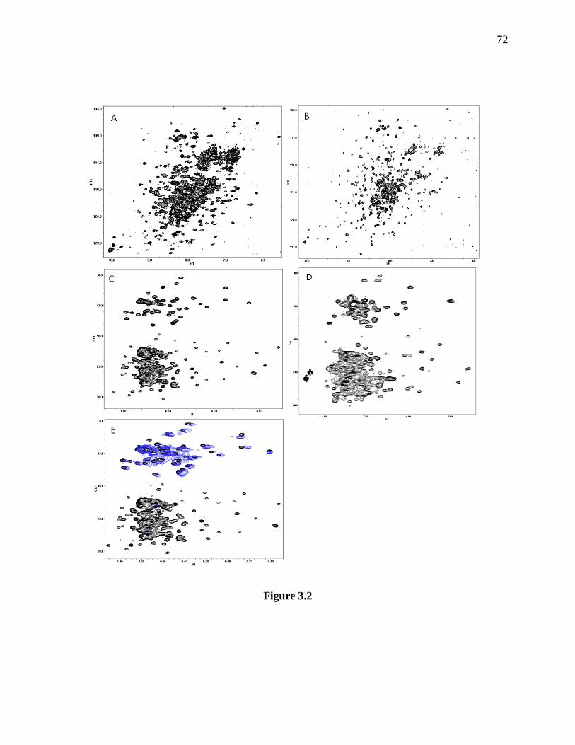
72
Figure 3.2
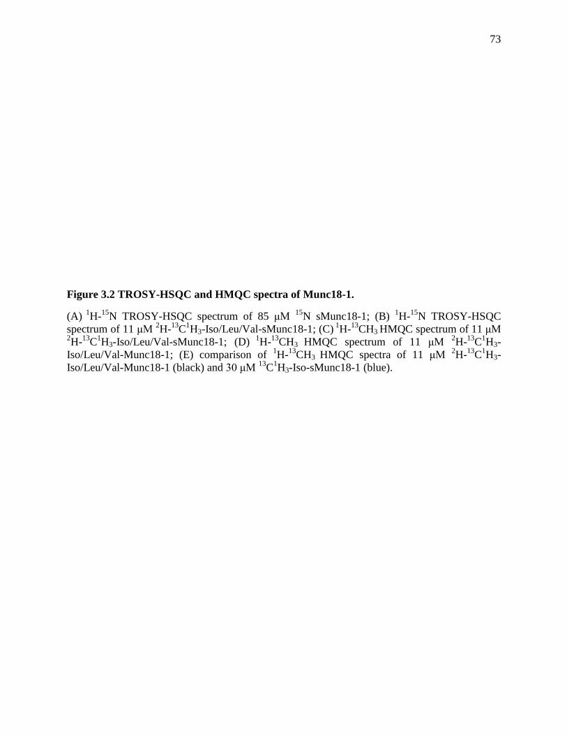
73
Figure 3.2 TROSY-HSQC and HMQC spectra of Munc18-1.
(A) 1
H-15
N TROSY-HSQC spectrum of 85 μM 15
N sMunc18-1; (B) 1H-
15N TROSY-HSQC
spectrum of 11 μM 2H-
13C
1H3-Iso/Leu/Val-sMunc18-1; (C)
1H-
13CH3 HMQC spectrum of 11 μM
2H-
13C
1H3-Iso/Leu/Val-sMunc18-1; (D)
1H-
13CH3 HMQC spectrum of 11 μM
2H-
13C
1H3-
Iso/Leu/Val-Munc18-1; (E) comparison of 1H-
13CH3 HMQC spectra of 11 μM
2H-
13C
1H3-
Iso/Leu/Val-Munc18-1 (black) and 30 μM 13
C1H3-Iso-sMunc18-1 (blue).
74
Figure 3.3 Binding of the SNARE four-helix bundle to Munc18-1 and chemical structures
of MTSL and DOTA-M8-Dy3+
.
(A) 1H-
13CH3 HMQC spectra of 30 μM
13C
1H3-Iso-sMunc18-1 in the absence (blue) and
presence (red) of 40 μM the SNARE four-helix bundle; (B) 1H-
13CH3 HMQC spectra of 11 μM
2H-
13C
1H3-Iso/Leu/Val-Munc18-1 in the absence (black) and presence of 30 μM (red) SNARE
four-helix bundle-synaptobrevin-61C-MTSL; (C) Dithionine addition (blue) to the sample in (B);
in (B) and (C), the blue rectangles highlight the cross-peaks that were broadened; those cross-
peaks were expanded in the windows on the upper right; (D) Chemical Structure of MTSL that
attaches to a cysteine through a disulfide bond; (E) Chemical Structure of DOTA-M8-Dy3+
.

75
3.3.3 To identify the residues of the SNARE four-helix bundle involved in Munc18-1binding.
To find out the residues of the SNARE four-helix bundle involved in Munc18-1 binding,
we need to label Munc18-1 with paramagnetic probes first, and then monitor the changes of the
NMR spectra of the SNARE four-helix bundle upon addition of paramagnetic probe labeled
Munc18-1. Experiments performed to label Munc18-1 with paramagnetic probes, such as MTSL,
MTS-EDTA-Gd3+
(S-Methanethiosulfonylcysteaminyl]ethylenediamine-N,N,N',N'-Tetraacetic
Acid -Gd3+
), maleimido-proxyl, and iodoacetamido-proxyl, all resulted in unfolding of Munc18-
1, probably because of the intrinsic instability of Munc18-1 and the hydrophobicity of these
probes that make them easy to go into Munc18-1 and unfold the protein.
One of our previous papers (Khvotchev et al. 2007) suggested that that Munc18-1 N-
terminal domain (Munc18N) interacts with the Habc domain of syntaxin-1 in the same way as
Munc18-1 interacts with syntaxin-1. The crystal structure of Munc18-1/syntaxin-1 showed that
Munc18N provides large contact surface for syntaxin-1 binding especially for syntaxin-1 Habc
binding (Burkhardt et al. 2008). Therefore, it would make sense that the way Munc18N interacts
with syntaxin-1 Habc domain is similar to the way Munc18-1 interacts with syntaxin-1. These
data led to the hypothesis that the mode of Munc18N binding to the SNARE four-helix bundle
may represent how Munc18-1 interacts with the SNARE four-helix bundle, since the SNARE
four-helix bundle binds to Munc18-1 through the central cavity where Munc18N provides a lot
of surface (Dulubova et al. 1999). If Munc18N indeed binds to the SNARE four-helix bundle, it
would greatly simplify our work since Munc18N is very small compared to Munc18-1. The
structure of Munc18N/SNARE four-helix bundle would help us define the structure of Munc18-
1/SNARE four-helix bundle.

76
1H-
15N HSQC experiments of 40 µM
15N-Munc18NL (1-139) in the absence and
presence of 10 μM, 20 μM, 40 μM the SNARE four-helix bundle showed gradual and specific
broadenings of several cross-peaks (Figure 3.4A), which indicates that Munc18-1 N-terminal
domain indeed binds to the SNARE four-helix bundle. ITC experiments revealed an affinity of
around 4 μM for this interaction, which is tighter than the complex of Munc18-1/SNARE four-
helix bundle (Kd~10 μM) (Figure 3.4B). The fact that the affinity is tighter makes the project
very interesting and exciting, because it could mean that Munc18-1 needs a regulator to change
its conformation in order to bind better to the SNARE four-helix bundle and exert its function.
To find out the residues of the SNARE four-helix bundle involved in Munc18N binding,
the four individual SNAREs were first 2H-
15N labeled and assembled into the SNARE complex,
and then Munc18N was titrated into the labeled SNARE complex. Chemical shifts changes or
broadening in the TROSY-HSQC spectra of the SNARE complexes were then monitored. 1H-
15N TROSY-HSQC experiments of 40 μM
2H-
15N-syntaxin-1 (191-253), SNN, synaptobrevin
(29-93) and SNC labeled SNARE four-helix bundle alone and in the presence of 40 μM
Munc18N suggest that the C-terminal part of synaptobrevin (77-86) and syntaxin-1 (237-242) on
the SNARE four helix-bundle are involved in binding to Munc18N (Figure 3.5A,B,C,D,E),
although the quality of the data is not very good and the chemical shifts changes are not very big.

77
Figure 3.4 Binding of the SNARE four-helix bundle to Munc18N.
(A) 1
H-15
N TROSY-HSQC spectra of 40 μM 15
N Munc18NL in the absence (black) and presence
of 10 μM (light blue), 20 μM (purple) and 40 μM (red) the SNARE four-helix bundle; some
cross-peaks were strongly broadened; (B) ITC analysis of binding of Munc18N to the SNARE
four-helix bundle; Kd=4 μM; N=1.04.
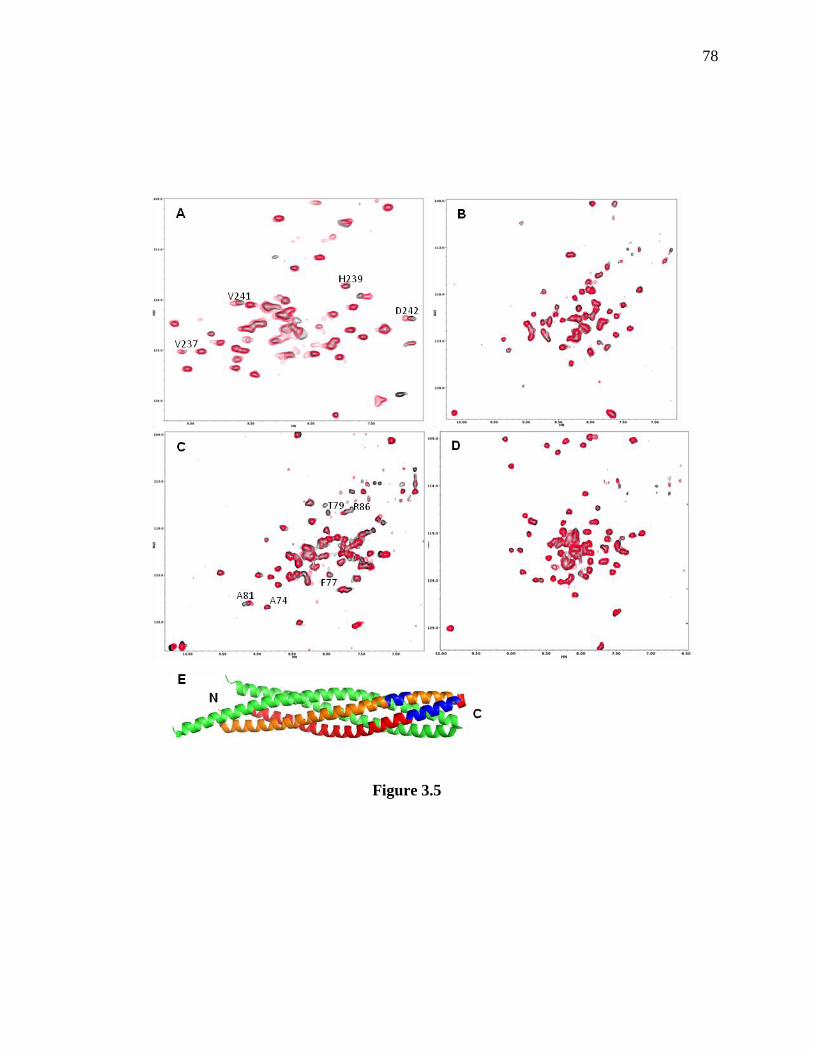
78
Figure 3.5

79
Figure 3.5 Titration of Munc18N to the 2H-
15N labeled SNARE four-helix bundle.
(A) 1
H-15
N TROSY-HSQC spectra of 40 μM 15
N 2H-
15N labeled syntaxin-1 (191-253)-SNARE
four-helix bundle in the absence (black) and presence (red) of 40 μM Munc18N; the cross-peaks
that were shifted are labeled in black; (B) 1H-
15N TROSY-HSQC spectra of 40 μM
15N
2H-
15N
labeled SNN-SNARE four-helix bundle in the absence (black) and presence (red) of 40 μM
Munc18N; (C) 1
H-15
N TROSY-HSQC spectra of 40 μM 15
N 2H-
15N labeled synaptobrevin (29-
93)-SNARE four-helix bundle in the absence (black) and presence (red) of 40 μM Munc18N; the
cross-peaks that were shifted are labeled in black; (D) 1H-
15N TROSY-HSQC spectra of 40 μM
15N
2H-
15N labeled SNC-SNARE four-helix bundle in the absence (black) and presence (red) of
40 μM Munc18N; (E) Ribbon diagram of the crystal structure of the SNARE four-helix bundle
with SNN and SNC in green, synaptobrevin (29-93) in red, and syntaxin-1 (191-253) in orange;
the dark blue helixes are residues shifted in (A) and (C).

80
3.3.4 To define the binding sites in the complex of Munc18N/SNARE four-helix bundle.
To define the structure of the complex of Munc18N/SNARE four-helix bundle, we need
better NMR data than the ones shown in 3.3.2 and 3.3.3. To identify the residues of Munc18N
involved in binding, the backbone cross-peaks of Munc18NL were first assigned. According to
the assignment, the data in Figure 3.4A showed broadenings everywhere in the structure of
Munc18N, especially broadenings in the core hydrophobic region (Figure 3.6A,B,C), which
suggest that there might be a slight conformational change of Munc18N when it binds to the
SNARE four-helix bundle. Therefore, we need to monitor chemical shift changes rather than
broadenings. I used the paramagnetic probe DOTA(1,4,7,10-tetraazacyclododecane-1,4,7,10-
tetraacetic acid)-M8-Dy3+
(Figure 3.3E), which induces pseudo-contact chemical shift to the
residues near the probe and causes chemical shift changes to the cross-peaks correspond to those
residues. The SNARE four-helix bundle labeled with DOTA-M8-Dy3+
in a place close to the
binding site would probably induce chemical shift changes in Munc18NL when it binds to
Munc18NL.
I mutated glutamate 183 of SNC, which is in the C-terminal part of the SNARE four-
helix bundle but not in the binding site according to Figure 3.5, to a cysteine (Figure 3.6E). 1H-
15N-SNC-E183C was labeled with DOTA-M8-Dy
3+ and then assembled into the SNARE four-
helix bundle. TROSY-HSQC experiments of 100 μM 1H-
15N-SNC-E183C SNARE four-helix
bundle before and after DOTA-M8-Dy3+
labeling suggested full labeling, although the quality of
the data was not very high (Figure 3.7A). 1H-
13CH3 HMQC experiments of 50 μM
2H (50%)-
13C
1H3-Iso/Leu/Val-Munc18NL alone and in the presence of 10 μM, 20 μM SNC-E183C-
DOTA-M8-Dy3+
SNARE four-helix bundle, and 20 μM the SNARE four-helix bundle with 0.3
μM TCEP were performed. The results in Figure 3.7B showed that there are several cross-peaks
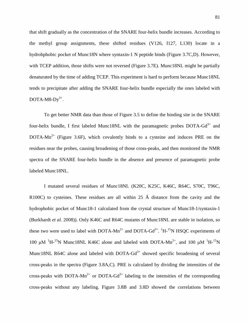
81
that shift gradually as the concentration of the SNARE four-helix bundle increases. According to
the methyl group assignments, these shifted residues (V126, I127, L130) locate in a
hydrohphobic pocket of Munc18N where syntaxin-1 N peptide binds (Figure 3.7C,D). However,
with TCEP addition, those shifts were not reversed (Figure 3.7E). Munc18NL might be partially
denaturated by the time of adding TCEP. This experiment is hard to perform because Munc18NL
tends to precipitate after adding the SNARE four-helix bundle especially the ones labeled with
DOTA-M8-Dy3+
.
To get better NMR data than those of Figure 3.5 to define the binding site in the SNARE
four-helix bundle, I first labeled Munc18NL with the paramagnetic probes DOTA-Gd3+
and
DOTA-Mn2+
(Figure 3.6F), which covalently binds to a cysteine and induces PRE on the
residues near the probes, causing broadening of those cross-peaks, and then monitored the NMR
spectra of the SNARE four-helix bundle in the absence and presence of paramagnetic probe
labeled Munc18NL.
I mutated several residues of Munc18NL (K20C, K25C, K46C, R64C, S70C, T96C,
R100C) to cysteines. These residues are all within 25 Å distance from the cavity and the
hydrophobic pocket of Munc18-1 calculated from the crystal structure of Munc18-1/syntaxin-1
(Burkhardt et al. 2008)). Only K46C and R64C mutants of Munc18NL are stable in isolation, so
these two were used to label with DOTA-Mn2+
and DOTA-Gd3+
. 1H-
15N HSQC experiments of
100 μM 1H-
15N Munc18NL K46C alone and labeled with DOTA-Mn
2+, and 100 μM
1H-
15N
Munc18NL R64C alone and labeled with DOTA-Gd3+
showed specific broadening of several
cross-peaks in the spectra (Figure 3.8A,C). PRE is calculated by dividing the intensities of the
cross-peaks with DOTA-Mn2+
or DOTA-Gd3+
labeling to the intensities of the corresponding
cross-peaks without any labeling. Figure 3.8B and 3.8D showed the correlations between

82
distances to the probe and PRE caused by DOTA-Mn2+
and DOTA-Gd3+
. The correlations
revealed that DOTA-Mn2+
and DOTA-Gd3+
give PRE up to 35 Å and 40 Å (Figure 3.8B,D),
which are good for our experiments. 1H-
13CH3 HMQC experiments of 20 μM
2H-
13C
1H3-
Iso/Leu/Val-syntaxin-1 (191-253) SNARE four-helix bundle alone and in the presence of 20 μM
Munc18N K46C-DOTA-Mn2+
or Munc18N R64C-DOTA-Mn2+
both showed that one valine of
syntaxin-1 (191-253) in the SNARE four-helix bundle was significantly affected (Figure 3.9
A,B). 1H-
13CH3 HMQC experiments of 15 μM
2H-
13C
1H3-Iso/Leu/Val-syntaxin-1 (191-253)
SNARE four-helix bundle alone and in the presence of 15 μM Munc18NL R64C-DOTA-Gd3+
also showed that the same valine was affected (Figure 3.9C). Although we do not have the
methyl group assignments of the SNARE four-helix bundle yet, based on the chemical shift of
the valine and the structure of the SNARE four-helix bundle (Sutton et al. 1998), We speculate
that the cross-peak corresponds to V241, which is in the C-terminal region of syntaxin-1 (Figure
3.9D). These data validate our results in 3.3.3 that the C-terminal part of the SNARE four-helix
bundle is involved in Munc18N binding.
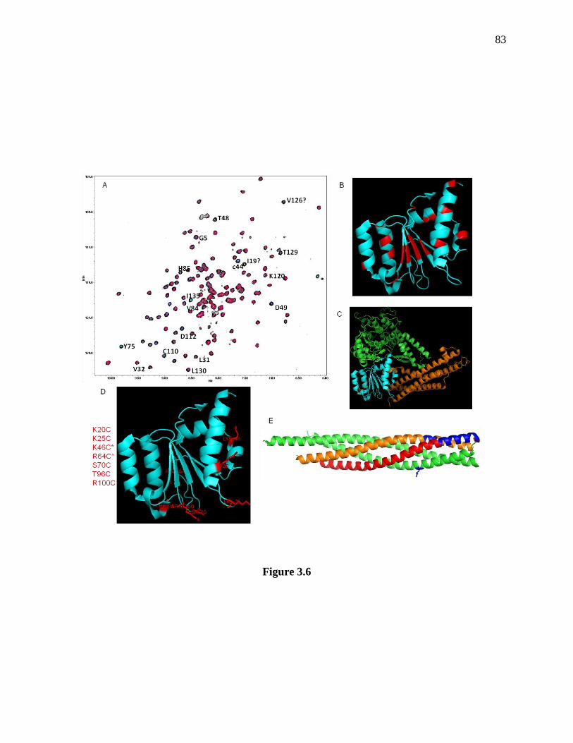
83
Figure 3.6

84
Figure 3.6 Potential residues of Munc18N involved in binding and paramagnetic probe
labeling places of Munc18N and the SNARE four-helix bundle.
(A) 1
H-15
N TROSY-HSQC spectra of 40 μM 15
N Munc18NL in the absence (black) and presence
of 10 μM (light blue), 20 μM (purple) and 40 μM (red) SNARE four-helix bundle; the cross-
peaks that were strongly broadened are labeled in black; (B) Ribbon diagram of the structure of
Munc18N in blue with residues that were broadened in (A) in red; (C) Ribbon diagram of crystal
structure of Munc18-1/syntaxin-1 (2-253) complex with syntaxin-1 (2-253) in orange, Munc18-1
domain 1 in blue and domain 2 and 3 in green; (D) Ribbon diagram of the structure of Munc18N
in blue with residues that were mutated to cysteins in red; the residue names are labeled on the
left; the residues with star labels are stable in isolation; (E) Ribbon diagram of the crystal
structure of the SNARE four-helix bundle with SNN and SNC in green, synaptobrevin (29-93) in
red, and syntaxin-1 (191-253) in orange; the dark blue stick represents SNC E183; the dark blue
helixes are residues involved in Munc18N binding from Figure 3.5. The structure of Munc18N
here is taken from the crystal structure of Munc18-1/syntaxin-1 (Burkhardt et al. 2008).

85
Figure 3.7
86
Figure 3.7 HMQC experiments of 2H-
13CH3-Iso/Leu/Val-Munc18NL in the presence of
DOTA-M8-Dy3+
labeled SNARE four-helix bundle
(A) 1H-
15N TROSY-HSQC spectra of 100 μM
15N SNCE183C-SNARE four-helix bundle in the
absence (black) and presence (red) of DOTA-M8 labeling; (B) 1H-
13CH3 HMQC spectra of 50
μM 2H (50%)-
13CH3-Iso/Leu/Val-Munc18NL in the absence (black) and presence of 10 μM (red),
20 μM (light blue) SNC-E183C- DOTA-M8-Dy3+
-SNARE four-helix bundle; the blue rectangle
highlights the residues shifted; (C) Ribbon diagram of the structure of Munc18N in blue with
residues (V126, I127, L130) that shifted in (B) in red; (D) Ribbon diagram of the crystal
structure of Munc18-1/syntaxin-1 (2-253) complex with syntaxin-1 (2-253) in orange, Munc18-1
domain 1 in blue and domain 2 and 3 in green; (E) 1
H-13
CH3 HMQC spectra of 50 μM 2H (50%)-
13CH3-Iso/Leu/Val-Munc18NL in the absence (black) and presence of 10 μM (red), 20 μM (light
blue) SNC-E183C- DOTA-M8-Dy3+
-SNARE four-helix bundle and with 0.3 mM TCEP addition
(dark blue); the blue rectangle highlights the residues shifted. The structure of Munc18N here is
taken from the crystal structure of Munc18-1/syntaxin-1 (Burkhardt et al. 2008).

87
Figure 3.8 PRE of Munc18NL caused by DOTA-Mn2+/Gd3+ labeling.
(A) 1H-
15N HSQC spectra of 100 μM
15N Munc18NLK46C in the absence (black) and presence
(red) of 500 μM DOTA-Mn2+
; many cross-peaks were significantly broadened; (B) Correlation
between Mn2+
PRE (paramagnetic resonance enhancement) and distance to K46C; Mn2+
can give
PRE up to 35 Å; (C) 1H-
15N HSQC spectra of 100 μM
15N Munc18NLR64C in the absence
(black) and presence (red) of 500 μM DOTA-Gd3+
; many cross-peaks were significantly
broadened; (D) Correlation between Gd3+
PRE and distance to R64C; Gd3+
can give PRE up to
40 Å.
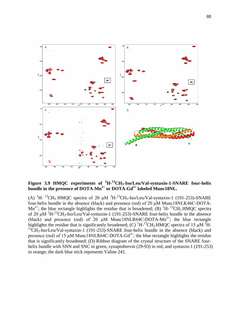
88
Figure 3.9 HMQC experiments of 2H-
13CH3-Iso/Leu/Val-syntaxin-1-SNARE four-helix
bundle in the presence of DOTA-Mn2+
or DOTA-Gd3+
labeled Munc18NL.
(A) 1H-
13CH3 HMQC spectra of 20 μM
2H-
13CH3-Iso/Leu/Val-syntaxin-1 (191-253)-SNARE
four-helix bundle in the absence (black) and presence (red) of 20 μM Munc18NLK46C-DOTA-
Mn2+
; the blue rectangle highlights the residue that is broadened; (B) 1H-
13CH3 HMQC spectra
of 20 μM 2H-
13CH3-Iso/Leu/Val-syntaxin-1 (191-253)-SNARE four-helix bundle in the absence
(black) and presence (red) of 20 μM Munc18NLR64C-DOTA-Mn2+
; the blue rectangle
highlights the residue that is significantly broadened; (C) 1H-
13CH3 HMQC spectra of 15 μM
2H-
13CH3-Iso/Leu/Val-syntaxin-1 (191-253)-SNARE four-helix bundle in the absence (black) and
presence (red) of 15 μM Munc18NLR64C-DOTA-Gd3+
; the blue rectangle highlights the residue
that is significantly broadened; (D) Ribbon diagram of the crystal structure of the SNARE four-
helix bundle with SNN and SNC in green, synaptobrevin (29-93) in red, and syntaxin-1 (191-253)
in orange; the dark blue stick represents Valine 241.
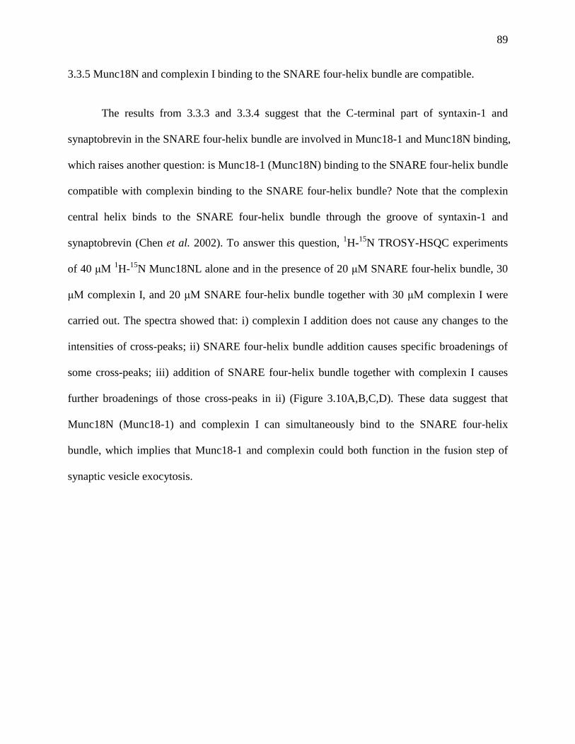
89
3.3.5 Munc18N and complexin I binding to the SNARE four-helix bundle are compatible.
The results from 3.3.3 and 3.3.4 suggest that the C-terminal part of syntaxin-1 and
synaptobrevin in the SNARE four-helix bundle are involved in Munc18-1 and Munc18N binding,
which raises another question: is Munc18-1 (Munc18N) binding to the SNARE four-helix bundle
compatible with complexin binding to the SNARE four-helix bundle? Note that the complexin
central helix binds to the SNARE four-helix bundle through the groove of syntaxin-1 and
synaptobrevin (Chen et al. 2002). To answer this question, 1H-
15N TROSY-HSQC experiments
of 40 μM 1H-
15N Munc18NL alone and in the presence of 20 μM SNARE four-helix bundle, 30
μM complexin I, and 20 μM SNARE four-helix bundle together with 30 μM complexin I were
carried out. The spectra showed that: i) complexin I addition does not cause any changes to the
intensities of cross-peaks; ii) SNARE four-helix bundle addition causes specific broadenings of
some cross-peaks; iii) addition of SNARE four-helix bundle together with complexin I causes
further broadenings of those cross-peaks in ii) (Figure 3.10A,B,C,D). These data suggest that
Munc18N (Munc18-1) and complexin I can simultaneously bind to the SNARE four-helix
bundle, which implies that Munc18-1 and complexin could both function in the fusion step of
synaptic vesicle exocytosis.

90
Figure 3.10 Binding of Munc18N and complexin I to the SNARE four-helix bundle.
(A) 1H-
15N TROSY-HSQC spectra of 40 μM
15N Munc18NL in the absence (black) and
presence (red) of 30 μM complexin I; (B) 1
H-15
N TROSY-HSQC spectra of 40 μM 15
N
Munc18NL in the absence (black) and presence (red) of 20 μM SNARE four-helix bundle; (C) 1H-
15N TROSY-HSQC spectra of 40 μM
15N Munc18NL in the absence (black) and presence
(red) of 30 μM complexin I and 20 μM SNARE four-helix bundle; (D) Ribbon diagram of the
crystal structure of the SNARE four-helix bundle and complexin I central helix with complexin I
central helix in purple, SNN and SNC in green, synaptobrevin (29-93) in red, and syntaxin-1
(191-253) in orange; the blue ellipse irises out the residues involved in Munc18N binding.
91
3.3.6 To define the structure of the complex of Munc18-1/SNARE four-helix bundle by X-ray
crystallography and SAXS.
Attempts to crystallize the complex of sMunc18-1/SNARE four-helix bundle yielded
either crystals of sMunc18-1 alone or crystals of the SNARE four-helix bundle alone, probably
because of the low affinity between the two proteins. SAXS (small angle X-ray scattering) could
give a low resolution solution structure of the complex of Munc18-1/SNARE four-helix bundle,
which in combination with the NMR results could define the complex structure.
SAXS experiments of 39 μM Munc18-1 alone, 120 μM SNARE four-helix bundle alone,
and 40 μM, 62 μM, 100 μM mixtures of Munc18-1 and SNARE four-helix bundle in a 1:1 ratio
were performed. The data were collected in house at Rigaku Americas Corporation, the
Woodlands, TX. The quality of the data was not high but was enough to generate some
preliminary results. Data analysis using ATSAS 2.4 revealed that Munc18-1 is more like a
globular protein in solution (Figure 3.11A), which is consistent with crystal structure of Munc18-
1 (Burkhardt et al. 2008). Moreover, superposition of the crystal structure and the envelope of
Munc18-1 generated from SAXS data suggest that domain1 and domain 3a are probably flexible
in solution, which might regulate the size of Munc18-1 cavity. The SAXS data of the SNARE
four-helix bundle showed an elongated envelope, which fits well with the crystal structure of the
SNARE four-helix bundle (Sutton et al. 1998) (Figure 3.11B). Since Munc18-1 interacts with the
SNARE four-helix bundle with weak affinity (Kd is around 10 μM), the mixtures of Munc18-
1/SNARE four-helix bundle contained three different species: Munc18-1, the SNARE four-helix
bundle, Munc18-1/SNARE four-helix bundle complex, and different concentrations of the
mixtures included different amounts of the three species (Table 3.1). Therefore, the data of the
mixtures were hard to interpret. By applying some scale factors to the data of the mixtures to
92
subtract the data from free Munc18-1 and free SNARE four-helix bundle, we obtained one
reasonable complex envelope, which can be fitted well with the crystal structures of Munc18-1
and the SNARE four-helix bundle (Figure 3.11C). The fitted complex envelope suggests that
Munc18-1 interacts with one end of the SNARE four-helix bundle through the cavity, which is
consistent with the NMR data that Munc18-1 binds to the C-terminal part of the SNARE four-
helix bundle. However, the data need further and careful analysis and interpretation.
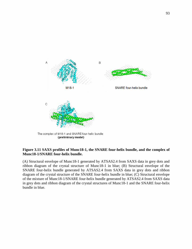
93
Figure 3.11 SAXS profiles of Munc18-1, the SNARE four-helix bundle, and the complex of
Munc18-1/SNARE four-helix bundle.
(A) Structural envelope of Munc18-1 generated by ATSAS2.4 from SAXS data in grey dots and
ribbon diagram of the crystal structure of Munc18-1 in blue; (B) Structural envelope of the
SNARE four-helix bundle generated by ATSAS2.4 from SAXS data in grey dots and ribbon
diagram of the crystal structure of the SNARE four-helix bundle in blue; (C) Structural envelope
of the mixture of Munc18-1/SNARE four-helix bundle generated by ATSAS2.4 from SAXS data
in grey dots and ribbon diagram of the crystal structures of Munc18-1 and the SNARE four-helix
bundle in blue.

94
Table 3.1 Different amounts of Munc18-1, the SNARE four-helix bundle, and the complex
of Munc18-1/SNARE four-helix bundle in the mixtures of Munc18-1 and the SNARE four-
helix bundle.
The concentrations of free Munc18-1, free SNARE four-helix bundle, and the complex of
Munc18-1/SNARE four-helix bundle were calculated from Kd (assume Kd=10 μM) and the
concentrations of the mixtures of the two proteins. If A represents Munc18-1, B represents the
SNARE four-helix bundle, AB represents the complex of Munc18-1/SNARE four-helix bundle.
, Kd=[A]free[B]free/[AB]=10 μM. The percentages of the complex of
Munc18-1/SNARE four-helix bundle are calculated by dividing the concentrations of this
complex to the concentrations of the mixtures of the two proteins.
Mixture of
Munc18-1 and
the SNARE four-
helix bundle (1:1)
concentration
(μM)
Complex of
Munc18-
1/SNARE four-
helix bundle
concentration
(μM)
Complex of
Munc18-
1/SNARE four-
helix bundle
percentage (%)
Free Munc18-1
concentration
(μM)
Free SNARE four-
helix bundle
concentration (μM)
40 24 60 16 16
62 42 68 20 20
100 73 73 27 27
95
3.4 Discussion
Despite intensive studies of SM proteins in the recent years, their key roles are largely
unclear. Our model (Figure 2.7) together with in vitro reconstitutions (Shen et al. 2007; Shen et
al. 2010) suggest that Munc18-1 might be directly involved in membrane fusion and that
interaction of Munc18-1 with the SNARE four-helix bundle might be key to uncover its crucial
role in neurotransmitter release. To investigate whether this interaction is physiologically
relevant, the best way is to understand the complex structure first, perform mutagenesis to
disrupt their interaction, and then test the impairments of neurotransmitter release of these
mutants in vivo. However, the interaction between Munc18-1 and the SNARE four-helix bundle
is weak, so it is technically hard to solve the complex structure.
By applying ITC and NMR spectroscopy, I was able to get some data that suggest that
Munc18N binds to the SNARE four-helix bundle tighter than Munc18-1, and that the C-terminal
part of the SNARE four-helix bundle is involved in binding, especially synaptobrevin and
syntaxin-1. NMR data also showed that Munc18N and complexin simultaneously bind to the
SNARE four-helix bundle. SAXS data suggest that Munc18-1 binds to one end of the SNARE
four-helix bundle, and that in solution the relative orientation between domain 1 and domain 3a
of Munc18-1 is flexible, which implies that there might be an allosteric regulation of Munc18-1
in vivo.
The finding that Munc18N binds tighter than Munc18-1 full length protein to the SNARE
four-helix bundle is very interesting. Data from our previous paper (Xu et al. 2010) suggest that
the binding of Munc18-1/SNARE four-helix bundle involves the Munc18-1 cavity. Munc18N
provides a major contact surface of the cavity but not all, so it would make sense that Munc18N
interacts with the SNARE four-helix bundle weaker than full length Munc18-1. However, the

96
result is opposite to our hypothesis, which implies that Munc18-1 needs a conformation change
in order to bind better to the SNARE four-helix bundle. This finding is consistent with SAXS
data that the relative orientation between domain 1 and domain 3a of Munc18-1 is flexible in
solution. Our data suggest a view that in vivo Munc18-1 might require an activator or a
modification to regulate its conformation so as to exert its critical function in fusion. In addition,
our results showed that Munc18N binds to the C-terminal part of syntaxin-1 and synaptobrevin
on the SNARE four-helix bundle, which is consistent with the SAXS result that Munc18-1 full
length protein binds to one end of the SNARE four-helix bundle. These results are intriguing
because they really place Munc18-1 right at the site where membrane fusion occurs, which
suggest that Munc18-1 might be directly involved in membrane fusion. Such a role together with
the allosteric regulation could certainly explain the critical nature of Munc18-1 and SM proteins
for membrane fusion in vivo.
The results that the C-terminal part of syntaxin-1 and synaptobrevin are involved in
Munc18N binding raise one question: does Munc18N (Munc18-1) and complexin bind together
to the SNARE four-helix bundle? The complexin central helix binds to the SNARE four-helix
bundle through the groove of syntaxin-1 and synaptobrevin (Chen et al. 2002), and complexin
plays important roles in late stage of neurotransmitter release. If complexin and Munc18-1
cannot simultaneously bind to the SNARE four-helix bundle, it would argue against our
hypothesis that Munc18-1 is directly involved in fusion through interaction to the SNARE four-
helix bundle. Our NMR results suggest that these three proteins bind together, which supports
our hypothesis.
Our data presented here are not very conclusive and they need further validation because
of several problems: i) Munc18-1 is a big molecule (70Kd) and it is destabilized when deuterated;

97
ii) The interaction between Munc18/SNARE four-helix bundle complex is weak; iii) Munc18N
is intrinsically unstable in isolation and undergoes some conformational change when it binds to
the SNARE four-helix bundle. These problems make it really hard to get good data by NMR
spectroscopy and to solve the structure of the complex. In this case of weak interaction, SAXS
and X-ray crystallography are not very ideal methods to tackle the problem, although I did not
try a lot in both methods. Further investigations need to be carried out to overcome the
difficulties, to validate the data presented here, and eventually to solve the complex structure and
to perform in vivo studies to uncover the key role of Munc18-1 in neurotransmitter release.
98
Chapter 4 Reconstitution of the vital functions of Munc18-1 and Munc13 in
neurotransmitter release
4.1 Introduction
The release of neurotransmitters by Ca2+
-triggered synaptic vesicle exocytosis is crucial
for neural function. The machinery that controls this process contains proteins that have
homologues in most types of intracellular membrane traffic and underlie a conserved mechanism
of membrane fusion, including N-ethylmaleimide sensitive factor (NSF), soluble NSF adaptor
proteins (SNAPs), the neuronal SNAP receptors (SNAREs) synaptobrevin, syntaxin-1 and
SNAP-25, and the Sec1/Munc18 (SM) protein Munc18-1 [reviewed in (Brunger 2005; Jahn et al.
2006; Rizo et al. 2008; Sudhof et al. 2009)]. The SNAREs form a tight four-helix bundle called
SNARE complex that brings the synaptic vesicle and plasma membranes together (Sollner et al.
1993; Hanson et al. 1997; Poirier et al. 1998; Sutton et al. 1998), which is key for fusion.
NSF/SNAPs disassemble the SNARE complex (Sollner et al. 1993) to recycle the SNAREs for
another round of fusion (Banerjee et al. 1996; Mayer et al. 1996). Munc18-1 binds to a self-
inhibited ‘closed’ conformation of syntaxin-1, an interaction that gates entry of syntaxin-1 into
SNARE complexes and is not conserved in some other systems (Carr et al. 1999; Dulubova et al.
1999; Misura et al. 2000; Sudhof 2004; Gerber et al. 2008). In addition, Munc18-1 binds to
SNARE complexes and likely cooperates with the SNAREs in membrane fusion by an as yet
unclear mechanism (Dulubova et al. 2007; Shen et al. 2007). Synaptic vesicle fusion also
depends critically on factors that are specialized for the exquisite regulation of neurotransmitter
release. Particularly important are the Ca2+
sensor synaptotagmin-1 (Fernandez-Chacon et al.
2001) and the large active zone proteins Munc13s, which play an essential role through their
MUN domain (Basu et al. 2005; Madison et al. 2005; Stevens et al. 2005) and modulate release
through other domains [e.g. (Rhee et al. 2002; Shin et al. 2010)]. The Munc13 MUN domain
99
catalyzes the opening of syntaxin-1 (Ma et al. 2011) and is related to tethering factors from other
membrane traffic systems, suggesting that the MUN domain may in addition have a universal
function in docking and/or fusion (Pei et al. 2009; Li et al. 2011).
Despite these advances, a coherent model that can explain the importance of all these
eight central components of the release machinery has not emerged yet. A particularly
fundamental question is why Munc18-1 and Munc13s are so crucial for exocytosis in vivo (Wu
et al. 1998; Aravamudan et al. 1999; Augustin et al. 1999; Richmond et al. 1999), which is
emphasized by the total abrogation of neurotransmitter release observed in the absence of
Munc18-1 or Munc13-1/2 [the strongest phenotypes observed so far with the high resolution of
electrophysiology in mammalian synapses (Verhage et al. 2000; Varoqueaux et al. 2002)].
Attempts to reconstitute synaptic vesicle fusion with purified components have provided seminal
insights into the mechanism of release, but they have not answered this vital question.
Reconstitution studies showed that the neuronal SNAREs alone can induce lipid mixing
between liposomes (Weber et al. 1998), and that this activity can be enhanced by Munc18-1
(Shen et al. 2007; Rodkey et al. 2008; Diao et al. 2010), by synaptotagmin-1 (Tucker et al. 2004),
and by Munc13-4 (Boswell et al. 2012) or the related protein CAPS (James et al. 2009), which
also contains a MUN domain. Moreover, evidence for content mixing was provided in
reconstitutions with neuronal SNAREs alone (Nickel et al. 1999; van den Bogaart et al. 2010)
and together with synaptotagmin-1 (Kyoung et al. 2011) or CAPS (James et al. 2009). However,
the combined effects of all these factors have not been studied, in contrast to the more
comprehensive reconstitutions performed in other membrane traffic systems (Mima et al. 2008;
Ohya et al. 2009; Stroupe et al. 2009). Particularly intriguing among the results obtained with the
neuronal proteins is the observation that the activity of the SNAREs in reconstitution assays is
100
dramatically stimulated by synaptotagmin-1 in the presence of Ca2+
and that some features of
such stimulation correlate with physiological observations (Stein et al. 2007; Chicka et al. 2008;
Xue et al. 2008; Lee et al. 2010; Kyoung et al. 2011; van den Bogaart et al. 2011). At the same
time, these findings are puzzling because they were obtained without Munc18-1 and Munc13s,
and hence contrast with the crucial importance of these proteins for neurotransmitter release.
It is also worth noting that most of these studies did not include NSF and SNAPs, and
used reconstituted syntaxin-1/SNAP-25 heterodimers (often referred to as t-SNARE complexes),
assuming that these heterodimers are obligatory intermediates in the membrane fusion pathway.
However, early data showed that NSF/SNAPs inhibit the activity of the SNAREs in inducing
lipid mixing because they disassemble the syntaxin-1/SNAP-25 heterodimers (Weber et al.
2000). Moreover, it is plausible that the correct starting point is the binary syntaxin-1/Munc18-1
complex, since syntaxin-1 and Munc18-1 stabilize each other in vivo (Verhage et al. 2000;
Gerber et al. 2008), and may travel together to the plasma membrane (Medine et al. 2007).
Interestingly, in reconstitutions started with syntaxin-1, Munc18-1 had inhibitory and stimulatory
effects of Munc18-1 on lipid mixing that may reflect at least in part the multiple roles of this
protein (Schollmeier et al. 2011), but the effects of other factors were not studied.
In the research presented here, we sought to develop a model of synaptic vesicle fusion
that integrates the functions of the neuronal SNAREs, Munc18-1, Munc13s, NSF, SNAPs and
synaptotagmin-1, expecting that such a model could explain why Munc18-1 and Munc13s are so
crucial for neurotransmitter release and could thus bridge the large gap existing between the
available reconstitution and physiological data. We show that Munc18-1 binding to syntaxin-1
displaces SNAP-25 from syntaxin-1/SNAP-25 heterodimers, which is aided by NSF/-SNAP on
membranes. We reconstituted co-expressed syntaxin-1/Munc18-1 into proteoliposomes and

101
observed efficient lipid and content mixing with synaptobrevin proteoliposomes that requires
Ca2+
, SNAP-25, and core fragments of Munc13-1 and synaptotagmin-1. We also found that the
efficient lipid mixing between liposomes containing syntaxin-1/SNAP-25 heterodimers and
synaptobrevin-liposomes occurring in the presence of Ca2+
/synaptotagmin-1 is strongly inhibited
by NSF/-SNAP, but is activated upon further addition of Munc18-1 and Munc13-1. These
results suggest that our system reconstitutes basic steps of synaptic vesicle fusion in a manner
that requires Munc18-1 and Munc13-1, as observed in vivo, and that in addition incorporates the
three neuronal SNAREs, NSF, -SNAP, synaptotagmin-1 and Ca2+
. We propose a model
whereby syntaxin-1/heterodimers are disassembled by NSF/SNAPs and synaptic vesicle fusion
starts with closed syntaxin-1 bound to Munc18-1, which orchestrates SNARE complex assembly
together with Munc13s through a pathway that is not inhibited by NSF/-SNAP and allows the
exquisite regulation of neurotransmitter release.
4.2 Materials and methods
4.2.1 Recombinant DNA constructs
Bacteria expression vectors to express full-length rat synaptobrevin and its cytoplasmic
domain (residues 29-93), full length rat syntaxin-1A and its cytoplasmic domain (2-253), human
SNAP-25B full-length (with its four cysteins mutated to serines), the SNARE motifs of human
SNAP-25B (SNN, residues 11-82; and SNC, residues 141-203), full-length rat Munc18-1, full-
length squid Munc18-1, rat Munc13-1 MUN domain (residues 859-1407,EF,1453-1531), rat
Munc13-1 fragment C1C2BMUN (residues 529-1407,EF,1453-1531), co-expressed Munc18-
1/syntaxin-1, co-expressed syntaxin-1/SNAP-25, NSF, α-SNAP, and the rat synaptotagmin-1
C2AB fragment (residues 140-421) were available in the lab. Note that the MUN domain and

102
C1C2BMUN contained a partial deletion in a long loop that promotes aggregation [see (Rizo et
al. 2008; Ma et al. 2011)].
4.2.2 Expression and purification of recombinant proteins
4.2.2.1 Expression and purification of C1C2BMUN
To generate a vector to express the C1C2BMUN fragment of rat Munc13-1 with the same
loop deletion as the MUN domain (residues 529-1407, EF, 1453-1531), we cloned a DNA
sequence encoding Munc13-1(529-1531) into the pFastBacTMHT B vector (invitrogen), which
contains a polyhedron promoter (for hight-level expression of recombinant protein in insect cell)
before the start codon and encodes an N-terminal TEV cleavable His6 tag. The loop deletion was
then performed using QickChange (Stratagene). The construct was used to generate a
baculovirus using the Bac-to-Bac system (Invitrogen). Insect cells (sf9) were infected with the
baculovirus, harvested about 68-72 hours post-infection, and resuspended in lysis buffer (50 mM
Tris 8.0, 250 mM Nacl, 10 mM imidazole). Cells were lysed through one freeze and thaw circle.
The cell lysate was centrifuged at 18,000 rpm for 45 minutes, and the clear supernatant was
incubated with Ni2+-NTA agarose at 4 °C for 2 hours. The beads were washed with: i) lysis
buffer; ii) lysis buffer containing 1% Triton-100; iii) lysis buffer containing 1 M NaCl; and iv)
lysis buffer. The protein was eluted with 200 mM imidazole and the His6-tag was removed by
incubation with TEV protease at 4 °C overnight. The protein was further purified by ion
exchange chromatography and gel filtration, and was concentrated to 9 mg/ml for storage in 10
mM Tris buffer (pH 8.0) containing 10% glycerol and 250 mM NaCl.
103
4.2.2.2 Expression and purification of NSF and α-SNAP
NSF and α-SNAP were cloned from a pQE9 vector into a pGEX-KG vector. Both
proteins were expressed in BL21 E.Coli cells in LB media. NSF was induced with 0.4 mM IPTG
at 20 °C for 20 hrs. α-SNAP was induced with 0.4 mM IPTG at 25 °C for 18 hrs. Cells were re-
suspended in a buffer containing 50 mM HEPES pH7.6, 400 mM KCl, 10% glycerol (v/v), and 2
mM DTT. The cells were lysed with an Avestin cell disruptor and the lysate was clarified by
centrifugation at 20,000 rpm for 30 minutes. The supernatant was incubated with Glutathione
Sepharose 4B (GE Healthcare) at 4 °C overnight. The bound proteins were washed with PBS,
PBS with 1% Triton X-100, and then PBS with 500 mM NaCl. The GST-tag was cleaved from
the proteins by thrombin cleavage at 4 °C overnight on the beads. The proteins were further
purified by sizeexclusion chromatography using a Superdex 75 (for α-SNAP) or Superdex 200
(for NSF) column in a buffer containing 20 mM HEPES pH7.6, 150 mM KCl, 10% glycerol
(v/v), 1 mM DTT. For NSF purification, all buffers contain 0.5 mM ATP.
4.2.2.3 Co-expression and purification of Munc18-1 and full-length syntaxin-1
A pET-DUET vector (Novagen) to co-express N-terminally His6-tagged full-length rat
Munc18-1 and full-length rat syntaxin-1A in BL21 E. coli cells was constructed using standard
recombinant DNA methods. Cells were grown in LB media and induced with 0.4 mM IPTG at
an optical density A600=1.0 for 18 hrs at 23 °C. Cells were harvested and lysed with an Avestin
cell disruptor in buffer containing 50 mM Tris pH 8.0, 300 mM KCl, 10% glycerol, 1% TX-100
and 0.3 mM TCEP. The cell lysate was centrifuged at 20,000 rpm for 30 minutes. The
supernatant was incubated with Ni2+
- nitrilotriacetic acid (NTA) Agarose (QIAGEN) at room
temperature for 1 hr. The bound proteins were first washed with buffer containing 50 mM Tris
pH 8.0, 300 mM KCl, 10% glycerol (v/v), 0.5% TX-100 and 0.3 mM TCEP, and then washed
104
with the same buffer containing 20 mM imidazole and 1% CHAPS instead of 0.5% TX-100. The
bound proteins were eluted in the same buffer containing 250 mM imidazole and 1% CHAPS.
The eluted proteins were purified by size-exclusion chromatography using a superdex 200
10/300 column in a buffer containing 20 mM Tris pH8.0, 150 mM KCl, 10% glycerol (v/v), 1%
CHAPS, and 0.5 mM TCEP.
4.2.2.4 Co-expression and purification of full-length syntaxin-1 and SNAP25
Recombinant rat full-length syntaxin and human SNAP25B were co-expressed and
purified basically as described (Misura et al. 2000), but with some modifications. BL21 E. Coli
cells were grown in Terrific Broth media. Cells were induced with 0.4 mM IPTG to an optical
density A600=1.0 for 20 hrs at 25 °C. Cells were lysed with an Avestin cell disruptor in buffer A
(50 mM Tris pH 8.0, 500 mM NaCl, 5% glycerol, 1% Triton X-100, 20 mM imidazole, and 10
mM 2-Mercaptoethanol). Cell lysate was clarified by centrifugation at 20,000 rpm for 30
minutes. The supernatant was incubated with Ni2+
-NTA Agarose (QIAGEN) at room
temperature for 1 hr. The bound proteins were first washed with buffer A, then washed with
buffer B (50 mM Tris pH 8.0, 200 mM NaCl, 1% w/v octyl-β-D-glucopyranoside (β-OG), 5%
glycerol (v/v), and 50 mM imidazole), and then eluted with buffer B containing 250 mM
imidazole instead of 50 mM imidazole. The eluted proteins were further purified by ion
exchange chromatography on Mono Q with buffers containing β-OG.
4.2.2.5 Expression and purification of other recombinant proteins
The expression and purification of other recombinant proteins were similar to those
described in previous chapter. Isotopic labeling to obtain 2H-Ile-
13CH3-syntaxin-1 was performed
using well-established procedures (Tugarinov et al. 2004).

105
4.2.3 NMR spectroscopy
1H-
13C HMQC spectra were acquired at 25
oC on a Varian INOVA800 spectrometer.
Samples contained 12-20 μM 2H-Ile-
13CH3-syntaxin-1 alone or with different additions
(Munc18-1, sMunc18-1 and/or SNAP-25 were added in a 20% excess). Samples were dissolved
in 20 mM Tris pH 8.0, 150 mM NaCl, 2 mM TCEP, using D2O as the solvent. Spectra were
acquired with a 2.3 hour total acquisition time.
4.2.4 Lipid mixing assay using syntaxin-1/Munc18-1 liposomes
All lipids were purchased from Avanti Polar Lipid. Donor (synaptobrevin) liposomes
contained 60% POPC, 17% POPE, 20% DOPS, 1.5% NBD-PE and 1.5% Rhodamine-PE.
Acceptor (syntaxin-1/Munc18-1) liposomes contained 58% POPC, 20% POPE, 18% DOPS, 2%
PIP2 and 2% DAG. Lipid mixtures were dried in glass tubes with nitrogen gas and under
vacuum overnight. Lipid films were re-suspended in buffer buffer C (25 mM HEPES, pH 7.4,
150 mM KCl, 1 mM DTT, 10% glycerol (v/v)) and vortexed for at least 5 min. The re-suspended
lipid films were frozen and thawed five times, and then extruded through a 50 nm polycarbonate
filter with an Avanti extruder for at least 29 times. Purified proteins in buffer containing 1%
CHAPS were added slowly to liposomes (5 mM lipids) to make the final concentration of
CHAPS to 0.4%. The P/L ratio ranged from 1:500 to 1:1000 for the synaptobrevin liposomes,
and from 1:1000 to 1:2000 for the syntaxin-1/Munc18-1 liposomes. The liposome protein
mixtures were incubated at room temperature for 40 minutes and then dialyzed extensively with
1g/L Bio-beads SM2 (Bio-Rad) 3 times in 20 mM HEPES, pH 7.4, 150 mM KCl, 1 mM DTT,
10% glycerol (v/v) at 4 °C. For lipid mixing assays, donor liposomes (0.25 mM lipids) were
mixed with acceptor liposomes (0.5 mM lipids) in the presence of 1 μM C1C2BMUN, 5 μM
SNAP-25, 2 μM C2AB fragment and/or 0.5 mM Ca2+ (as indicated in the figures) in a total

106
volume of 80 μl. NBD fluorescence emission at 538 nm (excitation 460 nm) was monitored with
a PTI Spectrofluorometer. All experiments were performed at 30 °C. At the end of each reaction,
1% w/v β-OG was added to solubilize the liposomes. Because of considerable variability in the
fluorescence intensity observed upon detergent addition, which may arise from partial
precipitation, we find that we obtain more consistent results by first converting the time traces to
F1/F0, where F1 is the observed fluorescence intensity and F0 is the initial intensity, and then we
average the F1/F0 value observed upon detergent addition for an entire set of experiments. This
average is then used to normalize all the traces to express the data as % of maximum
fluorescence.
4.2.5 Lipid mixing assay using syntaxin-1/SNAP-25 liposomes
Donor (synaptobrevin) liposomes and acceptor (syntaxin-1/SNAP25) liposomes
contained the same lipid compositions described above. Lipid mixtures were dried in glass tubes
with nitrogen gas and under vacuum overnight. Lipid films were re-suspended and dissolved in
buffer C with 1.2% β-OG. Purified proteins in buffer containing 1% β-OG were added to
liposomes (5 mM lipids) to make the P/L ratio 1:600. The mixtures were incubated at room
temperature for 40 minutes and dialyzed extensively with 1g/L Bio-beads SM2 (Bio-Rad) 3
times in buffer C. For lipid mixing assays, donor liposomes (0.25 mM ) were mixed with
acceptor liposomes (0.5 mM) with different additions in a total volume of 80 μl. For experiments
with NSF/α-SNAP, acceptor liposomes were first incubated with 0.1 μM NSF, 0.5 μM α-SNAP,
5 mM MgCl2 and 2 mM ATP at 37°C for 20minutes, and then mixed with donor liposomes with
different additions, which included 0.5 μM Munc18-1, 1 μM C1C2BMUN, 2 μM C2AB
fragment, 0.5 mM Ca2+
, and excess SNAP-25 as indicated in the figures. All experiments were

107
performed at 37°C. At the end of each reaction, 1% w/v β-OG was added to solubilize the
liposomes, and the data were analyzed as described above.
4.2.6 Content mixing assay
Donor (synaptobrevin) liposomes contained 44.5% POPC, 20% POPE, 12% DOPS, 20%
cholesterol, 3.5% DiD (Invitrogen). Lipid mixtures were dried in glass tubes with nitrogen gas
and under vacuum overnight. Lipid films were re-suspended and dissolved in buffer C containing
1% w/v β-OG and 40mM sulforhodamine B (Acros Organics). Purified full-length synaptobrevin
in buffer containing 1% w/v β-OG was added to the lipid mixtures to make the final
concentration of lipids 5 mM and the P/L ratio 1:500. The protein lipid mixtures were diluted 4
times (keeping the sulforhodamine concentration at 40 mM) to allow liposome formation, and
then were incubated at room temperature for 40 minutes. The liposomes were purified with a
Superose 6 column. The first 1 ml sample coming out in the void volume was collected and
dialyzed in 150 ml detergent free buffer (20 mM HEPES pH 7.4, 150 mM KCl, 1 mM DTT)
containing 10 g of Amberlite XAD2 3 times at 4 oC (2 times 2 hrs and one time overnight). The
resulting lipid concentration was determined by UV. Acceptor (syntaxin-1/Munc18-1) liposomes
were prepared with the same method described for the lipid mixing assays. Lipid mixing and
content mixing were monitored as described for the lipid mixing assays by measuring
dequenching of DiD (excited at 650 nm, emission at 675 nm) and sulforhodamine B (excited at
565 nm, emission at 587 nm).
4.2.7 Liposome co-floatation assays
Proteoliposomes containing syntaxin-1/Munc18-1 complex were prepared as described
below with a protein/lipid (P/L) ratio of 1:1000. Solutions containing 4 mM lipids (4 μM
108
syntaxin-1/Munc18-1 complex) were incubated with 10 μM SNAP-25 at room temperature for 1
hr. The proteoliposomes and bound proteins were isolated by floatation on a Histodenz density
gradient (40%:35%:30%) as described (Guan et al. 2008). Samples from the top of the gradient
(35 μl) were taken and analyzed by SDS-PAGE and Coomassie blue staining. For experiments
with reconstituted full-length syntaxin-1, proteoliposomes containing PC:PE:PS:PIP2
(60:18:20:2) at a P/L ratio of 1:500 were prepared as previously described for a syntaxin-1(183-
288) fragment (Chen et al. 2006). The syntaxin-1 proteoliposomes were then dialyzed with 1g/L
Bio-beads SM2 (Bio-Rad) 3 times in 25 mM HEPES pH 7.3, 150 mM KCl, 10% glycerol (v/v)
at 4°C overnight to remove the detergent. The syntaxin-1 liposomes were incubated with SNAP-
25 at 4°C for 2h and contained 3 μM syntaxin-1 and 6 μM SNAP-25 after addition of different
reagents, which included 5 μM Munc18-1, 0.5 μM NSF, 1 μM α-SNAP, 2 mM ATP, 5 mM
MgCl2 or 5 mM EDTA. After incubation at 37 °C for 1 hr, the proteoliposomes were isolated by
floatation on a Histodenz density gradient (40%:35%:30%) and samples at the top (35 μl) were
taken and analyzed by SDS-PAGE and Coomassie blue staining.
4.3 Results
4.3.1 Munc18-1 displaces SNAP-25 from syntaxin-1 in solution
This work stems from our previous efforts to understand the critical role of Munc13-1 in
neurotransmitter release and its relation to Munc18-1 and SNARE function. Dr. Cong Ma from
the lab showed that Munc13-1 MUN domain dramatically accelerates the transition from the
closed syntaxin-1/Munc18-1 complex to the SNARE complex (Ma et al. 2011). Since many
models of neurotransmitter release assume that syntaxin-1/SNAP-25 heterodimers are obligatory
intermediates that are formed downstream of the syntaxin-1/Munc18-1 complex, Dr. Cong Ma
investigated whether the MUN domain can also accelerate the transition from the latter complex
109
to the former. For this purpose, he used NMR spectroscopy, taking advantage of the power of
1H-
13C heteronuclear multiple quantum coherence (HMQC) spectra to distinguish between
different states of syntaxin-1 cytoplasmic region (residues 2-253) specifically 1H,
13C-labeled at
Ile methyl groups (2H-Ile-
13CH3-syntaxin-1) (Ma et al. 2011). Thus, multiple changes were
observed when 1H-
13C HMQC spectra were acquired on samples of
2H-Ile-
13CH3-syntaxin-1
alone, assembled in the SNARE complex, or bound to Munc18-1 or SNAP-25 (Figures 4.1A-C).
He acquired consecutive 2-hr 1H-
13C HMQC spectra of
2H-Ile-
13CH3-syntaxin-1 bound to
Munc18-1 after addition of the N- and C-terminal SNARE motifs of SNAP-25 (SNN and SNC,
respectively) to test whether SNN and SNC can displace Munc18-1 from syntaxin-1. However,
no spectral changes were observed after long incubations whether a fragment spanning the
Munc13-1 MUN was present (Figure 4.1D) or absent (Figure 4.1I). This result contrasts with the
efficient formation of the SNARE complex observed in analogous experiments performed in the
presence of synaptobrevin and the MUN domain (Ma et al. 2011), suggesting that synaptobrevin
(in addition to SNAP-25) is required to open the conformation of syntaxin-1 bound to Munc18-1.
To test whether the opposite reaction can occur, i.e. whether Munc18-1 can displace
SNAP-25 from syntaxin-1, he used squid Munc18-1 (sMunc18-1), since it is more soluble in
isolation and forms similar complexes with the mammalian SNAREs as rat Munc18-1 (Xu et al.
2010), as confirmed in part by the similarity of the 1H-
13C HMQC spectra of
2H-Ile-
13CH3-
syntaxin-1 bound to rat Munc18-1 or sMunc18-1 (Figures 4.1A,E). Importantly, 1H-
13C HMQC
spectra of 2H-Ile-
13CH3-syntaxin-1 bound to SNAP-25 showed gradual changes upon addition of
sMunc18-1 that revealed quantitative formation of the closed syntaxin-1/sMunc18-1 complex
after several hours (Figures 4.1F,G). To confirm this result by a different method, he used FRET
assays starting with complexes between the SNAP-25 SNARE motifs and syntaxin-1(2-253) that
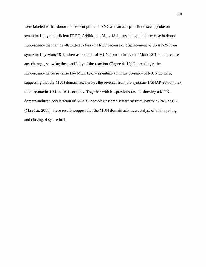
110
were labeled with a donor fluorescent probe on SNC and an acceptor fluorescent probe on
syntaxin-1 to yield efficient FRET. Addition of Munc18-1 caused a gradual increase in donor
fluorescence that can be attributed to loss of FRET because of displacement of SNAP-25 from
syntaxin-1 by Munc18-1, whereas addition of MUN domain instead of Munc18-1 did not cause
any changes, showing the specificity of the reaction (Figure 4.1H). Interestingly, the
fluorescence increase caused by Munc18-1 was enhanced in the presence of MUN domain,
suggesting that the MUN domain accelerates the reversal from the syntaxin-1/SNAP-25 complex
to the syntaxin-1/Munc18-1 complex. Together with his previous results showing a MUN-
domain-induced acceleration of SNARE complex assembly starting from syntaxin-1/Munc18-1
(Ma et al. 2011), these results suggest that the MUN domain acts as a catalyst of both opening
and closing of syntaxin-1.
111
Figure 4.1
I

112
Figure 4.1 Munc18-1 displaces SNAP-25 from syntaxin-1 in solution.
(A-G) 1H-
13C HMQC spectra of
2H-Ile-
13CH3-syntaxin-1 alone (orange) or bound to Munc18-1
(black) (A); alone (orange) or forming the SNARE complex with the synaptobrevin and SNAP-
25 SNARE motifs (blue) (B); forming the SNARE complex (blue) or bound to SNAP-25
(magenta) (C); bound to Munc18-1 after incubation with the SNAP-25 SNARE motifs (SNN and
SNC) for 11 hr in the presence of the Munc13-1 MUN domain (black) (D); bound to sMunc18-1
(E); bound to SNAP-25 (magenta) or bound to SNAP-25 after incubation with sMunc18-1 for
0.5 hr (black) (F); and bound to SNAP-25 (magenta) or bound to SNAP-25 after incubation with
sMunc18-1 for 9 hr (black) (G). Note that after 9 hr the spectrum is analogous to that in panel E,
showing that 2H-Ile-13CH3-syntaxin- 1 was released from SNAP-25 and bound to sMunc18-1,
and that only partial displacement was observed in the spectrum acquired after 0.5 hr incubation
with sMunc18-1 (black in panel F). The cross-peak assignments that are available for isolated
syntaxin-1 (Dulubova et al. 1999; Chen et al. 2008) are indicated in orange in A,B. Cross-peaks
that move considerably upon formation of the syntaxin-1/Munc18-1 complex or the SNARE
complex are indicated in black in A or in blue in B, respectively. H3 identifies cross-peaks that
are known to belong to the SNARE motif but do not have residue-specific assignment; *
indicates tentative assignments. (H) The Munc13-1 MUN domain accelerates the transition from
the syntaxin-1/SNAP-25 complex to the syntaxin-1/Munc18-1 complex. Syntaxin-1 cytoplasmic
region (residues 2-253) labeled with rhodamine at residue 249 was incubated with SNN and SNC
that was labeled with BODIPY at residue 187. The BODIPY fluorescence, which decreases upon
formation of the syntaxin-1/SNN/SNC complex due to efficient FRET with the rhodamine label
on syntaxin-1, was monitored and the sample was incubated until the BODIPY fluorescence
reached a plateau that indicated completion of the reaction. Munc18-1, Munc13-1 MUN domain
or both were added and then we monitored the increase in BODIPY fluorescence resulting from
loss of FRET as syntaxin-1 binds to Munc18-1 and SNN/SNC are released. The three curves
were normalized to the extrapolated maximum fluorescence in the presence of Munc18-1 and
MUN domain. (I) The SNAP-25 SNARE motifs cannot displace Munc18-1 from syntaxin-1. The
diagram shows a 1H-
13C HMQC spectrum of
2H-Ile-
13CH3-syntaxin-1 bound to Munc18-1 after
incubation with the SNAP-25 SNARE motifs (SNN and SNC) for 11 hr (black).
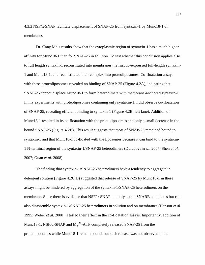
113
4.3.2 NSF/α-SNAP facilitate displacement of SNAP-25 from syntaxin-1 by Munc18-1 on
membranes
Dr. Cong Ma’s results show that the cytoplasmic region of syntaxin-1 has a much higher
affinity for Munc18-1 than for SNAP-25 in solution. To test whether this conclusion applies also
to full length syntaxin-1 reconstituted into membranes, he first co-expressed full-length syntaxin-
1 and Munc18-1, and reconstituted their complex into proteoliposomes. Co-floatation assays
with these proteoliposomes revealed no binding of SNAP-25 (Figure 4.2A), indicating that
SNAP-25 cannot displace Munc18-1 to form heterodimers with membrane-anchored syntaxin-1.
In my experiments with proteoliposomes containing only syntaxin-1, I did observe co-floatation
of SNAP-25, revealing efficient binding to syntaxin-1 (Figure 4.2B, left lane). Addition of
Munc18-1 resulted in its co-floatation with the proteoliposomes and only a small decrease in the
bound SNAP-25 (Figure 4.2B). This result suggests that most of SNAP-25 remained bound to
syntaxin-1 and that Munc18-1 co-floated with the liposomes because it can bind to the syntaxin-
1 N-terminal region of the syntaxin-1/SNAP-25 heterodimers (Dulubova et al. 2007; Shen et al.
2007; Guan et al. 2008).
The finding that syntaxin-1/SNAP-25 heterodimers have a tendency to aggregate in
detergent solution (Figure 4.2C,D) suggested that release of SNAP-25 by Munc18-1 in these
assays might be hindered by aggregation of the syntaxin-1/SNAP-25 heterodimers on the
membrane. Since there is evidence that NSF/α-SNAP not only act on SNARE complexes but can
also disassemble syntaxin-1/SNAP-25 heterodimers in solution and on membranes (Hanson et al.
1995; Weber et al. 2000), I tested their effect in the co-floatation assays. Importantly, addition of
Munc18-1, NSF/α-SNAP and Mg2+
-ATP completely released SNAP-25 from the
proteoliposomes while Munc18-1 remain bound, but such release was not observed in the

114
presence of EDTA and/or absence of Munc18-1 (Figure 4.2B). The natural explanation for these
results is that NSF/α-SNAP disassemble the syntaxin-1/SNAP-25 heterodimers, which requires
ATP hydrolysis, and such disassembly allows Munc18-1 to ‘capture’ the syntaxin-1 closed
conformation, thus preventing the re-binding of the released SNAP-25 to syntaxin-1.
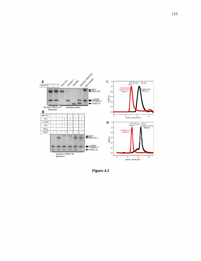
115
Figure 4.2
C
D
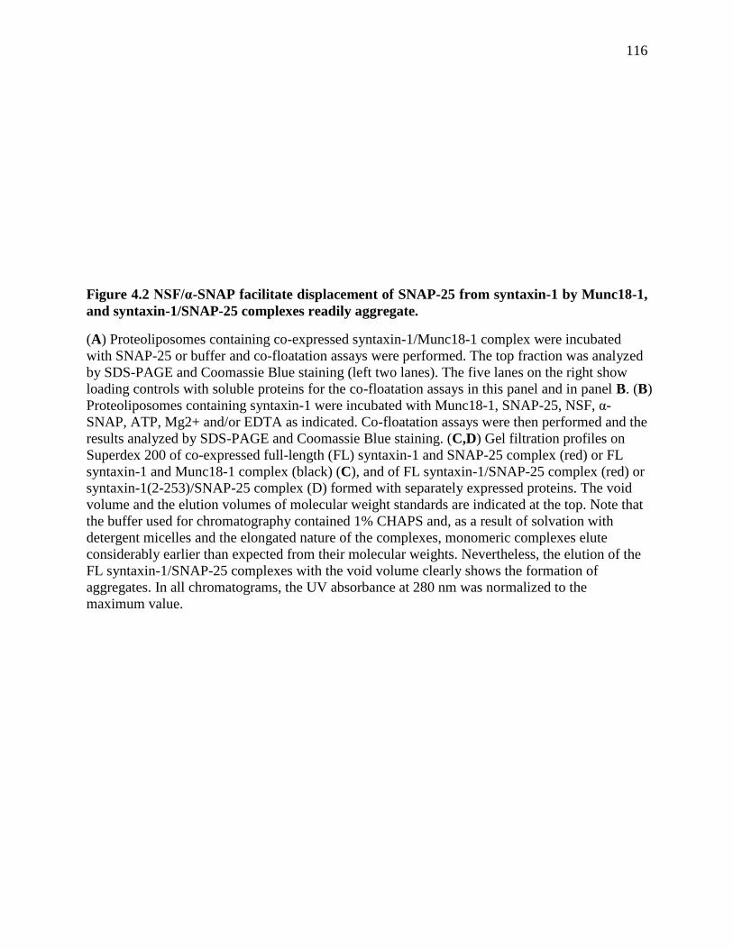
116
Figure 4.2 NSF/α-SNAP facilitate displacement of SNAP-25 from syntaxin-1 by Munc18-1,
and syntaxin-1/SNAP-25 complexes readily aggregate.
(A) Proteoliposomes containing co-expressed syntaxin-1/Munc18-1 complex were incubated
with SNAP-25 or buffer and co-floatation assays were performed. The top fraction was analyzed
by SDS-PAGE and Coomassie Blue staining (left two lanes). The five lanes on the right show
loading controls with soluble proteins for the co-floatation assays in this panel and in panel B. (B)
Proteoliposomes containing syntaxin-1 were incubated with Munc18-1, SNAP-25, NSF, α-
SNAP, ATP, Mg2+ and/or EDTA as indicated. Co-floatation assays were then performed and the
results analyzed by SDS-PAGE and Coomassie Blue staining. (C,D) Gel filtration profiles on
Superdex 200 of co-expressed full-length (FL) syntaxin-1 and SNAP-25 complex (red) or FL
syntaxin-1 and Munc18-1 complex (black) (C), and of FL syntaxin-1/SNAP-25 complex (red) or
syntaxin-1(2-253)/SNAP-25 complex (D) formed with separately expressed proteins. The void
volume and the elution volumes of molecular weight standards are indicated at the top. Note that
the buffer used for chromatography contained 1% CHAPS and, as a result of solvation with
detergent micelles and the elongated nature of the complexes, monomeric complexes elute
considerably earlier than expected from their molecular weights. Nevertheless, the elution of the
FL syntaxin-1/SNAP-25 complexes with the void volume clearly shows the formation of
aggregates. In all chromatograms, the UV absorbance at 280 nm was normalized to the
maximum value.
117
4.3.3 Reconstitution of a strict Munc13 requirement for lipid mixing
The above results suggest that the syntaxin-1/Munc18-1 complex may constitute the true
starting point for neurotransmitter release in vivo, rather than syntaxin-1/SNAP-25 heterodimers.
This postulate might explain the critical importance of Munc13s for neurotransmitter release,
since they mediate the transition from the syntaxin-1/Munc18-1 complex to the SNARE complex
(Ma et al. 2011). Thus, we decided to investigate whether basic features of synaptic vesicle
fusion, in particular the strict requirement for Munc13s, can be reproduced in reconstitution
experiments started with syntaxin-1/Munc18-1 proteoliposomes.
For the experiments described below we used a Munc13-1 fragment that includes the
MUN domain and its preceding C1 and C2B domains (residues 529-1531; referred to as
C1C2BMUN). These domains mediate binding to diacylglycerol (DAG) (Rhee et al. 2002) and
phosphatidylinositol- 4,5-bisphosphate (PIP2) (Shin et al. 2010), respectively. To perform some
basic characterization of the C1C2BMUN fragment, Dr. Cong Ma tested whether it can
accelerate the transition from the syntaxin-1/Munc18-1 complex to the SNARE complex using a
FRET assay (Ma et al. 2011), and found that the C1C2BMUN fragment has a similar activity as
the MUN domain in promoting this transition in solution (Figure 4.3A). We also tested whether
the C1C2BMUN fragment binds DAG and PIP2 using liposome co-floatation assays. These
experiments showed that both DAG and PIP2 enhance binding of the C1C2BMUN fragment to
liposomes, and that there is a clear synergy between both types of lipids in promoting such
binding (Figure 4.3B).
To test whether syntaxin-1/Munc18-1 complexes can provide an appropriate starting
point for synaptic vesicle fusion, we first used standard lipid mixing assays that monitor
dequenching of NBD fluorescence when the lipids of synaptobrevin donor liposomes containing

118
NBD- and rhodamine-labeled lipids dilute into unlabeled acceptor liposomes, similar to the
original reconstitutions with SNAREs alone (Weber et al. 1998) but using acceptor
proteolipomes that contained syntaxin-1/Munc18-1 complex instead of syntaxin-1/heterodimers
(Figure 4.4A). Throughout this work, we used low protein-to-lipid ratios (ranging from 1:500 to
1:2000) and relatively low concentrations of soluble proteins, and we routinely checked that the
observed increases in NBD fluorescence intensity reflected true dequenching rather than light
scattering caused by liposome clustering [see (Xu et al. 2011)]. Unless otherwise specified, the
syntaxin-1/Munc18-1 liposomes included DAG and PIP2 to facilitate binding of C1C2BMUN.
To have a quantitative idea of the results obtained under different conditions, we used the
NBD fluorescence intensity after 1000 s of reaction (expressed as % from the maximum
fluorescence observed upon detergent addition) as a compromise measurement that reflects in
part the initial slope and in part the level of completion of the reaction at longer times. The
values observed under identical conditions in experiments repeated with the same liposome
preparations were then averaged.
When we mixed donor liposomes containing synaptobrevin with the acceptor syntaxin-
1/Munc18-1-liposomes, no significant lipid mixing was observed even after addition of SNAP-
25 (Figure 4.4B,D). We did observe some (albeit inefficient) lipid mixing when SNAP-25 was
added together with C12BMUN either in the presence or absence of Ca2+
, and the rate of lipid
mixing was dramatically increased when we included a synaptotagmin-1 fragment containing its
two C2 domains (C2AB fragment) in addition to SNAP-25, C1C2BMUN and Ca2+
(Figure
4.4B,D). Control experiments showed that no lipid mixing occurs in the absence of SNAP-25 or
C1C2BMUN and that the synaptobrevin cytoplasmic domain (Sybcd) inhibits the reaction,
demonstrating that lipid mixing is SNARE-dependent and strictly requires the Munc13-1
119
C1C2BMUN fragment (Figures 4.4C,D). This strict requirement is particularly rewarding
because it mirrors the essential nature of Munc13s for neurotransmitter release.
To further explore the factors that influence the lipid mixing observed in the presence of
SNAP-25, C1C2BMUN and C2AB fragment, I performed a Ca2+
titration and found that the
efficiency of lipid mixing exhibits a high cooperativity with Ca2+
, with a Hill coefficient of 5.9
and half-maximal rate occurring at 250 μM Ca2+
(Figure 4.4E,F). These Ca2+
concentrations are
somewhat higher than those required for neurotransmitter release at the Calyx of Held but
comparable to those estimated for other systems, and the Ca2+
cooperativity resembles that of
neurotransmitter release, which occurs with a Hill coefficient of 5 (Meinrenken et al. 2003;
Schneggenburger et al. 2005). We also examined how the observed lipid mixing depends on
DAG and PIP2, agents that underlie the stimulation of neurotransmitter release during
presynaptic plasticity processes mediated by the Munc13 C1 and C2B domains. Removal of
either DAG or PIP2 led to a considerable decrease in the rate of lipid mixing, which was
accentuated when both DAG and PIP2 were absent (Figure 4.4G,H). These results suggest that
these two types of lipids play a synergistic role in attracting C1C2BMUN to the membrane, thus
enhancing its activity on the syntaxin-1/Munc18-1 complex to mediate SNARE complex
formation.
Finally, to establish an additional correlation with physiological data, we tested the
effects of a point mutation in Munc18-1 (E66A) that decreases neurotransmitter release in
neurons by about 50% (Deak et al. 2009). Reassuringly, this mutation led to a comparable
decrease in the lipid mixing observed in our reconstitution assays (Figures 4.4I,J). We would like
to emphasize that the syntaxin-1/Munc18-1 liposomes used to test the effects of removing DAG
or PIP2, as well as the effects of the Munc18-1 E66A mutation, were prepared side-by-side with
120
liposomes containing WT syntaxin-1/Munc18-1 and contained similar amounts of both proteins
(Figure 4.3C), ruling out the possibility that the differences observed arise from different
amounts of reconstituted proteins.
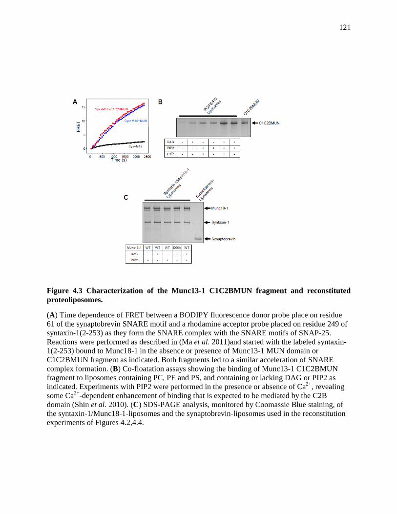
121
Figure 4.3 Characterization of the Munc13-1 C1C2BMUN fragment and reconstituted
proteoliposomes.
(A) Time dependence of FRET between a BODIPY fluorescence donor probe place on residue
61 of the synaptobrevin SNARE motif and a rhodamine acceptor probe placed on residue 249 of
syntaxin-1(2-253) as they form the SNARE complex with the SNARE motifs of SNAP-25.
Reactions were performed as described in (Ma et al. 2011)and started with the labeled syntaxin-
1(2-253) bound to Munc18-1 in the absence or presence of Munc13-1 MUN domain or
C1C2BMUN fragment as indicated. Both fragments led to a similar acceleration of SNARE
complex formation. (B) Co-floatation assays showing the binding of Munc13-1 C1C2BMUN
fragment to liposomes containing PC, PE and PS, and containing or lacking DAG or PIP2 as
indicated. Experiments with PIP2 were performed in the presence or absence of Ca2+
, revealing
some Ca2+
-dependent enhancement of binding that is expected to be mediated by the C2B
domain (Shin et al. 2010). (C) SDS-PAGE analysis, monitored by Coomassie Blue staining, of
the syntaxin-1/Munc18-1-liposomes and the synaptobrevin-liposomes used in the reconstitution
experiments of Figures 4.2,4.4.
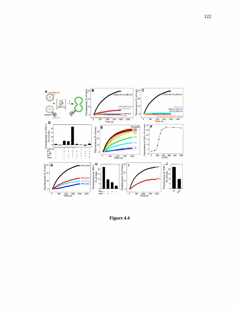
122
Figure 4.4
123
Figure 4.4 Requirement of Munc13 for lipid mixing of syntaxin-1/Munc18-1-liposomes with
synaptobrevin-liposomes.
(A) Diagram summarizing the lipid mixing experiments, which were performed with acceptor
liposomes containing syntaxin-1/Munc18-1, donor liposomes containing synaptobrevin and
NBD-lipids quenched by rhodamine-lipids, and addition of various reagents. Lipid mixing is
expected to result in increased NBD fluorescence. (B,C) Traces showing the lipid mixing
observed in the presence of SNAP-25, Munc13-1 C1C2BMUN (M13), synaptotagmin-1 C2AB
fragment (C2AB), synaptobrevin cytoplasmic domain (Sybcd) and/or 0.5 mM Ca2+
in various
combinations. The y axis represents NBD fluorescence normalized to the maximum fluorescence
observed upon detergent addition. (D) Quantification of the results obtained in the experiments
of (B,C). (E) Lipid mixing observed in the presence of SNAP-25, C1C2BMUN and C2AB
fragment as a function of Ca2+
concentration. (F) Plot of the normalized NBD fluorescence
intensity at 1000 s as a function of Ca2+
observed in (E). Curve fitting of the data (solid line)
fielded a Hill coefficient of 5.9 and half-maximal rate at 250 μM Ca2+
. (G,H) Fluorescence
traces showing the dependence of the lipid mixing observed on the presence of DAG and/or PIP2
in the acceptor syntaxin-1/Munc18-1 liposomes (G), and quantification of the results (H). (I,J)
Traces comparing lipid mixing obtained with acceptor liposomes that contained syntaxin-1
bound to WT or E66A mutant Munc18-1 (I) and quantification of the results (J). In (D, H and J),
bars represent averages of the normalized NBD fluorescence observed after 1000 s in repeated
experiments performed under the same conditions. Error bars represent standard deviations.

124
4.3.4 Lipid mixing is accompanied by content mixing
To demonstrate liposome fusion, it is important to show not only lipid mixing but also
content mixing. These experiments have commonly been hindered by the tendency of small
molecules to leak from SNARE-containing proteoliposomes [e.g. see (Dennison et al. 2006;
Diao et al. 2010; Zucchi et al. 2011)]. However, synaptobrevin-liposomes with encapsulated
sulforhodamine were recently reported to have very slow leakage rates that allowed monitoring
of content mixing from dequenching of sulforhodamine upon fusion with acceptor liposomes
(Kyoung et al. 2011). In our hands, a small amount of leakiness of sulforhodamine trapped into
synaptobrevin-liposomes was observed when standard methods were used to prepare the
proteoliposomes, and addition of C1C2BMUN exacerbated the leakiness (Figure 4.5A), likely
because of sensitivity to residual detergent. However, I was able to minimize this problem
through extensive dialysis of the liposomes against buffer with detergent-absorbing beads in
small volumes (see Experimental Procedures), resulting in a very slow degree of leakiness that
can be readily subtracted from signal increases due to content mixing (Figure 4.5B).
We adapted the sulforhodamine fluorescence de-quenching method to monitor content
mixing in our bulk assays, including self-quenched DiD lipids in the donor liposomes to allow
simultaneous monitoring of lipid mixing as described for the single-vesicle assays (Kyoung et al.
2011) (Figure 4.6A). In these experiments, two clearly distinct outcomes are expected if true
content mixing occurs or the content of the donor vesicles is released due to lysis. Thus, since
our NBD fluorescence de-quenching assays indicate that donor vesicles undergo an average of
1.5 to 2 rounds of fusion based on a standard conversion method (Parlati et al. 1999), content
release upon vesicle lysis should lead to a monotonous increase in sulforhodamine fluorescence
intensity that would saturate close to the maximal signal observed upon detergent addition. In
125
contrast, bona-fide content mixing should result in increases in sulforhodamine fluorescence
intensity that saturate at lower intensities and follow a similar time course as lipid mixing,
perhaps with some delay.
When we mixed the synaptobrevin donor liposomes with acceptor liposomes containing
syntaxin-1/Munc18-1 complex, we observed efficient sulforhodamine fluorescence de-quenching
upon addition of SNAP-25, C1C2BMUN and C2AB fragment/ Ca2+
(Figure 4.6B,C). Control
experiments showed that SNAP-25, C1C2BMUN and the C2AB fragment are each essential for
the de-quenching (Figure 4.6B,C). The lipid mixing measured simultaneously during these
experiments from the DiD fluorescence dequenching (Figure 4.6D,E) paralleled the data
obtained with the NBD fluorescence de-quenching assay (Figures 4.4B-D) and were similar to
the sulforhodamine de-quenching results, although in the absence of C2AB fragment there is a
small amount of lipid mixing but not sulforhodamine de-quenching. Importantly, the time course
of the sulforhodamine fluorescence de-quenching in the presence of SNAP-25, C1C2BMUN and
C2AB fragment/ Ca2+
closely followed the time course observed for lipid mixing through DiD
fluorescence de-quenching (see the comparison in Figure 4.6F). These results show that content
mixing accompanies lipid mixing in these experiments and hence that our system reconstitutes
membrane fusion with the three neuronal SNAREs, Munc18-1 and core fragments of Munc13-1
and synaptotagmin-1, although we cannot rule out that some degree of leakiness may occur
during fusion.

126
Figure 4.5 Analysis of donor liposome leakiness.
(A,B) The sulforhodamine fluorescence intensity of synaptobrevin-donor liposomes was
monitored as a function of time in the absence or presence of Munc13-1 C1C2BMUN fragment.
In (A), the liposomes were prepared by standard methods as described (Kyoung et al. 2011).In
(B), the same procedure was followed except that dialysis was performed against BioBeads
(BioRad) in small volumes to remove the detergent more efficiently.
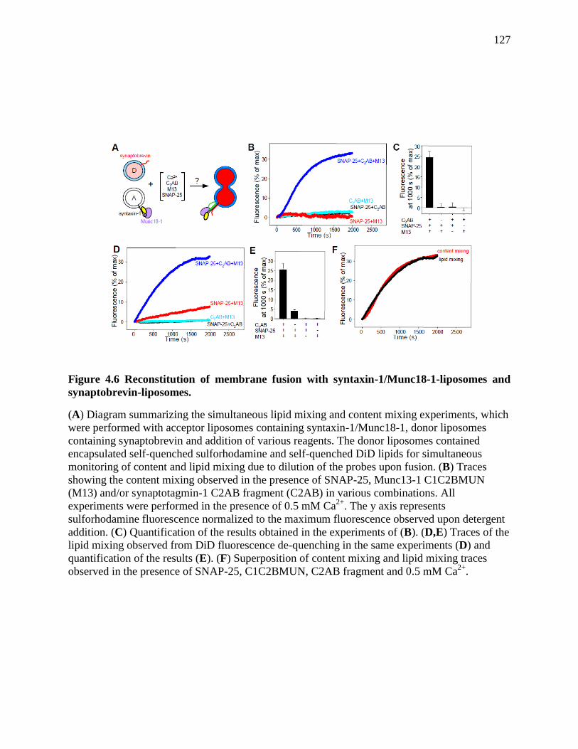
127
Figure 4.6 Reconstitution of membrane fusion with syntaxin-1/Munc18-1-liposomes and
synaptobrevin-liposomes.
(A) Diagram summarizing the simultaneous lipid mixing and content mixing experiments, which
were performed with acceptor liposomes containing syntaxin-1/Munc18-1, donor liposomes
containing synaptobrevin and addition of various reagents. The donor liposomes contained
encapsulated self-quenched sulforhodamine and self-quenched DiD lipids for simultaneous
monitoring of content and lipid mixing due to dilution of the probes upon fusion. (B) Traces
showing the content mixing observed in the presence of SNAP-25, Munc13-1 C1C2BMUN
(M13) and/or synaptotagmin-1 C2AB fragment (C2AB) in various combinations. All
experiments were performed in the presence of 0.5 mM Ca2+
. The y axis represents
sulforhodamine fluorescence normalized to the maximum fluorescence observed upon detergent
addition. (C) Quantification of the results obtained in the experiments of (B). (D,E) Traces of the
lipid mixing observed from DiD fluorescence de-quenching in the same experiments (D) and
quantification of the results (E). (F) Superposition of content mixing and lipid mixing traces
observed in the presence of SNAP-25, C1C2BMUN, C2AB fragment and 0.5 mM Ca2+
.
128
4.3.5 Munc18-1- and Munc13-1-dependent lipid mixing in the presence of NSF/α-SNAP
While membrane fusion in our reconstitution experiments exhibits a strict requirement for
Munc13-1, and uses Munc18-1 as an intrinsic component of the acceptor liposomes, our results
still do not explain why Munc18-1 and Munc13-1 are so crucial for neurotransmitter release in
vivo, since efficient lipid mixing and content mixing can also be obtained with SNAREs and
syntaptotagmin-1 in the absence of Munc18-1 and Munc13-1 (see Introduction). However,
almost all reconstitution studies of synaptic vesicle fusion described so far started with syntaxin-
1/SNAP-25 heterodimers and did not incorporate NSF/SNAPs, which play fundamental roles in
all SNARE-dependent forms of intracellular membrane traffic. One study that did incorporate
NSF/α-SNAP showed that these factors inhibit lipid mixing, likely because they disassemble the
syntaxin-1/SNAP-25 heterodimers (Weber et al. 2000). Since our biochemical results showed
that Munc18-1 displaces SNAP-25 from syntaxin-1 and that NSF/α-SNAP facilitate this
displacement on a membrane (Figures 4.1 and 4.2), we hypothesized that fusion might be
activated by Munc18-1 and Munc13-1 even in the presence of NSF/α-SNAP because Munc18-1
can ‘capture’ the syntaxin-1 released from the syntaxin-1/SNAP-25 heterodimers and lead to
membrane fusion with the help of Munc13-1 (Figures 4.4 and 4.6).
To test our hypothesis, we used the NBD fluorescence de-quenching assay as in the
experiments of Figure 4.4 but starting with acceptor liposomes that contained reconstituted
syntaxin-1/SNAP-25 heterodimers (Figure 4.7A). In one set of experiments, the heterodimers
were formed from separately expressed proteins. As expected, when the acceptor liposomes were
mixed with synaptobrevin-containing donor liposomes in the presence of Ca2+
and C2AB
fragment, highly efficient lipid mixing was observed (Figure 4.7B,C). The amount of lipid
mixing was not substantially altered by addition of Munc18-1 and C1C2BMUN, or by addition

129
of NSF/α-SNAP in the absence of ATP, but lipid mixing was almost completely abolished by
NSF/α-SNAP in the presence of ATP (Figure 4.7B,C). Interestingly, some lipid mixing was
observed when Munc18-1 and C1C2BMUN were added together with NSF/α-SNAP and ATP
(Figure 4.7B,C), suggesting that that Munc18-1 and C1C2BMUN can activate fusion through a
pathway that is resistant to NSF/α-SNAP, as predicted by our hypothesis.
We reasoned that, since the concentration of free SNAP-25 should favor the formation of
SNARE complexes without affecting the rate of syntaxin-1/SNAP-25 heterodimer dissociation
by NSF/α-SNAP, an excess of SNAP-25 might assist in the activation of the lipid mixing by
Munc18-1 and C1C2BMUN. Indeed, a titration with SNAP-25 showed increased efficiency of
the lipid mixing activated by Munc18-1 and C1C2BMUN, which saturated at an excess SNAP-
25 concentration of 2 μM (Figure 4.7D). Importantly, the 2 μM excess SNAP-25 did not affect
the overall lipid mixing observed upon addition of only C2AB fragment and Ca2+
(compare
black symbols in Figures 4.7B,E), and NSF/α-SNAP still inhibited lipid mixing strongly in the
presence of excess SNAP-25 and the absence of Munc18-1 and C1C2BMUN (Figures 4.7E,F).
With the 2 μM excess SNAP-25, Munc18-1 and C1C2BMUN stimulated lipid mixing to levels
comparable to those observed in the absence of NSF/α-SNAP (compare orange and black curves
in Figure 4.7E); Munc18-1 alone (but not C12C2BMUN alone) appeared to provide a small
amount of activation (Figure 4.7E, cyan and blue symbols; see also Figure 4.7F).
We repeated the same set of experiments using acceptor liposomes with reconstituted
syntaxin-1/SNAP-25 heterodimers that were co-expressed, since it was originally reported that
co-expression of syntaxin-1/SNAP-25 was necessary for lipid mixing induced by the neuronal
SNAREs (Weber et al. 1998). Under our conditions, the efficiency of lipid mixing between these
liposomes and synaptobrevin-containing donor liposomes in the presence of Ca2+
and C2AB
130
fragment (Figure 4.8A,B) was similar to those observed with the separately expressed syntaxin-1
and SNAP-25 (Figure 4.7B,C). Moreover, we again observed strong inhibition of lipid mixing by
NSF/α-SNAP in the presence of ATP, and activation of lipid mixing upon further addition of
Munc18-1, C1C2BMUN and excess SNAP-25 (Figure 4.8). Hence, the interplay between
NSF/α-SNAP and Munc18-1/Munc13-1 does not depend on the method of expression of
syntaxin-1 and SNAP-25. Overall, these results suggest that inclusion of NSF/α-SNAP is key to
unmask the crucial importance of Munc18-1 and Munc13-1 for membrane fusion in these
reconstituted systems.
To see whether we can get the same conclusions in the absence of synaptotagmin-1
C2AB fragment, I performed lipid mixing assays as in the experiments of Figure 4.7. As
expected, when syntaxin-1/SNAP-25-containing acceptor liposomes were mixed with
synaptobrevin-containig liposomes in the absence of C2AB, a small amount of lipid mixing was
observed (Figure 4.9A,B,C). Addition of C1C2BMUN/Ca2+
did not alter the amount of lipid
mixing (Figure 4.9A,C). Addition of Munc18-1 slightly enhanced the amount of lipid mixing,
while addition of both Munc18-1 and C1C2BMUN/Ca2+
increased amount of lipid mixing to a
larger amount (Figure 4.9A,C), suggesting that Munc18-1 and C1C2BMUN cooperate in
stimulating SNARE-dependent lipid mixing. Lipid mixing was almost completely abolished by
addition of NSF/α-SNAP/ATP in the absence or presence of Munc18-1 or C1C2BMUN (Figure
4.9A,B,C). Munc18-1 and C1C2BMUN together activated lipid mixing in the presence of
NSF/α-SNAP/ATP (Figure 4.9B,C), suggesting that in the absence of C2AB, Munc18-1 and
Munc13 still activate fusion that is inhibited by NSF/α-SNAP/ATP.
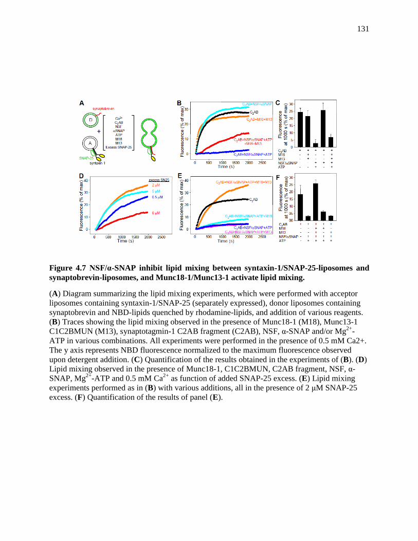
131
Figure 4.7 NSF/α-SNAP inhibit lipid mixing between syntaxin-1/SNAP-25-liposomes and
synaptobrevin-liposomes, and Munc18-1/Munc13-1 activate lipid mixing.
(A) Diagram summarizing the lipid mixing experiments, which were performed with acceptor
liposomes containing syntaxin-1/SNAP-25 (separately expressed), donor liposomes containing
synaptobrevin and NBD-lipids quenched by rhodamine-lipids, and addition of various reagents.
(B) Traces showing the lipid mixing observed in the presence of Munc18-1 (M18), Munc13-1
C1C2BMUN (M13), synaptotagmin-1 C2AB fragment (C2AB), NSF, α-SNAP and/or Mg2+
-
ATP in various combinations. All experiments were performed in the presence of 0.5 mM Ca2+.
The y axis represents NBD fluorescence normalized to the maximum fluorescence observed
upon detergent addition. (C) Quantification of the results obtained in the experiments of (B). (D)
Lipid mixing observed in the presence of Munc18-1, C1C2BMUN, C2AB fragment, NSF, α-
SNAP, Mg2+
-ATP and 0.5 mM Ca2+
as function of added SNAP-25 excess. (E) Lipid mixing
experiments performed as in (B) with various additions, all in the presence of 2 μM SNAP-25
excess. (F) Quantification of the results of panel (E).
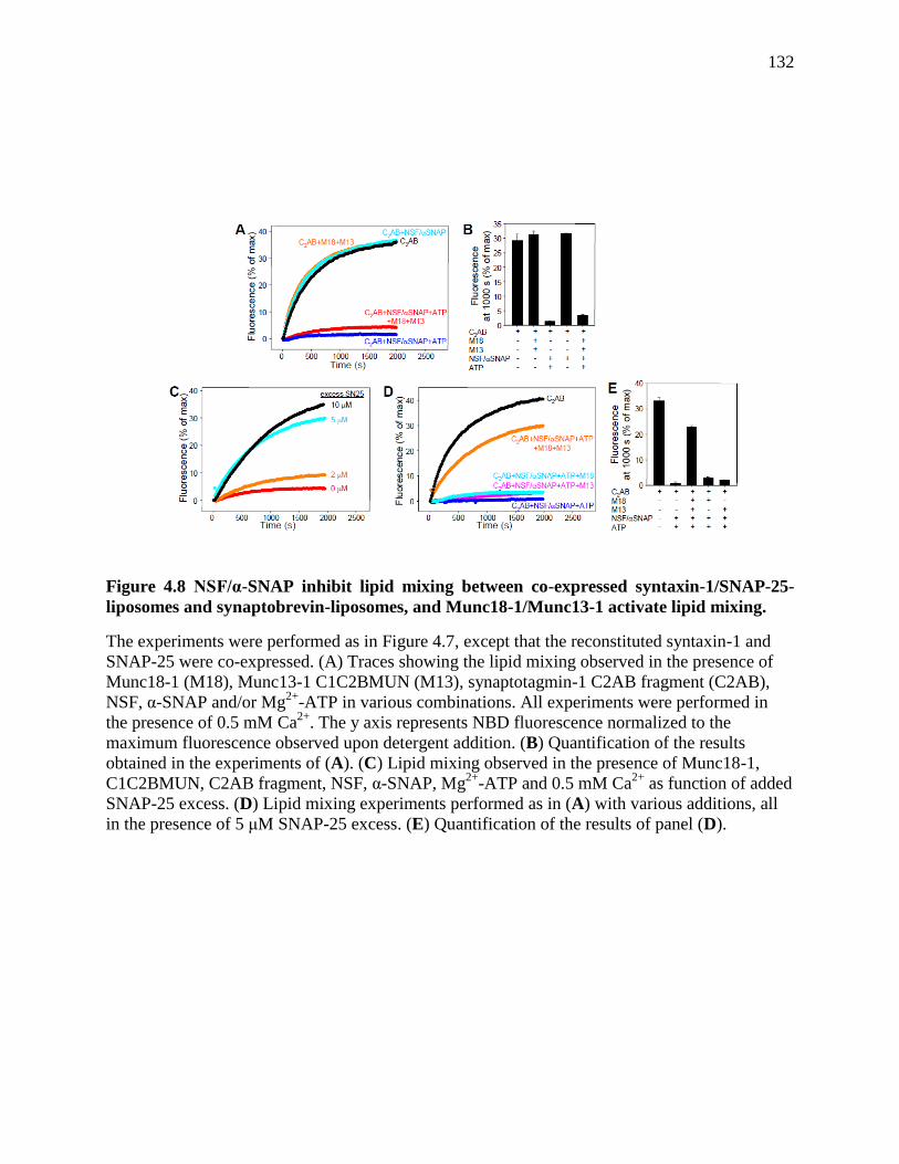
132
Figure 4.8 NSF/α-SNAP inhibit lipid mixing between co-expressed syntaxin-1/SNAP-25-
liposomes and synaptobrevin-liposomes, and Munc18-1/Munc13-1 activate lipid mixing.
The experiments were performed as in Figure 4.7, except that the reconstituted syntaxin-1 and
SNAP-25 were co-expressed. (A) Traces showing the lipid mixing observed in the presence of
Munc18-1 (M18), Munc13-1 C1C2BMUN (M13), synaptotagmin-1 C2AB fragment (C2AB),
NSF, α-SNAP and/or Mg2+
-ATP in various combinations. All experiments were performed in
the presence of 0.5 mM Ca2+
. The y axis represents NBD fluorescence normalized to the
maximum fluorescence observed upon detergent addition. (B) Quantification of the results
obtained in the experiments of (A). (C) Lipid mixing observed in the presence of Munc18-1,
C1C2BMUN, C2AB fragment, NSF, α-SNAP, Mg2+
-ATP and 0.5 mM Ca2+
as function of added
SNAP-25 excess. (D) Lipid mixing experiments performed as in (A) with various additions, all
in the presence of 5 μM SNAP-25 excess. (E) Quantification of the results of panel (D).
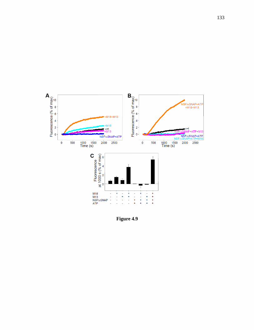
133
Figure 4.9

134
Figure 4.9 NSF-α-SNAP inhibit lipid mixing between syntaxin-1-SNAP-25-liposomes and
synaptobrevin-liposomes, and Munc18-1-Munc13-1 activate lipid mixing in the absence of
synaptotagmin-1.
(A,B) Traces showing the lipid mixing observed between acceptor liposomes containing
syntaxin-1-SNAP-25 heterodimers (separately expressed proteins) and synaptobrevin donor
liposomes containing NBD-lipids quenched by rhodamine-lipids in the presence of Munc18-1
(M18), Munc13-1 C1C2BMUN (M13), NSF, α-SNAP and/or Mg2+
-ATP in various
combinations (v+t indicates the lipid mixing observed with no additions). All samples contained
an excess of 2 μM SNAP-25. Samples containing C1C2BMUN also included 0.5 mM Ca2+
. The
y axis represents NBD fluorescence normalized to the maximum fluorescence observed upon
detergent addition. (C) Quantification of the results obtained in the experiments of (A,B). Error
bars represent standard deviations. In A, the black trace (v+t) shows that lipid mixing between
the synaptobrevin-liposomes and the syntaxin-1-SNAP-25 liposomes was inefficient under the
conditions of our experiments, that Munc18-1 enhanced lipid mixing moderately, and that
Munc18-1 together the Munc13-1 C1C2BMUN fragment stimulated lipid mixing more strongly,
revealing a synergy between them. NSF-α-SNAP completely abolished the lipid mixing induced
by the SNAREs alone. Panel B shows that Munc18-1 alone or the Munc13-1 C1C2BMUN
fragment alone could not overcome the inhibition caused by NSF-α-SNAP, but together they
dramatically activated lipid mixing. These results show that Munc18-1 and Munc13-1 can also
stimulate lipid mixing in an NSF-α-SNAP-resistant manner in the absence of the synaptotagmin-
1 C2AB fragment, although the overall lipid mixing is less efficient because of the lack of the
activity of the Ca2+
sensor (note the different scale of the y axis in panels A-C compared to those
in Figure 4.7).
135
4.4 Discussion
Multiple seminal contributions over the past three decades have identified the major
proteins that control neurotransmitter release, have shown that some of these proteins belong to
families with general roles in intracellular membrane fusion, have provided key insights into
their functions, and have revealed their three-dimensional structures. And yet, it has been
difficult to integrate all this knowledge into a coherent model of synaptic vesicle fusion that
explains all the most key findings. Perhaps most worrisome was the huge gap that emerged
between physiological data showing that Munc18-1 and Munc13s are crucial for
neurotransmitter release and in vitro reconstitution results revealing highly efficient membrane
fusion without these proteins (see Introduction). The results presented here now bridge this gap,
showing that multiple basic features of synaptic vesicle fusion, including critical roles for
Munc18-1 and Munc13s, can be reproduced when these two proteins and the other six major
components of the release machinery (syntaxin-1, synaptobrevin, SNAP-25, synaptotagmin-1,
NSF and α-SNAP) are included in the reconstitution system. Our results, together with the
available data, lead naturally to a model of synaptic vesicle fusion that readily explains the key
physiological importance of each one of these eight proteins (Figure 4.10). This model postulates
that: i) the syntaxin-1/Munc18- 1 complex constitutes the starting point for synaptic vesicle
fusion; ii) Munc13 opens syntaxin-1 and, together with Munc18-1, forms a template to bring the
three SNAREs together and initiate SNARE complex assembly; iii) the resulting Munc18-
1/Munc13/SNARE assembly is resistant to disassembly by NSF/SNAPs and underlies the
primed state that enables fast membrane fusion through the action of synaptotagmin-1/Ca2+
; iv)
syntaxin-1/SNAP-25 heterodimers constitute ‘off-pathway’ complexes that are disassembled by

136
NSF/SNAPs, and the released syntaxin-1 is captured in its closed conformation by Munc18-1,
leading to the productive pathway for membrane fusion.
Elucidating the basis for the key functions of Munc18-1 and other SM proteins is perhaps
the most fundamental question that needs to be answered to understand the mechanism of
neurotransmitter release and intracellular membrane fusion in general (Sudhof et al. 2011).
Addressing this question at the synapse also necessitates an understanding of the role of
Munc13s given the similarly drastic phenotypes observed in the absence of Munc18-1 or
Munc13s (Verhage et al. 2000; Varoqueaux et al. 2002). The recent finding of sequence and
structural similarity between the Munc13 MUN domain and subunits of tethering factors that
function in diverse membrane compartments (e.g. exocyst, GARP, Cog and Dsl1) indicated that
Munc13s have a general function that is shared with these factors (Pei et al. 2009; Li et al. 2011).
However, these tethering factors form part of large complexes with multiple subunits, and
tethering complexes from other membrane compartments that do not exhibit sequence homology
with Munc13s, such as HOPS and TRAPP, also contain multiple subunits. Hence, it was highly
unclear whether Munc13s by themselves could play similar roles as these tethering complexes.
Our results now uncover a clear similarity with reconstitutions of yeast vacuolar fusion,
where the HOPS complex orchestrates formation of trans-SNARE complexes (Mima et al. 2008)
and protects them from disassembly by Sec18p/Sec17p (Xu et al. 2010), the yeast homologues of
NSF/SNAPs. Although vacuolar fusion does not require synaptotagmin-1/ Ca2+
and some of the
details of the interplay between HOPS and Sec18p/Sec17p (Mima et al. 2008) differ from the
interplay between Munc18-1/Munc13 and NSF/α-SNAP described here, our data strongly
suggest that Munc18-1 and Munc13 must coordinate trans-SNARE complex assembly in an
NSF/α-SNAP-resistant manner to promote membrane fusion. It is rather remarkable that this
137
same task can be executed both by Munc18-1/Munc13 and by HOPS since, although HOPS
includes the SM protein Vps33p, it contains five additional large subunits unrelated to Munc13.
Considering these differences in architecture and the fact that synaptic exocytosis and yeast
vacuolar fusion are distantly related forms of membrane traffic, this similarity suggests that a
major general function of SM proteins and their associated factors is indeed the orchestration of
SNARE complex assembly in an NSF/SNAP-resistant manner. This notion does not preclude
other functions proposed for SM proteins and their co-factors, including the possibility that they
cooperate with the SNAREs in exerting force on the membranes to induce fusion much more
efficiently than SNAREs alone (Rizo et al. 2006; Dulubova et al. 2007).
The findings that NSF/α-SNAP disassemble syntaxin-1/SNAP-25 heterodimers [Figure
4.2 and (Weber et al. 2000)] and that Secp18p/Sec17p also act on vacuolar t-SNARE complexes
(Mima et al. 2008) suggest that the function of NSF/SNAPs is not limited to disassembling
SNARE complexes to recycle the SNAREs for another round of fusion. It is plausible that the
activity of NSF/SNAPs on syntaxin-1/SNAP-25 heterodimers and homologous complexes may
be an ‘accident’ reminiscent of its ‘true’ function on the SNARE complex. However, if syntaxin-
1/SNAP-25 heterodimers were bona-fide starting points for synaptic vesicle fusion, it seems that
their disassembly by NSF/SNAPs would involve a futile waste of energy. Hence, the activity of
NSF/SNAPs on syntaxin-1/SNAP-25 heterodimers is likely to serve an important function. In
this context, sequences that form long coiled coils have a natural promiscuity, which is
particularly emphasized by the tendency of the syntaxin-1 SNARE motif to bind to many
proteins (Jahn et al. 2006), to form a tetramer (Misura et al. 2001), and to associate with SNAP-
25 in different forms (Misura et al. 2001), including 2:1 complexes where synaptobrevin is
replaced by a second syntaxin-1 unit (Zhang et al. 2002). Moreover, oligomeric t-SNARE

138
complexes can be formed through bridging by the two SNARE motifs of SNAP-25 (Figure 4.10).
Hence, syntaxin-1/SNAP-25 heterodimers constitute a heterogeneous mixture of complexes that
probably provide a poor starting point for an exquisitely regulated process such as
neurotransmitter release.
We propose that direct displacement of SNAP-25 from syntaxin-1 by Munc18-1, and
disassembly of syntaxin-1/SNAP-25 heterodimers by NSF/SNAPs followed by capture of
syntaxin-1 by Munc18-1, provide two means toward the correct pathway for fusion, with a well
defined starting point (the syntaxin-1/Munc18-1 complex) that is amenable to tight regulation.
This proposal implies that the acceptor complex for synaptobrevin is not a syntaxin-1/SNAP-25
heterodimer but rather a complex of Munc18-1, Munc13-1 and syntaxin-1. Binding of SNAP-25
and synaptobrevin to this acceptor complex may occur concomitantly or sequentially but,
regardless of the mechanism, our data [(Figures 4.1,4.2) and (Ma et al. 2011)] suggest that both
SNAREs are required to favor the transition from closed to open syntaxin-1. It is also worth
noting that specific protein-protein interactions in presynaptic active zones may also favor this
pathway over the potential formation of trans-SNARE complexes containing only syntaxin-1,
SNAP-25 and synaptobrevin, including for instance the formation of tripartite complexes of
Munc13-1 with RIMs and Rab3s (Dulubova et al. 2005).
Based on the seminal work on yeast vacuolar fusion (Mima et al. 2008; Stroupe et al.
2009), the common features of synaptic vesicle fusion described here, and increasing evidence
for interactions of SNAREs with tethering factors [e.g. (Sivaram et al. 2005; Tripathi et al.
2009)], it is tempting to speculate that the acceptor complexes for v-SNAREs may generally be
formed by t-SNAREs, SM proteins and tethering factors, rather than by t-SNAREs alone. At the
same time, the diversity in the architecture of tethering complexes suggests that, even if they

139
share a common function in orchestrating SNARE complex assembly, they may accomplish this
function by different mechanisms that respond to different regulatory requirements. For instance,
the closed conformation of syntaxin-1 is not adopted by the vacuolar syntaxin Vam3p (Dulubova
et al. 2001), and the Munc18-1/syntaxin-1 complex appears to be a specialization of regulated
secretion (Carr et al. 1999; Dulubova et al. 1999). Correspondingly, the Munc13 MUN domain
may have a general role in coordinating SNARE complex assembly and a specific role in
opening syntaxin-1. This latter role appears to be catalytic in nature, since the MUN domain not
only accelerates the transition from the syntaxin-1/Munc18-1 complex to the SNARE complex
(Ma et al. 2011) but also the transition from syntaxin-1/SNAP-25 heterodimers to the syntaxin-
1/Munc18-1 complex (Figure 4.1H). Hence, Munc13s can provide an additional means to
facilitate disruption of syntaxin-1/SNAP-25 heterodimers in favor of the syntaxin-1/Munc18-1
complex.
Can we conclude that our system represents a bona-fide reconstitution of synaptic vesicle
fusion? More research will be necessary to fully answer this question, but it is reassuring that our
experiments were performed under mild conditions, with relatively low protein concentrations
and protein-to-lipid ratios, and reproduce multiple basic features of neurotransmitter release,
including a key function for Munc18-1, Munc13-1 and the other six proteins used. Moreover, the
effects of the Munc18-1 E66A mutation on lipid mixing (Figure 4.4I,J) correlate well with the
functional effects of this mutation in neurons (Deak et al. 2009), and our experiments reveal a
high cooperativity with Ca2+
that mirrors that of neurotransmitter release (Meinrenken et al. 2003;
Schneggenburger et al. 2005). The Ca2+
concentrations required for fusion in our experiments
are somewhat higher than those that trigger release, but this is not surprising given the natural
limitations of our reconstituted system. For instance, the use of reconstituted fulllength
140
synaptotagmin-1 may improve this behavior, although the C2AB fragment contains the key
Ca2+
-binding domains of synaptotagmin-1 and is sufficient to dramatically enhance
SNAREdependent lipid mixing due to its ability to bridge membranes (Xue et al. 2008; Hui et al.
2011). Likely a more important issue that hinders the fusion efficiency in our experiments is the
absence of a defined mechanism for vesicle docking, which depends on proteins such as
RIMs/unc10 and Rab3s (Weimer et al. 2006; Gracheva et al. 2008), and enables the high speed
of release. Addressing this issue is hampered by our limited understanding of the mechanism of
docking, but it will be interesting to extend our studies to single-vesicle fusion assays, which
allow measurement of the rates of individual fusion events after docking [e.g. (Lee et al. 2010;
Kyoung et al. 2011)]. We also note that our experiments did not include other proteins with
important roles in neurotransmitter release such as complexins. Nevertheless, despite these
limitations, the fundamental results obtained with this system can now be used as a basis for
further studies of the functions of these proteins with a variety of approaches.

141
Figure 4.10
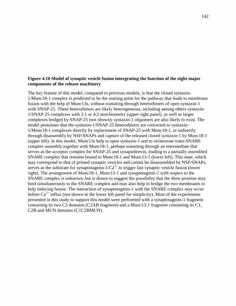
142
Figure 4.10 Model of synaptic vesicle fusion intergrating the function of the eight major
components of the release machinery
The key feature of this model, compared to previous models, is that the closed syntaxin-
1/Munc18-1 complex is predicted to be the starting point for the pathway that leads to membrane
fusion with the help of Munc13s, without transiting through heterodimers of open syntaxin-1
with SNAP-25. These heterodimers are likely heterogeneous, including among others syntaxin-
1/SNAP-25 complexes with 2:1 or 4:2 stoichiometry (upper right panel), as well as larger
complexes bridged by SNAP-25 (not shown); syntaxin-1 oligomers are also likely to exist. The
model postulates that the syntaxin-1/SNAP-25 heterodimers are converted to syntaxin-
1/Munc18-1 complexes directly by replacement of SNAP-25 with Munc18-1, or indirectly
through disassembly by NSF/SNAPs and capture of the released closed syntaxin-1 by Munc18-1
(upper left). In this model, Munc13s help to open syntaxin-1 and to orchestrate trans-SNARE
complex assembly together with Munc18-1, perhaps transiting through an intermediate that
serves as the acceptor complex for SNAP-25 and synaptobrevin, leading to a partially assembled
SNARE complex that remains bound to Munc18-1 and Munc13-1 (lower left). This state, which
may correspond to that of primed synaptic vesicles and cannot be disassembled by NSF/SNAPs,
serves as the substrate for synaptotagmin-1/Ca2+
to trigger fast synaptic vesicle fusion (lower
right). The arrangement of Munc18-1, Munc13-1 and synaptotagmin-1 with respect to the
SNARE complex is unknown, but is drawn to suggest the possibility that the three proteins may
bind simultaneously to the SNARE complex and may also help to bridge the two membranes to
help inducing fusion. The interaction of synaptotagmin-1 with the SNARE complex may occur
before Ca2+
influx (not shown in the lower left panel for simplicity). Most of the experiments
presented in this study to support this model were performed with a synaptotagmin-1 fragment
containing its two C2 domains (C2AB fragment) and a Munc13-1 fragment containing its C1,
C2B and MUN domains (C1C2BMUN).
143
Chapter 5 Summary and Future Directions
Munc18-1 is considered part of the fusion machinery for synaptic transmission. Several
functions of Munc18-1 have been discovered through knocking it out or expressing its mutants
of in neurons, but its key role has not been found yet. Most importantly, its mode of action is still
largely unknown. This dissertation provides insights into the roles of Munc18-1 in
neurotransmitter release, especially Chapter 4.
Chapter 4 demonstrates that: i) in the presence of NSF and α-SNAP, Munc18-1 interacts
with syntaxin-1 or breaks the syntaxin-1/SNAP-25 complex and replace SNAP-25 to hold
syntaxin-1 in a closed conformation; ii) the priming factor Munc13, opens syntaxin-1 that is held
by Munc18-1, and then Munc18-1and Munc13 guide the synaptic vesicle fusion through a tightly
regulated pathway that depends on SNARE-complex and synaptotagmin, and is NSF and α-
SNAP resistant (Figure 4.10). This is the first convincing evidence showing the molecular
mechanism of action of Munc18-1 in synaptic vesicle exocytosis, despite extensive studies of
this protein in the past decade. However, there are still questions to ask about this protein. i) Is
Munc18-1 directly involved in membrane fusion itself after SNARE complex formation? Our
model leaves the possibility open that Munc18-1 might also have a separate effect on the Ca2+
-
triggered membrane fusion step. Chapters 2 and 3 showed that Munc18-1 interacts with the
SNARE motifs of synaptobrevin and syntaxin-1 respectively, and that Munc18-1 interacts with
the C-terminal part of synaptobrevin and syntaxin-1 on the SNARE four-helix bundle, which is
right adjacent to the place where membrane fusion occurs. Moreover, the interaction of Munc18-
1 with the SNARE four-helix bundle is compatible with binding of the SNARE four-helix bundle
to complexin-I, which functions in a late stage of synaptic vesicle exocytosis. These data suggest
144
that Munc18-1 might be directly involved in membrane fusion itself. A high resolution structure
of the complex of Munc18-1/SNARE four-helix bundle will be needed for further in vivo
investigation to provide answers to this question. Moreover, capturing the intermediate steps of
synaptic vesicle fusion by Cryo-EM and identifying Munc18-1 in those intermediate fusion steps
would also be great evidence to prove that Munc18-1 is directly involved in the Ca2+
-triggered
membrane fusion step. ii) Does Munc18-1 cooperate with Munc13 to play a direct role in the
membrane fusion step? The results in Figure 4.9 showed that Munc18-1 cooperates with Munc13
in stimulation of SNARE-dependent lipid mixing in the absence of synaptotagmin, although the
amount of stimulation is small. In vivo investigation suggests that Munc13 might have a role
downstream of priming. It is hard to provide direct evidence that Munc18-1 and Munc13 are
involved in Ca2+
-triggered membrane fusion step so far. Studying Munc18-1 and Munc13
together might be easier to uncover their roles in the fusion step. Cryo-EM would be a great
method to identify whether these two proteins are together in the fusion intermediates, which
would provide important information for further in vitro cooperativity studies. iii) Does the
glass-hour shape fusion pore proposed by the stalk model (Figure 1.4) really exist? Is it regulated
by Munc18-1 or Munc18-1 together with Munc13-1? Recent cryo-EM studies of intermediates
of SNARE-mediated vesicle fusion (Hernandez et al. 2012) showed that fusion proceeds through
membrane contact, membrane flattening, membrane merging, and fusion pore formation from
one end, which is very different from the stalk model. Does Munc18-1 regulated or Munc18-1
and Munc13 regulated membrane fusion proceed through the stalk model? Cryo-EM to capture
the intermediates of Munc18-1 and Munc13 regulated membrane fusion, and comparison of
these intermediates with those observed with the SNARE alone would provide answers. iv)
Structurally, how are Munc18-1, Munc13, complexin, and synaptotagmin arranged on the
145
SNARE four-helix bundle? These proteins all bind to the SNARE four-helix bundle, and most
probably they all participate in the membrane fusion step. Understanding their spatial
arrangement is also important for understanding the whole system. In vitro studies of the
structures of the complexes will be needed. v) Do Munc18-1 homologs function in the same way
in other intracellular membrane fusion events? This question remains to be tested in other
systems. The answers to questions i, ii and iii will shed lights on the answer to question iv.
146
BIBLIOGRAPHY
Arac, D., X. Chen, H. A. Khant, J. Ubach, S. J. Ludtke, M. Kikkawa, A. E. Johnson, W. Chiu, T.
C. Südhof and J. Rizo (2006). "Close membrane-membrane proximity induced by
Ca(2+)-dependent multivalent binding of synaptotagmin-1 to phospholipids." Nat Struct
Mol Biol 13(3): 209-217.
Arac, D., X. Chen, H. A. Khant, J. Ubach, S. J. Ludtke, M. Kikkawa, A. E. Johnson, W. Chiu, T.
C. Sudhof and J. Rizo (2006). "Close membrane-membrane proximity induced by
Ca(2+)-dependent multivalent binding of synaptotagmin-1 to phospholipids." Nat Struct
Mol Biol 13(3): 209-217.
Aravamudan, B., T. Fergestad, W. S. Davis, C. K. Rodesch and K. Broadie (1999). "Drosophila
UNC-13 is essential for synaptic transmission." Nat Neurosci 2(11): 965-971.
Arunachalam, L., L. Han, N. G. Tassew, Y. He, L. Wang, L. Xie, Y. Fujita, E. Kwan, B.
Davletov, P. P. Monnier, H. Y. Gaisano and S. Sugita (2008). "Munc18-1 is critical for
plasma membrane localization of syntaxin1 but not of SNAP-25 in PC12 cells." Mol Biol
Cell 19(2): 722-734.
Augustin, I., A. Betz, C. Herrmann, T. Jo and N. Brose (1999). "Differential expression of two
novel Munc13 proteins in rat brain." Biochem J 337 ( Pt 3): 363-371.
Augustin, I., C. Rosenmund, T. C. Sudhof and N. Brose (1999). "Munc13-1 is essential for
fusion competence of glutamatergic synaptic vesicles." Nature 400(6743): 457-461.
Bai, J. and E. R. Chapman (2004). "The C2 domains of synaptotagmin--partners in exocytosis."
Trends Biochem Sci 29(3): 143-151.
Banerjee, A., V. A. Barry, B. R. DasGupta and T. F. Martin (1996). "N-Ethylmaleimide-
sensitive factor acts at a prefusion ATP-dependent step in Ca2+-activated exocytosis." J
Biol Chem 271(34): 20223-20226.
Basanez, G., F. M. Goni and A. Alonso (1998). "Effect of single chain lipids on phospholipase
C-promoted vesicle fusion. A test for the stalk hypothesis of membrane fusion."
Biochemistry 37(11): 3901-3908.
Basu, J., N. Shen, I. Dulubova, J. Lu, R. Guan, O. Guryev, N. V. Grishin, C. Rosenmund and J.
Rizo (2005). "A minimal domain responsible for Munc13 activity." Nat Struct Mol Biol
12(11): 1017-1018.
Bennett, M. K., N. Calakos and R. H. Scheller (1992). "Syntaxin: a synaptic protein implicated
in docking of synaptic vesicles at presynaptic active zones." Science 257(5067): 255-259.
Bennett, M. K. and R. H. Scheller (1993). "The molecular machinery for secretion is conserved
from yeast to neurons." Proc Natl Acad Sci U S A 90(7): 2559-2563.
Betz, A., M. Okamoto, F. Benseler and N. Brose (1997). "Direct interaction of the rat unc-13
homologue Munc13-1 with the N terminus of syntaxin." J Biol Chem 272(4): 2520-2526.
Boswell, K. L., D. J. James, J. M. Esquibel, S. Bruinsma, R. Shirakawa, H. Horiuchi and T. F.
Martin (2012). "Munc13-4 reconstitutes calcium-dependent SNARE-mediated membrane
fusion." J Cell Biol 197(2): 301-312.
Bowen, M. E., K. Weninger, J. Ernst, S. Chu and A. T. Brunger (2005). "Single-molecule studies
of synaptotagmin and complexin binding to the SNARE complex." Biophys J 89(1): 690-
702.
Bracher, A., T. Dresbach, H. Betz and W. Weissenhorn (2000). "Crystallization and preliminary
X-ray analysis of squid neuronal Sec1." Acta Crystallogr D Biol Crystallogr 56(Pt 4):
501-503.
147
Bracher, A., A. Perrakis, T. Dresbach, H. Betz and W. Weissenhorn (2000). "The X-ray crystal
structure of neuronal Sec1 from squid sheds new light on the role of this protein in
exocytosis." Structure 8(7): 685-694.
Bracher, A. and W. Weissenhorn (2001). "Crystal structures of neuronal squid Sec1 implicate
inter-domain hinge movement in the release of t-SNAREs." J Mol Biol 306(1): 7-13.
Bracher, A. and W. Weissenhorn (2002). "Structural basis for the Golgi membrane recruitment
of Sly1p by Sed5p." EMBO J 21(22): 6114-6124.
Brenner, S. (1974). "The genetics of Caenorhabditis elegans." Genetics 77(1): 71-94.
Brose, N. (2008). "For better or for worse: complexins regulate SNARE function and vesicle
fusion." Traffic 9(9): 1403-1413.
Brose, N., K. Hofmann, Y. Hata and T. C. Sudhof (1995). "Mammalian homologues of
Caenorhabditis elegans unc-13 gene define novel family of C2-domain proteins." J Biol
Chem 270(42): 25273-25280.
Brose, N., A. G. Petrenko, T. C. Sudhof and R. Jahn (1992). "Synaptotagmin: a calcium sensor
on the synaptic vesicle surface." Science 256(5059): 1021-1025.
Brunger, A. T. (2005). "Structure and function of SNARE and SNARE-interacting proteins." Q
Rev Biophys 38(1): 1-47.
Burkhardt, P., D. A. Hattendorf, W. I. Weis and D. Fasshauer (2008). "Munc18a controls
SNARE assembly through its interaction with the syntaxin N-peptide." EMBO J 27(7):
923-933.
Carpp, L. N., L. F. Ciufo, S. G. Shanks, A. Boyd and N. J. Bryant (2006). "The Sec1p/Munc18
protein Vps45p binds its cognate SNARE proteins via two distinct modes." J Cell Biol
173(6): 927-936.
Carr, C. M., E. Grote, M. Munson, F. M. Hughson and P. J. Novick (1999). "Sec1p binds to
SNARE complexes and concentrates at sites of secretion." J Cell Biol 146(2): 333-344.
Carr, C. M. and J. Rizo (2010). "At the junction of SNARE and SM protein function." Curr Opin
Cell Biol 22(4): 488-495.
Chapman, E. R., S. An, N. Barton and R. Jahn (1994). "SNAP-25, a t-SNARE which binds to
both syntaxin and synaptobrevin via domains that may form coiled coils." J Biol Chem
269(44): 27427-27432.
Chapman, E. R., P. I. Hanson, S. An and R. Jahn (1995). "Ca2+ regulates the interaction between
synaptotagmin and syntaxin 1." J Biol Chem 270(40): 23667-23671.
Chen, X., D. Arac, T. M. Wang, C. J. Gilpin, J. Zimmerberg and J. Rizo (2006). "SNARE-
mediated lipid mixing depends on the physical state of the vesicles." Biophys J 90(6):
2062-2074.
Chen, X., J. Lu, I. Dulubova and J. Rizo (2008). "NMR analysis of the closed conformation of
syntaxin-1." Journal of biomolecular NMR 41(1): 43-54.
Chen, X., J. Lu, I. Dulubova and J. Rizo (2008). "NMR analysis of the closed conformation of
syntaxin-1." J Biomol NMR 41(1): 43-54.
Chen, X., D. Tomchick, E. Kovrigin, D. Ara , M. Machius, T. Südhof and J. Rizo (2002).
"Three-dimensional structure of the complexin/SNARE complex." Neuron 33(3): 397-
409.
Chen, X., D. R. Tomchick, E. Kovrigin, D. Arac, M. Machius, T. C. Sudhof and J. Rizo (2002).
"Three-dimensional structure of the complexin/SNARE complex." Neuron 33(3): 397-
409.
148
Chen, Y. A., S. J. Scales and R. H. Scheller (2001). "Sequential SNARE assembly underlies
priming and triggering of exocytosis." Neuron 30(1): 161-170.
Chernomordik, L. (1996). "Non-bilayer lipids and biological fusion intermediates." Chem Phys
Lipids 81(2): 203-213.
Chernomordik, L. V., G. B. Melikyan and Y. A. Chizmadzhev (1987). "Biomembrane fusion: a
new concept derived from model studies using two interacting planar lipid bilayers."
Biochim Biophys Acta 906(3): 309-352.
Chicka, M. C., E. Hui, H. Liu and E. R. Chapman (2008). "Synaptotagmin arrests the SNARE
complex before triggering fast, efficient membrane fusion in response to Ca2+." Nat
Struct Mol Biol 15(8): 827-835.
Clary, D. O. and J. E. Rothman (1990). "Purification of three related peripheral membrane
proteins needed for vesicular transport." J Biol Chem 265(17): 10109-10117.
Dai, H., N. Shen, D. Arac and J. Rizo (2007). "A quaternary SNARE-synaptotagmin-Ca2+-
phospholipid complex in neurotransmitter release." J Mol Biol 367(3): 848-863.
Deák, F., Y. Xu, W. Chang, I. Dulubova, M. Khvotchev, X. Liu, T. Südhof and J. Rizo (2009).
"Munc18-1 binding to the neuronal SNARE complex controls synaptic vesicle priming."
The Journal of cell biology 184(5): 751.
de Wit, H., A. Walter, I. Milosevic, A. Gulyás-Kovács, D. Riedel, J. Sørensen and M. Verhage
(2009). "Synaptotagmin-1 docks secretory vesicles to syntaxin-1/SNAP-25 acceptor
complexes." Cell 138(5): 935-946.
Deak, F., Y. Xu, W. P. Chang, I. Dulubova, M. Khvotchev, X. Liu, T. C. Sudhof and J. Rizo
(2009). "Munc18-1 binding to the neuronal SNARE complex controls synaptic vesicle
priming." J Cell Biol 184(5): 751-764.
Dennison, S. M., M. E. Bowen, A. T. Brunger and B. R. Lentz (2006). "Neuronal SNAREs do
not trigger fusion between synthetic membranes but do promote PEG-mediated
membrane fusion." Biophys J 90(5): 1661-1675.
Diao, J., Z. Su, Y. Ishitsuka, B. Lu, K. S. Lee, Y. Lai, Y. K. Shin and T. Ha (2010). "A single-
vesicle content mixing assay for SNARE-mediated membrane fusion." Nat Commun 1:
54.
Diao, J., Z. Su, X. Lu, T. Y. Yoon, Y. K. Shin and T. Ha (2010). "Single-Vesicle Fusion Assay
Reveals Munc18-1 Binding to the SNARE Core Is Sufficient for Stimulating Membrane
Fusion." ACS Chem Neurosci 1(3): 168-174.
Dulubova, I., M. Khvotchev, S. Liu, I. Huryeva, T. C. Südhof and J. Rizo (2007). "Munc18-1
binds directly to the neuronal SNARE complex." Proc Natl Acad Sci U S A 104(8):
2697-2702.
Dulubova, I., M. Khvotchev, S. Liu, I. Huryeva, T. C. Sudhof and J. Rizo (2007). "Munc18-1
binds directly to the neuronal SNARE complex." Proc Natl Acad Sci U S A 104(8):
2697-2702.
Dulubova, I., X. Lou, J. Lu, I. Huryeva, A. Alam, R. Schneggenburger, T. C. Sudhof and J. Rizo
(2005). "A Munc13/RIM/Rab3 tripartite complex: from priming to plasticity?" EMBO J
24(16): 2839-2850.
Dulubova, I., S. Sugita, S. Hill, M. Hosaka, I. Fernandez, T. C. Südhof and J. Rizo (1999). "A
conformational switch in syntaxin during exocytosis: role of munc18." EMBO J 18(16):
4372-4382.
149
Dulubova, I., S. Sugita, S. Hill, M. Hosaka, I. Fernandez, T. C. Sudhof and J. Rizo (1999). "A
conformational switch in syntaxin during exocytosis: role of munc18." EMBO J 18(16):
4372-4382.
Dulubova, I., T. Yamaguchi, Y. Wang, T. C. Sudhof and J. Rizo (2001). "Vam3p structure
reveals conserved and divergent properties of syntaxins." Nat Struct Biol 8(3): 258-264.
Fasshauer, D., D. Bruns, B. Shen, R. Jahn and A. T. Brunger (1997). "A structural change occurs
upon binding of syntaxin to SNAP-25." J Biol Chem 272(7): 4582-4590.
Fasshauer, D. and M. Margittai (2004). "A transient N-terminal interaction of SNAP-25 and
syntaxin nucleates SNARE assembly." J Biol Chem 279(9): 7613-7621.
Fasshauer, D., R. B. Sutton, A. T. Brunger and R. Jahn (1998). "Conserved structural features of
the synaptic fusion complex: SNARE proteins reclassified as Q- and R-SNAREs." Proc
Natl Acad Sci U S A 95(26): 15781-15786.
Fernandez-Chacon, R., A. Konigstorfer, S. H. Gerber, J. Garcia, M. F. Matos, C. F. Stevens, N.
Brose, J. Rizo, C. Rosenmund and T. C. Sudhof (2001). "Synaptotagmin I functions as a
calcium regulator of release probability." Nature 410(6824): 41-49.
Fernandez-Chacon, R., O. H. Shin, A. Konigstorfer, M. F. Matos, A. C. Meyer, J. Garcia, S. H.
Gerber, J. Rizo, T. C. Sudhof and C. Rosenmund (2002). "Structure/function analysis of
Ca2+ binding to the C2A domain of synaptotagmin 1." J Neurosci 22(19): 8438-8446.
Fernandez, I., D. Arac, J. Ubach, S. H. Gerber, O. Shin, Y. Gao, R. G. Anderson, T. C. Sudhof
and J. Rizo (2001). "Three-dimensional structure of the synaptotagmin 1 C2B-domain:
synaptotagmin 1 as a phospholipid binding machine." Neuron 32(6): 1057-1069.
Fernandez, I., J. Ubach, I. Dulubova, X. Zhang, T. C. Sudhof and J. Rizo (1998). "Three-
dimensional structure of an evolutionarily conserved N-terminal domain of syntaxin 1A."
Cell 94(6): 841-849.
Fleming, K. G., T. M. Hohl, R. C. Yu, S. A. Muller, B. Wolpensinger, A. Engel, H. Engelhardt,
A. T. Brunger, T. H. Sollner and P. I. Hanson (1998). "A revised model for the
oligomeric state of the N-ethylmaleimide-sensitive fusion protein, NSF." J Biol Chem
273(25): 15675-15681.
Fujiwara, T., T. Mishima, T. Kofuji, T. Chiba, K. Tanaka, A. Yamamoto and K. Akagawa (2006).
"Analysis of knock-out mice to determine the role of HPC-1/syntaxin 1A in expressing
synaptic plasticity." J Neurosci 26(21): 5767-5776.
Gengyo-Ando, K., Y. Kamiya, A. Yamakawa, K. Kodaira, K. Nishiwaki, J. Miwa, I. Hori and R.
Hosono (1993). "The C. elegans unc-18 gene encodes a protein expressed in motor
neurons." Neuron 11(4): 703-711.
Geppert, M., B. T. Archer, 3rd and T. C. Sudhof (1991). "Synaptotagmin II. A novel
differentially distributed form of synaptotagmin." J Biol Chem 266(21): 13548-13552.
Geppert, M., Y. Goda, R. E. Hammer, C. Li, T. W. Rosahl, C. F. Stevens and T. C. Sudhof
(1994). "Synaptotagmin I: a major Ca2+ sensor for transmitter release at a central
synapse." Cell 79(4): 717-727.
Gerber, S. H., J. C. Rah, S. W. Min, X. Liu, H. de Wit, I. Dulubova, A. C. Meyer, J. Rizo, M.
Arancillo, R. E. Hammer, M. Verhage, C. Rosenmund and T. C. Südhof (2008).
"Conformational switch of syntaxin-1 controls synaptic vesicle fusion." Science
321(5895): 1507-1510.
Gerber, S. H., J. C. Rah, S. W. Min, X. Liu, H. de Wit, I. Dulubova, A. C. Meyer, J. Rizo, M.
Arancillo, R. E. Hammer, M. Verhage, C. Rosenmund and T. C. Sudhof (2008).
150
"Conformational switch of syntaxin-1 controls synaptic vesicle fusion." Science
321(5895): 1507-1510.
Grabowski, R. and D. Gallwitz (1997). "High-affinity binding of the yeast cis-Golgi t-SNARE,
Sed5p, to wild-type and mutant Sly1p, a modulator of transport vesicle docking." FEBS
Lett 411(2-3): 169-172.
Gracheva, E. O., G. Hadwiger, M. L. Nonet and J. E. Richmond (2008). "Direct interactions
between C. elegans RAB-3 and Rim provide a mechanism to target vesicles to the
presynaptic density." Neurosci Lett 444(2): 137-142.
Grote, E., C. M. Carr and P. J. Novick (2000). "Ordering the final events in yeast exocytosis." J
Cell Biol 151(2): 439-452.
Guan, R., H. Dai and J. Rizo (2008). "Binding of the Munc13-1 MUN domain to membrane-
anchored SNARE complexes." Biochemistry 47(6): 1474-1481.
Gulyas-Kovacs, A., H. de Wit, I. Milosevic, O. Kochubey, R. Toonen, J. Klingauf, M. Verhage
and J. B. Sorensen (2007). "Munc18-1: sequential interactions with the fusion machinery
stimulate vesicle docking and priming." J Neurosci 27(32): 8676-8686.
Hammarlund, M., M. T. Palfreyman, S. Watanabe, S. Olsen and E. M. Jorgensen (2007). "Open
syntaxin docks synaptic vesicles." PLoS Biol 5(8): e198.
Hanson, P. I., H. Otto, N. Barton and R. Jahn (1995). "The N-ethylmaleimide-sensitive fusion
protein and alpha-SNAP induce a conformational change in syntaxin." J Biol Chem
270(28): 16955-16961.
Hanson, P. I., R. Roth, H. Morisaki, R. Jahn and J. E. Heuser (1997). "Structure and
conformational changes in NSF and its membrane receptor complexes visualized by
quick-freeze/deep-etch electron microscopy." Cell 90(3): 523-535.
Hashizume, K., Y. S. Cheng, J. L. Hutton, C. H. Chiu and C. M. Carr (2009). "Yeast Sec1p
functions before and after vesicle docking." Mol Biol Cell 20(22): 4673-4685.
Hata, Y., C. A. Slaughter and T. C. Sudhof (1993). "Synaptic vesicle fusion complex contains
unc-18 homologue bound to syntaxin." Nature 366(6453): 347-351.
Hayashi, T., H. McMahon, S. Yamasaki, T. Binz, Y. Hata, T. C. Sudhof and H. Niemann (1994).
"Synaptic vesicle membrane fusion complex: action of clostridial neurotoxins on
assembly." EMBO J 13(21): 5051-5061.
Hazzard, J., T. C. Sudhof and J. Rizo (1999). "NMR analysis of the structure of synaptobrevin
and of its interaction with syntaxin." J Biomol NMR 14(3): 203-207.
Helm, C. A. and J. N. Israelachvili (1993). "Forces between phospholipid bilayers and
relationship to membrane fusion." Methods Enzymol 220: 130-143.
Hernandez, J. M., A. Stein, E. Behrmann, D. Riedel, A. Cypionka, Z. Farsi, P. J. Walla, S.
Raunser and R. Jahn (2012). "Membrane fusion intermediates via directional and full
assembly of the SNARE complex." Science 336(6088): 1581-1584.
Hui, E., J. D. Gaffaney, Z. Wang, C. P. Johnson, C. S. Evans and E. R. Chapman (2011).
"Mechanism and function of synaptotagmin-mediated membrane apposition." Nat Struct
Mol Biol 18(7): 813-821.
Jahn, R. and D. Fasshauer (2012). "Molecular machines governing exocytosis of synaptic
vesicles." Nature 490(7419): 201-207.
Jahn, R., T. Lang and T. C. Sudhof (2003). "Membrane fusion." Cell 112(4): 519-533.
Jahn, R. and R. H. Scheller (2006). "SNAREs--engines for membrane fusion." Nat Rev Mol Cell
Biol 7(9): 631-643.
151
James, D. J., J. Kowalchyk, N. Daily, M. Petrie and T. F. Martin (2009). "CAPS drives trans-
SNARE complex formation and membrane fusion through syntaxin interactions." Proc
Natl Acad Sci U S A 106(41): 17308-17313.
Khvotchev, M., I. Dulubova, J. Sun, H. Dai, J. Rizo and T. C. Südhof (2007). "Dual modes of
Munc18-1/SNARE interactions are coupled by functionally critical binding to syntaxin-1
N terminus." J Neurosci 27(45): 12147-12155.
Khvotchev, M., I. Dulubova, J. Sun, H. Dai, J. Rizo and T. C. Sudhof (2007). "Dual modes of
Munc18-1/SNARE interactions are coupled by functionally critical binding to syntaxin-1
N terminus." J Neurosci 27(45): 12147-12155.
Kozlov, M. M. and V. S. Markin (1983). "[Possible mechanism of membrane fusion]." Biofizika
28(2): 242-247.
Kummel, D., S. S. Krishnakumar, D. T. Radoff, F. Li, C. G. Giraudo, F. Pincet, J. E. Rothman
and K. M. Reinisch (2011). "Complexin cross-links prefusion SNAREs into a zigzag
array." Nat Struct Mol Biol 18(8): 927-933.
Kweon, D. H., C. S. Kim and Y. K. Shin (2003). "Regulation of neuronal SNARE assembly by
the membrane." Nat Struct Biol 10(6): 440-447.
Kyoung, M., A. Srivastava, Y. Zhang, J. Diao, M. Vrljic, P. Grob, E. Nogales, S. Chu and A. T.
Brunger (2011). "In vitro system capable of differentiating fast Ca2+-triggered content
mixing from lipid exchange for mechanistic studies of neurotransmitter release." Proc
Natl Acad Sci U S A 108(29): E304-313.
Latham, C. F., J. A. Lopez, S. H. Hu, C. L. Gee, E. Westbury, D. H. Blair, C. J. Armishaw, P. F.
Alewood, N. J. Bryant, D. E. James and J. L. Martin (2006). "Molecular dissection of the
Munc18c/syntaxin4 interaction: implications for regulation of membrane trafficking."
Traffic 7(10): 1408-1419.
Lee, H. K., Y. Yang, Z. Su, C. Hyeon, T. S. Lee, H. W. Lee, D. H. Kweon, Y. K. Shin and T. Y.
Yoon (2010). "Dynamic Ca2+-dependent stimulation of vesicle fusion by membrane-
anchored synaptotagmin 1." Science 328(5979): 760-763.
Lee, J. and B. R. Lentz (1997). "Evolution of lipidic structures during model membrane fusion
and the relation of this process to cell membrane fusion." Biochemistry 36(21): 6251-
6259.
Li, C., B. Ullrich, J. Z. Zhang, R. G. Anderson, N. Brose and T. C. Sudhof (1995). "Ca(2+)-
dependent and -independent activities of neural and non-neural synaptotagmins." Nature
375(6532): 594-599.
Li, W., C. Ma, R. Guan, Y. Xu, D. R. Tomchick and J. Rizo (2011). "The crystal structure of a
Munc13 C-terminal module exhibits a remarkable similarity to vesicle tethering factors."
Structure 19(10): 1443-1455.
Li, Y., G. J. Augustine and K. Weninger (2007). "Kinetics of complexin binding to the SNARE
complex: correcting single molecule FRET measurements for hidden events." Biophys J
93(6): 2178-2187.
Lin, R. C. and R. H. Scheller (1997). "Structural organization of the synaptic exocytosis core
complex." Neuron 19(5): 1087-1094.
Link, E., L. Edelmann, J. H. Chou, T. Binz, S. Yamasaki, U. Eisel, M. Baumert, T. C. Sudhof, H.
Niemann and R. Jahn (1992). "Tetanus toxin action: inhibition of neurotransmitter release
linked to synaptobrevin proteolysis." Biochem Biophys Res Commun 189(2): 1017-1023.
152
Liu, J., T. Guo, Y. Wei, M. Liu and S. F. Sui (2006). "Complexin is able to bind to SNARE core
complexes in different assembled states with distinct affinity." Biochem Biophys Res
Commun 347(2): 413-419.
Liu, J., T. Guo, J. Wu, X. Bai, Q. Zhou and S. F. Sui (2007). "Overexpression of complexin in
PC12 cells inhibits exocytosis by preventing SNARE complex recycling." Biochemistry
(Mosc) 72(4): 439-444.
Lu, J., M. Machius, I. Dulubova, H. Dai, T. C. Sudhof, D. R. Tomchick and J. Rizo (2006).
"Structural basis for a Munc13-1 homodimer to Munc13-1/RIM heterodimer switch."
PLoS Biol 4(7): e192.
Ma, C., W. Li, Y. Xu and J. Rizo (2011). "Munc13 mediates the transition from the closed
syntaxin-Munc18 complex to the SNARE complex." Nat Struct Mol Biol 18(5): 542-549.
Ma, C., W. Li, Y. Xu and J. Rizo (2011). "Munc13 Mediates the Transition from the Closed
Syntaxin/Munc18 complex to the SNARE complex." Nature structural & molecular
biology, in press.
Mackler, J. M., J. A. Drummond, C. A. Loewen, I. M. Robinson and N. E. Reist (2002). "The
C(2)B Ca(2+)-binding motif of synaptotagmin is required for synaptic transmission in
vivo." Nature 418(6895): 340-344.
Madison, J. M., S. Nurrish and J. M. Kaplan (2005). "UNC-13 interaction with syntaxin is
required for synaptic transmission." Curr Biol 15(24): 2236-2242.
Martens, S., M. M. Kozlov and H. T. McMahon (2007). "How synaptotagmin promotes
membrane fusion." Science 316(5828): 1205-1208.
Maximov, A., J. Tang, X. Yang, Z. P. Pang and T. C. Sudhof (2009). "Complexin controls the
force transfer from SNARE complexes to membranes in fusion." Science 323(5913):
516-521.
Mayer, A., W. Wickner and A. Haas (1996). "Sec18p (NSF)-driven release of Sec17p (alpha-
SNAP) can precede docking and fusion of yeast vacuoles." Cell 85(1): 83-94.
McMahon, H. T., M. Missler, C. Li and T. C. Sudhof (1995). "Complexins: cytosolic proteins
that regulate SNAP receptor function." Cell 83(1): 111-119.
Medine, C. N., C. Rickman, L. H. Chamberlain and R. R. Duncan (2007). "Munc18-1 prevents
the formation of ectopic SNARE complexes in living cells." J Cell Sci 120(Pt 24): 4407-
4415.
Meinrenken, C. J., J. G. Borst and B. Sakmann (2003). "Local routes revisited: the space and
time dependence of the Ca2+ signal for phasic transmitter release at the rat calyx of
Held." J Physiol 547(Pt 3): 665-689.
Mima, J., C. M. Hickey, H. Xu, Y. Jun and W. Wickner (2008). "Reconstituted membrane fusion
requires regulatory lipids, SNAREs and synergistic SNARE chaperones." EMBO J
27(15): 2031-2042.
Misura, K. M., L. C. Gonzalez, Jr., A. P. May, R. H. Scheller and W. I. Weis (2001). "Crystal
structure and biophysical properties of a complex between the N-terminal SNARE region
of SNAP25 and syntaxin 1a." J Biol Chem 276(44): 41301-41309.
Misura, K. M., R. H. Scheller and W. I. Weis (2000). "Three-dimensional structure of the
neuronal-Sec1-syntaxin 1a complex." Nature 404(6776): 355-362.
Misura, K. M., R. H. Scheller and W. I. Weis (2001). "Self-association of the H3 region of
syntaxin 1A. Implications for intermediates in SNARE complex assembly." J Biol Chem
276(16): 13273-13282.
153
Nagiec, E. E., A. Bernstein and S. W. Whiteheart (1995). "Each domain of the N-
ethylmaleimide-sensitive fusion protein contributes to its transport activity." J Biol Chem
270(49): 29182-29188.
Neher, E. (2010). "Complexin: does it deserve its name?" Neuron 68(5): 803-806.
Nichols, B. J., J. C. Holthuis and H. R. Pelham (1998). "The Sec1p homologue Vps45p binds to
the syntaxin Tlg2p." Eur J Cell Biol 77(4): 263-268.
Nickel, W., T. Weber, J. A. McNew, F. Parlati, T. H. Sollner and J. E. Rothman (1999). "Content
mixing and membrane integrity during membrane fusion driven by pairing of isolated v-
SNAREs and t-SNAREs." Proc Natl Acad Sci U S A 96(22): 12571-12576.
Nishiki, T. and G. J. Augustine (2004). "Dual roles of the C2B domain of synaptotagmin I in
synchronizing Ca2+-dependent neurotransmitter release." J Neurosci 24(39): 8542-8550.
Novick, P., C. Field and R. Schekman (1980). "Identification of 23 complementation groups
required for post-translational events in the yeast secretory pathway." Cell 21(1): 205-215.
Ohya, T., M. Miaczynska, U. Coskun, B. Lommer, A. Runge, D. Drechsel, Y. Kalaidzidis and M.
Zerial (2009). "Reconstitution of Rab- and SNARE-dependent membrane fusion by
synthetic endosomes." Nature 459(7250): 1091-1097.
Pabst, S., J. W. Hazzard, W. Antonin, T. C. Sudhof, R. Jahn, J. Rizo and D. Fasshauer (2000).
"Selective interaction of complexin with the neuronal SNARE complex. Determination of
the binding regions." J Biol Chem 275(26): 19808-19818.
Pabst, S., M. Margittai, D. Vainius, R. Langen, R. Jahn and D. Fasshauer (2002). "Rapid and
selective binding to the synaptic SNARE complex suggests a modulatory role of
complexins in neuroexocytosis." J Biol Chem 277(10): 7838-7848.
Parlati, F., T. Weber, J. A. McNew, B. Westermann, T. H. Sollner and J. E. Rothman (1999).
"Rapid and efficient fusion of phospholipid vesicles by the alpha-helical core of a
SNARE complex in the absence of an N-terminal regulatory domain." Proc Natl Acad
Sci U S A 96(22): 12565-12570.
Pei, J., C. Ma, J. Rizo and N. V. Grishin (2009). "Remote homology between Munc13 MUN
domain and vesicle tethering complexes." J Mol Biol 391(3): 509-517.
Peng, R. and D. Gallwitz (2002). "Sly1 protein bound to Golgi syntaxin Sed5p allows assembly
and contributes to specificity of SNARE fusion complexes." J Cell Biol 157(4): 645-655.
Pevsner, J., S. C. Hsu, J. E. Braun, N. Calakos, A. E. Ting, M. K. Bennett and R. H. Scheller
(1994). "Specificity and regulation of a synaptic vesicle docking complex." Neuron 13(2):
353-361.
Poirier, M. A., W. Xiao, J. C. Macosko, C. Chan, Y. K. Shin and M. K. Bennett (1998). "The
synaptic SNARE complex is a parallel four-stranded helical bundle." Nat Struct Biol 5(9):
765-769.
Rathore, S. S., E. G. Bend, H. Yu, M. Hammarlund, E. M. Jorgensen and J. Shen (2010).
"Syntaxin N-terminal peptide motif is an initiation factor for the assembly of the
SNARE-Sec1/Munc18 membrane fusion complex." Proc Natl Acad Sci U S A 107(52):
22399-22406.
Reim, K., M. Mansour, F. Varoqueaux, H. T. McMahon, T. C. Sudhof, N. Brose and C.
Rosenmund (2001). "Complexins regulate a late step in Ca2+-dependent neurotransmitter
release." Cell 104(1): 71-81.
Reim, K., H. Wegmeyer, J. H. Brandstatter, M. Xue, C. Rosenmund, T. Dresbach, K. Hofmann
and N. Brose (2005). "Structurally and functionally unique complexins at retinal ribbon
synapses." J Cell Biol 169(4): 669-680.
154
Rhee, J. S., A. Betz, S. Pyott, K. Reim, F. Varoqueaux, I. Augustin, D. Hesse, T. C. Sudhof, M.
Takahashi, C. Rosenmund and N. Brose (2002). "Beta phorbol ester- and diacylglycerol-
induced augmentation of transmitter release is mediated by Munc13s and not by PKCs."
Cell 108(1): 121-133.
Richmond, J. E., W. S. Davis and E. M. Jorgensen (1999). "UNC-13 is required for synaptic
vesicle fusion in C. elegans." Nat Neurosci 2(11): 959-964.
Richmond, J. E., R. M. Weimer and E. M. Jorgensen (2001). "An open form of syntaxin
bypasses the requirement for UNC-13 in vesicle priming." Nature 412(6844): 338-341.
Rickman, C., F. A. Meunier, T. Binz and B. Davletov (2004). "High affinity interaction of
syntaxin and SNAP-25 on the plasma membrane is abolished by botulinum toxin E." J
Biol Chem 279(1): 644-651.
Riento, K., T. Galli, S. Jansson, C. Ehnholm, E. Lehtonen and V. M. Olkkonen (1998).
"Interaction of Munc-18-2 with syntaxin 3 controls the association of apical SNAREs in
epithelial cells." J Cell Sci 111 ( Pt 17): 2681-2688.
Rizo, J., X. Chen and D. Ara (2006). "Unraveling the mechanisms of synaptotagmin and SNARE
function in neurotransmitter release." Trends in cell biology 16(7): 339-350.
Rizo, J., X. Chen and D. Arac (2006). "Unraveling the mechanisms of synaptotagmin and
SNARE function in neurotransmitter release." Trends Cell Biol 16(7): 339-350.
Rizo, J. and C. Rosenmund (2008). "Synaptic vesicle fusion." Nature structural & molecular
biology 15(7): 665-674.
Rizo, J. and C. Rosenmund (2008). "Synaptic vesicle fusion." Nat Struct Mol Biol 15(7): 665-
674.
Rizo, J. and T. Südhof (2002). "Snares and Munc18 in synaptic vesicle fusion." Nature Reviews
Neuroscience 3(8): 641-653.
Rizo, J. and T. C. Sudhof (2002). "Snares and Munc18 in synaptic vesicle fusion." Nat Rev
Neurosci 3(8): 641-653.
Robinson, I. M., R. Ranjan and T. L. Schwarz (2002). "Synaptotagmins I and IV promote
transmitter release independently of Ca(2+) binding in the C(2)A domain." Nature
418(6895): 336-340.
Rodkey, T. L., S. Liu, M. Barry and J. A. McNew (2008). "Munc18a scaffolds SNARE assembly
to promote membrane fusion." Mol Biol Cell 19(12): 5422-5434.
Schiavo, G., F. Benfenati, B. Poulain, O. Rossetto, P. Polverino de Laureto, B. R. DasGupta and
C. Montecucco (1992). "Tetanus and botulinum-B neurotoxins block neurotransmitter
release by proteolytic cleavage of synaptobrevin." Nature 359(6398): 832-835.
Schneggenburger, R. and E. Neher (2005). "Presynaptic calcium and control of vesicle fusion."
Curr Opin Neurobiol 15(3): 266-274.
Schoch, S., F. Deak, A. Konigstorfer, M. Mozhayeva, Y. Sara, T. C. Sudhof and E. T. Kavalali
(2001). "SNARE function analyzed in synaptobrevin/VAMP knockout mice." Science
294(5544): 1117-1122.
Schollmeier, Y., J. M. Krause, S. Kreye, J. Malsam and T. H. Sollner (2011). "Resolving the
function of distinct Munc18-1/SNARE protein interaction modes in a reconstituted
membrane fusion assay." J Biol Chem 286(35): 30582-30590.
Shen, J., S. S. Rathore, L. Khandan and J. E. Rothman (2010). "SNARE bundle and syntaxin N-
peptide constitute a minimal complement for Munc18-1 activation of membrane fusion."
J Cell Biol 190(1): 55-63.
155
Shen, J., D. Tareste, F. Paumet, J. Rothman and T. Melia (2007). "Selective activation of cognate
SNAREpins by Sec1/Munc18 proteins." Cell 128(1): 183-195.
Shen, J., D. C. Tareste, F. Paumet, J. E. Rothman and T. J. Melia (2007). "Selective activation of
cognate SNAREpins by Sec1/Munc18 proteins." Cell 128(1): 183-195.
Shin, O. H., J. Lu, J. S. Rhee, D. R. Tomchick, Z. P. Pang, S. M. Wojcik, M. Camacho-Perez, N.
Brose, M. Machius, J. Rizo, C. Rosenmund and T. C. Sudhof (2010). "Munc13 C2B
domain is an activity-dependent Ca2+ regulator of synaptic exocytosis." Nat Struct Mol
Biol 17(3): 280-288.
Shin, O. H., J. S. Rhee, J. Tang, S. Sugita, C. Rosenmund and T. C. Sudhof (2003). "Sr2+
binding to the Ca2+ binding site of the synaptotagmin 1 C2B domain triggers fast
exocytosis without stimulating SNARE interactions." Neuron 37(1): 99-108.
Siksou, L., F. Varoqueaux, O. Pascual, A. Triller, N. Brose and S. Marty (2009). "A common
molecular basis for membrane docking and functional priming of synaptic vesicles." Eur
J Neurosci 30(1): 49-56.
Sivaram, M. V., J. A. Saporita, M. L. Furgason, A. J. Boettcher and M. Munson (2005).
"Dimerization of the exocyst protein Sec6p and its interaction with the t-SNARE Sec9p."
Biochemistry 44(16): 6302-6311.
Sollner, T., M. K. Bennett, S. W. Whiteheart, R. H. Scheller and J. E. Rothman (1993). "A
protein assembly-disassembly pathway in vitro that may correspond to sequential steps of
synaptic vesicle docking, activation, and fusion." Cell 75(3): 409-418.
Sorensen, J. B. (2009). "Conflicting views on the membrane fusion machinery and the fusion
pore." Annu Rev Cell Dev Biol 25: 513-537.
Starai, V., C. Hickey and W. Wickner (2008). "HOPS proofreads the trans-SNARE complex for
yeast vacuole fusion." Molecular biology of the cell 19(6): 2500.
Stein, A. and R. Jahn (2009). "Complexins living up to their name--new light on their role in
exocytosis." Neuron 64(3): 295-297.
Stein, A., A. Radhakrishnan, D. Riedel, D. Fasshauer and R. Jahn (2007). "Synaptotagmin
activates membrane fusion through a Ca2+-dependent trans interaction with
phospholipids." Nat Struct Mol Biol 14(10): 904-911.
Stein, A., G. Weber, M. C. Wahl and R. Jahn (2009). "Helical extension of the neuronal SNARE
complex into the membrane." Nature 460(7254): 525-528.
Stevens, C. F. and J. M. Sullivan (2003). "The synaptotagmin C2A domain is part of the calcium
sensor controlling fast synaptic transmission." Neuron 39(2): 299-308.
Stevens, D. R., Z. X. Wu, U. Matti, H. J. Junge, C. Schirra, U. Becherer, S. M. Wojcik, N. Brose
and J. Rettig (2005). "Identification of the minimal protein domain required for priming
activity of Munc13-1." Curr Biol 15(24): 2243-2248.
Stroupe, C., K. M. Collins, R. A. Fratti and W. Wickner (2006). "Purification of active HOPS
complex reveals its affinities for phosphoinositides and the SNARE Vam7p." EMBO J
25(8): 1579-1589.
Stroupe, C., C. M. Hickey, J. Mima, A. S. Burfeind and W. Wickner (2009). "Minimal
membrane docking requirements revealed by reconstitution of Rab GTPase-dependent
membrane fusion from purified components." Proc Natl Acad Sci U S A 106(42): 17626-
17633.
Sudhof, T. C. (2002). "Synaptotagmins: why so many?" J Biol Chem 277(10): 7629-7632.
Sudhof, T. C. (2004). "The synaptic vesicle cycle." Annu Rev Neurosci 27: 509-547.

156
Sudhof, T. C. and J. Rizo (2011). "Synaptic vesicle exocytosis." Cold Spring Harb Perspect Biol
3(12).
Sudhof, T. C. and J. E. Rothman (2009). "Membrane fusion: grappling with SNARE and SM
proteins." Science 323(5913): 474-477.
Sutton, R. B., D. Fasshauer, R. Jahn and A. T. Brunger (1998). "Crystal structure of a SNARE
complex involved in synaptic exocytosis at 2.4 A resolution." Nature 395(6700): 347-353.
Tagaya, M., D. W. Wilson, M. Brunner, N. Arango and J. E. Rothman (1993). "Domain structure
of an N-ethylmaleimide-sensitive fusion protein involved in vesicular transport." J Biol
Chem 268(4): 2662-2666.
Tellam, J. T., S. McIntosh and D. E. James (1995). "Molecular identification of two novel Munc-
18 isoforms expressed in non-neuronal tissues." J Biol Chem 270(11): 5857-5863.
Thurmond, D. C., M. Kanzaki, A. H. Khan and J. E. Pessin (2000). "Munc18c function is
required for insulin-stimulated plasma membrane fusion of GLUT4 and insulin-
responsive amino peptidase storage vesicles." Mol Cell Biol 20(1): 379-388.
Togneri, J., Y. S. Cheng, M. Munson, F. M. Hughson and C. M. Carr (2006). "Specific SNARE
complex binding mode of the Sec1/Munc-18 protein, Sec1p." Proc Natl Acad Sci U S A
103(47): 17730-17735.
Toonen, R. F. and M. Verhage (2007). "Munc18-1 in secretion: lonely Munc joins SNARE team
and takes control." Trends Neurosci 30(11): 564-572.
Tripathi, A., Y. Ren, P. D. Jeffrey and F. M. Hughson (2009). "Structural characterization of
Tip20p and Dsl1p, subunits of the Dsl1p vesicle tethering complex." Nat Struct Mol Biol
16(2): 114-123.
Tucker, W. C., T. Weber and E. R. Chapman (2004). "Reconstitution of Ca2+-regulated
membrane fusion by synaptotagmin and SNAREs." Science 304(5669): 435-438.
Tugarinov, V., R. Sprangers and L. E. Kay (2004). "Line narrowing in methyl-TROSY using
zero-quantum 1H-13C NMR spectroscopy." J Am Chem Soc 126(15): 4921-4925.
Ubach, J., X. Zhang, X. Shao, T. C. Sudhof and J. Rizo (1998). "Ca2+ binding to synaptotagmin:
how many Ca2+ ions bind to the tip of a C2-domain?" EMBO J 17(14): 3921-3930.
Ullrich, B., C. Li, J. Z. Zhang, H. McMahon, R. G. Anderson, M. Geppert and T. C. Sudhof
(1994). "Functional properties of multiple synaptotagmins in brain." Neuron 13(6): 1281-
1291.
van den Bogaart, G., M. G. Holt, G. Bunt, D. Riedel, F. S. Wouters and R. Jahn (2010). "One
SNARE complex is sufficient for membrane fusion." Nat Struct Mol Biol 17(3): 358-364.
van den Bogaart, G., S. Thutupalli, J. H. Risselada, K. Meyenberg, M. Holt, D. Riedel, U.
Diederichsen, S. Herminghaus, H. Grubmuller and R. Jahn (2011). "Synaptotagmin-1
may be a distance regulator acting upstream of SNARE nucleation." Nat Struct Mol Biol
18(7): 805-812.
Varoqueaux, F., A. Sigler, J. S. Rhee, N. Brose, C. Enk, K. Reim and C. Rosenmund (2002).
"Total arrest of spontaneous and evoked synaptic transmission but normal synaptogenesis
in the absence of Munc13-mediated vesicle priming." Proc Natl Acad Sci U S A 99(13):
9037-9042.
Verhage, M., A. S. Maia, J. J. Plomp, A. B. Brussaard, J. H. Heeroma, H. Vermeer, R. F. Toonen,
R. E. Hammer, T. K. van den Berg, M. Missler, H. J. Geuze and T. C. Südhof (2000).
"Synaptic assembly of the brain in the absence of neurotransmitter secretion." Science
287(5454): 864-869.
157
Verhage, M., A. S. Maia, J. J. Plomp, A. B. Brussaard, J. H. Heeroma, H. Vermeer, R. F. Toonen,
R. E. Hammer, T. K. van den Berg, M. Missler, H. J. Geuze and T. C. Sudhof (2000).
"Synaptic assembly of the brain in the absence of neurotransmitter secretion." Science
287(5454): 864-869.
Verhage, M. and R. Toonen (2007). "Regulated exocytosis: merging ideas on fusing
membranes." Current opinion in cell biology 19(4): 402-408.
Verhage, M. and R. F. Toonen (2007). "Regulated exocytosis: merging ideas on fusing
membranes." Curr Opin Cell Biol 19(4): 402-408.
Voets, T., R. F. Toonen, E. C. Brian, H. de Wit, T. Moser, J. Rettig, T. C. Sudhof, E. Neher and
M. Verhage (2001). "Munc18-1 promotes large dense-core vesicle docking." Neuron
31(4): 581-591.
Washbourne, P., P. M. Thompson, M. Carta, E. T. Costa, J. R. Mathews, G. Lopez-Bendito, Z.
Molnar, M. W. Becher, C. F. Valenzuela, L. D. Partridge and M. C. Wilson (2002).
"Genetic ablation of the t-SNARE SNAP-25 distinguishes mechanisms of
neuroexocytosis." Nat Neurosci 5(1): 19-26.
Weber, T., F. Parlati, J. A. McNew, R. J. Johnston, B. Westermann, T. H. Sollner and J. E.
Rothman (2000). "SNAREpins are functionally resistant to disruption by NSF and
alphaSNAP." J Cell Biol 149(5): 1063-1072.
Weber, T., B. V. Zemelman, J. A. McNew, B. Westermann, M. Gmachl, F. Parlati, T. H. Sollner
and J. E. Rothman (1998). "SNAREpins: minimal machinery for membrane fusion." Cell
92(6): 759-772.
Weimer, R. M., E. O. Gracheva, O. Meyrignac, K. G. Miller, J. E. Richmond and J. L. Bessereau
(2006). "UNC-13 and UNC-10/rim localize synaptic vesicles to specific membrane
domains." J Neurosci 26(31): 8040-8047.
Weimer, R. M., J. E. Richmond, W. S. Davis, G. Hadwiger, M. L. Nonet and E. M. Jorgensen
(2003). "Defects in synaptic vesicle docking in unc-18 mutants." Nat Neurosci 6(10):
1023-1030.
Whiteheart, S. W., I. C. Griff, M. Brunner, D. O. Clary, T. Mayer, S. A. Buhrow and J. E.
Rothman (1993). "SNAP family of NSF attachment proteins includes a brain-specific
isoform." Nature 362(6418): 353-355.
Williams, R. W. and K. Herrup (1988). "The control of neuron number." Annu Rev Neurosci 11:
423-453.
Wilson, D. W., S. W. Whiteheart, M. Wiedmann, M. Brunner and J. E. Rothman (1992). "A
multisubunit particle implicated in membrane fusion." J Cell Biol 117(3): 531-538.
Wu, M. N., J. T. Littleton, M. A. Bhat, A. Prokop and H. J. Bellen (1998). "ROP, the Drosophila
Sec1 homolog, interacts with syntaxin and regulates neurotransmitter release in a dosage-
dependent manner." EMBO J 17(1): 127-139.
Xu, H., Y. Jun, J. Thompson, J. Yates and W. Wickner (2010). "HOPS prevents the disassembly
of trans-SNARE complexes by Sec17p/Sec18p during membrane fusion." EMBO J
29(12): 1948-1960.
Xu, Y., A. B. Seven, L. Su, Q. X. Jiang and J. Rizo (2011). "Membrane bridging and hemifusion
by denaturated Munc18." PLoS One 6(7): e22012.
Xu, Y., L. Su and J. Rizo (2010). "Binding of Munc18-1 to synaptobrevin and to the SNARE
four-helix bundle." Biochemistry 49(8): 1568-1576.
158
Xue, M., T. K. Craig, J. Xu, H. T. Chao, J. Rizo and C. Rosenmund (2010). "Binding of the
complexin N terminus to the SNARE complex potentiates synaptic-vesicle fusogenicity."
Nat Struct Mol Biol 17(5): 568-575.
Xue, M., C. Ma, T. K. Craig, C. Rosenmund and J. Rizo (2008). "The Janus-faced nature of the
C(2)B domain is fundamental for synaptotagmin-1 function." Nat Struct Mol Biol 15(11):
1160-1168.
Xue, M., K. Reim, X. Chen, H. T. Chao, H. Deng, J. Rizo, N. Brose and C. Rosenmund (2007).
"Distinct domains of complexin I differentially regulate neurotransmitter release." Nat
Struct Mol Biol 14(10): 949-958.
Yang, B., M. Steegmaier, L. C. Gonzalez, Jr. and R. H. Scheller (2000). "nSec1 binds a closed
conformation of syntaxin1A." J Cell Biol 148(2): 247-252.
Yang, X., Y. J. Kaeser-Woo, Z. P. Pang, W. Xu and T. C. Sudhof (2010). "Complexin clamps
asynchronous release by blocking a secondary Ca(2+) sensor via its accessory alpha
helix." Neuron 68(5): 907-920.
Zhang, F., Y. Chen, D. H. Kweon, C. S. Kim and Y. K. Shin (2002). "The four-helix bundle of
the neuronal target membrane SNARE complex is neither disordered in the middle nor
uncoiled at the C-terminal region." J Biol Chem 277(27): 24294-24298.
Zhao, C., E. C. Smith and S. W. Whiteheart (2012). "Requirements for the catalytic cycle of the
N-ethylmaleimide-Sensitive Factor (NSF)." Biochim Biophys Acta 1823(1): 159-171.
Zucchi, P. C. and M. Zick (2011). "Membrane fusion catalyzed by a Rab, SNAREs, and SNARE
chaperones is accompanied by enhanced permeability to small molecules and by lysis."
Mol Biol Cell 22(23): 4635-4646.