Insert to October 2014 Sponsored by OCULUS …crstoday.com/wp-content/themes/crst/assets/... ·...
12
Insert to October 2014 Sponsored by OCULUS Optikgeräte GmbH EXPERT STRATEGIES FOR YOUR DAILY PRACTICE Pentacam – Take a closer look.
Transcript of Insert to October 2014 Sponsored by OCULUS …crstoday.com/wp-content/themes/crst/assets/... ·...

Insert to October 2014
Sponsored by OCULUS Optikgeräte GmbH
EXPERT STRATEGIES FOR YOUR DAILY PRACTICEPentacam – Take a closer look.

2 INSERT TO CATARACT & REFRACTIVE SURGERY TODAY/EUROPE OCTOBER 2014
Expert Strategies For Your Daily Practice
Improving Quality of Vision After Cataract Surgery . . . . . . . . . . . . . . . . . . 3BY H. JOHN SHAMMAS, MD
The Belin/Ambrósio Enhanced Ectasia Display . . . . . . . . . . . . . . . . . . . . . . . . . 5BY RENATO AMBRÓSIO JR, MD, PhD; AND MICHAEL W. BELIN, MD
Optimizing Toric IOL Implantation With the Pentacam . . . . . . . . . . . . . . . . . . . . . 7BY IQBAL IKE K. AHMED, MD, FRCSC; AND XAVIER “XAVO” CAMPOS-MÖLLER, MD
The Pentacam: A Comprehensive Diagnostic Tool . . . . .10BY THOMAS KOHNEN, MD, PhD, FEBO
Contents

OCTOBER 2014 INSERT TO CATARACT & REFRACTIVE SURGERY TODAY/EUROPE 3
Expert Strategies For Your Daily Practice
Improving Quality of Vision After Cataract SurgeryClearer vision requires more than successful surgery and accurate IOL power calculations.
BY H. JOHN SHAMMAS, MD
A nterior segment imaging with the Pentacam (Oculus) can play a vital role in preoperative evalu-ation of cataract patients. This tomographer uses
Scheimpflug technology to acquire detailed images of the cornea and the anterior segment, scanning up to 138,000 true elevation points to form a three-dimensional (3-D) image. The Cataract Pre-Op Display (Figure 1) has proven to be a useful tool for preoperative cataract evaluations, not only for corneal topography and anterior segment imaging but also for precise astigmatism calculation, deter-mination of pupil decentration, and evaluation of corneal optical quality with wavefront analysis.
MEASUREMENT OF CORNEAL POWERThe new software of the Pentacam HR allows measure-
ment of the corneal power taken from three different power maps: sagittal, true net, and total corneal refractive.
Sagittal (axial) power map. This Placido-style map of the front corneal surface (KF) uses a 1.3375 index of refraction to estimate the corneal power.
True net power map. The optical power of the cornea (KTNP) is shown on this map by using sagittal curvature values from the anterior and posterior surfaces and the correct refractive indices for the anterior and posterior corneal interfaces (1.376/1 and 1.336/1.376, respectively).
Total corneal refractive power map. With this map, ray tracing is used to calculate the total corneal power. Parallel light beams are refracted according to the cor-rect refractive indices (1.376/1.336), the slope of the sur-faces, and the exact location of the refraction to yield the total corneal refractive power (KRP) value.
In a prospective observational study of 50 cataractous eyes scheduled for surgery, we analyzed the KF, KTNP, and KRP values from the Pentacam HR taken at the 2-, 3-, 4-, and 5-mm zones and the 2-, 3-, 4-, and 5-mm rings, respectively, and compared them with the automated keratometry (K) values from the IOLMaster (KIOLM; Carl Zeiss Meditec).1 KF averaged 0.08 D higher than KIOLM, whereas KTNP averaged 1.20 D lower than KIOLM, and KRP averaged 0.45 D lower than KIOLM. We then
Figure 1 . The Cataract Pre-Op Display of the Pentacam .

4 INSERT TO CATARACT & REFRACTIVE SURGERY TODAY/EUROPE OCTOBER 2014
Expert Strategies For Your Daily Practice
evaluated these measurements in the commonly used IOL power calculation formulas in routine cataract sur-gery. The Pentacam KF values from the 2-mm ring were the closest to KIOLM and yielded the best results for IOL power calculation. KTNP and KRP values from the 2- and 3-mm rings can also be used for excellent results. However, using the Pentacam K readings requires opti-mization of the formulas’ constants (Table 1).
POWER CALCULATIONS AFTER REFRACTIVE CORRECTION
IOL power calculations have always been more chal-lenging following corneal refractive surgery. In eyes that have previously undergone radial keratotomy (RK), the 2-mm KF ring values can be used with the Haigis for-mula. A recent study by Potvin and Hill2 has shown that more accurate results in IOL power calculation can be achieved in post-RK eyes by taking into consideration two factors: (1) mean K value at the 4-mm zone centered over the pupil (PWR_SF_40) from the Pentacam sagittal front curvature power distribution display and (2) mini-mum central corneal thickness in microns as displayed by the Pentacam (CT_MIN). The formula is available on the ASCRS online IOL calculator.
A similar study by the same authors (Potvin; personal communication; April 20, 2014) has shown better results in post-myopic LASIK eyes using a modification of the Shammas-PL formula by substituting the K readings with an algorithm that uses the Pentacam KTNP taken at the 4-mm zone over the corneal apex and the anterior chamber depth.
All keratometers, including those of optical biometers such as the IOLMaster 500 and the Lenstar (Haag-Streit), measure K readings only from the anterior corneal sur-face and do not take into account the amount of astig-matism produced by the posterior corneal surface. The corneal refractive power map of the Pentacam HR, how-ever, does account for posterior corneal astigmatism. Such precise astigmatism determination is important if astigmatism correction, whether with a toric IOL or relaxing corneal incisions, is part of the surgical plan.
The pupil is not always centered in the visual axis. In most eyes, pupil decentration is usually within 0.40 mm and is of no major importance if a monofocal IOL is implanted. In some hyperopic eyes, however, decentra-tion exceeds 0.40 mm and potentially causes visual aber-rations if a multifocal IOL is implanted.
SPHERICAL ABERRATIONS AND IOL SELECTION
Assessment of corneal optical quality with wavefront analysis is fast becoming an important part of the pre-operative evaluation in cataract surgery, especially in
eyes that have had prior refractive corneal surgery and in patients who select a premium IOL. The Pentacam HR evaluates the total corneal aberrations, including spheri-cal aberrations and higher-order aberrations (HOAs), and displays the corresponding Zernike polynomials.
Eyes with spherical aberration (Z4,0) of 0.10 µm or higher do well with aspheric IOLs. Commonly used aspheric IOLs include the AcrySof IQ (Alcon), which cor-rects -0.20 µm of spherical aberration, and the Tecnis (Abbott Medical Optics), which corrects -0.27 µm of spherical aberration. On the other hand, eyes with a Z4,0 of less than 0.10 µm might do better with the Akreos (Bausch + Lomb), an aberration-free IOL, or a spherical IOL, as most of these add 0.18 µm of spherical aberra-tion. The evaluation of spherical aberration is especially important in patients who have previously undergone hyperopic LASIK.
Eyes with a HOA index exceeding 0.35 µm might not be suitable for a multifocal IOL. This is especially true in some patients who have previously undergone myopic LASIK.
CONCLUSIONThe Pentacam HR has added new dimensions to
the preoperative evaluation of patients with cataract, especially patients who have had corneal refractive surgery and those scheduled to receive a premium IOL. n
H. John Shammas, MD, is a Clinical Professor of Ophthalmology at the Keck School of Medicine of USC, Los Angeles, California, and the Medical Director of the Shammas Eye Medical Center and M/S Surgery Center, Lynwood, California. Dr. Shammas states that he has no financial inter-est in the products or companies mentioned. He may be reached at e-mail: [email protected].
1. Saad E, Shammas MC, Shammas HJ. Scheimpflug corneal power measurements for intraocular lens power calculation in cataract surgery. Am J Ophthalmol. 2103;156(3):460-467.2. Potvin R, Hill W. New algorithm for post-radial keratotomy intraocular lens power calculations based on rotating Scheimpflug camera data. J Cataract Refract Surg. 2013;38(3):358.
K READINGS A SF pACD
KF +0.07 +0.04 +0.04
KTNP -1.09 -0.64 -0.62
KRP -0.40 -0.22 -0.23
TABLE 1. SUGGESTED ADJUSTMENTS IN THE FORMULAS’ CONSTANTS WHEN USING THE PENTACAM K READINGS INSTEAD OF THE
IOLMASTER K READINGS
A = A-constant; SF = surgeon factor; pACD = personalized anterior chamber depth

OCTOBER 2014 INSERT TO CATARACT & REFRACTIVE SURGERY TODAY/EUROPE 5
Expert Strategies For Your Daily Practice
The Belin/Ambrósio Enhanced Ectasia DisplayApplications beyond refractive surgery.
BY RENATO AMBRÓSIO JR, MD, PhD; AND MICHAEL W. BELIN, MD
T he Belin/Ambrósio Enhanced Ectasia Display (BAD) was initially developed for two purposes: preoperative refractive screening and utilization
of additional information that the Pentacam (Oculus) offered over conventional Placido-based imaging.1 It was designed to present comprehensive data based on anterior and posterior corneal elevation and a com-prehesive pachymetric evaluation. Elevation maps from the front and back surfaces of the cornea are displayed with their respective best-fit-spheres (BFSs) and with a new enhanced reference surface.
IDENTIFYING ECTATIC REGIONSBoth the BFS and enhanced reference surface are
generated from a fixed central 8-mm zone; however, the enhanced reference zone, designed to amplify ectatic regions, excludes a variable area of 3.5 to 4 mm in diam-eter centered at the thinnest point. Because the numeri-cal difference (ie, positive elevation change) between the standard and enhanced BFS of the anterior and posterior corneal surfaces was found to be highly statistically dif-ferent between normal and ectatic corneas, the BAD can be used to identify ectatic (protruded) regions.2-4
The pachymetric data available with the BAD includes a full corneal thickness map, which identifies the value and location of the thinnest point, and thickness pro-file graphs that indicate the rate of change in corneal pachymetry from the thinnest point to the limbus. Thickness progression indices are calculated based on the relative normal increment of thickness on each cor-neal meridian. The ratios between the thinnest point and the average and maximal progression are calculated as Ambrósio’s relational thickness (ART).5 The corneal thickness spatial profile (CTSP) and percentage thickness increase (PTI) are displayed as graphs and contain data from the mean and 95% confidence interval of a normal population.6 This comprehensive pachymetric assess-ment offers a wealth of information compared with the single-point analysis offered by ultrasonic pachymetry.5,6
Nine indices are reported as a deviation from normality toward disease: change in anterior and posterior elevation with the enhanced reference shape, anterior and posterior elevation at the thinnest point, corneal thinnest value and
its displacement in the vertical direction, pachymetric progression, ART, and maximum keratometry [Kmax].7 These indices are computed in a regression analysis to gen-erate a final overall map reading, referred to as final D. The final D is calculated based on a regression analysis, opti-mizing the sensitivity and specificity to detect ectasia.4,7-9
USED IN REFRACTIVE SCREENINGThe test population for the BAD included normal eyes
and those with known cases of keratoconus and subclini-cal (subtopographic) ectasia (ie, eyes with evidence of posterior ectasia and pachymetric change but a normal anterior surface; Figure 1), eyes that developed ectasia after laser vision correction with unidentified risk factors based on earlier testing parameters, and the second eyes of patients with very asymmetric keratoconus.8 The final D is the only parameter designed to have adequate discrimi-natory potential and is the one most efficiently used in refractive screening.9 Additionally, when combined with
Figure 1 . BAD demonstrating subclinical keratoconus: A
prominent posterior ectasia is seen on the standard posterior
elevation map and becomes more pronounced with the
enhanced reference surface . The pachymetric progression
graphs are abnormal, and the thinnest pachymetry reading
is 489 µm . The final D is more than 3 .50 standard deviations
from the norm despite a near-normal anterior surface and
excellent BCVA .

6 INSERT TO CATARACT & REFRACTIVE SURGERY TODAY/EUROPE OCTOBER 2014
Expert Strategies For Your Daily Practice
other clinical parameters, such as age and residual stromal bed, this is the most efficient contributor for detecting ectasia susceptibility.10
The BAD has been validated with an independent patient population of normal and keratoconic eyes that were not used in the original regression analysis. It has illustrated excellent false positive rates for refractive screening and eliminated 99% of keratoconus corneas. A false positive rate of 0% is achieved with a final D of 2.69, meeting the more stringent criteria for treatment studies.9
ADDITIONAL APPLICATIONSAlthough the value of preoperative screening for ectatic
disease in refractive surgery is obvious, the significance in preoperative cataract assessment is less appreciated but equally important. Typical cataract diagnostic testing is limited to axial length measurements and anterior corneal curvature. As more cataract patients are being treated with advanced lens designs including multifocal and toric IOLs, obtaining additional measurements is crucial. The BAD may be utilized preoperatively to identify those patients who may be poor candidates for a multifocal lens. In subclinical keratoconus (Figure 1), for instance, the abnormal posterior surface can lead to significant higher-order aberrations that may increase with a multifocal IOL. Likewise, it can be useful to identify astigmatic patients whose axis orientation may be significantly altered by the posterior surface contribution (Figure 2).
Most toric IOL formulas compensate in magnitude for
the lack of a posterior corneal measurement, but they cannot adjust for the axis deviation. This is often more clinically significant than the magnitude contribution.11
CONCLUSION Both refractive and cataract surgery patients expect a high
degree of accuracy. Such accuracy is only obtainable by using all available information. Surgeons should no longer rely sim-ply on anterior measurements and single-point pachymetric readings, and therefore the Pentacam is an indispensable instrument for today’s modern refractive cataract surgeon. n
Renato Ambrósio Jr, MD, PhD, is an Associate Professor of Ophthalmology, Pontific Catholic University of Rio de Janeiro and Federal University of São Paulo, Brazil. Dr. Ambrósio states that he is a consultant to Oculus. He may be reached at e-mail: [email protected].
Michael W. Belin, MD, is a Professor of Ophthalmology and Vision Science, University of Arizona, Tucson. Dr. Belin states that he is a consultant to Oculus. He may be reached at e-mail: [email protected].
1. Ambrósio R Jr, Belin MW. Imaging of the cornea: topography vs tomography. J Refract Surg. 2010;26(11):847-849.2. Belin MW, Ambrósio R Jr. Scheimpflug imaging for keratoconus and ectatic disease. Indian J Ophthal-mol. 2013;61(8):401-406.3. Belin MW, Asota IM, Ambrósio R Jr, Khachikian SS. What’s in a name: keratoconus, pellucid marginal degenera-tion, and related thinning disorders. Am J Ophthalmol. 2011;152(2):157-162.4. Faria-Correia F, Ramos I, Lopes B, et al. Topometric and tomographic parameters for keratoconus diagnosis. Int J Keratoconus Ectatic Corneal Dis. 2012;1(2):92-99.5. Ambrósio R Jr., Klyce SD, Wilson SE. Corneal topographic and pachymetric screening of keratorefractive patients. J Refract Surg. 2003;19(1):24-29.6. Ambrósio R Jr, Caiado AL, Guerra FP, et al. Novel pachymetric parameters based on corneal tomography for diagnosing keratoconus. J Refract Surg. 2011;27(10):753-758.7. Gilani F, Cortese M, Ambrosio R Jr, et al. Comprehensive anterior segment normal values generated by rotating Scheimpflug tomography. J Cataract Refract Surg. 2013;39(11):1707-1712.8. Ambrósio R Jr, Dawson DG, Salomão M, et al. Corneal ectasia after LASIK despite low preoperative risk: tomo-graphic and biomechanical findings in the unoperated, stable, fellow eye. J Refract Surg. 2010;26(11):906-911.9. Villavicencio OF, Gilani F, Henriquez MA, et al. Independent population validation of the Belin/Ambrosio Enhanced Ectasia Display: Implications for keratoconus studies and screening. Int J Keratoconus Ectatic Dis. 2014;3(1):1-8.10. Ambrósio Jr R, Ramos I, Lopes B, et al. Assessing ectasia susceptibility prior to LASIK: the role of age and residual stromal bed (RSB) in conjunction to Belin-Ambrósio deviation index (BAD-D). Revista Brasileira de Oftalmologia. 2013;73:75-80.11. Koch DD, Jenkins RB, Weikert MP, et al. Correcting astigmatism with toric intraocular lenses: effect of posterior corneal astigmatism. J Cataract Refract Surg. 2013;39(12):1803-1809.
Figure 2 . The anterior curvature map with orthogonal principal meridians and minor asymmetry with a normal corneal
thickness map (A) . This BAD (B) shows ectatic changes on the posterior cornea and abnormal pachymetric progression
parameters . The contribution of the posterior cornea alters the axis of astigmatism by 7 .7º (total corneal refractive power) rela-
tive to the anterior curvature axis (C) .
Surgeons should no longer rely simply on anterior measurements and
single-point pachymetric readings, and therefore the Pentacam is an
indispensable instrument for today’s modern refractive cataract surgeon.
A B C

OCTOBER 2014 INSERT TO CATARACT & REFRACTIVE SURGERY TODAY/EUROPE 7
Expert Strategies For Your Daily Practice
Optimizing Toric IOL Implantation With the PentacamThe advantages of using Scheimpflug corneal imaging and ray-tracing technology for toric IOL power calculation and alignment.
BY IQBAL IKE K. AHMED, MD, FRCSC; AND XAVIER “XAVO” CAMPOS-MÖLLER, MD
W ith the increased safety of modern cataract surgery and the advent of premium
IOLs, patient expectations in relation to outstanding uncorrected vision and spectacle independence are higher than ever before.
Toric IOL calculations have come a long way, and now we can account for posterior corneal astigmatism by directly measuring it rather than fudging it—thanks to devices like the Pentacam HR (Oculus). The true net power function provides a keratometry (K) measurement that accounts for both the anterior and posterior corneal powers and axes, and the total corneal refractive power function incorporates ray tracing, rendering more precise and accurate power calculation and alignment of toric IOLs.
In our experience, the two biggest advantages of using the Pentacam HR as part of the preoperative work-up for cataract extraction with toric IOL implantation are its ability to obtain direct keratometric measurements of the anterior and posterior cor-nea and the option to incorporate these measurements into the PhacoOptics software platform designed by Thomas Olsen, MD, for IOL power calculation.
DIRECT MEASUREMENTSHistorically, toric IOL calculations relied on anterior
K measurements taken with manual or automated keratometers, topographers, and optical biometers. The problem with the aforementioned devices is that they measure only the anterior radius of curvature of the cornea and estimate the corneal power in diopters by assuming a corneal refractive index of 1.3375 and a fixed anterior-to-posterior corneal curvature ratio. This,
however, is inaccurate; both the power and axis of pos-terior corneal astigmatism influence the total power of the cornea, which typically shows negative with-the-rule cylinder (WTR).1 This explains why many patients who undergo toric IOL implantation and have WTR cylinder are overcorrected, whereas those with against-the-rule (ATR) cylinder are undercorrected.
Attempting to adjust for posterior corneal cylinder, surgeons have relied on the Baylor nomogram, devel-oped by Douglas D. Koch, MD. When using this nomo-gram, we purposely overcorrected patients with anterior ATR cylinder (knowing they already had some amount of unmeasurable negative WTR posterior cylinder) and
Figure 1 . Patient No . 1 presented with typical WTR astigmatism .
Figure 2 . Patient No . 2 presented with typical ATR astigmatism .
8 INSERT TO CATARACT & REFRACTIVE SURGERY TODAY/EUROPE OCTOBER 2014
Expert Strategies For Your Daily Practice
undercorrected patients who presented with anterior WTR corneal astigmatism. Calculations using the nomo-gram were more predictable than the standard approach of measuring anterior Ks only, but we were still doing a lot of guesswork. The downside of this approach is that not all patients have the same magnitude and direction of posterior astigmatism, and it was still unclear what was best for patients exhibiting oblique astigmatism.
Now, with the latest version of the Pentacam HR’s software, we can exploit the capabilities of Scheimpflug imaging to accurately obtain direct measurements of both surfaces of the cornea and also use ray tracing to most accurately predict the true corneal power and all of its components, like astigmatism magnitude and direc-tion. Ray tracing involves propagation of incoming paral-lel rays, and using Snell’s law to refract these rays through the anterior and posterior corneal surfaces, instead of assuming that rays approaching the posterior corneal surface are parallel.
PHACOOPTICS SOFTWAREPhacoOptics is a powerful IOL power calculation and
data management system that improves the refractive outcome of IOL implantation. Based on exact ray trac-ing (Snell’s law of refraction) and paraxial ray tracing
(Gaussian optics), it incorporates the latest generation of effective lens position (ELP) prediction algorithms based on the complex relationship between the preopera-tive ocular dimensions—in particular anterior chamber depth and lens thickness—and the postoperative posi-tion of the IOL.
In this way, the IOL power can be predicted not only in long and short eyes but also in eyes with steep or flat corneas, with shallow or deep anterior chambers, and with thick or thin crystalline lenses.
The option to integrate the PhacoOptics soft-ware with the Pentacam HR further enhances our ability to predict the toric IOL power and the ELP. Measurements of the anterior and posterior corneal curvatures and conic coefficients (Q-values) obtained by the Pentacam HR can be automatically exported into PhacoOptics and used directly, without any equivalent K reading.
In our early experience, we found the PhacoOptics
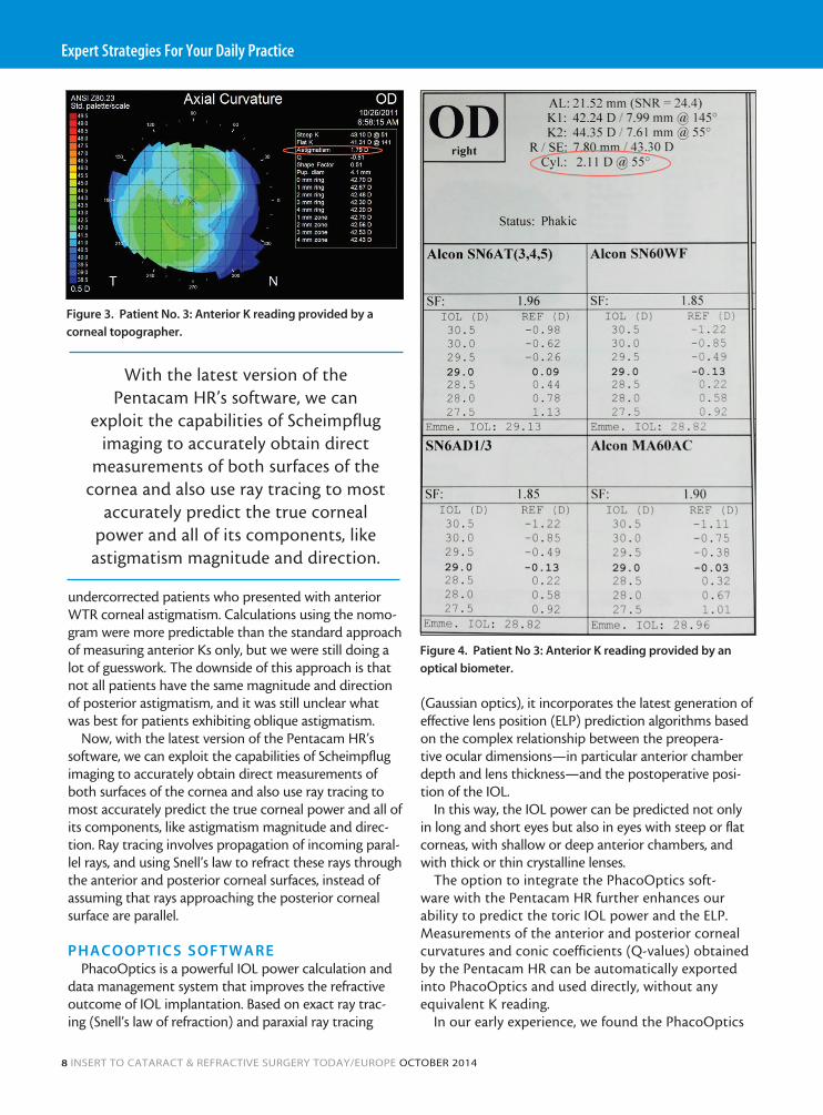
Figure 3 . Patient No . 3: Anterior K reading provided by a
corneal topographer .
Figure 4 . Patient No 3: Anterior K reading provided by an
optical biometer .
With the latest version of the Pentacam HR’s software, we can
exploit the capabilities of Scheimpflug imaging to accurately obtain direct
measurements of both surfaces of the cornea and also use ray tracing to most
accurately predict the true corneal power and all of its components, like
astigmatism magnitude and direction.

OCTOBER 2014 INSERT TO CATARACT & REFRACTIVE SURGERY TODAY/EUROPE 9
Expert Strategies For Your Daily Practice
software to be helpful in deducing the actual spherocylindrical correction by toric IOLs, and also to deduce the IOL composition overall, including ELP.
CASE EXAMPLESBelow are three patient examples to exemplify the use-
fulness of the Pentacam HR and PhacoOptics software in toric IOL implantation.
Patient No. 1. A patient with typical WTR astig-matism exhibited higher total astigmatism than the
anterior Ks revealed (Figure 1). If the K readings from an optical biometer only were used in the manufacturers’ online calculators, Patient No. 1’s cylinder would have been overcorrected.
Patient No. 2. A patient with typical ATR astigmatism exhibited higher total astigmatism than the anterior Ks revealed (Figure 2). If the K readings from an optical biometer only were used in the manufactur-ers’ online calculators, Patient No. 2’s cylinder would have been undercor-rected.
Patient No. 3. In a patient who exhibited oblique astigmatism, note the dif-ference between what two anterior-K measuring devices measured (topographer, Figure 3; optical biometer, Figure 4), compared with the total corneal refractive astigmatism measured by the Pentacam (Figure 5). Furthermore, when automatically export-ing the Pentacam data to PhacoOptics, the previously described principles are applied and we can obtain an accurate prediction of what the postoperative refrac-tion will look like (Figure 6). This is done without hav-ing to do any guesswork and without any extra calcula-tions or external webpages.
CONCLUSIONThe latest version of the Pentacam HR software allows
us to accurately obtain direct measurements of both the anterior and posterior cornea and also to use ray tracing to predict not only the true corneal power but all of its components, including the magnitude and direction of astigmatism. n
Iqbal Ike K. Ahmed, MD, FRCSC, is an Assistant Professor and Director of the Glaucoma and Advanced Anterior Surgical Fellowship at the University of Toronto and a Clinical Assistant Professor at the University of Utah in Salt Lake City. Dr. Ahmed states that he is a consultant to Oculus, Abbott Medical Optics, and Alcon. He may be reached at tel: +1 905 820 3937 ext. 161; e-mail: [email protected].
Xavier “Xavo” Campos-Möller, MD, is an anterior segment specialist and clinical research fellow with Dr. Ahmed. Dr. Campos-Möller states that he has no finan-cial interests to disclose. He may be reached at e-mail: [email protected].
1. Koch DD, Jenkins RB, Weikert MP, et al. Correcting astigmatism with toric intraocular lenses: effect of posterior corneal astigmatism. J Cataract Refract. Surg. 2013;39(12):1803-1809.
Figure 5 . Patient No . 3: The true net power and the total corneal refractive power
as determined by the Pentacam HR .
Figure 6 . The PhacoOptics software provides an accurate
prediction of what the postoperative refraction will look like
in Patient No . 3 .

10 INSERT TO CATARACT & REFRACTIVE SURGERY TODAY/EUROPE OCTOBER 2014
Expert Strategies For Your Daily Practice
The Pentacam: A Comprehensive Diagnostic ToolThis diagnostic device enhances the preoperative exam in refractive corneal and lens-based surgeries.
BY THOMAS KOHNEN, MD, PhD, FEBO
A nterior segment Scheimpflug tomographers, such as the Pentacam HR (Oculus), offer surgeons a variety of sophisticated diagnostic tools. Due to the need
for comprehensive preoperative diagnoses before refractive surgical procedures such as LASIK, PRK, phakic IOL implan-tation, and refractive lens exchange (RLE), Scheimpflug tomography is the standard clinical tool in my clinic.
My research in the past few years has utilized Scheimpflug diagnostic options in screening for subclinical forme fruste keratoconus (FFK), optimizing phakic IOL size according to anterior chamber dimensions, analyzing total corneal refractive power (TCRP), and performing lens den-sitometry. Each area is discussed in detail below.
ECTASIA SCREENING PRIOR TO LASER ABLATION
Subclinical FFK is one of the remaining risk factors for post-LASIK ectasia. Its preoperative diagnosis is difficult, as patients usually do not have any visual disturbances and classical keratoconus indicators like keratometry (K) values are not identifiable in the very early stages of keratoconus.1
Analyses of spatially resolved corneal thickness profiles have been shown to offer sensitive and specific options to discriminate subclinical FFK from a normal eye.2,3 The Pentacam, for instance, offers a multifactorial keratoconus screening display based on anterior and posterior elevation maps and pachymetry progression analysis that are com-bined into one final factor, final D (Figure 1; see The Belin/Ambrósio Enhanced Ectasia Display, pg 5, for more informa-tion). The Pentacam’s software also allows surgeons to identify other more classic predictors, like anterior corneal surface curvature patterns, and newer methods, like the analysis of corneal wavefront aberrations.4 The Pentacam uses a large set of well-established patient data to safely indicate deviations from these normal values displayed in the indices report.5
PHAKIC LENS IMPLANTATIONHorizontal white-to-white (HWTW). Although the cor-
neal diameter does not directly reflect the dimensions of the interior anterior chamber diameter or the sulcus diameter, HWTW measurements still represent the clinically most fea-
sible method to estimate these dimensions and determine the appropriate sizes of phakic IOLs. In a recent trial submit-ted for publication, we compared the HWTW measure-ments derived from three different devices and found that the Pentacam HR had a better coefficient of repeatability (0.08) than either the IOLMaster 500 (Carl Zeiss Meditec; 0.18) or the Lenstar 900 (Haag-Streit; 0.19).
The systematic difference in results between methods accounts for mean differences of -0.2 mm between the Pentacam HR and IOLMaster 500 and 0.2 mm between the Lenstar 900 and IOLMaster 500. Devices should not be used interchangeably without compensation for this offset.
Anterior chamber dimensions. Phakic IOLs, especially those designed for anterior chamber implantation, must be implanted only in eyes that have sufficient long-term postoperative central and peripheral clearance from the phakic IOL to the corneal endothelium.6,7 Imaging with the Pentacam offers preoperative measurement of anterior chamber metrics, such as central and peripheral anterior chamber depth and volume, and postoperative evaluation of the lens position and central and peripheral clearance (Figure 2).
REFRACTIVE LENS EXCHANGEOptical success with premium IOLs, especially mul-
tifocals, depends on regular corneal conditions. Large
Figure 1 . The Belin/Ambrósio Display of a keratoconic eye .

OCTOBER 2014 INSERT TO CATARACT & REFRACTIVE SURGERY TODAY/EUROPE 11
Expert Strategies For Your Daily Practice
irregularities, unfavorable spherical aberrations, and uncompensated corneal astigmatism can lead to major image distortions. The Pentacam accounts for this need of precise corneal diagnostics in several ways.
Summary for total corneal spherical aberration and total corneal higher order aberrations root mean square (HOA-RMS) values. With implantation of aspheric IOLs, spherical aberration can be chosen accord-ing to the total corneal spherical aberration. With mul-tifocal IOLs, besides clearly irregular curvature patterns, total corneal HOA-RMS of more than 0.3 µm can be interpreted as a contraindication for implantation. Both metrics are no longer limited to anterior surface measure-ment only but are derived from corneal tomography and, thus, are based on ray-tracing calculations that take into consideration corneal anterior and posterior surface and spatially resolved pachymetry values.
Analysis of TCRP. Keratometers and topographers use only the central anterior corneal curvature for empirical estimation of TCRP; however, the Pentacam also con-siders the anterior and posterior corneal surfaces and pachymetry values, which are available in the Cataract Pre-Op Display. Tonn et al, from our group, recently reported that, especially in corneas with against-the-rule or oblique astigmatism on the anterior surface, empiri-cal evaluation is flawed.8 When the analysis of corneal astigmatism is based on anterior surface astigmatism measurements only, refractive surprises after toric IOL implantation are more likely.
Another benefit of Scheimpflug imaging compared with classical K is the superior repeatability of both mea-surement of magnitude and axis of corneal astigmatism. Pentacam simulated K and TCRP have proven to be about 20% more repeatable than classical automated measurements when it comes to the magnitude of astig-matism (Fityo et al, submitted for publication). Last but not least, individualized IOL power requires precise eval-uation of corneal asphericity and irregularities; both of these analyses are offered with the Pentacam or topogra-phers but cannot be provided by keratometers.
LENS DENSITOMETRYScheimpfug images from the Pentacam provide surgeons
with a detailed look at the structure and size and the cata-ract status (Figure 3). Weiner et al, from our group, recently evaluated the feasibility of lens densitometry measurements with the Pentacam HR and found that three-dimensional evaluation ofnucleus opacity was repeatable and valid.9
Mayer et al, from our group, then used Pentacam Nucleus Staging (PNS) metrics to evaluate the impact of densitometry on phaco time in manual and laser-assisted cataract surgery.10 Increasing densitometry was shown to correspond with increasing effective phaco time; addition-ally, patients with higher PNS values profited most from laser phacofragmentation, as they benefit from the largest reduction of effective phaco time.10
CONCLUSIONThe Pentacam offers a broad variety of diagnostics for
various kinds of refractive surgery and has become a stan-dard tool that we use prior to every refractive and cataract surgical procedure we perform. n
Thomas Kohnen, MD, PhD, FEBO, is Professor and Chairman of the Department of Ophthalmology at Goethe-University, Frankfurt, Germany. He states that he is a consultant to Oculus. He may be reached at tel: +49 69 6301 3945; e-mail: [email protected].
1. Bühren J, Kook D, Kohnen T. Suitability of various topographic corneal parameters for diagnosis of early keratoconus. Ophthalmologe. 2012;109:37-44.2. Bühren J, Kook D, Yoon G, Kohnen T. Detection of subclinical keratoconus by using corneal anterior and posterior surface aberrations and thickness spatial profiles. Invest Ophthalmol Vis Sci. 2010;51:3424-3432.3. Ambrósio R Jr, Klyce SD, Wilson SE. Corneal topographic and pachymetric screening of keratorefractive patients. J Refract Surg. 2003;19:24-29.4. Bühren J, Kühne C, Kohnen T. Defining subclinical keratoconus using corneal first-surface higher-order aberra-tions. Am J Ophthalmol. 2007;143:381-389.5. Gilani F, Cortese M, Ambrósio R Jr, et al. Comprehensive anterior segment normal values generated by rotating Scheimpflug tomography. J Cataract Refract Surg. 2013;39:1707-1712.6. Doors M, Berendschot TTJM, Webers CAB, Nuijts RMMA. Model to predict endothelial cell loss after iris-fixated phakic intraocular lens implantation. Invest Ophthalmol Vis Sci. 2010;51:811-815.7. Kohnen T, Klaproth OK. Three-year stability of an angle-supported foldable hydrophobic acrylic phakic intraocular lens evaluated by Scheimpflug photography. J Cataract Refract Surg. 2010;36:1120-1126.8. Tonn B, Kohnen T, Klaproth O. Impact of corneal thickness and posterior corneal curvature on total corneal astigmatism. Paper presented at: the ARVO; May 4-8, 2014; Orlando, Florida.9. Weiner X, Baumeister M, Kohnen T, Bühren J. Repeatability of lens densitometry using Scheimpflug imaging. J Cataract Refract Surg. 2014;40:756-763.10. Mayer WJ, Klaproth OK, Hengerer FH, Kohnen T. Impact of crystalline lens opacification on effective phacoemul-sification time in femtosecond laser-assisted cataract surgery. Am J Ophthalmol. 2013;157(2):426-432.
Figure 2 . Scheimpflug image of a phakic anterior chamber angle-
supported IOL . The blue arrows indicate central clearance from
the natural lens to the phakic IOL and the central and peripheral
clearances from the implant to the corneal endothelium .
Figure 3 . Lens densitometry and thickness evaluation before
refractive lens exchange .

OCULUS Optikgeräte GmbHPostfach • 35549 Wetzlar • GERMANY
Tel. +49 641 2005 0 • Fax +49 641 2005 295Email: [email protected] • www.oculus.de
OCULUS USA, [email protected]
OCULUS Asia, [email protected] Czechia, [email protected]
OCULUS Iberia, [email protected] Poland, [email protected]
The availability of products and features may vary by country.
OCULUS reserves the right to change product specifications and design.
P/SD/030/EN