INNOVATIONS IN PEDIATRIC CARDIOPULMONARY - RePub
160
INNOVATIONS IN PEDIATRIC CARDIOPULMONARY BYPASS A CONTINUOUS PROCESS OF QUALITY IMPROVEMENT Hanna Dorota Golab
Transcript of INNOVATIONS IN PEDIATRIC CARDIOPULMONARY - RePub
InnovatIons In pedIatrIc cardIopulmonary bypass
a contInuous process of qualIty Improvement
Innovations in pediatric cardiopulmonary bypass A
continuous process of quality improvem
ent H
anna Dorota G
olab
Hanna dorota Golab
Hanna Golab cover.indd 1 04-04-11 12:00

InnovatIons In pedIatrIc cardIopulmonary bypass
a contInuous process of qualIty Improvement
Hanna Dorota Golab

ISBN 978-94-6169-050-0
Cover design: H.D. GolabLayout and Print by: Optima Grafische Communicatie, Rotterdam, The Netherlansds
© 2011 H.D. GolabAll rights reserved. No part of this thesis may be reproduced, distributed, stored in a retrieval system or transmitted in any form or by any means, without permission of the author, or when appropriate, of the publishers of the publications.
InnovatIons In pedIatrIc cardIopulmonary bypassa contInuous process of qualIty Improvement
InnovatIes In pedIatrIsche cardIopulmonale bypasseen contInu proces van kwalIteItsverbeterIng
Proefschrift
ter verkrijging van de graad van doctor aan deErasmus Universiteit Rotterdam
op gezag van derector magnificus
Prof.dr. H.G. Schmidt
en volgens besluit van het College voor Promoties.De openbare verdediging zal plaatsvinden op
Woensdag 8 juni 2011 om 11: 30 uur
door
Hanna Dorota Golab
geboren te Gliwice

Promotiecommissie
Promotor: Prof.dr. A.J.J.C. Bogers
Overige leden: Prof.dr. W.A. Helbing Dr. A.P. Kappetein Prof.dr. B. Mochtar
Copromotor: Dr. J.J.M. Takkenberg
Financial support by the Netherlands Heart Foundation for the publication of this thesis is gratefully acknowledged.

contents
chapter 1 7General introduction, background on cardiopulmonary bypass, aim and the structure of the thesis.
chapter 2 15The effect of temperature management during cardiopulmonary bypass on clinical outcome in pediatric patients undergoing correction of ventricular septal defect. Golab HD, Wijers MJ, Witsenburg M, Bol Raap G, Cruz E, Bogers AJJC.Journal of Extra-Corporeal Technology 2000; 32: 89-94
chapter 3 29Effects of cardiopulmonary bypass circuit reduction and residual volume salvage on allogeneic transfusion requirements in infants undergoing cardiac surgery. Golab HD, Takkenberg JJM, van Gerner-Weelink GL, Wijers MJ, Scohy TV, de Jong PL, Bogers AJJC. Interact Cardiovasc Thorac Surg. 2007; 6: 335-9
chapter 4 43Intraoperative cell salvage in infants undergoing elective cardiac surgery: a prospective trial. Golab HD, Scohy TV, de Jong PL, Takkenberg JJM, Bogers AJJC. Eur J Cardiothorac Surg. 2008; 34: 354-9
chapter 5 59Small, smaller, smallest. Steps towards bloodless neonatal and infant cardio-pulmonary bypass. Golab HD, Bogers AJJC. Perfusion 2009; 24: 239-42
chapter 6 67Specific requirements for bloodless cardiopulmonary bypass in neonates and infants; a review.Golab HD, Takkenberg JJM, Bogers AJJC. Perfusion 2010; 25: 237-43

chapter 7 81Risk factors for low colloid osmotic pressure during infant cardiopulmonary bypass with colloidal prime. Golab HD, Takkenberg JJM, Bogers AJJC. Interact Cardiovasc Thorac Surg. 2009; 8: 512-6
chapter 8 93Relevance of colloid oncotic pressure regulation during neonatal and infant cardiopulmonary bypass: a prospective randomised study.Golab HD, Scohy TV, Jong de PL, Kissler J, Takkenberg JJM, Bogers AJJC. Eur J Cardiothorac Surg. 2010; Nov 3. [Epub ahead of print]PMID: 21055963
chapter 9 109Intraoperative glycemic control during pediatric cardiac surgery for congenital heart disease. Scohy TV, Golab HD, Egal M, Takkenberg JJM, Bogers AJJC.Submitted
chapter 10 125General discussion, conclusions and prospects.
chapter 11 137Summary / SamenvattingAcknowledgementCurriculum vitaeList of publicationsPortfolio

Chapter 1General introduction


General introduction 9
Cardiopulmonary bypass (CPB) is defined as a technique that temporarily replaces the function of the heart and lungs, maintaining an adequate blood circulation and oxygen content of the body during surgery of the heart and great vessels.
The current practice of cardiopulmonary bypass was achieved through the efforts of a numbers of individuals who believed that artificial cardiopulmonary support could replace the body’s own circulatory and respiratory systems. Collaborative efforts of physiologists, chemists, physicists, engineers, and physicians led to design and develop-ment of synthetic devices that could effectively sustain patients who required corrective surgical procedures of treatable lesions of the heart, lungs or other vital organs.
In 1937, Gibbon reported the first successful total cardiopulmonary bypass of an animal (cat) with the use of heparin, a Dale-Schuster-type pump, and a vertical rotating cylinder oxygenator [1]. However, it was not until 1951 that cardiopulmonary bypass was applied in a patient, enabling an unobstructed view of intracardiac lesions. In Min-neapolis on the 5th of April 1951, Dennis and his associates performed the first total cardiopulmonary bypass in a 6-year old patient with an endocardial cushion defect [2]. Unfortunately, the patient could not be weaned from the bypass and died on the table because of cardiac failure. Gibbon, who considered to be the “father of cardiopulmo-nary bypass”, together with his wife Mary refined the work of Dennis and successfully performed on the 20th of May 1953 in Philadelphia the closure of an atrial septal defect in an 18-years old patient [3]. This moment marked in the history of cardiopulmonary bypass the beginning of the “applied technology period” that lasted from 1950 to 1970. The “refinement period” has followed and lasts until present [4].
Cardiopulmonary bypass is still an unavoidable prerequisite for complete repair or palliation of many congenital cardiac defects. The current policy in cardiac surgery is to perform a complete repair of congenital heart defects early in life, preferably in neonate and infant patients, before the heart and other organs adapt and change according to the abnormal physiology. In the past 30 years, mortality after pediatric cardiac surgery in Europe and the United States decreased from greater than 50% to less than 2% [5]. This exceptional reduction was achieved through improvements in CPB, and the evolu-tion of diagnostic and surgical techniques as well as perioperative care. However, the exposure of the young and small patients to the CPB still may result in major morbidity and represents a serious challenge for all involved healthcare professionals.
Attempts to reduce postoperative mortality and morbidity related to CPB contain a continuous process of quality improvement (CQI) concerning perfusion technique. Dif-ferent aspects of quality improvement can be recognized: product improvement, pro-cess improvement and people based improvement. The concept of CQI was introduced in the 1920s by Shewhart. Working in the Bell Telephone Laboratories on methods to improve quality and lower costs, he developed the concept of control with regard to process variation [6]. CQI has been used in the manufacturing world more extensively

10 Chapter 1
than in healthcare, despite of being equally well applicable to processes in medicine. Control of process variation includes the observation of a phenomenon, the isolation of influential variables, and changing the process. If a change proves to be beneficial, it will be implemented. If it does not result in advancement, new solutions are explored. Moreover, the CQI concept emphasises strongly that there is always the next area to improve [7].
On institutional level, since many years, perfusionists in the ErasmusMC have been applying the guidelines of CQI into CPB technique. Firstly, the specific CPB registry was developed to automatically collect relevant perfusion and patient data. Secondly, audits of existing data were performed to recognize CPB risk factors and areas of improvement. The evidence-based process innovations, in terms of employed materials and strategies, were introduced and subsequently the results were evaluated. All those efforts were taken to model the CPB strategy to become as beneficial as possible for pediatric patient.
Recently, worldwide healthcare professionals recognized the need of evidence–based perfusion and quality improvement in the field. That led to the formation of the Interna-tional Consortium for Evidence-Based Perfusion (ICEBP) in October 2006. The mission of the Pediatric Process Improvement Subcommittee of the ICEBP is to “develop, foster, and promote process improvement in the delivery of perfusion services for neonatal, infant and pediatric patients. The objectives of this committee include the development of pediatric practice guidelines and a registry specific to pediatric perfusion. Additionally, the Committee has started collaboration with other congenital heart disease societies such as the World Society for Pediatric and Congenital Heart Disease.”[8, 9].
Background on cardiopulmonary bypass
Cardiopulmonary bypass provides a complex set of nonphysiologic circumstances un-der which patients are subjected to severe physiologic alterations. The systemic inflam-matory response syndrome, also named postperfusion syndrome, is recognized as the most deleterious effect of CPB. Its multifactorial etiology is associated with generalized activation of the immune system [10, 11], direct tissue trauma and ischemia-reperfusion injury to the vital organs [12]. Consequently, capillary leak, acute lung injury, coagu-lopathy and multiorgan failure may occur to some extend in the postoperative period. In neonates and infants, those deleterious effects of CPB are often more pronounced than in adults. One of the reasons is immaturity of vital organs, as well as coagulation and the endocrine system [13]. Additionally, due to the higher metabolic demands, neonates and infants require higher perfusion flow rate per body surface area and this may produce higher sheer stress on the blood cell components. Another negative factor is an extreme disproportion between the CPB circuit size (surface area and volume) in relation to patient size. This results in significant dilution of clotting factors, red blood cells and plasma proteins. As compensation, homologous blood products are transfused

General introduction 11
that may exacerbate the inflammatory response [14]. Finally, surgical corrective tech-niques for some of congenital heart defects still require deep hypothermia with low flow perfusion rates or even circulatory arrest to allow repair and to facilitate adequate exposure [15]. In the last two decades, technological development and intensive clinical research on CPB helped to establish basic guidelines to enhance the outcome for neonatal and infant patients. An effort has been made to ameliorate the adverse CPB effects related to hypothermia used purposely during the bypass [16]. Technical improvements of the CPB circuit and evolution of the surgical skills allowed for temperature strategy changes, the reduction of depth of hypothermia from moderate (± 28 ° C) to mild (± 32 ° C) or to normothermia in most pediatric cardiac procedures [17].
Continuous product improvement has resulted in a miniaturized CPB circuit, tailored to the size of the patient. Manufacturers of circuit elements (oxygenators, arterial filters, ultrafiltration devices) downsized the new models without compromising on the safety and performance quality. Additionally, new pediatric dedicated heart-long machines with pole-mounted pump-heads gave the opportunity to redesign the customary circuits to reduce the volumes and contact surface area and therefore diminished he-modilution and inflammatory response [18].
Another technological innovation that has been successfully introduced as a routine treatment into pediatric CPB is the use of coating materials on the circuit surface. Primarily, heparin- bounded coating layers were designed to allow the reduction of heparinization level during CPB. Its biocompatible quality recognized in clinical studies stimulated further development of different materials with specific bio-neutral quali-ties [19]. Although the clinical relevance of the biocompatible coatings in neonatal and infant CPB is still a point of discussion [20] the results from clinical studies showed a significant decrease of cytokine and interleukin level in plasma when CPB circuit was coated [21].
Furthermore, conventional ultrafiltration and modified ultrafiltration during- and post-bypass gained lately on popularity as a powerful anti-inflammatory and anti –he-modilutinal strategy. This was possible due to the availability of a wide range of small pediatric hemoconcentration devices [22, 23].
Miniaturization of the CPB circuit and optional use of hemofiltration facilitate a basis for substantial reduction of homologous blood products transfusion during CPB [24].
Additionally, intraoperative blood salvage, that proved a very effective strategy to reduce perioperative transfusion demand in adult patients [25], was introduced into pediatric cardiac surgery. The feasibility of this step depended on the development of a small, high quality salvage system [26]. Not only blood loss during surgery was collected into the cell saving device, also the residual volume of CPB circuit could be processed. In that manner the total blood loss was reduced, requirement for homologous red blood

12 Chapter 1
cell concentrate was diminished and retransfusion of unprocessed residual volume containing inflammatory mediators was avoided [27].
Despite of miniaturization of the bypass circuit and diminishing of the CPB related hemodilution, the composition of the priming solutions is still studied as an important aspect of CPB quality improvement process. The post CPB capillary leak with an unde-sirable extravasation of the fluids and postoperative weight gain are documented and studied in neonates and infants undergoing CPB [28]. Lowered plasma colloid osmotic pressure (COP) during CPB is pointed as the culprit of the fluids shift and postoperative oedema [29].
In addition, results of clinical studies established that during CPB the level of most stress hormones raises and that hypothermia during the bypass enhances this effect [30, 31]. The insulin level in plasma can be decreased as well as a peripheral response to this hormone, therefore the plasma glucose level may rise [32]. On the other hand, the glycemic control in the perioperative period, aiming at avoidance of hyperglycemia by maintening normal glucose levels, may be a risk for hypoglycaemia and adverse outcomes [33].
Aim and the structure of the thesis
The aim of this thesis was to investigate the results of specific innovations that were tested and implemented to contain advers effects of neonatal and infant CPB in an at-tempt to reduce CPB related morbidity and to improve patient outcome.
In Chapter 2, we studied the clinical relevance of the temperature management dur-ing CPB on clinical outcome in pediatric patients undergoing correction of ventricular septal defect.
The effects of CPB circuit reduction and processing of the residual volume after the bypass were retrospectively studied in Chapter 3. The results of this retrospective audit encouraged the prospective trial on further circuit reduction and adjuvant perioperative blood salvage in neonates and infants (Chapter 4). The consequences of stepwise circuit miniaturization as a prerequisite for a bloodless neonatal and infant cardiopulmonary bypass are presented in Chapter 5. Chapter 6 reviews different strategies and future possibilities to achieve fully bloodless cardiopulmonary bypass in neonates and infants.
We recognized and retrospectively studied the risk factors for the occurrence of low COP during the CPB in Chapter 7. Subsequently, in a prospective study (Chapter 8) we examined in a randomized fashion the impact of different COP regulatory strategies on postoperative weight gain, fluid balance and clinical outcomes.
In Chapter 9, we present results of the retrospective data audit related to the intraop-erative glycemia control strategy.
Chapter 10 contains the general discussion and conclusions to this thesis as well as the recommendations concerning innovative CPB strategy for pediatric patients.

General introduction 13
references
1. Gibbon JH. Artificial maintenance of circulation during experimental occlusion of the pulmonary artery. Arch Surg 1937; 34: 1105-1131
2. Dennis C, Spreng DS, Nelson GE, Karlson KE, Nelson RM, Thomas JV, Eder WP, Varco RL. Develop-ment of a pump-oxygenator to replace the heart and lungs: An apparatus applicable to human patients and an application to one case. Ann Surg 1951; 134: 709-721
3. Gibbon JH. Application of the mechanical heart and lung apparatus to cardiac surgery. Minn Med 1954; 36: 171-185
4. Stammers AH. Historical Aspects of Cardiopulmonary Bypass: From Antiquity to Acceptance. J Cardiothorac Vasc Anesth 1997; 11: 266-274
5. Freedom RM, Lock J, Bricker JT. Pediatric cardiology and cardiovascular surgery: 1950-2000. Circulation 2000;102: IV58-IV68
6. Shewhart WA. Economic Control of Quality of Manufactured Product. Republished by the Ameri-can Society for Quality Control, Milwaukee, WI: 1981; pp.501, ISBN 0-87389-076-0
7. Donabedian A. Evalueting the quality of medical care. Milbank Mem Fund Q. 1966; 44: 166-206 8. Likosky DS. A primer on reviewing and synthesizing evidence. J Extra Corpor Technol 2006; 38:
112-5 9. Likosky DS. Integrating evidence-based perfusion into practices: the International Consortium
for Evidence-Based Perfusion. J Extra Corpor Technol 2006; 38: 297-301 10. Levy JH, Tanaka KA. Inflamatory response to cardiopulmonary bypass. Ann Thorac Surg 2003; 75:
715-720 11. Seghaye MC, Duchateau J, Grabitz RG, Faymonville ML, Messmer BJ, Buro-Rathsmann K, von Ber-
nuth G. Complement activation during cardiopulmonary bypass in infants and children: relation to postoperative multiple organ failure. J Thorac Cardiovasc Surg 1993; 106: 978–987
12. Butler J, Pathi VL, Paton RD, Logan RW, MacArthur KJD, Jamieson MPG, Pollock JCS. Acute phase responses to cardiopulmonary bypass in children weighing less than 10 kilograms. Ann Thorac Surg 1996; 62: 538–542
13. Jensen E, Bengtsson A, Berggren H, Ekroth R, Andreasson S. Clinical variables and pro-inflamatory activation in paediatric heart surgery. Scand Cardiovasc J 2001; 35: 201-206
14. Hickey E, Karamlou T, You J, Ungerleider R.M. Effects of circuit miniaturization in reducing inflam-matory response to infant cardiopulmonary bypass by elimination of allogeneic blood products. Ann Thoracic Surg 2006; 81: 2367-2372
15. Ungerleider RM. Gaynor J.W. The Boston circulatory arrest study:An analysis. J Thorac Cardiovasc Surg 2004; 127:1256-1261
16. Eggum R, Ueland T, Mollnes TE, Videm V, Aukrust P, Fiene AE, Lindberg HL. Effect of perfusion temperature onthe inflammatory response during pediatric cardiac surgery. Ann Thorac Surg 2008; 85: 611-7
17. Groom RC, Froebe S, Martin J, Manfra MJ, Cormack JE, Morse C, Taenzer AH, Quinn RD. Update on pediatric perfusion practice in North America: 2005 survey. J Extra Corpor Technol. 2005; 37: 343-50
18. Ando M, Takahashi Y, Suzuki N. Open heart surgery for small children without homologous blood transfusion by using remote pump head system. Ann Thorac Surg 2004; 78: 1717-22
19. Boning A, Scheewe J, Ivers T, Friedrich C, Stieh J, Freitag S, Cremer JT. Phosphorylcholine coating or heparin coating for pediatric extracorporeal circulation causes similar effects in neonates and infants. J Thorac Cardiovasc Surg 2004; 127: 1458-65

14 Chapter 1
20. Draaisma AM, Hazekamp MG, Anes N, Schoof PH, Hack CE, Sturk A, Dion RAE. Phosphorylcholine coating of bypass systems used for young infants does not attenuate the inflammatory response. Ann Thorac Surg 2006; 81: 1455-9
21. Miyaji K. Miyamoto T, Kohira S, Nakashima K, Inoue N, Sato H, Ohara K. Miniaturized cardiopulmo-nary bypass system in neonates and small infants. Interact Cardivasc Thorac Surg 2008; 7: 75-79
22. Draaisma AM, Hazekamp MG, Frank M, Annes N, Schoof PH, Huysmans HA. Modified ultrafiltra-tion after cardiopulmonary bypass in pediatric cardiac surgery. Ann Thorac Surg 1997; 64: 521-5
23. Allen M, Sundararajan S, Pathan N, Burmester M, Macrae D. Anti-inflammatory modalities: Their current use in pediatric cardiac surgery in the United Kingdom and Ireland. Pediatr Crit Care Med 2009; 10: 341-345
24. Durandy Y. Usfulnes of low prime perfusion pediatric circuit in decreasing blood transfusion. ASAIO Journal 2007; 53: 659-661
25. Daane CR, Golab HD, Wijers MJ, Bogers AJJC. Processing and transfusion of residual cardiopulmo-nary bypass volume; effects on haemostasis, complement activation, postoperative blood loss and transfusion volume. Perfusion 2003; 18: 115-21
26. Melo A, Serrick CJ, Scholz M, Singh O, Noel D. Quality of red blood cells using the Dideco Electa autotransfusion device. J Extra Corpor Technol. 2005; 37: 58-59
27. Booke M, Hagemann O, Van Aken H, Erren M, Wullenweber J, Bone HG. Intraoperative autotrans-fusion in small children: an in vitro investigation to study its feasibility. Anesth Analg 1999; 88: 763-5
28. Rigger LQ, Voepel-Lewis T, Kulik TJ, Malviya S, Tait AR, Mosca RS, Bove EL. Albumin versus crys-talloid prime solution for cardiopulmonary bypass in young children. Crit Care Med 2002; 30: 2649-2654
29. Loeffelbein F, Zirell U, Benk C, Schlensak C, Dittrich S. High colloid ocotic pressure priming of cardiopulmonary bypass in neonates and infants: implications on haemofiltration, weight gain and renal function. Eur J Cardiothorac Surg 2008; 34: 648-52
30. Gajarski RJ, Stefanelli CB, Graziano JN, Kaciroti N, Charpie JR, Vazquez D. Adrenocortical response in infants undergoing cardiac surgery with cardiopulmonary bypass and circulatory arrest. Pedi-atr Crit Care Med 2010; 11: 44-51
31. Jakob SM, Stanga Z. Perioperative metabolic changes in patients undergoing cardiac surgery. Nutrition 2010; 26: 349–353
32. Piper HG, Alexander JL, Shukla A, Pigula F, CostelloJM, Laussen PC, Jaksic T, Agus MS. Real time continous glucose monitoringin pediatric patients during and after cardiac suegery. Pediatrics 2006; 118: 1176-1184
33. Paliato A, Thiagarajan RR, Laussen PC, Gauvreau K, Agus MS, Scheurer MA, Pigula FA, Costello JM. Association between intraoperative and early postoperative glucose levels and adverse outcomes after complex congenital heart surgery. Circulation 2008; 118: 2235-2242

Chapter 2The effect of temperature management during cardiopulmonary bypass on clinical outcomes in pediatric patients undergoing correction of ventricular septal defect
Golab HD, Wijers MJ, Witsenburg M, Bol Raap G, Cruz E, Bogers AJJC.
Journal of Extra-Corporeal Technology 2000; 32: 89-94

16 Chapter 2
AbstrAct
background: Moderate hypothermia of 28 °C is widely accepted in cardiac surgery with cardiopulmonary bypass (CPB). Recently however, several studies suggested that nor-mothermic or “tepid” bypass techniques may improve the clinical outcomes for patients undergoing cardiac operations.
methods: To assess the effect of bypass temperature management strategy in pediat-ric patients undergoing correction of ventricular septal defect, 26 patients with body weight under 10 kg were randomly assigned to two treatment groups: Group 1, mild hypothermia , patients cooled to nasopharyngeal temperature of 32 °C during the by-pass; or Group 2, moderate hypothermia of 28 °C. Clinical parameters were recorded and blood samples were obtained just before, during and 24 hours after operation.
results: All the population characteristics and intraoperative variables were similar in the two groups. Hematologic data after CPB and protamine administration revealed a significantly (p<0.05) longer activated partial thromboplastin time in the 32 °C group, however the difference in blood loss did not reach significance.
conclusion: Our study shows that both perfusion temperatures equally well facilitated CPB for this type intracardiac surgery.

Temperature management during pediatric cardiopulmonary bypass 17
introDuction
Until recently, intracardiac repair in pediatric cardiac surgery was mostly performed with moderate (27 - 30 °C) to profound (< 25 °C) systemic hypothermia. The exceptions to this rule were short procedures for which only mild (30 - 32 °C) hypothermia was used.
Systemic hypothermia reduces tissue metabolic rate and oxygen demand and is used to protect the myocardium and the brain against potential ischemic insult [1-3]. Further-more, it enhances protection of the other organs. Systemic hypothermia allows to lower the arterial blood flow and promotes better conditions in the operation field as well as less blood trauma occurrence. Hypothermia also causes marked changes in the periph-eral circulation, hormonal body response and alterations in organ function. Generalized inflammatory response, involving the complement, coagulation, kallikrein and fibrino-lytic cascades, is often a dominant feature of hypothermic bypass [4-6]. Undoubtedly, hypothermia has its positive as well as negative aspects during cardiopulmonary bypass (CPB). Therefore, normothermic CPB with warm cardioplegia in adult cardiac surgery is becoming increasingly popular [7]. In pediatric heart surgery, technical developments and enhanced surgical skills also allow reduction of depth of hypothermia in most of the procedures. Still comparatively few publications have addressed the issues raised by maintenance of mild or moderate hypothermia during pediatric bypass.
The aim of this study was to compare clinical results between two groups of pediatric patients, operated with short aorta occlusion times and mild or moderate hypothermic CPB.
mAteriAls AnD metHoDs
Patients
The study population comprised 26 consecutive pediatric patients who underwent closure of ventricular septal defect (VSD) with use of a Gore - Tex® patch. Only children with body weight under 10 kg and without concomitant heart disease were included. Exclusion criteria were: respiratory insufficiency with need of respiratory support, kidney failure, liver failure and neurological impairment. Patients were equally randomized into two groups: Group 1, mild hypothermia (nasopharyngeal temperature ≥ 32 °C during CPB); Group 2, moderate hypothermia (nasopharyngeal temperature ≥ 28 °C during CPB). The same surgeon performed all operations. The study was conducted according to the regulations of the hospital medical ethical committee. Informed parental consent was obtained for all patients.

18 Chapter 2
Anesthesia
Patients were premedicated with 0.3 mg/kg midazolam suppositorium 1 hour before induction of anesthesia. Induction of anesthesia was done by inhalation (halothane or sevoflurane) or intravenously (midazolam, pavulon, fentanyl). Patients were intubated with a nasal - endotracheal tube and ventilated with a minute volume of 10 ml/kg/min. Heart rate, ECG, arterial and right atrial blood pressure, nasopharyngeal and rectal temperature were continuously measured. A bladder catheter was inserted to monitor urine production during and after the operation.
Cardiopulmonary bypass
All patients were operated upon with CPB and cardioplegic arrest. In all patients the ascending aorta was cannulated with either an 8 or 10 Fr standard straight-tip cannula, depending upon patient size. In all cases, venous return was provided by bicaval can-nulation of the superior and inferior caval veins with angled metal tip 12 Fr cannulae.
CPB circuit consisted of a membrane oxygenator with integrated venous-cardiotomy reservoir, roller pump with silicone tubing and an arterial line filter. The circuit was primed with Ringer’s solution and whole blood to achieve an intraoperative hematocrit of 28% during the bypass period. During CPB nonpulsatile pump flow with rates of 1.8 to 2.4 L/min/m2 was maintained adequate to metabolic needs of the patient. In-line moni-toring of arterial oxygen tension (PaO2) and venous oxygen saturation (SvO2) provided conditions for alpha-stat strategy during the whole period of bypass.
Anticoagulation of the patient and the CPB circuit were achieved with initial patient heparin dose of 300 IU / kg body weight and prime heparin dose of 4.2 IU/ml total prime volume. Assessment of anticoagulation during operation was done by measurement of the kaolin activated clotting time (ACT). The ACT values were maintained ≥480 sec by administration of additional heparin when necessary. After discontinuation of the CPB heparin was neutralized by protamine chloride with standard dose of 4 to 5 mg/ kg body weight. Adequacy of protamine reversal was ascertained with the use of heparin-protamine titration.
No aprotinin was administrated to patients in the study population.Patient mean arterial blood pressure was maintained between 30 - 65 mmHg during
the bypass. Continuous recording of the pump flow, arterial blood temperature along with patient nasopharyngeal and rectal temperatures, oxygen and air flow, PaO2, SvO2, arterial line pressure as well as patient pressures, was provided by “Odis” - perfusion registration system [8, 9]. Myocardial preservation was achieved by antegrade admin-istration of cold (4 °C) St.Thomas Hospital cardioplegic solution delivered by gravity at the dose of 10 - 15 ml/kg of body weight after application of the aortic cross-clamp. No topical cooling was used.

Temperature management during pediatric cardiopulmonary bypass 19
All patients were weaned from bypass with infusion of dopamine 2 mg/ kg/ min and nitroglycerin 1 mg/ kg/ min.
Measurements and calculations
In both groups arterial and venous blood gas samples were obtained before bypass, during CPB after 5 min - 20 min - and at the end (± 50 min). The last sample was obtained after administration of protamine chloride. During CPB oxygen consumption index (VO2 I) and systemic vascular resistance (SVR) were calculated as follows:
SVR =mean arterial pressure - central venous pressure
x 80 [dynes sec cm-5 ]cardiac output
VO2 I =cardiac index x {(art. saturation - ven. saturation) x haemoglobin content x 2.32}[ml/min m2 ]
Coagulation factors: platelet count, fibrinogen, activated partial thromboplastin time (APTT), thrombin time (TT) and were measured before the bypass, after protamine chloride administration and 24 hours after the operation.
Other measured and recorded variables include occurrence of electrical activity (ECG) during the aortic cross-clamping, spontaneous cardiac conversion after releasing of the clamp and existence of any kind of the rhythm disturbances. In addition, the amount of administrated blood products, diuresis, blood loss, length of the respiratory support and stay in the intensive care unit (ICU) were recorded.
Intraoperative post-correction epicardial echocardiography [10] was carried out with colour-Doppler studies to assess left-to-right shunting by echo-contrast injection into the left atrium. After 24 hours post-operative left ventricular function was determined by echocardiography and follow-up was completed at the discharge visit.
Data analysis
All values are presented as mean ± standard deviation (SD) of the mean. Two-way analy-sis of variance (ANOVA) for repeated measurements was used for comparison between the groups at specific points in time. p - Values were obtained for the overall group ef-fect. Other data were compared by unpaired t - test between the two groups. p -Values ≤ 0.05 were considered statistically significant.
20 Chapter 2
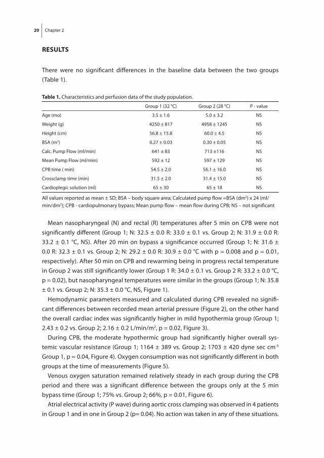
results
There were no significant differences in the baseline data between the two groups (Table 1).
table 1. Characteristics and perfusion data of the study population.
Group 1 (32 °C) Group 2 (28 °C) P - value
Age (mo) 3.5 ± 1.6 5.0 ± 3.2 NS
Weight (g) 4250 ± 817 4958 ± 1245 NS
Height (cm) 56.8 ± 15.8 60.0 ± 4.5 NS
BSA (m2) 0.27 ± 0.03 0.30 ± 0.05 NS
Calc. Pump Flow (ml/min) 641 ± 83 713 ±116 NS
Mean Pump Flow (ml/min) 592 ± 12 597 ± 129 NS
CPB time ( min) 54.5 ± 2.0 56.1 ± 16.0 NS
Crossclamp time (min) 31.5 ± 2.0 31.4 ± 15.0 NS
Cardioplegic solution (ml) 65 ± 30 65 ± 18 NS
All values reported as mean ± SD; BSA – body square area; Calculated pump flow =BSA (dm2) x 24 (ml/min/dm2); CPB - cardiopulmonary bypass; Mean pump flow – mean flow during CPB; NS – not significant
Mean nasopharyngeal (N) and rectal (R) temperatures after 5 min on CPB were not significantly different (Group 1; N: 32.5 ± 0.0 R: 33.0 ± 0.1 vs. Group 2; N: 31.9 ± 0.0 R: 33.2 ± 0.1 °C, NS). After 20 min on bypass a significance occurred (Group 1; N: 31.6 ± 0.0 R: 32.3 ± 0.1 vs. Group 2; N: 29.2 ± 0.0 R: 30.9 ± 0.0 °C with p = 0.008 and p = 0.01, respectively). After 50 min on CPB and rewarming being in progress rectal temperature in Group 2 was still significantly lower (Group 1 R: 34.0 ± 0.1 vs. Group 2 R: 33.2 ± 0.0 °C, p = 0.02), but nasopharyngeal temperatures were similar in the groups (Group 1; N: 35.8 ± 0.1 vs. Group 2; N: 35.3 ± 0.0 °C, NS, Figure 1).
Hemodynamic parameters measured and calculated during CPB revealed no signifi-cant differences between recorded mean arterial pressure (Figure 2), on the other hand the overall cardiac index was significantly higher in mild hypothermia group (Group 1; 2.43 ± 0.2 vs. Group 2; 2.16 ± 0.2 L/min/m2, p = 0.02, Figure 3).
During CPB, the moderate hypothermic group had significantly higher overall sys-temic vascular resistance (Group 1; 1164 ± 389 vs. Group 2; 1703 ± 420 dyne sec cm-5 Group 1, p = 0.04, Figure 4). Oxygen consumption was not significantly different in both groups at the time of measurements (Figure 5).
Venous oxygen saturation remained relatively steady in each group during the CPB period and there was a significant difference between the groups only at the 5 min bypass time (Group 1; 75% vs. Group 2; 66%, p = 0.01, Figure 6).
Atrial electrical activity (P wave) during aortic cross clamping was observed in 4 patients in Group 1 and in one in Group 2 (p= 0.04). No action was taken in any of these situations.

Temperature management during pediatric cardiopulmonary bypass 21
27
29
31
33
35
37
5 min 20 min 50 minCPB time
Temperature [°C]
*
**
figure 1. Changes of temperature during cardiopulmonary bypass. Group 32 °C - N Group 32 °C - R Group 28 °C - N Group 28 °C – R
*p = 0.01**p = 0.0008
0
10
20
30
40
50
60
70
80
90
5 min 20 min 50 min CPB time
MAP [mmHg]
figure 2. Mean arterial pressure during cardiopulmonary bypass. Group 32 °C Group 28 °C

22 Chapter 2
0
0,5
1
1,5
2
2,5
3
3,5
5 min. 20 min. 50 min. CPB time
CI [l/min/m2]
*
figure 3. Cardiac Index during cardiopulmonary bypass. Group 32 °C Group 28 °C
* p = 0. 0006
0
500
1000
1500
2000
2500
3000
5 min. 20 min. 50 min. CPB time
SVR [dynxsecxcm-5]
*
figure 4. Systemic vascular resistance during cardiopulmonary bypass. Group 32 °C Group 28 °C
* p = 0. 01

Temperature management during pediatric cardiopulmonary bypass 23
0
20
40
60
80
100
120
140
5 min 20 min 50 min CPB time
VO2 [ml/min/m2]
figure 5. Oxygen consumption during cardiopulmonary bypass. Group 32 °C Group 28 °C
0,5
0,55
0,6
0,65
0,7
0,75
0,8
0,85
0,9
5 min 20 min 50 min CPB time
SVO2 [%]
*
figure 6. Oxygen saturation in venous blood during cardiopulmonary bypass. Group 32 °C Group 28 °C
* p = 0. 01

24 Chapter 2
All patients in both groups converted spontaneously to sinus rhythm after aorta declamp-ing. AV block was temporally observed in 2 patients in Group 1. The external pacing was applied before sinus rhythm returned.
Initial dose of heparin used before the start of CPB was not significantly different between both groups (Group 1; 1222 ± 290 vs. Group 2; 1500 ± 409 IU, NS). During CPB two patients from Group 1 and one from Group 2 required additional heparin to sustain ACT ≥480 sec. Standard protamine dose given in compliance with protocol was not significantly different. In each group, 4 patients required additional protamine (Group 1; 2.2 ± 3.4 mg vs. Group 2; 2.4 ± 3.5 mg, NS). Pre-operative measurement of coagulation factors as well as base line ACT in both groups were not significantly different. After administration of protamine the ACT values were still higher if compared with base line values, but did not show any significant difference between groups; whereas, APTT values (Group 1; 55 ± 18 vs. Group 2; 41 ± 3 s, p = 0.045) were significantly higher in mild hypothermia group (Table 2).
table 2. ACT values and plasma coagulation factors pre- and post CPB.
Group 1 (32 °C) Group 2 (28 °C) P value
Pre cPb
ACT (sec) 127 ± 16 132 ± 9 NS
APTT (sec) 48 ± 11 51 ± 17 NS
APTT ratio 1.6 ± 0.4 1.7 ± 0.6 NS
TT (sec) 19 ± 7 16 ± 2 NS
Fibrinogen (mg/dl) 220 ± 70 220 ± 50 NS
Platlets count (x1000/mm3) 287 ± 55 315 ± 78 NS
Post cPb
ACT (sec) 144 ± 35 141 ± 23 NS
APTT (sec) 55 ± 18 41 ± 3 NS
APTT ratio 1.5 ± 0.1 1.4 ± 0.1 NS
TT (sec) 15 ± 2 15.9 ± 3 NS
Fibrinogen ( mg/dl) 160 ± 30 180 ± 40 NS
Platelets count (x1000/mm3) 177 ± 61 183 ± 50 NS
All values reported as mean ± SD; ACT – activated clotting time; APTT – activated partial thromboplastin time; APTT ratio - patient’s APTT/ normal population APTT; NS – not significant; TT – thrombin time
There was no difference in diuresis, blood loss and the amount of blood products used during the period of 24 hours post CPB (Table 3). Additionally, the mean duration of post - operative ventilatory support, as well as the highest percentage of oxygen used in inspiratory fraction (FiO2 %) and applied positive end expiratory pressure (PEEP) were similar (Table 4). The mean duration of stay at the ICU by patients in both groups was not significantly different.

Temperature management during pediatric cardiopulmonary bypass 25
Intraoperative epicardial echocardiography revealed a residual VSD with insignificant leakage of contrast in 2 patients from Group 1 and 3 patients from Group 2. At discharge 2 patients in Group 1 displayed a hemodynamically insignificant residual VSD as well as 1 patient in Group 2. No residual VSDs were observed on subsequent follow-up visits. In-traoperative post-correction epicardial echocardiography assessed left ventricular func-tion in all patients as normal. Echocardiographic examination after 24 hours estimated left ventricular function as “diminished” in 3 patients in Group 1 and 1 patient in Group 2, nevertheless at discharge all the patients’ regained normal left ventricular function.
table 3. Urine production and blood balance in 24 hours after CPB.
Group 1 (32 °C) Group 2 (28 °C) P - value
Urine output (ml) 345 ± 44 350 ± 111 NS
Blood loss (ml) 109 ± 106 95 ± 24 NS
Homologous blood (ml) 175 ± 39 145 ± 53 NS
FFP (ml) 63 ±117 106 ± 90 NS
All values reported as mean ± SD; CPB – cardiopulmonary bypass; FFP – fresh frozen plasma; NS – not significant
table 4. Postoperative use of the respiratory support.
Group 1 (32 °C) Group 2 (28 °C) P - value
Respiratory supp.(h) 22.8 ± 18 16.5 ± 19 NS
Highest FiO2 (%) 48.8 ± 11 46.0 ± 10 NS
PEEP (cm H2O) 3.5 ± 0.8 3.7 ± 1.0 NS
All values reported as mean ± SD; FiO2 – inspiratory fraction of oxygen; NS – not significant; PEEP – positive end expiratory pressure
Discussion
Use of moderate (28 °C) systemic hypothermia improves operating conditions and allows lower arterial flow rates. This, in turns, reduces collateral coronary circulation and con-tributes to myocardial protection as well as protection of other vital organs. Therefore, moderate hypothermia is widely carried out, although the effects of skin and perhaps muscle ischemia related to this temperature and the increased sympathetic effects may balance out any potential advantages [11]. Another effect of hypothermia is that blood viscosity increases so that use of appropriate hemodilution is required to reduce the systemic vascular resistance during CPB [12]. Hypothermia together with hemodilution disturbs coagulation and fibrinolitic cascades, therefore is assumed to be responsible for enhanced blood loss during and after operation [13]. In certain operations, the selection of CPB temperature is dependent on the complexity of the operation; for example, deep

26 Chapter 2
hypothermia during circulatory arrest. Therefore, questions may be raised as to what the best temperature is in case of short (under 1 hour of CPB time) surgical procedures for small pediatric patients with body weight under 10 kg. [14]. According to the surgeon’s opinion, use of moderate (28 °C) or mild (32 °C) hypothermia during correction of the ventriculr septal defect had no influence on the technical complexity of the operation.
The hemodynamic data obtained from both groups showed no differences in adequa-cy of the CPB. Mean arterial pressure and venous oxygen saturation were kept constant during the bypass without any difficulty, although patients from Group 1 (32 °C) required significantly higher cardiac index to achieve this. On the other hand Group 2 (28 °C) had significantly higher overall systemic vascular resistance during the CPB, which might be caused by moderate hypothermia. Oxygen consumption in the two groups was not significantly different, which can be explained by the negligible differences of patient’s temperatures during long time on CPB.
Myocardial protection in both groups was the same and there was spontaneous return of sinus rhythm in all the cases. In Group 1 (32 °C) twice atrio- ventricular block occurred, which had to be resolved by temporarily use of pacemaker. Those adverse events could be related to less adequate myocardial protection, but also could be associated with the surgical procedure itself. There were no clinical consequences of these events and the echocardiographic control of the left ventricular function showed no difference between the patients from both groups. Kidney function assessed by urine output and lung function assessed by duration of ventilation support did not differ in both groups.
The amount of heparin and protamine used showed no difference in the two groups. Postoperative, after administration of protamine, both groups still had prolonged ACT values and significantly lower plasma fibrinogen concentration when compared to the “ base line “ values, but Group 1 (32 °C) also had significantly longer APTT in comparison to Group 2 (28 °C). Mean blood loss was least in the moderate hypothermia group but the difference did not reach significance. Hematologic data suggested increased fibrinolitic potential in the mild hypothermia group [15].
conclusions
Our study documented no difference in organ preservation depending on type of hypo-thermia, mild or moderate, used during the reconstruction of VSD in pediatric patients. The chosen temperatures did not impair adequacy of CPB. There was no difference in technical complexity of the operation. Moreover, the clinical outcome of the patients did not depend on the type of hypothermia. There was suggestion of more activation of fibrinolitic potential in the 32 °C group.
Temperature management during pediatric cardiopulmonary bypass 27
references
1. Cameron DE, Gardner TJ. Principles of clinical hypothermia. Card Surg State Art Revi.1988; 2:13-25 2. Hickely RF, Hoar PF. Whole body oxygen consumption during low-flow hypothermic cardiopul-
monary bypass. J Thorac Cardiovasc Surg 1983; 86: 903-906 3. Swain JA, McDonald TJ Jr, Balaban RS, et al. Metabolism of the heart and brain during hypother-
mic cardiopulmonary bypass. Ann Thorac Surg 1991; 51:105-109 4. Moore FD, Warner KG, Assousa S, et al. The effects of complement activation during cardiopulmo-
nary bypass: attenuation by hypothermia, heparin and hemodilution. Ann Surg 1988; 208: 95-103 5. Utley JR, Wachtel C, Cain RB, et al. Effects of hypothermia, hemodilution and pump oxygenation
on organ water conyent, blood flow and oxygen delivery, and renal function. Ann Thorac Surg 1981; 31: 121-133
6. Ohata T, Sawa Y, Kadoba K, et al. Effect of cardiopulmonary bypass under tepid temperature on inflammatory reactions. Ann Thorac Surg 1997; 64: 124-8
7. Lichtenstain SV, Ashe KA, El-Dalati H, et al. Warm heart surgery. J Thorac Cardiovasc Surg 1991; 101: 269-274
8. Oomen JA, Wijers M, Bos E, Schuurbier J. Automated bookkeeping in the operating room. Thorax-centre Journal 1994; 6: 23-26
9. Burgerhout A. Electronic hearts controlling lung machines. Thoraxcentre Journal 1993; 5: 44-48 10. Bol-Raap G, Bogers AJJC, Boersma H, et al.Temporary tricuspid valva detachment in closure of
congenital ventricular septal defect. Eur J Cardio Thorac Surg 1994; 8: 145-148 11. Zwischenberger JB, Kirsch MM, Dechter RE. Suppression of shivering decreases oxygen consump-
tion and improves hemodynamic stability during postoperative rewarming. Ann Thorac Surg 1987; 43: 428-432
12. Matsuda H, Sasako Y, Susune N, at al. Determination of optimal perfusion flow rate for deep hy-pothermia cardiopulmonary bypass in the adults based on distribution of blood flow and oxygen consumption. J Thorac Cardiovasc Surg 1992; 103: 541-548
13. Dale J, Lilleasson P, Erikssen J. Hemostasis after open - heart surgery with extreme or moderate hemodilution. European Surg Reaserch 1987; 19: 339-342
14. Mora CT, Henson MB, Weintraub WS, at al. The effect of temperature management during car-diopulmonary bypass on neurologic and neuropsychologic outcome in patients undergoing coronary revaculaization. J Thorac Cardiovasc Surg 1996; 112: 514-522
15. Engelman RM, Pleet AB, Rousou JA, et al. What is the best perfusion temperature for coronary revascularization. J Thorac Cardiovasc Surg 1996; 112:1162-1163

Chapter 3Effects of cardiopulmonary bypass circuit reduction and residual volume salvage on allogeneic transfusion requirements in infants undergoing cardiac surgery
Golab HD, Takkenberg JJM, van Gerner-Weelink GL, Wijers MJ, Scohy TV, de Jong PL, Bogers AJJC.
Interact Cardiovasc Thorac Surg. 2007; 6: 335-9

30 Chapter 3
AbstrAct
background: Cardiopulmonary bypass in children may cause severe hemodilution and can lead to excessive perioperative blood loss and high transfusion requirements. Minimization of cardiopulmonary bypass circuit and salvage of red blood cells from the residual volume after the procedure are widely utilised to reduce allogeneic transfusion. We evaluated the effectiveness of those measures introduced in infant cardiac surgery in our institution.
methods: This retrospective observational study included 148 consecutive infants between 1 to12 months of age, with a body weight less than 10 kg, who underwent an elective cardiac operation between 1997 and 2005. Patients were divided into three groups defined by the circuit prime volume; 700 mL (Group 1), 450 mL (Group 2) and 330 mL (Group3). In Group 1 residual volume after perfusion was discarded and in Group 2 and 3 was processed in cell saving device. Analyzed variables were: perioperative blood loss, urine production, transfusion of homologous blood products and cell salvage product, and haematology data.
results: Reduction of the circuit volume significantly diminished use of red blood cells concentrate from 1.6 units to 0.8 units (p<0.0001) and fresh frozen plasma from 1.3 units to 0.4 units (p<0.0001). Utilization of the cell salvage product reduced significantly (p=0,023) the postoperative need for homologous blood transfusion.
conclusions: Reduction of cardiopulmonary bypass circuit together with cell salvage from the residual volume of hart-lung machine proved to be effective in reducing of homologous blood transfusion in infant cardiac surgery.

Cardiopulmonary bypass circuit reduction and residual volume salvage 31
introDuction
Cardiopulmonary bypass (CPB) during open-heart surgery remains a non-physiological technique that may cause severe hemodilution and an acute inflammatory body re-sponse [1,2,3,4]. In children, who undergo cardiac surgery, this alone can lead to exces-sive perioperative blood loss and high transfusion requirements. Additionally, nowadays smaller and younger patients are undergoing more complex procedures and there is strong evidence of enhanced blood loss and blood transfusion associated with these patients [5-8]. Awareness that allogeneic blood may transmit known and unknown pathogens and cause alloimmunization on future transfusion and pregnancies, stimu-lated development of new blood conservation policies.
Use of smaller CPB circuits and perioperative salvage of red blood cells (RBC) dimin-ished some of the adverse effects of CPB and consequently reduced transfusion demand. Despite recent advances in technology, the majority of neonates and infants still require perioperative transfusion of different homologous blood components [9,10,11]. In the past decade, an effort was made in our institution to reduce the exposure of pediatric patients to homologous blood products. First, we minimized the extracorporeal circuit used during CPB and secondly, a cell saving device was routinely used to process re-sidual blood from the extracorporeal circuit. The present study evaluates the effects of those blood conservation measures.
mAteriAl AnD metHoDs
Population
From January 1997 to March 2005, 166 consecutive infants between 1 to 12 months of age, with body weight of less than 10 kg, underwent an elective cardiac operation with CPB at the Erasmus University Medical Center, Rotterdam. During this period, three different types of CPB circuits were used; each specific type was solitary utilized within the defined period. For the purpose of the retrospective study patients were assigned into three groups according to the type of CPB circuit and how the residual volume after CPB was processed. Excluded were patients who had preoperatively known clotting disorders, patients on systemic anticoagulant drugs preoperatively, re-do procedures with perioperative cell salvage and postoperative reexplorations. Patients with aorta oc-clusion time longer than 90 minutes were also excluded from the study to homogenize the patient’s groups according to the type and complexity of the operation.
In Group 1(patients operated from 1997 to 1999), the CPB system consisted of a Cobe VPCML flat-sheet membrane oxygenator with hard-shell reservoir (Cobe, Denver, CO),

32 Chapter 3
with a priming volume 450 mL. The total priming volume of the CPB system was 700 mL. The residual volume of the system was discarded after the procedure.
In Group 2 (patients operated from 2000 to 2003), a Polystan Safe Mini hollow-fibre membrane oxygenator with hard-shell reservoir (Maquet Cardiopulmonary, Hirrlingen, Germany) was used, with a priming volume 160 mL. The total priming volume of the CPB system was 450 mL. Residual volume after the procedure was processed by HaemoLite 2 plus (Haemonetics, Bothwell, UK) cell-saving device with a pediatric centrifugal bowl of 100 mL. Red blood cell concentrate obtained through cell salvage was postoperatively transfused to the patient.
Group 3 (patients operated from 2004 till March 2005), had a Capiox Baby Rx hollow-fibre oxygenator with hard-shell reservoir (Terumo, Tokyo, Japan), with priming volume 60 mL. The total CPB system volume was reduced to 330 mL. The residual volume was as in Group 2, processed under the same conditions by the cell-saving device.
All the CPB systems utilized a roller pump with ¼” silicone tubing (Raumedic REHAU, Muri, Switzerland), a D736-40 Micron (Dideco, Mirandola, Italy) arterial filter and PVC ¼” arterial and venous tubing. None of the CPB systems was coated.
Patients gender, cardiac anomaly, age, body weight (BW), body surface area (BSA), CPB time, aorta cross-clamp time (AoX) and time at the Intensive Care Unit (ICU) were noted.
Anesthesia, Anticoagulation and Cardiopulmonary Bypass
All patients received standard anesthesia with midazolam, pancuronium bromide (Organon, Oss, The Netherlands) and fentanyl (Janssen-Cilag, Tilburg, The Netherlands). Anticoagulation was established with an initial bolus 300 IU/kg BW of porcine heparin (Leo Pharmaceutical Products, Weesp, The Netherlands). Additional heparin was ad-ministrated to maintain activated clotting time higher than 480 sec during the whole procedure. Initial protamine hydrochloride dose was 5 mg/kg BW (ICN Pharmaceutic, Zoetermeer, The Netherlands). Control of the heparin neutralization was performed with the HepCon HMS device (Medtronic HemoTec, Englewood, CO) and if necessary an extra protamine was given.
The CPB prime contained always red blood cells (RBC), fresh-frozen plasma (FFP) and Ringer’s Solution (Baxter, Utrecht, The Netherlands) or Gelofusine (B. Braun, Melsungen, Germany). The amount of RBC product added to the priming was calculated to achieve a hematocrit of 0.28 L/L during CPB. The prime was completed with 0.5 g/kg BW mannitol (NPBI, Emmer, The Netherlands), 0.5 g/kg BW human albumin (Sanquin CLB, Amsterdam, The Netherlands), 4.2 IU heparine / mL priming volume and 2-5 ml NaHCO3
8.4% (Frese-nius Nederland, s’Hertogenbosch, The Netherlands).
Nonpulsatile CPB, with mild hypothermia of 28 °C to 32° C, was performed with blood flow rates between 1.8 L/min/m2 to 3.2 L/min/m2 to maintain venous oxygen satura-

Cardiopulmonary bypass circuit reduction and residual volume salvage 33
tion above 70% and mean arterial pressure between 40 to 60 mmHg. In accordance with our infant CPB protocol no modified ultrafiltration was used and no aprotinin was administrated in any of the cases.
Administrations of RBC products and additional crystalloids or colloids during CPB were at the discretion of the perfusionist, based upon the working volumes and hema-tocrit levels. Cardioplegic cardiac arrest was obtained by antegrade infusion of 10-15 mL/kg BW St. Thomas Hospital Solution I á 4 °C (Apotheek ErasmusMC, Rotterdam, The Netherlands). Laboratory tests, urine production, blood loss and blood products transfusion
Hemoglobin concentration (Hb), hematocrit (Ht) and platelet count (Thr) were mea-sured one day before the operation, at the start and end of the operation, during the CPB at the 5 min on bypass and at the end of the CPB, and after 24 hours postoperatively. Urine production and blood loss was noted at the end of the operation and as the total volume collected at the ICU.
Blood loss in the operation room (OR) represented the sum of blood loss calculated from swabs, discarded suction volumes and the chest drains output. Postoperative blood loss was calculated as the total loss from the chest tubes during the ICU stay.
In Group 1, an additional blood loss was the discarded residual volume from the CPB circuit (RES). In Group 2 and 3 residual volumes were processed by a cell saving device. Discarded plasma fraction separated during the cell saving and a minimal RES were than accounted for a blood loss. The decision to transfuse blood products was based upon measured blood loss, patient clinical status and laboratory tests. In general, acyanotic patients were transfused to maintain a hemoglobin level of 6.0 mmol/L. Platelet transfu-sion was considered if the platelet count at the end of CPB was less than 100 x 109/L. The volume of blood products transfused in the OR, including blood products added to the circuit prime and during the CPB, and administrated at the ICU was noted.
Data analysis
Continuous data are presented as mean ± standard error of the mean, categorical data are presented as proportions. Continuous independent data were compared with one-way analysis of variance ANOVA and Bonferroni (in case of equal variances) or Tamhane T2 (in case of unequal variances) post-hoc corrections were applied to the p values for multiple comparisions. Categorical data were compared with the chi-square test. A p value less than 0.05 was considered statistically significant. All statistical analyses were performed using SPSS 13.0 statistical software (SPSS, Chicago, IL).

34 Chapter 3
results
Demographics and CPB
A total of 148 consecutive infants fulfilling the inclusion criteria were enrolled in this study. Fifty-two patients were selected into Group 1, 54 patients in Group 2 and 42 in Group 3. Thirteen patients were excluded due to an aorta occlusion time longer than 90 minutes. Five patients with re-do procedures were also excluded. The same surgical team performed all the operations. Postoperatively there were no complications, no re-explorations and all patients survived. Population and CPB data are presented in Table1.
table 1. Population and CPB data.
Variable Group 1 Group 2 Group 3
n = 52 n = 54 n = 42 P value
Mean ± SE Mean ± SE Mean ± SE
Gender (female/male) 27/25 28/26 17/25 0,76
Age (mo) 6,2 ± 0,4 5,5 ± 0,3 4,4 ± 0,4 0,004ab
Weight (kg) 6,2 ± 0,3 5,7 ± 0,2 5,3 ± 0,2 0,001ab
BSA (m2) 0,35 ± 0,02 0,33 ± 0,01 0,31 ± 0,01 <0,001a
CPB time (min) 76 ± 4 91 ± 6 91± 4 0,03ab
Cross-clamp time (min) 43 ± 3 51 ± 2 58 ± 3 <0,001ab
ICU time (hours) 33 ± 3 31 ± 2 22 ± 0,4 0,001b
Anomaly - correction
ASD - closure 4 2 0
VSD - closure 9 11 16
AVSD - correction 19 19 14
F4 - correction 11 11 4
Other 9 11 8
a p=ns Group2 vs. Group 3; b p=ns Group 1 vs. Group 2; ASD - atrial septal defec; AVSD – atrioventriculair septal defect; BSA - body surface area; CPB-cardiopulmonary bypass; F4 – tetralogy of Fallot; ICU - Intensive Care Unit; SE - standard error of the mean VSD – ventricular septal defect
Laboratory tests results, urine production and blood loss
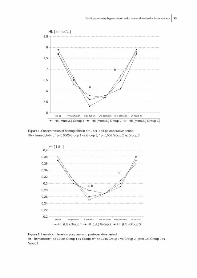
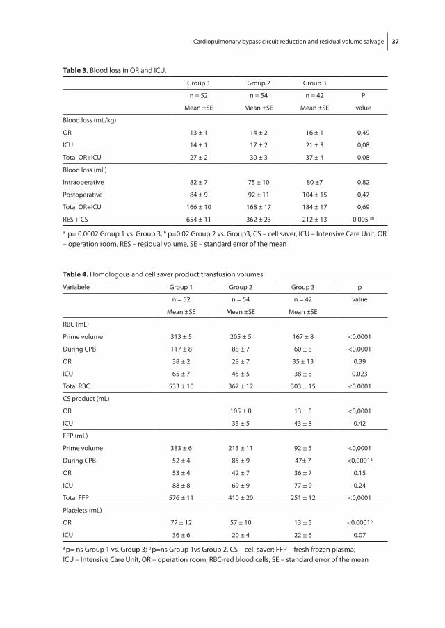
Measurements of hemoglobin concentration (Hb), hematocrit (Ht) and platelet count (Thr) are presented in Figures 1, 2 and 3. There were no significant differences with regard to Hb and Ht values measured the day before operation and pre-perfusion. At the end of the operation, Group 2 compared to Group 3 had significantly lower values of Hb (p=0,006) and Ht (p=0,023). After 24 hours at the ICU there were no significant differences between the groups.
Cardiopulmonary bypass circuit reduction and residual volume salvage 35
5
5,5
6
6,5
7
7,5
8
8,5
Pre-op Pre-perfusion 5' perfusion End perfusion Post perfusion 24 hours IC
Hb (mmol/L) Group 1 Hb (mmol/L) Group 2 Hb (mmol/L) Group 3
Hb [ mmol/L ]
a
b
3.1
figure 1. Concentration of hemoglobin in pre-, per- and postoperative period.Hb – haemoglobin; a p<0.0005 Group 1 vs. Group 3; b p=0,006 Group 2 vs. Group 3
0,2
0,22
0,24
0,26
0,28
0,3
0,32
0,34
0,36
0,38
0,4
Pre-op Pre-perfusion 5' perfusion End perfusion Post perfusion 24 hours IC
Ht (L/L) Group 1 Ht (L/L) Group 2 Ht (L/L) Group 3
Ht [ L/L ]
c
a, b
3.2
figure 2. Hematocrit levels in pre-, per- and postoperative period.Ht – hematocrit; a p<0.0005 Group 1 vs. Group 3; b p=0.016 Group 1 vs. Group 2; c p=0.023 Group 2 vs. Group3

36 Chapter 3
The platelet count the day before operation was significantly lower in the Group 3 compared to the Group 1 (p=0,021) and 2 (p=0,006), but already preoperatively was not significantly different. Urine production and blood loss are presented in Table 2 and 3 respectively. Postoperatively, Group 1 had significantly higher urine output than both other groups. There were no significant differences in the blood loss.
Blood products transfusion
The amount of homologous blood products and cell salvage product transfused in the OR and the ICU are presented in Table 4. Significantly more allogeneic RBC (p<0.0001) was used in the prime and during CPB in Group 1 (1.6 units) compared to Group 2 (1.1 units) and 3 (0.8 unit). Group 2 required significantly more RBC in the prime (p<0.0001) and during the bypass (p=0.024) than Group 3.
0
50
100
150
200
250
300
350
400
450
500
Pre-op Pre-perfusion 5' perfusion End perfusion Postperfusion
24 hours IC
Thr Group 1 Thr Group 2 Thr Group 3
Thr [ x 10E9/L ]
a
3.3
figure 3. Platelets count in pre-, per- and postoperative period.Thr – thrombocytes; a p=0.021 Group 1 vs. Group 3; b p=0.006 Group 2 vs. Group 3
table 2. Urine output during and post –operation.
Urine output Group 1 Group 2 Group 3
mL/kg/h n = 52 n = 54 n = 42 P
Mean ±SE Mean ±SE Mean ±SE value
OR 8 ± 1 6 ± 1 7 ±1 0,54
ICU 6 ± 0,2 3 ± 0,2 4 ± 0,2 <0,001a
a p =ns Group 2 vs. Group 3; ICU – Intensive Care Unit, OR – operation room, SE – standard error of the mean
Cardiopulmonary bypass circuit reduction and residual volume salvage 37
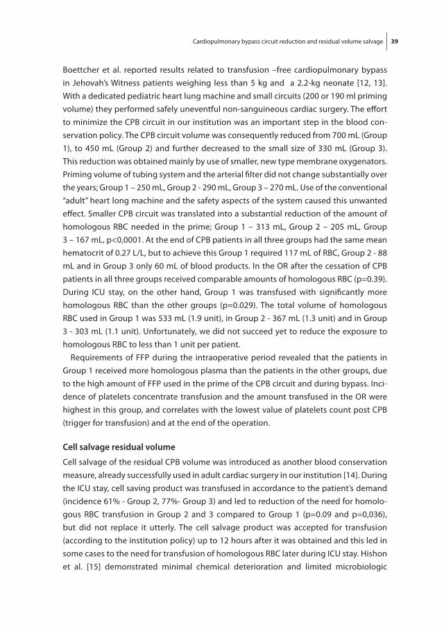
table 3. Blood loss in OR and ICU.
Group 1 Group 2 Group 3
n = 52 n = 54 n = 42 P
Mean ±SE Mean ±SE Mean ±SE value
Blood loss (mL/kg)
OR 13 ± 1 14 ± 2 16 ± 1 0,49
ICU 14 ± 1 17 ± 2 21 ± 3 0,08
Total OR+ICU 27 ± 2 30 ± 3 37 ± 4 0,08
Blood loss (mL)
Intraoperative 82 ± 7 75 ± 10 80 ±7 0,82
Postoperative 84 ± 9 92 ± 11 104 ± 15 0,47
Total OR+ICU 166 ± 10 168 ± 17 184 ± 17 0,69
RES + CS 654 ± 11 362 ± 23 212 ± 13 0,005 ab
a p= 0.0002 Group 1 vs. Group 3, b p=0.02 Group 2 vs. Group3; CS – cell saver, ICU – Intensive Care Unit, OR – operation room, RES – residual volume, SE – standard error of the mean
table 4. Homologous and cell saver product transfusion volumes.
Variabele Group 1 Group 2 Group 3 p
n = 52 n = 54 n = 42 value
Mean ±SE Mean ±SE Mean ±SE
RBC (mL)
Prime volume 313 ± 5 205 ± 5 167 ± 8 <0.0001
During CPB 117 ± 8 88 ± 7 60 ± 8 <0.0001
OR 38 ± 2 28 ± 7 35 ± 13 0.39
ICU 65 ± 7 45 ± 5 38 ± 8 0.023
Total RBC 533 ± 10 367 ± 12 303 ± 15 <0.0001
CS product (mL)
OR 105 ± 8 13 ± 5 <0,0001
ICU 35 ± 5 43 ± 8 0.42
FFP (mL)
Prime volume 383 ± 6 213 ± 11 92 ± 5 <0,0001
During CPB 52 ± 4 85 ± 9 47± 7 <0,0001a
OR 53 ± 4 42 ± 7 36 ± 7 0.15
ICU 88 ± 8 69 ± 9 77 ± 9 0.24
Total FFP 576 ± 11 410 ± 20 251 ± 12 <0,0001
Platelets (mL)
OR 77 ± 12 57 ± 10 13 ± 5 <0,0001b
ICU 36 ± 6 20 ± 4 22 ± 6 0.07
a p= ns Group 1 vs. Group 3; b p=ns Group 1vs Group 2, CS – cell saver; FFP – fresh frozen plasma; ICU – Intensive Care Unit, OR – operation room, RBC-red blood cells; SE – standard error of the mean

38 Chapter 3
The FFP was used in significantly (p=0.0001) larger volume in the prime of Group 1 (1.2 units) versus Group 2 (0.7 unit) versus Group 3 (0.3 unit). The cell saving product, available for Group 2 and 3, was transfused in the OR in a significantly larger volume (p<0.0001) in Group 2 than in Group 3. Platelets concentrate transfusion was significantly higher in de Group1 in the OR (p<0.0001) but at the ICU were no differences between the groups.
Table 5 shows transfusion incidence of homologous blood products (RBC, FFP and platelets) and cell salvage product. In all groups 100% of patients were exposed to the homologous RBC and FFP in the prime of the CPB circuit.
table 5. Percentage of patients’ transfused with homologous blood and cell saver product.
Group 1 Group 2 Group 3
Incidence (%) n = 52 n = 54 n = 42 p
value
RBC Prime 100 100 100 ns
FFP Prime 100 100 100 ns
RBC CPB 100 89 79 <0.01
FFP CPB 52 46 71 0.03
RBC OR 100 67 71 <0.001
FFP OR 77 70 71 ns
CS OR 96 24 <0.001
Platelets OR 52 39 17 <0.001
RBC ICU 98 77 58 <0,001
FFP ICU 69 79 95 0.03
CS ICU 61 77 ns
Platelets ICU 54 41 29 0.03
CPB – cardiopulmonary bypass; CS – cell saver; FFP – fresh frozen plasma; ICU – Intensive Care Unit; ns – not significant; OR – Operation Room; RBC – red blood cells
Discussion
This study shows that consequent minimization of the bypass circuit and cell salvage from circuit residual volume were effective measures in reducing the need for ho-mologous blood products transfusion in infants younger than 12 months of age, who underwent an elective cardiac operation in our institution.
Circuit reduction and homologous transfusion requirements
It is undoubtedly true that the volume of the CPB circuit determines the exposure to allogeneic blood products in the majority of children weighting less than 10 kg. In 2005
Cardiopulmonary bypass circuit reduction and residual volume salvage 39
Boettcher et al. reported results related to transfusion –free cardiopulmonary bypass in Jehovah’s Witness patients weighing less than 5 kg and a 2.2-kg neonate [12, 13]. With a dedicated pediatric heart lung machine and small circuits (200 or 190 ml priming volume) they performed safely uneventful non-sanguineous cardiac surgery. The effort to minimize the CPB circuit in our institution was an important step in the blood con-servation policy. The CPB circuit volume was consequently reduced from 700 mL (Group 1), to 450 mL (Group 2) and further decreased to the small size of 330 mL (Group 3). This reduction was obtained mainly by use of smaller, new type membrane oxygenators. Priming volume of tubing system and the arterial filter did not change substantially over the years; Group 1 – 250 mL, Group 2 - 290 mL, Group 3 – 270 mL. Use of the conventional “adult” heart long machine and the safety aspects of the system caused this unwanted effect. Smaller CPB circuit was translated into a substantial reduction of the amount of homologous RBC needed in the prime; Group 1 – 313 mL, Group 2 – 205 mL, Group 3 – 167 mL, p<0,0001. At the end of CPB patients in all three groups had the same mean hematocrit of 0.27 L/L, but to achieve this Group 1 required 117 mL of RBC, Group 2 - 88 mL and in Group 3 only 60 mL of blood products. In the OR after the cessation of CPB patients in all three groups received comparable amounts of homologous RBC (p=0.39). During ICU stay, on the other hand, Group 1 was transfused with significantly more homologous RBC than the other groups (p=0.029). The total volume of homologous RBC used in Group 1 was 533 mL (1.9 unit), in Group 2 - 367 mL (1.3 unit) and in Group 3 - 303 mL (1.1 unit). Unfortunately, we did not succeed yet to reduce the exposure to homologous RBC to less than 1 unit per patient.
Requirements of FFP during the intraoperative period revealed that the patients in Group 1 received more homologous plasma than the patients in the other groups, due to the high amount of FFP used in the prime of the CPB circuit and during bypass. Inci-dence of platelets concentrate transfusion and the amount transfused in the OR were highest in this group, and correlates with the lowest value of platelets count post CPB (trigger for transfusion) and at the end of the operation.
Cell salvage residual volume
Cell salvage of the residual CPB volume was introduced as another blood conservation measure, already successfully used in adult cardiac surgery in our institution [14]. During the ICU stay, cell saving product was transfused in accordance to the patient’s demand (incidence 61% - Group 2, 77%- Group 3) and led to reduction of the need for homolo-gous RBC transfusion in Group 2 and 3 compared to Group 1 (p=0.09 and p=0,036), but did not replace it utterly. The cell salvage product was accepted for transfusion (according to the institution policy) up to 12 hours after it was obtained and this led in some cases to the need for transfusion of homologous RBC later during ICU stay. Hishon et al. [15] demonstrated minimal chemical deterioration and limited microbiologic

40 Chapter 3
contamination in blood salvaged from the CPB circuit and stored at room temperature for an 18-hour period. Therefore, prolongation of acceptable transfusion period for cell salvage product could be beneficial. In the OR 96% patients from Group 2 received cell salvage product versus 24% from Group 3, but this was not translated to a reduction of homologous blood transfusion in the OR. The cell salvage product was available not earlier than at the end of the procedure, a practical limitation to the transfusion.
Comparison between groups
Our study shares that in recent years children who undergo cardiac surgical operation are becoming younger and smaller. The mean age and weight of infants that were oper-ated in our institution decreased steadily over the years of this study, while CPB and aortic cross clamp times increased. This illustrates that younger and smaller children un-dergo procedures that are more complex. However, ICU stay in the most recent years has declined. The ICU length of stay was significantly longer in Group 1 compared to Group 2 and 3 (33 h vs 31 h vs 22 h respectively, p=0,001) but most likely related to the logistic at the ICU than to the type of the corrections. This requires further study. Pre-perfusion hemoglobin concentration, hematocrit and platelet count, intraoperative and postop-erative blood loss were not significantly different between three groups. Slightly higher total blood loss in Group 3, the youngest group, (37 mL/kg BW) compared to Group 2 (30 mL/kg BW) and Group 1 (27 mL/kg BW) was only borderline significant (p=0.08). In this regard our study results were well within the ranges presented by others [6,7].
The mean age differences between three groups were small and all patients were older than 1 month that excluded influence of the immaturity of the coagulation system, mostly associated with age younger than 1 month [8]. In relation to urine production during the OR stay we have found no significant differences between the groups, but postoperatively Group 1 compared to Group 2 and 3 had enhanced urine output (6 mL/kg vs 3 mL/kg vs 4 mL/kg respectively). Although, we have not specific data to validate our hypothesis, use of the Ringer’s Solution in the prime of the CPB circuit could be associated with this occurrence.
Study design and limitations
Our project was designed as a retrospective observational study to evaluate the conse-quences of policy changing through the years in the infant cardiac surgery in our institu-tion. Due to the retrospective character of our study and 8 years time span involved, we were not able to apply a more demanding protocol (for example for retransfusion of cell saving product) and homogenized our study population. In addition, a formal cost-benefit analysis was not possible. We expect that a follow up prospective study, which is already under way, will give more conclusive information on cell salvage product utilization.

Cardiopulmonary bypass circuit reduction and residual volume salvage 41
conclusions
Minimization of the CPB circuit significantly reduced the demand of homologous blood products, both RBC and FFP, during the infant cardiac surgery. This reduction diminished patient exposure to the number of donors with all well-known benefits of this. We also showed the beneficial effect of the cell salvage from residual volume of the CPB circuit. The timing of the cell saving product availability in the OR, the amount of the product and acceptable transfusion period after the operation are of crucial importance for it effective utilization. In conclusion, allogeneic transfusion data and cell salvage data obtained in this study were important for modifying of the clinical practice in infant cardiac surgery in our institution.

42 Chapter 3
references
1. Seghaye MC, Duchateau J, Grabitz RG, Faymonville ML, Messmer BJ, Buro-Rathsmann K, von Ber-nuth G. Complement activation during cardiopulmonary bypass in infants and children: relation to postoperative multiple organ failure. J Thorac Cardiovasc Surg 1993; 106: 978–987
2. Butler J, Pathi VL, Paton RD, Logan RW, MacArthur KJD, Jamieson MPG, Pollock JCS. Acute phase responses to cardiopulmonary bypass in children weighing less than 10 kilograms. Ann Thorac Surg 1996; 62: 538–542
3. Karamlou T, Schultz JM, Silliman C, Sandquist C,You J, Shen I, Ungerleider RM. Using a miniaturized circuit and an asanguineous prime to reduce neutrophil-mediated organ dysfunction following infant cardiopulmonary bypass. Ann Thorac Surg 2005; 80: 6-14
4. Hickey E, Karamlou T, You J, Ungerleider RM. Effects of circuit miniaturization in reducing inflam-matory response to infant cardiopulmonary bypass by elimination of allogeneic blood products. Ann Thorac Surg 2006; 81: S2367-S2372
5. Andrew M, Vegh P, Johnston M, Bowker J, Ofosu F, Mitchell L. Maturation of the hemostatic system during childhood. Blood 1992; 80: 1998–2005
6. Chambers LA, Cohen DM, Davis JT, Transfusion patterns in pediatric open heart surgery. Transfu-sion 1996; 36: 150–154
7. Kwiatkowski JL, Manno CS. Blood transfusion support in pediatric cardiovascular surgery. Trans-fusion Science1999; 21: 63-72
8. Williams GD, Bratton SL, Riley EC, Ramamoorthy C. Association between age and blood loss in children undergoing open heart operations. Ann Thorac Surg 1998; 66: 870-876
9. Petaja J, Lundstrom U, Leijala M, Peltola K, Siimes MA. Bleeding and the use of blood products after heart operations in infants. J Thorac Cardiovasc Surg 1995; 109: 524–529
10. Friesen RH, Tornabene MA, Coleman SP. Blood conservation during pediatric cardiac surgery: ultrafiltration of the extracorporeal circuit volume after cardiopulmonary bypass. Anesth Analg 1993; 77: 702-7
11. Van Son JAM, Hovaguimian H, Rao IM, He G, Meiling GA, King DH, Starr A., Strategies for repair of congenital heart defects in infants without the use of blood. Ann Thorac Surg 1995; 59: 384–8
12. Boettcher W, Merkle F, Huebler M, Koster A, Schulz F, Kopitz M, Kuppe H, Lange P, Hetzer R. Transfusion-free cardiopulmonary bypass in Jehovah’s Witness patients weighing less than 5 kg. J Extra Corpor Technol 2005; 37: 282-5
13. Hubler M, Boettcher W, Koster A, Redlin M, Stiller B, Lange P, Hetzer R. Transfusion-free cardiac surgery with cardiopulmonary bypass in a 2.2-kg neonate. J Card Surg 2005; 20:180-2
14. Daane CR, Golab HD, Wijers MJ, Bogers AJJC. Processing and transfusion of residual cardiopulmo-nary bypass volume; effects on haemostasis, complement activation, postoperative blood loss and transfusion volume. Perfusion 2003; 18: 115-21
15. Hishon ML, Ryan A, Lithgow P, Butt W. An evaluation of changes in composition and contamina-tion of salvaged blood from the cardiopulmonary bypass circuit of pediatric patients. Heart Lung 1995; 24: 307–311
Chapter 4Intraoperative cell salvage in infants undergoing elective cardiac surgery: a prospective trial
Golab HD, Scohy TV, de Jong PL, Takkenberg JJM, Bogers AJJC.
Eur J Cardiothorac Surg. 2008; 34: 354-9

44 Chapter 4
AbstrAct
background: For a long time intraoperative cell salvage was considered to be not ap-plicable in pediatric patients due to technical limitations. Recently, new autotransfusion devices with small volume centrifugal bowls and dedicated pediatric systems allow ef-ficient blood salvage in small children. The purpose of this prospective non-randomized study was to determine the impact of intraoperative cell salvage on postoperative al-logeneic blood products transfusion in infant patients undergoing cardiac surgery with cardiopulmonary bypass.
methods: Two consecutive cohorts (122 patients) were studied. The first cohort under-went between January 2004 and July 2005 procedures with only blood salvage from the residual volume. The second cohort consisted of patients operated on from August 2005 until December 2006, with additional use of intraoperative cell salvage. The following variables were analyzed: peri- and postoperative blood loss, transfusion of homologous blood products and cell salvage product, haematological and coagulation data, mea-sured before, during and after the operation.
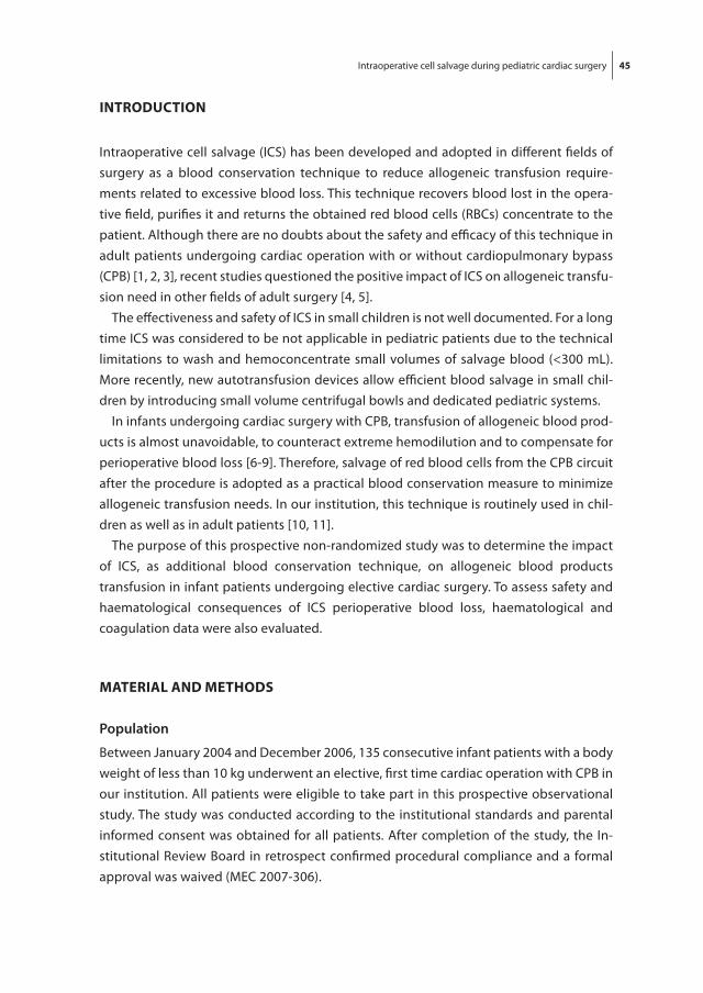
results: Additional intraoperative cell-salvage significantly enhanced the amount of cell saving product available for transfusion (183 ± 56 vs. 152 ± 57 mL, p = 0.003) and sig-nificantly more patients in this group received the cell saving product postoperatively. Consequently, allogeneic blood transfusion was significantly reduced in volume as well as in frequency. We did not observe any adverse effects of intraoperative cell-salvage.
conclusions: Intraoperative cell-salvage, employed as an adjuvant technique to the residual volume salvage in infants undergoing first time cardiac surgery with cardio-pulmonary bypass, was a save and effective method to reduce postoperative alloge-neic blood transfusion. Considering current cell salvage related expense and the cost reduction achieved by diminished allogeneic transfusion, intraoperative cell-salvage in infants demonstrated no economic benefit.
Intraoperative cell salvage during pediatric cardiac surgery 45
introDuction
Intraoperative cell salvage (ICS) has been developed and adopted in different fields of surgery as a blood conservation technique to reduce allogeneic transfusion require-ments related to excessive blood loss. This technique recovers blood lost in the opera-tive field, purifies it and returns the obtained red blood cells (RBCs) concentrate to the patient. Although there are no doubts about the safety and efficacy of this technique in adult patients undergoing cardiac operation with or without cardiopulmonary bypass (CPB) [1, 2, 3], recent studies questioned the positive impact of ICS on allogeneic transfu-sion need in other fields of adult surgery [4, 5].
The effectiveness and safety of ICS in small children is not well documented. For a long time ICS was considered to be not applicable in pediatric patients due to the technical limitations to wash and hemoconcentrate small volumes of salvage blood (<300 mL). More recently, new autotransfusion devices allow efficient blood salvage in small chil-dren by introducing small volume centrifugal bowls and dedicated pediatric systems.
In infants undergoing cardiac surgery with CPB, transfusion of allogeneic blood prod-ucts is almost unavoidable, to counteract extreme hemodilution and to compensate for perioperative blood loss [6-9]. Therefore, salvage of red blood cells from the CPB circuit after the procedure is adopted as a practical blood conservation measure to minimize allogeneic transfusion needs. In our institution, this technique is routinely used in chil-dren as well as in adult patients [10, 11].
The purpose of this prospective non-randomized study was to determine the impact of ICS, as additional blood conservation technique, on allogeneic blood products transfusion in infant patients undergoing elective cardiac surgery. To assess safety and haematological consequences of ICS perioperative blood loss, haematological and coagulation data were also evaluated.
mAteriAl AnD metHoDs
Population
Between January 2004 and December 2006, 135 consecutive infant patients with a body weight of less than 10 kg underwent an elective, first time cardiac operation with CPB in our institution. All patients were eligible to take part in this prospective observational study. The study was conducted according to the institutional standards and parental informed consent was obtained for all patients. After completion of the study, the In-stitutional Review Board in retrospect confirmed procedural compliance and a formal approval was waived (MEC 2007-306).
46 Chapter 4
Exclusion criteria were preoperatively known clotting disorders and procedures that required deep hypothermic circulatory arrest (DHCA).
Two consecutive cohorts were studied. The first cohort (control group) underwent procedures with blood salvage from the residual volume of CPB circuit, between January 2004 and July 2005. At that point, additionally to the residual volume salvage, ICS was introduced and used in all infant patients operated on thenceforth. The second cohort (ICS group) consisted of patients operated on from August 2005 until December 2006. The same surgical team performed all the operations.
Anesthesia, Anticoagulation, Cardiopulmonary Bypass and Cell Salvage
Before induction of anesthesia, all patients were monitored with a five-lead, two-channel electrocardiogram, non-invasive blood pressure measurement, and pulse oximetry. After the insertion of a peripheral venous line, general anesthesia was in-duced with midazolam 0.2 mg/kg, sufentanil 2 mcg/kg and pancuronium 0.15 mg/kg. Patients were nasotracheally intubated and pressure controlled ventilated (PCV) using a Siemens 900C ventilator. Anesthesia was maintained with midazolam 0.1 mg/kg/h and sufentanil 1 mcg/kg/h. Invasive monitoring via a femoral arterial line and an internal jugular central venous catheter was performed, and a Foley bladder catheter and rectal temperature probe were inserted. Before going on CPB all patients received 30 mg/kg iv methylprednisolone, 30 mg/kg iv magnesium sulfate, 1 mg/kg iv furosemide, 40 mg/kg iv cefazoline and 1 mg/kg iv ranitidine.
Anticoagulation was established with an initial bolus 300 IU/kg BW of porcine heparin and additional heparin was administrated to maintain activated clotting time higher than 480 sec during the whole procedure. Initial protamine hydrochloride dose was 4 mg/kg BW. Control of the heparin neutralization was performed and if necessary ad-ditional protamine was given.
The CPB circuit consisted of a Capiox Baby Rx hollow-fiber oxygenator with hard-shell reservoir (Terumo, Tokyo, Japan), a roller pump with ¼” silicone tubing (Raumedic RE-HAU, Muri, Switzerland), a D736-40 Micron (Dideco, Mirandola, Italy) arterial filter and PVC ¼” arterial and venous tubing. The CPB systems were not coated. In accordance with our infant CPB protocol no modified ultrafiltration and no antifibrinolitic medica-tion was used.
CPB prime contained homologous red blood cells concentrate (RBCs), fresh-frozen plasma (FFP) and Gelofusine (B.Braun, Melsungen, Germany). The amount of RBCs added to the priming was calculated to achieve a hematocrit of 28% during CPB. The prime was completed with 0.5 g/kg mannitol, 0.5 g/kg human albumin 20% solution, 4.2 IU heparine / mL priming volume and 2-5 mL NaHCO3
8.4%.Nonpulsatile CPB, with mild hypothermia of 28° C to 32° C, was performed with blood
flow rates between 1.8 L/min/m2 to 3.2 L/min/m2 to maintain venous oxygen saturation

Intraoperative cell salvage during pediatric cardiac surgery 47
above 70% and mean arterial pressure between 40 to 60 mmHg. During CPB α – stat regulation was used. Myocardial protection was achieved with crystalloid cardioplegia.
Administration of RBCs, FFP, crystalloids or colloids during CPB was based upon the system working volumes, target values for hematocrit (not lower than 28% for acyanotic as well as cyanotic patients) and colloid osmotic pressure (not lower than 15 mmHg).
After CPB, residual volume of the circuit was in all cases processed by HaemoLite 2 plus (Haemonetics, Bothwell, UK) cell-saving device with a centrifugal bowl of 100 mL. In the ICS group all blood loss from skin incision to commencement of CPB and then after administration of protamine to skin closure was also salvaged. The HaemoLite 2 plus uti-lized an automatic protocol with centrifuge speed of 8000 rpm, pump speed of 300 mL/min by filling, washing and transfusing, and pump speed of 150 mL/min by concentrat-ing. Washing volume was 500 mL. All those parameters were not manually interrupted during the study. Only completely filled bowls were automatically processed (in the last cycle the “concentrate” option was used to fill the bowl completely) and accepted for transfusion. In all cases the entire salvage volume was processed. The harvested cell saving (CS) product had a hematocrit of 60%.
Laboratory tests, blood loss and blood products transfusion
The hematocrit (Hct) and platelet (Plt) count were measured one day before the op-eration, at the start and end of the operation, during the CPB at the 5 min on bypass and at the end. During the postoperative period, measurements were performed at 6 hours and at 24 hours. Fibrinogen (Fib) concentration, prothrombin time (expressed as international normalized ratio, PT ratio) and activated partial thromboplastin time (expressed as APTT ratio to a normalized control value) were measured at the start and end of the operation and 6 and 24 hours postoperatively.
Blood loss was recorded at the end of the operation and at 6 and 24 hours postopera-tively.
In the operation room (OR) blood loss represented the sum of blood loss calculated from swabs, discarded suction volumes (control group) or collected volumes (ICS group) and the chest drain output. In the intensive care unit (ICU), the volume of chest tube drainage was counted as blood loss. Post CPB and postoperatively the decision to trans-fuse RBCs, CS product, FFP or platelet concentrate was based upon the patient’s clinical status and laboratory tests. Acyanotic as well as cyanotic patients were transfused to maintain a level of Hct above 30%. The CS product, if available, was always considered first line blood replacement therapy. Institutional transfusion policy allowed transfusion of this product only up to 6 hours and overdue remnants of the CS product were always discarded. Transfusion of FFP was administrated in case of enhanced blood loss and prolonged PT values (PT ratio >1.5). Platelet concentrate was administrated if the Plt count at the end of CPB was less than 100 x 109/L.

48 Chapter 4
The volume of blood products transfused in the OR, including blood products added to the circuit prime and during the CPB, and administrated at the ICU was recorded.
Data analysis
Continuous data are presented as a mean ± standard deviation (SD); categorical data are presented as proportions. All data were assessed for normality of distribution and equality of variance. Continuous independent data were compared with unpaired t test and one-way analysis of variance ANOVA (in case of normally distributed data) or Mann – Whitney test (in case of non-normally distributed data). Repeated measures of continuous variables were compared using repeated measures ANOVA. Categorical data were compared with the chi-square test. Pearson correlation test quantified the rela-tion between two variables where appropriate. A p value less than 0.05 was considered statistically significant. All statistical analyses were performed using SPSS 13.0 statistical software (SPSS, Chicago, IL). In order to detect 50% reduction in postoperative RBCs transfusion with a power of 80% and an alpha value of 0.05, 60 patients in each group were required.
results
Population and CPB data
One hundred twenty two patients completed the study. Twelve patients who under-went DHCA were excluded as well as one patient with delayed sternum closure (1 day after operation). There were no patients with preoperative clotting disorders and no re-thoracotomies were required during the study period. All patients survived. Patient’s gender, age, body weight, body surface area, CPB time, aorta cross-clamp time, time at the ICU and cardiac anomaly are presented in Table 1.
In the control group 38% of patients was diagnosed with cyanotic type of congenital heart disease versus 42% in the ICS group (p = 0.76).
Laboratory tests result and blood loss
In the control group, mean Hct values measured the day before operation and before start of the CPB were lower than the ICS group, but results showed no significance (Fig-ure 1). Otherwise, the Hct values were not significantly different between the groups. There was no evidence for preoperative secondary erythrocytosis related to cyanotic type of heart disease (Table 2).
The Plt count was not significantly different between the groups at the times of measurements (Figure 2), as well as the mean values of PT ratio, APTT ratio and Fib concentration (Table 3).

Intraoperative cell salvage during pediatric cardiac surgery 49
table 1. Population and CPB data.
Control ICS
Variable N = 63 N = 59 P value
Mean ± SD Mean ± SD
Gender (female/male) 34/29 26/33 0.27
Age (months) 4.2 ± 2.0 4.1 ± 2.1 0.93
Weight (kg) 4.8 ± 1.6 5.2 ± 1.8 0.19
BSA (m2) 0.29 ± 0.06 0.30 ± 0.07 0.23
CPB time (min) 99 ± 37 99 ± 45 0.92
Cross-clamp (min) 64 ± 32 57 ± 32 0.21
ICU time (hours) 23 ± 5 25 ± 9 0.17
Anomaly - correction
ASD - closure 0 2
VSD - closure 9 6
AVSD - correction 26 21
F4 - correction 9 12
TGA - Switch 4 7
TAPVR - correction 2 3
PCPC 4 3
Other 9 5
ASD - atrial septal defec; AVSD – atrioventriculair septal defect; BSA - body surface area; CPB-cardiopulmonary bypass; F4 – tetralogy of Fallot; ICS – intraoperative cell-salvage; ICU - Intensive Care Unit; PCPC – partial cavopulmonary connection; SD - standard deviation; TAPVR – total anomalous pulmonary venous return; TGA – transposition great arteries; VSD – ventricular septal defect
0,20,220,240,260,28
0,30,320,340,360,38
0,40,420,440,46
preop start OR CPB1 CPB2 end OR 6h 24h
Control ICS
p=0.07
p=0.05
Hct [ L/L ]
4.1
figure 1. Hematocrit levels in pre-, per- and postoperative period.CPB – cardiopulmonary bypass; Hct – hematocrit; ICS – intraoperative cell-salvage; OR – Operation Room

50 Chapter 4
After 6 hours postoperatively the mean values of Fib concentration were within the normal range, APTT and PT values were normalised at 24 hours. There were no signifi-cant differences in terms of a total blood loss (Table 3) at any indicated time.
Blood salvage and blood products transfusion
Results of blood salvage and CS product transfusion are presented in Figure 3.The mean amount of CS product, generated from residual volume of CPB circuit and intraoperative blood loss, in the ICS group was significantly more than in the control group (183 ± 56 mL versus 152 ± 57 mL, p = 0.003). The mean volume of shed blood collected in the autotransfusion device was 66 ± 25 mL, with an estimated mean Hct of 30%. Only in 10 out of 59 patients in the ICS group, collected shed blood had adequate quantity and quality to produce full bowl at the end of the CPB without additional salvage of the CPB residual volume.
table 2. Preoperative values of hematocrit in cyanotic and acyanotic patients.
Control N = 63 ICS N = 59
Variable (%) Cyanotic Acyanotic p Cyanotic Acyanotic p
N = 24 N = 39 value N = 25 N = 34 value
Hct preop 41 ± 9 36 ± 6 0.03 43 ± 9 38 ± 6 0.02
Hct start OR 32 ± 5 30 ± 5 0.14 36 ± 10 31 ± 6 0.04
ICS – intraoperative cell-salvage; Hct – hematocrit; OR – Operation Room
0
100
200
300
400
500
600
preop start OR CPB1 CPB2 end OR 6h 24h
Control ICS
Plt [ x 10E9/L ]
4.2
figure 2. Platelets count in pre-, per- and postoperative period.CPB – cardiopulmonary bypass; ICS – intraoperative cell-salvage; OR – Operation Room; Plt – platelets count
Intraoperative cell salvage during pediatric cardiac surgery 51
In the OR after the cessation of CPB, there was no significant difference in the amount of transfused CS product or allogeneic RBCs between two groups and there was no significant difference in the frequency of transfusion (Table 4 and Table 5). On the other hand, during the first 6 postoperative hours 79% of patients in the ICS group received CS product versus 46% (p<0.0001) in the control group. The mean volume transfused was not significantly different between the groups (ICS group: 52 ± 45 mL versus control group: 43 ± 50 mL, p = 0.32). Frequency of allogeneic RBCs transfusion in the ICU during the first 6 hours was in ICS group significantly lower than in the control group (27% versus 59%, p<0.0001) as well as the mean volume transfused (14 ± 15 mL versus 37 ± 35 mL, p = 0.008).
In relation to the FFP and Plt concentrate transfusion, no significant differences be-tween the groups were observed considering volume and transfusion frequency at any time of measurements.
comment
This prospective observational study investigated the effects of ICS on allogeneic blood transfusion in infants, who underwent an elective first time cardiac operation with CPB. Our results demonstrated that ICS significantly enhanced the amount of CS product available for transfusion. In the postoperative period, significantly more patients in the ICS group received CS product, therefore the frequency of allogeneic RBCs transfusion
0
20
40
60
80
100
120
140
160
180
200
Total volume OR transfusion ICU transfusion
Control ICS
p=0.003
CS product [ mL ]
4.3
figure 3. Use of the cell saving product.CS – cell saving product; ICS – intraoperative cell-salvage; ICU – Intensive Care Unit; OR – Operation Room

52 Chapter 4
was significantly reduced as well as the mean transfused volume. We did not observe in the ICS group any adverse effects of CS product transfusion in terms of increased postoperative bleeding, derangement of clotting pathway or enhanced transfusion of platelets concentrate and FFP.
Prospective studies addressing the effects of intraoperative cell salvage in adult cardiac surgery have been performed since the late seventies. Results proved that intraoperative and postoperative cell salvage was effective, safe and that autotransfusion of washed cells was not associated with clinically significant derangement of clotting profiles [2, 11], although mechanochemical activation of platelets and leucocytes was found in the salvage blood [12]. Initially, cell salvage of the residual CPB volume was successfully introduced in our institution in adult cardiac surgery. Later on intraoperative cell sal-vage was subsequently combined with it as a routine blood conservation measure [13]. Availability of the new autotransfusion system, which processes small volumes of blood (100 mL bowl), gave us the opportunity to adapt this technique also in small children
table 3. Blood loss, APTT, PT and fibrinogen concentration during the study period.
Control ICS
Variable N = 63 N = 59 P value
Mean ± SD Mean ± SD
Blood loss (mL/kg)
End operation 21 ± 13 27 ± 10 0.06
6 hours 13 ± 9 11 ± 8 0.23
24 hours 8 ± 5 7 ± 3 0.08
PT ratio
Start operation 1.1 ± 0.1 1.0 ± 0.1 0.72
End operation 1.4 ± 0.2 1.5 ± 0.3 0.31
6 hours 1.2 ± 0.3 1.4 ± 0.2 0.26
24 hours 1.0 ± 0.2 1.1 ± 0.1 0.38
APTT ratio
Start operation 1.4 ± 0.3 1.3 ± 0.2 0.24
End operation 1.9 ± 0.2 1.5 ± 0.2 0.06
6 hours 1.3 ± 0.5 1.0 ± 0.1 0.23
24 hours 1.1 ± 0.2 1.0 ± 0.2 0.34
Fibrinogen (g/L)
Start operation 2.2 ± 0.7 2.1 ± 0.3 0.41
End operation 1.4 ± 0.4 1.6 ± 0.3 0.33
6 hours 2.1 ± 0.5 2.3 ± 0.3 0.22
24 hours 2.5 ± 0.7 2.3 ± 0.5 0.31
APTT – activated partial thromboplastin time; ICS – intraoperative cell-salvage; PT – prothrombin time; SD – standard deviation

Intraoperative cell salvage during pediatric cardiac surgery 53
undergoing cardiac surgery. Primarily, we investigated the impact of the cell salvage from the residual volume of CPB circuit and the results showed a significant reduction of allogeneic RBCs transfusion in postoperative period [14]. Although, recent studies in infants undergoing surgical correction of craniosynostosis or acetabuloplasty found intraoperative cell salvage effective in limiting allogeneic blood transfusion [15], it is obvious that ICS is not equally useful for all types of surgery. Its effectiveness strongly depends on the ability to collect shed blood from the operating field, the hematocrit of collected blood as well as on the transfusion trigger.
In this study, in infants with a body weight less than 10 kg who underwent cardiac surgery with CPB, intraoperative blood loss collected in the autotransfusion device was in 83% of the cases not sufficient to fill completely the bowl of 100 mL, without ad-ditional salvage from the residual volume of the CPB circuit. We did not accept partially filled bowls for further processing to avoid an unpredictable and too low hematocrit
table 4. Transfusion of blood products.
Control ICS p
Variable (mL) N = 63 N = 59 value
Mean ± SD Mean ± SD
RBCs
Prime 170 ± 48 148 ± 47 0.01
CPB 62 ± 50 62 ± 49 0.93
OR 36 ± 33 36 ± 31 0.98
6h ICU 37 ± 35 14 ± 15 0.008
24h ICU 11 ± 9 8 ± 10 0.39
CS
OR 14 ± 32 15 ± 34 0.82
6h ICU 43 ± 50 52 ± 45 0.32
FFP
Prime 92 ± 32 88 ± 30 0.45
CPB 52 ± 49 42 ± 37 0.20
OR 34 ± 30 41 ± 37 0.32
6h ICU 59 ± 52 65 ± 47 0.53
24h ICU 19 ± 25 27 ± 29 0.11
Plt. Concentrate
OR 19 ± 38 15 ± 40 0.59
6h ICU 26 ± 35 22 ± 39 0.78
24h ICU 3 ± 7 1 ± 3 0.46
CPB – cardiopulmonary bypass; CS – cell saving; FFP – fresh frozen plasma; ICS – intraoperative cell-salvage; ICU – Intensive Care Unit; OR – Operation Room; Plt – platelets; RBCs - red blood cells concentrate; SD – standard deviation

54 Chapter 4
[16]. Because of that, the CS product was no sooner available for transfusion until the salvage of CPB residual volume had been completed, usually at the end of the opera-tion. Accordingly, patients in both groups received in the OR transfusion of allogeneic RBCs with a frequency and amount that was not significantly different. In the OR use of the ICS had no effect on allogeneic blood transfusion. On the contrary, during the first 6 hours in the ICU, only 27% of patients from the ICS group received transfusion of allogeneic RBCs compared to as much as 59% of patients in the control group. Patients in the ICS group received on average 14 ± 15 mL of allogeneic RBCs and patients in the control group; 37 ± 35 mL. After 6 hours allogeneic RBCs were routinely administrated in the both groups, because the CS product acceptance for transfusion elapsed after this time. The study of Hishon et al. and Amand et al. [17,18] demonstrated minimal chemical deterioration and limited microbiologic contamination in blood salvaged from the CPB circuit and stored at room temperature for an 18-hour period. Therefore, prolongation
table 5. Percentage of patients transfused with blood products.
Variable (%) Control ICS
N = 63 N = 59 p
value
RBCs
Prime 97 98 0.48
CPB 79 87 0.13
OR 70 63 0.49
6h ICU 59 27 <0.001
24h ICU 34 23 0.42
CS
OR 21 28 0.50
6h ICU 46 79 <0.001
FFP
Prime 98 100 0.55
CPB 75 75 0.98
OR 70 66 0.53
6h ICU 84 89 0.68
24h ICU 57 62 0.59
Plt. concentrate
OR 29 21 0.44
6h ICU 39 35 0.63
24h ICU 2 2 0.98
CPB – cardiopulmonary bypass; CS – cell saving product; FFP – fresh frozen plasma; ICS – intraoperative cell-salvage; ICU – Intensive Care Unit; OR – Operation Room; Plt – platelets; RBCs – red blood cells concentrate

Intraoperative cell salvage during pediatric cardiac surgery 55
of the acceptable transfusion period for CS product could be beneficial with regard to further reduction of allogeneic blood requirements. Use of the ICS had no consequence for the FFP and platelet concentrate transfusion as well as for the coagulation pathways and Fib concentration changes in the perioperative period.
Additionally, our study results revealed that allogeneic blood was in many cases transfused in the ICU even though there was sufficient amount of CS product available (Figure 3). This phenomenon was considered to be a major study limitation.
Study design and limitations
The study compared two cohorts of patients that underwent operations in different periods, nevertheless there were no differences in perioperative management, CPB and population data as well as the ratio between cyanotic and acyanotic patients in each group.
On the other hand, preoperative laboratory data revealed unexplained lower Hct values in the control group (for cyanotic and acyanotic patients, Table 2). Consequently, significantly more allogeneic RBCs were used in the prime of the CPB circuit and a sec-ond unit of allogeneic blood was required for transfusion after the cessation of CPB. This occurred in 48% of patients in control group versus 23% in ICS group (p = 0.04). Both groups showed a significant correlation (control group r = 0.86, ICS group r = 0.78, both p = 0.05) between transfusion of a second unit allogeneic RBCs in the OR and use of allogeneic blood during the first six postoperative hours. These findings support the hypothesis that, against the protocol, homologous blood transfusion was continued in the ICU if the previous exposure to the specific unit of blood took place in the OR. Consequently, postoperative use of the CS product was limited (Figure 3). After reconsideration of those findings, the study data on allogeneic RBCs transfusion during the first 6 postoperative hours were recalculated. To correct for this bias, patients who utilized the whole volume of the first unit allogeneic RBCs before the end of the CPB were excluded from the recalculation. Even so, frequency of allogeneic RBCs transfusion in the ICS group was significantly lower than in the control group (23% versus 41%, p = 0.03), but difference in the average transfused volume did not show significance (12 ± 11 versus 23 ± 32 mL, p=0.11).
We also performed a financial cost-benefit analysis based upon the reduction of allo-geneic RBCs transfused in the ICS group compared to the control group. Introduction of the ICS reduced transfusion of allogeneic RBCs by eighteen units (total cost of 3348.- €), but required an investment of 4725.- € (75.- € per patient to collect shed blood). If the primary bias was removed from the study the reduction of allogeneic transfusion was only three units (total cost 560.-€) but required an investment of 2665.-€. Therefore, the cost made to collect shed blood was higher than the financial savings associated with the reduction of allogeneic blood transfusion.

56 Chapter 4
conclusions
Autologous transfusion is considered to be worthy as a prevention of transfusion-transmitted diseases, transfusion reactions and immune-related problems; although a unit of CS product costs slightly more than a unit of allogeneic blood.
The ICS used, as an adjuvant technique to the cell salvage from the CPB residual volume, in infants undergoing first time cardiac surgery proved to be save and effec-tive to produce more CS product available for transfusion. The ICS was also effective in reducing postoperative allogeneic blood transfusion in terms of frequency as well as volume. Therefore, ICS was validated to be a useful blood conservation measure in infant cardiac surgery.
Prolongation of the acceptable transfusion time for the CS product, strict realisation of the postoperative transfusion protocol (CS product as the first line transfusion therapy) and adjustment of the transfusion trigger would be beneficial for the effectiveness of ICS. Under those conditions, ICS potentially could be an economic benefit.
AcknowleDGements
The authors would like to express their gratitude to Mrs. Marianne Wijers, Chief Perfu-sionist, to all members of the Department of Extracorporeal Circulation and to the OR and ICU staff for their cooperation during this study.

Intraoperative cell salvage during pediatric cardiac surgery 57
references
1. McGill N, O’Shaughnessy D, Pickering R, Herbertson M, Gill R. Mechanical methods of reducing blood transfusion in cardiac surgery: randomized controlled trial. BMJ 2002; 324:1299
2. Murphy GJ, Allen SM, Unsworth-White J, Lewis CT, Dalrymple-Hay MJR. Safety and efficacy of perioperative cell salvage and autotransfusion after coronary artery bypass grafting; a random-ized trial. Ann Thorac Surg 2004; 77: 1553-9
3. Niranjan G, Asimakopoulos G, Karagounis A, Cockerill G, Thompson M, Chandrasekaran V. Effects of cell saver autologous blood transfusion on blood loss and homologous blood transfusion requirements in patients undergoing cardiac surgery on- versus off- cardiopulmonary bypass: a randomized trial. Eur J Cardiothorac Surg 2006; 30: 271-7
4. Alvarez GG, Fergusson DA, Neilipovitz DT, Hebert PC. Cell salvage does not minimize periop-erative allogeneic blood transfusion in abdominal vascular surgery: a systematic review. Can J Anesth 2004; 51: 425-431
5. Ikeda S, Johnston MFM, Keiichi Y, Gillespie KN, Schweiss JF, Homan SM. Intraoperative autologous blood salvage with cardiac surgery: an analysis of five years experience in more than 3000 pa-tients. J Clin Anesth 1992; 4: 359-366
6. Chambers LA, Cohen DM, Davis JT, Transfusion patterns in pediatric open heart surgery. Transfu-sion 1996; 36: 150–154
7. Kwiatkowski JL, Manno CS. Blood transfusion support in pediatric cardiovascular surgery. Trans-fusion Science1999; 21: 63-72
8. Williams GD, Bratton SL, Riley EC, Ramamoorthy C. Association between age and blood loss in children undergoing open heart operations. Ann Thorac Surg 1998; 66: 870-876
9. Petaja J, Lundstrom U, Leijala M, Peltola K, Siimes MA.. Bleeding and the use of blood products after heart operations in infants. J Thorac Cardiovasc Surg 1995; 109: 524–529
10. Friesen RH, Tornabene MA, Coleman SP. Blood conservation during pediatric cardiac surgery: ultrafiltration of the extracorporeal circuit volume after cardiopulmonary bypass. Anesth Analg 1993; 77: 702-7
11. Davies L, Brown TJ, Haynes S, Payne K, Elliott RA, McCollum C. Cost-effectiveness of cell salvage and alternative methods of minimizing perioperative allogeneic blood transfusion : a systemic review and economic model. Health Technol Assess 2006; 10: 1-228
12. Bull BS. The salvage blood syndrome: a sequel to mechanochemical activation of platelets and leucocytes? Blood Cells 1990; 16: 5-20
13. Daane CR, Golab HD, Wijers MJ, Bogers AJJC. Processing and transfusion of residual cardiopulmo-nary bypass volume; effects on haemostasis, complement activation, postoperative blood loss and transfusion volume. Perfusion 2003; 18: 115-21
14. Hanna D. Golab, Johanna J.M. Takkenberg, Gerri L. van Gerner-Weelink, Marianne J. Wijers, Thierry V. Scohy, Peter L. de Jong, and Ad J.J.C. Bogers. Effects of cardiopulmonary bypass circuit reduc-tion and residual volume salvage on allogeneic transfusion requirements in infants undergoing cardiac surgery. Interactive CardioVascular and Thoracic Surgery 2007; 6: 335-339
15. Woloszczuk – Gebicka B. How to limit allogenic blood transfusion in children. Pediatric Anesthe-sia 2005; 15: 913-924
16. Booke M, Hagemann O, Van Aken H, Erren M, Wullenweber J, Bone HG. Intraoperative autotrans-fusion in small children: an in vitro investigation to study its feasibility. Anesth Analg 1999; 88: 763-5

58 Chapter 4
17. Hishon ML, Ryan A, Lithgow P, Butt W. An evaluation of changes in composition and contamina-tion of salvaged blood from the cardiopulmonary bypass circuit of pediatric patients. Heart Lung 1995; 24: 307–311
18. Amand T, Pincemail J, Blaffart F, Larbuisson R, Limet R, Defraigne JO. Levels of inflammatory mark-ers in the blood processed by autotransfusion devices during cardiac surgery associated with cardiopulmonary bypass circuit. Perfusion 2002; 17: 117-123

Chapter 5Small, smaller, smallest. Steps towards bloodless neonatal and infant cardiopulmonary bypass
Golab HD, Bogers AJJC.
Perfusion 2009; 24: 239-42

60 Chapter 5
AbstrAct
In open heart surgery in neonates and small children, the cardiopulmonary bypass circuit surface and the priming volume are relatively large in relation to the patient’s size and blood volume. Therefore, the use of allogeneic blood is inevitable to maintain the optimal hematocrit level during the bypass. To avoid the deleterious effects of blood transfusion, as well as to reduce the contact surface of blood with artificial materials, we stepwise reduced the bypass circuit size. Use of the commercially available minimized elements and an adjusted set-up of the system allowed us to reduce usage of allogeneic blood in the prime and during the bypass. However, other supplemental measures are needed to obtain asnguineous cardiopulmonary bypass for neonatal and infant patients.

Small, smaller, smallest. Steps towards bloodless pediatric cardiopulmonary bypass 61
introDuction
Well-known adverse effects of neonatal and infant cardiopulmonary bypass (CPB) are related to a systemic inflammatory response. This complex process is induced by many factors such as the exposure of blood to foreign surfaces, temperature fluctuations, ischemia-reperfusion injury and is enhanced by patient organ immaturity and congeni-tal defects [1].
In neonates and small children, the CPB circuit surface and the priming volume are relatively large in relation to the patient’s size and blood volume. Therefore, the use of al-logeneic blood is inevitable to maintain the optimal hematocrit level during the bypass. The deleterious effects of allogeneic blood transfusion are well documented in adult patients and in animal models. Silliman et al have shown that blood transfusion is a major factor for postinjury multiorgan failure and that the transfusion related morbidity highly correlated with the duration of blood storage [2].
Recent miniaturizations of a CPB circuit enable bloodless open heart surgery to be performed in selected pediatric patients [3]. However, the asanguineous prime com-position for neonates and infants is not yet a routine clinical practice. In keeping with safety standards and institutional protocols, we achieved the CPB circuit reduction from 700 ml to 300 ml during the past years. In this brief report, we present a retrospective analysis of the effect of stepwise miniaturization of CPB circuit on homologous blood requirement during the neonatal and infant CPB.
mAteriAl AnD metHoDs
This retrospective audit was performed in compliance with the Hospital Data Protec-tion Policy. All patients, between 1-to12 months of age and weighing less than 10 kg, who underwent an elective, first time open heart operation at the Erasmus University Medical Center, Rotterdam, from January 1997 to July 2008, were included in the study. Procedures that required deep hypothermic circulatory arrest and postoperative reex-plorations were excluded from the audit. We divided the entire cohort into four groups based on the CPB system:Group 1 - (52 patients, operated from 1997 till 1999) the CPB system total priming volume was 700 ml and a Cobe VPCML flat-sheet membrane oxygenator with hard-shell reservoir (Cobe, Denver, Colorado, USA) was used, with a priming volume 450 ml.
Group 2 - (54 patients, from 2000 to 2003) the total priming volume of 450 ml, a Polystan Safe Mini hollow-fiber membrane oxygenator with hard-shell reservoir (Maquet Cardio-pulmonary, Hirrlingen, Germany), with a priming volume 160 ml.

62 Chapter 5
Group 3 - (42 patients, from 2004 till March 2005) the total priming volume of 330 ml, a Capiox Baby Rx hollow-fiber oxygenator with hard-shell reservoir (Terumo, Tokyo, Japan), with priming volume 60 ml.
Group 4 - (38 patients, from March 2005 till July 2008) the total priming volume of 300 ml, a Capiox Baby Rx oxygenator.
All the CPB systems utilized a roller pump with ¼” silicone tubing (Raumedic REHAU, Muri, Switzerland), an arterial filter and PVC ¼” arterial and venous tubing. The compo-nents of the CPB systems used in Groups 1, 2 and 3 were not coated. The CPB system used in the Group 4 had a phosphorylcholine coating.
All patients received standard anesthesia (as described before [4]). The CPB prime contained red blood cells (RBC); the amount was calculated to achieve a hematocrit of 0.28 L/L during CPB for acyanotic as well as cyanotic patients. Other, main prime components were: fresh-frozen plasma (FFP) and Ringer’s Solution (Baxter, Utrecht, The Netherlands) or Gelofusine (B. Braun, Melsungen, Germany). Administrations of RBC products during CPB were at the discretion of the perfusionist, based upon the working volumes and hematocrit levels. Nonpulsatile CPB, with mild hypothermia of 28 °C to 32° C, was performed with blood flow rates between 1.8 L/min/m2 to 3.2 L/min/m2 to maintain venous oxygen saturation above 70% and mean arterial pressure between 40 to 60 mmHg. Details of CPB technique and postoperative transfusion criteria were described previously [4].
The total amount of the priming volume and the RBC used in the prime and during CPB were compared between the groups. Hematocrit (Ht), platelet count and fibrinogen concentration were measured one day before the operation, after the induction of an-esthesia and during the CPB at the 5 min on bypass and at the end. Blood loss and urine production were noted at the end of the operation and postoperatively (the total chest tube output and urine output during the first 24 hours post operation). The volumes of blood products transfused peri- and postoperatively were recorded.
Data analysis
Continuous data are presented as mean ± standard deviation; categorical data - as proportions. Statistical significance of differences between the groups was determined by one-way analysis of variance and Bonferroni (in case of equal variances) or Tamhane T2 (in case of unequal variances) post-hoc corrections were applied to the p values for multiple comparisions. Categorical data were compared with the chi-square test. A p value less than 0.05 was considered statistically significant.

Small, smaller, smallest. Steps towards bloodless pediatric cardiopulmonary bypass 63
results
The baseline results are presented in Table1. Group 3 was significantly younger and lighter than Group 1, 2 and 4. The CPB and cross-clamp time were significantly shorter
table 1. Population and CPB data.
Variabele Group 1 Group2 Group 3 Group 4 p
n = 52 n = 54 n = 42 n = 38 value
Mean ± SD Mean ± SD Mean ± SD Mean ± SD
Age (mo) 6.2 ± 4.2 5.5 ± 3.0 4.9 ± 3.9* 5.8 ± 4.0 0.07
Weight (kg) 6.2 ± 1.9 5.7 ± 1.8 5.5 ± 2.0* 6.5 ± 2.1 0.09
CPB time (min) 76 ± 16# 91 ± 28 91 ± 28 81 ± 30 0.06
Cross-clamp time (min) 43 ± 29# 51 ± 19 58 ± 27 53 ± 25 0.04
Anomaly - correction
ASD - closure 4 2 0 0
VSD - closure 9 11 16 3
AVSD - correction 19 19 14 15
F4 - correction 11 11 4 6
Other 9 11 8 14
* p<0.05 group 3 vs. group 1, 2 and 4 (Bonferroni correction); # p<0.05 group 1 vs. group 2, 3 and 4 (Bonferroni correction); ASD - atrial septal defec; AVSD – atrioventriculair septal defect; CPB-cardiopulmonary bypass; F4 – tetralogy of Fallot; IC – intensive care; SD - standard deviation; VSD – ventricular septal defect
table 2. Homologous RBC transfusion (volume and incidence) and changes in hematocrit.
Variabele Group 1 Group 2 Group 3 Group 4 p
n = 52 n = 54 n = 42 n = 38 value
Mean ± SD Mean ± SD Mean ± SD Mean ± SD
Total prime volume (ml) 718 ± 15 458 ± 15 334 ± 8 305 ± 10 <0.001
RBC prime (ml) 313 ± 5 205 ± 5 167 ± 8 116 ± 7 <0.001
RBC CPB (ml) 117 ± 8 88 ± 7 60 ± 8 51 ± 6 <0.001
RBC Prime (%) 100 100 100 94 <0.01
RBC CPB (%) 100 89 79 80 0.03
Ht pre OP (l/l) 0.38 ± 0.03 0.37 ± 0.01 0.38 ± 0.02 0.39 ± 0.05 NS
Ht pre CPB (l/l) 0.31 ± 0.02 0.31 ± 0.02 0.31 ± 0.03 0.35 ± 0.04 NS
Ht 5’ CPB(l/l) 0.25 ± 0.02 0.26 ± 0.01 0.28 ± 0.01 0.27 ± 0.02 NS
Ht end CPB (l/l) 0.27 ± 0.02 0.27 ± 0.03 0.27 ± 0.02 0.26 ± 0.02 NS
Ht end IC (l/l) 0.37 ± 0.04 0.38 ± 0.03 0.36 ± 0.04 0.36 ± 0.02 NS
CPB – cardiopulmonary bypass; Ht - hematocrit; IC – intensive care; NS – not significant; OP – operation; RBC-red blood cells; SD – standard deviation

64 Chapter 5
in Group 1 compared to the other groups. The hematocrit values during the study pe-riod did not differ among the groups; however, there was significant difference for RBC amount used in the priming, as well as during the CPB (Table 2).
The outcome variables are listed in the Table 3. Group 4 (the smallest priming volume) stayed significantly shorter at the Intensive Care unit and had the shortest time until the extubation. On the other hand, group 1 (the biggest priming volume) had significantly lower count of platelets after the CPB and received significantly more transfusion of FFP and platelets concentrate during the peri- and postoperative periods.
table 3. Outcome variables
Variabele Group 1 Group 2 Group 3 Group 4 p
n = 52 n = 54 n = 42 n = 38 value
Mean ± SD Mean ± SD Mean ± SD Mean ± SD
IC stay (h) 33 ± 22 31 ± 18 23 ± 5* 20 ± 8† 0.03
Till extubation (h) 21 ± 15 20 ± 15 18 ± 15 10 ± 6†† <0.001
Blood loss (mL/kg)
Perioperative 13 ± 7 14 ± 14 16 ± 7 15 ± 12 0.07
Postoperative 14 ± 7 17 ± 14 21 ± 19 18 ± 11 0.08
Urine output (ml/kg/h)
Perioperative 8 ± 1 6 ± 1 7 ± 1 5 ± 1 0.15
Postoperative 6 ± 1# 3 ± 1 4 ± 1 3 ± 1 0.001
Platelet count (109/l)
Pre-perfusion 357 ± 137 345 ± 125 321 ± 84 303 ± 77 0.35
End perfusion 132 ± 65# 147 ± 59 139 ± 39 157 ± 60 0.02
Fibrinogen (g/l)
Pre-perfusion 2.2 ± 0.7 2.1 ± 0.5 2.3 ± 0.5 2.2 ± 0.5 0.87
End perfusion 1.4 ± 0.4 1.6 ± 0.3 1.3 ± 0.4 1.5 ± 0.4 0.76
RBC (ml)
Perioperative 38 ± 14 28 ± 51§ 48 ± 75 50 ± 51 <0.01
Postoperative 65 ± 50 79 ± 37 76 ± 52 71 ± 42 0.06
FFP (ml)Perioperative 53 ± 29# 42 ± 51 36 ± 45 38 ± 50 <0.01
Postoperative 88 ± 57# 69 ± 66 77 ± 58 72 ± 73 <0.01
Platelets (ml)Perioperative 77 ± 86# 57 ± 73 13 ± 32 12 ± 29 <0.001
Postoperative 36 ± 43# 20 ± 29 22 ± 39 12 ± 33† <0.001
* p<0.05 group 3 vs. group 1, 2 and 4 (Bonferroni correction); †p< 0.05 group 4 vs. group 1, 2 and 3 (Bonferroni correction); ††p< 0.05 group 4 vs. group 1, 2 and 3 (Tamhane T2 correction); # p<0.05 group 1 vs. group 2, 3 and 4 (Bonferroni correction); §p<0.05 group 2 vs. group 1, 3, and 4( Bonferroni correction); CPB – cardiopulmonary bypass; FFP – fresh frozen plasma; Ht - hematocrit; IC – intensive care; RBC-red blood cells; SD – standard deviation

Small, smaller, smallest. Steps towards bloodless pediatric cardiopulmonary bypass 65
Discussion
The current data demonstrate that consequent reduction of the CPB circuit size is a right step towards the transfusion – free cardiac surgery. By downsizing the oxygenator and reducing the circuit tubing length, we significantly reduced the priming volume and subsequently the total use of blood. The incidence of RBC transfusion in the priming and during the CPB was decreased from 100% to 94% and from 100% to 80% respectively. The hematocrit values during the CPB were within the expected range. Still, our CPB circuit volume of approximately 300 ml was higher than reported by others [5, 6].
The use of newly developed oxygenator and repositioning of the circuit elements to eliminate even more the tube length allow us priming reduction to 250 ml for the smallest children. Further reduction would be possible if we consider a closer position of the circuit to the patient, diminishing tube diameter and applying of vacuum–assisted venous drainage [7]. Other measures leading towards the asanguineous neonatal CPB could be the preoperative strategies to maximize the patient’s RBC mass [8] and ac-ceptance of lower hematocrit levels during the CPB [9, 10]. This multifactorial approach might contribute to essential reduction (40% to 60% reduction) in the total number of homologous blood exposures and finally asanguineous neonatal and infant CBP [3, 7].
Recent, stepwise miniaturization of the CPB circuit and reduction of RBC amount used during the bypass did not result in decrease of the total blood loss and peri- and post-operative RBC transfusion requirement. Otherwise, volume of peri- and postoperative transfusion of FFP and platelets concentrate was significantly higher in the group with the biggest CPB circuit. This group had also the lowest count of platelets after the bypass compared to the other groups.
Neonates operated with the smallest CPB circuits stayed significantly shorter at the ICU and were extubated after the shortest duration of ventilatory support. Those im-proved clinical outcomes indicate superior postoperative cardiopulmonary function of neonates operated with use of minimized CPB circuit [1, 5].
Major limitations of this study include its retrospective nature and absence of inflam-matory endpoints. However, our results permit the investigation of the clinical benefits of a CPB circuit reduction in the prospective randomized study in the near future.
conclusion
In summary, miniaturization of the CPB circuit size reduces the overall use of blood transfusion, and possibly, decreases patient inflammatory response. Together with the determination of the safe hematocrit strategy, this may render asanguinouse neonatal CPB.

66 Chapter 5
references
1. Hickey E, Karamlou T, You J, Ungerleider RM. Effects of circuit miniaturization in reducing inflam-matory response to infant cardiopulmonary bypass by elimination of allogeneic blood products. Ann Thorac Surg 2006; 81: S2367-72
2. Silliman CC, Voelkel NF, Allard JD, Elzi DJ, Tuder RM, Johnson JL, Ambruso DR. Plasma and lipids from stored packed red blood cells cause acute lung injury in an animal model. J Clin Invest. 1998; 101: 1458- 67
3. Durandy Y. Usfulnes of low prime perfusion pediatric circuit in decreasing blood transfusion. ASAIO journal 2007; 53: 659-661
4. Golab HD, Takkenberg JJ, van Gerner-Weelink GL, Wijers MJ, Scohy TV, de Jong PL, Bogers AJ. Effects of cardiopulmonary bypass circuit reduction and residual volume salvage on allogeneic transfusion requirements in infants undergoing cardiac surgery. Interact Cardiovasc Thorac Surg. 2007; 6: 335-9
5. Ando M, Takahashi Y, Suzuki N. Open heart surgery for small children without homologous blood transfusion by using remote pump head system. Ann Thorac Surg 2004; 78: 1717-22
6. Merkle F, Boettcher W, Schulz F, Koster A, Huebler M, Hetzer R. Perfusion technique for nonhaemic cardiopulmonary bypass prime in neonates and infants under 6 kg body weight. Perfusion 2004; 19: 229-237
7. Durandy Y. The impact of vacuum-assisted venous drainage and miniaturized bypass circuits on blood transfusion in pediatric cardiac surgery. ASAIO journal 2009; 55: 117-120
8. Ging, Onge JR St, Fitzgerald DC, Collazo LR, Bower LS, Shen I. Bloodless cardiac surgery and the pediatric patient: a case study. Perfusion 2008; 23: 131-134
9. Miyaji K, Kohira S, Miyamoto T, Nakashima K, Sato H, Ohara K, Yoshimura H. Pediatric cardiac surgery without homologous blood transfusion, using a miniaturized bypass system in infants with lower body weight. J Thorac Cardiovasc Surg. 2007; 134: 284-9
10. Kotani Y, Honjo O, Nakakura M, Fujii Y, Ugaki S, Oshima Y, Yoshizumi K, Kasahara S, Sano S. Single Center Experience With a Low Volume Priming Cardiopulmonary Bypass Circuit for Preventing Blood Transfusion in Infants and Small Children. ASAIO J. 2009; 55: 296-299

Chapter 6Specific requirements for bloodless cardiopulmonary bypass in neonates and infants; a review
Golab HD, Takkenberg JJM, Bogers AJJC.
Perfusion. 2010; 25: 237-43

68 Chapter 6
AbstrAct
A miniaturized cardiopulmonary bypass circuit enables the safe performance, in selected pediatric patients, bloodless open-heart surgery. As the latest survival rates in neonatal and infant cardiac surgery have become satisfactory, investigators concentrate upon the improvement of existing procedures. Institutional guidelines and multidisciplinary efforts undertaken in the pre- and postoperative period are of a great importance con-cerning the bloodless cardiopulmonary bypass and should be seriously pursued by all involved caregivers.
This review reflects upon the selective, most relevant requirements for success of asanguineous neonatal and infant CPB: acceptable level of hemodilution during the CPB, patient preoperative hematocrit value and volume of CPB circuit. We present an assessment of practical measures that were also adapted in our institution to achieve an asanguineous CPB for neonatal and infant patients.

Specific requirements for bloodless pediatric cardiopulmonary bypass 69
introDuction
In open-heart surgery for neonates and small children, the cardiopulmonary bypass (CPB) circuit surface and the priming volume are relatively large in relation to patient size and blood volume, which exacerbate the systemic inflammatory response syndrome (SIRS) [1]. In addition, patient related factors including organ immaturity, complex congenital defects and the specific CPB techniques required during the surgical correction (deep hypothermic circulatory arrest, hypothermic low- flow perfusion) contribute to that response [2, 3]. Consequently, postoperative capillary leak syndrome, acute lung injury and multiorgan failure may occur [4, 5]. There is also evidence that SIRS aggravates post-ischemic cerebral injury [6, 7]. In the past years, multiple efforts were attempted to diminish those deleterious effects of CPB through circuit miniaturization. Reduction of the contact surface area and use of diverse coating materials proved to be efficient to attenuate negative effects of systemic inflammatory response [8, 9, 10].
Yet another goal has to be achieved by circuit miniaturization that is the reduction of the allogeneic blood in the bypass prime. In the conventional neonatal and infant circuit, use of allogeneic blood is inevitable to maintain an optimal hematocrit level dur-ing bypass. The deleterious effects of allogeneic blood transfusion are well documented in adult patients. Studies in adults demonstrated that low hematocrit was associated with postoperative renal dysfunction, but transfusion of red blood cells concentrate (RBCs) to compensate for hemodilution significantly increased the risk of renal impair-ment and failure [11,12, 13]. This suggests that severe hemodilution during CPB may diminish systemic oxygen delivery and that allogeneic blood transfusion may worsen ischemic organ injury. Silliman et al. have shown that blood transfusion is a major factor for postinjury multiorgan failure and that the transfusion related morbidity positively correlated with the duration of blood storage [14]. Potassium level and free hemoglobin level from erythrocyte lysis increase with duration of blood storage as well as activated complement fragments and inflammatory cytokine levels (IL-8 and TNF-α) [15]. The importance of the elevated lactate levels in the stored blood in relation to the clinical outcome has not been established [16], although high levels of lactate during CPB are associated with increased morbidity and mortality [17]. Therefore, total avoidance of allogeneic blood during pediatric open heart surgery became strongly advocated. This is supported by recent investigation in an animal model, which suggested that exclu-sion of allogeneic blood from the circuit prime might significantly reduce CPB – induced inflammation [18].
Miniaturized CPB circuit enables to perform safely, in selected pediatric patients, bloodless open heart surgery [19 - 25]. Still, the consequent effort toward the circuit reduction and diminishing of blood transfusion are of a great importance. This review reflects upon the most important requirements for success of asanguineous neonatal

70 Chapter 6
and infant CPB: (1) acceptable level of hemodilution during the CPB, (2) patient pre-operative hematocrit value, and (3) volume of CPB circuit. We also present a practical assessment of measures that should be adapted to achieve an asanguineous CPB.
HemoDilution DurinG tHe cPb
The prime composition in neonates and infants is primarily calculated to achieve a re-quired hematocrit during CPB and this parameter determines the amount of allogeneic blood, which is added to the priming solution. Practically, the hematocrit level during the CPB is commonly calculated with empirical formula:Ht cpb = (Ht pat x BV pat) / (BV pat + V prime), that can be rewritten in the equivalent form:
Ht cpb =Ht patient
1 +V prime
BV patient
Ht cpb – expected hematocrit during the bypass, Ht patient – preoperative patient hematocrit, BV patient – patient total blood volume, V prime – priming volumeThe relationships between the hematocrit during the bypass and the prime volume for 3 kg neonate are presented in the Figure 1.
Hematocrit value during the bypass is directly proportional to the value of preopera-tive hematocrit of the patient and inversely proportional to the prime volume, therefore these factors must be re-evaluated in a context of asanguinous prime concept. Still, the most important issue is a definition of acceptable hemodilution level during CPB.
Hemodilutional anemia is an inevitable consequence of asanguineous CPB in neo-nates and small children and adequacy of hemodilution level during CPB is still a matter of controversy. Lowering the hematocrit during the bypass decreases blood viscosity and perfusion pressure, this consequently increases microcirculation and improves tis-sue perfusion. On the other hand, excessive hemodilution may compromise systemic oxygen delivery at the tissue level and cause hypotension during the bypass. There are no data available to define a safe threshold at which the benefits of RBCs transfusion outweight the risk of hemodilution. Therefore, hemodilution during CPB should be employed cautiously.
Jonas and associates investigated in randomized clinical trails the effect of a hemato-crit level at the onset of low-flow hypothermic cardiopulmonary bypass on Psychomotor Development Index (PDI) and Mental Development Index (MDI) scores [26, 27]. The conclusion based upon data of 271 infants, was that a hematocrit level of approximately 24% or higher was associated with higher PDI at 1 year and with reduced lactate levels

Specific requirements for bloodless pediatric cardiopulmonary bypass 71
after the bypass. The effect of hemodilution level on PDI scores was not linear; PDI scores were improving with increase of hematocrit up to 24%, followed by a plateau. There was no association between increasing hematocrit level and MDI scores. Although results of Jonas and associates were related to low-flow deep hypothermia CPB, they are still frequently cited and deterred to a significant hemodilution. Respondents to “Survey 2005 on pediatric perfusion practice in North America” offered as minimal acceptable hemodilution level during mild hypothermia a hematocrit of 27,3%, hematocrit of 34% at the end of CPB for cyanotic defects and hematocrit of 28,8% for noncyanotic patients [29].
Observational studies on asanguinous minimal CPB for pediatric Jehovah’s Witness patients demonstrated that low hematocrit during the bypass (between 18% to 20%) was safe and did not result in relevant complications [23, 30]. The availability of the latest miniaturized CPB circuit and recent refinement of perfusion strategy (use of continuous in-line monitoring systems and near infrared spectroscopy) allowed large data clinical studies on the asanguinous CPB. Ando and associates reported in retrospective cohort of 158 patients weighing 5 kg or less, no neurological squealae, no renal and liver function impairment and no diminishing of Japanese psychomotor development scale scores related to the low hematocrit during CPB (nadir hematocrit approx. 16, 7%, target hematocrit 15%). However, the studied group included only acyanotic patients with simple heart lesion; mostly ventricular septal defect [20].
0
0,05
0,1
0,15
0,2
0,25
0,3
0,35
0,4
0,45
0,5
0 40 80 120 160 200 240 280 320 360 400Prime volume (ml)
Ht [ l/l ]
Ht.pat 0.45 Ht.pat 0.40 Ht.pat 0.35 Ht.pat 0.30 Ht.pat 0.25
BV pat = 270 ml
6.1
figure 1. Hemodilution with asanguineous prime for 3 kg neonate.BV pat – patient blood volume; Ht. – hematocrit; Ht. pat – patient preoperative hematocrit

72 Chapter 6
Study results of Miyaji and co-workers showed that they were able to safely perform transfusion free complex heart surgery in 45 out of 70 patients (64%) weighing between 4 and 7 kg. The use of a minimal CPB system of 140 ml priming volume produced the lowest hematocrit level during bypass of 22,2% .The limiting factor for bloodless heart surgery with minimal CPB circuit was bypass time and patient body weight, not preop-erative hematocrit or complexity of procedure [24]. Other investigators concluded that open heart surgery without blood transfusion could be achieved in half of the patients weighing 5-20 kg without any major complications. For patients with a bodyweight less than 10 kg bloodless cardiac surgery was successfully perform in 29% of cases. The nadir hematocrit during bypass was for the nontransfusion group 20, 9% [25].
Owing to general availability of continuous monitoring of mixed venous oxygen saturation (Svo2), regional cerebral and systemic oxygenation (rSo2) and frequent measurement of plasma lactate level during bypass, investigators were able to accept a hemodilution levels as low as 20% during CPB for biventricular corrections and 25% for univentricular corrections.
If there were difficulties to maintain the Svo2 above 70% and rSo2 above 50%, despite of increased pump flow and oxygen concentration during the bypass, allogeneic blood transfusion was administrated. Similarly, when plasma lactate level was increased above 4.0 mmol/l blood transfusion was performed [23, 25].
Depending on those new technical possibilities and the positive results from clinical studies a lower level of hemodilution during CPB was easily accepted and this allowed to perform a bloodless CPB and even bloodless open heart surgery in pediatric patients.
PreoPerAtive PAtient HemAtocrit
Preoperative patient hematocrit is an important factor that strongly influences level of hemodilution during asanguinous CPB. Higher level of preoperative hematocrit is as-sociated with higher hematocrit during the bypass (Fig.1). Since recombinant human erythropoietin (EPO) has become available for clinical use, it is successfully administrat-ed in adult patients undergoing open-heart surgery to avoid transfusion of allogeneic blood [31].
In preparation for bloodless heart surgery, especially in Jehovah’s Witness pediatric patients, the strategy to maximize patient red cell mass by administrating erythropoi-etin (EPO) and iron supplement is recorded [23]. Shimpo and collaborators proved that EPO is an effective adjuvant therapy for open heart surgery without blood transfusion in children [32]. They assessed efficacy and determined the effective dose of EPO in 48 children between 3 and 13 years of age. EPO was administrated on the day of hospital

Specific requirements for bloodless pediatric cardiopulmonary bypass 73
admission (6 to 7 days prior to surgery), on the following day, immediately after surgery and on the following day.
A relatively high dose of EPO (300 U/kg) was effective for pediatric patients. Open-heart surgery in the high dose EPO group was completed without blood transfusion in all patients (100%) and there were no adverse reactions to the drug administration. Other authors applied an autologous predonation protocol using EPO in pediatric car-diac surgery [33]. In the group that included 69 children weighing 8 kg or more, twice 8 ml/kg of blood was taken before operation. At each donation, EPO was given subcu-taneously. Although between donations the hematocrit decreased, one day after the operation it normalized. No harmful events occurred during the donation period. From the study group 93,5% patients completed heart surgery without homologous blood transfusion. Perioperative administration of EPO, with or without predonation protocol, was concluded to be an effective strategy for bloodless heart surgery. On the downside, there are costs involved with EPO administration and predonation strategy (need for prolonged hospitalization) as well as physical and psychological stress for the patient. In addition, some patients, due to their cardiac conditions, cannot wait for this strategy to result in the desired, higher level of hematocrit.
circuit minimAlizAtion AnD volume reDuction
In recent years, commercially available oxygenators, arterial line filters and hemofilters, designed to serve pediatric patient underwent consequent miniaturization (Table 1). This process of technical refinement is still going on and the latest products are very promising. The newest oxygenators incorporate an arterial line filter thus further limit-ing priming volume (Terumo Capiox Fx 0.5, Maqet Quadrox –i Neonate and Pediatric).
Despite of the availability of minimized system elements also the rest of CPB circuit should be drastically reduced in volume. For the circuit lines this means reduction of length and diameter. Length reduction of arterial, venous, suction and venting lines is possible by positioning the circuit into close proximity to the operating table and short-ening the distances between the circuit elements. A dedicated neonatal pump console is of great help to achieve this goal. Conventional mast mounted, remote pump heads consoles are reported to reduce the priming volume with 29% [35]. To position the CPB circuit close to the operating table, a protective sterilized sheet is often utilized to create a barrier between the operators and the heart-lung machine [20, 24]. Diameter of most commonly used tubes in neonatal and infant CPB systems varies from ¼” throughout 3/16” to 1/8”. Although the smallest size of tubing considerably reduces the priming volume of the system (10 cm of 1/8” tubing have a volume of 0.75 ml, for 3/16” tubing it is 1.73 ml and for ¼” tubing it is 3.11 ml) there are practical limits for its utilization. In the

74 Chapter 6
3/16”arterial line the unwanted effect of flow change from laminar into turbulent can occur when the flow reaches a velocity of 1.8 l/min (at this flow it is established that the Reynolds number reaches a value of 1000) [21]. Therefore, it is very important to esti-mate the expected arterial pump flow under every condition, for example, the reduction of oxygen carrying capacity and the decline of arterial pressure due to hemodilutional effect of bloodless priming.
The 3/16”diameter venous tube can accommodate gravity drainage up to approxi-mately 550 ml/min. A higher arterial pump flow requires a supported rate of venous drainage by vacuum assisted venous drainage technique (VAVD). As the result of VAVD, downsizing of the tubing reduces priming volume as well as the need for allogeneic blood transfusion in pediatric patients [19, 36]. Advantages of VAVD must be always balanced against potential risks of embolization in the arterial line or overestimation of arterial pump flow [37, 38]. The safe use of VAVD necessitates refinement of perfusion techniques, adequate choice of application and further development of the CPB circuit. Latest in vitro studies proved that VAVD in combination with unprimed venous line could also be safely performed [39]. The results of Hudecko and co-workers suggest that, if an oxygenator and arterial filter with sufficient air handling capabilities are used, this method to reduce prime volume does not increase gaseous microemboli in the arterial line distal to the arterial filter [39].
All technical measures to reduce priming volume result in such small neonatal and infant CPB circuits that their hemodilutional effects are markedly diminished. For ex-
table l. Commercially available pediatric products.
type blood flow(l/min)
surface Area(m 2 )
volume(ml)
terumoCapiox Fx 05Capiox Rx 05 Arterial filter Pall AL3
1.1 - 1.50.1 – 1.5
< 3
0.53 + 0.0130.53
0.0175
434328
sorinKids 100Arterial filter D130Kids 101 Arterial filter D 131
< 0.7
< 2.5
0.22
0.61
31168728
sorinLilliput 1Lilliput 2Arterial filter Micro
< 0.8< 2.3< 2.5
0.360.66
0.014
6010540
maquetSafe MicroSafe MiniQuadrox –i NeonateQuadrox- i Pediatric
0.80.3 – 2.30.2 – 1.50.2 – 2.8
0.420.830.380.8
4690
38 +281 + 18

Specific requirements for bloodless pediatric cardiopulmonary bypass 75
ample, Durandy and co-operators created a system of 120 ml volume for patients up to 10 kg [22], Merkle and associates used a circuit of 190 ml volume for children with a body weight under 6 kg [21], and Charette with associates reduced their CPB system to 172 ml [40].
institutionAl GuiDelines for AsAnGuineous neonAtAl AnD infAnt cPb
As there is a general agreement that bloodless CPB for small children could be safely and successfully performed, a specific centre - related program is required to achieve this objective. In that regard, the institutional guidelines must include aspects of:- CPB circuit reduction- increasing of preoperative patient hematocrit- agreement on acceptable safe hematocrit during CPB - assessment of expected reduction of blood transfusion during CPBIn the Erasmus University Medical Center of Rotterdam, from January 1997 until present, volume of pediatric CPB system was significantly reduced from 700 ml to 300 ml in 2008, and recently even further to 280 ml. All that volume reduction was mainly achieved by implementation of the latest smaller oxygenators and arterial filters, and from the year 2008 through utilisation of the Stöckert S5 heart-lung machine with mast mounted pump heads (Stöckert, Munich, Germany). During all those years there was no change in the tubing diameter of arterial-venous loop: ¼ x ¼ inch, and pump boot: ¼ inch. Only the suction lines were at some point in time converted from ¼ inch diameter in to 3/16 inch. There was also not much of change in the circuit design as to the elements position and distances. Therefore, total priming volume with regard to the lines did not change significantly (Figure 2 and Figure 3).
Additionally, our hematocrit target during CPB was invariably established at the same, relatively high level of 28%. This approach, based solely upon the CPB circuit reduction, resulted in significant reduction of RBCs volume used in the priming and during the CPB, but only in minimal decrease of transfusion incidence. In the year 1997 100% patients with the body weight up to 10 kg received blood in the priming and 100% required additional blood transfusion during the CPB, this percentage was in 2008 reduced to 94% and 80% respectively [34].
At present, pediatric oxygenators with integrated arterial line filters offer a new opportunity to create a smaller CPB circuit. Newly developed, customized CPB circuit based upon the Terumo Capiox Fx 0.5 oxygenator, with redesigned tubing set of existing diameter, will reduce our prime volume to 230 ml without any concession to safety of CPB performance.

76 Chapter 6
0
100
200
300
400
500
600
700
800
1997 - 1999 2000 - 2003 2004 - 2005 2005 - 2008 Years
Volume (ml)
Total prime volume Oxy. + art.filter Total lines volume
6.2
figure 2. Volume changes of CPB circuit.CPB – cardiopulmonary bypass; Oxy. + art. filter – oxygenator + arterial filter
0
100
200
300
400
500
600
700
800
1997 - 1999 2000 - 2003 2004 - 2005 2005 - 2008 Years
Volume (ml)
Total prime volume Oxy. + art.filter Total lines volume
6.3
figure 3. Miniaturized circuit with Sorin Kids 100 oxygenator.

Specific requirements for bloodless pediatric cardiopulmonary bypass 77
In absence of an active pediatric predonation and EPO administration program in Erasmus University Medical Center, our institutional guideline concerning preoperative patient hematocrit emphasises the importance of limitation of all undesired hemodilu-tion during direct preoperative period. Multidisciplinary efforts undertaken in our clinic to reduce homologous blood transfusion in patients with bodyweight less than 10 kg, already resulted in limitation of induction related hemodilution. Between year 1997 and 2005 the mean preoperative hematocrit of 38% (measured day before operation) was decreased during the induction of anesthesia to the value of 31%, yet between 2006 and 2008 induction only diminished hematocrit value from 39% to 35%. That consequently allowed less homologous blood transfusion during CPB [34].
New agreement on acceptable safe hematocrit during CPB, due to present availability of near infrared spectroscopy for measurements of regional tissue oxygenation (cerebral and systemic monitoring) together with on-line continuous measurement of mixed venous blood oxygen saturation, allows to establish target hematocrit value during CPB down to 24% .
To evaluate the prospective positive results of institutional guidelines on RBCs require-ment in the CPB priming, we recalculated the new bypass conditions within the existing pediatric database from year 2008 and 2009. Our presumption was that bloodless prime would be more frequently achieved than the present 6% cases. Reduction of CPB circuit volume to 230 ml with an unchanged hematocrit target at 28% during CPB would only increase the frequency of bloodless prime to 10%. However, reduction of the CPB circuit volume together with a decreased hematocrit target at 24% would result in 31% of bloodless prime cases.
Certainly, bloodless prime is no guarantee for bloodless CPB or for bloodless open heart surgery. Still, it establishes a satisfactory onset for asanguineous pediatric cardiac surgery.
conclusions
The reduction of CPB prime volume together with an adequate level of hemodilution during the bypass are simple and safe techniques to reduce allogeneic blood transfu-sion in neonatal and infant open heart surgery. Adherence to institutional guidelines and multidisciplinary efforts undertaken in the pre- and postoperative period are of a great importance concerning the bloodless CPB and should be seriously pursued by all involved caregivers.

78 Chapter 6
references
1. Seghaye MC, Grabitz RG, Duchateau J, Busse S, Däbritz S, Koch D, Alzen G, Hörnchen H, Messmer BJ, Von Bernuth G. Inflammatory reaction and capillary leak syndrome related to cardiopulmonary bypass in neonates undergoing cardiac operations.J Thorac Cardiovasc Surg. 1996; 112 :687-97
2. Seghaye MC, Duchateau J, Grabitz RG, Faymonville ML, Messmer BJ, Buro-Rathsmann K, von Ber-nuth G. Complement activation during cardiopulmonary bypass in infants and children. Relation to postoperative multiple system organ failure. J Thorac Cardiovasc Surg. 1993; 106: 978-87
3. Jansen E, Bengtsson A, Berggren H, Ekroth R, Andreasson S. Clinical variables and pro-inflamma-tory activation in paediatric heart surgery. Scand Cardiovasc J 2001; 35: 201-6
4. Tassani P, Schad H, Schreiber C, Zaccaria F, Haas F, Mössinger H, Altmeyer S, Köhler R, Seghaye MC, Lange R. Extravasation of albumin after cardiopulmonary bypass in newborns. J Cardiothorac Vasc Anesth. 2007; 21: 174-8
5. Brudney CS, Gosling P, Manji M. Pulmonary and renal function following cardiopulmonary bypass is associated with systemic capillary leak.J Cardiothorac Vasc Anesth. 2005; 19: 188-92
6. Hickey E, Karamlou T, You X, Komanapalli C, Person T, Wehrley K, Ungerleider R, Hawley H. The use of a miniaturized circuit and bloodless prime to avoid cerebral no-reflow after neonatal cardiopulmonary bypass. Ann Thorac Surg. 2007; 83: 895-901
7. Karamlou T, Hickey E, Silliman C, Shane I, Ungerleider R. Reducing risk in infant cardiopulmonary bypass: the use of a miniaturized circuit and a crystalloid prime improves cardiopulmonary func-tion and increases cerebral blood flow. Semin Thorac Cardiovasc Surg Pediatr Card Surg Annu 2005; 8: 3-11
8. Miyaji K, Miyamoto T, Kohira S, Nakashima K, Inoue N, Sato H, Ohara K. Miniaturized cardiopul-monary bupass system in neonates and small children. Interact Cardio Vasc Thorac Surg 2008; 7: 75-79
9. Draaisa AM, Hazekamp MG, Schoof PH, Hack E, Sturk A, Dion RAE. Phosphorylcholine coating of bypass system used for young infants does not attenuate the inflammatory response. Ann Thorac Surg 2006; 81: 1455-1459
10. Jensen E, Andreasson S, Bengtsson A, Berggren H, Ekroth R, Larsson LE, Ouchterlony J. Changes in hemostasis during pediatric heart surgery: impact of biocompatible heparin – coated perfusion system. Ann Thorac Surg 2004; 77: 962-967
11. Karkouti K, Beattie WS, Wijeysundera DN, Rao V, Chan C, Dattilo KM, Djaiani G, Ivanov J, Karaski J, David TE. Hemodilution during cardiopulmonary bypass is an independent risk factor for acute renal failure in adult cardiac surgery. J Thorac Cardiovasc Surg 2005; 129: 391-400
12. Swaminathan M, Philips – Bute BG, Conlon PJ, Smith PK, Newman MF, Stafford – Smith M. The association of lowest hematocrit during cardioplulmonary bypass with acute renal injury after coronary artery bypass surgery. Ann Thorac Surg 2003; 76: 784-92
13. Habib RH, Zacharias A, Schwann TA, Riordan CJ, Durham SJ, Shah A. Adverse effects of low hema-tocrit during cardiopulmonary bypass in the adult: should current practice be changed? J Thorac Cardiovasc Surg 2003; 125: 1438-50
14. Silliman CC, Voelkel NF, Allard JD, Elzi DJ, Tuder RM, Johnson JL, Ambruso DR. Plasma and lipids from stored packed red blood cells cause acute lung injury in an animal model. J Clin Invest. 1998 1; 101: 1458-67
15. Jacobi KE, Wanke C, Jacobi A, Weisbach V, Hemmerling TM. Determination of eicosanoid and cytokine production in salvage blood, stored red blood cell concentrates, and whole blood. J clin Anesth 2000; 12: 94-9

Specific requirements for bloodless pediatric cardiopulmonary bypass 79
16. Schroeder TH, Hansen M. Effects of fresh versus old stored blood in the priming solution on whole blood lactate levels during paediatric cardiac surgery. Perfusion. 2005; 20: 17-9
17. Munoz R, Laussen PC, Palacio G, Zienko L, Piercey G, Wessel DL. Changes in whole blood lactate levels during cardiopulmonary bypass for surgery for congenital cardiac disease: an early indica-tor of morbidity and mortality.J Thorac Cardiovasc Surg. 2000; 119: 155-62
18. Hickey E, Karamlou T, You J, Ungerleider RM. Effects of circuit miniaturization in reducing inflam-matory response to infant cardiopulmonary bypass by elimination of allogeneic blood products. Ann Thorac Surg 2006; 81: S2367-72
19. Durandy Y. Usfulnes of low prime perfusion pediatric circuit in decreasing blood transfusion. ASAIO journal 2007; 53: 659-661
20. Ando M, Takahashi Y, Suzuki N. Open heart surgery for small children without homologous blood transfusion by using remote pump head system. Ann Thorac Surg 2004; 78: 1717-22
21. Merkle F, Boettcher W, Schulz F, Koster A, Huebler M, Hetzer R. Perfusion technique for nonhaemic cardiopulmonary bypass prime in neonates and infants under 6 kg body weight. Perfusion 2004; 19: 229-237
22. Durandy Y. The impact of vacuum-assisted venous drainage and miniaturized bypass circuits on blood transfusion in pediatric cardiac surgery. ASAIO journal 2009; 55: 117-120
23. Ging AL, Onge JR St, Fitzgerald DC, Collazo LR, Bower LS, Shen I. Bloodless cardiac surgery and the pediatric patient: a case study. Perfusion 2008; 23: 131-134
24. Miyaji K, Kohira S, Miyamoto T, Nakashima K, Sato H, Ohara K, Yoshimura H. Pediatric cardiac surgery without homologous blood transfusion, using a miniaturized bypass system in infants with lower body weight. J Thorac Cardiovasc Surg. 2007; 134: 284-9
25. Kotani Y, Honjo O, Nakakura M, Fujii Y, Ugaki S, Oshima Y, Yoshizumi K, Kasahara S, Sano S. Single Center Experience With a Low Volume Priming Cardiopulmonary Bypass Circuit for Preventing Blood Transfusion in Infants and Small Children. ASAIO J. 2009; 55: 296-299
26. Jonas RA, Wypij D, Roth SJ, Bellinger DC, Visconti KJ, du Plessis AJ, Goodkin H, Laussen PC, Farrell DM, Bartlett J, McGrath E, Rappaport LJ, Bacha EA, Forbess JM, del Nido PJ, Mayer JE Jr, Newburger JW. The influence of hemodilution on outcome after hypothermic cardiopulmonary bypass: results of a randomized trial in infants. J Thorac Cardiovasc Surg. 2003; 126: 1765-74
27. Newburger JW, Jonas RA, Soul J, Kussman BD, Bellinger DC, Laussen PC, Robertson R, Mayer JE Jr, del Nido PJ, Bacha EA, Forbess JM, Pigula F, Roth SJ, Visconti KJ, du Plessis AJ, Farrell DM, McGrath E, Rappaport LA, Wypij D. Randomized trial of hematocrit 25% versus 35% during hypothermic cardiopulmonary bypass in infant heart surgery. Thorac Cardiovasc Surg. 2008; 135: 347-54
28. Wypij D, Jonas RA, Bellinger DC, Del Nido PJ, Mayer JE Jr, Bacha EA, Forbess JM, Pigula F, Laussen PC, Newburger JW. The effect of hematocrit during hypothermic cardiopulmonary bypass in infant heart surgery: results from the combined Boston hematocrit trials. J Thorac Cardiovasc Surg. 2008; 135: 355-60
29. GroomRC, Froebe S, Martin J, Manfra MJ, Cormack JE, Morse C, Taenzer AH, Quinn RD. Update on pediatric perfusion practice in North America: 2005 survey. J Extra Corpor Technol. 2005; 37: 343-50
30. Boettcher W, Merkel F, Huebler M, Koster A, Schulz F, Kopitz M, Kippe H, Lange P, Hetzer R. Transfusion-free cardiopulmonary bypass in Jehovah’s Witness patients weighing less then 5 kg. J Extracorpor Technol 2005; 37: 282-5
31. Watanabe Y, Fuse K, Konishi T, Autologous blood transfusion wit recombinant human erythropoi-etin in heart operations. Ann Thorac Surg 1991;51:767-72

80 Chapter 6
32. Shimpo H, Mizumoto T, Onoda K, Yuasa H, Yada I. Erythropietin in pediatric cardiac surgery. Chest 1997; 111: 1565-70
33. Komai H, Naito Y, Fujiwara H, Suzuki H, Uemura S. Preliminary study of autologous blood pre-donation in pediatric open heart surgery: impact of advance infusion of recombinant human erythropoietin. Pediatr Cardiol 2005; 26: 50-55
34. Golab HD, Bogers JJ. Small, smaller, smallest. Steps towards bloodless neonatal and infant cardio-pulmonary bypass. Perfusion. 2009; 24: 239-42
35. Horisberger J, Jegger D, Boone Y, Seigneul I, Pierrel N, Hurni M, Corno AF, von Segesser LK. Impact of a remote pump head on neonatal priming volumes. Perfusion 1999; 14: 351-56
36. Nakanishi K, Shichijo T, Shinkawa Y, Takeuchi S, Nakai M, Kato G, Oba O.Usefulness of vacuum-assisted cardiopulmonary bypass circuit for pediatric open-heart surgery in reducing homolo-gous blood transfusion. Eur J Cardiothorac Surg. 2001; 20: 233-8
37. Willcox TW. Vacuum assisted venous drainage: to air or not to air, that is the question. Has the bubble burst? Extra Corpor Technol. 2002; 34: 24-8
38. Fiorucci A, Gerometta PS, DeVecchi M, Guzman C, Costantino ML, Arena V. In vitro assessment of the vacuum assisted venous drainage (VAVD) system: risks and benefits. Perfusion. 2004; 19: 113-7
39. Hudacko A, Sievert A, Sistino J. Gaseous microemboli in a pediatric bypass circuit with an unprimed venous line: an in vitro study. Extra Corpor Technol. 2009; 41: 166-71
40. Charette K, Hirata Y, Bograd A, Mongero L, Chen J, Quaegebeur J, Mosca R. 180 ml and less: Cardiopulmonary bypass techniques to minimize hemodilution for neonates and small infants. Perfusion 2007; 22: 327-31

Chapter 7Risk factors for low colloid osmotic pressure during infant cardiopulmonary bypass with colloidal prime
Golab HD, Takkenberg JJM, Bogers AJJC.
Interact Cardiovasc Thorac Surg. 2009; 8: 512-6

82 Chapter 7
AbstrAct
background: Extensive variations of colloid osmotic pressure (COP) measured in the priming as well as during infant cardiopulmonary bypass motivated us to audit a clinical and laboratory data to identify the risk factors for low COP at the end of bypass.
methods: Data of 73 consecutive infant patients with body weight less than 10 kg, who underwent elective, first time open-heart surgery between March 2005 and December 2006 were examined. The following variables were analyzed: COP, blood loss, transfusion requirements and haematological data. Univariate and multivariate analysis of risk fac-tors for low COP (< 15 mmHg) was performed.
results: Forty-eight percent of patients had COP <15 mmHg at the end of bypass. Those patients had significantly lower COP before start of bypass, during and at the end of the operation. Significant univariate predictors of low COP at the end of bypass were; lower patient weight; lower COP before start of bypass, lower priming COP and larger volume of cardioplegia received into the circulation. After multivariable analysis lower patient COP before bypass remained the only significant predictor for low COP at the end of bypass.
conclusions: Pre-bypass crystalloid dilution during induction should be avoided, as this is the most important cause of low COP during the bypass. Priming COP and COP management strategy should be adapted to the individual patient demand.

Risk factors for low colloid osmotic pressure in infant cardiopulmonary bypass 83
introDuction
Despite the latest minimization of extracorporeal circuit for infant cardiopulmonary bypass (CPB) and reduction of its priming volume, severe hemodilution with dilution of plasma proteins may occur. Decrease of plasma colloid osmotic pressure (COP) favours a fluid shift from the intravascular space into the interstitial space that consequently augments total body water and enhances the risk of multiple organ failure [1]. During the perioperative period, any estimation of the COP from the total plasma protein or albumin level would be altered by infusion therapy [2, 3, 4]. Therefore, the importance of direct measurement of COP during open heart surgery in children is widely acknowl-edged [5]. Still, the effects of colloidal or crystalloid priming solutions are predominantly studied in adult patients [6, 7] and remarkably few data address this issue in pediatric patients [8, 9]. In our institution, priming of infant CPB circuit constituted of different colloidal solutions and the COP values were routinely measured. Accordingly to the pediatric protocol, human albumin solution was administrated during the bypass to manage COP at appropriate level.
Extensive variations of COP values measured in the priming as well as during the infant CPB motivated us to audit all clinical and laboratory data. The primary objective was to recognize patients prone to low COP at the end of bypass. Secondarily, we validated the existing protocol to propose the future adjustments of infant priming composition and COP regulation during the bypass.
mAteriAl AnD metHoDs
An audit of clinical and laboratory data of consecutive infant patients with body weight (BW) less than 10 kg was conducted. All patients underwent elective, first time cardiac operation with CPB between March 2005 and December 2006. Patients with known clotting disorders (n = 0) and procedures that required deep hypothermic circulatory arrest (n = 6) were excluded from the study. Patients with COP measured at the end of CPB lower than 15 mmHg were appointed to Group Low COP, whereas all other patients were assigned to Group High COP. The same surgical, anaesthesia and perfusion team performed all the operations. Collection and audit of the data was performed in compli-ance with the Hospital Data Protection Policy.
Infant CPB, Anesthesia and Anticoagulatin
Infant CPB circuits consisted of a Capiox Baby Rx hollow-fiber oxygenator with hard-shell reservoir (Terumo, Tokyo, Japan), a roller pump and arterial filter. Tubing internal diameter was ¼ inch. The circuits were not coated. Circuit prime volume was 330 ml and

84 Chapter 7
contained homologous red blood cells (RBCs), fresh-frozen plasma (FFP) and Gelofusine (B.Braun, Melsungen, Germany). The amount of RBCs in the priming was calculated to achieve a hematocrit of 0.28 L/L during CPB. The prime was always completed with 0.5 g/kg BW mannitol, 0.5 g/kg BW human albumin 20% solution (Sanquin, Amsterdam, The Netherlands), 4.2 IU heparine / mL priming volume and 2-5 ml NaHCO3
8.4%.Administration of RBCs, FFP, crystalloids or colloids during CPB was based upon the
system working volumes and target values for hematocrit (not lower than 0.28 L/L for acyanotic as well as cyanotic patients). Human albumin was added to maintain the COP ≥15 mmHg. In accordance with protocol no modified ultrafiltration and no antifibrinol-itic medication was used.
All patients received standard general anesthesia. Activated clotting time was moni-tored during the bypass and maintained above 480 sec. Nonpulsatile CPB, with mild hypothermia of 28 °C to 32° C, was performed with blood flow rates between 1.8 L/min/m2 to 3.2 L/min/m2 to maintain venous oxygen saturation above 70% and mean arterial pressure between 40 to 60 mmHg. During CPB α – stat regulation was used. Myocardial protection was achieved with crystalloid cardioplegia. Cardioplegic solution was prefer-ably sucked into the cell-saving device (CS). Perioperative blood loss was collected and processed by CS device, together with residual volume of the CPB circuit. After the CPB, acyanotic as well as cyanotic patients received RBCs transfusion to maintain hematocrit above 0.30 L/L and the CS product was always considered first line blood replacement therapy. Transfusion of FFP was administrated in case of enhanced blood loss and pro-thrombin time ratio >1.5). Platelet concentrate was administrated if the platelet count at the end of CPB was less than 100 x 109/L.
Data collection
Hematocrit (Hct), platelet count (Plt) and fibrinogen concentration (Fib) were measured one day before the operation, at the start and end of the operation, during the CPB after 5 min on bypass and at the end, postoperatively at the 6 and at 24 hours.
COP measurements were taken from the CPB circuit before start of bypass, after the 5 min on bypass and at the end of the CPB. Additionally, patient’s COP was measured at the start and at the end of the operation. Plasma COP was determined by the commer-cially available membrane osmometer Osmomat 050 (Gonotec, Berlin, Germany) using a membrane with molecular mass cut-off at 20 kDa.
Blood loss, urine production and volume of perioperatively transfused blood products were recorded.
Data analysis
Continuous data are presented as a mean ± standard deviation (SD); categorical data - as proportions. Continuous independent data were compared with unpaired t test and one-

Risk factors for low colloid osmotic pressure in infant cardiopulmonary bypass 85
way analysis of variance ANOVA (in case of normally distributed data) or Mann – Whitney test (in case of non-normally distributed data). Repeated measures of continuous variables were compared using repeated measures ANOVA. Categorical data were compared with the chi-square test. The Cox proportional hazards regression analysis (uni- and multivari-ate) was used to evaluate variables as predictors for low COP at the end of bypass. All tests were two-sided and a p value less than 0.05 were considered statistically significant. All statistical analyses were performed using SPSS 13.0 statistical software (SPSS, Chicago, IL).
results
Data from a 73 infants were evaluated in the study. Patient characteristics and CPB data are presented in Table 1. There were no postoperative re-explorations and all patients survived.
table 1. Population and CPB data.
Variable All patients(n = 73)
Group Low COP(n = 35)
Group High COP(n = 38)
Mean ± SD Mean ± SD Mean ± SD p
Range Range Range
Male (%) 57 57 58 0.25
Age (months) 4.9 ± 4.6(0.03 – 23.0)
4.1 ± 4.4(0.03 –20.6)
5.6 ± 4.8(0.2 – 23.0)
0.90
Weight (kg) 5.5 ±1.8(2.5 – 9.5)
4.9 ± 1.7(2.5 – 9)
6.0 ± 1.8(3 – 9.5)
0.10
CPB time (min) 96 ± 42(34 – 224)
99 ± 42(34 – 176)
94 ± 43(39 – 224)
0.42
Cross-clamp time (min) 52 ± 33(0 – 156)
58 ± 30(0 – 126)
47 ± 34(0 – 156)
0.14
Lowest temperature (°C)NasopharyngealRectal
28,228,7
28,228,7
28,428,8
ICU time (hours) 25 ± 11(6 – 92)
25 ± 13(6 – 92)
25 ± 9(18 – 64)
0.67
Cyanotic anomaly (%) 52 46 58 0.31
ASD – closure (n) 2 0 2
VSD – closure (n) 6 1 5
AVSD – correction (n) 21 14 7
F4 – correction (n) 12 6 6
TGA – switch (n) 7 6 1
Other (n) 25 8 17
ASD - atrial septal defect; AVSD - atrioventriculair septal defect; CPB - cardiopulmonary bypass; F4 - tetralogy of Fallot; ICU - Intensive Care Unit; SD - standard deviation; TGA - transposition great arteries; VSD- ventricular septal defect

86 Chapter 7
Thirty-five patients had COP lower than 15 mmHg at the end of CPB and were therefore assigned to Group Low COP. Group Low COP had significantly lower value of COP before the bypass, during the CPB and at the end of the operation. Additionally, value of the priming COP in this group was also significantly lower than in Group High COP (Table 2).
Absolute amount of priming used in both groups was not significantly different, Group Low COP: 349 ± 15 ml versus Group High COP: 342 ± 16 ml, p=0,09.The relative amount
table 2. COP changes during operation.
COP (mmHg) All patients(n = 73)
Group Low COP(n = 35)
Group High COP(n = 38)
P value
Mean ± SDRange
Mean ± SDRange
Mean ± SDRange
Priming 23 ± 5(15 – 36)
22 ± 4(15 – 29)
24 ± 6(15 – 36)
0.027
Before CPB 16 ± 3(10 – 22)
15 ± 2(10 – 19)
18 ± 3(13 – 22)
<0.001
CPB start 17 ± 3(11 – 23)
15 ± 2(11 – 19)
18 ± 3(12 – 23)
<0.001
CPB end 16 ± 3(12 – 23)
14 ± 1(12 –15)
18 ± 2(16 – 23)
<0.001
End operation 16 ± 2(12 – 23)
15 ± 2(12 – 19)
17 ± 2(14 – 23)
<0.001
COP - colloid osmotic pressure; CPB - cardiopulmonary bypass; SD - standard deviation
table 3. Priming components and fluids addition during cardiopulmonary bypass.
All patientsn =73
Group Low COPn = 35
Group High COPn = 38
(ml/kg BW) Mean ± SD Mean ± SD Mean ± SD P value
Range Range Range
Priming volume 69 ± 21 (36 – 139) 79 ± 25 (39 – 139) 63 ± 21 (36 – 110) 0.006
RBCs 29 ± 16 (0 – 69) 34 ± 15 (0 – 63) 25 ± 15 (6 – 69) 0.013
FFP 19 ± 11 (7 – 80) 21 ± 13 (7 – 80) 17 ± 8 (7 – 36) 0.13
Gelofusine 16 ± 6 (5 – 33) 17± 6 (6 – 32) 16 ± 6 (5 – 33) 0.33
Albumin 20% 2.7 ± 0.4 (2 – 4) 2.8 ± 0.5 (2 – 4) 2.6 ± 0.3 (2 – 3) 0.13
Mannitol 20% 2.8 ± 0.3 (2 – 4) 2.7 ± 0.4 (2 – 3) 2.8 ± 0.5 (2 – 4) 0.21
CPB
RBCs 13 ± 10 (0 – 52) 14 ± 10 (0 – 52) 13 ± 10 (0 – 43) 0.58
FFP 8 ± 7 (0 – 29) 8 ± 8 (0 – 29) 8 ± 6 (0 – 19) 0.95
Gelofusine 6 ± 8 (0 – 37) 5 ± 5 (0 – 37) 8 ± 9 (0 – 18) 0.09
Albumin 20% 1.0 ± 2 (0 – 8) 1.5 ± 1.8 (0 – 8) 0.6 ± 1.3 (0 – 6) 0.03
CP in circulation 12 ± 10 (0 – 39) 14 ± 10 (0 –32) 9 ± 10 (0 – 39) 0.06
CP - cardioplegia; CPB - cardiopulmonary bypass; FFP - fresh frozen plasma; RBCs - red blood cells concentrate

Risk factors for low colloid osmotic pressure in infant cardiopulmonary bypass 87
(ml/kg BW) of priming components and fluids added into the circulation during CPB are presented in Table 3. Group Low COP received significantly more priming volume per kg BW than Group High COP. Volume of human albumin used during the bypass in Group Low COP was also significantly bigger than in Group High COP.
Figure 1 presents total blood loss and transfusion requirements after termination of CPB. Group Low COP received after the cessation of bypass significantly more FFP transfusion then Group High COP (33 ±18 ml/kg BW versus 24 ± 15 ml/kg BW, p= 0.03).
Urine output after CPB till 24 hours postoperatively was not significantly different between the groups (Group Low COP: 7 ml/kg BW/h versus Group High COP: 6 ml/kg BW/h, p=0.08).
Figures 2, 3 and 4 display the time course of hematocrit, platelet count and fibrinogen concentration.
Significant univariate predictors of the COP value lower than 15 mmHg at the end of bypass were patient weight and COP before start of CPB, priming COP, volume of human albumin added into the circulation during CPB and volume of cardioplegia received into the circulation during CPB. After multivariate analysis patient COP before CPB remained the only significant predictor of lower COP at the end of CPB (Table 4).
0
10
20
30
40
50
60
BL RBCs CS FFP PLT GPO
[ ml/kg BW ]
Group Low COP Group High COP
p=0.03
7.1
figure 1. Blood loss and transfusion after cardiopulmonary bypass.BL - blood loss; COP – colloid osmotic pressure; CS - cell saving product; FFP – fresh frozen plasma; GPO – pasteurised plasma protein solution; PLT – platelet concentrate; RBCs – red blood cells

88 Chapter 7
0,2
0,25
0,3
0,35
0,4
0,45
0,5
Pre OR Pre CPB CPBstart
CPB end PostCPB
IC 6h IC 24h
Ht [ l/l ]
Group Low COP Group High COP
p=0,045
7.2
figure 2. Perioperative values of hematocrit.CPB – cardiopulmonary bypass; COP – colloid osmotic pressure; IC 4h and IC 6h – Intensive Care unit at 4 and 6 hours; Post CBP – at the end of the operation; Pre CBP – start of the operation; Pre OR – 1day before operation.
0
100
200
300
400
500
600
Pre OR Pre CPB CPBstart
CPBend
PostCPB
IC 6h IC 24h
Plt [ x10E9/l ]
Group Low COP Group High COP
p=0,04
7.3
figure 3. Perioperative values of platelet concentration.CPB – cardiopulmonary bypass; COP – colloid osmotic pressure; IC 4h and IC 6h – Intensive Care unit at 4 and 6 hours; Post CBP – at the end of the operation; Pre CBP – start of the operation; Pre OR – 1day before operation.
Risk factors for low colloid osmotic pressure in infant cardiopulmonary bypass 89
0
100
200
300
400
500
600
Pre OR Pre CPB CPBstart
CPBend
PostCPB
IC 6h IC 24h
Plt [ x10E9/l ]
Group Low COP Group High COP
p=0,04
0
0,5
1
1,5
2
2,5
3
3,5
4
4,5
5
Pre OR Pre CPB Post CPB IC 6h IC 24h
Fibr [ g/l ]
Group Low COP Group High COP
7.4
figure 4. Perioperative values of fibrinogen concentration. CPB – cardiopulmonary bypass; COP – colloid osmotic pressure; IC 4h and IC 6h – Intensive Care unit at 4 and 6 hours; Post CBP – at the end of the operation; Pre CBP – start of the operation; Pre OR – 1day before operation.
table 4. Risk factors for COP lower than 15 mmHg at the end of CPB.
Risk factors Univariate analysisOR (95% CI) P value
Multivariate analysisOR (95% CI) P value
Age (months) 0.92 (0.84-1.04) 0.19 1.16 (0.93-1.44) 0.19
Weight (kg) 0.70 (0.53-0.93) 0.014 0.77 (0.42-1.42) 0.41
Gender (male) 0.66 (0.26-1.67) 0.36
Cyanotic disease 0.61 (0.24-1.55) 0.30
CPB time 1.00 (0.99-1.01) 0.58
COP patient before CPB (mmHg) 0.68 (0.54-0.84) 0.001 0.71 (0.53-0.95) 0.02
COP priming (mmHg) 0.89 (0.79-0.99) 0.04 0.90 (0.76-1.05) 0.18
Albumin 20% (ml/kg) 1.48 (1.03-2.12) 0.04 0.96 (0.61-1.59) 0.95
FFP (ml/kg) 1.00 (0.94-1.07) 0.95
Gelofusine (ml/kg) 0.95 (0.88-1.01) 0.11 0.94 (0.86-1.02) 0.12
Cardioplegia (ml/kg) 1.05 (1.00-1.11) 0.05 1.06 (0.99-1.13) 0.12
CI – confidence interval; COP- colloid osmotic pressure; CPB – cardiopulmonary bypass; FFP – fresh frozen plasma; OR – odds ratio
90 Chapter 7
Discussion
Our results show that despite colloidal prime and volume replacement therapy, guided by COP measurements, the desired level of COP was not always achieved at the end of bypass. The significant risk factor for low COP at the end of bypass was low level of patient’s COP measured before the start of CPB.
Although the impact of the priming components and priming volume can have sig-nificant effect on neonates and infants, there is no consensus about the choice of the priming solution and intravascular volume replacement regime as well as the optimal level of COP.
Meta-analysis of control adult trials by Russell at al [7] concluded that albumin prime favourably influenced COP, on-bypass positive fluid balance, postoperative weight gain, and colloid usage, than crystalloid priming. Eising et al. established that hyperoncotic CPB prime (COP = 48 mmHg) prevented long water accumulation in the early post-pump period, while pulmonary function was unchanged [6]. Haneda et al. advocated addition of colloids to the prime in the pediatric patients [5]. Their retrospective study suggested that maintaining COP during CPB around 19 mmHg by using colloid hemodi-lution prime reduced significantly fluid balance at the end of CPB compared to crystal-loid hemodilution. More recent study of Aukerman et al [11] provided information that albumin in the prime resulted in less weight gain in children after bypass. Riegger et al [8] presented the same conclusions concerning weight gain after pediatric CPB, but they associated albumin prime with an increase transfusion rate (RBCs). The optimal COP level during bypass remains equivocal. An animal model suggested that COP of 16 mmHg is optimal [10]. Ekblad et al [12] also concluded in their study that infants with a COP of umbilical cord plasma greater than 16 mmHg are unlikely to develop respiratory distress syndrome.
In our institution, COP target value during bypass was agreed at 15 mmHg based upon the results from the literature studies [13]. Still, forty-eight percent of patients from the study population had at the end of bypass COP of 14 ± 1 mmHg. Those patients had already before the start of bypass significantly lower COP than the others (15 ± 2 mmHg versus 18 ± 3 mmHg, p<0.001), presumptively due to the crystalloid infusion during induction. Since that was the only significant risk factor for low COP at the end of bypass (multivariate analysis OR=0.71, p= 0.02) it is advisable to replace the crystalloids by col-loidal solutions to avoid this unwanted dilution effect. All patients received colloidal prime, but prime COP in Group Low COP was significantly lower than in Group High COP (22 ± 4 mmHg versus 24 ± 6 mmHg, p=0.027). Prime components were routinely calculated to target required hematocrit, with no specific regard to the achieved level of COP. In consequence, our colloidal priming was not able to counterbalance for patient’s low COP before the bypass. During the CPB Group Low COP received significantly more

Risk factors for low colloid osmotic pressure in infant cardiopulmonary bypass 91
human albumin than the other group (Table 3). Even so, the COP level in this group significantly dropped at the end of bypass (from 15 ± 2 mmHg to 14 ± 1 mmHg, p=0.04). Transfusion of albumin solution during CPB to increase COP level was not effective (mul-tivariate analysis OR=0.96, p=0.95) and therefore the COP management strategy during the bypass should be reconsidered. On the other hand, the amount of cardioplegic solution returned to the circulation during the bypass showed tendency for association with low COP level (OR=1.06, p=0.12) and should be avoided.
Clinical relevance of low COP during the bypass was less evident, as both groups showed no significant differences in postoperative blood loss, urine production and length of stay in the ICU. Postoperative transfusion requirements with exception of FFP were also not significantly different. Group Low COP received in postoperative period significantly more FFP than Group High COP (33 ± 18 ml/kg BW versus 24 ± 15 ml/kg BW, p=0.03), which could be related to the extravasation of fluids caused by low levels of COP.
Study limitations
The main limitation of this single center study was the retrospective character and the lack of definite study endpoints related to changes of COP. A prospective randomized study with clinical endpoints as; body weight gain, perioperative plasma albumin con-centration and fluid balance, is scheduled to evaluate new composition of colloid prime and volume replacement therapy during CPB to achieve COP not lower than 18 mmHg at the end of bypass.
conclusions AnD recommenDAtions
Results of our study demonstrated that it is advisable to evaluate the customary protocol and identified existing risk factors in regard to protocol adjustments. Pre-bypass crystal-loid dilution during induction should be avoided as well as return of the crystalloid cardioplegic solution into the circulation. Priming COP and COP management strategy during bypass should be tailored to individual patient demand.

92 Chapter 7
references
1. Seghaye MC, Grabitz RG, Duchateau J, Busse S, Däbritz S, Koch D, Alzen G, Hörnchen H, Messmer BJ, Von Bernuth G. Inflammatory reaction and capillary leak syndrome related to cardiopulmo-nary bypass in neonates undergoing cardiac operations. J Thorac Cardiovasc Surg. 1996; 112: 687-97
2. Barclay SA, Bennett D. The direct measurement of plasma colloid osmotic pressure is superior to colloid osmotic pressure derived from albumin or total protein. Intensive Care Med. 1987; 13: 114-18
3. Loeb P, Leslie GI, McDevitt M, Cassady G. Colloid osmotic pressure at birth. Effect of sample site, type and mode of delivery. Am J Dis Child. 1983; 137: 674-7
4. Sussmane JB, de Soto M, Torbati D. Plasma osmotic pressure in healthy infants. Critical Care. 2001;5: 261-4
5. Haneda K, Sato S, Ishizawa E, Horiuchi T. The importance of colloid osmotic preassure during open heart surgery in infants. J Exp Med. 1985; 147: 65-71
6. Eising GP, Niemeyer M, Günther T, Tassani P, Pfauder M, Schad H, Lange R. Does a hyperoncotic cardiopulmonary bypass prime affect extravascular lung water and cardiopulmonary function in patients undergoing coronary artery bypass surgery? Eur J Cardiothorac Surg. 2001; 20: 282-9
7. Russell JA, Navickis RJ, Wilkes MM. Albumin versus crystalloid for pump priming in cardiac sur-gery: meta-analysis of controlled trials. J Cardiothorac Vasc Anesth. 2004; 18: 429-37
8. Rigger LQ, Voepel-Lewis T, Kulik TJ, Malviya S, Tait AR, Mosca RS, Bove EL. Albumin versus crys-talloid prime solutiom for cardiopulmonary bypass in young children. Crit Care Med. 2002; 30: 2649-54
9. Darling E, Harris-Holloway S, Kern FH, Ungerleider R, Jaggers J, Lawson S, Shearer I. Impact of modifying priming components and fluid administration using miniaturized circuitry in neonatal cardiopulmonary bypass. Perfusion 2000; 15: 3-12
10. Schüpbach P, Pappova E, Schilt W, Kollar J, Kollar M, Sipos P, Vucic D. Perfusate oncotic pressure during cardiopulmonary bypass. Optimum level as determined by metabolic acidosis, tissue edema, and renal function. Vox Sang. 1978; 35(5): 332-44
11. Aukerman J, Voepel-Lewis T, Riegger LQ, Siewert M, Shayevits JR, Mosca RS. The relationship between extracorporeal circuit prime, albumin, and postoperative weight gain in children. J Cardiothorac Vasc Anesth 1998; 12: 408-14
12. Ekblad H, Kero P, Korvenranta H, Erkkola R, Välimäki I. Colloid osmotic pressure of umbilical cord plasma in healthy and sick newborn infants. Pediatrics. 1985; 75: 764-9
13. Abbott MSA, McLaren AD, Algie T. Intraocular pressure during cardiopulmonary bypass- a com-parison of crystalloid and colloid priming solutions. Anaesthesia 1994; 49: 343-46
Chapter 8Relevance of colloid oncotic pressure regulation during neonatal and infant cardiopulmonary bypass: a prospective randomised study
Golab HD, Scohy TV, Jong de PL, Kissler J, Takkenberg JJM, Bogers AJJC.
Eur J Cardiothorac Surg. 2010; Nov 3. [Epub ahead of print]PMID: 21055963

94 Chapter 8
AbstrAct
background: In neonatal and infant cardiac surgery with cardiopulmonary bypass (CPB) hemodilution with reduction of plasma albumin concentration and low colloid oncotic pressure (COP) are the main factors associated with tissue edema and postoperative weight gain. The aim of our study was to evaluate the influence of two different COP regulatory strategies on post bypass body weight gain, fluid balance and clinical out-comes.
methods: Seventy elective patients with body weight < 10 kg underwent first time cardiac surgery with CPB and were randomized into two groups. The standard COP group received a 0.5 g/kg of human albumin in the priming and during CPB albumin was added to maintain the COP >15 mmHg. In the high COP group albumin concentration in the priming was 5% and during CPB the COP was maintained above 18 mmHg. All patients were monitored before, during and until 24 hours postoperatively. Data were collected on: body weight gain, colloid oncotic pressure, albumin concentration, fluids transfusion, blood loss, urine production and laboratory results.
results: Patient’s demographics and operative data were comparable. Although the high COP group had perioperatively significantly higher COP and albumin concentration than the standard COP group, no significant difference was found in the body weight gain. There were also no significant differences between the groups in fluid balance, urine output and blood loss. However, the high COP group had significantly shorter postoperative duration of mechanical ventilation (10 h versus 14 h, p = 0.02) and lower plasma lactate concentration post operation (1.1 mmol/l versus 1.4 mmol/l, p = 0.046).
conclusions: The COP regulatory strategy for neonatal and infant CPB, based upon the 5% concentration of albumin in the priming and a COP target at 18 mmHg during the bypass, better preserves the plasma albumin concentration within the physiological range and stabilizes the colloid pressure than the standard strategy (0.5 g/kg albumin in the priming, bypass COP target at 15 mmHg). Nevertheless, only the lower postopera-tive plasma lactate concentration and the shorter duration of mechanical ventilation in the high COP group pointed at the potential clinical benefit of this new strategy.

Relevance of colloid oncotic pressure regulation during pediatric cardiopulmonary bypass 95
introDuction
Colloid oncotic pressure (COP) and hydrostatic pressure are the primary forces that control the exchange between the aqueous plasma solutions and the interstitial fluids. Proteins concentration in plasma generates a pressure that draws water out of the inter-stitium into the plasma, and is known as the COP. The capillary wall acts in this process as a filtration barrier.
Under physiological conditions, fluid gain and loss from the plasma are closely bal-anced and there is little or no change in plasma and interstitial fluid volumes [1].
Although albumin in plasma comprises only 50-60% of the total protein content it generates about 80% of intravascular COP. The albumin concentration in plasma in healthy humans ranges between 33 and 52 g/l and there is a steady state between its synthesis and metabolism. The COP in healthy adult subjects is about 25 mmHg [1,2]. There have been controversies about the normal COP range in newborns and infants [3], but the study results from Sussmane and co-workers [4] showed that in healthy male and female infants (n=37) the 95% confidence interval for COP was between 24.3 to 26.0 mmHg.
Open heart surgery with cardiopulmonary bypass (CPB) disturbs the balance between the regulatory forces of plasma – interstitial fluid exchange. Especially in neonatal and infant patients this results in tissue edema and weight gain [5] directly after the CPB. Inflammatory response to CPB [6], hypothermia [7] and hemodilution with reduction of plasma albumin concentration [8] are the main contributing factors in this regard.
In our institution, priming of neonatal and infant CPB circuit constitutes of different colloidal solutions with addition of hyperoncotic 20% human albumin (HA) to regulate COP at an appropriate level. Extensive variations of COP values measured in the priming as well as during the CPB motivated us to audit all clinical and laboratory data. Results of our previous study [9] demonstrated that pre-bypass crystalloid dilution during induc-tion as well as return of the crystalloid cardioplegic solution into the circulation were as-sociated with lower level of COP at the end of CPB. On the other hand, the existing COP regulatory strategy was not adequate to compensate for this unwanted hemodilution. In present study, we examined in a randomized fashion the impact of “up regulation” of COP in the priming and during the CPB on postoperative weight gain, fluid balance and clinical outcomes.

96 Chapter 8
mAteriAl AnD metHoDs
Study design
Between April 2008 and December 2009, with approval from the institutional Medical Ethics Committee (MEC-2008-037) and written parental consent, 70 elective patients with body weight less than 10 kg who underwent first time cardiac surgery with CPB entered the study. Exclusion criteria were: prematurity, preoperatively recognized metabolic disorders leading to hypo- or hyperalbuminemia and procedures with deep hypothermic circulatory arrest. Patients were randomly allocated (block randomization per 10) into two groups: standard COP and high COP group. Each patient in the stan-dard COP group received a 0.5 g/kg body weight of HA in the priming and during the CPB supplementary albumin was added to maintain the COP >15 mmHg. In the high COP group volume of HA was calculated to achieve 5% albumin concentration in the priming solution and during the CPB additional albumin was used to preserve the COP > 18 mmHg. The same surgical team performed all the operations. Surgeons, anaes-thesiologists and all personnel at the Intensive Care Unit (ICU) except the perfusionist were blinded to the study group. Study abortion criteria were allergic reaction to HA or postoperative hemodynamic instability. The following variables were primarily investi-gated: (1) relative and absolute body weight gain directly after CPB, (2) COP value at the end of CPB and (3) plasma albumin concentration at the end of operation. Patients were weighted immediately before CPB and at the end of surgery using Sartorius QS 16 digital scale (DWS, Elk Grove, IL). COP measurements were taken from the CPB priming, before the start of CPB, at 5 min on bypass, at the end of CPB and at the end of operation. All measurements were performed by Osmomat 050 (Gonotec, Berlin, Germany) with a 20 kDa membrane. Additionally we studied (1) postoperative plasma albumin (Alb) level, (2) postoperative fluid balance and (3) mechanical ventilation support duration. Blood analysis included hematocrit (Hct), platelet count (Plt) and lactate concentration (Lac). Laboratory data were obtained starting one day before operation until 24 hours after surgery. Blood loss and urine production were recorded as well as the volume of crystalloids, colloids and blood products transfused in the OR and at the ICU.
CPB, Anesthesia and Anticoagulation
Infant CPB circuits consisted of a Capiox Baby Rx hollow-fiber oxygenator with hard-shell reservoir (Terumo, Tokyo, Japan), a roller pump and arterial filter. Tubing internal diameter was ¼ inch. The circuits were phosphorylcholine (Phisio) coated (Sorin Group, Mirandola, Italy). Priming volume was 300 ml and contained homologous red blood cells (RBCs), fresh-frozen plasma (FFP) and Gelofusine (B.Braun, Melsungen, Germany). The amount of RBCs in the priming was calculated to achieve a hematocrit of 28% during CPB. The prime was always completed with an amount of HA determined by the group

Relevance of colloid oncotic pressure regulation during pediatric cardiopulmonary bypass 97
assignment, 0.5 g/kg BW mannitol, 4.2 IU heparine / mL priming volume and 2-5 ml NaHCO3
8.4%. All patients received standard general anesthesia as previously reported [10]. Activated clotting time was monitored during the bypass and preserved above 480 sec. Nonpulsatile CPB, with mild hypothermia of 28 °C to 32 °C, was performed with blood flow rates between 1.8 L/min/m2 to 3.2 L/min/m2 to maintain venous oxygen saturation above 70% and mean arterial pressure between 40 to 60 mmHg. During CPB α – stat regulation was used. Myocardial protection was achieved with crystalloid cardioplegia. Cardioplegic solution was preferably aspired into the cell-saving device (CS). Perioperative blood loss was collected and processed by CS device, together with residual volume of the CPB circuit. Administration of RBCs, FFP, crystalloids or colloids during CPB was based upon the system working volumes, target values for hematocrit and COP. For patients with acyanotic as well as cyanotic diseases required hematocrit on bypass was not lower than 28%. In accordance with our institutional protocol no modified ultrafiltration and no antifibrinolitic medication was used. After the CPB, all fluids transfusion inclusive RBCs, CS product, FFP and platelet concentrate was based upon patient’s clinical status and laboratory results.
Data analysis
One of the primary interests of this study was a comparison of patient weight gain after the CPB with standard COP or with high COP during the bypass. A total sample size of 64 patients was capable of detecting a difference in body weight gain of 100 g, with 80% power using a cut-off for statistical significance of 0.05, assuming a standard deviation of 140 g (data from the pilot). Continuous data are presented as a mean ± standard deviation (SD); categorical data are presented as proportions. Continuous independent data were compared with unpaired t test (in case of normally distributed data) or Mann – Whitney test (in case of non-normally distributed data). Categorical data were compared with the chi-square test. Repeated measures of continuous variables were compared using paired t-test. All tests were two-sided and a p value less than 0.05 were considered statistically significant. All statistical analyses were performed using SPSS 15.0 statistical software (SPSS, Chicago, IL).
results
Population and CPB data
A total number of 69 patients completed the study. All patients survived. One patient with delayed sternum closure (1 day after operation) survived but was excluded from postoperative follow-up with regard to our study. There were no patients excluded due to preoperative metabolic disorders and allergic reactions to HA. Demographics and

98 Chapter 8
intraoperative data were comparable between the groups. Preoperative patient charac-teristics, CPB time, aorta cross-clamp time and lowest temperature on CPB are presented in Table 1.
table 1. Population and CPB data.
Standard COP High COP
Variable N = 35 N = 34 P value
Mean ± SD Mean ± SD
Gender (female/male) 10/25 13/21 0.29
Age (months) 5.3 ± 3.8 5.0 ± 4.1 0.75
Weight (kg) 6.3 ± 1.7 5.9 ± 2.1 0.44
BSA (m2) 0.35 ± 0.06 0.33 ± 0.08 0.37
CPB time (min) 88 ± 42 89 ± 36 0.88
Cross-clamp (min) 58 ± 37 59 ± 30 0.93
Lowest temp. on CPB (C°) 29.1 ± 2.5 28.6 ± 3.7 0.65
Anomaly - correction
ASD - closure 1 2
VSD - closure 5 4
ASD + VSD - closure 8 6
AVSD - correction 6 3
F4 - correction 8 9
TGA – Arterial Switch 1 4
TAPVR - correction 2 3
UVH - PCPC 1 1
Other 3 2
ASD - atrial septal defec; AVSD – atrioventriculair septal defect; BSA - body surface area; CPB-cardiopulmonary bypass; F4 – tetralogy of Fallot; PCPC – partial cavopulmonary connection; SD - standard deviation; TAPVR – total anomalous pulmonary venous return; TGA – transposition great arteries; UVH – univentricular heart; VSD – ventricular septal defect
Study variables and additional clinical data
Table 2 shows for both groups the total priming volume together with specification of all components and additional fluids transfusion during and post - CPB. The total prim-ing volume was not significantly different between the groups. In compliance with the study design in the high COP group significantly larger volume of HA was used and that resulted in the significantly smaller volume of FFP and Gelofusine. In each group the ratio between FFP and Gelofusine was not significantly different (p= 0.89). The achieved albumin concentration in the priming in the high COP group was significantly higher than in the standard COP group (4.7 ± 0.4% versus 3.1 ± 0.3% respectively, p < 0.001). To maintain the COP during CPB significantly more HA was added to the circulating volume during the bypass in the high COP group. After the CPB there was a significantly smaller

Relevance of colloid oncotic pressure regulation during pediatric cardiopulmonary bypass 99
volume of pasteurised plasma protein solution (GPO) used in the high COP group, but no significant difference between the groups in total transfuse volume was noted.
Before CPB, patients in the both groups had not significantly different plasma albumin concentration and COP (Fig.1 and Fig.2). Mean priming COP in high COP group was sig-nificantly higher (31.7 mmHg) than in the standard COP group (25.2 mmHg, p = 0.0001) and during the CPB this significant difference in the COP level was sustained. The high COP group maintained albumin concentration within the physiological range during the whole study period.
Table 3 presents the study variables. At the end of the operation, patient body weight increased significantly (p<0.001) within the both groups compared to the preoperative measurement. On the other hand, relative and absolute body weight gain was not sig-nificantly different between the groups. However, the high COP group had a significantly higher COP at the end of bypass as well as a higher albumin concentration at the end of operation comparing to the standard COP group. In addition, duration of mechanical ven-tilation was significantly shorter in the high COP group than in the standard COP group.
table 2. Priming composition, fluid transfusion on CPB and until 24 hours post CPB.
Standard COP High COP
Variable (ml) N = 35 N = 34 P value
Mean ± SD Mean ± SD
Total priming volume 299 ± 14 301 ± 15 0.62
RBCs 111 ± 47 120 ± 45 0.45
FFP 77 ± 26 64 ± 27 0.04
Gelofusine 75 ± 19 61 ± 20 0.005
HA 17 ± 4 37 ± 4 <0.001
Mannitol 17 ± 5 15 ± 5 0.28
Total volume during CPB 172 ± 41 154 ± 42 0.04
RBCs 59 ± 42 61 ± 44 0.64
FFP 30 ± 31 25 ± 26 0.51
Gelofusine 37 ± 40 22 ± 26 0.05
HA 1 ± 4 5 ± 8 0.01
Cardioplegia 42 ± 44 40 ± 36 0.88
Total volume during 24 h post CPB 347 ± 66 365 ± 75 0.47
RBCs + CS 115 ± 48 123 ± 65 0.57
FFP 152 ± 66 181 ± 96 0.18
GPO 65 ± 77 32 ± 41 0.05
Platelets concentrate 16 ± 39 33 ± 51 0.17
CPB – cardiopulmonary bypass; CS – cell saving product; FFP – fresh frozen plasma; GPO – pasteurised plasma protein solution; HA – 20% human albumin solution; ICS – intraoperative cell-salvage; RBCs - red blood cells concentrate; SD – standard deviation

100 Chapter 8
0
5
10
15
20
25
30
35
40
Priming pre CPB 5'CPB end CPB end OR 4h postOR
24h postOR
COP [mmHg]
Standard COP High COP
p=0.0001
p=0.001 p=0.005 p=0.04 p=0.56p=0.33 p=0.095
8.1
figure 1. Colloid osmotic pressure during the study period.CPB – cardiopulmonary bypass; OR – operation
20
25
30
35
40
45
pre CPB end OR 4h post OR 24h post OR
Plasma albumin [g/l]
Standard COP High COP
p = 0.0001
p = 0.03
p = 0.42
p = 0.33
8.2
figure 2. Plasma albumin concentration during the study period.CPB – cardiopulmonary bypass; OR – operation

Relevance of colloid oncotic pressure regulation during pediatric cardiopulmonary bypass 101
Table 4 shows a supplementary clinical data. From all collected data the platelets count at 24 hours post operation and plasma lactate concentration at the end of opera-tion showed significant differences between the groups, with clinically better results in the high COP group.
table 3. Primary and secondary study variables.
Standard COP High COP
Variable N = 35 N = 34 P value
Mean ± SD Mean ± SD
Relative weight gain (%) 5.2 ± 4 5.3 ± 4 0.67
Absolute weight gain (g) 310 ± 243 285 ± 179 0.66
COP end CPB (mmHg) 16 ± 2 18 ± 2 0.005
Albumin end OR (g/l) 29 ± 5 34 ± 3 0.0001
Albumin 4 h post OR (g/l) 36 ± 5 38 ± 4 0.026
Albumin 24 h. post OR (g/l) 38 ± 4 39 ± 3 0.42
Fluid balance end OR (ml) 424 ± 68 449 ± 79 0.79
Fluid balance 24 h (ml) - |110 ± 115| - |98 ± 95| 0.83
Mechanical ventilation (h) 14 ± 7 10 ± 5 0.02
COP – colloid osmotic pressure; CPB – cardiopulmonary bypass; OR – operation; SD- standard deviation
table 4. Supplementary clinical results.
Standard COP High COP
Variable N = 35 N = 34 P value
Mean ± SD Mean ± SD
Blood loss end OR (ml) 113 ± 110 122 ± 128 0.45
Blood loss 24 h (ml) 72 ± 38 87 ± 72 0.77
Urine output end OR (ml) 120 ± 101 79 ± 77 0.07
Urine output 24 h (ml) 366 ±190 355 ± 201 0.57
Hct pre CPB (%) 33 ± 4 33 ± 6 0.75
Hct end OR 29 ± 3 29 ± 3 0.78
Hct 24 h 34 ± 3 35 ± 5 0.32
Plt pre CPB (x 10E9/l) 298 ± 73 322 ± 83 0.21
Plt end OR 121 ± 35 139 ± 57 0.11
Plt 24 h post CPB 182 ± 55 220 ± 63 0.01
Lac pre CPB (mmol/l) 0.8 ±0.07 0.9 ± 0.05 0.86
Lac end OR 1.4 ± 0.1 1.1 ± 0.1 0.046
Lac 24 h 1.9 ± 0.3 1.7 ± 0.5 0.38
CPB – cardiopulmonary bypass; Hct – hematocrit; Lac – plasma lactate concentration; OR – operation; Plt – platelets count; SD- standard deviation

102 Chapter 8
Discussion
The relevance of COP during hypothermic CPB is since long documented and studied [12, 13]. Furthermore, the influence of different priming solutions, with associated COP, on perioperative fluid balance was also previously examined [14, 15]. Our study results confirm that the increased oncotic pressure during the CPB, based upon the 5% human albumin concentration in the priming and the COP target value above 18 mmHg during the bypass, maintained the albumin concentration within the physiological limits in the direct postoperative period. On the contrary, the priming albumin concentration of 0.5 g/kg body weight and the COP target of 15 mmHg during the bypass resulted in a low postoperative albumin concentration, well under the lowest physiological limit, and the low postoperative COP level. Although there was no significant difference in patients body weight gain after the bypass between the both study groups, the high COP group had significantly lower level of plasma lactate concentration at the end of operation, shorter duration of mechanical ventilation and higher platelets count 24 h after CPB.
Clinical [15, 17] and animal [16] studies have previously shown that increased oncotic prime pressure (albumin or hydroxyethyl starch priming solutions) prevents water ac-cumulation and body weight gain after CPB [17]. Latest miniaturization of neonatal and infant CPB circuit together with introduction of modified ultrafiltration subsequently ameliorated deleterious effects of extreme hemodilution and systemic inflammatory response [13]. Still there is no consensus about the optimal priming solution for neo-natal and infant CPB. Riegger and collaborators studied the effects of crystalloid prime solution versus albumin prime in young children and concluded that albumin may attenuate the extravasation of fluid out of the vascular space, but it may be associated with an increased transfusion rate [14]. Additionally, the recent studies of Tassani and co-workers did not support the hypothesis of capillary leak and increased escape rate of albumin triggered by the inflammatory response after CPB in newborns [6] and adult pa-tients [18], but they concluded that fluids shift might be a consequence of hypo-oncotic hemodilution by crystalloid priming and use of crystalloid cardioplegic solutions. This led us to study the clinical relevance of higher oncotic pressure and plasma albumin concentration during neonate and infant CPB (up regulation) comparing to the existing regulatory strategy.
In our previous studies, we recognized that general anesthesia lowers plasma colloid pressure in infant patients [9]. The animal studies of Dismukes and associates concluded that in healthy dogs general anesthesia may decrease COP by 5 mmHg on average and that this decrease may not be predicted by the volume of administrated fluids or by the measuring of total plasma protein concentration [19]. Additionally, Solo and Gregory reported in 60 pre-term, sick infants significantly lower COP of 12.5 ± 2.5 mmHg com-pared to the mean COP of healthy full-term 99 infants (19.4 ± 2.2 mmHg, p < 0.001)

Relevance of colloid oncotic pressure regulation during pediatric cardiopulmonary bypass 103
[20]. Therefore, we hypothesised that higher COP in the priming could compensate for this initial COP drop at the start of CPB and the priming albumin concentration at 5% would maintain plasma albumin within the physiological range. Those circumstances should decrease extravasation of fluids during the CPB and reduce the body weight gain directly after CPB. With utilization of our new protocol, we achieved a stable, high level of COP during CPB (average 18 mmHg) and plasma albumin concentration at the end of operation of on average 34 g/l. Nevertheless, there was no significant difference in the body weight gain after the CPB between the study groups. On the other hand, within the groups we recorded significant relative body weight gain of 5% after the CPB. That com-pares to the study results of Loeffelbein and co-workers [21]. In their data, relative body weight gain in the HA group (albumin priming with mean COP value of 27.9 mmHg) was 2% and the FFP group (no- albumin priming with mean COP value of 5.8 mmHg) gained 8% of body mass after the CPB. Apparently, in our study the difference in the COP values between the groups during the whole CPB was too small (on average the COP difference between the groups during the CPB was 1.5 mmHg) to manifest itself in a difference in postoperative body weight gain. Results of the other authors [14, 21], that recorded a significant differences in the body weight gain post perfusion, were associated with the extreme differences between the groups COP value.
Although high oncotic pressure is considered to be nephrotoxic [22], we did not recog-nize any significant difference in urine production between the study groups. The urine output during CPB was decreased in the high COP group, but in the 24 hours after the CPB no significant difference in urine output was observed between the groups. Also, Rigger and co-workers recorded higher urine output in the crystalloid group through postoperative day 3 compared to the albumin group, but they did not elaborate on this finding [14]. On the other hand, Loeffelbein and associates found more physiological creatinine clearances in the HA group versus FFP group and no significant differences in renal protein loss per gram creatinine between the groups [21]. There were also no sig-nificant differences in per- and postoperative blood loss and total fluid balance between our study groups. The high COP group received less GPO transfusion in the postopera-tive period. This observation corresponds with result of Russell and associates [17] that described less colloidal transfusion in patients with high oncotic pressure during CPB. In addition, the significantly higher platelet count found in our high COP group at 24 hours post operation also agrees with their conclusion that albumin prime better preserves platelet counts than crystalloid [17]. In contrary, Loeffelbbein noted that in his study the absolute thrombocyte count in the HA group at 6 hours post CPB was lower than in the FFP group, but did not reach significance [21].
Significantly shorter duration of mechanical ventilation and significantly lower plasma lactate concentration at the end of the operation recorded in the high COP group could indicate positive effects of higher oncotic pressure on the water accumulation in the

104 Chapter 8
lungs and whole tissue perfusion. Considering we did not measured the lung water and tissue micro-perfusion during our study this results required further investigation. Eising and associates found also an improvement of cardiopulmonary function in relation to the hyperoncotic CPB prime using hydroxyethyl starch 10% [15]. In contrast, there were no differences in duration of ventilation between the study groups from the Riegger trial [14] and Loeffelbein and co-workers did not find differences in the lactate level at 6 h post CPB in their study groups [21]. Those contradictory study results are characteristic for pediatric trials that are mostly single center, performed with small groups of patients and very often over a longer period of time. Therefore, due to the specific, institutional protocols and study conditions, the large variability of cardiac pathology and surgical techniques comparison of those study results is very difficult if not impossible.
Study limitations
This prospective, randomized study has some limitations. In the both groups, priming of the CPB circuit as well as the fluids transfused during the CPB (in compliance with the institutional protocol) were mainly colloidal solutions. That greatly diminished the differences in COP value between the study groups and in consequence attenuated the expected differences between the study variables. Due to the logistic and technical problems, the body weight was only measured directly before and after the operation, and there was no follow up for this variable. On the other hand, the redistribution of water largely takes place during CPB and is less prominent afterward.
conclusions
Results of our study permit the conclusion that the priming for neonatal and infant CPB, based upon the 5% concentration of human albumin, better counteracts the preopera-tive decrease in patient colloid oncotic pressure than priming with albumin dose of 0.5 g/kg body weight. Additional supplementation of human albumin during the CPB to maintain colloid oncotic pressure at least at 18 mmHg preserves the plasma albumin concentration within the physiological range and stabilizes the colloid pressure during the bypass better than regulatory strategy with target at 15 mmHg. As there was no significant difference between the study groups in the postoperative body weight gain, only the lower plasma lactate concentration post-operation and the shorter duration of mechanical ventilation in the high COP group indicate the prospect of clinical benefit of this new strategy. To ameliorate the outcome of neonatal and infant CPB the clinical relevance of these results should be further investigated in relation to the inflammatory response mechanisms.

Relevance of colloid oncotic pressure regulation during pediatric cardiopulmonary bypass 105
AcknowleDGments
The authors would like to express their gratitude to all members of the Department of Extracorporeal Circulation and to the OR and ICU staff for their cooperation during this study.

106 Chapter 8
references
1. Boldt J. Use of albumin: an update. Br J Anaesth 2009; 04: 276-284 2. Boldt J. New Light on Intravascular Volume Replacement Regimens: What Did We Learn From the
Past Three Years? Anesth Analg 2003; 97: 1595-604 3. Loeb P, Leslie GI, McDevitt M, Cassady G. Effect of sample site, type and mode of delivery. Am J Dis
Child 1983; 137: 674-7 4. Sussmane JB, de Soto M, Torbati D. Plasma colloid osmotic pressure in healthy infants. Critical
Care 2001; 5: 2610164 5. Aukerman J, Voepel-Lewis T, Riegger LQ, Siewert M, Shayevits JR, Mosca RS. The relationship
between extracorporeal circuit prime, albumin, and postoperative weight gain in children. J Cardiothorac Vasc Anesth 1998; 12: 408-14
6. Tassani P, Schad H, Schreiber C, ZaccariaF, Haas F, Mossinger H, Altmeyer S, Kohler R, Seghaye M-C, Lange R. Extravasation of albumin after cardiopulmonary bypass in newborns. J Cardiothorac Vasc Anesth. 2007; 21:174-178
7. Farstad M, Kvalheim VL, Husby P. Cold-induced fluid extravasation during cardiopulmonary by-pass in piglets can be counteracted by use of iso-oncotic prime. J Thorac Cardiovasc Surg 2005; 130: 287-94
8. Jonas RA, Wypij D, Roth SJ, Bellinger DC, Visconti KJ, du Plessis AJ, Goodkin H, Laussen PC, Farell DM, Bartlett J, McGrath E, Rappaport LJ, Bacha EA, Forbes JM, del Nido PJ, Mayer JE, Newburger JW. The influence of hemodilution on outcome after hypothermic cardiopulmonary bypass: results of randomized trial in infants. J Thorac Cardiovasc Surg 2003; 126: 1765-74
9. Golab HD, Takkenberg JJ, Bogers AJ. Risk factors for low colloid osmotic pressure during infant cardiopulmonary bypass with a colloidal prime. Interact Cardiovasc Thorac Surg. 2009; 8: 512-6
10. Golab HD, Takkenberg JJ, van Gerner-Weelink GL, Wijers MJ, Scohy TV, de Jong PL, Bogers AJ. Effects of cardiopulmonary bypass circuit reduction and residual volume salvage on allogeneic transfusion requirements in infants undergoing cardiac surgery. Interact Cardiovasc Thorac Surg. 2007; 6: 335-9
11. Haneda K, Sato S, Ishizawa E, Horiuchi T. The importance of colloid osmotic pressure during open heart surgery in infants. Tohoku J Exp Med 1985; 147: 65-71
12. Shinoka T, Shum-Tim D, Laussen PC, Zinkovsky SM, Lidov HG, du Plessis A, Jonas RA. Effects of oncotic pressure and hematocrit on outcome after hypothermic circulatory arrest. Ann Thorac Surg 1998; 65: 155-64
13. Darling E, Harris-Holloway S, Kern FH, Ungerleider R, Jaggers J, Lawson S, Shearer I. Impact of modifying priming components and fluid administration using miniaturized circuitry in neonatal cardiopulmonary bypass. Perfusion 2000; 15: 3-12
14. Rigger LQ, Voepel-Lewis T, Kulik TJ, Malviya S, Tait AR, Mosca RS, Bove EL. Albumin versus crys-talloid prime solutiom for cardiopulmonary bypass in young children. Crit Care Med. 2002; 30: 2649-54
15. Eising GP, Niemeyer M, Gunther T, Tassani P, Pfauder M, Schad H, Lange R. Does a hyperoncotic cardiopulmonary bypass prime affect extravascular lung water and cardiopulmonary function in patients undergoing coronary artery bypass surgery? Eur J Cardiothorac Surg 2001; 20: 282-9
16. Mehlhorn U, Allen SJ, Davis KL, Geissler HJ, Warters RD, de VivieER. Increasing the colloid osmotic pressure of cardiopulmonary bypass prime and normotheric blood cardioplegia minimizes myo-cardial oedema and prevents cardiac dysfunction. Cardiovasc Surg 1998; 6: 274-81

Relevance of colloid oncotic pressure regulation during pediatric cardiopulmonary bypass 107
17. Russell JA, Navickis RJ, Wilkes MM. Albumin versus crystalloid for pump priming in cardiac sur-gery: meta-analysis of control trials. J Cardiothorac Vasc Anesth. 2004; 18: 429-37
18. Tassani P, Schad H, Winkler C, Bernhard A, Ettner U, Braun SL, Eising GP, Kochs E, Lange R, Richter JA. Capillary leak syndrome after cardiopulmonary bypass in elective, uncomplicated coronary artery bypass grafting operations: does it exist? J Thorac Cardiovasc Surg 2002; 123: 735-41
19. Dismukes DI, Thomovsky EJ, Mann FA, Middleton JR. Effects of general anesthesia on plasma colloid oncotic pressure in dogs. J Am Vet Med Assoc 2010: 236; 309-11
20. Solo A, Gregory GA. Colloid osmotic pressure of normal newborns and premature infants. Crit Care Med 1981; 9: 568-72
21. Loeffelbein F, Zirell U, Benk C, Schlensak C, Dittrich S. High colloid ocotic pressure priming of cardiopulmonary bypass in neonates and infants: implications on haemofiltration, weight gain and renal function. Eur J Cardiothorac Surg 2008; 34: 648-52
22. Bartels C, Hadzik B, Abel M, Roth B, Diefenbach C, de Vivie R. The significance of oncometry for infusion therapy during pediatric heart surtgery. J Cardiovasc Surg 1998; 56: 1515-7


Chapter 9Intraoperative Glycemic Control during Pediatric Cardiac Surgery for Congenital Heart Disease
Scohy TV, Golab HD, Egal M, Takkenberg JJM, Bogers AJJC.
Submitted

110 Chapter 9
AbstrAct
background: Many studies are reporting that the occurrence of hyperglycemia in the postoperative period is associated with increased morbidity and mortality rates in children after cardiac surgery for congenital heart disease. This study sought to deter-mine blood glucose levels in standard pediatric cardiac anesthesiological management without insulin infusions.
methods: The study population consisted of 204 consecutive pediatric patients aged from 3 days to 15.4 years undergoing open cardiac surgery for congenital heart disease between June 2007 and January 2009. Glucose containing fluids were not administrated intraoperatively, all patients received high dose of opioids (sufentanil 10mcg/kg) and steroids (30mg/kg methylprednisolone) iv. Glucose levels were measured before CPB, 10 minutes after initiation of CPB, every hour on CPB, post CPB and on arrival at Intensive Care Unit (ICU).
results: Intraoperatively only 1 patient had a glucose level < 50 mg/dl (=34.2 mg/dl), 57/204 patients (27.9%) had at least one intraoperative glucose > 180 mg/dl, but only 12 patients (5.8%) had a glucose level > 180 mg/dl at ICU arrival. 30 days mortality was 1.5% (3/204). Younger age, lower body weight and lower CPB temperature were associated with hyperglycemia at ICU arrival, as were higher pre CPB blood glucose and higher RACHS and Aristotle severity scores.
conclusion: A conventional (no insulin, no glucose) anesthetic management seems suf-ficient in the vast majority of patients (95%). Special attention should be paid to small neonates with complex congenital heart surgery, in whom insulin treatment may be contemplated

Intraoperative glycemic control during pediatric cardiac surgery 111
introDuction
Several studies report that the occurrence of hyperglycemia in the postoperative pe-riod is associated with increased morbidity and mortality rates in children after cardiac surgery for congenital heart disease [1-4]. However, no association with intraoperative management or complexity of congenital heart disease has yet been assessed.
Lately there is concern that glycemic control in the peri-operative period, aiming at avoiding hyperglycemia while maintaining a strict euglycemic target, could place pa-tients at risk for hypoglycemia and hereby enhance the risk for adverse outcome [1,2,5-7]. In addition, the NICE-sugar Study [6] found that postoperative intensive glucose control (81 to 108 mg/dl; 4.5 to 6.0 mmol/l) increased mortality and a moderate blood glucose target <180mg/dl (<10mmol/l) resulted in lower mortality in adults.
Infants often have reduced glycogen reserve, especially in the setting of physiological stress, and insulin therapy in this setting may place them at risk for developing hypogly-cemia [8]. To our knowledge there is only one published report on adverse outcome and intraoperative glucose levels during complex congenital heart surgery in children [2].
In this light we report on our pediatric cardiac anesthesiological management and the blood glucose levels in 204 consecutive children during open cardiac surgery for con-genital heart disease, exploring the association between blood glucose and complexity of congenital heart disease patient and preoperative characteristics.
metHoDs
The study population consisted of 204 consecutive pediatric patients aged from 3 days to 15.4 years undergoing open cardiac surgery for congenital heart disease between June 2007 and January 2009. The Erasmus MC internal review board approved the retro-spective study and the need for informed consent was waived.
Pre-operatively children were not allowed any food at least four hours before induc-tion of anesthesia, but were allowed glucose containing clear liquids until two hours before anesthesia. None of the patients received premedication. Before induction of an-esthesia, all patients were monitored with a five-lead, two-channel electrocardiogram, non-invasive blood pressure measurement, and pulse oximetry. All patients without a peripheral intravenous (iv) access had an inhalation induction with sevoflurane 8%. After insertion of iv catheter, midazolam 0.3 mg/kg, sufentanil 2 mcg/kg, pancuronium 0.15 mg/kg and 40mg/kg cefazoline were administrated and sevoflurane was stopped. Patients were nasotracheally intubated and pressure controlled ventilated after an alve-olar recruitment strategy [9]. Anesthesia was maintained with midazolam 0.1 mg/kg/h. Invasive monitoring via 20G arterial line in the femoral artery, an internal jugular central

112 Chapter 9
venous catheter, a Foley bladder catheter, a rectal temperature probe and the Oldelft MicroMultiplane TEE probe (Oldelft, Delft, The Netherlands) were routinely inserted [10].
Before incision we administered another 0.2mg/kg midazolam, 2mcg/kg sufentanil together with 30mg/kg magnesium sulfate, 30mg/kg methylprednisolone, 1 mg/kg ranitidine and 1mg/kg furosemide. All patients received a continuous infusion of NaCl 0.9% during the pre-incision period. Glucose containing fluids were not administrated intraoperatively. In case of normovolemic hypotension we administered 1 mcg/kg phenylephrine, in case of hypertension sevoflurane was used. Before initiation of CPB we administered 2mcg/kg sufentanil, 0.1 mg/kg midazolam, 300 IU heparine and 20mg/kg cefazoline,. Another bolus of 2mcg/kg sufentanil and 0.1 mg/kg midazolam was ad-ministrated during rewarming phase of CPB and dobutamine 4 mcg/kg/min was started. After declamping of the aorta enoximone 0.2mg/kg was administrated.
Pediatric CPB circuits consisted always of hollow-fiber oxygenator with hard-shell reservoir, a roller pump and arterial filter. The circuits were phosphorylcholine coated. Prime volumes varied depending on patient’s body weight; for neonates and infants up to 10 kg - 300 ml, for patients up to 25 kg - 450 ml and for patients up to 45 kg – 850 ml. Prime was composed of homologous red blood cells (RBCs), fresh-frozen plasma (FFP) and Gelofusine (B.Braun, Melsungen, Germany) and was always completed with 0.5 g/kg BW mannitol, 0.5 g/kg BW human albumin 20% solution (Sanquin, Amsterdam, The Netherlands), 4.2 IU heparine / mL priming volume and 2-5 ml NaHCO3
8.4%.Administration of RBCs, FFP, crystalloids or colloids during CPB was based upon the
system working volumes and target values for hematocrit during the CPB (not lower than 0.28 L/L for acyanotic as well as cyanotic patients). Human albumin was added to maintain the COP ≥15 mmHg. In accordance with protocol no modified ultrafiltration and no antifibrinolitic medication was used.
Activated clotting time was monitored during the bypass and maintained above 480 sec. Nonpulsatile CPB, with mild hypothermia of 28 °C to 32 °C, was performed with blood flow rates between 1.8 L/min/m2 to 3.2 L/min/m2 to maintain venous oxygen saturation above 70% and mean arterial pressure between 40 to 60 mmHg. In case of a deep hypothermic circulatory arrest (DHCA) patients were cooled till 18 °C nasopharyn-geal and 21 °C rectal temperature. Anterograde cerebral perfusion was utilized when appropriate. During CPB α – stat regulation was employed together with target value for arterial oxygen tension of 20 kPa. Myocardial protection was achieved with non-glucose containing crystalloid cardioplegia. Cardioplegic solution was preferably sucked into the cell-saving device (CS). Perioperative blood loss was collected and processed by CS device, together with residual volume of the CPB circuit. The CS product was always considered first line blood replacement therapy.

Intraoperative glycemic control during pediatric cardiac surgery 113
After weaning from CPB another 2 mcg/kg sufentanil and 0.1 mg/kg midazolam was administered. All patients received a total of 10 mcg/kg of sufentanil throughout the whole surgical procedure.
Glucose levels were measured before CPB, 10 minutes after initiation of CPB, every hour on CPB, 10 minutes after administrating 4 mg/kg Protamine and on arrival at Inten-sive Care Unit (ICU).
In line with previous publications, we defined the following blood glucose categories: severe hypoglycemia (< 30mg/dl; 1.7 mmol/l), moderate hypoglycemia (30-60mg/dl; 1.7-3.3 mmol/l)), euglycemia (60-125 mg/dl; 3.3-6.9 mmol/l), mild hyperglycemia (126-139 mg/dl; 6.9-7.7 mmol/l), moderate hyperglycemia (140-179 mg/dl; 7.7-9.9 mmol/L), or severe hyperglycemia (>180 mg/dl, >9.9 mmol/l) [2-5,11].
For reasons of safety, 2 ml/kg glucose 10% solution was given when blood glucose level dropped under 50 mg/dl (2.8 mmol/l).
Statistical analyses
All statistical analyses were performed with SPSS 15.0 (SPSS Inc, Chicago, IL, USA). Continuous variables are displayed as mean (SD), median and interquartile range, cat-
egorical variables are displayed as proportions. The One-Sample Kolmorogov-Smirnov Test was used to analyse distribution of continuous variables. In case of a normal distri-bution comparison of continuous variables was done using the independent samples T-test, otherwise the Mann-Whitney U-test was used. The Spearman Correlation test was applied to assess correlation between blood glucose level on arrival at ICU and age, weight, Aristotle complex score, RACHS score, CPB time, aortic cross clamp time (AoX), lowest intraoperative boby temperature, blood glucose level before CPB.
Univariable binary logistic regression analysis was used to study potential determi-nants of blood glucose level > 180 mg/dl on arrival at ICU. The following factors were considered as potential determinants: patient age, weight, Aristotle score, RACHS score, CPB time, AoX time, lowest intraoperative boby temperature, blood glucose level before CPB as covariates was used.
Graphs were constructed with Graphpad software package (version 4.0, Graphpad Software Inc., San Diego, USA).
results
204 consecutive pediatric patients from 2.4 kg to 45 kg (130 male/ 74 female) operated for congenital heart disease with use of CPB at the Thoraxcenter Rotterdam, The Neth-erlands, were included. Patients demographics, surgical procedure code by Aristotle Complexity Score [12], RACHS (risk adjustment for surgery for congenital heart disease)

114 Chapter 9
table 1. Patients demographics and operative details.
Age: n = 204
Mean (days;SD) 726 (1060)
Median (IQR) 221 (81-965)
< 30 days (N) 22
>30 days < 1 year (N) 101
>1 year 81
weight (kg): n = 204
Mean (days;SD) 10.2 (7.9)
Median (IQR) 8 (4.5-13.3)
<5kg (N) 59
>5kg (N) 145
Aristotle score: n = 204
Mean (SD) 7.9 (3.01)
Median (IQR) 8 (6-9)
|rAcHs score: n = 204
Mean (SD) 2.4 (1.03)
Median (IQR) 2 (2-3)
cPb (min): n = 204
Mean (SD) 92.58 (61.47)
Median (IQR) 82 (52.5-107)
AoX (min): n = 204
Mean (SD) 52.86 (37.06)
Median (IQR) 47 (26-67.5)
DHcA (min): n = 9
Mean (SD) 47 (32.4)
Median (IQR) 53 (21.5-70.5)
cPb temp (celsius): n = 204
Mean (SD) 30.48 (3.66)
Median (IQR) 30.3 (28.7-33.5)
extubation (hours): n = 142
Mean (SD) 8.4 (4.9)
Median (IQR) 7.1 (4.9-10.18)
blood glucose pre cPb (mg/dl): n = 204
Mean (SD) 92 (25)
Median (IQR) 86 (75-103)
blood glucose icu (mg/dl): n = 204
Mean (SD) 129 (35)
Median (IQR) 126 (106-151)
SD= standard deviation; IQR= Interquartile range; RACHS= risk adjustment for surgery for congenital heart disease; CPB= cardiopulmonary bypass; AoX= aortic cross clamp time; DHCA= deep hypothermic cardiac arrest; ICU= intensive care unit.

Intraoperative glycemic control during pediatric cardiac surgery 115
[13], CPB time, AoX time and lowest body temperature are listed in Table 1. According to the Kolmogorov-Smirnov Test all groups of Table 1 are not normally distributed except for blood glucose level on arrival at ICU. Operations by RACHS-1 category are shown in Table 2.
Box plots of per-operative and on arrival at ICU blood glucose levels are displayed in Figure 1. No patients received insulin during surgery.
Fifty-seven of the two-hunderd-four patients (27.9%) had at least one intraoperative glucose level > 180 mg/dl (10 mmol/l), 132/204 patients (64%) had at least one intraop-erative glucose level > 140 mg/dl (7.7 mmol/l), but only 12 patients (5.8%) had a glucose level > 180 mg/dl (10 mmol/l) on arrival at ICU, 59 patients (28.9%) had a glucose level > 140 mg/dl (7.7 mmol/l) on arrival at ICU (Table 3). Intraoperatively only 1 patient (a 2 year old boy with ASD) had a blood glucose level < 50 mg/dl (2.8 mmol/l). We measured 34.2 mg/dl and after administration of 2ml/kg glucose 10% blood glucose level rose to 48.6 mg/dl where after another glucose bolus was administrated and blood glucose level rose to 120 mg/dl.
Nine out of 204 patients had Deep Hypothermic Cardiac Arrest (DHCA), of which 5/9 (56%) had blood glucose levels > 180 mg/dl (10 mmol/l) on arrival at ICU, while only 7 of 195 (3.5%) of the patients who did not undergo DHCA had blood glucose levels > 180 mg/dl on arrival at ICU.
Of the 204 patients 142 had representative extubation times (Table 1), the other 62 patients received supplemental sedation for early transportation to the children ICU (logistic problem), or needed supplemental sedation because of their actual clinical condition or both.
30 days mortality was 1.5% (3/204); one sudden death 23 days postoperative after good recovery from TOF repair, one death a week after Norwood procedure due to respiratory infections complicated with necrotic enterocolitis and 1 death due to a mediastinale hemorrhage after Norwood procedure which needed postoperative ECMO support.
Factors potentially associated with hyperglycemia (blood glucose level > 180 mg/dl) on arrival at ICU are shown in Table 4. Patients age < 30 days had higher glucose levels on ICU arrival compared to patients age > 30 days (156.1 + 54.1 versus 126.2 + 30.5; p=0.001). Patients weighing 5 kilos or less had higher glucose levels on ICU arrival com-pared to patients weighing more than 5 kilos (139.0 + 44.1 versus 125.5 + 29.8; p=0.008).
Table 5 displays the correlation results between intraoperative blood glucose levels and LOS, ICU days, extubation time and mortality. All correlations were either nonsignifi-cant or at the most very weak (Spearman´s Rho (0.30).
Linear regression suggested that neither lower intraoperative blood glucose levels (<60 or <75 mg/dl) nor higher intraoperative blood glucose levels (>140 or >180mg/dl) had influence on LOS, ICU stay, mortality or extubation time (Table 6). Blood glucose levels at ICU arrival were associated with longer ICU stay (p=0.04) (Figure 2).

116 Chapter 9
table 2. Individual procedures by RACHS-1 category.
Risk Category 1
ASD closure (29)
Coarctation repair with left-left shunt at age > 30d (1)
Partially anomalous pulmonary venous return repair (2)
Sinus venosus atrial septal defect closue (2)
Risk Category 2
Aortic-pulmonary window repair and ASD closure (1)
Aortic valvuloplasty at age > 30d (1)
Partial cavo-pulmonary connection (16)
Pulmonary valvuloplasty (5)
Pulmonary valve replacement (1)
Total repair of TOF (21)
VSD closure (29)
VSD and ASD closure (9)
VSD closure and pulmonary valvuloplasty (2)
VSD closure and right vemtricle infundibulectomy (5)
VSD closure and subaortic stenosis (3)
Risk Category 3
Anomalous left coronary artery from pulmonary artery reimplantation (1)
Aortic- and mitral valvuloplasty (1)
Aortic-pulmonary window (2)
Arterial switch operation (3)
CAVSD repair (19)
Coarctation repair and VSD closure (2)
Heart transplantation (2)
Intracardiac tumor excision (1)
Left Ventricular Assist Device (1)
Mitral valvuloplasty (1)
Pulmonary artery banding and VSD closure (1)
Pulmonary valve replacement and VSD closure (1)
Ross procedure (1)
Tricuspid valvuloplasty (1)
TOF repair with pulmonary atresia (1)
Total cavo-pulmonary connection (16)
Tricuspid valve repositioning for Ebstein anomaly at age > 30d (1)
Risk Category 4
Atrial septectomy (1)
Arterial switch operation and VSD closure (6)
Hypoplastic aortic arch repair and VSD closure (2)
Interrupted aortic arch repair (1)
TAPVR repair at age < 30d (5)
TAPVR, CAVSD and TGA repair (1)
Risk Category 5
Damus-Kaye-Stansel procedure (1)
Norwood operation (5)
ASD=atrial septal defct, CAVSD=complete atrioventricular septal defect, TAPVR=Total anomalous pulmonary venous return, TGA=transposition of great arteries, TOF=tetralogy of Fallot, VSD=ventricular septal defect.

Intraoperative glycemic control during pediatric cardiac surgery 117
intraoperative blood glucose
pre CPB CPB 1 CPB 2 CPB 3 CPB 4 CPB 5 post CPB ICU0
50
100
150
200
250
300
350
time
bloo
d gl
ucos
e (m
g/dl
)
9.1
figure 1. Intraoperative and ICU arrival blood glucose levels.
table 3. Data of patients with hyperglycemia > 180mg/dl at ICU arrival.
Gender Age Weight (kg)Aristotle
ScoreRACHS score
CPB time (min)
AOX (min)
CPB temp
DHCA/ACP
(min)
glc IC (mg/
dl)
M 1m3d 13.4 ASD 3 1 45 20 35.1 181.8
F 4d 2.5 Switch+ASD 13 4 148 93 26.2 4/0 196.2
M 3m3d 5.9 ALCAPA 10 3 224 156 25.8 185.4
M 14d 3.8 VSD,ASD,CoA,PDA 15 3 104 53 26.1 21/0 199.8
M 2m 2.5
PDA, AP-window, Atrial Septal Fenestartion. 9.8 2 65 30 28.4 201.6
M 1m16d 4.3 F4 8 2 156 94 28.6 219.6
M 5m2d 5.2 F4 8 2 85 45 30.7 190.8
M 6d 3.8 VSD+corr. Ao Arch 13 4 126 56 22.1 187.2
M 4d 3.4 Norwood 14.5 6 197 110 17.2 82/59 275.4
F 3m11d 5.4 F4+corr. Ao Arch 14 5 111 66 21.5 24/0 181.8
M 7d 3 Norwood 14.5 6 162 100 25.5 54/34 286.2
M 13d 3.1 ASD+PDA 6 1 102 45 29.3 196.2
ASD=atrial septal defect; Switch= arterial switch operation; ALCAPA= abnormal left coronary artery from the pulmonary artery; VSD= ventricular septal defect; CoA=aortic coarctation; PDA= patent ductus arteriosus; AP-window=aortic pulmonary window; F4= tetralogy of Fallot; corr. Ao Arch= correction aortic arch, RACHS= risk adjustment for surgery for congenital heart disease, CPB= cardiopulmonary bypass, AoX= Aortic crossclamping, DHCA= deep hypothermic cardiac arrest, ACP= antegrade cerebral perfusion, glc IC= blood glucose on Intensive Care arrival.

118 Chapter 9
table 4. Logistic regression analysis (blood glucose >180 mg/dl at ICU arrival as dependent variable)
B S.E. Sig. Exp(B) 95.0% C.I.for EXP(B)
Lower Upper
Weight (kg) -0.358 0.144 0.013 0.699 0.527 0.927
Age (days) -0.018 0.007 0.008 0.982 0.969 0.995
Aristotle Score 0.317 0.101 0.002 1.373 1.126 1.675
RACHS Score 0.633 0.223 0.004 1.884 1.217 2.916
CPB time (min) 0.006 0.003 0.07 1.006 1 1.012
AoX (min) 0.12 0.006 0.068 1.012 0.999 1.024
Gluc pre (mg/dl) 0.028 0.01 0.004 1.028 1.009 1.048
CPB temp (°C) -0.296 0.066 0 0.744 0.654 0.846
DHCA 0.4 0.014 0.004 0.5 1.012 1.069
RACHS= risk adjustment for surgery for congenital heart disease, CPB= cardiopulmonary bypass, AoX= Aortic crossclamping, DHCA= deep hypothermic cardiac arrest, ACP= antegrade cerebral perfusion, glc IC= blood glucose on Intensive Care arrival. B=sample regression coefficient, S.E.= standard error, Sig.= two-sided P value or observed significance level, Exp(B)= exponentiation of the B coefficient, C.I.=confidence interval.
table 5. Correlation analysis results of intraoperative blood glucose versus LOS, ICU days, extubation time and mortality.
Correlation Spearman’s rho Significance (2-tailed)
Highest intraop Glc / ICU days 0.199 0.005
Highest intraop Glc / LOS 0.197 0.005
Highest intraop Glc / ICU glc 0.284 0.0001
Highest intraop Glc / Extubation time 0.259 0.0001
Highest intraop Glc / mortality 0.132 NS
> 140 mg/dl intraop glc / LOS 0.14 NS
> 140 mg/dl intraop glc / ICU days 0.102 NS
> 140 mg/dl intraop glc / extubation time 0.143 NS
> 140 mg/dl intraop glc / mortality 0.09 NS
> 180 mg/dl intraop glc / LOS 0.137 NS
> 180 mg/dl intraop glc / ICU days 0.147 0.038
> 180 mg/dl intraop glc / extubation time 0.203 0.015
> 180 mg/dl intraop glc / mortality 0.175 0.012

Intraoperative glycemic control during pediatric cardiac surgery 119
Discussion
This study shows that with a conventional (no insulin, no glucose) pediatric cardiac anesthetic approach there is little risk for intraoperative hypoglycaemia during pediatric cardiac surgery for congenital heart disease. In addition, severe hyperglycemia on ICU arrival was uncommon, but younger patients, lower weights, complex operations, high pre-CPB blood glucoses and DHCA representing a higher risk for hyperglycemia on ICU arrival.
Comparable with the paper of DeCampli et al. [14] significant higher glucose levels on ICU arrival were observed in patients age < 30 days compared to those older than 30 days, and in patients weighing 5 kilos or less compared to those weighing more than 5 kilos.
table 6. Linear regression analysis results: Lower (<60 and <75mg/dl) and higher intraoperative blood glucose levels (>140 and >180mg/dl) versus LOS, ICU stay, mortality or extubation time.
95% C.I.
Linear Regression B Std Error Beta Sign. lower higher
<60mg/dl intraop glc / LOS -1.949 4.285 -0.035 0.65 -10.399 6.501
<60mg/dl intraop glc / ICU days 0.154 1.863 0.006 0.934 -3.519 3.828
<60mg/dl intraop glc / extubation 1.715 2.2 0.071 0.437 -2.635 6.066
<60mg/dl intraop glc / mortality -0.004 0.038 -0.009 0.91 -0.08 0.071
<75mg/dl intraop glc / LOS 1.884 2.711 0.063 0.488 -3.461 7.23
<75mg/dl intraop glc / ICU days 0.795 1.178 0.06 0.501 -1.529 3.118
<75mg/dl intraop glc / extubation 0.741 1.169 0.667 0.528 -1.572 3.053
<75mg/dl intraop glc / mortality -0.025 0.024 -0.089 0.325 -0.072 0.024
>140mg/dl intraop glc / LOS 2.492 1.991 0.088 0.212 -1.435 6.418
>140mg/dl intraop glc / ICU days -0.943 1.214 -0.77 0.438 -3.337 1.45
>140mg/dl intraop glc / extubation 1.258 0.852 0.124 0.142 -0.426 2.942
>140mg/dl intraop glc / mortality 0.023 0.018 0.9 0.199 -0.12 0.58
>180mg/dl intraop glc / LOS 2.45 2.05 0.84 0.233 -1.593 6.493
>180mg/dl intraop glc / ICU days 0.015 0.01 0.118 0.126 -0.004 0.062
>180mg/dl intraop glc / extubation 1.319 0.877 0.126 0.135 -0.414 3.053
>180mg/dl intraop glc / mortality 0.027 0.17 0.106 0.11 -0.006 0.06
B=sample regression coefficient, S.E.= standard error, Sig.= two-sided P value or observed significance level, Beta=standardized coefficient, C.I.=confidence interval.

120 Chapter 9
Balancing between perioperative hypoglycaemia and hyperglycemia
Our study did not support the hypothesis that lower- or higher intraoperative blood glu-cose levels would be associated with a longer duration of postoperative hospitalisation.
According to the results of Polito et al.[2] 23% of our patients are at greater risk for adverse outcome (mortality and composite morbidity variable) because of an intraop-erative glucose level < 75 mg/dl (4.1 mmol/l). Our results do not support this, patients whose minimum intraoperative glucose level was < 75 mg/dl or even < 60 mg/dl did not show any association with LOS, ICU stay, extubation time or mortality. Avoiding lower glucose levels (< 75 mg/dl) can only be achieved by administrating glucose containing fluids in the pre CPB period, which will increase the occurance of hyperglycemic periods [15].
Twenty-eight percent of our patients had at least one intraoperative glucose level > 180 mg/dl (10 mmol/l) and 64% had at least one episode of blood glucose levels > 140 mg/dl which, in accordance to the results of Polito et al.[2], is not associated with adverse events.
On the other hand various other reports have shown an association between postop-erative hyperglycemia and extubation time, LOS, mortality [1,3,4]. We also found a weak association between blood glucose levels at ICU arrival and ICU days.
Factors influencing blood glucose levels during cardiac surgery
Stress, steroids and administration of glucose are conditions that are associated with hyperglycemia in the neonate. High circulating levels of cortisol and catecholamines
Linear Regression
100.00 150.00 200.00 250.00
glucoseICUarrival
0.00
10.00
20.00
30.00
40.00
ICU
days
ICUdays = -1.83 + 0.04 * glucoseICUarrivalR-Square = 0.06
9.2
figure 2. Blood glucose levels at ICU arrival versus ICU days.

Intraoperative glycemic control during pediatric cardiac surgery 121
(adrenaline and noradrenaline) have the reverse action to insulin, rendering the baby insulin resistant and catabolic, and in addition the latter directly suppress insulin re-lease. Since all patients received 30mg/kg methylprednisolone iv before going on CPB and since steroids use in pediatric cardiac surgery is still controversial [20,21], additional benefit in preventing hyperglycemia might be achieved by withholding steroids in pediatric cardiac surgery.
High doses of opioids (sufentanil 10mcg/kg) attenuate the physiologic response to stress in infants undergoing cardiac surgery [16-18]. High doses of sufentanil together with non-glucose iv fluids contributed to the high percentage (95%) of our patients that had glucose level < 180 mg/dl (10 mmol/l) on arrival at ICU. The NICE-SUGAR study [6], blood glucose target of < 180 mg/dl (<10 mmol/l) resulted in lower mortality in adult patients than did a more strict target, endorsed the concern that glycemic control in the peri-operative period could place patients at risk for hypoglycemia and hereby enhance the risk for adverse outcome and mortality. In our study we observed an overall 30 days postoperative mortality of 1.5% (3/204), if we only take into account the patients < 10 kg then mortality would be 3/131 (2.3%), were the now reported mortality in the European Association for Cardio-thoracic Surgery and the Society of Thoracic Surgeons Congenital Heart Surgery Database is 4% [19].
Since only 3.5% of our patients who did not undergo DHCA had blood glucose levels > 180 mg/dl on arrival at ICU, a conventional (no insulin, no glucose) anesthetic manage-ment seems sufficient in the vast majority of patients (96.5%) [6]. But special attention should be paid to neonates who need DHCA for surgical repair and are at an increased risk of postoperative hyperglycemia (5 out of 9 DHCA patients had blood glucose levels > 180 mg/dl at ICU arrival). In these patients a more strict intraoperative glycemic target ((90-140 mg/dl; 5-7.7 mmol/l) [5], (110-126 mg/dl; 6.1-7 mmol/l) [2]) might be useful, requiring insulin infusions and Real-Time continuous glucose monitoring [8].
High dose opioids may influence extubation time, but even with high dose opioids all of the 142 pediatric patients in our study, that did not received additional sedations, were all extubated within 21.1 hours after arrival at ICU.
Limitations
This is a non-blinded retrospective study. Because of logistic problems at our ICU 30% of our patients had to be transported to another department for which they received additional sedation, the reason for additional sedation whether it was for transportation or for medical reasons was not traceable.

122 Chapter 9
conclusions AnD recommenDAtions
Since the optimal intraoperative blood glucose target range in pediatric patients during cardiac surgery for congenital heart disease remains unclear, a conventional intraopera-tive high dose opioids and withholding glucose in intravenous fluids permits for a safe and moderate glucose control during pediatric cardiac surgery for congenital heart disease”.
Intraoperative glycemic control during pediatric cardiac surgery 123
references
1. Wintergerst KA, Buckingham B, Gandrud L, Wong BJ, Kache S, Wilson DM: Association of hypogly-cemia, hyperglycemia, and glucose variability with morbidity and death in the pediatric intensive care unit. Pediatrics 2006, 118: 173-179
2. Polito A, Thiagarajan RR, Laussen PC, Gauvreau K, Agus MS, Scheurer MA et al.: Association between intraoperative and early postoperative glucose levels and adverse outcomes after complex congenital heart surgery. Circulation 2008, 118: 2235-2242
3. Vlasselaers D, Milants I, Desmet L, Wouters PJ, Vanhorebeek I, van dH, I et al.: Intensive insulin therapy for patients in paediatric intensive care: a prospective, randomised controlled study. Lancet 2009, 373: 547-556
4. Yates AR, Dyke PC, Taeed R, Hoffman TM, Hayes J, Feltes TF et al.: Hyperglycemia is a marker for poor outcome in the postoperative pediatric cardiac patient. Pediatr Crit Care Med 2006, 7: 351-355
5. Ulate KP, Lima Falcao GC, Bielefeld MR, Morales JM, Rotta AT: Strict glycemic targets need not be so strict: a more permissive glycemic range for critically ill children. Pediatrics 2008, 122: e898-e904
6. Finfer S, Chittock DR, Su SY, Blair D, Foster D, Dhingra V et al.. Intensive versus conventional glu-cose control in critically ill patients. N Engl J Med 2009, 360: 1283-1297
7. Floyd TF, Horak J: Con: Tight perioperative glycemic control. J Cardiothorac Vasc Anesth 2009, 23: 906-908
8. Piper HG, Alexander JL, Shukla A, Pigula F, Costello JM, Laussen PC et al.: Real-time continuous glucose monitoring in pediatric patients during and after cardiac surgery. Pediatrics 2006, 118: 1176-1184
9. Scohy TV, Bikker IG, Hofland J, de Jong PL, Bogers AJ, Gommers D: Alveolar recruitment strategy and PEEP improve oxygenation, dynamic compliance of respiratory system and end-expiratory lung volume in pediatric patients undergoing cardiac surgery for congenital heart disease. Pae-diatr Anaesth 2009, 19: 1207-1212
10. Scohy TV, Gommers D, Jan ten Harkel AD, Deryck Y, McGhie J, Bogers AJ: Intraoperative evaluation of micromultiplane transesophageal echocardiographic probe in surgery for congenital heart disease. Eur J Echocardiogr 2007, 8: 241-246
11. Preissig CM, Rigby MR, Maher KO: Glycemic control for postoperative pediatric cardiac patients. Pediatr Cardiol 2009, 30: 1098-1104
12. Lacour-Gayet F, Clarke D, Jacobs J, Comas J, Daebritz S, Daenen W et al.: The Aristotle score: a complexity-adjusted method to evaluate surgical results. Eur J Cardiothorac Surg 2004, 25: 911-924
13. Jenkins KJ, Gauvreau K, Newburger JW, Spray TL, Moller JH, Iezzoni LI: Consensus-based method for risk adjustment for surgery for congenital heart disease. J Thorac Cardiovasc Surg 2002, 123: 110-118
14. DeCampli WM, Olsen MC, Munro HM, Felix DE: Perioperative hyperglycemia: effect on outcome after infant congenital heart surgery. Ann Thorac Surg 2010, 89: 181-185
15. Bell C, Hughes CW, Oh TH, Donielson DW, O’Connor T: The effect of intravenous dextrose infusion on postbypass hyperglycemia in pediatric patients undergoing cardiac operations. J Clin Anesth 1993, 5: 381-385

124 Chapter 9
16. Duncan HP, Cloote A, Weir PM, Jenkins I, Murphy PJ, Pawade AK et al.: Reducing stress responses in the pre-bypass phase of open heart surgery in infants and young children: a comparison of different fentanyl doses. Br J Anaesth 2000, 84: 556-564
17. Anand KJ, Hickey PR: Halothane-morphine compared with high-dose sufentanil for anesthesia and postoperative analgesia in neonatal cardiac surgery. N Engl J Med 1992, 326: 1-9
18. Ellis DJ, Steward DJ: Fentanyl dosage is associated with reduced blood glucose in pediatric patients after hypothermic cardiopulmonary bypass. Anesthesiology 1990, 72: 812-815
19. Jacobs JP, Jacobs ML, Maruszewski B, Lacour-Gayet FG, Clarke DR, Tchervenkov CI et al. Current status of the European Association for Cardio-Thoracic Surgery and the Society of Thoracic Sur-geons Congenital Heart Surgery Database. Ann Thorac Surg 2005, 80: 2278-2283
20. Brunow dC, Fonseca MC: Steroids use in pediatric cardiac surgery: more questions. Pediatr Crit Care Med 2007, 8: 503-504
21. Robertson-Malt S, Afrane B, El Barbary M: Prophylactic steroids for pediatric open heart surgery. Cochrane Database Syst Rev 2007, CD005550

Chapter 10General discussion, conclusions and prospects

General discussion, conclusions and prospects 127
Postoperative multiple organ dysfunction is a major contributor to morbidity and mor-tality for patients who underwent cardiac surgery. Risk factors in this regard are divers and generally include infection, non- infectious inflammatory conditions, tissue trauma, burn injury, ischemia, toxin exposure and immune system activation [1]. In neonatal and infant congenital heart surgery, the early mortality and morbidity significantly improved during the last decades [2]. Multivariate analysis of the data collected in the European Association for Cardiothoracic Surgery (EACTS) Congenital Database between 1999 and 2008 confirmed that lower body weight, higher Aristotle Basic Score (ABS), longer CPB time, longer aortic cross-clamp time, longer circulatory arrest time and univentriculair physiology are risk factors for hospital mortality[3]. Higher ABS as well as longer CPB time and circulatory arrest time are associated with an increased rate of complications [3]. Attempts to reduce postoperative CPB related morbidity primarily focused on sup-pressing of the systemic inflammatory response to the extracorporeal circuit. The aim of this thesis was to investigate the results of specific innovations that were tested and implemented in our institution to harness deleterious effects of neonatal and infant CPB. These specific innovations concerning optimization of temperature management, circuit minimization and blood salvage, regulation of colloid oncotic pressure and gly-cemia control, are addressed in more detail in the following sections. Finally, general conclusions are formulated and future prospects are presented.
temPerAture mAnAGement DurinG cPb
In pediatric heart surgery, technical developments and enhanced surgical skills allowed for reduction of depth of hypothermia in most procedures. Previously, intracardiac repairs were mostly performed with moderate (28 - 30 °C) to profound (< 25 °C) sys-temic hypothermia with exception for short procedures that utilized mild (30 - 32 °C) hypothermia. Systemic hypothermia reduces tissue metabolic rate and oxygen demand and is used to protect the myocardium, the brain and other organs against potential ischemic insult [4, 5]. Systemic hypothermia allows lowering the arterial blood flow that may be used to promote better visibility in the operation field and that is assumed to result in less blood trauma. On the other hand, a generalized inflammatory response is often a dominant feature of hypothermic bypass [6, 7]. Chapter 2 presents the results of our study on clinical outcome of pediatric patients, operated with short aorta occlusion time with mild (32 °C) or moderate (28 °C) hypothermic CPB [8]. Our study documented no difference in organ preservation depending on the type of hypothermia used during VSD closure in pediatric patients. The chosen temperatures did not impair the adequacy of CPB. There was no difference in technical complexity of the operation. Moreover, the clinical outcome of the patients did not depend on the type of hypothermia. Our results
128 Chapter 10
correlate with the results of Eggum and associates, who did not measure any differences in levels of inflammatory mediators between pediatric patients operated with mild or with moderate hypothermia, with exception for an enhanced interleukin-8 response in the moderate hypothermia group [9].
Based upon the results of our study, mild hypothermia has been applied in the wide spectrum of corrective operations for congenital heart problems in pediatric patients in our institution.
circuit minimizAtion AnD blooD sAlvAGe
One of the measures to reduce the inflammatory response caused by CPB is to reduce both the blood contact surface area of the circuit as well as the priming volume [10, 11, 12]. In neonates and small children, the CPB circuit surface and the priming volume are relatively large in relation to the size and blood volume of the patient. Therefore, the use of allogeneic blood products is often inevitable to maintain an optimal hemato-crit during the bypass. On the other hand, the deleterious effects of allogeneic blood transfusion are well documented in adult patients and Silliman et al. have shown that blood transfusion is a major contributing factor for multiorgan failure [13]. In Chapter 3 and 4 we investigated the effects of CPB circuit reduction, residual volume salvage and intraoperatieve blood salvage on allogeneic transfusion requirements in neonates and infants undergoing cardiac surgery. Our results showed that transfusion of homologous blood product during and after infant cardiac surgery can be efficiently diminished by minimization of the CPB circuit and cell saving of the residual volume after operation [14]. Especially, minimization of the CPB circuit (priming volume reduction from 700 ml, to 450 ml and further down to 330 ml) significantly reduced the demand of homologous blood products, both red blood cell concentrate and fresh frozen plasma, during the operation. As an advantage, patients were exposed to a decreased number of donors as well. Additionally, there was a significant reduction in the homologous blood products transfusion postoperatively due to the use of the cell saving product obtained by the processing of the residual CPB volume. To realize the benefits of this blood conservation policy the availability of the cell saving product as soon as possible after the cessation of CPB, the amount of the product and the commitment of the team to the policy goals are of crucial importance.
The results of our retrospective audit encouraged the prospective trial on further circuit reduction and adjuvant intraoperative blood salvage in neonates and infants (Chapter 4). For a long time blood salvage was considered to be not applicable in pediatric patients due to the technical limitations to wash and hemoconcentrate small volumes of salvage blood (<300 ml). More recently, new autotransfusion devices allow
General discussion, conclusions and prospects 129
efficient blood salvage in small children by employing small volume centrifugal bowls (<100 ml) and dedicated pediatric systems. Prospective studies addressing the effects of intraoperative cell salvage in adult cardiac surgery have been performed since the late seventies, showing that intraoperative and postoperative cell salvage is effective, safe and that autotransfusion of washed cells is not associated with clinically significant derangement of clotting profiles [15], although activation of platelets and leucocytes was found in the salvage blood [16]. In our institution, cell salvage of the residual circuit volume was successfully introduced in adult cardiac surgery and intraoperative cell salvage was subsequently combined with it as a routine blood conservation measure [17]. It is obvious that intraoperative blood salvage is not equally useful for all types of surgery and its effectiveness strongly depends on the ability to collect shed blood from the operating field, the hematocrit of collected blood as well as on the local re-transfusion policy. In the prospective non-randomized study, we determined the impact of intraoperative blood salvage, as an additional blood conservation technique, on allogeneic blood products transfusion in infant patients undergoing elective cardiac surgery [18].
Our results demonstrated that intraoperative cell salvage significantly enhanced the amount of salvage product available for transfusion. Consequently, significantly more patients received salvage product in the postoperative period and the frequency of allogeneic blood transfusion was significantly reduced as well as the mean transfused volume. We did not observe any adverse effects of salvage product transfusion in terms of increased postoperative bleeding, derangement of clotting pathways or enhanced transfusion of platelet concentrate and FFP.
The intraoperative blood salvage had the benefit of reducing postoperative alloge-neic blood transfusion, but was not cost effective (the cost made to collect shed blood was higher than the financial savings associated with the reduction of allogeneic blood transfusion).
Further benefit would be enhanced by prolongation of the acceptable transfusion time for salvage product, strict application of a postoperative transfusion protocol with the salvage product as the first line transfusion therapy and adjustment of the transfu-sion trigger.
The latest miniaturizations of a CPB circuit enable to perform bloodless open heart surgery in selected pediatric patients [19]. Still, the asanguineous prime composition for neonates and infants is not yet a routine clinical practice. In keeping with safety standards and institutional protocols, we finally achieved the CPB circuit reduction from 700 ml to 300 ml during the past years (from 1997 until 2006). In Chapter 5, we pres-ent a retrospective analysis of the effect of stepwise miniaturization of CPB circuit on homologous blood requirement during the neonatal and infant CPB. The incidence of RBC transfusion in the priming decreased from 100% to 94% and during the CPB from

130 Chapter 10
100% to 80% [20]. Moreover, the volume of peri- and postoperative transfusion of fresh frozen plasma and platelet concentrate was significantly higher in the group with the largest CPB circuit. This group also had the lowest platelet counts after bypass compared to the other groups. Neonates operated with the smallest CPB circuits stayed signifi-cantly shorter in the ICU and were extubated sooner. Those improved clinical outcomes indicate superior postoperative cardiopulmonary function of neonates operated with the use of a minimized CPB circuit [12, 21]. We could conclude that miniaturization of the CPB circuit size reduced the overall use of blood products. This facilitates a decreased patient inflammatory response. Still, our CPB circuit with the volume of approximately 300 ml (the circuit type that was used during the trial) was higher than reported by others [21, 22] and required further reductions.
As the concept has emerged that bloodless CPB for small children could be safely and successfully performed, a specific centre - related program is required to achieve this objective. As discussed in Chapter 6, the institutional guidelines must consider reduction of CPB circuit, enhancement of preoperative patient hematocrit, evaluation of acceptable safe CPB hematocrit and assessment of expected reduction of blood transfu-sion during CPB [23].
Accordingly, with the recommendations from our studies, we now routinely use intraoperative blood salvage together with residual volume proceeding during pedi-atric cardiac surgery. Introduction of a dedicated cell-saving device with small pediatric centrifugal bowl of 55 ml allows us to obtain a useful salvage product from the reduced amount of blood loss and the minimal residual volume. We additionally redesigned our CPB circuit and incorporated the new type of oxygenator with integrated arterial filter. This rendered the reduction of CPB priming volume down to 250 ml for the smallest pa-tients. The impact of those innovations on the transfusion of allogeneic blood products during pediatric cardiac surgery will be evaluated in future audits.
The future volume reduction together with the implementation of a safe hematocrit strategy may render asanguinouse neonatal CPB.
reGulAtion of colloiD osmotic Pressure DurinG cPb
Minimization of CPB circuit and elimination of allogeneic blood from the priming solu-tion to diminish inflammatory response to CPB do not solve completely the problem of fluid extravasation and body weight gain after CPB. Dilution depended decrease of plasma colloid osmotic pressure (COP) favours a fluid shift from the intravascular space into the interstitial space that consequently augments total body water and enhances the risk of multiple organ failure [10].

General discussion, conclusions and prospects 131
A meta-analysis of controled adult trials by Russell et al. [24] concluded that albu-min added to the priming of the CPB favourably influenced COP, on-bypass positive fluid balance, postoperative weight gain, and colloid usage. Although the impact of the priming components and volume have significant effect on neonates and infants [25, 26], there is no consensus about the choice of the priming formula and intravascular volume replacement regime as well as the optimal level of COP. Chapter 7 presents study results on the risk factors for low COP during infant CPB [27]. In our institution, priming of infant CPB circuit constituted of different colloidal solutions, the COP values were routinely measured and human albumin solution was administrated during the bypass to manage COP at appropriate level. Extensive variations of COP values of the priming and during the infant CPB motivated us to audit clinical and laboratory data to recognize the patients prone to low COP at the end of bypass. We also validated the existing protocol to propose the future adjustments of infant priming composition and COP regulation strategy. Results of our study demonstrated that pre-bypass crystalloid dilution during induction as well as return of the crystalloid cardioplegic solution into the circulation should be avoided as the significant risk factors for low COP during the bypass. Additionally, we concluded that priming COP and COP management strategy during bypass should be tailored to individual patient demand, as the existing regula-tory protocol was not adequate.
In a prospective randomized study (Chapter 8), we evaluated the influence of two different COP regulatory strategies on post bypass body weight gain, fluid balance and clinical outcomes [28]. Results of our study permit the conclusion that the priming for neonatal and infant CPB with a 5% albumin solution (achieved by addition of 20% albumin into the priming volume to acquire a concentration of 5 g per 100 ml), better counteracts the preoperative decrease in patient colloid osmotic pressure than priming with an albumin dose of 0.5 g/kg body weight. Additional supplementation of human albumin during the CPB to maintain colloid osmotic pressure at least at 18 mmHg pre-serves the plasma albumin concentration within the physiological range and stabilizes the colloid pressure during the bypass better than regulatory strategy with target at 15 mmHg. The lower postoperative plasma lactate concentration and the shorter dura-tion of mechanical ventilation in the high colloid osmotic pressure group suggests a clinical benefit of this new strategy in neonates and infants, even though there were no significant differences between the study groups with regard to postoperative body weight gain.
The new strategy of COP regulation during CPB already has been implemented and the effort to avoid any additional perioperative crystalloid dilution intensified.

132 Chapter 10
intrAoPerAtive GlycemiA control
Infants often have reduced glycogen reserve and especially under the physiological stress and insulin therapy they may be at risk for developing hypoglycemia [29]. Study results of Polito et al. reported on adverse outcomes and intraoperative glucose levels during complex congenital heart surgery in children [30]. Therefore, we audited our pediatric cardiac perioperative management and the blood glucose levels in 204 con-secutive children during open cardiac surgery for congenital heart defects, exploring the association between blood glucose and complexity of congenital heart disease, and preoperative patient characteristics. Younger age, lower body weight and lower CPB temperature were associated with postoperative hyperglycemia, as were higher pre CPB blood glucose level, higher Risk Adjustments Congenital Heart Surgery (RACHS) scores and Aristotle severity scores. Still, a conventional (no insulin, no glucose) anaesthesia management was sufficient in the vast majority of patients (95%) to control the glucose level within the normal ranges [31].
Therefore, this strategy concerning the glucose control during CPB is continued.
GenerAl conclusions
In the past two decades, different technological as well as procedural innovations were implemented in CPB to improve its quality. The efficacy of those innovations was vali-dated in clinical trails and the results of our studies demonstrated that implementation of novel concepts in CPB produced measurable improvement of its quality, although not always a significant benefit in terms of patient outcome could be detected.
Minimization of the circuit together with the avoidance of allogeneic blood during the CPB showed the most positive impact on postoperative course in the neonates and in-fants undergoing cardiac surgery. The pursuit for asanguineous neonatal and infant CPB will be continued, as new, extremely small devices will be developed and implemented in clinical practice. Conditional innovations presented in the thesis may be regarded as the “fine tuning” of CPB strategy, but their additional value must be not underestimated. All those steps are essential in optimisation of CPB and tailoring of the individual strat-egy for every single patient in the ongoing process of quality improvement.
Performing clinical research concerning CPB, we realized that some of our studies demonstrated typical limitations representative for pediatric CPB trials. Publications in peer-reviewed journals often disclose that they are; single centre, not randomized, ret-rospective and observational. Moreover, the study groups frequently comprise a small number of cases so a reliable statistical assessment may be extremely difficult. Reasons for that are obvious for anyone involved in a pediatric clinical trial. Most institutions, if

General discussion, conclusions and prospects 133
not all, performing pediatric cardiac surgery with CPB have a caseload that does not match numbers required for clinical trials and therefore any method of risk adjustment for congenital heart disease is highly challenging. Inclusion of patients in randomized tri-als and obtaining of signed informed consent from the parents or guardians is a difficult task that limits the enrolment of patients. Logistics of the chain of care often requires a cooperative effort of different professionals with their own departmental protocols that must be temporally adjusted or abandoned for the study protocol. Even systematic literature studies on pediatric cardiac surgery related subjects are difficult to conduct as the specific institutional policies frustrate the comparison of the study results.
Being aware of the existence of limiting factors that influence the results of pediatric clinical trails we need to view proposed innovations with caution. We need to choose carefully the innovations that we consider applicable, safe and potentially beneficial for patients under local institutional circumstances. Only a continuous scrupulous audit of local results could objectively assess the efficacy of the innovations made.
Continuous quality improvement of CPB is a process that is not yet completed. With active involvement of industrial partners, technological progress can be achieved in the nearby future. Optimisation of the collaborative effort of all professionals involved in the multidisciplinary application of CPB is a prerequisite for further improvement in the pediatric cardiac surgery.
ProsPects
More profound understanding of specific CPB aspects that was obtained during the clinical trails presented in this thesis consequently led to the initiation of new research to improve the clinical outcomes. Continuous quality improvement strongly depends on the adequate and reliable data collection system as well as the distinctive defini-tion of influential variables and measurable outcomes relevant for the process. These requirements apply to the improvement of the CPB process as well. Therefore, we will persist in our efforts to expand the existing data collection system with new monitor-ing possibilities, as for example the Near Infra Red Spectroscopy (NIRS). This technique allows us to collect data on the regional oxygenation in relation to the level of dilution during the CPB as well as the abnormal blood flow patterns during the operation. We expect that additional information provided by the NIRS will support us in the pursuit of asanguineous neonatal and infant CPB.
Recently, manufacturers and the users of pediatric CPB equipment concluded that industrial process of minimization has reached its limits. Without new technological discoveries, further reduction of CPB circuit elements seems not possible. Therefore, rethinking and redesigning of concepts of CPB circuit to reduce the total system volume

134 Chapter 10
is indicated. With use of the advanced last generation neonatal and infant oxygenators, with integrated arterial filter, we still will be able to reduce our circuit.
To diminish deleterious effects of allogeneic blood transfusion during the CPB, the efficacy of pre- washing of red blood cells concentrate in a cell saving device is already studied. Additionally, the relevance of the electrolytes balanced washing solutions would be evaluated. However, this innovative approach requires a clinical audit to con-firm it relevance for the quality improvement in CPB.
Furthermore, the ongoing progress of the surgical skill and development of the advanced monitoring techniques, make the normothermic CPB for complex cardiac corrections a realistic possibility. This eventually will require new myocardial protec-tive strategies, as for example minimal warm cardioplegia. To determine the quality improvement related to this innovation the reliable marker for the myocardial impair-ment is required. Additionally, inter -institutional benchmarking would be purposeful to recognize the beneficial value of different operative en protective strategies.
Attention to these future prospects will enable an excellence in the field and achieve-ment of the optimal results.

General discussion, conclusions and prospects 135
references
1. Proulx F, Joyal JS, Mariscalco MM, Leteurtre S, Leclerc F, Lacroix J. The pediatric multiple organ dysfunction syndrome. Pediatr Crit Care Med. 2009;10:12-22
2. European Association for Cardiothoracic Surgery Congenital Database. Available at: www.eactscongenitaldb.org
3. Kansy A, Tobota Z, Maruszewski P, Maruszewski B. Analysis of 14843 neonatal congenital heart surgical procedures in the European Association of Cardiothoracic Surgery Congenital Database. Ann Thorac Surg 2010; 89:1255-9
4. Hickely RF, Hoar PF. Whole body oxygen consumption during low-flow hypothermic cardiopul-monary bypass. J Thorac Cardiovasc Surg 1983; 86: 903-906
5. Swain JA, McDonald TJ Jr, Balaban RS, Robbins RC. Metabolism of the heart and brain during hypothermic cardiopulmonary bypass. Ann Thorac Surg 1991; 51: 105-109
6. Moore FD, Warner KG, Assousa S, Valeri CR, Khuri SF. The effects of complement activation during cardiopulmonary bypass: attenuation by hypothermia, heparin and hemodilution. Ann Surg 1988; 208: 95-103
7. Utley JR, Wachtel C, Cain RB, Spaw EA, Collins JC, Stephens DB. Effects of hypothermia, hemodilu-tion and pump oxygenation on organ water content, blood flow and oxygen delivery, and renal function. Ann Thorac Surg 1981; 31: 121-133
8. Golab HD, Wijers MJ, Witsenburg M, Bol Raap G, Cruz E, Bogers AJJC. The effect of temperature management during cardiopulmonary bypass on clinical outcome in pediatric patients undergo-ing correction of ventricular septal defect. Journal of Extra-Corporeal Technology 2000; 32: 89-94
9. Eggum R, Ueland T, Mollnes TE, Videm V, Aukrust P, Fiene AE, Lindberg HL. Effect of perfusion temperature onthe inflammatory response during pediatric cardiac surgery. Ann Thorac Surg 2008; 85: 611-7
10. Seghaye MC, Grabitz RG, Duchateau J, Busse S, Dabritz S, Koch D, Alzen G, Hornchen H, Messme BJ, von Bernuth G. Inflammatory reaction and capillary leak syndrome related to cardiopulmonary bypass in neonates undergoing cardiac operations. J Thorac Cardiovasc Surg 1996; 112: 687–697
11. Seghaye MC, Duchateau J, Grabitz RG, Faymonville ML, Messmer BJ, Buro-Rathsmann K, von Ber-nuth G. Complement activation during cardiopulmonary bypass in infants and children: relation to postoperative multiple organ failure. J Thorac Cardiovasc Surg 1993; 106:978–987
12. Hickey E, Karamlou T, You J, Ungerleider RM. Effects of circuit miniaturization in reducing inflam-matory response to infant cardiopulmonary bypass by elimination of allogeneic blood products. Ann Thorac Surg 2006; 81: S2367-72
13. Silliman CC, Voelkel NF, Allard JD, Elzi DJ, Tuder RM, Johnson JL, Ambruso DR. Plasma and lipids from stored packed red blood cells cause acute lung injury in an animal model. J Clin Invest. 1998;101:1458-67
14. Golab HD, Takkenberg JJ, van Gerner-Weelink GL, Wijers MJ, Scohy TV, de Jong PL, Bogers AJ. Effects of cardiopulmonary bypass circuit reduction and residual volume salvage on allogeneic transfusion requirements in infants undergoing cardiac surgery. Interact Cardiovasc Thorac Surg. 2007; 6:335-9
15. Murphy GJ, Allen SM, Unsworth-White J, Lewis CT, Dalrymple-Hay MJR. Safety and efficacy of perioperative cell salvage and autotransfusion after coronary artery bypass grafting; a random-ized trial. Ann Thorac Surg 2004; 77:1553-9
16. Bull BS. The salvage blood syndrome: a sequel to mechanochemical activation of platelets and leucocytes? Blood Cells 1990; 16:5-20

136 Chapter 10
17. Daane CR, Golab HD, Wijers MJ, Bogers AJJC. Processing and transfusion of residual cardiopulmo-nary bypass volume; effects on haemostasis, complement activation, postoperative blood loss and transfusion volume. Perfusion 2003; 18: 115-21
18. Golab HD, Scohy TV, de Jong PL, Takkenberg JJ, Bogers AJ. Intraoperative cell salvage in infants undergoing elective cardiac surgery: a prospective trial. Eur J Cardiothorac Surg. 2008; 34: 354-9
19. Durandy Y. Usfulnes of low prime perfusion pediatric circuit in decreasing blood transfusion. ASAIO journal 2007; 53: 659-661
20. Golab HD, Bogers JJ. Small, smaller, smallest. Steps towards bloodless neonatal and infant cardio-pulmonary bypass. Perfusion 2009; 24: 239-42
21. Ando M, Takahashi Y, Suzuki N. Open heart surgery for small children without homologous blood transfusion by using remote pump head system. Ann Thorac Surg 2004; 78: 1717-22
22. Merkle F, Boettcher W, Schulz F, Koster A, Huebler M, Hetzer R. Perfusion technique for nonhaemic cardiopulmonary bypass prime in neonates and infants under 6 kg body weight. Perfusion 2004; 19, 229-237
23. Golab HD, Takkenberg JJM, Bogers AJJC. Specific requirements for bloodless cardiopulmonary bypass in neonates and infants; a review. Perfusion 2010; 25: 237-43
24. Russell JA, Navickis RJ, Wilkes MM. Albumin versus crystalloid for pump priming in cardiac sur-gery: meta-analysis of controlled trials. J Cardiothorac Vasc Anesth. 2004; 18: 429-37
25. Darling E, Harris-Holloway S, Kern FH, Ungerleider R, Jaggers J, Lawson S, Shearer I. Impact of modifying priming components and fluid administration using miniaturized circuitry in neonatal cardiopulmonary bypass. Perfusion 2000; 15: 3-12
26. Rigger LQ, Voepel-Lewis T, Kulik TJ, Malviya S, Tait AR, Mosca RS, Bove EL. Albumin versus crys-talloid prime solutiom for cardiopulmonary bypass in young children. Crit Care Med. 2002; 30: 2649-54
27. Golab HD, Takkenber JJM, Bogers AJJC. Risk factors for low colloid osmotic pressure during infant cardiopulmonary bypass with colloidal prime. Interact Cardiovasc Thorac Surg 2009; 8: 512-6
28. Golab HD, Scohy TV, Jong de PL, Kissler J, Takkenberg JJM, Bogers AJJC. Relevance of colloid oncotic pressure regulation during neonatal and infant cardiopulmonary bypass: a prospective randomized study. In press Eur J Cardiothorac Surg. 2010
29. Piper HG, Alexander JL, Shukla A, Pigula F, Costello JM, Laussen PC, Jaksic T, Augus MS. Real-time continuous glucose monitoring in pediatric patients during and after cardiac surgery. Pediatrics 2006; 118: 1176-84
30. Polito A, Thiagarajan RR, Laussen PC, Gauvreau K, Agus MS, Scheurer MA, Pigula FA, Costello JM. Association between intraoperative and early postoperative glucose levels and adverse outcomes after complex congenital heart surgery. Circulation 2008; 118: 2235-2242
31. Scohy TV, Golab HD, Egal M, Takkenberg JJM, Bogers AJJC. Intraoperative Glycemic Control during Pediatric Cardiac Surgery for Congenital Heart Disease: Does it really make a difference? Submited

Chapter 11Summary / Samenvatting
Acknowledgement
Curiculum vitae
List of publication
Portofolio
Summary


Summary 139
chapter 1 contains the introduction to the thesis and presents the idea of innovation in cardiopulmonary bypass as a continuous quality improvement process. It describes spe-cific aspects of cardiopulmonary bypass that are in the focus of this thesis and presents latest innovations introduced to attenuate the deleterious effects of it.
chapter 2 compares the clinical results between two groups of pediatric patients, operated with short aorta occlusion times and mild or moderate hypothermic cardio-pulmonary bypass, to assess the effect of temperature management on the clinical outcome. Although, moderate hypothermia of 28 °C was widely accepted in cardiac surgery with cardiopulmonary bypass, several studies suggested that normothermic or “tepid” bypass techniques might improve the clinical outcomes for patients undergoing cardiac operations. To assess the effect of bypass temperature management strategy in pediatric patients undergoing correction of ventricular septal defect, 26 patients with body weight under 10 kg were randomly assigned to two treatment groups: Group 1, mild hypothermia, patients cooled to nasopharyngeal temperature of 32 °C during the bypass; or Group 2, moderate hypothermia of 28 °C. Hematologic data after bypass and protamine administration revealed a significantly longer activated partial thromboplastin time in the 32 °C group; however, the difference in blood loss did not reach significance. This study showed that both perfusion temperatures equally well facilitated cardiopulmonary bypass for this type of intracardiac surgery.
chapter 3 evaluates the effects of cardiopulmonary bypass circuit reduction and salvage of red blood cells from the residual volume after the procedure on allogeneic transfusion requirements in infants undergoing cardiac surgery. The retrospective observational study included 148 consecutive infants between 1 to12 months of age, with a body weight less than 10 kg, who underwent an elective cardiac operation between 1997 and 2005. Patients were divided into three groups depending on the circuit prime volume used during the operation; 700 ml (Group 1), 450 ml (Group 2) and 330 ml (Group3). In Group 1 residual volume after perfusion was fully discarded and in Group 2 and 3 was processed in cell saving device.
Reduction of the circuit volume significantly diminished transfusion of homologous blood products in the prime and during the bypass. Total perioperative use of red blood cells concentrate was reduced from 1.9 units to 1.1 units (p<0.0001) and fresh frozen plasma from 1.8 units to 0.8 units (p<0.0001). Despite of the cell salvage product avail-ability there were no significant differences between the groups in amount of homolo-gous red blood cells transfused directly after the cessation of bypass. However, the cell salvage product transfused in the Intensive Care Unit significantly (p=0.02) diminished use of homologous red blood cells. Therefore, we concluded that consequent minimiza-tion of the bypass circuit was extremely effective in reduction of homologous blood

140 Chapter 11
products transfusion requirements. Cell salvage product from residual volume reduced homologous blood transfusion in the postoperative period, but intraoperatively, had no effect on allogeneic transfusion demand.
chapter 4 presents the results of the prospective non-randomized study undertaken as the continuation of the retrospective audit described in Chapter 3. The impact of intraoperative cell salvage on the allogeneic blood products transfusion requirements in infant patients undergoing cardiac surgery with cardiopulmonary bypass was studied in two consecutive cohorts (122 patients). The first cohort underwent between January 2004 and July 2005 procedures with only blood salvage from the residual volume. The second cohort consisted of patients operated on from August 2005 until December 2006, with additional use of intraoperative cell salvage. Additional intraoperative cell-salvage significantly enhanced the amount of cell saving product available for transfusion (183 ± 56 vs. 152 ± 57 mL, p = 0.003) and significantly more patients received the cell sav-ing product postoperatively. We did not observe any adverse effects of intraoperative cell-salvage. Accordingly, we stated that intraoperative cell-salvage, employed as an adjuvant technique to the residual volume salvage in infants undergoing first time cardiac surgery with cardiopulmonary bypass, was a save method of reducing the use of allogeneic blood in postoperative period, but was not cost effective.
chapter 5 summarizes in a retrospective analysis the effect of stepwise miniaturiza-tion of the cardiopulmonary bypass circuit on homologous blood requirement during neonatal and infant cardiac surgery. All patients, between 1 to12 months of age and weighing less than 10 kg, who underwent an elective first time open heart operation at the Erasmus MC, between January 1997 and July 2008, were included in the study. Our results demonstrate that consequent reduction of the circuit size was a right step towards the transfusion – free cardiac surgery. By downsizing the oxygenator and reducing the circuit tubing length, we significantly reduced the priming volume and subsequently the total use of blood. The incidence of homologous blood transfusion in the priming was decreased from 100% to 94% and during the bypass from 100% to 80%. Still, there were additional measures needed to obtain asnguineous cardiopulmonary bypass for neonatal and infant patients.
chapter 6 reflects upon the most relevant requirements for success of asanguineous neonatal and infant cardiopulmonary bypass: an acceptable level of hemodilution during the bypass, the preoperative patient hematocrit value and the volume of cardiopulmonary bypass circuit. It presents an assessment of practical measures that were adapted in our institution to achieve an asanguineous CPB for neonatal and infant patients. The reduction of the prime volume together with an adequate level of hemo-

Summary 141
dilution during the bypass are simple and safe techniques to reduce allogeneic blood transfusion in neonatal and infant open heart surgery. Adherence to institutional guide-lines and multidisciplinary efforts undertaken in the pre- and postoperative period are of a great importance concerning the bloodless CPB and should be seriously pursued by all involved caregivers.
chapter 7 contains the results of the clinical and laboratory data audit that was performed to identify the risk factors for low colloid osmotic pressure at the end of cardiopulmonary bypass. Data of 73 consecutive infant patients with body weight less than 10 kg, who underwent elective, first time open heart surgery between March 2005 and December 2006 were examined. Univariate and multivariate analysis of risk factors for low colloid osmotic pressure (< 15 mmHg) was performed. Forty-eight percent of patients had low colloid osmotic pressure at the end of bypass. The significant univari-ate predictors of low oncotic pressure at the end of bypass were: lower patient weight, lower oncotic pressure before start of bypass, lower priming oncotic pressure and larger volume of cardioplegia received into the circulation. After multivariable analysis lower patient’s oncotic pressure before bypass remained the only significant predictor for low oncotic pressure at the end of bypass.
Therefore, pre-bypass crystalloid dilution during induction should be avoided as this is the most important cause of low oncotic pressure during the bypass. Oncotic pressure of the priming and management strategy during the bypass should be adapted to the individual patient demand.
chapter 8 continues the subject of oncotic pressure regulation during the neonatal and infant cardiopulmonary bypass. In neonatal and infant cardiac surgery with car-diopulmonary bypass hemodilution with reduction of plasma albumin concentration and low colloid oncotic pressure are the main factors associated with tissue edema and postoperative weight gain. The aim of this study was to evaluate the influence of two different oncotic pressure regulatory strategies on post bypass body weight gain, fluid balance and clinical outcomes.The oncotic pressure regulatory strategy for neonatal and infant cardiopulmonary by-pass, based upon the 5% concentration of albumin in the priming and a pressure target at 18 mmHg during the bypass, better preserved the plasma albumin concentration within the physiological range and stabilized the colloid pressure than the standard strategy (0.5 g/kg albumin in the priming, bypass pressure target at 15 mmHg). Nevertheless, to ameliorate the outcome of neonatal and infant cardiopulmonary bypass the clinical relevance of these results should be further investigated in relation to the inflammatory response mechanisms.

142 Chapter 11
chapter 9 provides the information about the study results related to the glycemia control during pediatric cardiac surgery for congenital heart diseases. Many studies are reporting that the occurrence of hyperglycemia in the postoperative period is as-sociated with increased morbidity and mortality rates in children after cardiac surgery for congenital heart disease. This study sought to determine blood glucose levels in standard pediatric cardiac anesthesiological management without insulin infusion. The study population consisted of 204 consecutive pediatric patients aged from 3 days to 15.4 years undergoing open cardiac surgery for congenital heart disease between June 2007 and January 2009. Younger age, lower body weight and lower cardiopulmonary bypass temperature were associated with hyperglycemia at Intensive Care Unit arrival, as well as were higher pre - bypass blood glucose level and higher RACHS and Aristotle severity scores. A conventional (no insulin, no glucose) anesthetic management seems sufficient in the vast majority of patients (95%). Still, intensive monitoring is indicated, especially in small neonates with complex congenital heart surgery, in whom insulin treatment may be contemplated.
chapter 10 contains the discussion and conclusions to this thesis. The findings of this thesis were that minimization of the circuit together with the avoid-ance of allogeneic blood during the cardiopulmonary bypass showed a positive impact on postoperative course in the neonates and infants undergoing cardiac surgery. In the future, the pursuit for asanguineous neonatal and infant CPB will be continuing. With involvement of industrial partners, technological progress can be easily achieved. Still, the further optimization of collaboration between all professionals involved in the multidisciplinary effort of cardiopulmonary bypass should be an additional goal of improvement and should receive the required attention.

Samenvatting 143
sAmenvAttinG
Hoofdstuk 1 bevat de inleiding van dit proefschrift en beschrijft innovatie van cardio-pulmonale bypass (CPB) als een continu proces van kwaliteitsverbetering. Specifieke aspecten van CPB die de focus van dit proefschrift vormen, worden toegelicht, alsmede de nieuwste innovaties om de schadelijke effecten van de CPB te verminderen.
Hoofdstuk 2 vergelijkt de klinische resultaten tussen twee groepen van pediatrische patiënten, geopereerd met korte aorta occlusietijden met milde versus matige hypo-thermie tijdens de CPB. Hoewel matige koeling van 28 °C tijdens CPB algemeen werd geaccepteerd binnen de hartchirurgie, suggereerden verschillende studies dat normo-therme bypasstechnieken of milde hypothermie de klinische uitkomsten voor patiënten mogelijk verbeteren. Om het effect van de bypasstemperatuur management strategie op klinische uitkomsten te bestuderen bij pediatrische patiënten, die een correctie van ventrikelseptumdefect ondergaan, werden 26 patiënten met een lichaamsgewicht onder de 10 kg toegewezen aan een van twee behandelgroepen: Groep 1, lichte koeling tot een nasofaryngeale temperatuur van 32 °C tijdens de bypass, of Groep 2, matige koe-ling van 28 °C. Hematologische parameters na het beëindigen van CPB en na protamine toediening toonden een significant langere geactiveerde partiële tromboplastinetijd in Groep 1, maar het verschil in bloedverlies tussen de groepen was niet significant. Conclusie van deze studie is dat beide perfusietemperaturen adequate condities voor uitvoering van de cardiopulmonale bypass faciliteren, voor dit type van intracardiale chirurgie.
Hoofdstuk 3 evalueert de effecten van reductie van het cardiopulmonale bypass circuit en het bewerken van het CPB circuit restvolume, op allogene transfusiebehoefte van zuigelingen die hartchirurgie ondergaan. In deze retrospectieve observationele studie werden 148 opeenvolgende zuigelingen tussen 1 tot 12 maanden oud en met een lichaamsgewicht van minder dan 10 kg werden geincludeerd. Alle patiënten ondergin-gen een electieve hartoperatie tussen 1997 en 2005. Patiënten werden verdeeld over drie groepen, afhankelijk van het circuitvolume dat werd gebruikt tijdens de operatie; 700 ml (Groep 1), 450 ml (Groep 2) en 330 ml (Group3). In Groep 1 werd het restvolume na perfusie volledig verwijderd terwijl in Groep 2 en 3 het restvolume werd verwerkt in het autologe bloedtransfusie-apparaat. De vermindering van het volume van het circuit was geassocieerd met een aanzienlijke reductie van transfusie van homologe bloed producten in de priming en tijdens de bypass: Totaal gebruik van rode bloedcellen concentraat tijdens operatie daalde van gemiddeld 1,9 eenheden naar 1,1 eenheden (p <0,0001) en van vers ingevroren plasma van 1,8 eenheden naar 0,8 eenheden (p <0,0001). Ondanks de beschikbaarheid van het autologe product waren er geen signifi-

144 Chapter 11
cante verschillen tussen de groepen in de hoeveelheid van homologe rode bloedcellen transfusie direct na de beëindiging van CPB. Echter, autologe product transfusie in de Intensive Care Unit was geassocieerd met significant minder gebruik van homologe rode bloedcelproducten. Geconcludeerd werd dat minimalisering van het cardiopulmo-nale bypass circuit zeer effectief was in het verminderen van de behoefte aan homologe bloedproductentransfusies. Het autologe product uit restvolume verlaagt homologe bloedtransfusie in de postoperatieve periode, maar intra-operatief had het geen effect op de allogene transfusie vraag.
Hoofdstuk 4 bevatte prospectieve niet-gerandomiseerde studie die werd verricht als voortzetting van de retrospectieve audit beschreven in hoofdstuk 3. De impact van intraoperatieve autologe bloedbewerking op de allogene bloedproducten transfusie behoefte van de patiënten die hartchirurgie met CPB hebben ondergaan werd on-derzocht in twee opeenvolgende cohorten ( totaal 122 patiënten). Het eerste cohort onderging tussen januari 2004 en juli 2005 procedures met alleen cel-salvage van het restvolume. Het tweede cohort bestond uit patiënten geopereerd vanaf augustus 2005 tot december 2006, met extra gebruik van intraoperatieve autologe bloedbewerking techniek. Aanvullende autologe bloedbewerking was geassocieerd met een significant hoger volume van het autologe product beschikbaar voor transfusie (183 ± 56 versus 152 ± 57 ml, p = 0,003); aanzienlijk meer patiënten ontvingen het autologe product postoperatief. Er werden geen nadelige gevolgen gevonden als gevolg van de intraope-ratieve bloed bewerkingstechniek. Daarom werd geconcludeerd dat intraoperatieve autologe bloedbewerking, toegepast als een adjuvante techniek op de bloedbewerking van het restvolume uit de CPB circuit, een veilige methode is om het gebruik van al-logene bloedtransfusie in de postoperatieve periode te verminderen. Deze methode bleek helaas niet kosteneffectief.
Hoofdstuk 5 betreft een retrospectieve analyse van het effect van stapsgewijze minia-turisatie van CPB circuit op homoloog bloed gebruik tijdens hartchirurgie van neonaten en zuigelingen. Alle patiënten, tussen 1 tot 12 maanden oud en met het lichaamsge-wicht minder dan 10 kg, die een electieve primaire open hartoperatie in het Erasmus MC ondergingen tussen januari 1997 en juli 2008, werden in de studie opgenomen. Onze resultaten tonen aan dat reductie van het CPB circuit, een goede stap is richting de transfusie – vrije hartchirurgie. Door de keuze van de kleinste oxygenator en het reduceren van de lengte van CPB circuitlijnen, wordt een significante vermindering van het vulvolume bereikt en een vermindering van het totale bloedgebruik. De incidentie van homologe bloedtransfusie in de priming werd verlaagd van 100% naar 94%, en tijdens de cardiopulmonale bypass van 100% naar 80%. Toch blijven aanvullende maat-

Samenvatting 145
regelen nodig om bloedvrije cardiopulmonale bypass te verkrijgen voor neonatale en zuigelingen patiënten.
Hoofdstuk 6 beschrijft de meest relevante factoren voor succesvolle bloedvrije cardio-pulmonale bypass bij kinderen die hartchirurgie ondergaan: een acceptabel niveau van hemodilutie tijdens de bypass, de waarde van preoperatief hematocriet van de patiënt, en het volume van de cardiopulmonale bypass circuit. Het presenteert een evaluatie van de praktische maatregelen die werden toegepast in onze instelling om een bloed-vrije CPB bereiken voor neonatale en zuigelingen patiënten. De vermindering van het priming volume gekoppeld aan een adequaat niveau van hemodilutie tijdens de bypass zijn eenvoudige en veilige technieken om allogene bloedtransfusies te beperken. Na-leving van de institutionele richtlijnen en multidisciplinaire inspanningen in de pre- en postoperatieve periode zijn van groot belang om bloedeloze CPB waar te kunnen maken en moeten serieus worden nagestreefd door alle betrokken zorgverleners.
Hoofdstuk 7 beschrijft de in ons instituut uitgevoerde audit van klinische- en labora-toriumgegevens die als doel had om risicofactoren te identificeren voor lage colloïd osmotische druk aan het eind van CPB in kinderen met een lichaamsgewicht van minder dan 10 kilo. Gegevens van 73 opeenvolgende pediatrische patiënten met een lichaams-gewicht minder dan 10 kg, die electieve, primaire open hartchirurgie ondergingen tussen maart 2005 en december 2006 werden onderzocht. Univariate en multivariate analyse van risicofactoren voor lage colloïd osmotische druk (<15 mmHg) aan het einde van CPB werd uitgevoerd. Achtenveertig procent van de patiënten had een lage col-loïd osmotische druk aan het eind van CPB. De significante univariate voorspellers van lage osmotische druk aan het eind van CPB zijn: een lager lichaamsgewicht, een lagere osmotische druk voor aanvang van CPB, een lagere osmotische druk van de priming, en een groter cardioplegievolume. Na multivariate analyse, bleek de lagere colloid osmotische druk voor de bypass de enige onafhankelijke voorspeller voor een lage colloid-osmotische druk aan het eind van CPB. Derhalve dient pre-bypass kristalloïde verdunning tijdens de inductie te worden vermeden. Colloid-osmotische druk van de priming en de managementstrategie van colloid-osmotische druk tijdens de bypass dient aangepast te worden aan de individuele patiënt.
Hoofdstuk 8 borduurt voort op het onderwerp van colloid-osmotische druk regulatie tijdens de neonatale en zuigelingen CPB. Bij neonatale en zuigelingen hartchirurgie met cardiopulmonale bypass, zijn hemodilutie met een reductie van plasma albumine concentratie en een lage colloïd osmotische druk zijn de belangrijkste twee factoren die weefseloedeem en postoperatieve gewichtstoename voorspellen. Het doel van deze studie was om de resultaten te evalueren van twee verschillende colloid-osmotische

146 Chapter 11
druk regulatie strategieën op post-CPB gewichtstoename, vochtbalans en klinisch eindresultaat. De colloid-osmotische druk regulatie strategie voor neonatale en zuige-lingen cardiopulmonale bypass, gebaseerd op de concentratie van 5% van albumine in de priming en een osmotische druk target op 18 mmHg tijdens de bypass, resulteert in een betere handhaving van de plasma-albumine concentratie binnen de fysiologi-sche grenzen en stabiliseert de colloïd druk beter dan de standaard strategie (0,5 g / kg albumine in de priming, druk target bypass bij 15 mmHg) . Desalniettemin, dient de klinische relevantie van deze resultaten verder te worden onderzocht in relatie tot de inflammatoire respons mechanismen, met het uiteindelijke doel om de klinische uitkomsten te verbeteren.
Hoofdstuk 9 betreft een studie naar bloedsuiker controle tijdens hartchirurgie voor aan-geboren hartziekten bij kinderen. De literatuur suggereert dat het optreden van hyper-glycemie in de postoperatieve periode is geassocieerd met een verhoogde morbiditeit en mortaliteit bij kinderen na een hartoperatie voor aangeboren hartafwijkingen. De studie beschreven in hoofdstuk 9 onderzocht de bloedsuikerspiegel tijdens standaard pediatrisch cardio - anesthesie management zonder insuline-infusie. De onderzoekspo-pulatie bestond uit 204 opeenvolgende pediatrische patiënten in de leeftijd van 3 dagen tot 15,4 jaar die een open hart chirurgie voor aangeboren hartafwijkingen ondergingen tussen juni 2007 en januari 2009. Jongere leeftijd, een lager lichaamsgewicht en een lagere cardiopulmonale bypass temperatuur waren geassocieerd met hyperglycemie bij aankomst in de Intensive Care Unit, evenals hogere pre - bypass bloedsuikerspiegels en hogere RACHS en Aristoteles scores. Conventioneel (geen insuline, geen glucose) anesthesie management lijkt voldoende in de overgrote meerderheid van de patiënten (95%). Desalniettemin is intensieve monitoring aangewezen, met name in kleine neona-ten die een complexe aangeboren hartoperatie ondergaan, en bij wie behandeling met insuline kan worden overwogen.
Hoofdstuk 10 bevat de discussie en conclusies van dit proefschrift. De belangrijkste bevindingen van dit proefschrift zijn dat minimalisatie van het circuit samen met het vermijden van allogene bloed tijdens de cardiopulmonale bypass een positief effect hebben op het postoperatieve beloop in de pasgeborenen en zuigelingen die cardiale chirurgie ondergingen. In de toekomst zal het nastreven van bloedloze neonatale en zuigelingen CPB verder worden voortgezet. Met betrokkenheid van de industriële part-ners kan de technologische vooruitgang gemakkelijk worden bereikt. Essentieel is de verdere optimalisatie van de samenwerking tussen alle professionals betrokken bij de multidisciplinaire inspanning, om uiteindelijk een optimaal CPB beleid te realiseren, dat is toegespitst op de individuele eigenschappen van de patiënt.

Acknowledgements 147
AcknowleDGements
During the writing of the thesis I often thought about my professional “past and pres-ent” and many different situations and people that influenced my career. Some of those people were very helpful and stimulated me to pursue my scientific goals, some were discouraging and always negatively critical and the others were merely indifferent. Nevertheless, I am very thankful to all of them. The interaction with them taught me to ask the right questions and looked for the adequate answers and solutions.
To me, the scientific involvement means to understand the surrounding world, to search for the significant patterns in it and to use the acquired knowledge for solving the problems. Because of that I appreciate contact and interaction with all living creatures and nature elements throughout my whole life.
This thesis is the product of a teamwork that facilitated all clinical research. Therefore, I would like to express my gratitude to all cardiac surgeons, anesthetists, perfusionists, personnel of an Operation Room and Intensive Care Unit for their cooperation during the various studies.
However, the names of two special persons, who played the key role in the performing of the studies and writing of the thesis, must be individually mentioned.
I would like to thank my promoter Prof. Ad Bogers for asking me to consider the option of the thesis writing. He ignited the spark. Without his enduring support and counsel in the whole process I would not have been able to reach the finish so straightforward.
To my co-promoter, Hanneke Takkenberg, I am grateful for her advice, comments and help with all the publishing work.
Although, this thesis may be seen as a final achievement in my scientific career, I still expect in the future an opportunity to perform more research work to improve the qual-ity of pediatric crdiopulmonary bypass.


Curriculum Vitae 149
curriculum vitAe
Hanna Dorota Golab (maiden name: Schwarz) was born on 9th November 1956 in the industrial city of Gliwice in the south of Poland. After finishing primary school she con-tinued her education at the 2nd Liceum in Gliwice, where above the general courses she took the facultative ones in mathematic, physic and biology. Having past the final ex-amination “cum laude” she was awarded the free entrance to any polish university of her choice. As she was hesitating between the biology studies or mathematics, the decision to study bio – electronics with specialization in medical electronic devices was a logical one. In 1980 she obtained her Master of Science Degree in Electronics and graduated as a certified Engineer at the Silesian University of Technology in Gliwice. Her graduation work was related to the Doppler measurements of the blood flow in the blood vessels. This field of the interest brought her in contact with the medical professionals and a clinical research. She started her employment as an engineer in the Thoracic and Vascu-lar Surgery Department of University Hospital of Silesian Medical University in Zabrze and afterward she moved to the department of Cardio - Thoracic Surgery of the same hospital. From the beginning, she was involved with the development and utilisation of extracorporeal circulation systems, at first as a technical specialist and gradually as a perfusionist. She followed “in service” perfusion education in Poland, and additionally she took the scholarships in England and the Netherlands. In 1986 she was a member of the team of Prof. Z. Religa that performed the first human heart transplantation in Poland.
Since 1988 till present she works in the Department of Cardio-Thoracic Surgery of Erasmus MC, University Hospital of Rotterdam, in the position of certified clinical perfu-sionist and scientific researcher. In 1990 she was certificated as an “Erkend Klinisch Per-fusionist” by the Dutch Society for Extracorporeal Circulation (NeSECC). The certification was based upon the research study”Clinical comparison on arterial line filters: Bently 1025 C and Swank HF 6000 used during cardiopulmonary bypass for CABG operations.” In 1996 the Dutch Hospitals Association (NVZ) and the European Board of Cardiovascular Perfusion registered her as a Clinical Perfusionist.
She is an active member of the NeSECC and between 2002 and 2006 she chaired the Scientific Committee of this perfusion organisation. From 1995 till present she teaches the subjects of perfusion technology at the national Perfusion Education Program in LUMC in Leiden.


List of Publications 151
list of PublicAtions
1. Golab HD, Scohy TV, Jong de PL, Kissler J, Takkenberg JJM, Bogers AJJC. Relevance of colloid oncotic pressure regulation during neonatal and infant cardiopulmonary by-pass: a prospective randomised study. Eur J Cardiothorac Surg. 2010; Nov 3. [PMID: 21055963]
2. Scohy TV, Golab HD, Egal M, Takkenberg JJM, Bogers AJJC. Intraoperative Glycemic Control during Pediatric Cardiac Surgery for Congenital Heart Disease: Does it really make a difference? Pediatric Anaesthesia 2010; submitted
3. Golab HD, Takkenberg JJM, Bogers AJJC. Specific requirements for bloodless cardio-pulmonary bypass in neonates and infants; a review. Perfusion. 2010; 25: 237-43
4. Golab HD, Bogers AJ. Small, smaller, smallest. Steps towards bloodless neonatal and infant cardiopulmonary bypass. Perfusion 2009; 24: 239-42
5. Golab HD, Takkenberg JJ, Bogers AJ. Risk factors for low colloid osmotic pressure during infant cardiopulmonary bypass with a colloidal prime. Interact Cardiovasc Thorac Surg. 2009; 8: 512-6.
6. Golab HD, Scohy TV, de Jong PL, Takkenberg JJ, Bogers AJ. Intraoperative cell salvage in infants undergoing elective cardiac surgery: a prospective trial. Eur J Cardiothorac Surg. 2008; 34: 354-9.
7. Golab HD, Takkenberg JJ, van Gerner-Weelink GL, Wijers MJ, Scohy TV, de Jong PL, Bogers AJ. Effects of cardiopulmonary bypass circuit reduction and residual volume salvage on allogeneic transfusion requirements in infants undergoing cardiac sur-gery. Interact Cardiovasc Thorac Surg. 2007; 6:335-339
8. Van Akkooi AC, Golab-Schwarz HD, Eggermont AM, van Geel AN. Isolated limb perfusion for an irresectable melanoma recurrence in a Jehovah’s Witness. Eur J Cardiothorac Surg. 2006; 30: 408-10
9. Daane CR, Golab HD, Wijers MJ, Bogers AJJ. Processing and transfusion of residual cardiopulmonary bypass volume: effects on haemostasis, complement activation, postoperative blood loss and transfusion volume. Perfusion 2003; 18: 115-21
10. Golab HD, Wijers MJ, Witsenburg M, Bol-Raap G, Cruz E, Bogers AJ. The effect of temperature management during cardiopulmonary bypass on clinical outcome in pediatric patients undergoing correction of ventricular septal defect. Journal of Extra-Corporeal Technology 2000; 32: 89-94
11. Golab HD, Bos E, Quaegebeur J, Hess J, Wijers MJ. Clinical experience in the Low-Flow perfusion technique for neonates. Proceedings of 4th European Congress on Extra-Corporeal Circulation Technology, Noordwijk June 1991
12. Golab HD. Clinical comparision on arterial line filters Bentley 1025C and Swank HF 6000 used during CPB for CABG operations. The NeSECC Journaal 1990; 3: 16- 19

152 Chapter 11
13. Gołab K, Gołab H, Dobosz J. Ultrasonic evaluation of blood flow in arteriovenous fistulas for the purpose of hemodialysis. Wiad Lek. 1984; 37:1665-8, (in Polish)
14. Schwarz H, Wos S, Bochenek A. Diagnostic value of ultrasonic methods used in pe-ripheral vascular diseases. Polski Przeglad Chirurgiczny 1982; 11: 817-822 (in Polish)

PhD Portfolio 153
PHD Portfolio
Summary of PhD training and teaching
Name PhD student: Golab Hanna Dorota Erasmus MC Department: Cardiothoracic Surgery Research School: COEUR
PhD period: 2006 - 2010 Promotor(s):Prof. dr. A.J.J.C. Bogers Supervisor: Dr. J.J.M. Takkenberg
1. PhD training year workload Hours/ects
General courses- PAOG Erasmus “Effective presentation in
English”5 en 11.11.1998 20
- PAOG Erasmus” How to write a readable scientific article”
10.03 -29.04.1998 40
- PAOG Erasmus “Statistic methods in health sciences”
21.11 – 12.12.1998 20
- BROK (‘Basiscursus Regelgeving Klinisch Onderzoek’
10 - 14.11.2008 40
- LUMC “Training competentiegericht onderwijs geven”
16.05.2008 10
- LUMC “Cursus werkbegeleiding voor klinisch perfusionisten”
14 -15.02.2001 20
specific courses (e.g. research school, medical training)- COEUR : Congenital Heart Disease 05 - 06.02.2009 1.5- COEUR: Intensive Care Research 17 - 18.09.2009 1.5- Righospitalet European Pediatric Perfusion
Course, Kopenhagen27 - 28.11.2008 1.5
-
seminars and workshops- COEUR :Tetralogy of Fallot 25.01.2008 0.4- COEUR: Surgical and percutaneous aortic valve
implantation; indications, techniques and follow-up
30.01.2009 0.4
- Pediatric Perfusion Workshop , Munchen 22 - 23.10.2010 1- Symposium “Over hartkleppen en hartpompen”,
Utrecht10.09.2009 0.4
154 Chapter 11
1. PhD training year workload Hours/ects
Presentations- “Effects of cardiopulmonary bypass circuit
reduction, residual volume salvage and intraoperative cell salvage on allogeneic transfusion requirements in infants undergoing cardiac surgery.” 10th International Cardio-surgical meeting in Gdansk
18.01.2008 3
- “Small, smaller, smallest; relevance of cpb circuit downsizing for neonates and infants”. Themadag Kinderhartchirurgie Opleiding tot Klinisch Perfusionist, Leiden
05.10.2009 3
- “Bloodless cardiopulmonary bypass for neonates and infants; state of the art in cardiovascular perfusion” NeSECC 35th Anniversary Jubilee Symposium, Nunspeet
05.02.2011 3
(inter)national conferences- 12th International Symposium on perfusion,
Brussels02.10.2010 1
- 5 International Conference Pediatric Mechanical Circulatory Support Systems and Pediatric Cardiopulmonary Bypass, Dallas
27 - 30.05.2009 1
- Onderwijsconferentie “Over meesters en gezellen; vakbekwaamheid in praktijk”, LUMC
24.05.2009 1
- Wetenschappelijke bijeenkomst NeSECC “Cardiac assist”, Amsterdam
15.05.2009 1
- 10th International Cardio surgical meeting in Gdansk
18 -19.01.2008 1
- 11th International Symposium on perusion, Brussels
18.10.2008 1
- Wetenschappelijke bijeenkomst NeSECC “Opleiding en juridische aspecten van het vakgebied van de klinisch perfusionist”, Leiden
24.11.2007 1
- Wetenschappelijke bijeenkomst NeSECC “Reductie van donorbloedgebruik in de cardiochirurgie”, Breda
21.04.2007 1

PhD Portfolio 155
1. PhD training year workload Hours/ects
- Wetenschappelijk bijeenkomst NeSECC “Gebruik van de antifibrinolitica tijdens CPB; de plaats van aprotinine.” Nieuwegein
07.06.2006 1
- The 30th Anniversary Internarional Symposium NeSECC, Ermelo
04 - 05.02.2006 1
2. teaching year workload Hours/ects
lecturing- Teacher “Opleiding tot Klinisch Perfusionist”,
Directoraat Onderwijs en Opleidingen, LUMC1996 – present 30/y
- Teaching activities “Opleidingsinstituut Erasmus MC” Nursing School.
2000 – present 10/y
- Teaching 3rd year medical students ( minor Congenital Cardiology Erasmus MC)
2010 5
supervising practicals and excursions, tutoring- Practical supervisor of perfusionists in training
in Erasmus MC1995 - present 50/y
supervising master’s theses- Supervisor of research project and final
research paper for the School of Clinical Perfusionists, LUMC
1995 - present 16/y
other- Reviewer “European Journal of Cardiothoracic
Surgery”2007- present 1
- Reviewer “Interactive Cardio Vascular and Thoracic Surgery”
2007- present 0.6
- Reviewer “Perfusion” 2008 - present 0.6

156 Chapter 11
Additional financial support for the publication of this thesis by the following companies is gratefully acknowledged:
MAQUET Netherlands B.V.Sorin Group Nederland N.V.
Terumo Europe N.V.Krijnen Medical Innovations B.V.


InnovatIons In pedIatrIc cardIopulmonary bypass
a contInuous process of qualIty Improvement
Innovations in pediatric cardiopulmonary bypass A
continuous process of quality improvem
ent H
anna Dorota G
olab
Hanna dorota Golab
Hanna Golab cover.indd 1 04-04-11 12:00