“Inner Life of a Cell” Inner Life Of A Cell - Full Version
25
“Inner Life of a Cell”
Transcript of “Inner Life of a Cell” Inner Life Of A Cell - Full Version

EPISODE 1
Proof of LifeHOW THE SCIENTIFIC METHOD AND MICROSCOPE TECHNOLOGY HELPED
FORM THE BASIS OF CELL THEORY
~ SCIENCE 10 ~

Life Before the Scientific Method…
OBSERVATION: maggots appear on raw meat if it is left out too long
OBSERVATION: mice eventually appear in open vases filled with wheat and sweaty underwear… (wut)
Aristotle,
300s BCE

CONCLUSION: Living organisms arise from non-living matter. I shall call this: SPONTANEOUS GENERATION!!!
Aristotle
…the idea of spontaneous generation persisted for approximately 2,000 years…
Life Before the Scientific Method…

The Scientific Method
➢ Without complicated equipment, can you design an experiment to disprove spontaneous generation?

The Scientific Method
➢ In the 1700s, Louis Pasteur provided strong evidence against spontaneous generation through experimentation with soup broth…
➢ Can you identify the following elements of the scientific method in Pasteur’s experiment?o manipulated variableo responding variableo controlled variables

● Today, the process of eliminating bacteria from food products through the use of heat is referred to as pasteurization.
Pasteurization
Raw unpasteurized milk is not
recommended because it can contain
harmful bacteria

Discovering Cells● Our current knowledge of cells
is based on observations that scientists made upon the invention of the microscope.
● Microscope technology has made significant advances in the last few decades, allowing us to visualize cellular components & processes that we never could have imagined possible
You may owe your life to the development of the
MICROSCOPE
“ “

Discovering CellsHANS & ZACHARIAS JANSSEN (1595)
● The first microscope was created by this father & son team. They were lens makers by trade.
● The Janssens’ microscope had more than one lens, making it a COMPOUND microscope.
● It could magnify objects to approximately 20X their original size.

● In 1665, Robert Hooke invented a three-lens system for magnifying objects.
● Hooke studied cork, and seeing the empty spaces coined the term “cells”, from the Latin “cellula,” meaning “small compartment.”
Cork under a microscope
The not-at-all-
creepy-looking
Robert Hooke
Discovering Cells

● In the early 1670’s, Anton van Leeuwenhoek used a microscope to observe drops of pond water.
● He was the first person to see living unicellular organisms.
Discovering Cells
Van Leeuwenhoek’s
“animalcules”
Anton van Leeuwenhoek

● Our buddy Anton also looked at various human tissues… things got a little weird along the way…
Discovering Cells

● Matthias Schleiden was a botanist who used the microscope to look at plant tissues
● Theodor Schwann was a physician who used the microscope to look at animal tissues
● Together, Schleiden and Schwann conclude that all animals and plants are composed of cells.
→ CELL THEORY IS BORN!!
Schwann’s animal cellsSchleiden’s plant cells
Discovering Cells

The Modern Compound Light Microscope
● A compound light microscope uses two or more lenses placed on top of each other to magnify objects, which are illuminated by light that is projected from the base.
● Commonly used to study prepared slides of stained cells as well as living cells.

Microscope Lab1. Carry with both hands: one on the arm and the
other on the base
2. Do not put your gross oily fingers anywhere
near the lenses. Use tweezers to position
specimens.
3. You will need to use textbook pages 478-481 to
guide you through the basics of microscopy.

STRUCTURE FUNCTION
Eye piece (Ocular lens) Observe the specimen by looking through
Coarse adjustment knob Moves the stage up and down. Use only with low power.
Fine adjustment knob Makes the image sharper and clearer. Use with medium and low power.
Revolving nosepiece Holds(supports) the objective lenses.
Body tube Supports the ocular lens.
Objective lenses Different levels of magnification power (low 4x, medium 10x, high 40x)
Stage Is where the slide is placed for support and viewing.
Stage clips Holds the slide in place
Diaphragm Regulates the amount of light passing through the stage opening.
Light source Supplies the light for viewing the specimen on the slide.
Arm Supports the revolving nosepiece & body tube. Used to carry the microscope.
Base Supports the entire microscope and is also used when carrying the microscope.
Parts of a Compound Light Microscope

Cell Theory● Matthias Schleiden was a botanist
who used the microscope to look at plant tissues
● Theodor Schwann was a physician who used the microscope to look at animal tissues
● Together, Schleiden and Schwann conclude that all animals and plants are composed of cells.
→ CELL THEORY IS BORN!!
Schwann’s animal cellsSchleiden’s plant cells

Cell Theory
Principle
All living things are
made of one or more
cells
The cell is the
fundamental unit of
life; there is no smaller
unit that is considered
to be alive.
All new cells come
from pre-existing cells
through cell division.

Cell DiversityAnimal Cells
● Not all cells are the same! Many different types of cells exist, each with a unique function.
Viruses (?) Bacterium Cell
E. coliPlant Cells

Are Viruses Living Cells?● Although we typically think of
bacteria and viruses as being the same, they are very different from each other.
● Viruses look like cells, and they DO contain genetic material, but they cannot perform the functions of life without a host.

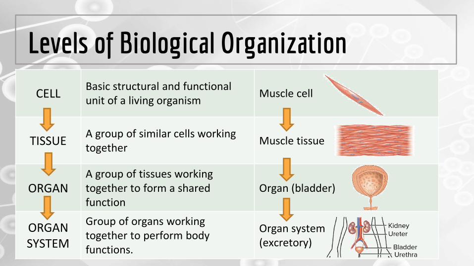
Levels of Biological Organization
Organism
Levels of Biological Organization
CELLBasic structural and functional unit of a living organism
Muscle cell
TISSUEA group of similar cells working together
Muscle tissue
ORGANA group of tissues working together to form a shared function
Organ (bladder)
ORGAN SYSTEM
Group of organs working together to perform body functions.
Organ system(excretory)

Improving Microscope Technology
● The addition of a stain to a sample of cells can help to increase the contrastof cellular structures under a microscope.
Staining
Stained onion cells
Unstained onion cells
Staining procedure

Improving Microscope Technology
Electron Microscopes
● Illuminates specimens with beams of electrons instead of a beam of light
● Used to view objects that are too small to see with a light microscope (provides a higher resolution)
● High-powered electron microscopes recently used to obtain images of atoms!

Improving Microscope Technology
● Fluorescence is another imaging technique that can be used to show molecules on the membrane of cells in more detail.
● Can even be used to “label” cancerous cells in order to distinguish them from healthy cells.
Fluorescence