Innate Immunity-Based Immunotherapy of Cancer
36
6 Innate Immunity-Based Immunotherapy of Cancer Kouji Maruyama et al. * Experimental Animal Facility, Shizuoka Cancer Center Research Institute, Japan 1. Introduction The immune system protects against invading pathogens and transformed cells, including cancer. Mammalian immune system is divided into two major categories, i.e., innate and adaptive immunity. Innate immunity consists of cellular and biochemical defense mechanisms that respond in the early phase after harmful events, such as encounters with microbes or transformed cells. The cellular components of innate immunity include dendritic cells (DCs), macrophages and monocytes, polynuclear cells (e.g. neutrophils and mast cells), natural killer (NK) cells, DŽDž T cells and natural killer T (NKT) cells. Adaptive immunity consists of T and B lymphocytes and their humoral mediators, including cytokines and antibodies, and achieves excellent antigen specificity by somatic rearrangement of the antigen receptor genes of each lymphocyte lineage; T cell receptor for T lymphocytes and immunoglobulin for B lymphocytes, respectively. Furthermore, another excellent characteristic of adaptive immunity is a “memory system” to maintain antigen- specific lymphocytes in a functionally quiescent or slowly cycling state for many years. The memory system enables host organisms to respond to the second and subsequent exposure to the same or related antigens in a more rapid and effective manner. Dr. William Coley, an American bone surgeon, is credited with pioneering work in the field of cancer immunotherapy. In the 1890s, he noted that patients with sarcoma who developed bacterial infections after surgery had visible regression of their cancer. At first, Dr. Coley injected live cultures of streptococcus to produce erysipelas in cancer patients and assessed their responses (Bickels et al., 2002; MacCarthy, 2006). He found that the antitumor effects depended upon the toxins of the bacteria, and eventually developed mixed toxins of a Gram-positive and -negative bacterium called Coley toxins (also called “mixed bacterial vaccines”). A compilation of Dr. Coley's clinical observations indicated that in certain tumor * Hidee Ishii 1 , Sachiko Tai 1 , Jinyan Cheng 2 , Takatomo Satoh 2 , Sachiko Karaki 2 , Shingo Akimoto 3 and Ken Yamaguchi 4 1 Experimental Animal Facility, Shizuoka Cancer Center Research Institute, 2 Advanced Analysis Technology Department, Corporate R&D Center, Olympus Corporation, 3 Department of Pediatric Hematology and Oncology Research, National Medical Center for Children and Mothers Research Institute, 4 Shizuoka Cancer Center Hospital and Research Institute, Japan www.intechopen.com
Transcript of Innate Immunity-Based Immunotherapy of Cancer
6
Innate Immunity-Based Immunotherapy of Cancer
Kouji Maruyama et al.* Experimental Animal Facility,
Shizuoka Cancer Center Research Institute, Japan
1. Introduction
The immune system protects against invading pathogens and transformed cells, including cancer. Mammalian immune system is divided into two major categories, i.e., innate and adaptive immunity. Innate immunity consists of cellular and biochemical defense mechanisms that respond in the early phase after harmful events, such as encounters with microbes or transformed cells. The cellular components of innate immunity include dendritic cells (DCs), macrophages and monocytes, polynuclear cells (e.g. neutrophils and mast cells), natural killer (NK) cells, ┛├ T cells and natural killer T (NKT) cells. Adaptive immunity consists of T and B lymphocytes and their humoral mediators, including cytokines and antibodies, and achieves excellent antigen specificity by somatic rearrangement of the antigen receptor genes of each lymphocyte lineage; T cell receptor for T lymphocytes and immunoglobulin for B lymphocytes, respectively. Furthermore, another excellent characteristic of adaptive immunity is a “memory system” to maintain antigen-specific lymphocytes in a functionally quiescent or slowly cycling state for many years. The memory system enables host organisms to respond to the second and subsequent exposure to the same or related antigens in a more rapid and effective manner.
Dr. William Coley, an American bone surgeon, is credited with pioneering work in the field of cancer immunotherapy. In the 1890s, he noted that patients with sarcoma who developed bacterial infections after surgery had visible regression of their cancer. At first, Dr. Coley injected live cultures of streptococcus to produce erysipelas in cancer patients and assessed their responses (Bickels et al., 2002; MacCarthy, 2006). He found that the antitumor effects depended upon the toxins of the bacteria, and eventually developed mixed toxins of a Gram-positive and -negative bacterium called Coley toxins (also called “mixed bacterial vaccines”). A compilation of Dr. Coley's clinical observations indicated that in certain tumor
*Hidee Ishii1, Sachiko Tai1, Jinyan Cheng2, Takatomo Satoh2, Sachiko Karaki2, Shingo Akimoto3 and Ken Yamaguchi4
1Experimental Animal Facility, Shizuoka Cancer Center Research Institute, 2Advanced Analysis Technology Department, Corporate R&D Center, Olympus Corporation, 3Department of Pediatric Hematology and Oncology Research, National Medical Center for Children and Mothers Research Institute, 4Shizuoka Cancer Center Hospital and Research Institute, Japan
www.intechopen.com

Advancements in Tumor Immunotherapy and Cancer Vaccines
108
types, such as soft tissue sarcoma and lymphoma, the response is marked even by today’s standards (Tsung & Norton, 2006). Following the pioneering works by Dr. Coley and his daughter, Dr. Helen Coley Nauts, efforts to treat cancer based on the function of the immune system, i.e., immunotherapy, have continued. As with Coley’s toxins, the innate immune response against microbes could provoke anti-tumor effects as a secondary response. Although the exact molecular mechanisms of innate immune cells to recognize the components of microorganisms have not been fully understood, the substances such as bacteria-derived materials which evoke tumor immunity have been categorized as ‘biological response modifier (BRM)’.
In Japan, several original BRMs have been developed since 1950’s. Dr Chisato Maruyama
noticed that there were few cancer patients in sanatoriums for tuberculosis or Hansen's
disease, and he started research to apply extracts from Mycobacterium tuberculosis to cancer
treatment. His preparation, named Specific Substance MARUYAMA (SSM, also called the “MARUYAMA vaccine”) うSuzuki et al., 1986a; Suzuki et al., 1986b; Sasaki et al., 1990え
was composed of deproteinized extracts, and contained lipoarabinomannan, a kind of
polysaccharide, as the main component. SSM received much attention from the public as a
miracle drug for cancer treatment before being approved by the Ministry of Health and
Welfare of Japan at that time. SSM has not been approved to date as a anti-neoplastic drug,
but its related preparation, referred to as Z-100 or Ancer, has been approved since 1991 as a
drug for radiotherapy associated leucopenia. Ttwo other BRM drugs, krestin (PS-K,
polysaccharide-protein complexes extracted from basidiomycetes, Trametes versicolor)
(Akiyama et al., 1977; Mizushima et al., 1982) and picibanil (OK-432, a lyophilized
preparation of attenuated group A Streptococcus haemolyticus) (Kai et al., 1979; Kataoka et al.,
1979) were approved by the Ministry of Health and Welfare of Japan as anti-neoplastic
drugs in 1975. Another example of Japanese BRMs is BCG-CWS, a cell wall skeleton
preparation of Mycobacterium bovis bacillus Calmette-Guérin, a tuberculosis vaccine strain
which is almost nonpathogenic yet retains the immunogenic properties of tuberculosis
(Tsuji et al., 2000). BCG-CWS contains a peptidoglycan that is covalently linked to
arabinogalactan and mycolic acids (Azuma et al., 1974). Although BCG-CWS has been
clinically used for a long time (Hayashi et al., 2009; Kodama et al., 2009), it has not been
approved by the Ministry of Health and Welfare of Japan.
One of the characteristics of classical BRMs is that they are crude products which are not fully purified. Their multiple components seem to be important for the induction of efficient anti-tumor effects as seen with Coley toxins.
2. Stimulators of innate immunity
2.1 Toll-like receptors (TLRs)
The cells of the innate immune system recognize infectious agents by receptors for characteristic components of pathogenic microorganisms. The structures of these components are highly conserved and are called pathogen-associated molecular patterns (PAMPs). The receptors for PAMPs, referred to as pattern recognition receptors (PRRs), are germline-encoded and highly conserved across species. The innate immune system detects various classes of pathogens and abnormal cells through PRRs, and serves as a first line of defense against microbes.
www.intechopen.com
Innate Immunity-Based Immunotherapy of Cancer
109
Toll, a gene involving to the establishment of dorsal-ventral pattern of Drosophila embryo
(Hashimoto et al., 1988), other function in immune response against fungus in Drosophila
adults (Lemaitre et al., 1996). In 1997, a cloning of human homologue of Drosophila Toll and
its function as an antigen receptor was reported (Medzhitov et al., 1997). At present, 10
molecules have been identified in humans and are referred to as Toll-like receptors (TLRs)
(Takeda et al., 2003; Iwasaki & Medzhitov, 2004). The identification of TLRs in mammalian
species could give us clues to understand the molecular basis for the early phase events of
host defense against microbial infection. In addition, accumulating evidence has shed light
on the broad range of their roles in various biological processes. The TLR family is one of the
most extensively characterized families of PRRs, and is also regarded as the most rapidly
growing research field in immunology.
TLRs are type I transmembrane proteins containing repeated leucine-rich motifs in their
extracellular domains similar to such motifs in other PRRs, and have a conserved
intracellular motif (i.e. TIR domain, which initiates signal transduction). The typical
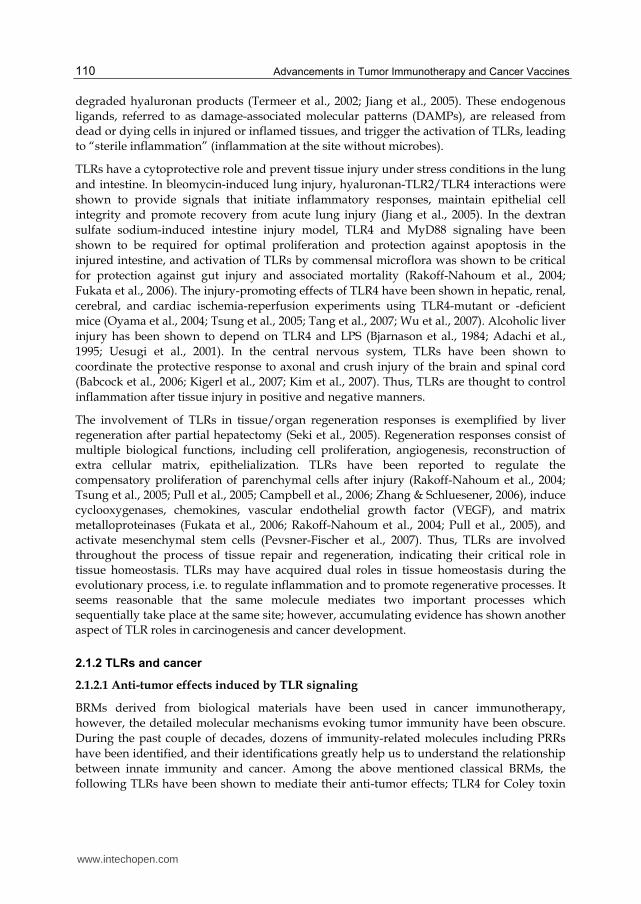
microbial ligands for TLRs are summarized in Table 1. The initial step of signal transduction
from TLRs is mediated through several adaptor molecules, including myeloid
differentiation factor 88 (MyD88), Toll receptor-associated activator of interferon (TRIF),
MyD88-adaptor-like/TIR-associated protein (MAL/TIRAP) and Toll receptor-associated
molecule (TRAM), relayed to the inflammatory pathways involving NF-κB, Janus kinase
(JNK)/p38 kinase and interferon regulatory factor (IRF) 3, 5 and 7. Finally, TLR signal
transduction induces various transcripts, including cytokines such as tumor necrosis factor-
(TNF-) and interferon (IFN)-inducible genes.
TLR1/TLR2/TLR6 Bacterial lipoproteins and lipotechoic acid, fungal zymosan
TLR3 Double stranded RNA
TLR4 Lipopolysaccharide (LPS) from Gram-negative bacteria
TLR5 Bacterial fragelin
TLR7/TLR8 Single-stranded RNA
TLR9 Unmethylated CpG motifs in DNA
TLR10 Unknown
Table 1. Typical ligands for human TLRs known as pathogen-associated molecular patterns (PAMPs)
2.1.1 Roles of TLRs in tissue homeostasis
In addition to their role in the host defense system, TLRs have been shown to be involved in various aspects of tissue homeostasis, including tissue repair and regeneration (van Noort & Bsibsi, 2009; Li et al., 2010). TLRs have also been reported to recognize various host-derived endogenous ligands, including cellular proteins such as heat shock proteins (Asea et al., 2002; Dybdahl et al., 2002; Ohashi et al., 2000; Roelofs et al., 2006; Vabulas et al., 2001; Vabulas et al., 2002a; Vabulas et al., 2002b) and high mobility group box1 (HMGB1) (Park et al., 2004; Park et al., 2006), uric acid crystal うLiu-Bryan et al., 2005a; Liu-Bryan et al.,
2005bえ, surfactant protein A (Guillot et al., 2002), and products of the extracellular matrix,
such as fibronectin (Okamura et al., 2001), heparan sulfate (Johnson et al., 2002), biglycan (Schaefer et al., 2005), fibrinogen (Smiley et al., 2001), oligosaccharides of hyaluronan and
www.intechopen.com
Advancements in Tumor Immunotherapy and Cancer Vaccines
110
degraded hyaluronan products (Termeer et al., 2002; Jiang et al., 2005). These endogenous ligands, referred to as damage-associated molecular patterns (DAMPs), are released from dead or dying cells in injured or inflamed tissues, and trigger the activation of TLRs, leading to “sterile inflammation” (inflammation at the site without microbes).
TLRs have a cytoprotective role and prevent tissue injury under stress conditions in the lung
and intestine. In bleomycin-induced lung injury, hyaluronan-TLR2/TLR4 interactions were
shown to provide signals that initiate inflammatory responses, maintain epithelial cell
integrity and promote recovery from acute lung injury (Jiang et al., 2005). In the dextran
sulfate sodium-induced intestine injury model, TLR4 and MyD88 signaling have been
shown to be required for optimal proliferation and protection against apoptosis in the
injured intestine, and activation of TLRs by commensal microflora was shown to be critical
for protection against gut injury and associated mortality (Rakoff-Nahoum et al., 2004;
Fukata et al., 2006). The injury-promoting effects of TLR4 have been shown in hepatic, renal,
cerebral, and cardiac ischemia-reperfusion experiments using TLR4-mutant or -deficient
mice (Oyama et al., 2004; Tsung et al., 2005; Tang et al., 2007; Wu et al., 2007). Alcoholic liver
injury has been shown to depend on TLR4 and LPS (Bjarnason et al., 1984; Adachi et al.,
1995; Uesugi et al., 2001). In the central nervous system, TLRs have been shown to
coordinate the protective response to axonal and crush injury of the brain and spinal cord
(Babcock et al., 2006; Kigerl et al., 2007; Kim et al., 2007). Thus, TLRs are thought to control
inflammation after tissue injury in positive and negative manners.
The involvement of TLRs in tissue/organ regeneration responses is exemplified by liver regeneration after partial hepatectomy (Seki et al., 2005). Regeneration responses consist of multiple biological functions, including cell proliferation, angiogenesis, reconstruction of extra cellular matrix, epithelialization. TLRs have been reported to regulate the compensatory proliferation of parenchymal cells after injury (Rakoff-Nahoum et al., 2004; Tsung et al., 2005; Pull et al., 2005; Campbell et al., 2006; Zhang & Schluesener, 2006), induce cyclooxygenases, chemokines, vascular endothelial growth factor (VEGF), and matrix metalloproteinases (Fukata et al., 2006; Rakoff-Nahoum et al., 2004; Pull et al., 2005), and activate mesenchymal stem cells (Pevsner-Fischer et al., 2007). Thus, TLRs are involved throughout the process of tissue repair and regeneration, indicating their critical role in tissue homeostasis. TLRs may have acquired dual roles in tissue homeostasis during the evolutionary process, i.e. to regulate inflammation and to promote regenerative processes. It seems reasonable that the same molecule mediates two important processes which sequentially take place at the same site; however, accumulating evidence has shown another aspect of TLR roles in carcinogenesis and cancer development.
2.1.2 TLRs and cancer
2.1.2.1 Anti-tumor effects induced by TLR signaling
BRMs derived from biological materials have been used in cancer immunotherapy,
however, the detailed molecular mechanisms evoking tumor immunity have been obscure.
During the past couple of decades, dozens of immunity-related molecules including PRRs
have been identified, and their identifications greatly help us to understand the relationship
between innate immunity and cancer. Among the above mentioned classical BRMs, the
following TLRs have been shown to mediate their anti-tumor effects; TLR4 for Coley toxin
www.intechopen.com

Innate Immunity-Based Immunotherapy of Cancer
111
(Garay et al., 2007) and OK-432 (Hironaka et al., 2006; Okamoto et al., 2006), TLR2/TLR4 for
BCG-CWS (Uehori et al., 2005), respectively. TLR agonists mediate anti-tumor activity by
multiple mechanisms (Rakoff-Nahoum & Medzhitov, 2008). TLR agonists have been shown
to directly kill both tumor cells and ancillary cells of the tumor microenvironment, such as
vascular endothelium (Salaun et al., 2006; El Andaloussi et al., 2006; Haimovitz-Friedman et
al., 1997; Nogueras et al., 2008). TLR activation may also lead to tumor regression directly or
indirectly (TNF-mediated) by increasing vascular permeability (Garay et al., 2007),
recruitment of leukocytes, activation of the tumor lytic activity of NK cells and cytotoxic T
lymphocytes (CTL), and increasing the sensitivity of tumor cells to elimination mechanisms
by effector molecules, such as TRAIL, TNF, and granzyme B/perforin (Smyth et al., 2006;
Akazawa, 2007).
In cancer patients with a growing tumor, immune tolerance between the host immune
system and tumor may have already been established. Agonists of TLRs induce intrinsic
innate immune responses against microbial infection, leading to adaptive immune
responses also to microbial pathogens. At the same time, the triggered innate immune
response may also act to break the established tolerance between host and tumor, and
induce an adaptive immune response against the tumor, mainly through activated antigen-
presenting cell (APC) functions. Breaking the tolerance to tumor self-antigens is a property
known as adjuvanticity. The mechanisms by which TLRs induce effective antitumor
adaptive immune responses include the uptake, processing and presentation of tumor
antigens, enhancement of survival, and induction of costimulatory molecules on
professional APCs, induction of Th1 and CTL responses, and the inhibition of regulatory T
cell activity (Garay et al., 2007; Smyth et al., 2006; Akazawa et al., 2007).
2.1.2.2 Positive effects of TLRs in cancer development
In the 19th century, Rudolf Virchow, one of the founding fathers of modern pathology, postulated a link between chronic inflammation and cancer (Kluwe et al., 2009). Chronic inflammatory diseases, including inflammatory bowel diseases, hepatitis and Helicobacter pyroli infection, have been shown to be associated with cancer development (Chen et al., 2007). Although the detailed mechanisms of tumor-promoting effects by chronic inflammation are far from understood, TLRs are likely candidates as mediators of the effects of the innate immune system on carcinogenesis (Kluwe et al., 2009).
The tumor-promoting effects of TLRs have been shown in studies using adoptively transferred tumor models. Lipopolysaccharide (LPS) enhanced tumor growth in a lung metastasis model of mouse mammary carcinoma cells (Pidgeon et al., 1999; Harmey et al., 2002) and mouse colon cancer cells (Luo et al., 2004). Direct injection of Listeria monocytogenes, a Gram-positive facultative intracellular bacterium, promoted tumor growth in a subcutaneous tumor model of hepatocarcinoma cells (Huang et al., 2007). In a study using the mouse mammary tumor cell line and its highly antigenic subline D2F2/E2, which stably expresses human ErbB-2, Salmonella typhimurium flagellin, a TLR5 ligand, showed complicated effects in the subcutaneous tumor setting; flagellin inhibited tumor growth only in the D2F2/E2 cells when administered 8-10 days after tumor inoculation, but it accelerated tumor growth in both cell lines when administered at the time of inoculation (Sfondrini et al., 2006) suggesting that the status of interactions between the tumor and its microenvironment and/or host immune system are critical for the effects of flagellin.
www.intechopen.com

Advancements in Tumor Immunotherapy and Cancer Vaccines
112
Furthermore, the combination of falagellin and TLR9 agonist, CpG containing oligodeoxynucleotide (CpG ODN) administered 8-10 days after tumor inoculation, abrogated D2F2/E2 tumor growth.
The involvement of TLR signaling in caricinogenesis has been shown in studies using TLR-deficient or its signaling molecule-deficient mice (Fukata et al., 2007; Rakoff-Nahoum & Medzhitov, 2007; Naugler et al., 2007). TLR4-deficient mice were protected from colon tumorigenesis in an azoxymethan-induced chronic inflammation model, and TLR4 signaling was suggested to be involved in tumorigenesis via cyclooxygenase-2 (Cox-2) expression and prostaglandin (PG) E2 production, leading to the activation of EGFR signaling (Fukata et al., 2007). MyD88 deficiency resulted in decreased colon tumor development in mice with heterozygous mutation of the adenomatous polyposis coli gene (spontaneous model), and also in an azoxymethane-induced colon tumor model (Rakoff-Nahoum & Medzhitov, 2007). Significant reduction of tumor formation was observed in MyD88 or interleukin (IL)-6 deficient male but not female mice in a diethylnitrosamine-induced hepatocellular carcinoma model (Naugler et al., 2007).
A wide range of human cancer cells have been shown to express various TLRs (Sato et al., 2009). For example, nine TLRs are expressed in normal colon epithelial cells, and three TLRs (TLR2-4) are elevated in most colorectal cancer cell lines (Sfondrini et al., 2006). Four TLRs (TLR2-5) are expressed in both normal ovary epithelial cells and ovarian cancer cell lines (Zhou et al., 2009). The significance of the expression of multiple TLRs in a wide range of cancer cells is not fully understood; however, simultaneous activation of multiple TLR types might be able to enhance the biological response, as in the case of synergistic effects by combined TLR ligation reported for cytokine production in DCs (Whitmore et al., 2004; Roelofs et al., 2005; Warger et al., 2006; Gautier et al., 2005; Theiner et al., 2007; Zhu et al., 2008; Krummen et al., 2010) and macrophages (Sato et al., 2000; Ouyang et al., 2007). It is possible that tumor cells express multiple TLRs to recognize various DAMPs in their microenvironment, and thereby enhance the biological process triggered by TLR activation to produce favorable conditions for growth and survival. Furthermore, ligation of TLRs in tumor cells has also been shown to increase the production of immunosuppressive
cytokines, such as interleukin (IL)-10 and transforming growth factor (TGF)- (Sato et al., 2009), suggesting that tumor cells also utilize the TLR functions to escape from the tumor immunosurveillance.
2.1.3 TLRs as target molecules for drug development
In simple terms, TLR signaling pathway can be facilitated by agonistic agents to enhance the immune response, and inhibited by antagonistic agents to attenuate a hyper-immune reaction. The former can be applied to diseases including cancer and infectious diseases, and the latter to autoimmune diseases, excessive inflammation, and bacteria-induced pathological conditions, such as sepsis. TLR ligands and their related compounds which can bind to TLRs directly are the primary candidates as therapeutic agent. Most of the TLR-targeting drugs under development are agonistic agents, and a few antagonists for TLR4 seems to be developed as therapeutics for sepsis, such as Eritoran (Eisai Co. Ltd, Tokyo, Japan) (Rossignol et al., 2004). These drugs include various types of compounds, including single and double-stranded RNA, CpG ODN, small compounds, etc., but agonistic agents could be roughly divided into two categories in terms of their usage; an immunomodulatory
www.intechopen.com

Innate Immunity-Based Immunotherapy of Cancer
113
agent and an adjuvant for vaccination. Agonists of TLRs produce their therapeutic efficacy via stimulation of APCs of innate immunity, such as DCs and macrophages, as an initial step, and then their activation/maturation trigger secondary immune responses, including the activation of effector cells of both innate and adaptive immunity, such as NK and T cells; therefore, TLR activation is an indirect stimulus of adaptive immunity. Antigens in vaccines are incorporated by APCs at first, and then presented to T lymphocytes to evoke specific adaptive immune responses. TLR agonists could be one of the best candidates for a vaccine adjuvant, because they are well characterized, and their extensive scientific backgrounds make it possible to produce novel compounds with favorable properties, such as less toxicity, stronger immunostimulatory effects, and longer half life. In the TLR-targeting drugs under development, more than half of compounds seem to be targeting TLR9 (Krishnan et al., 2009; Romagne, 2007), and the reasons for that could be their chemical nature (ready to synthesize and handle) and the economic benefit of DNA and its derivatives in addition to their features as strong inducers of Th1 immune responses.
2.1.4 TLR-targeting drugs for cancer
Here, the most advanced two examples of TLR-targeting drug development will be
discussed; i.e. agonists for TLR9 and TLR7. A group, so-called antiviral TLRs, including
TLR3 and TLR7-9 are predominantly localized intracellularly to endosomal membranes, and
recognize nucleic acids and related compounds (Table 1).
2.1.4.1 TLR9 agonists: CpG ODN
TLR9 detects the unmethylated CpG dinucleotides prevalent in bacterial and viral DNA but
not in vertebrate genomes (Krieg, 2007). TLR9 has been shown to have the narrowest
expression profile among all the TLRs; among resting immune cells, B cells and
plasmacytoid DCs (pDCs) seem to exclusively express TLR9 (Iwasaki & Medzhitov, 2004).
pDCs are extremely important cells in host defense as they produce most of the type I IFN
that is made in response to viral infection and that is essential to control viral replication and
to promote the development of an immune response to eradicate infected cells and prevent
recurrence (Liu, 2005). One of the most notable features of TLR9 activation is that strong Th1
responses are triggered. In synthetic CpG ODN used in vivo applications, including animal
experiments and clinical trials, native phosphodiester backbones are replaced partially or
fully with phosphorothioate backbones to improve stability.
In animal experiments, significant anti-tumor effects of TLR9 agonists have been
demonstrated in a variety of settings; not only in monotherapy (Lonsdorf et al., 2003; Baines
& Celis, 2003), but also in combination therapy with monoclonal antibody therapy
(Buhtoiarov et al., 2006; Daftarian et al., 2004; Davila et al., 2003; Dercamp et al., 2005;
Guiducci et al., 2005; Vicari et al., 2002; Wooldridge et al., 1997), cytokines, chemokines and
related factors (Chaudhry et al., 2006; Ishii et al., 2003; Merad et al., 2002; Okano et al., 2005),
TLR3 and TLR5 ligands (Sfondrini et al., 2006; Whitmore et al., 2004), EGFR-related
signaling and angiogenesis inhibition (Damiano et al., 2006), radiotherapy (Mason et al.,
2005), surgery (Ohashi et al., 2006; Weigel et al., 2003), cryotherapy (den Brok et al., 2006),
and chemotherapy (Balsari et al., 2004; Pratesi et al., 2005; Taieb et al., 2006; van der Most et
al., 2006; H. Wang et al. 2006; X.S. Wang et al., 2005; Weigel et al., 2003). TLR9 has been
thought to be the only TLR for which a systemically administered specific agonist has
www.intechopen.com

Advancements in Tumor Immunotherapy and Cancer Vaccines
114
shown substantial evidence of anti-tumor activity in human clinical trials (reviewed in
Krieg, 2007). CPG 7909 (also called PF-3512676), a CpG ODN with a fully modified
phosphorothioate backbone, has been thought to be one of the most promising drug
candidates for cancer therapy.
Unfortunately, it has been announced that the sponsor pharmaceutical company made the
decision to terminate phase III clinical trials of CPG 7907. In these phase III clinical trials, the
efficacy of CPG 7909 had been tested in combination with gemcitabine/cisplatin or
paclitaxel/carboplatin in patients with advanced non-small-cell lung cancer.
2.1.4.2 TLR7 agonists: Imiquimod
TLR7 recognizes single-stranded RNA, and acts as a potent activator of innate immune
responses to viral infections (Diebold et al., 2004; Heil et al., 2004). Other than the natural
ligands, several distinct classes of low-molecular-weight compounds have been shown to
effectively and selectively activate this receptor (Hemmi et al., 2002; Lee et al., 2003).
Imiquimod (also called Aldara, R-837, and S-28463), a member of the imidazoquinoline
family, has antiviral activity in guinea pigs infected with herpes simplex virus (Harrison et
al., 1988), and against arbovirus and cytomegalovirus (Akira & Hemmi, 2003). The first
report on the anti-tumor activity of imiquimod was published in 1992, using subcutaneous
tumor models of mouse colon carcinoma and mouse sarcoma and a lung metastasis model
of mouse Lewis lung carcinoma (Sidky et al., 1992); however, the mechanism of imiquimod
action as TLR agonist was totally unknown at that time. Subsequently, anti-tumor effects of
imiquimod have been shown to be mediated by the induction of cytokines, including IFN- and IL-12 (Hemmi et al., 2002), the enhancement of the activation of tumor antigen-specific
CTLs in vaccination studies (Prins et al., 2006; Rechtsteiner et al., 2005), and the activation of
a myeloid DC subset with cytotoxic activity (Stary et al., 2007). Because of the intracellular
localization of TLR7, small molecules such as imiquimod might have an advantage in terms
of cellular uptake (Schön & Schön, 2008).
Imiquimod is a success story of TLR-targeting drugs. A topical cream formulation of imiquimod has been approved to treat actinic keratosis and external genital warts, and its
additional indication for the treatment of superficial basal cell carcinoma was approved by the United States Food and Drug Administration in 2004. At present, 20 clinical oncology
trials with imiquimod are ongoing (Maruyama et al., 2011). In these clinical trials, new target
cancers are included such as basal cell carcinoma of different types/stages, melanoma, cancers of the breast, ovary, uterine cervix, and lung, glioblastoma, neuroblastoma,
rhabdomyosarcoma, and osteogenic sarcoma. In addition, imiquimod is used as a vaccine adjuvant in 10 out of 20 trials. Vaccines in these clinical trials include DC-based vaccines,
peptides or proteins of tumor associated antigens, human papilloma virus related DNA vaccines (Maruyama et al., 2011). Thus, imiquimod has already been established as a
therapeutic product, and is under development as a vaccine adjuvant.
2.1.5 Perspectives: TLRs as double-edged swords in cancer therapy
TLRs have dual important roles in mammals; host defense from infection and maintaining
tissue homeostasis by regulating inflammatory and tissue repair responses to damage.
Antitumor effects of TLR agonists, including classical BRMs, depend on the former role of
www.intechopen.com

Innate Immunity-Based Immunotherapy of Cancer
115
TLRs in the immune systems. TLRs initiate innate immune responses, bridge the response
from innate to adaptive immunity, and lead to the elimination of transformed cancer cells;
however, cancer cells themselves utilize the latter role of TLRs in tissue homeostasis for their
growth and survival by means of a variety of strategies; suppression of host immune
responses by the production of various immunosuppressive factors, promotion of cell
growth and angiogenesis, induction of cytoprotective and anti-apoptotic factors. These
findings suggest that TLR signaling acts as a double-edged sword in cancer therapy.
Administration routes and doses may be critical issues to develop TLR-targeting drugs for
cancer treatment. Systemic administration of TLR agonists may include the risks of
promoting growth and survival of tumor. On the other hand, local application, especially
transcutaneous or intradermal administration could be a promising modality to lower the
risks. In addition, the dose of the TLR agonist may be an important issue. It has been
suggested that exceptionally high doses of TLR agonists appear to have an antitumor effect,
whereas low doses of TLR agonists promote tumor growth (Kluwe et al., 2009). It is critical
to know how we can dominantly use the unilateral antitumor edge of the TLR sword.
Furthermore, another point to be tested in clinical studies may be the combinatorial usage of
agonists for different types of TLRs, such as TLR4 agonist plus TLR9 agonist, as tested in
animal experiments. As mentioned above, the mixed nature of classical BRMs seems to
contribute to efficient antitumor effects. The evidence on the role of TLRs in tissue
homeostasis has been obtained mainly from the studies focused on TLR2 and TLR4. For a
better understanding of the role of TLRs in tissue homeostasis, carcinogenesis, and tumor
progression, more research data on other types of TLR are necessary.
2.2 Fms-like tyrosine kinase 3 ligand (FL)
2.2.1 Cloning of FL as a ligand for fms-like tyrosine kinase-3 (Flt3-R)
FL is a transmembrane or soluble protein and is expressed by a variety of cells including
hematopoietic and bone marrow stromal cells. In synergy with other cytokines and growth
factors, FL stimulates proliferation and development of a wide range of hematopoietic cells
including stem cells, myeloid and lymphoid progenitor cells, DCs and NK cells (Drexler &
Quentmeier, 2004; Lyman & Jacobsen, 1998). FL was cloned as a ligand for Flt3-R, a tyrosine
kinase receptor belongs to the class III receptor tyrosine kinase family structurally related to
macrophage colony-stimulating factor receptor (c-fms) and to mast stem cell factor (also
known as steel factor, stem cell factor, and c-kit ligand) receptor (c-kit) (Matthews et al.,
1991; Rosnet et al., 1991a; Rosnet et al., 1991b). Lyman et al. isolated a murine Flt3L cDNA
by screening an expression library with a fusion protein consisting of the extra cellular
domain of the Flt3-R (Lyman et al., 1993). Hannum et al. purified FL from conditioned
medium of a murine thymic stromal cell line using an affinity column made with Flt3-R
extracellular domain (Hannum et al., 1994). Subsequently, a human counterpart of a murine
FL was cloned using a mouse cDNA as a probe (Hannum et al., 1994; Lyman et al., 1994b).
The mouse and human FL proteins are 72% identical at the amino acid level with a greater
homology in the extracellular region than in the cytoplasmic domain.
Multiple isoforms of FL have been reported for mouse and human; the first one is a
transmebrane protein which is a predominant isoform in human (Hannum et al., 1994;
Lyman et al., 1993; Lyman et al., 1994b), and is converted to biologically active soluble form
www.intechopen.com

Advancements in Tumor Immunotherapy and Cancer Vaccines
116
by proteolytic cleavage (Lyman et al., 1993). The second biologically active isoform, the most
abundantly found in mouse (Lyman et al., 1995b), is a membrane bound form which lacks a
normal transmembrane structure but has a hydrophobic amino acid stretch in N-terminus
(Hannum et al., 1994; Lyman et al., 1995a). The third isoform identified in mouse and
human is a soluble form generated by alternative splicing (Lyman et al., 1995a, Lyman et al.,
1995b).
2.2.2 Biological activity of FL
It is known that FL is interactive between different species, mouse and human FL can be
fully active on cells expressing either the mouse or human Flt3-R (Lyman et al., 1994a).
Although physiological importance for the ubiquitous expression is not clear, FL is widely
expressed in murine and human tissues, the transcripts have been detected almost every
tissue tested (Hannum et al., 1994; Lyman et al., 1994b; Lyman et al., 1995a). In contrast to
FL, its receptor Flt3-R seems to have a relatively limited expression profile. Flt3-R expression
in hematopoietic system appear predominantly restricted to the progenitor/stem cell
compartment (Drexler et al., 2004).
FL stimulates proliferation and development of a wide range of hematopoietic cells
including hematopoietic stem cells, myeloid and lymphoid progenitor cells (reviewed in
Drexler et al., 2004). In this section, effects of in vivo administered FL on DCs and NK cells
will be discussed.
2.2.2.1 Effects of FL on DC development
DCs express CD45 and arise from BM progenitor cells; evidence suggests that DCs are
derived from myeloid and lymphoid progenitor cells (Caux et al., 1995; Peters et al., 1996).
Early studies of mouse in vivo experiments identified that FL as a cytokine that could affect
DC proliferation (Maraskovsky et al., 1996; Pulendran et al., 1999). In vivo administration of
FL in mouse results in a dramatic increase in the number of myeloid- and lymphoid-derived
functional DC in BM, spleen, thymus, peripheral blood, and other tissues indicating an
absolute increase in functionally mature DC (Maraskovsky et al., 1996). In contrast,
polyethylene glycol-modified granulocyte-macrophage colony-stimulating factor (GM-CSF)
into mice only expands the myeloid-related DC subset (Pulendran et al., 1999).
In human, two distinct classes of blood DC subsets, CD11c+ immature DCs and CD11c-
pDCs with different morphologies, phenotypes, and functional properties, have been
identified (Liu, 2001). Blood CD11c+ DCs are considered to be myeloid derived, whereas
pDCs, also known as type 1 interferon-producing cells as mentioned in section 5.1, are
considered to be lymphoid related (Liu, 2001). Administration of FL to healthy volunteers
significantly expands the number of circulating CD11c+ and CD11c- DC subsets and DC
precursors (Maraskovsky, 2000).
2.2.2.2 Effects of FL on NK cells
FL administered to mice increased number of NK cells (Brasel et al., 1996; Shaw et al., 1998).
Brasel et al. reported that daily subcutaneous injection of recombinant FL (10 g) for 10 days
resulted in 2.5-fold increase in NK cell absolute number in spleen (Brasel et al., 1996). Shaw
et al. also showed daily intraperitoneal administration of recombinant FL resulted in
www.intechopen.com

Innate Immunity-Based Immunotherapy of Cancer
117
increase in the absolute number of CD3¯ NK1.1+ NK cell especially in spleen and liver (Shaw
et al., 1998). Similar effect of FL on NK expansion was reported by He et al. using
intravenous injection of a plasmid expression vector for FL cDNA encoding extra cellular
domain (He et al., 2000). These in vivo effects of FL on NK expansion was supported by the
evidence obtained from mice lacking FL in which a marked deficiency of NK cells in the
spleen was noted (McKenna et al., 2000). The ability of FL to expand NK cells in vivo has
been confirmed by subsequent studies (Guimond et al., 2010; Péron et al., 1998; Smith et
al., 2001).
In contrast to the mouse findings in vivo, few are available on the effect of FL on human NK
cells in vivo. In a phase I clinical trial, FL was subcutaneously injected into cancer patients
including Hodgkin disease, non-Hodgikin lymphoma, or advanced-stage breast cancer after
autologous hematopoietic cell transplantation (Chen et al., 2005). Injection of FL was safe
and well tolerated and significantly increased blood DC subsets without affecting other
mature cell lineages including NK cells (Chen et al., 2005). According to the findings
obtained, the activity of FL alone to induce cellular expansion is higher in DCs than that in
NK cells. It appears necessary to combine FL with other cytokines to induce effective NK
cell expansion in vivo especially in human.
2.2.3 Anti-tumor effects evoked by FL
The finding that FL administration resulted in dramatic expansion of DCs led to the studies
to test anti-tumor effect of FL. Early study demonstrated that FL alone induced the
regression of methylcholanthrene (MCA)-induced fibrosarcoma (Lynch et al., 1997).
Subsequently, anti-tumor effect of systemically administered FL has been demonstrated in a
lot of syngeneic murine tumor models of melanoma, lymphoma (Esche et al., 1998), Lewis
lung carcinoma (Chakravarty et al., 1999), liver metastasis (Péron et al., 1998), mesothelioma
(Fernandez et al., 1999), breast cancer (Chen et al., 1997), prostate cancer (Ciavarra et al.,
2000). The anti-tumor effect of FL has been shown also in rat syngeneic colon cancer model
(Favre-Felix et al., 2000) and xenograft model of human ovarian cancer (Silver et al., 2000). In
these tumor models, NK cells seem to have a critical role in the anti-tumor effect induced by
FL administration, because depletion of NK cells resulted in abrogation of anti-tumor effect
(Fernandez et al., 1999; Péron et al., 1998; Silver et al., 2000). However, the anti-tumor effects
induced by FL have also been shown to be T-cell mediated in several mouse models (Chen
et al., 1997; Lynch et al., 1997). Fernandez et al. reported that in mice with MHC class I-
negative tumors, adoptively transferred- or FL-expanded DCs promoted NK cell-dependent
anti-tumor effects (Fernandez et al., 1999). Kelly et al. demonstrated that primary rejection of
MHC-class I negative-CD70 expressing RMA-S tumor cells by NK cells efficiently evoked
the subsequent development of tumor-specific cytotoxic and T helper type 1 responses to
the parental MHC class I-sufficient RMA tumor cells (Kelly et al., 2002). Activated NK cells
can facilitate adaptive immune responses via induction of DC maturation by secretion of
cytokines and a cell-cell contact (Walzer et al., 2005). These findings suggest that interaction
between NK cells and DCs induces not only anti-tumor effects by NK cells, but also
adaptive anti-tumor immunity.
At present, limited information are available on clinical studies using FL administration.
Marroquin et al. reported that FL administration resulted in a 19-fold increase in DC
www.intechopen.com

Advancements in Tumor Immunotherapy and Cancer Vaccines
118
number in the peripheral blood of patients with melanoma and renal cancer, however, DC
generated in vivo appeared only partially activated. This partial activation might account
for the lack of enhanced immune responses to melanoma antigens and absence of clinical
response in the patients even in combination with antigen immunization (Marroquin et al.,
2002). Rini et al. reported that administration of FL either alone or in combination with
interleukin-2 (IL-2), although capable of inducing expansion of circulating myeloid and
plasmacytoid DCs in patients with metastatic renal cell carcinoma, lacks significant clinical
activity (Rini et al., 2002). The above mentioned report by Chen et al. showed that FL
administration significantly increased the frequency and absolute number of myeloid DCs,
plasmacytoid DCs, and monocytes in the patients with Hodgkin disease, non-Hodgikin
lymphoma, or advanced-stage breast cancer after autologous hematopoietic cell
transplantation (Chen et al., 2005). In the clinical studies mentioned here, expected clinical
responses were not obtained.
The clinical application of DCs for cancer vaccines, however, has been moderately successful
(Rosenberg et al., 2004). In the clinical procedure to prepare DC vaccines, monocytes
harvested from cancer patients by leukocyte apheresis are cultured in the presence of
cytokines to generate DCs. The generated DCs are matured in vitro, treated with tumor
antigens such as peptides and tumor lysates, and then injected into patients. The course to
prepare DC vaccines needs a lot of times, efforts, and costs. If the in vivo expansion of DCs
by FL could be induced effectively and appropriately, it is possible that there is no need to
generate DC vaccines ex vivo. Furthermore, ability to expand NK cells is another important
point in the clinical application of FL. In the clinical trial mentioned above (Chen et al.,
2005), administration of FL to cancer patients did not result in apparent expansion of NK
cells. Combination of FL with other cytokines like IL-15 may be the promising strategy to
expand human NK cells in vivo (Yu et al., 1998).
2.3 Alpha-galactosylceramide (-GalCer)
2.3.1 Isolation, identification, and synthesis of -GalCer
Glycosphingolipid compounds, named agelasphins, were isolated from an extract of the
Okinawan marine sponge, Agelas mauritianus, by Kirin Pharmaceuticals, and some of them
were found to possess anti-tumor activity in mice (Natori et al., 1994; Tsuji, 2006). Because of
the low contents of agelasphins in the marine sponge, and the difficulty of their scale-up
synthesis, the structure of agelasphins was modified, and a novel compound, α-GalCer (also
called KRN7000) was synthesized (Kobayashi et al., 1995). This glycolipid was shown to
bind non-classical MHC molecule CD1d of human and mice (Gansert et al., 2003; Taniguchi
& Nakayama, 2000), and activate natural killer T (NKT) cells of both species in vitro and in
vivo when it is presented on CD1d molecule (Brossay et al., 1998; Burdin et al., 1998;
Kawano et al., 1998; Spada et al., 1998) . To date,-GalCer has been the most extensively
studied ligand for CD1d molecules and stimulant for NKT cells.
2.3.2 NKT cells
NKT cells are a unique subpopulation of T cells which play important role in regulating immune responses by bridging innate and adaptive immune systems. The term ‘NKT cells’
www.intechopen.com

Innate Immunity-Based Immunotherapy of Cancer
119
was first used in 1995 (Makino et al., 1995) to define a subset of mouse T cells that share some characteristics with NK cells, particularly expression of the NK1.1 marker (Nkrp1c or CD161c). At present, the term NKT cells is well accepted and broadly applied to mice, humans, and other species (Godfrey et al., 2004). NKT cells express an antigen-specific T cell receptor (TCR), but unlike with conventional T cells that detect peptide antigens presented by MHC molecules, NKT cells recognize lipid antigens presented by CD1d molecule. NKT cells have been shown to involve wide spectrum of disorders including infectious diseases caused by bacteria, parasites, and virus, autoimmunity, and cancer (Terabe & Berzofsky, 2008).
Accumulating several lines of evidences which obtained from the late 80’s to 90’s had been integrated into the establishment of novel immune cell lineage ‘NKT cells’ (Bendelac et al., 1997; Bendelac et al., 2007; Godfrey et al., 2004; Godfrey et al., 2010; Macdonald., 2007; Taniguchi et al., 2003; Terabe. & Berzofsky, 2008). However, an ambiguous definition of NKT cells has caused confusion in our understanding of their biological roles. At present, it has been proposed to classify NKT cells into two types (Godfrey et al., 2010).
Type I NKT cells, also called invariant NKT cells or iNKT cells, are defined by their expression of the canonical invariant TCRα chain of Vα14Jα18 in mouse (Vα24Jα18 in
human) with a limited number of TCR chains of V8, V7, and V2 in mouse (V11 in human). Type I NKT cells are detected with α-GalCer-loaded CD1d tetramers, however, other CD1d restricted T cells exist (Godfrey et al., 2004; Godfrey et al., 2010; Terabe & Berzofsky, 2007). On stimulation with α-GalCer, type I NKT cells produce a large amount of
TH1 (IFN-) and Th2 (IL-4 and IL-13) cytokines. Although type I NKT cells have NK-like cytolytic activity, they are considered to be regulators of immune responses because they rapidly produce large amount of both Th1 and Th2 cytokines in autoimmune diseases, infectious diseases, and cancer. Another subset of CD1d-restricted NKT cells which does not respond to α-GalCer, seems to recognize a range of hydrophobic antigens including sulfatide (Jahng et al., 2004), lysophosphatidylcholine (Chang et al., 2008), and small aromatic (non-lipid) molecules (Van Rhijn et al., 2004). This NKT subset, called ‘type II NKT cells’, is present in humans and mice, and has been shown to be more heterogeneous than
type I in both TCR and TCR chain usage (Godfrey et al., 2004; Godfrey et al., 2010; Terabe & Berzofsky, 2007; Terabe & Berzofsky, 2008). In this chapter, the findings on type I NKT cells and their ligands will be discussed hereafter.
2.3.3 Anti-tumor effects of-GalCer
Originally, a mother compound of α-GalCer, agelasphins, was selected from several glycosphingolipids extracted from marine sponge depending on their anti-tumor effects in mouse tumor models (Natori et al., 1994). Anti-tumor effect caused by α-GalCer injection has been shown in mouse tumor models (Kobayashi et al., 1995; Motoki et al., 1995; Morita et al., 1995). Subsequently, protective roles of NKT cells stimulated by α-GalCer were confirmed by liver and lung metastasis models in mice using B16 melanoma cells and Lewis lung carcinoma cells, respectively (Kawano et al., 1998). A complete inhibition of B16 melanoma metastasis in the liver was observed when α-GalCer-pulsed DCs were injected 7 days after transfer of tumor cells to mice where metastatic nodules were already formed (Toura et al., 1999). In addition, long-term administration of α-GalCer inhibited primary tumor formation in the tumor models of MCA-induced sarcoma, mammary carcinomas in Her-2/neu transgenic mice, and spontaneous sarcomas in p53 deficient mice (Hayakawa et al., 2003).
www.intechopen.com

Advancements in Tumor Immunotherapy and Cancer Vaccines
120
Anti-tumor effects of α-GalCer depend on the production of IFN from activated type I
NKT cells (Terabe & Berzofsky, 2007). Crowe et al. showed that IFN- production by NKT cells was essential for protection against tumor, and perforin production by effector cells, but not NKT cells, was also critical in MCA-induced sarcoma models (Crowe et al., 2002). Smyth et al. reported that both NK cells and NKT cells were essential and collaborate in host protection from MCA-induced sarcoma (Smyth et al., 2001), and sequential production of
IFN- by NKT cells and NK cells was essential for the antimetastatic effect of α-GalCer in B16 melanoma metastasis models (Smyth et al., 2002).
In addition to the essential role of IFN-, IL-12 was shown to be critical for the anti-tumor
effect of NKT cells. Kitamura et al. demonstrated that production of IFN- by NKT cells in response to α-GalCer required IL-12 produced by DCs and direct contact between NKT cells and DCs through CD40-CD40 ligand interactions (Kitamura et al., 1999). Hayakawa et al. showed that both CD28-CD80/CD86 and CD40-CD40 ligand costimulatory pathways are
essential for α-GalCer-induced IFN- production by NKT cells (Hayakawa et al., 2001). Furthermore, administration of α-GalCer induced not only innate immune response, but also adaptive immune response. A single dose of α-GalCer rapidly stimulated the full maturation of DCs in NKTcell-dependent manner, and this maturation accounted for the induction of combined Th1 CD4+ and CD8+ T cell immunity to a coadministered antigen (Fujii et al., 2003).
Taken together, anti-tumor effects of α-GalCer are thought to be evoked by the following sequential processes; α-GalCer presented on CD1d of DCs is recognized by NKT cells, and up-regulates CD40 ligand on NKT cells. Activated DCs by CD40 and CD28 costimulatory
pathways produce IL-12, and activate NKT cells by IL-12. IFN-produced by fully activated NKT cells trigger DC maturation, activation and recruitment of NK cells, Th1 CD4+ and CD8+ T cell leading to direct anti-tumor effects.
2.3.4 Clinical trials using -GalCer in cancer patients
At present, a number of clinical trials of α-GalCer have been conducted in multiple institutes, and their results have been published since 2002. Early study of phase I clinical trial in which α-GalCer was injected intravenously reported that α-GalCer was well tolerated in cancer patients over a wide range of doses (Giaccone et al., 2002). Phase I studies of α-GalCer-pulsed DCs have been conducted in patients with various solid tumors and myeloma (Chang et al., 2005; Dhodapkar et al., 2004; Ishikawa et al., 2005; Nieda et al., 2004; Okai et al., 2002; Uchida et al., 2008). However, none of these clinical trials reported significant efficacy against tumors.
Recently, the research team of Chiba University in Japan reported the summary of their clinical trials (Motohashi et al., 2011). The research team has been well known by their pioneering works on α-GalCer, and has been conducted multiple clinical trials with enthusiasm (Ishikawa et al., 2005; Kunii et al., 2009; Kurosaki et al., 2011; Motohashi et al., 2006; Motohashi et al., 2009; Uchida et al., 2008; Yamasaki et al., 2011). The team conducted the α-GalCer-NKT cell-based clinical trials in patients with non-small cell lung cancer and head and neck squamous cell carcinoma (Motohashi et al., 2011). Alpha-GalCer-pulsed APCs seemed to produce better clinical outcomes in the patients with head and neck squamous cell carcinoma than in those with non-small cell lung cancer. The cells were
www.intechopen.com

Innate Immunity-Based Immunotherapy of Cancer
121
injected into nasal submucosa of patients with head and neck squamous cell carcinoma, whereas intravenous injection was performed in the non-small cell lung cancer patients. Furthermore, the anti-tumor effect of α-GalCer-pulsed APCs seemed to be augmented by the combined adoptive transfer of α-GalCer-activated NKT cells. Thus, clinical efficacy of α-GalCer-based immunotherapy may depend on the tumor type and treatment settings. Further efforts are necessary to develop an effective treatment modality.
3. Conclusion
In cancer patients with a growing tumor, immune tolerance between the host immune system and tumor may have already been established. Agents mentioned in this chapter induce inherent innate immune responses intrinsically against microbial infection. The anti-tumor effect via activation of innate immune system may depend on the action to break the established tolerance between host and tumor leading to adaptive immune responses (Figure.1). In other words, the activation of innate immune system acts like as a ‘reset
Fig. 1. In the cancer patients with a growing tumor, immune tolerance between host immune system and tumor may have already been established. Stimulants of innate immunity such as biological response modifiers (BRMs) or ligands for toll-like receptors induce inherent innate immune responses intrinsically against microbial infection. The anti-tumor effect via activation of innate immune system may depend on the action to break the established tolerance between host and tumor leading to adaptive immune responses.
www.intechopen.com
Advancements in Tumor Immunotherapy and Cancer Vaccines
122
button’ in cancer patients to restart the immune responses. Administration of cytokines such as FL which induce expansion of immune cells can potentiate subsequent immune responses, and it may be the ideal pretreatment for cancer patients with immunosuppressive status. With regards to TLRs agonist, FL, and α-GalCer, the efforts to develop them as vaccine adjuvant have already been started. At present, the efficacy of a prophylactic vaccination against human papillomavirus has been established in the prevention of cervical cancer. Although tough and long-lasting works and huge research fund are required to perform clinical trials, one of the most promising immunotherapeutic modalities for cancer may be cancer prevention. Above mentioned three agents may be excellent vaccine adjuvants to enhance tumor immunity in cancer prevention. Further research efforts are required to establish more effective immunotherapy which will also achieve a better quality of life for cancer patients.
4. Acknowledgment
The authors wish to thank Dr. Masatoshi Kusuhara, Dr. Ken-ichi Urakami, Dr. Vincent Zangiacomi, and Ms. Yoko Masuda of Regional Resources Division of Shizuoka Cancer Center Research Institute, and Dr. Christophe Borg and Dr. Zohair Selmani of INSERM UMR 645, Besançon, France, for their kind collaborations.
5. References
Adachi, Y., Moore, L.E., Bradford, B.U., Gao, W. & Thurman, R.G. (1995). Antibiotics
prevent liver injury in rats following long-term exposure to ethanol.
Gastroenterology, Vol.108, No.1, (January), pp. 218–224.
Akiyama, J., Kawamura, T., Gotohda, E., Yamada, Y., Hosokawa, M., Kodama, T. &
Kobayashi, H. (1977). Immunochemotherapy of transplanted KMT-17 tumor in
WKA rats by combination of cyclophosphamide and immunostimulatory protein-
bound polysaccharide isolated from basidiomycetes. Cancer Research., Vol.37, No.9,
(September), pp. 3042-3045.
Akira, S. & Hemmi, H. (2003). Recognition of pathogen-associated molecular patterns by
TLR family. Immunology Letters, Vol.85, No.2, (January), pp. 85-95.
Akazawa, T., Ebihara, T., Okuno, M., Okuda, Y., Shingai, M., Tsujimura, K., Takahashi, T.,
Ikawa, M., Okabe, M., Inoue, N., Okamoto-Tanaka, M., Ishizaki, H., Miyoshi, J.,
Matsumoto, M. & Seya, T. (2007). Antitumor NK activation induced by the Toll-like
receptor 3-TICAM-1 (TRIF) pathway in myeloid dendritic cells. Proceedings of the
National Academy of Sciences of the United States of America, Vol.104, No.1, (January),
pp. 252-257.
Asea, A., Rehli, M., Kabingu, E., Boch, J.A., Bare, O., Auron, P.E., Stevenson, M.A. &
Calderwood, S.K. (2002). Novel signal transduction pathway utilized by
extracellular HSP70: role of toll-like receptor (TLR) 2 and TLR4. The Journal of
Biological Chemistry, Vol.277, No.17, (April), 15028–15034.
Azuma, I., Ribi, E.E., Meyer, T.J. & Zbar, B. (1974). Biologically active components from
mycobacterial cell walls. I. Isolation and composition of cell wall skeleton and
component P3. Journal of the National Cancer Institute, Vol.52, No.1, (January), pp.
95–101.
www.intechopen.com

Innate Immunity-Based Immunotherapy of Cancer
123
Babcock, A.A., Wirenfeldt, M., Holm, T., Nielsen, H.H., Dissing-Olesen, L., Toft-Hansen, H.,
Millward, J.M., Landmann, R., Rivest, S., Finsen, B. & Owens, T. (2006). Toll-like
receptor 2 signaling in response to brain injury: an innate bridge to
neuroinflammation. The Journal of Neuroscience, Vol.26, No.49, (December), pp.
12826-12837.
Baines, J. & Celis, E. (2003). Immune-mediated tumor regression induced by CpG-containing
oligodeoxynucleotides. Clinical Cancer Research, Vol.9, No.7, (July), pp. 2693–2700.
Balsari, A., Tortoreto, M., Besusso, D., Petrangolini, G., Sfondrini, L., Maggi, R., Ménard, S.
& Pratesi, G. (2004). Combination of a CpG-oligodeoxynucleotide and a
topoisomerase I inhibitor in the therapy of human tumour xenografts. European
Journal of Cancer, Vol.40, No.8, (May), pp. 1275–1281.
Bendelac, A., Rivera, M.N., Park, S.H. & Roark, J.H. (1997). Mouse CD1-specific NK1 T cells:
development, specificity, and function. Annual Review of Immunology, Vol.15,
(April), pp. 535-562.
Bendelac, A., Savage, P.B. & Teyton, L. (2007). The biology of NKT cells. Annual Review of
Immunology, Vol.25, (April), pp. 297-336.
Bickels, J., Kollender, Y., Merinsky, O. & Meller, I. (2002). Coley's toxin: historical
perspective. The Israel Medical Association Journal, Vol.4, (June), pp. 471-472.
Bjarnason, I., Peters, T.J. & Wise, R.J. (1984). The leaky gut of alcoholism: possible route of
entry for toxic compounds. Lancet, Vol.1, No.8370, (January), pp. 179–182.
Brasel, K., McKenna, H.J., Morrissey, P.J., Charrier, K., Morris, A.E., Lee, C.C., Williams, D.E.
& Lyman, S.D. (1996). Hematologic effects of flt3 ligand in vivo in mice. Blood,
Vol.88, No.6, (September), pp. 2004-2012.
Brossay, L., Chioda, M., Burdin, N., Koezuka, Y., Casorati, G., Dellabona, P. & Kronenberg,
M. (1998). CD1d-mediated recognition of an alpha-galactosylceramide by natural
killer T cells is highly conserved through mammalian evolution. The Jounal of
Experimental Medicine , Vol.188, No.8, (October), pp. 1521-1528.
Buhtoiarov, I.N., Lum, H.D., Berke, G., Sondel, P.M. & Rakhmilevich, A.L. (2006).
Synergistic activation of macrophages via CD40 and TLR9 results in T cell
independent antitumor effects. The Journal of Immunology, Vol.176, No.1, (January),
pp. 309–318.
Burdin, N., Brossay, L., Koezuka, Y., Smiley, S.T., Grusby, M.J., Gui, M., Taniguchi, M.,
Hayakawa, K. & Kronenberg, M. (1998). Selective ability of mouse CD1 to present
glycolipids: alpha-galactosylceramide specifically stimulates V alpha 14+ NK T
lymphocytes. The Journal of Immunology, Vol.161, No.7, (October), pp. 3271–3281.
Campbell, J.S., Riehle, K.J., Brooling, J.T., Bauer, R.L., Mitchell, C. & Fausto, N. (2006).
Proinflammatory cytokine production in liver regeneration is Myd88-dependent,
but independent of Cd14, Tlr2, and Tlr4. The Journal of Immunology, Vol.176, No.4,
(February), pp. 2522-2528.
Caux, C., Liu, Y.J. & Banchereau, J. (1995). Recent advances in the study of dendritic cells
and follicular dendritic cells. Immunology Today, Vol.16, No.1, (January), pp. 2-4.
Chakravarty, P.K., Alfieri, A., Thomas, E.K., Beri, V., Tanaka, K.E., Vikram, B. & Guha, C.
(1999). Flt3-ligand administration after radiation therapy prolongs survival in a
www.intechopen.com
Advancements in Tumor Immunotherapy and Cancer Vaccines
124
murine model of metastatic lung cancer. Cancer Research, Vol.59, No.24,
(December), pp. 6028-6032.
Chang, D.H., Deng, H., Matthews, P., Krasovsky, J., Ragupathi, G., Spisek, R., Mazumder,
A., Vesole, D.H., Jagannath, S. & Dhodapkar, M.V. (2008). Inflammation-associated
lysophospholipids as ligands for CD1d-restricted T cells in human cancer. Blood,
Vol.112, No.4, (August), pp. 1308-1316.
Chang, D.H., Osman, K., Connolly, J., Kukreja, A., Krasovsky, J., Pack, M., Hutchinson, A.,
Geller, M., Liu, N., Annable, R., Shay, J., Kirchhoff, K., Nishi, N., Ando, Y., Hayashi,
K., Hassoun, H., Steinman, R.M. & Dhodapkar, M.V. (2005). Sustained expansion of
NKT cells and antigen-specific T cells after injection of alpha-galactosyl-ceramide
loaded mature dendritic cells in cancer patients. The Journal of Experimental
Medicine, Vol.201, No.9, (May), pp. 1503-1517.
Chaudhry, U.I., Kingham, T.P., Plitas, G., Katz, S.C., Raab, J.R. & DeMatteo, R.P. (2006).
Combined stimulation with interleukin-18 and CpG induces murine natural killer
dendritic cells to produce IFN-gamma and inhibit tumor growth. Cancer Resesrch,
Vol.66, No.2, (November), pp. 10497–10504.
Chen, K., Braun, S., Lyman, S., Fan, Y., Traycoff, C.M., Wiebke, E.A., Gaddy, J., Sledge, G.,
Broxmeyer, H.E. & Cornetta, K. (1997). Antitumor activity and immunotherapeutic
properties of Flt3-ligand in a murine breast cancer model. Cancer Research, Vol.57,
No.16, (August), pp. 3511-3516.
Chen, K., Huang, J., Gong, W., Iribarren, P., Dunlop, N.M. & Wang J.M. (2007). Toll-like
receptors in inflammation, infection and cancer. International Immunopharmacology,
Vol.7, No.10, (October), pp. 1271-1285.
Chen, W., Chan, A.S., Dawson, A.J., Liang, X., Blazar, B.R. & Miller, J.S. (2005). FLT3 ligand
administration after hematopoietic cell transplantation increases circulating
dendritic cell precursors that can be activated by CpG oligodeoxynucleotides to
enhance T-cell and natural killer cell function. Biology of Blood and Marrow
Transplantation, Vol.11, No.1, (January), pp. 23-34.
Ciavarra, R.P., Somers, K.D., Brown, R.R., Glass, W.F., Consolvo, P.J., Wright, G.L. &
Schellhammer, P.F. (2000). Flt3-ligand induces transient tumor regression in an
ectopic treatment model of major histocompatibility complex-negative prostate
cancer. Cancer Research, Vol.60, No,8, (April), pp. 2081-2084.
Crowe, N.Y., Smyth, M.J. & Godfrey, D.I. (2002). A critical role for natural killer T cells in
immunosurveillance of methylcholanthrene-induced sarcomas. The Journal of
Experimental Medicine, Vol.196, No.1, (July), pp. 119-127.
Daftarian, P., Song, G.Y., Ali, S., Faynsod, M., Longmate, J., Diamond, D.J. & Ellenhorn, J.D.
(2004). Two distinct pathways of immuno-modulation improve potency of p53
immunization in rejecting established tumors. Cancer Research, Vol.64, No.15,
(August), pp. 5407–5414.
Damiano, V., Caputo, R., Bianco, R., D'Armiento, F.P., Leonardi, A., De Placido, S., Bianco,
A.R., Agrawal, S., Ciardiello, F. & Tortora, G. (2006). Novel toll-like receptor 9
agonist induces epidermal growth factor receptor (EGFR) inhibition and synergistic
antitumor activity with EGFR inhibitors. Clinical Cancer Research, Vol.12, No.2,
(January), pp. 577–583.
www.intechopen.com

Innate Immunity-Based Immunotherapy of Cancer
125
Davila, E., Kennedy, R. & Celis, E. (2003). Generation of antitumor immunity by cytotoxic T
lymphocyte epitope peptide vaccination, CpG-oligodeoxynucleotide adjuvant, and
CTLA4 blockade. Cancer Research, Vol.63, No.12, (June), pp. 3281–3288.
den Brok, M.H., Sutmuller, R.P., Nierkens, S., Bennink, E.J., Toonen, L.W., Figdor, C.G.,
Ruers, T.J. & Adema, G.J. (2006). Synergy between in situ cryoablation and TLR9
stimulation results in a highly effective in vivo dendritic cell vaccine. Cancer
Research, Vol.66, No.14, (July), pp. 7285–7292.
Dercamp, C., Chemin, K., Caux, C., Trinchieri, G. & Vicari, A.P. (2005). Distinct and
overlapping roles of interleukin-10 and CD25+ regulatory T cells in the inhibition
of antitumor CD8 T-cell responses. Cancer Research, Vol.65, No. 18, (September), pp.
8479–8486.
Dhodapkar, K.M., Cirignano, B., Chamian, F., Zagzag, D., Miller, D.C., Finlay, J.L. &
Steinman, R.M. (2004). Invariant natural killer T cells are preserved in patients with
glioma and exhibit antitumor lytic activity following dendritic cell-mediated
expansion. International Journal of Cancer, Vol.109, No.6, (May), pp. 893-899.
Diebold, S.S., Kaisho, T., Hemmi, H., Akira, S. & Reis e Sousa, C. (2004). Innate antiviral
responses by means of TLR7-mediated recognition of single-stranded RNA. Science,
Vol.303, No.5683, (March), pp. 1529-1531.
Drexler, H.G. & Quentmeier, H. (2004). FLT3: receptor and ligand. Growth Factors, Vol.22,
No.2, (June), pp. 71-73.
Dybdahl, B., Wahba, A., Lien, E., Flo, T.H., Waage, A., Qureshi, N., Sellevold, O.F., Espevik,
T. & Sundan, A. (2002). Inflammatory response after open heart surgery: release of
heat-shock protein 70 and signaling through Toll-like receptor-4. Circulation,
Vol.105, No.6, (February), pp. 685–690.
El Andaloussi, A., Sonabend, A.M., Han, Y. & Lesniak, M.S. (2006). Stimulation of TLR9
with CpG ODN enhances apoptosis of glioma and prolongs the survival of mice
with experimental brain tumors. Glia, Vol.54, No.6, (November). pp. 526-535.
Esche, C., Subbotin, V.M., Maliszewski, C., Lotze, M.T., Shurin, M.R. (1998). FLT3 ligand
administration inhibits tumor growth in murine melanoma and lymphoma. Cancer
Research, Vol.58, No.3, (February), pp. 380-383.
Favre-Felix, N., Martin, M., Maraskovsky, E., Fromentin, A., Moutet, M., Solary, E., Martin,
F. & Bonnotte, B. (2000). Flt3 ligand lessens the growth of tumors obtained after
colon cancer cell injection in rats but does not restore tumor-suppressed dendritic
cell function. International Journal of Cancer, Vol.86, No.6, (June), pp. 827-834.
Fernandez, N.C., Lozier, A., Flament, C., Ricciardi-Castagnoli, P., Bellet, D., Suter, M.,
Perricaudet, M., Tursz, T., Maraskovsky, E. & Zitvogel, L. (1999). Dendritic cells
directly trigger NK cell functions: cross-talk relevant in innate anti-tumor immune
responses in vivo. Nature Medicine, Vol.5, No.4, (April), pp. 405-411.
Fujii, S., Shimizu, K., Smith, C., Bonifaz, L. & Steinman, R.M. (2003). Activation of natural
killer T cells by alpha-galactosylceramide rapidly induces the full maturation of
dendritic cells in vivo and thereby acts as an adjuvant for combined CD4 and CD8
T cell immunity to a coadministered protein. The Journal of Experimental Medicine,
Vol.198, No.2, (July), pp. 267-279.
www.intechopen.com
Advancements in Tumor Immunotherapy and Cancer Vaccines
126
Fukata, M., Chen, A., Klepper, A., Krishnareddy, S., Vamadevan, A.S., Thomas, L.S., Xu, R.,
Inoue, H., Arditi, M., Dannenberg, A.J. & Abreu, M.T. (2006). Cox-2 is regulated by
Toll-like receptor-4 (TLR4) signaling: Role in proliferation and apoptosis in the
intestine. Gastroenterology, Vol.131, No.3, (September), 862–877.
Fukata, M., Chen, A., Vamadevan, A.S., Cohen, J., Breglio, K., Krishnareddy, S., Hsu, D., Xu,
R., Harpaz, N., Dannenberg, A.J., Subbaramaiah, K., Cooper, H.S., Itzkowitz, S.H.
& Abreu, M.T. (2007). Toll-like receptor-4 promotes the development of colitis-
associated colorectal tumors. Gastroenterology, Vol.133, No.6, (December), pp. 1869–
1881.
Gansert, J.L., Kiessler, V., Engele, M., Wittke, F., Rollinghoff, M., Krensky, A.M., Porcelli,
S.A., Modlin, R.L. and Stenger, S. (2003). Human NKT cells express granulysin and
exhibit antimycobacterial activity. The Journal of Immunology, Vol.170, No.6,
(March), pp. 3154–3161.
Garay, R., Viens, P., Bauer, J., Normier, G., Bardou, M., Jeannin, J.F. & Chiavaroli, C. (2007).
Cancer relapse under chemotherapy: why TLR2/4 receptor agonists can help.
European Journal of Pharmacology, Vol.563, No.1-3, (June), pp. 1-17.
Gautier, G., Humbert, M., Deauvieau, F., Scuiller, M., Hiscott, J., Bates, E.E., Trinchieri, G.,
Caux, C. & Garrone, P. (2005). A type I interferon autocrine-paracrine loop is
involved in Toll-like receptor-induced interleukin-12p70 secretion by dendritic
cells. The Journal of Experimantal Medicine, Vol.201, No.9, (May), 1435–1446.
Giaccone, G., Punt, C.J., Ando, Y., Ruijter, R., Nishi, N., Peters, M., von Blomberg, B.M.,
Scheper, R.J., van der Vliet, H.J., van den Eertwegh, A.J., Roelvink, M., Beijnen, J.,
Zwierzina, H. & Pinedo, H.M. (2002). A phase I study of the natural killer T-cell
ligand alpha-galactosylceramide (KRN7000) in patients with solid tumors. Clinical
Cancer Research, Vol.8, No.12, (December), pp. 3702-3709.
Guiducci, C., Vicari, A.P., Sangaletti, S., Trinchieri, G. & Colombo M.P. (2005). Redirecting in
vivo elicited tumor infiltrating macrophages and dendritic cells towards tumor
rejection. Cancer Research, Vol.65, No.8, (April), 3437–3446.
Guillot, L., Balloy, V., McCormack, F.X., Golenbock, D.T., Chignard, M. & Si-Tahar, M.
(2002). Cutting edge: the immunostimulatory activity of the lung surfactant
protein-A involves Toll-like receptor 4. The Journal of Immunology, Vol.168, No.12,
(June), pp. 5989–5992.
Guimond, M., Freud, A.G., Mao, H.C., Yu, J., Blaser, B.W., Leong, J.W., Vandeusen, J.B.,
Dorrance, A., Zhang, J., Mackall, C.L. & Caligiuri, M.A. (2010). In vivo role of Flt3
ligand and dendritic cells in NK cell homeostasis. The Journal of Immunology,
Vol.184, No.6, (February), pp. 2769-2775.
Godfrey, D.I., MacDonald, H.R., Kronenberg, M., Smyth, M.J. & Van Kaer, L. (2004). NKT
cells: what's in a name? Nature Review of Immunology, Vol.4, No.3, (March), pp. 231-
237.
Godfrey, D.I., Stankovic, S. & Baxter, A.G. (2010). Raising the NKT cell family. Nature
Immunology, Vol.11, No.3, (March), pp. 197-206.
Haimovitz-Friedman, A., Cordon-Cardo, C., Bayoumy, S., Garzotto, M., McLoughlin, M.,
Gallily, R., Edwards, C.K. 3rd., Schuchman, E.H., Fuks, Z. & Kolesnick, R. (1997).
Lipopolysaccharide induces disseminated endothelial apoptosis requiring
www.intechopen.com

Innate Immunity-Based Immunotherapy of Cancer
127
ceramide generation. The Journal of Experimental Medicine, Vol.186, No.11,
(December), pp. 1831-1841.
Hannum. C., Culpepper. J., Campbell, D., McClanahan, T., Zurawski, S., Bazan, J.F.,
Kastelein, R., Hudak, S., Wagner, J., Mattson, J., Luh, J., Duda, G., Martina, N.,
Peterson, D., Menon, S., Shanafelt, A.M., Muench, A.M., Kelner, G., Namikawa, R.,
Rennick, D., Roncarolo, M-G., Zlotnik, A., Rosnet, O., Dubreuil, P., Birnbaum, D. &
Lee, F. (1994). Ligand for FLT3/FLK2 receptor tyrosine kinase regulates growth of
haematopoietic stem cells and is encoded by variant RNAs. Nature, Vol.368,
No.6472, (April), pp. 643-648.
Harmey, J.H., Bucana, C.D., Lu, W., Byrne, A.M., McDonnell, S., Lynch, C., Bouchier-Hayes,
D. & Dong, Z. (2002). Lipopolysaccharide-induced metastatic growth is associated
with increased angiogenesis, vascular permeability and tumor cell invasion.
International Journal of Cancer, Vol.101, No.5, (October), pp. 415–422.
Harrison, C.J., Jenski, L., Voychehovski, T. & Bernstein, D.I. (1988). Modification of
immunological responses and clinical disease during topical R-837 treatment of
genital HSV-2 infection. Antiviral Research, Vol.10, No.4-5, (December), pp. 209-223.
Hashimoto, C., Hudson, K.L. & Anderson, K.V. (1988). The Toll gene of Drosophila,
required for dorsal-ventral embryonic polarity, appears to encode a
transmembrane protein. Cell, Vol.52, No.2, (January), pp. 269-279.
Hayakawa, Y., Rovero, S., Forni, G. & Smyth, M.J. (2003). Alpha-galactosylceramide
(KRN7000) suppression of chemical- and oncogene-dependent carcinogenesis.
Proceedings of the National Academy of Sciences of the United States of America, Vol.100,
No.16, (August), pp. 9464-9469.
Hayakawa, Y., Takeda, K., Yagita, H., Van Kaer, L., Saiki, I. & Okumura K. (2001).
Differential regulation of Th1 and Th2 functions of NKT cells by CD28 and CD40
costimulatory pathways. The Journal of Immunology, Vol.166, No.10, (May), pp. 6012-
6018.
Hayashi, A., Nishida, Y., Yoshii, S., Kim, S.Y., Uda, H. & Hamasaki, T. (2009).
Immunotherapy of ovarian cancer with cell wall skeleton of Mycobacterium bovis
Bacillus Calmette-Guérin: effect of lymphadenectomy. Cancer Science, Vol.100,
No.10, (October), pp. 1991-1995.
He, Y., Pimenov, A.A., Nayak, J.V., Plowey, J., Falo, L.D. Jr. & Huang, L. (2000). Intravenous
injection of naked DNA encoding secreted flt3 ligand dramatically increases the
number of dendritic cells and natural killer cells in vivo. Human Gene Therapy.
Vol.11, No.4, (March), pp. 547-554.
Heil, F., Hemmi, H., Hochrein, H., Ampenberger, F., Kirschning, C., Akira, S., Lipford, G.,
Wagner, H. & Bauer, S. (2004). Species-specific recognition of single-stranded RNA
via toll-like receptor 7 and 8. Science, Vol.303, No.5663, (March), pp. 1526-1529.
Hemmi, H., Kaisho, T., Takeuchi, O., Sato, S., Sanjo, H., Hoshino, K., Horiuchi, T.,
Tomizawa, H., Takeda, K. & Akira, S. (2002). Small anti-viral compounds activate
immune cells via the TLR7 MyD88-dependent signaling pathway. Nature
Immunology, Vol.3, No.2, (February), pp. 196-200.
Hironaka, K., Yamaguchi, Y., Okita, R., Okawaki, M. & Nagamine, I. (2006). Essential
requirement of toll-like receptor 4 expression on CD11c+ cells for locoregional
www.intechopen.com

Advancements in Tumor Immunotherapy and Cancer Vaccines
128
immunotherapy of malignant ascites using a streptococcal preparation OK-432.
Anticancer Research, Vol.26, No.5B, (September-October), pp. 3701-3707.
Huang, B., Zhao, J., Shen, S., Li, H., He, K.L., Shen, G.X., Mayer, L., Unkeless, J., Li, D., Yuan,
Y., Zhang, G.M., Xiong, H. & Feng, Z.H. (2007). Listeria monocytogenes promotes
tumor growth via tumor cell toll-like receptor 2 signaling. Cancer Research, Vol. 67,
No.9, (May), pp. 4346–4352.
Ishii, K.J., Kawakami, K., Gursel, I., Conover, J., Joshi, B.H., Klinman, D.M. & Puri, R.K.
(2003). Antitumor therapy with bacterial DNA and toxin: complete regression of
established tumor induced by liposomal CpG oligodeoxynucleotides plus
interleukin-13 cytotoxin. Clinical Cancer Research, Vol.9, No.17, (December), pp.
6516–6522.
Ishikawa, A., Motohashi, S., Ishikawa, E., Fuchida, H., Higashino, K., Otsuji, M., Iizasa, T.,
Nakayama, T., Taniguchi, M. & Fujisawa, T. (2005). A phase I study of alpha-
galactosylceramide (KRN7000)-pulsed dendritic cells in patients with advanced
and recurrent non-small cell lung cancer. Clinical Cancer Research, Vol.11, No.5,
(March), pp. 1910-1917.
Iwasaki, A. & Medzhitov, R. (2004). Toll-like receptor control of the adaptive immune
responses. Nature Immunology, Vol.5, No.10, (October), pp. 987-995.
Jahng, A., Maricic, I., Aguilera, C., Cardell, S., Halder, R.C. & Kumar, V. (2004). Prevention
of autoimmunity by targeting a distinct, noninvariant CD1d-reactive T cell
population reactive to sulfatide. The Journal of Experimental Medicine, Vol.199, No.7,
(April), pp. 947-957.
Jiang, D., Liang, J., Fan, J., Yu, S., Chen, S., Luo, Y., Prestwich, G.D., Mascarenhas, M.M.,
Garg, H.G., Quinn, D.A., Homer, R.J., Goldstein, D.R., Bucala, R., Lee, P.J.,
Medzhitov, R. & Noble, P.W. (2005). Regulation of lung injury and repair by Toll-
like receptors and hyaluronan. Nature Medicine, Vol.11, No.11, (November), pp.
1173–1179.
Johnson, G.B., Brunn, G.J., Kodaira, Y. & Platt, J.L. (2002). Receptor-mediated monitoring of
tissue well-being via detection of soluble heparan sulfate by Toll-like receptor 4.
The Journal of Immunology, Vol.168, No.10, (May), pp. 5233–5239.
Kai, S., Tanaka, J., Nomoto, K. & Torisu, M. (1979). Studies on the immunopotentiating
effects of a streptococcal preparation, OK-432. I. Enhancement of T cell-mediated
immune responses of mice. Clinical & Experimental Immunology, Vol.37, No.1, (July),
pp. 98-105.
Kataoka, T., Oh-hashi, F. & Sakurai, Y. (1979). Immunotherapeutic response of concanavalin
A-bound L1210 vaccine enhanced by a streptococcal immunopotentiator, OK-432.
Cancer Research, Vol.39, No.7, Part 1, (July), pp. 2807-2810.
Kawano, T., Cui, J., Koezuka, Y., Toura, I., Kaneko, Y., Sato, H., Kondo, E., Harada, M.,
Koseki, H., Nakayama, T., Tanaka, Y. & Taniguchi, M. (1998). Natural killer-like
nonspecific tumor cell lysis mediated by specific ligand-activated Valpha14 NKT
cells. Proceedings of the National Academy of Sciences of the United States of America,
Vol.95, No.10, (May), pp. 5690-5693.
www.intechopen.com

Innate Immunity-Based Immunotherapy of Cancer
129
Kelly, J.M., Darcy, P.K., Markby, J.L., Godfrey, D.I., Takeda, K., Yagita, H. & Smyth, M.J.
(2002). Induction of tumor-specific T cell memory by NK cell-mediated tumor
rejection. Nature Immunology, Vol.3, No.1, (January), pp. 83-90.
Kigerl, K.A., Lai, W., Rivest, S., Hart, R.P., Satoskar, A.R. & Popovich, P.G. (2007). Toll-like
receptor (TLR)-2 and TLR-4 regulate inflammation, gliosis, and myelin sparing
after spinal cord injury. Journal of Neurochemistry, Vol.102, No.1, (July), pp. 37-50.
Kim, D., Kim, M.A., Cho, I.H., Kim, M.S., Lee, S., Jo, E.K., Choi, S.Y., Park, K., Kim, J.S.,
Akira, S., Na, H.S., Oh, S.B. & Lee, S.J. (2007). A critical role of toll-like receptor 2 in
nerve injury-induced spinal cord glial cell activation and pain hypersensitivity. The
Journal of Biological Chemistry, Vol.282, No.20, (May), pp. 14975-14983.
Kitamura, H., Iwakabe, K., Yahata, T., Nishimura, S., Ohta, A., Ohmi, Y., Sato, M., Takeda,
K., Okumura, K., Van Kaer, L., Kawano, T., Taniguchi, M. & Nishimura, T. (1999).
The natural killer T (NKT) cell ligand alpha-galactosylceramide demonstrates its
immunopotentiating effect by inducing interleukin (IL)-12 production by dendritic
cells and IL-12 receptor expression on NKT cells. The Journal of Experimental
Medicine, Vol.189, No.7, (April), pp. 1121-1128.
Kluwe, J., Mencin, A. & Schwabe, R.F. (2009). Toll-like receptors, wound healing, and
carcinogenesis. Journal of Molecular Medicine, Vol.87, No.2, (February), pp. 125-138.
Kobayashi, E., Motoki, K., Uchida, T., Fukushima, H. & Koezuka, Y. (1995). KRN7000, a
novel immunomodulator, and its antitumor activities. Oncology Research, Vol.7,
No.10-11, pp. 529-534.
Kodama. K., Higashiyama, M., Takami, K., Oda, K., Okami, J., Maeda, J., Akazawa, T.,
Matsumoto, M., Seya, T., Wada, M. & Toyoshima, K. (2009). Innate immune
therapy with a Bacillus Calmette-Guérin cell wall skeleton after radical surgery for
non-small cell lung cancer: a case-control study. Surgery Today, Vol.39, No.3, pp.
194-200.
Krieg, A.M. (2007). Development of TLR9 agonists for cancer therapy. The Journal of Clinical
Investigation, Vol.117, No.5, (May), pp. 1184-1194.
Krishnan, J., Lee, G. & Choi, S. (2009). Drugs targeting Toll-like receptors. Archives of
Pharmcal Research, Vol.32, No.11, (November), pp. 1485-1502.
Krummen, M., Balkow, S., Shen, L., Heinz, S., Loquai, C., Probst, H.C. & Grabbe, S. (2010).
Release of IL-12 by dendritic cells activated by TLR ligation is dependent on
MyD88 signaling, whereas TRIF signaling is indispensable for TLR synergy. Journal
of Leukocyte Biology, Vol.88, No.1, (July), pp. 189-199.
Kunii, N., Horiguchi, S., Motohashi, S., Yamamoto, H., Ueno, N., Yamamoto, S., Sakurai, D.,
Taniguchi, M., Nakayama, T. & Okamoto, Y. (2009). Combination therapy of in
vitro-expanded natural killer T cells and alpha-galactosylceramide-pulsed antigen-
presenting cells in patients with recurrent head and neck carcinoma. Cancer Science,
Vol.100, No.6, (June), pp. 1092-1098.
Kurosaki, M., Horiguchi, S., Yamasaki, K., Uchida, Y., Motohashi, S., Nakayama, T.,
Sugimoto, A. & Okamoto, Y. (2011). Migration and immunological reaction after
the administration of αGalCer-pulsed antigen-presenting cells into the submucosa
of patients with head and neck cancer. Cancer Immunology, Immunotherapy, Vol.60,
No.2, (February), pp. 207-215.
www.intechopen.com

Advancements in Tumor Immunotherapy and Cancer Vaccines
130
Lee, J., Chuang, T.H., Redecke, V., She, L., Pitha, P.M., Carson, D.A., Raz, E. & Cottam, H.B.
(2003). Molecular basis for the immunostimulatory activity of guanine nucleoside
analogs: activation of Toll-like receptor 7. Proceedings of the National Academy of
Sciences of the United States of America, Vol.100, No.11, (May), pp. 6646-6651.
Lemaitre, B., Nicolas, E., Michaut, L., Reichhart, J.M. & Hoffmann, J.A. (1996). The
dorsoventral regulatory gene cassette spätzle/Toll/cactus controls the potent
antifungal response in Drosophila adults. Cell, Vol.86. No.6, (September), 973-983.
Li, X., Jiang, S. & Tapping, R.I. (2010). Toll-like receptor signaling in cell proliferation and
survival. Cytokine, Vol.49, No.1, (January), pp. 1-9.
Liu, Y.J. (2001). Dendritic cell subsets and lineages, and their functions in innate and
adaptive immunity. Cell., Vol.106, No.3, (August), pp. 259-262.
Liu Y.J. (2005). IPC: professional type 1 interferon-producing cells and plasmacytoid
dendritic cell precursors. Annual Review of Immunology, Vol.23, (April), pp. 275-306.
Liu-Bryan, R., Scott, P., Sydlaske, A., Rose, D.M. & Terkeltaub, R. (2005a). Innate immunity
conferred by Toll-like receptors 2 and 4 and myeloid differentiation factor 88
expression is pivotal to monosodium urate monohydrate crystal-induced
inflammation. Arthritis & Rheumatism, Vol.52, No.9, (September), pp. 2936–2946.
Liu-Bryan, R., Pritzker, K., Firestein, G.S. & Terkeltaub, R. (2005b). TLR2 signaling in
chondrocytes drives calcium pyrophosphate dihydrate and monosodium urate
crystal-induced nitric oxide generation. The Journal of Immunology, Vol.174, No.8,
(April), pp. 5016–5023.
Lonsdorf, A.F., Kuekrek, H., Stern, B.V., Boehm, B.O., Lehmann, P.V. & Tary-Lehmann, M.
(2003). Intratumor CpG-oligodeoxynucleotide injection induces protective
antitumor T cell immunity. The Journal of Immunology, Vol.171, No.8, (October), pp.
3941–3946.
Luo, J.L., Maeda, S., Hsu, L.C., Yagita, H. & Karin, M. (2004). Inhibition of NF-kappaB in
cancer cells converts inflammation-induced tumor growth mediated by TNFalpha
to TRAIL-mediated tumor regression. Cancer Cell, Vol.6, No.3, (September), pp.
297–305.
Lyman, S.D., Brasel, K., Rousseau, A.M. & Williams, D.E. (1994a). The flt3 ligand: a
hematopoietic stem cell factor whose activities are distinct from steel factor. Stem
Cells, Vol.12, Supplement 1, pp. 99-110.
Lyman, S.D. & Jacobsen, S.E. (1998). c-kit ligand and Flt3 ligand: stem/progenitor cell
factors with overlapping yet distinct activities. Blood, Vol.91, No.4, (February), pp.
1101-1134.
Lyman, S.D., James, L., Escobar, S., Downey, H., de Vries, P., Brasel, K., Stocking, K.,
Beckmann, M.P., Copeland, N.G., Cleveland, L.S., Jenkins, N.A., Belmont, J.W. &
Davison, B.L. (1995a). Identification of soluble and membranebound isoforms of
the murine flt3 ligand generated by alternative splicing of mRNAs. Oncogene,
Vol.10, No.1, (January), pp. 149-157.
Lyman, S.D., James, L., Johnson, L., Brasel, K., de Vries, P., Escobar, S.S., Downey H., Splett,
R.R., Beckmann, M.P. & McKenna, H.J. (1994b). Cloning of the human homologue
of the murine flt3 ligand: a growth factor for early hematopoietic progenitor cells.
Blood, Vol.83, No.10, (May), pp. 2795-2801.
www.intechopen.com

Innate Immunity-Based Immunotherapy of Cancer
131
Lyman, S.D., James, L., Vanden Bos, T., de Vries, P., Brasel, K., Gliniak, B., Hollingsworth,
L.T., Picha, K.S., McKenna, H.J., Splett, R.R., Fletcher, F.A., Maraskovsky, E.,
Farrah, T., Foxworthe, D., Williams, D.E. & Beckmann, M.P. (1993). Molecular
cloning of a ligand for the flt3/flk-2 tyrosine kinase receptor: a proliferative factor
for primitive hematopoietic cells. Cell, Vol.75, No.6, (December), pp. 1157-1167.
Lyman, S.D., Stocking, K., Davison, B., Fletcher, F., Johnson, L. & Escobar, S. (1995b).
Structural analysis of human and murine flt3 ligand genomic loci. Oncogene. Vol.11,
No.6, (September), pp. 1165-1172.
Lynch, D.H., Andreasen, A., Maraskovsky, E., Whitmore, J., Miller, R.E. & Schuh, J.C. (1997).
Flt3 ligand induces tumor regression and antitumor immune responses in vivo.
Nature Medicine, Vol.3, No.6, (June), pp. 625-631.
Macdonald, H.R. (2007). NKT cells: In the beginning... European Journal of Immunology,
Vol.37, Supplement 1, S111-115.
Makino, Y., Kanno, R., Ito, T., Higashino, K. & Taniguchi, M. (1995). Predominant
expression of invariant V alpha 14+ TCR alpha chain in NK1.1+ T cell populations.
International Immunology, Vol.7, No.7, (July), pp. 1157-1161.
Maraskovsky, E., Brasel, K., Teepe, M., Roux, E.R., Lyman, S.D., Shortman, K. & McKenna,
H.J. (1996). Dramatic increase in the numbers of functionally mature dendritic cells
in Flt3 ligand-treated mice: multiple dendritic cell subpopulations identified. The
Journal of Experimental Medicine, Vol.184, No.5, (November), pp. 1953-1962.
Maraskovsky, E., Daro, E., Roux, E., Teepe, M., Maliszewski, C.R., Hoek, J., Caron, D.,
Lebsack, M.E. & McKenna, H.J. (2000). In vivo generation of human dendritic cell
subsets by Flt3 ligand. Blood, Vol.96, No.3, (August), pp. 878-884.
Marroquin, C.E., Westwood, J.A., Lapointe, R., Mixon, A., Wunderlich, J.R., Caron, D.,
Rosenberg, S.A. & Hwu, P. (2002). Mobilization of dendritic cell precursors in
patients with cancer by flt3 ligand allows the generation of higher yields of
cultured dendritic cells. Journal of Immunotherapy, Vol.25, No.3, (May-June), pp. 278-
288.
Maruyama, K., Selmani, Z., Ishii, H. & Yamaguchi, K. (2011). Innate immunity and cancer
therapy. International Immunopharmacology, Vol.11, No.3, (March), pp. 350-357.
Mason, K.A., Ariga, H., Neal, R., Valdecanas, D., Hunter, N., Krieg, A.M., Whisnant, J.K. &
Milas, L. (2005). Targeting toll-like receptor 9 with CpG oligodeoxynucleotides
enhances tumor response to fractionated radiotherapy. Clinical Cancer Research,
Vol.11, No.1, (January), pp. 361–369.
Matthews, W., Jordan, C.T., Wiegand, G.W., Pardoll, D. & Lemischka, I.R. (1991). A receptor
tyrosine kinase specific to hematopoietic stem and progenitor cell-enriched
populations. Cell, Vol.65, No.7, (June), pp. 1143-1152.
McCarthy, E.F. (2006). The toxins of William B. Coley and the treatment of bone and soft-
tissue sarcomas. The Iowa Orthopaedic Journal, Vol.26, pp. 154-158.
McKenna, H.J., Stocking, K.L., Miller, R.E., Brasel, K., De Smedt, T., Maraskovsky, E.,
Maliszewski, C.R., Lynch, D.H., Smith, J., Pulendran, B., Roux, E.R, Teepe, M.,
Lyman, S.D. & Peschon, J.J. (2000). Mice lacking flt3 ligand have deficient
hematopoiesis affecting hematopoietic progenitor cells, dendritic cells, and natural
killer cells. Blood, Vol.95, No.11, (June), pp. 3489-3497.
www.intechopen.com

Advancements in Tumor Immunotherapy and Cancer Vaccines
132
Medzhitov, R., Preston-Hurlburt, P. & Janeway, C.A. Jr. (1997). A human homologue of the
Drosophila Toll protein signals activation of adaptive immunity. Nature, Vol.388,
No.6640, (July), pp. 394-397.
Merad, M., Sugie, T., Engleman, E.G. & Fong, L. (2002). In vivo manipulation of dendritic
cells to induce therapeutic immunity. Blood, Vol.99, No.5, (March), pp. 1676–1682.
Mizushima, Y., Yuhki, N., Hosokawa, M., & Kobayashi, H. (1982). Diminution of
cyclophosphamide-induced suppression of antitumor immunity by an
immunomodulator PS-K and combined therapeutic effects of PS-K and
cyclophosphamide on transplanted tumor in rats. Cancer Research, Vol.42, No.12,
(December), pp. 5176-5180.
Morita, M., Motoki, K., Akimoto, K., Natori, T., Sakai, T., Sawa, E., Yamaji, K., Koezuka, Y.,
Kobayashi, E. & Fukushima, H. (1995). Structure-activity relationship of alpha-
galactosylceramides against B16-bearing mice. Journal of Medicinal Chemistry,
Vol.38, No.12, (June), pp. 2176-2187.
Motohashi, S., Ishikawa, A., Ishikawa, E., Otsuji, M., Iizasa, T., Hanaoka, H., Shimizu, N.,
Horiguchi, S., Okamoto, Y., Fujii, S., Taniguchi, M., Fujisawa, T. & Nakayama, T.
(2006). A phase I study of in vitro expanded natural killer T cells in patients with
advanced and recurrent non-small cell lung cancer. Clinical Cancer Research, Vol. 12,
No.20, (October), pp. 6079-6086.
Motohashi, S., Nagato, K., Kunii, N., Yamamoto, H., Yamasaki, K., Okita, K., Hanaoka, H.,
Shimizu, N., Suzuki, M., Yoshino, I., Taniguchi, M., Fujisawa, T. & Nakayama, T.
(2009). A phase I-II study of alpha-galactosylceramide-pulsed IL-2/GM-CSF-
cultured peripheral blood mononuclear cells in patients with advanced and
recurrent non-small cell lung cancer. The Journal of Immunology, Vol.182, No.4,
(February), pp. 2492-2501.
Motohashi, S., Okamoto, Y., Yoshino, I. & Nakayama, T. (2011). Anti-tumor immune
responses induced by iNKT cell-based immunotherapy for lung cancer and head
and neck cancer. Clinical Immunology, Vol.140, No.2, (August), pp. 167-176.
Motoki, K., Morita, M., Kobayashi, E., Uchida, T., Akimoto, K., Fukushima, H. & Koezuka,
Y. (1995). Immunostimulatory and antitumor activities of monoglycosylceramides
having various sugar moieties. Biological & Pharmaceutical Bulletin, Vol.18, No.11,
(November), pp. 1487-1491.
Natori, T., Morita, M., Akimoto, K. & Koezuka, Y. (1994). Agelasphins, novel antitumor and
immunostimulatory cerebrosides from the marine sponge Agelas mauritianus.
Tetrahedron, Vol.50, No.9, (February), pp. 2771–2784.
Naugler, W.E., Sakurai, T., Kim, S., Maeda, S., Kim, K., Elsharkawy, A.M. & Karin, M.
(2007). Gender disparity in liver cancer due to sex differences in MyD88-dependent
IL-6 production. Science, Vol.317, No.5834, (July), pp. 121-124.
Nieda, M., Okai, M., Tazbirkova, A., Lin, H., Yamaura, A., Ide, K., Abraham, R., Juji, T.,
Macfarlane, D.J. & Nicol, A.J. (2004). Therapeutic activation of Valpha24+Vbeta11+
NKT cells in human subjects results in highly coordinated secondary activation of
acquired and innate immunity. Blood, Vol.103, No.2, (January), pp. 383-389.
Nogueras, S., Merino, A., Ojeda, R., Carracedo, J., Rodriguez, M., Martin-Malo, A., Ramírez,
R. & Aljama, P. (2008). Coupling of endothelial injury and repair: an analysis using
www.intechopen.com

Innate Immunity-Based Immunotherapy of Cancer
133
an in vivo experimental model. American Journal of Physiology Heart and Circulatory
Physiology, Vol.294, No.2, (February), pp. H708-713.
Ohashi, K., Burkart, V., Flohe, S. & Kolb, H. (2000). Cutting edge: heat shock protein 60 is a
putative endogenous ligand of the toll-like receptor-4 complex. The Journal of
Immunology, Vol.164, No.2, (January), 558–561.
Ohashi, K., Kobayashi, G., Fang, S., Zhu, X., Antonia, S.J., Krieg, A.M. & Sandler, A.D. (2006)
Surgical excision combined with autologous whole tumor cell vaccination is an
effective therapy for murine neuroblastoma. Journal of Pediatric Surgery, Vol.41,
No.8, (August), pp. 1361–1368.
Okai, M., Nieda, M., Tazbirkova, A., Horley, D., Kikuchi, A., Durrant, S., Takahashi, T.,
Boyd, A., Abraham, R., Yagita, H., Juji, T. & Nicol, A. (2002). Human peripheral
blood Valpha24+ Vbeta11+ NKT cells expand following administration of alpha-
galactosylceramide-pulsed dendritic cells. Vox Sanguinis, Vol.83, No.3, (October),
pp. 250-253.
Okamoto, M., Oshikawa, T., Tano, T., Ahmed, S.U., Kan, S., Sasai, A., Akashi, S., Miyake, K.,
Moriya, Y., Ryoma, Y., Saito, M. & Sato, M. (2006). Mechanism of anticancer host
response induced by OK-432, a streptococcal preparation, mediated by
phagocytosis and Toll-like receptor 4 signaling. Journal of Immunotherapy, Vol.29,
No.1, (January-February), pp. 78-86.
Okamura, Y., Watari, M., Jerud, E.S., Young, D.W., Ishizaka, S.T., Rose, J., Chow, J.C. &
Strauss, J.F. (2001). The extra domain A of fibronectin activates Toll-like receptor 4.
The Journal of Biological Chemistry, Vol.276, No.13, (March), pp. 10229–10233.
Okano, F., Merad, M., Furumoto, K. & Engleman, E.G. (2005). In vivo manipulation of
dendritic cells overcomes tolerance to unmodified tumor-associated self antigens
and induces potent antitumor immunity. The Journal of Immunology, Vol.174, No.5,
(March), pp. 2645–2652.
Ouyang, X., Negishi, H., Takeda, R., Fujita, Y., Taniguchi, T. & Honda, K. (2007).
Cooperation between MyD88 and TRIF pathways in TLR synergy via IRF5
activation. Biochemical Biophysical Research Communications, Vol.354, No.4, (March),
pp. 1045-1051.
Oyama, J., Blais, C. Jr., Liu, X., Pu, M., Kobzik, L., Kelly, R.A. & Bourcier, T. (2004). Reduced
myocardial ischemiareperfusion injury in toll-like receptor 4-deficient mice.
Circulation, Vol.109, No.6, (February), 784–789.
Park, J.S., Gamboni-Robertson, F., He, Q., Svetkauskaite, D., Kim, J.Y., Strassheim, D., Sohn,
J.W., Yamada, S., Maruyama, I., Banerjee, A., Ishizaka, A. & Abraham, E. (2006).
High mobility group box 1 protein interacts with multiple Toll-like receptors.
American Journal of Cell Physiology, Vol.290, No.3, (March), C917–924.
Park, J.S., Svetkauskaite, D., He, Q., Kim, J.Y., Strassheim, D., Ishizaka, A. & Abraham, E.
(2004). Involvement of toll-like receptors 2 and 4 in cellular activation by high
mobility group box 1 protein. The Journal of Biological Chemistry, Vol.279, No.9,
(February), 7370–7377.
Péron, J.M., Esche, C., Subbotin, V.M., Maliszewski, C., Lotze, M.T. & Shurin, M.R. (1998).
FLT3-ligand administration inhibits liver metastases: role of NK cells. The Journal of
Immunology, Vol.161, No.11, (December), pp. 6164-6170.
www.intechopen.com

Advancements in Tumor Immunotherapy and Cancer Vaccines
134
Peters, J.H., Gieseler, R., Thiele, B. & Steinbach, F. (1996). Dendritic cells: from ontogenetic
orphans to myelomonocytic descendants. Immunology Today, Vol.17, No.6, (June),
pp. 273-278.
Pevsner-Fischer, M., Morad, V., Cohen-Sfady, M., Rousso-Noori, L., Zanin-Zhorov, A.,
Cohen, S., Cohen, I.R. & Zipori, D. (2007). Toll-like receptors and their ligands
control mesenchymal stem cell functions. Blood, Vol.109, No.4, (February), pp. 1422-
1432.
Pidgeon, G.P., Harmey, J.H., Kay, E., Da Costa, M., Redmond, H.P. & Bouchier-Hayes, D.J.
(1999). The role of endotoxin/lipopolysaccharide in surgically induced tumour
growth in a murine model of metastatic disease. British Journal of Cancer, Vol.81,
No.8, (December), pp. 1311–1317.
Pratesi, G., Petrangolini, G., Tortoreto, M., Addis, A., Belluco, S., Rossini, A., Selleri, S.,
Rumio, C., Menard, S. & Balsari, A. (2005). Therapeutic synergism of gemcitabine
and CpG-oligodeoxynucleotides in an orthotopic human pancreatic carcinoma
xenograft. Cancer Research, Vol.65, No.14, (July), pp. 6388–6393.
Pulendran, B., Smith, J.L., Caspary, G., Brasel, K., Pettit, D., Maraskovsky, E. & Maliszewski,
C.R. (1999). Distinct dendritic cell subsets differentially regulate the class of
immune response in vivo. Proceedings of the National Academy of Sciences of the United
States of America, Vol.96, No.3, (February), pp. 1036-1041.
Pull, S.L., Doherty, J.M., Mills, J.C., Gordon, J.I. & Stappenbeck, T.S. (2005). Activated
macrophages are an adaptive element of the colonic epithelial progenitor niche
necessary for regenerative responses to injury. Proceedings of the National Academy of
Sciences of the United States of America, Vol.102, No.1, (January), pp. 99-104.
Prins, R.M., Craft, N., Bruhn, K.W., Khan-Farooqi, H., Koya, R.C., Stripecke, R., Miller, J.F. &
Liau, L.M. (2006). The TLR-7 agonist, imiquimod, enhances dendritic cell survival
and promotes tumor antigen-specific T cell priming: relation to central nervous
system antitumor immunity. The Journal of Immunology, Vol.176, No.1, (January),
pp. 157–164.
Rakoff-Nahoum, S. & Medzhitov, R. (2007). Regulation of spontaneous intestinal
tumorigenesis through the adaptor protein MyD88. Science, Vol.317, No.5834,
(July), pp. 124–127.
Rakoff-Nahoum, S. & Medzhitov, R. (2008). Role of toll-like receptors in tissue repair and
tumorigenesis. Biochemistry (Mosc), Vol.73, No.5, (May), pp. 555-561.
Rakoff-Nahoum, S., Paglino, J., Eslami-Varzaneh, F., Edberg, S. & Medzhitov, R. (2004).
Recognition of commensal microflora by toll-like receptors is required for intestinal
homeostasis. Cell, Vol.118, No.2, (July), pp. 229–241.
Rechtsteiner, G., Warger, T., Osterloh, P., Schild, H. & Radsak, M.P. (2005). Cutting edge:
priming of CTL by transcutaneous peptide immunization with imiquimod. The
Journal of Immunology, Vol.174, No.5, (March), pp. 2476–2480.
Rini, B.I., Paintal, A., Vogelzang, N.J., Gajewski, T.F. & Stadler, W.M. (2002). Flt-3 ligand and
sequential FL/interleukin-2 in patients with metastatic renal carcinoma: clinical
and biologic activity. The Journal of Immunotherapy, Vol.25, No.3, (May-June), pp.
269-277.
www.intechopen.com

Innate Immunity-Based Immunotherapy of Cancer
135
Roelofs, M.F., Joosten, L.A., Abdollahi-Roodsaz, S., van Lieshout, A.W., Sprong, T., van den
Hoogen, F.H., van den Berg, W.B. & Radstake, T.R. (2005). The expression of Toll-
like receptors 3 and 7 in rheumatoid arthritis synovium is increased and
costimulation of Toll-like receptors 3, 4, and 7/8 results in synergistic cytokine
production by dendritic cells. Arthritis & Rheumatism, Vol.52, No.8, (August), pp.
2313–2322.
Roelofs, M.F., Boelens, W.C., Joosten, L.A., Abdollahi-Roodsaz, S., Geurts, J., Wunderink,
L.U., Schreurs, B.W., van den Berg, W.B. & Radstake, T.R. (2006). Identification of
small heat shock protein B8 (HSP22) as a novel TLR4 ligand and potential
involvement in the pathogenesis of rheumatoid arthritis. The Journal of Immunology,
Vol.176, No.11, (June), pp. 7021–7027.
Romagne, F. (2007). Current and future drugs targeting one class of innate immunity
receptors: the Toll-like receptors. Drug Discovery Today, Vol.12, No.1-2, (January),
pp. 80-87.
Rosenberg, S.A., Yang, J.C. & Restifo, N.P. (2004). Cancer immunotherapy: moving beyond
current vaccines. Nature Medicine, Vol.10, No.9, (September). pp. 909-915.
Rosnet, O., Marchetto, S., deLapeyriere, O. & Birnbaum, D. (1991a). Murine Flt3, a gene
encoding a novel tyrosine kinase receptor of the PDGFR/CSF1R family. Oncogene,
Vol.9, No.9, (September), pp. 1641-1650.
Rosnet, O., Matteï, M.G., Marchetto, S. & Birnbaum, D. (1991b). Isolation and chromosomal
localization of a novel FMS-like tyrosine kinase gene. Genomics, Vol.9, No.9,
(February), pp. 380-385.
Rossignol, D.P., Wasan, K.M., Choo, E., Yau, E., Wong, N., Rose, J., Moran, J. & Lynn, M.
(2004). Safety, pharmacokinetics, pharmacodynamics, and plasma lipoprotein
distribution of eritoran (E5564) during continuous intravenous infusion into
healthy volunteers. Antimicrobial Agents and Chemotherapy, Vol.48, No.9,
(September), pp. 3233-3240.
Salaun, B., Coste, I., Rissoa, M.C., Lebecque S.J. & Renno, T. (2006). TLR3 can directly trigger
apoptosis in human cancer cells. The Journal of Immunology, Vol.176, No.8, (April),
pp. 4894-4901.
Sasaki, H., Schmitt, D., Hayashi, Y., Pollard, R.B. & Suzuki, F. (1990). Induction of
interleukin 3 and tumor resistance by SSM, a cancer immunotherapeutic agent
extracted from Mycobacterium tuberculosis. Cancer Research. Vol.50, No.13, (July),
pp. 4032-4037.
Sato, S., Nomura, F., Kawai, T., Takeuchi, O., Mühlradt, P.F., Takeda, K. & Akira, S. (2000).
Synergy and cross-tolerance between toll-like receptor (TLR) 2- and TLR4-mediated
signaling pathways. The Journal of Immunology, Vol.165, No.12, (December), 7096-
7101.
Sato, Y., Goto, Y., Narita, N. & Hoon, D.S. (2009). Cancer cells expressing Toll-like receptors
and the tumor microenvironment. Cancer Microenvironment, Suppl. 1, (September),
pp. 205-214.
Schaefer, L., Babelova, A., Kiss, E., Hausser, H.J., Baliova, M., Krzyzankova, M., Marsche, G.,
Young, M.F., Mihalik, D., Götte, M., Malle, E., Schaefer, R.M. & Gröne, H.J. (2005).
The matrix component biglycan is proinflammatory and signals through Toll-like
www.intechopen.com

Advancements in Tumor Immunotherapy and Cancer Vaccines
136
receptors 4 and 2 in macrophages. The Journal of Clinical Investigation, Vol.115, No.8,
(August), pp. 2223–2233.
Schön, M.P. & Schön, M. (2008). TLR7 and TLR8 as targets in cancer therapy. Oncogene,
Vol.27, No.2, (January), pp. 190–199.
Seki, E., Tsutsui, H., Iimuro, Y., Naka, T., Son, G., Akira, S., Kishimoto, T., Nakanishi, K. &
Fujimoto, J. (2005). Contribution of Toll-like receptor/myeloid differentiation factor
88 signaling to murine liver regeneration. Hepatology, Vol.41, No.3, (March), pp.
443–450.
Sfondrini, L., Rossini, A., Besusso, D., Merlo, A., Tagliabue, E., Mènard, S. & Balsari, A.
(2006). Antitumor activity of the TLR-5 ligand flagellin in mouse models of cancer.
The Journal of Immunology, Vol.76, No.11, (June), pp. 6624-6630.
Shaw, S.G., Maung, A.A., Steptoe, R.J., Thomson, A.W. & Vujanovic, N.L. (1998). Expansion
of functional NK cells in multiple tissue compartments of mice treated with Flt3-
ligand: implications for anti-cancer and anti-viral therapy. The Journal of
Immunology, Vol.161, No.6, (September), pp. 2817-2824.
Sidky, Y.A., Borden, E.C., Weeks, C.E., Reiter, M.J., Hatcher, J.F. & Bryan, G.T. (1992).
Inhibition of murine tumor growth by an interferon-inducing
imidazoquinolinamine. Cancer Research, Vol.52, No.13, (July), pp. 3528-3533.
Silver, D.F., Hempling, R.E., Piver, M.S. & Repasky, E.A. (2000). Flt-3 ligand inhibits growth
of human ovarian tumors engrafted in severe combined immunodeficient mice.
Gynecologic Oncology , Vol.77, No.3, (June), pp. 377-382.
Smiley, S.T., King, J.A. & Hancock, W.W. (2001). Fibrinogen stimulates macrophage
chemokine secretion through toll-like receptor 4. The Journal of Immunology, Vol.167,
No.5, (September), pp. 2887–2894.
Smith, J.R., Thackray, A.M. & Bujdoso, R. (2001). Reduced herpes simplex virus type 1
latency in Flt-3 ligand-treated mice is associated with enhanced numbers of natural
killer and dendritic cells. Immunology, Vol.102, No.3, (March), pp. 352-358.
Smyth, M.J., Crowe, N.Y. & Godfrey, D.I. (2001). NK cells and NKT cells collaborate in host
protection from methylcholanthrene-induced fibrosarcoma. International
Immunology, Vol.13, No.4, (April), pp. 459-463.
Smyth, M.J., Crowe, N.Y., Pellicci, D.G., Kyparissoudis, K., Kelly, J.M., Takeda, K., Yagita, H.
& Godfrey, D.I. (2002). Sequential production of interferon-gamma by NK1.1+ T
cells and natural killer cells is essential for the antimetastatic effect of alpha-
galactosylceramide. Blood, Vol.99, No.4, (February), pp. 1259-1266.
Smyth, M.J., Dunn, G.P. & Schreiber, R.D. (2006). Cancer immunosurveillance and
immunoediting: the roles of immunity in suppressing tumor development and
shaping tumor immunogenicity. Advances in Immunology, Vol.90, pp. 1-50.
Spada, F.M., Koezuka, Y. & Porcelli, S.A. (1998). CD1d-restricted recognition of synthetic
glycolipid antigens by human natural killer T cells. The Journal of Experimental
Medicine, Vol.188, No.8, (October), pp. 1529–1534.
Stary, G., Bangert, C., Tauber, M., Strohal, R., Kopp, T. & Stingl, G. (2007). Tumoricidal
activity of TLR7/8-activated inflammatory dendritic cells. The Journal of
Experimental Medicine, Vol.204, No.6, (June), pp. 1441–1451.
www.intechopen.com

Innate Immunity-Based Immunotherapy of Cancer
137
Suzuki, F., Brutkiewicz, R.R. & Pollard, R.B. (1986a). Importance of Lyt 1+ T-cells in the
antitumor activity of an immunomodulator, SSM, extracted from human-type
Tubercle bacilli. Journal of the National Cancer Institute, Vol.77, No.2, (August), pp.
441-447.
Suzuki, F., Brutkiewicz, R.R. & Pollard, R.B. (1986b). Lack of correlation between antitumor
response and serum interferon levels in mice treated with SSM, an
immunotherapeutic anticancer agent. British Journal of Cancer, Vol.53, No.4, (April).
pp. 567-570.
Taieb, J., Chaput, N., Schartz, N., Roux, S., Novault, S., Ménard, C., Ghiringhelli, F., Terme,
M., Carpentier, A.F., Darrasse-Jèze, G., Lemonnier, F. & Zitvogel, L. (2006).
Chemoimmunotherapy of tumors: cyclophosphamide synergizes with exosome
based vaccines. The Journal of Immunology, Vol.176, No.5, (March), pp. 2722–2729.
Takeda, K., Kaisho, T. & Akira, S. (2003). Toll-like receptors. Annual Review of Immunology,
Vol.21, 335-376.
Tang, S.C., Arumugam, T.V., Xu, X., Cheng, A., Mughal, M.R., Jo, D.G., Lathia, J.D., Siler,
D.A., Chigurupati, S., Ouyang, X., Magnus, T., Camandola, S. & Mattson, M.P.
(2007). Pivotal role for neuronal Toll-like receptors in ischemic brain injury and
functional deficits. Proceedings of the National Academy of Sciences of the United States
of America, Vol.104, No.34, (August), pp. 13798–13803.
Taniguchi, M., Harada, M., Kojo, S., Nakayama, T. & Wakao, H. (2003). The regulatory role
of Valpha14 NKT cells in innate and acquired immune response. Annual Review of
Immunology, Vol.21, (April), pp. 483-513.
Taniguchi, M. & Nakayama, T. (2000). Recognition and function of Valpha14 NKT cells.
Seminars in Immunology, Vol.12, No.6, (December), pp. 543–550.
Terabe, M. & Berzofsky, J.A. (2007). NKT cells in immunoregulation of tumor immunity: a
new immunoregulatory axis. Trends in Immunology, Vol.28, No.11, (November), pp.
491-496.
Terabe, M. & Berzofsky, J.A. (2008). The role of NKT cells in tumor immunity. Advances in
Cancer Research, Vol.101, pp. 277-348.
Termeer, C., Benedix, F., Sleeman, J., Fieber, C., Voith, U., Ahrens, T., Miyake, K.,
Freudenberg, M., Galanos, C. & Simon, J.C. (2002). Oligosaccharides of Hyaluronan
activate dendritic cells via toll-like receptor 4. The Journal of Experimental Medicine,
Vol.195, No.1, (January), pp. 99–111.
Theiner, G., Rossner, S., Dalpke, A., Bode, K., Berger, T., Gessner, A. & Lutz, M.B. (2007).
TLR9 cooperates with TLR4 to increase IL-12 release by murine dendritic cells.
Molecular Immunology, Vol.45, No.1, (January), pp. 244–252.
Toura, I., Kawano, T., Akutsu, Y., Nakayama, T., Ochiai, T. & Taniguchi, M. (1999). Cutting
edge: inhibition of experimental tumor metastasis by dendritic cells pulsed with
alpha-galactosylceramide. The Journal of Immunology, Vol.163, No.5, (September),
pp. 2387-2391.
Tsuji, M. (2006). Glycolipids and phospholipids as natural CD1d-binding NKT cell ligands.
Cellular and Molecular Life Sciences, Vol.63, No.16, (August), pp. 1889-1898.
Tsuji, S., Matsumoto, M., Takeuchi, O., Akira, S., Azuma, I., Hayashi, A., Toyoshima, K. &
Seya, T. (2000). Maturation of human dendritic cells by cell wall skeleton of
www.intechopen.com
Advancements in Tumor Immunotherapy and Cancer Vaccines
138
Mycobacterium bovis bacillus Calmette-Guérin: involvement of toll-like receptors.
Infection and Immunity, Vol.68, No.12, (December), pp. 6883-6890.
Tsung, A., Sahai, R., Tanaka, H., Nakao, A., Fink, M.P., Lotze, M.T., Yang, H., Li, J., Tracey,
K.J., Geller, D.A. & Billiar, T.R. (2005). The nuclear factor HMGB1 mediates hepatic
injury after murine liver ischemiareperfusion. The Journal of Experimental Medicine,
Vol.201, No.7, (April), pp. 1135–1143.
Tsung, K. & Norton, J.A. (2006). Lessons from Coley's Toxin. Surgical Oncology, Vol. 15,
No.1, (July), pp. 25-28.
Uchida, T., Horiguchi, S., Tanaka, Y., Yamamoto, H., Kunii, N., Motohashi, S., Taniguchi,
M., Nakayama, T. & Okamoto, Y. (2008). Phase I study of alpha-
galactosylceramide-pulsed antigen presenting cells administration to the nasal
submucosa in unresectable or recurrent head and neck cancer. Cancer Immunology,
Immunotherapy, Vol.57, No.3, (March), pp. 337-345.
Uehori, J., Fukase, K., Akazawa, T., Uematsu, S., Akira, S., Funami, K., Shingai, M.,
Matsumoto, M., Azuma, I., Toyoshima, K., Kusumoto, S. & Seya, T. (2005).
Dendritic cell maturation induced by muramyl dipeptide (MDP) derivatives:
monoacylated MDP confers TLR2/TLR4 activation. The Journal of Immunology,
Vol.174, No.11, (June), pp. 7096-7103.
Uesugi, T., Froh, M., Arteel, G.E., Bradford, B.U. & Thurman, R.G. (2001). Toll-like receptor 4
is involved in the mechanism of early alcohol-induced liver injury in mice.
Hepatology, Vol.34, No.1, (July), pp. 101–108.
Vabulas, R.M., Ahmad-Nejad, P., da Costa, C., Miethke, T., Kirschning, C.J., Häcker, H. &
Wagner, H. (2001). Endocytosed HSP60s use toll-like receptor 2 (TLR2) and TLR4
to activate the Toll/interleukin-1 receptor signaling pathway in innate immune
cells. The Journal of Biological Chemistry, Vol.276, No.33, (August), pp. 31332–
31339.
Vabulas, R.M., Ahmad-Nejad, P., Ghose, S., Kirschning, C.J., Issels, D. & Wagner, H. (2002a).
HSP70 as endogenous stimulus of the Toll/interleukin-1 receptor signal pathway.
The Journal of Biological Chemistry, Vol.277, No.17, (April), pp. 15107–15112.
Vabulas, R.M., Braedel, S., Hilf, N., Singh-Jasuja, H., Herter, S., Ahmad-Nejad, P.,
Kirschning, C.J., Da Costa, C., Rammensee, H.G., Wagner, H. & Schild, H. (2002b).
The endoplasmic reticulum-resident heat shock protein Gp96 activates dendritic
cells via the Toll-like receptor 2/4 pathway. The Journal of Biological Chemistry;
Vol.277, No.23, (June), pp. 20847–20853.
van der Most, R.G., Himbeck, R., Aarons, S., Carter, S.J., Larma, I., Robinson, C., Currie, A.
& Lake, R.A. (2006). Antitumor efficacy of the novel chemotherapeutic agent
coramsine is potentiated by cotreatment with CpG-containing
oligodeoxynucleotides. Journal of Immunotherapy, Vol.29, No.2, (March-April), pp.
134–142.
van Noort, J.M. & Bsibsi, M. (2009). Toll-like receptors in the CNS: implications for
neurodegeneration and repair. Progress in Brain Research, Vol.175, pp. 139-48.
Vicari, A.P., Chiodoni, C., Vaure, C., Aït-Yahia, S., Dercamp, C., Matsos, F., Reynard, O.,
Taverne, C., Merle, P., Colombo, M.P., O'Garra, A., Trinchieri, G. & Caux, C. (2002).
Reversal of tumor-induced dendritic cell paralysis by CpG immunostimulatory
www.intechopen.com

Innate Immunity-Based Immunotherapy of Cancer
139
oligonucleotide and anti-interleukin 10 receptor antibody. The Journal of
Experimental Medicine, Vol.196, No.4, (August), pp. 541–549.
Van Rhijn, I., Young, D.C., Im, J.S., Levery, S.B., Illarionov, P.A., Besra, G.S., Porcelli, S.A.,
Gumperz, J., Cheng, T.Y. & Moody, D.B. (2004). CD1d-restricted T cell activation by
nonlipidic small molecules. Proceedings of the National Academy of Sciences of the
United States of America, Vol.101, No.37, (September), pp. 13578-13583.
Walzer, T., Dalod, M., Robbins, S.H., Zitvogel, L. & Vivier, E. (2005). Natural-killer cells and
dendritic cells: "l'union fait la force". Blood, Vol.106, No.7, (October), pp. 2252-2258.
Wang, H., Rayburn, E.R., Wang, W., Kandimalla, E.R., Agrawal, S. & Zhang, R. (2006).
Chemotherapy and chemosensitization of non-small cell lung cancer with a novel
immunomodulatory oligonucleotide targeting Toll-like receptor 9. Molecular Cancer
Therapeutics, Vol.5, No.6, (June), pp. 1585–1592.
Wang, X.S., Sheng, Z., Ruan, Y.B., Guang, Y. & Yang, M.L. (2005). CpG
oligodeoxynucleotides inhibit tumor growth and reverse the immunosuppression
caused by the therapy with 5-fluorouracil in murine hepatoma. World Journal of
Gastroenterology, Vol.11, No. 8, (February), pp. 1220–1224.
Warger, T., Osterloh, P., Rechtsteiner, G., Fassbender, M., Heib, V., Schmid, B., Schmitt, E.,
Schild, H. & Radsak, M.P. (2006). Synergistic activation of dendritic cells by
combined Toll-like receptor ligation induces superior CTL responses in vivo. Blood,
Vol.108, No.2, (July), pp. 544–550.
Weigel, B.J., Rodeberg, D.A., Krieg, A.M. & Blazar, B.R. (2003). CpG oligodeoxynucleotides
potentiate the antitumor effects of chemotherapy or tumor resection in an
orthotopic murine model of rhabdomyosarcoma. Clinical Cancer Research, Vol.9,
No.8, (August), pp. 3105–3114.
Whitmore, M.M., DeVeer, M.J., Edling, A., Oates, R.K., Simons, B., Lindner, D. & Williams,
B.R. (2004). Synergistic activation of innate immunity by double-stranded RNA and
CpG DNA promotes enhanced antitumor activity. Cancer Resesrch, Vol.64, No.16,
(August), pp. 5850–5860.
Wooldridge, J.E., Ballas, Z., Krieg, A.M. & Weiner, G.J. (1997). Immunostimulatory
oligodeoxynucleotides containing CpG motifs enhance the efficacy of monoclonal
antibody therapy of lymphoma. Blood, Vol.89, No.8, (April), pp. 2994–2998.
Wu, H., Chen, G., Wyburn, K.R., Yin, J., Bertolino, P., Eris, J.M., Alexander, S.I., Sharland,
A.F. & Chadban, S.J. (2007). TLR4 activation mediates kidney ischemia/reperfusion
injury. The Journal of Clinical Investigation, Vol.117, No.10, (October), pp. 2847–2859.
Yamasaki, K., Horiguchi, S., Kurosaki, M., Kunii, N., Nagato, K., Hanaoka, H., Shimizu, N.,
Ueno, N., Yamamoto, S., Taniguchi, M., Motohashi, S., Nakayama, T. & Okamoto,
Y. (2011). Induction of NKT cell-specific immune responses in cancer tissues after
NKT cell-targeted adoptive immunotherapy. Clinical Immunology, Vol.138, No.6,
(June), pp. 255-265.
Yu, H., Fehniger, T.A., Fuchshuber, P., Thiel, K.S., Vivier, E., Carson, W.E. & Caligiuri, M.A.
(1998). Flt3 ligand promotes the generation of a distinct CD34(+) human natural
killer cell progenitor that responds to interleukin-15. Blood, Vol.92, No.10,
(November), pp. 3647-3657.
www.intechopen.com
Advancements in Tumor Immunotherapy and Cancer Vaccines
140
Zhang, Z. & Schluesener, H.J. (2006). Mammalian toll-like receptors: from endogenous
ligands to tissue regeneration. Cellular and Molecular Life Sciences, Vol.63, No.24,
(December), pp. 2901-2907.
Zhou, M., McFarland-Mancini, M.M., Funk, H.M., Husseinzadeh, N., Mounajjed, T. & Drew,
A.F. (2009). Toll-like receptor expression in normal ovary and ovarian tumors.
Cancer Immunology, Immunotherapy, Vol.5, No.9, (September), pp. 1375-1385.
Zhu, Q., Egelston, C., Vivekanandhan, A., Uematsu, S., Akira, S., Klinman, D., Belyakov, I. &
Berzofsky, J. (2008). Toll-like receptor ligands synergize through distinct dendritic
cell pathways to induce T cell responses: implications for vaccines. Proceedings of the
National Academy of Sciences of the United States of America, Vol.105, No.42, (October),
pp. 16260–16265.
www.intechopen.com
Advancements in Tumor Immunotherapy and Cancer VaccinesEdited by Dr. Hilal Arnouk
ISBN 978-953-307-998-1Hard cover, 218 pagesPublisher InTechPublished online 03, February, 2012Published in print edition February, 2012
InTech EuropeUniversity Campus STeP Ri Slavka Krautzeka 83/A 51000 Rijeka, Croatia Phone: +385 (51) 770 447 Fax: +385 (51) 686 166www.intechopen.com
InTech ChinaUnit 405, Office Block, Hotel Equatorial Shanghai No.65, Yan An Road (West), Shanghai, 200040, China
Phone: +86-21-62489820 Fax: +86-21-62489821
Harnessing the potential of the human body's own immune system to attack malignant tumor cells has beenthe goal of many scientific investigators in recent years, with advances in cancer biology and immunologyenabling cancer immunotherapy to become a reality. World-class bench and clinical researchers have joinedforces to collaborate and review current developments and trends in cancer immunology for the purposes ofthis book, and the result is a promising review of contemporary clinical treatments. In each chapter the authorspresent the scientific basis behind such therapeutic approaches, including cancer vaccines with special focuson prostate cancer, melanoma and novel approaches utilizing both innate and adaptive immune responses.
How to referenceIn order to correctly reference this scholarly work, feel free to copy and paste the following:
Kouji Maruyama, Hidee Ishii, Sachiko Tai, Jinyan Cheng, Takatomo Satoh, Sachiko Karaki, Shingo Akimotoand Ken Yamaguchi (2012). Innate Immunity-Based Immunotherapy of Cancer, Advancements in TumorImmunotherapy and Cancer Vaccines, Dr. Hilal Arnouk (Ed.), ISBN: 978-953-307-998-1, InTech, Availablefrom: http://www.intechopen.com/books/advancements-in-tumor-immunotherapy-and-cancer-vaccines/-innate-immunity-based-cancer-therapy

© 2012 The Author(s). Licensee IntechOpen. This is an open access articledistributed under the terms of the Creative Commons Attribution 3.0License, which permits unrestricted use, distribution, and reproduction inany medium, provided the original work is properly cited.