Inmunology Glossary & Terminology
23
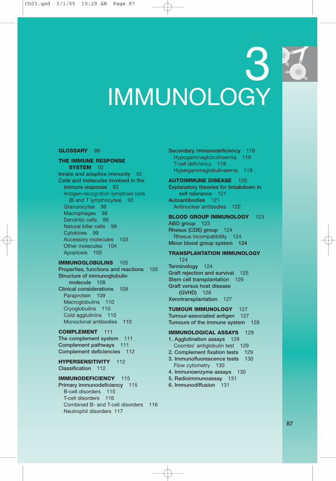
87 3 IMMUNOLOGY GLOSSARY 88 THE IMMUNE RESPONSE SYSTEM 92 Innate and adaptive immunity 92 Cells and molecules involved in the immune response 93 Antigen-recognition lymphoid cells (B and T lymphocytes) 93 Granulocytes 98 Macrophages 98 Dendritic cells 98 Natural killer cells 99 Cytokines 99 Accessory molecules 103 Other molecules 104 Apoptosis 105 IMMUNOGLOBULINS 105 Properties, functions and reactions 105 Structure of immunoglobulin molecule 108 Clinical considerations 109 Paraprotein 109 Macroglobulins 110 Cryoglobulins 110 Cold agglutinins 110 Monoclonal antibodies 110 COMPLEMENT 111 The complement system 111 Complement pathways 111 Complement deficiencies 112 HYPERSENSITIVITY 112 Classification 112 IMMUNODEFICIENCY 115 Primary immunodeficiency 115 B-cell disorders 115 T-cell disorders 116 Combined B- and T-cell disorders 116 Neutrophil disorders 117 Secondary immunodeficiency 118 Hypogammaglobulinaemia 118 T-cell deficiency 118 Hypergammaglobulinaemia 119 AUTOIMMUNE DISEASE 120 Explanatory theories for breakdown in self tolerance 121 Autoantibodies 121 Antinuclear antibodies 122 BLOOD GROUP IMMUNOLOGY 123 ABO group 123 Rhesus (CDE) group 124 Rhesus incompatibility 124 Minor blood group system 124 TRANSPLANTATION IMMUNOLOGY 124 Terminology 124 Graft rejection and survival 125 Stem cell transplantation 126 Graft versus host disease (GVHD) 126 Xenotransplantation 127 TUMOUR IMMUNOLOGY 127 Tumour-associated antigen 127 Tumours of the immune system 128 IMMUNOLOGICAL ASSAYS 129 1. Agglutination assays 129 Coombs’ antiglobulin test 129 2. Complement fixation tests 129 3. Immunofluorescence tests 130 Flow cytometry 130 4. Immunoenzyme assays 130 5. Radioimmunoassay 131 6. Immunodiffusion 131 Ch03.qxd 3/1/05 10:29 AM Page 87
description
Glossary Inmunology
Transcript of Inmunology Glossary & Terminology
87
3IMMUNOLOGY
GLOSSARY 88
THE IMMUNE RESPONSE SYSTEM 92
Innate and adaptive immunity 92Cells and molecules involved in the
immune response 93Antigen-recognition lymphoid cells
(B and T lymphocytes) 93Granulocytes 98Macrophages 98Dendritic cells 98Natural killer cells 99Cytokines 99Accessory molecules 103Other molecules 104Apoptosis 105
IMMUNOGLOBULINS 105Properties, functions and reactions 105Structure of immunoglobulin
molecule 108Clinical considerations 109
Paraprotein 109Macroglobulins 110Cryoglobulins 110Cold agglutinins 110Monoclonal antibodies 110
COMPLEMENT 111The complement system 111Complement pathways 111Complement deficiencies 112
HYPERSENSITIVITY 112Classification 112
IMMUNODEFICIENCY 115Primary immunodeficiency 115
B-cell disorders 115T-cell disorders 116Combined B- and T-cell disorders 116Neutrophil disorders 117
Secondary immunodeficiency 118Hypogammaglobulinaemia 118T-cell deficiency 118Hypergammaglobulinaemia 119
AUTOIMMUNE DISEASE 120Explanatory theories for breakdown in
self tolerance 121Autoantibodies 121
Antinuclear antibodies 122
BLOOD GROUP IMMUNOLOGY 123ABO group 123Rhesus (CDE) group 124
Rhesus incompatibility 124Minor blood group system 124
TRANSPLANTATION IMMUNOLOGY124
Terminology 124Graft rejection and survival 125Stem cell transplantation 126Graft versus host disease
(GVHD) 126Xenotransplantation 127
TUMOUR IMMUNOLOGY 127Tumour-associated antigen 127Tumours of the immune system 128
IMMUNOLOGICAL ASSAYS 1291. Agglutination assays 129
Coombs’ antiglobulin test 1292. Complement fixation tests 1293. Immunofluorescence tests 130
Flow cytometry 1304. Immunoenzyme assays 1305. Radioimmunoassay 1316. Immunodiffusion 131
Ch03.qxd 3/1/05 10:29 AM Page 87
GLOSSARY
Adaptive The transfer of immune cells for therapeutic benefit.immunotherapy
ADCC, antibody- A cytotoxic reaction in which the Fc receptor-bearing dependent cellular killer cells recognize target cells via specific antibodies.cytotoxicity
Adhesion Cell surface molecules involved in cell–cell interaction ormolecules the binding of cells to extracellular matrix, where the
principal function is adhesion rather than cell activation,e.g. integrins and selectins.
Adjuvant Any foreign material introduced with an antigen toenhance its immunogenecity, e.g. killed bacteria,(mycobacteria), emulsions (Freund’s adjuvant) orprecipitates (alums).
Alloantibody Antibody raised in one individual and directed againstan antigen (primarily on cells) of another individual ofthe same species.
Allogeneic See page 124.
Allotypes The protein of an allele which may be detectable as anantigen by another member of the same species.Plasma proteins are an example of antigenicallydissimilar variants.
Alternative pathway The activation pathways of the complement systeminvolving C3 and factors B, D, P, H and I, which interactin the vicinity of an activator surface to form analternative pathway C3 convertase.
Anaphylatoxins Complement peptides (C3a and C5a) which cause mastcell degranulation and smooth muscle contraction.
Anchor residues Certain amino acid residues of antigenic peptides arerequired for interaction in the binding pocket of MHCmolecules.
Antigenic peptides Peptide fragments of proteins which bind to MHCmolecules and induce T-cell activation.
APCs (antigen- A variety of cell types which carry antigen in a form that presenting cells) can stimulate lymphocytes.
Apoptosis Programmed cell death: a mode of cell death whichoccurs under physiological conditions and is controlledby the dying cell itself (‘cell suicide’).
Autologous Originating from the same individual.
�2-microglobulin A polypeptide which constitutes part of some membraneproteins including the class I MHC molecules.
Bcl-2 A molecule expressed transiently on activated B cellswhich have been rescued from apoptosis.
CD markers Used as a prefix (and number). Cell surface molecules (cluster of of lymphocytes and platelets that are distinguishable differentiation) with monoclonal antibodies, and may be used to
distinguish different cell populations.
3
88
IMM
UN
OLO
GY
Ch03.qxd 3/1/05 10:29 AM Page 88
Cell adhesion A group of proteins of the immunoglobulin supergene molecules (CAMs) family involved in intercellular adhesion, including
ICAM-1, ICAM-2, ICAM-3, VCAM-1, MAd CAM-1 andPECAM.
Class I/II restriction The observation that immunologically active cells willonly operate effectively when they share MHChaplotypes of either the class I or class II loci.
Class switching The process by which B cells can express a new heavychain isotype without altering the specificity of theantibody produced. This occurs by gene rearrangement.
Clonal selection The fundamental basis of lymphocyte activation inwhich antigen selectively causes activation, division anddifferentiation only in those cells which expressreceptors with which it can combine.
Collectins A group of large polymeric proteins includingconglutinin and mannose-binding lectin (MBL) that canopsonize microbial pathogens.
Colony-stimulating A group of cytokines which control the differentiation of factors (CSFs) haemopoetic stem cells.
Constant regions The relatively invariant parts of the immunoglobulinheavy and light chains, and the α, β, γ and δ chains ofthe T-cell receptor.
Co-stimulation The signals required for the activation of lymphocytes inaddition to the antigen-specific signal delivered via theirantigen receptors. CD28 is an important costimulatingmolecule for T cells and CD40 for B cells.
Defensins A group of small antibacterial proteins produced byneutrophils.
Dendritic cells Derived from either the lymphoid or mononuclearphagocyte lineages. A set of cells present in tissues,which capture antigen and migrate to lymph nodes andspleen, where they are particularly active in presentingthe processed antigen to T cells.
Domain Segments or loops on heavy and light chains formed byintrachain disulphide bonds. Each immunoglobulindomain consists of about 110 amino acids.
Epitope Part of an antigen that binds to an antibody-combiningsite or a specific T-cell surface receptor, and determinesspecificity. Usually about 9–20 amino acids in size.
Fas ligand The ligand that binds to the cell surface molecule Fas(CD95) which is normally found on the surface oflymphocytes. When Fas ligand binds to its receptor, celldeath (apoptosis) is triggered.
Genetic restriction Describes the phenomenon where lymphocytes andantigen-presenting cells interact more effectively whenthey share particular MHC haplotypes.
Gut-associated Accumulations of lymphoid tissue associated with the lymphoid tissue gastrointestinal tract.(GALT)
3
89
IMM
UN
OLO
GY
Ch03.qxd 3/1/05 10:29 AM Page 89
Haplotype A set of genetic determinants coded by closely linkedgenes on a single chromosome.
Hapten A substance of low molecular weight which is not itselfimmunogenic, but which can bind to an antibodymolecule and produce a new antigenic determinant.
Helper (TH cells) A functional subclass of T cells which can help generate cytotoxic T cells and cooperate with B cells in the production of antibody responses. Helper cellsrecognize antigen in association with class II molecules.
Heterologous Originating from a different individual or different inbredline.
Heterophile antigen Antigen which occurs in tissues of many differentspecies and is therefore highly crossreactive, e.g.Paul–Bunnell antigen which reacts with both sheep andbeef erythrocytes.
HLA See page 36.
Idiotype Unique antigenic determinant on the antigen-bindingregion of an immunoglobulin molecule.
Hypervariable Amino acid sequences within the variable regions of regions heavy and light immunoglobulin chains and of the T-cell
receptor which show the most variability and contributemost to the antigen-binding site.
Immunoglobulin Immunoglobulin of the same class that is detectable in subclass the constant heavy chain region, and differs in
electrophoretic mobility and antigenic determinant, andfunction, e.g. IgG1, IgG2, IgG3 and IgG4.
Immunoglobulin Molecules which have domains homologous to those supergene seen in immunoglobulins, including MHC class I and II family (lgSF) molecules, the T-cell receptor, CD2, CD3, CD4, CD8
ICAMs, VCAM and some of the Fc receptors.
Intercellular Cell surface molecules found on a variety of leucocytes adhesion molecules and non-haematogenous cells which interact with
leucocyte functional antigen (LFA-1); e.g. ICAM-1 (CD54),ICAM-2 (CD102) and ICAM-3 (CD50).
Integrins One of the ‘families’ of adhesion molecules, some ofwhich interact with cell adhesion molecules, and otherswith components of the extracellular matrix.
Isologous Originating from the same individual or member of thesame inbred strain.
Isotype The class or subclass of an immunoglobulin common toall members of that species. Each isotype is encodedby a separate immunoglobulin constant region genesequence that is carried by all members of a species.
Killer (K) cells Type of cytotoxic lymphocyte that is able to mediateantibody-dependent cellular cytotoxicity (ADCC).
Langerhans’ cells Antigen-presenting cells of the skin which emigrate tolocal lymph nodes to become dendritic cells; they arevery active in presenting antigen to T cells.
3
90
IMM
UN
OLO
GY
Ch03.qxd 3/1/05 10:29 AM Page 90
Lectin pathway A pathway of complement activation, initiated bymannose-binding lectin (MBL) which intersects theclassical pathway.
Leucocyte A group of three molecules (LFA-1 (CD11a/CD18), functional antigens LFA-2 (CD2) and LFA-3 (CD58)), which mediate (LFAs) intercellular adhesion between leucocytes and other
cells in an antigen non-specific fashion.
Linkage The association of two linked alleles more frequently disequilibrium than would be expected by chance.
Memory cells Long-lived lymphocytes which have already beenprimed with antigen but have not yet undergoneterminal differentiation into effector cells. They reactmore readily than naïve lymphocytes when restimulatedwith the same antigen.
Mixed lymphocyte Proliferative response when lymphocytes from two reaction (MLR) genetically different (i.e. allogeneic) persons are mixed
in cell culture. A vital test in matching donor andrecipient prior to bone marrow transplantation.
Mucosa-associated Lymphoid tissue associated with the bronchial tree, lymphoid tissue gastrointestinal tract and other mucosa.(MALT)
Natural killer Type of cytotoxic lymphocyte that has the intrinsic (NK) cell ability to recognize and destroy virally infected cells and
some tumour cells. Specializes in killing cells thatexpress little or no MHC molecule.
NfkB A transcription factor which is widely used by differentleucocyte populations to signal activation.
Perforin A granule-associated molecule of cytotoxic cells,homologous to complement C9. It can form pores onthe membrane of a target cell.
Reactive oxygen/ Bactericidal metabolites produced by phagocytic cells, nitrogen including hydrogen peroxide, hypophalites and nitric intermediates acid.(ROIs/RNIs)
Selectins Three adhesion molecules, P-selectin (CD62P), E-selectin (CD62E), and L-selectin (CD62L) involved inslowing leucocytes during their transit through venules.
Superantigens Antigens (often bacterial, e.g. staphylococcalenterotoxins) which bind to the MHC outside thepeptide-binding groove and stimulate all or most of theT cells bearing particular T-cell receptor V regions.Antigens must normally be processed in order to triggerthe T-cell receptor. Superantigens are not processed butbind directly to class II and Vβ.
Suppressor (TS) Functionally defined populations of T cells which reducecell the immune responses of other T cells or B cells, or
switch the response into a different pathway to thatunder investigation.
Syngeneic Genetically identical or closely related, so as to allowtissue transplant.
3
91
IMM
UN
OLO
GY
Ch03.qxd 3/1/05 10:29 AM Page 91
TAP transporters A group of molecules which transport proteins andpeptides between intracellular compartments.
T-cell receptor The T-cell antigen receptor consists of either an (TCR) αβ dimer (TCR-2) or a γδ dimer (TCR-1) associated with
the CD3 molecular complex.
T-dependent Require recognition by both T and B cells to produce anantigens immune response.
T-independent Can directly stimulate B cells to produce specific antigens antibody.
Titre The highest dilution of a given substance, e.g. antibody,that will still produce a reaction with another substance,e.g. antigen.
Toll receptors A group of evolutionarily ancient cell surface molecules,e.g. the IL-1 receptor, some of which are involved intransducing signals for inflammation.
Transforming A group of cytokines, identified by their ability to growth factors promote fibroblast growth, that are also (TGFs) immunosuppressive.
Tumour necrosis See page 101.factor (TNF)
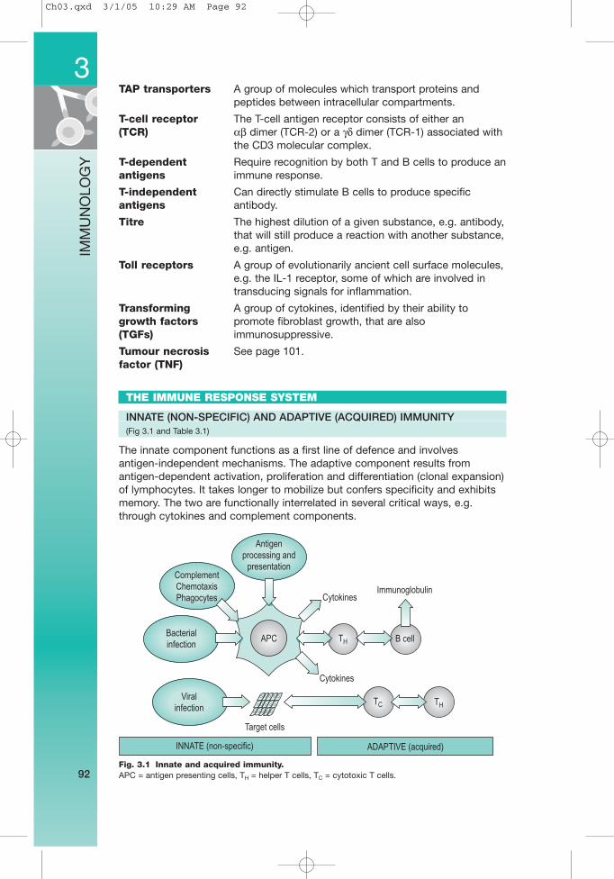
THE IMMUNE RESPONSE SYSTEM
INNATE (NON-SPECIFIC) AND ADAPTIVE (ACQUIRED) IMMUNITY (Fig 3.1 and Table 3.1)
The innate component functions as a first line of defence and involves antigen-independent mechanisms. The adaptive component results fromantigen-dependent activation, proliferation and differentiation (clonal expansion)of lymphocytes. It takes longer to mobilize but confers specificity and exhibitsmemory. The two are functionally interrelated in several critical ways, e.g.through cytokines and complement components.
3
92
IMM
UN
OLO
GY
Fig. 3.1 Innate and acquired immunity.APC = antigen presenting cells, TH = helper T cells, TC = cytotoxic T cells.
Antigen
processing and
presentation
Immunoglobulin
Complement
Chemotaxis
Phagocytes
Bacterial
infection
Viral
infection
APC
Target cells
Cytokines
Cytokines
TH B cell
TC TH
INNATE (non-specific) ADAPTIVE (acquired)
Ch03.qxd 3/1/05 10:29 AM Page 92
CELLS AND MOLECULES INVOLVED IN THE IMMUNE RESPONSE
1. Antigen-recognition lymphoid cells 5. Natural killer cells(B and T lymphocytes) 6. Cytokines
2. Granulocytes 7. Accessory molecules3. Macrophages 8. Other molecules4. Dendritic cells
1. ANTIGEN-RECOGNITION LYMPHOID CELLS (B AND T LYMPHOCYTES)
B lymphocytes (see also Immunoglobulins, p. 105).Functions: Humoral immunity – antibody production; control of
pyogenic bacteria; prevention of blood-borne infections;neutralization of toxins.
% of total 12%; mainly fixed.lymphocytes:Site of production: Produced in germinal centre of lymph nodes and
spleen.Assessment of Serum specific immunoglobulin levels; specific function: antibodies; immunoglobulin response to pokeweed
mitogen; endotoxin and EBV.
T lymphocytesFunctions: Cell-mediated immunity; protection against intracellular
organisms, protozoa and fungi; graft rejection; control ofneoplasms.
% of total 70–80%; mainly circulating; long-lived memory cells.lymphocytes:Site of production: Produced in paracortical region of lymph nodes and
spleen.
3
93
IMM
UN
OLO
GY
Table 3.1 Differences between the innate and adaptive immune response systems
Innate (non-specific system) Adaptive (acquired system)
Components Components1. Anatomical and physiological barriers 1. Cell-mediated response effected by T cells2. Inflammatory response with leakage of 2. Humoral immune response effected by B cells
antibacterial serum proteins (acute-phase proteins) and phagocytic cells
3. Phagocytosis by neutrophils and macrophages
4. Complement system
Properties Properties1. Rapid: responds within minutes to infection 1. Slow: response over days to weeks2. No antigenic specificity, i.e. the same 2. Antigenic specificity i.e. each cell is
molecules and cells respond to a programmed genetically to respond to a range of pathogens single antigen
3. No memory, i.e. the response does not 3. Immunological memory, i.e. on repeated change after repeated exposure exposure the response is faster, stronger and
qualitatively different4. Preformed or rapidly formed components 4. Diversity: ability to recognize and respond to
a vast number of different antigens5. Self/non-self recognition: i.e. lack of response
(tolerance) to self-antigens but response to foreign antigens
Ch03.qxd 3/1/05 10:29 AM Page 93
Assessment of Delayed hypersensitivity skin reactions using candida, function: mumps and purified protein derivative (PPD); active
sensitization with dinitrochlorobenzene (DNCP);lymphocyte transformation: mitogenic response tophytohaemagglutinin (PHA) and concanavalin-A; mixedlymphocyte reaction (MLR); lymphokine release.
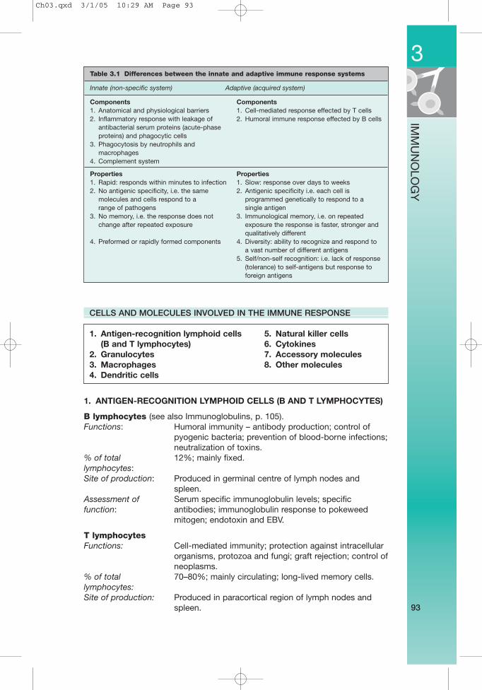
Identified by: T-cell surface phenotypes identified by reaction withmonoclonal Abs (Table 3.2 and Fig. 3.2).
3
94
IMM
UN
OLO
GY
Fig. 3.2 T-cell CD markers.
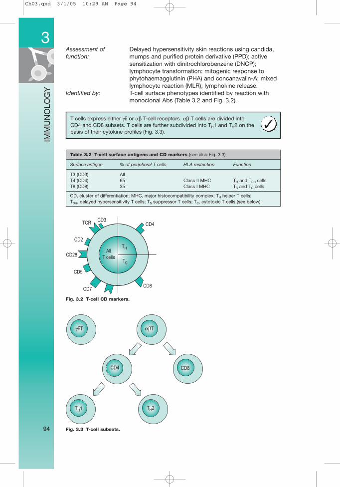
Fig. 3.3 T-cell subsets.
γδT αβT
CD4 CD8
TH2TH1
CD4
CD8
CD3TCR
CD2
CD28
CD5
CD7
TH
TC
All
T cells
T cells express either γδ or αβ T-cell receptors. αβ T cells are divided into CD4 and CD8 subsets. T cells are further subdivided into TH1 and TH2 on thebasis of their cytokine profiles (Fig. 3.3).
✓
Table 3.2 T-cell surface antigens and CD markers (see also Fig. 3.3)
Surface antigen % of peripheral T cells HLA restriction Function
T3 (CD3) AllT4 (CD4) 65 Class II MHC TH and TDH cellsT8 (CD8) 35 Class I MHC TS and TC cells
CD, cluster of differentiation; MHC, major histocompatibility complex; TH helper T cells; TDH, delayed hypersensitivity T cells; TS suppressor T cells; TC, cytotoxic T cells (see below).
Ch03.qxd 3/1/05 10:29 AM Page 94
T-cell subpopulations
Regulatory and effector T cellsRegulatory cells:1. TH helper T cells CD4+: recognize antigen by means of the T-cell receptors
in association with macrophage receptors. Produces cytokines and helpsgenerate cytotoxic T cells and cooperates with B cells in production ofantibody responses. Recognizes antigen in association with class II MHCmolecules on the surface of antigen-presenting cells.
2. TS suppressor T cells: interfere with the development of an immuneresponse of other T cells or B cells, either directly or via suppressor factors.
Effector cells:3. TC cytotoxic T cells CD8+: regulate the immune response and can lyse
target cells, e.g. viral or tumour antigens expressing antigen peptidespresented by MHC class I molecules on the surface of all nucleated cells.Interleukin-2 (IL-2) is responsible for the generation of cytotoxic T cells.
4. TDH delayed hypersensitivity T cells: release mediators that cause aninflammatory response attracting macrophages, neutrophils and otherlymphocytes to the site.
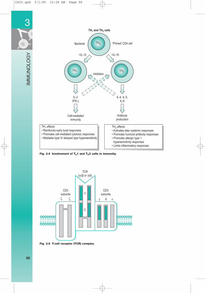
TH1 and TH2 populations (Fig. 3.4)
• CD4+ MHC class II-restricted T cells can also be subdivided into TH1 and TH2populations based on their profiles of cytokine production.
• The TH1 profile is associated with production of IL-2, tumour necrosis factor(TNF)-β and interferon (IFN)-γ and is driven by IL-12.
• The TH2 profile is associated with IL-4, IL-5, IL-6 and IL-13 and is driven byIL-10.
• TH1 cytokines are involved in helping cell-mediated immunity and the TH2cytokines mediate humoral immunity.
• TH1 cells can downregulate TH2 cells and vice versa.
T-cell antigen receptor (TCR) (Fig. 3.5)
TCR complex comprises a disulphide-linked heterodimeric glycoprotein thatenables T cells to recognize a diverse array of antigens in association with MHCmolecules. It consists of α and β subunits or occasionally γ and δ subunits. It isassociated at the cell surface with a complex of polypeptides knowncollectively as CD3 which is required for activation of T cells.• Consists of α, β subunits or, less commonly, γ or δ subunits.• Differences in the variable regions of the TCR subunits account for the
diversity of antigenic specificity among T cells.• TCRs only recognize antigenic peptides bound to class I or class II MHC
molecules.• T cells can be divided into different subsets based on the expression of one
or other T-cell receptor (TCR-1 or TCR-2).
3
95
IMM
UN
OLO
GY
Other selected important CD markersCD28: Present in highest amounts in activated T cells. It is a T-cellcostimulatory molecule which plays a major role in T cell activation.CD45RA: An isoform of CD45 associated with active T cells that respondpoorly to recall antigen.CD45RO: An isoform associated with memory T cells. Responds well torecall antigen.CD95: Also known as Fas, binds Fas ligand and mediates apoptosis ofactivated T cells.
Ch03.qxd 3/1/05 10:29 AM Page 95
3
96
IMM
UN
OLO
GY
Fig. 3.4 Involvement of TH1 and TH2 cells in immunity.
TH0
TH1 and TH2 cells
Bacterial 'Primed' CD4 cell
+IL-12 +IL-10
TH1 TH2Inhibition
IL-2
IFN-γIL-4, IL-5,
IL-6
Cell-mediated
immunity
TH1 effects
• Reinforces early local responses
• Promotes cell-mediated cytotoxic responses
• Mediates type IV delayed type hypersensitivity
TH2 effects
• Activates later systemic responses
• Promotes humoral antibody responses
• Promotes allergic type 1
hypersensitivity responses
• Limits inflammatory responses
Antibody
production
Fig. 3.5 T-cell receptor (TCR) complex.
CD3
subunits V
TCR
(α/β or γ/δ)
CD3
subunits
γ δ εζ ζ
C
Ch03.qxd 3/1/05 10:29 AM Page 96
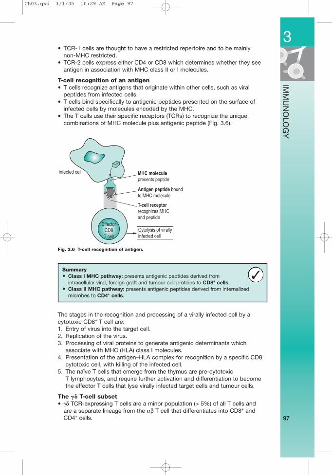
• TCR-1 cells are thought to have a restricted repertoire and to be mainly non-MHC restricted.
• TCR-2 cells express either CD4 or CD8 which determines whether they seeantigen in association with MHC class II or I molecules.
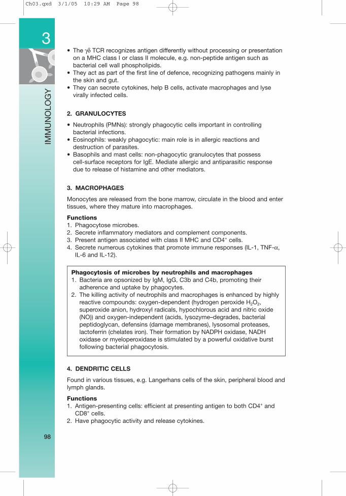
T-cell recognition of an antigen• T cells recognize antigens that originate within other cells, such as viral
peptides from infected cells.• T cells bind specifically to antigenic peptides presented on the surface of
infected cells by molecules encoded by the MHC.• The T cells use their specific receptors (TCRs) to recognize the unique
combinations of MHC molecule plus antigenic peptide (Fig. 3.6).
The stages in the recognition and processing of a virally infected cell by acytotoxic CD8+ T cell are:1. Entry of virus into the target cell.2. Replication of the virus.3. Processing of viral proteins to generate antigenic determinants which
associate with MHC (HLA) class I molecules.4. Presentation of the antigen–HLA complex for recognition by a specific CD8
cytotoxic cell, with killing of the infected cell.5. The naïve T cells that emerge from the thymus are pre-cytotoxic
T lymphocytes, and require further activation and differentiation to becomethe effector T cells that lyse virally infected target cells and tumour cells.
The �� T-cell subset• γδ TCR-expressing T cells are a minor population (> 5%) of all T cells and
are a separate lineage from the αβ T cell that differentiates into CD8+ andCD4+ cells.
3
97
IMM
UN
OLO
GY
Summary• Class I MHC pathway: presents antigenic peptides derived from
intracellular viral, foreign graft and tumour cell proteins to CD8+ cells.• Class II MHC pathway: presents antigenic peptides derived from internalized
microbes to CD4+ cells.
✓
Fig. 3.6 T-cell recognition of antigen.
Infected cell
Effector
CD8
T cell
MHC molecule
presents peptide
Antigen peptide bound
to MHC molecule
T-cell receptor
recognizes MHC
and peptide
Cytolysis of virally
infected cell
Ch03.qxd 3/1/05 10:29 AM Page 97
• The γδ TCR recognizes antigen differently without processing or presentationon a MHC class I or class II molecule, e.g. non-peptide antigen such asbacterial cell wall phospholipids.
• They act as part of the first line of defence, recognizing pathogens mainly inthe skin and gut.
• They can secrete cytokines, help B cells, activate macrophages and lysevirally infected cells.
2. GRANULOCYTES
• Neutrophils (PMNs): strongly phagocytic cells important in controllingbacterial infections.
• Eosinophils: weakly phagocytic: main role is in allergic reactions anddestruction of parasites.
• Basophils and mast cells: non-phagocytic granulocytes that possess cell-surface receptors for IgE. Mediate allergic and antiparasitic response due to release of histamine and other mediators.
3. MACROPHAGES
Monocytes are released from the bone marrow, circulate in the blood and entertissues, where they mature into macrophages.
Functions1. Phagocytose microbes.2. Secrete inflammatory mediators and complement components.3. Present antigen associated with class II MHC and CD4+ cells.4. Secrete numerous cytokines that promote immune responses (IL-1, TNF-α,
IL-6 and IL-12).
4. DENDRITIC CELLS
Found in various tissues, e.g. Langerhans cells of the skin, peripheral blood andlymph glands.
Functions1. Antigen-presenting cells: efficient at presenting antigen to both CD4+ and
CD8+ cells.2. Have phagocytic activity and release cytokines.
3
98
IMM
UN
OLO
GY
Phagocytosis of microbes by neutrophils and macrophages1. Bacteria are opsonized by IgM, IgG, C3b and C4b, promoting their
adherence and uptake by phagocytes.2. The killing activity of neutrophils and macrophages is enhanced by highly
reactive compounds: oxygen-dependent (hydrogen peroxide H2O2,superoxide anion, hydroxyl radicals, hypochlorous acid and nitric oxide(NO)) and oxygen-independent (acids, lysozyme–degrades, bacterialpeptidoglycan, defensins (damage membranes), lysosomal proteases,lactoferrin (chelates iron). Their formation by NADPH oxidase, NADHoxidase or myeloperoxidase is stimulated by a powerful oxidative burstfollowing bacterial phagocytosis.
Ch03.qxd 3/1/05 10:29 AM Page 98
Antigen-presenting cells• Include macrophages, monocytes or their derivatives (microglial cells, Kupffer
cells and skin Langerhans cells).• Characterized by their ability to phagocytose, internalize and process antigen.• Possess Ia antigen, Fc receptors and C3b receptors and produce interleukin 1.
5. NATURAL KILLER (NK) CELLS
Functions1. Similar function to lymphocytes – kill virus-infected cells and some tumour
cells, and produce cytokines.2. Recognition of target differs from lymphocytes – they do not bind MHC, and
a carbohydrate receptor selects target. NK cells express two major classesof inhibitory receptors for MHC molecules: lectin-like receptors of the CD94family and immunoglobulin superfamily molecules (KIRs).
3. Act rapidly, and constitute an early antiviral defence.4. Identified by: Fc receptor for IgG.5. Previously referred to as large granular lymphocytes (IGL) because of their
appearance.
Mechanisms of NK cell killing• Direct cytotoxicity involving contact with target cell and lysis by
perforin-mediated mechanism similar to that used by TC cells, except it isantigen independent and non-MHC restricted.
• Antibody-dependent cellular (ADCC) cytotoxicity. Binding of Fc receptors onNK cells to antibody-coated target cells initiates killing. (Neutrophils,eosinophils and macrophages also exhibit ADCC).
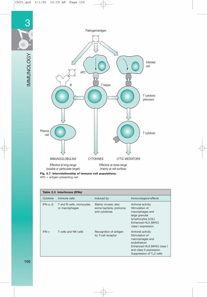
6. CYTOKINES (Fig. 3.7)
• Small protein signalling molecules (usually glycoproteins) of relatively lowmolecular weight.
• They regulate important biological processes: proliferation and differentiation,growth inhibition, apoptosis, chemotaxis and chemokinesis, resistance toviral infection, induction of cytotoxic effector cells, induction of phagocytes,promotion of intercellular adhesions and regulation of adhesion toextracellular matrix.
• Many cytokines act by causing aggregation of receptors at the cell surface,which leads to activation of second messenger system.
• The main cytokines are interferons, interleukins, tumour necrosis factor,growth factors, colony stimulating factors and chemokines.
1. Interferons (IFNs) (Table 3.3)
These glycoproteins are produced by virus-infected cells.• Three species of interferon:
1. Alpha-interferon (IFN-α) produced by human leucocytes2. Beta-interferon (IFN-β) produced by human fibroblasts3. Gamma-interferon (IFN-γ) produced by human T lymphocytes in response
to antigenic stimulation.• Properties:
1. Prevent viral replication2. Antitumour activity3. Activate macrophages and natural killer (NK) cells.
3
99
IMM
UN
OLO
GY
Ch03.qxd 3/1/05 10:29 AM Page 99
3
100
IMM
UN
OLO
GY
Fig. 3.7 Interrelationship of immune cell populations.APC = antigen-presenting cell.
Pathogen/antigen
APC
B
Plasma
cell
Infected
cell
T cytotoxic
precursor
T cytotoxic
T helper
CYTOKINES LYTIC MEDIATORSIMMUNOGLOBULINS
Effective at close-range
(mainly at cell surface)
Effective at long-range
(soluble or particulate target)
Table 3.3 Interferons (IFNs)
Cytokine Immune cells Induced by Immunological effects
IFN-α,-β T and B cells, monocytes Mainly viruses; also Antiviral activity or macrophages some bacteria, protozoa Stimulation of
and cytokines macrophages and large granular lymphocytes (LGL)Enhanced HLA (MHC)class I expression
IFN-γ T cells and NK cells Recognition of antigen Antiviral activityby T-cell receptor Stimulation of
macrophages and endotheliumEnhanced HLA (MHC) class I and class II expressionSuppression of TH2 cells
Ch03.qxd 3/1/05 10:29 AM Page 100
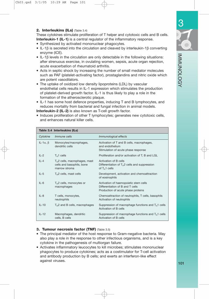
2. Interleukins (ILs) (Table 3.4)
These cytokines stimulate proliferation of T helper and cytotoxic cells and B cells.Interleukin-1 (IL-1) is a central regulator of the inflammatory response.• Synthesized by activated mononuclear phagocytes.• IL-1β is secreted into the circulation and cleaved by interleukin-1β converting
enzyme (ICE).• IL-1β levels in the circulation are only detectable in the following situations:
after strenuous exercise, in ovulating women, sepsis, acute organ rejection,acute exacerbation of rheumatoid arthritis.
• Acts in septic shock by increasing the number of small mediator moleculessuch as PAF (platelet-activating factor), prostaglandins and nitric oxide whichare potent vasodilators.
• The uptake of oxidized low density lipoproteins (LDL) by vascular endothelial cells results in IL-1 expression which stimulates the production of platelet-derived growth factor. IL-1 is thus likely to play a role in theformation of the atherosclerotic plaque.
• IL-1 has some host defence properties, inducing T and B lymphocytes, andreduces mortality from bacterial and fungal infection in animal models.
Interleukin-2 (IL-2) is also known as T-cell growth factor.• Induces proliferation of other T lymphocytes; generates new cytotoxic cells,
and enhances natural killer cells.
3. Tumour necrosis factor (TNF) (Table 3.5)
• The principal mediator of the host response to Gram-negative bacteria. Mayalso play a role in the response to other infectious organisms, and is a keycytokine in the pathogenesis of multiorgan failure.
• Activates inflammatory leucocytes to kill microbes; stimulates mononuclearphagocytes to produce cytokines; acts as a costimulator for T-cell activationand antibody production by B cells; and exerts an interferon-like effectagainst viruses.
3
101
IMM
UN
OLO
GY
Table 3.4 Interleukins (ILs)
Cytokine Immune cells Immunological effects
IL-1α, β Monocytes/macrophages, Activation of T and B cells, macrophages,dendritic cells and endothelium
Stimulation of acute phase response
IL-2 TH1 cells Proliferation and/or activation of T, B and LGL
IL-4 TH2 cells, macrophages, mast Activation of B cellscells and basophils, bone Differentiation of TH2 cells and suppressionmarrow stroma of TH1 cells
IL-5 TH2 cells, mast cells Development, activation and chemoattractionof eosinophils
IL-6 TH2 cells, monocytes or Activation of haemopoietic stem cellsmacrophages Differentiation of B and T cells
Production of acute phase proteins
IL-8 T cells, monocytes, Chemoattraction of neutrophils, T cells, basophilsneutrophils Activation of neutrophils
IL-10 TH2 and B cells, macrophages Suppression of macrophage functions and TH1 cells Activation of B cells
IL-12 Macrophages, dendritic Suppression of macrophage functions and TH1 cells cells, B cells Activation of B cells
Ch03.qxd 3/1/05 10:29 AM Page 101
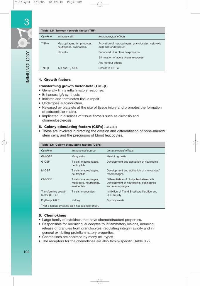
4. Growth factors
Transforming growth factor-beta (TGF-� )• Generally limits inflammatory response.• Enhances IgA synthesis.• Initiates and terminates tissue repair.• Undergoes autoinduction.• Released by platelets at the site of tissue injury and promotes the formation
of extracellular matrix.• Implicated in diseases of tissue fibrosis such as cirrhosis and
glomerulosclerosis.
5. Colony stimulating factors (CSFs) (Table 3.6)
• These are involved in directing the division and differentiation of bone-marrowstem cells, and the precursors of blood leucocytes.
6. Chemokines• Large family of cytokines that have chemoattractant properties.• Responsible for recruiting leucocytes to inflammatory lesions, inducing
release of granules from granulocytes, regulating integrin avidity and ingeneral exhibiting proinflammatory properties.
• Chemokines are secreted by many cell types.• The receptors for the chemokines are also family-specific (Table 3.7).
3
102
IMM
UN
OLO
GY
Table 3.6 Colony stimulating factors (CSFs)
Cytokine Immune cell source Immunological effects
GM-GSF Many cells Myeloid growth
G-CSF T cells, macrophages, Development and activation of neutrophils neutrophils
M-CSF T cells, macrophages, Development and activation of monocytes/neutrophils macrophages
GM-CSF T cells, macrophages, Differentiation of pluripotent stem cells mast cells, neutrophils, Development of neutrophils, eosinophils eosinophils and macrophages
Transforming growth T cells, monocytes Inhibition of T and B cell proliferation and factor (TGF)-β LGL activity
Erythropoietin* Kidney Erythropoiesis
*Not a typical cytokine as it has a single origin.
Table 3.5 Tumour necrosis factor (TNF)
Cytokine Immune cells Immunological effects
TNF-α Macrophages, lymphocytes, Activation of macrophages, granulocytes, cytotoxic neutrophils, eosinophils, cells and endothelium
NK cells Enhanced HLA class I expression
Stimulation of acute phase response
Anti-tumour effects
TNF-β TH1 and TC cells Similar to TNF-α
Ch03.qxd 3/1/05 10:29 AM Page 102
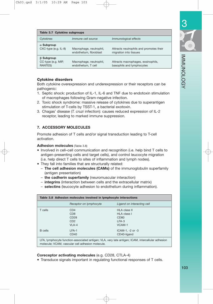
Cytokine disordersBoth cytokine overexpression and underexpression or their receptors can bepathogenic:1. Septic shock: production of IL-1, IL-6 and TNF due to endotoxin stimulation
of macrophages following Gram-negative infection.2. Toxic shock syndrome: massive release of cytokines due to superantigen
stimulation of T-cells by TSST-1, a bacterial exotoxin.3. Chagas’ disease (T. cruzi infection): causes reduced expression of IL-2
receptor, leading to marked immune suppression.
7. ACCESSORY MOLECULES
Promote adhesion of T cells and/or signal transduction leading to T-cellactivation.
Adhesion molecules (Table 3.8)
• Involved in cell–cell communication and recognition (i.e. help bind T cells toantigen-presenting cells and target cells), and control leucocyte migration (i.e. help direct T cells to sites of inflammation and lymph nodes).
• They fall into families that are structurally related:– The cell adhesion molecules (CAMs) of the immunoglobulin superfamily
(antigen presentation)– the cadherin superfamily (neuromuscular interaction)– integrins (interaction between cells and the extracellular matrix)– selectins (leucocyte adhesion to endothelium during inflammation).
Coreceptor activating molecules (e.g. CD28, CTLA-4)• Transduce signals important in regulating functional responses of T cells.
3
103
IMM
UN
OLO
GY
Table 3.8 Adhesion molecules involved in lymphocyte interactions
Receptor on lymphocyte Ligand on interacting cell
T cells CD4 HLA class IICD8 HLA class ICD28 CD80CD2 LFA-3VLA-4 VCAM-1
B cells LFA-1 ICAM-1, -2 or -3CD40 CD40-ligand
LFA, lymphocyte function-associated antigen; VLA, very late antigen; ICAM, intercellular adhesionmolecule; VCAM, vascular cell adhesion molecule.
Table 3.7 Cytokine subgroups
Cytokines Immune cell source Immunological effects
� SubgroupCXC-type (e.g. IL-8) Macrophage, neutrophil, Attracts neutrophils and promotes their
endothelium, fibroblast migration into tissues
� SubgroupCC-type (e.g. MIP, Macrophage, neutrophil, Attracts macrophages, eosinophils, RANTES) endothelium, T cell basophils and lymphocytes
Ch03.qxd 3/1/05 10:29 AM Page 103
8. OTHER MOLECULES
Heat shock proteinsThe heat shock response is a highly conserved and phylogenetically ancientresponse to tissue stress that is mediated by activation of specific genes. Thisleads to the production of specific heat shock proteins that alter the phenotypeof the cell and enhance its resistance to stress. Their principal function appearsto be to act as molecular chaperones for damaged protein to direct it intodegradation pathways such as ubiquitination.
Free radicals• A free radical is literally any atom or molecule which contains one or more
unpaired electrons, making it more reactive than the native species.• Free radical species produced in the human body are:
– OOH• (peroxide radical) – O2• (superoxide radical)
– OH• (hydroxyl radical) – NO• (nitric oxide).• The hydroxyl radical is by far the most reactive species, but the others can
generate more reactive species as breakdown products.• When a free radical reacts with a non-radical, a chain reaction ensues which
results in the formation of further free radicals and direct tissue damage bylipid peroxidation of membranes (particularly implicated in atherosclerosisand ischaemic reperfusion injury within tissues).
• Free radical scavengers bind reactive oxygen species.• Principal dietary antioxidants:
– Vitamin E – β-Carotene– Vitamin C – Flavonoids.
• Patients with dominant familial forms of amyotrophic lateral sclerosis (motorneuron disease) have mutations in the gene for Cu–Zn SOD-1, suggesting alink between failure of free radical scavenging and neurodegeneration.Protection against heart disease and cancer may be conferred by dietaryantioxidants.
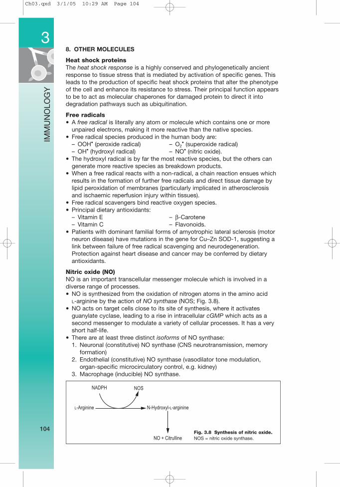
Nitric oxide (NO)NO is an important transcellular messenger molecule which is involved in adiverse range of processes.• NO is synthesized from the oxidation of nitrogen atoms in the amino acid
L-arginine by the action of NO synthase (NOS; Fig. 3.8).• NO acts on target cells close to its site of synthesis, where it activates
guanylate cyclase, leading to a rise in intracellular cGMP which acts as asecond messenger to modulate a variety of cellular processes. It has a veryshort half-life.
• There are at least three distinct isoforms of NO synthase:1. Neuronal (constitutive) NO synthase (CNS neurotransmission, memory
formation)2. Endothelial (constitutive) NO synthase (vasodilator tone modulation,
organ-specific microcirculatory control, e.g. kidney)3. Macrophage (inducible) NO synthase.
3
104
IMM
UN
OLO
GY
Fig. 3.8 Synthesis of nitric oxide. NOS = nitric oxide synthase.
NADPH NOS
L-Arginine N-Hydroxyl-L-arginine
NO + Citrulline
Ch03.qxd 3/1/05 10:29 AM Page 104
APOPTOSIS (see also p. 35)
• Is the process of programmed cell death, and is a mechanism for theelimination of excess or damaged cells.
• Several genes have been identified that either promote (bax, bak, bcl-Xs) orinhibit (bcl-2, bcl-XL, bcl-w) apoptosis. Antiapoptotic genes could confercharacteristics such as longer survival.
• It is mainly triggered through the Fas–Fas ligand interaction. Binding of Fasligand (expressed on a killer T cell) to Fas expressed on a target cell triggers acascade of intracellular biochemical changes in the target cell. Fas interactswith several proteins in the ‘death pathway’ to activate a proteolytic enzyme,caspase. The caspase proteolytic cascade then activates a cytoplasmicenzyme (caspase-activatable DNAase (CAD)) which can then migrate to thenucleus and cleave DNA into small fragments, which are the end-point ofapoptosis.
• It has several important roles in shaping the adaptive immune response, e.g. after an immune response to a pathogen, redundant lymphocytes arecleared by apoptosis.
• It is also involved in some pathological processes, e.g. destruction of CD4+
cells in HIV infection; can lead to the production of autoantibodies againstDNA and result in autoimmune disease; clones of B cells that have increasedlevels of bcl-2 through mutations may be protected from apoptosis anddevelop into a B-cell malignancy.
IMMUNOGLOBULINS
PROPERTIES, FUNCTIONS AND REACTIONS
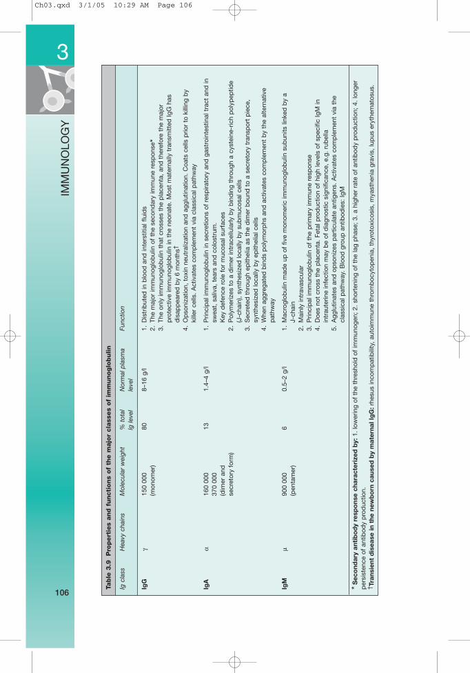
The properties and functions of the major classes of immunoglobulins areshown in Table 3.9, and the immunological reactions of IgG, IgA and IgM aresummarized in Table 3.10.
3
105
IMM
UN
OLO
GY
Clinical relevance of NO1. Septic shock (NO is released in massive amounts and results in decreased
vascular tone, cardiac output with low BP). This is because endotoxinrelease triggers the innate immune response when macrophages are directlyactivated through Toll-like receptors. Macrophage activation results in thesecretion of TNF, prostaglandins and NO. There have been three main approachesto preventing septic shock:(i) Blocking nitric oxide production by macrophages, endothelium and smooth
muscle.(ii) Blocking TNF with monoclonal antibodies.(iii) Recombinant bactericidal protein to bind to endotoxin and prevent
macrophage activation. All have failed in clinical trials, probably because theinnate immune response has already mediated its damage by the timesymptoms develop.
2. Atherosclerosis (where NO synthesis may be impaired, leading to tonicvasoconstriction and vasospasm).
3. 1° and 2° pulmonary hypertension (inhaled NO reverses pulmonary hypertension).4. Hepatorenal syndrome and the hypertension of chronic renal failure.5. Glutamate-mediated excitotoxic cell death in the CNS, such as in Alzheimer’s
disease, and also in acute brain injury, such as stroke.6. Tissue damage in acute and chronic inflammation (probably by interacting with
oxygen-derived free radicals).7. ARDS (adult respiratory distress syndrome).
✓
Ch03.qxd 3/1/05 10:29 AM Page 105
3
106
IMM
UN
OLO
GY
Table
3.9
Pro
pert
ies
and f
uncti
ons
of
the m
ajo
r cla
sses
of
imm
unoglo
bulin
Ig c
lass
Hea
vy c
hain
sM
olec
ular
wei
ght
% t
otal
N
orm
al p
lasm
aFu
nctio
nlg
leve
lle
vel
IgG
γ15
0 00
080
8–16
g/l
1.D
istr
ibut
ed in
blo
od a
nd in
ters
titia
l flu
ids
(mon
omer
)2.
The
maj
or im
mun
oglo
bul
in o
f th
e se
cond
ary
imm
une
resp
onse
*3.
The
only
imm
unog
lob
ulin
tha
t cr
osse
s th
e p
lace
nta,
and
the
refo
re t
he m
ajor
p
rote
ctiv
e im
mun
oglo
bul
in in
the
neo
nate
. M
ost
mat
erna
lly t
rans
mitt
ed Ig
G h
as
dis
app
eare
d b
y 6
mon
ths†
4.O
pso
niza
tion,
tox
in n
eutr
aliz
atio
n an
d a
gglu
tinat
ion.
Coa
ts c
ells
prio
r to
kill
ing
by
kille
r ce
lls.
Act
ivat
es c
omp
lem
ent
via
clas
sica
l pat
hway
IgA
α16
0 00
013
1.4–
4 g/
l1.
Prin
cip
al im
mun
oglo
bul
in in
sec
retio
ns o
f re
spira
tory
and
gas
troi
ntes
tinal
tra
ct a
nd in
37
0 00
0sw
eat,
sal
iva,
tea
rs a
nd c
olos
trum
.(d
imer
and
Key
def
ence
rol
e fo
r m
ucos
al s
urfa
ces
secr
etor
y fo
rm)
2.P
olym
eriz
es t
o a
dim
er in
trac
ellu
larly
by
bin
din
g th
roug
h a
cyst
eine
-ric
h p
olyp
eptid
e (J
-cha
in),
synt
hesi
zed
loca
lly b
y su
bm
ucos
al c
ells
3.S
ecre
ted
thr
ough
ep
ithel
ia a
s th
e d
imer
bou
nd t
o a
secr
etor
y tr
ansp
ort
pie
ce,
synt
hesi
zed
loca
lly b
y ep
ithel
ial c
ells
4.W
hen
aggr
egat
ed b
ind
s p
olym
orp
hs a
nd a
ctiv
ates
com
ple
men
t b
y th
e al
tern
ativ
e p
athw
ay
IgM
µ90
0 00
06
0.5–
2 g/
l1.
Mac
rogl
obul
in m
ade
up o
f fiv
e m
onom
eric
imm
unog
lob
ulin
sub
units
link
ed b
y a
(pen
tam
er)
J-ch
ain
2.M
ainl
y in
trav
ascu
lar
3.P
rinci
pal
imm
unog
lob
ulin
of
the
prim
ary
imm
une
resp
onse
4.D
oes
not
cros
s th
e p
lace
nta.
Fet
al p
rod
uctio
n of
hig
h le
vels
of
spec
ific
IgM
in
intr
aute
rine
infe
ctio
n m
ay b
e of
dia
gnos
tic s
igni
fican
ce,
e.g.
rub
ella
5.A
gglu
tinat
es a
nd o
pso
nize
s p
artic
ulat
e an
tigen
s. A
ctiv
ates
com
ple
men
t vi
a th
e cl
assi
cal p
athw
ay.
Blo
od g
roup
ant
ibod
ies:
IgM
*Sec
ond
ary
anti
bo
dy
resp
ons
e ch
arac
teri
zed
by:
1. lo
wer
ing
of t
he t
hres
hold
of
imm
unog
en;
2. s
hort
enin
g of
the
lag
pha
se;
3. a
hig
her
rate
of
antib
ody
pro
duc
tion;
4.
long
erp
ersi
sten
ce o
f an
tibod
y p
rod
uctio
n.† T
rans
ient
dis
ease
in t
he n
ewb
orn
cau
sed
by
mat
erna
l Ig
G:r
hesu
s in
com
pat
ibili
ty,
auto
imm
une
thro
mb
ocyt
open
ia,
thyr
otox
icos
is,
mya
sthe
nia
grav
is,
lup
us e
ryth
emat
osus
.
Ch03.qxd 3/1/05 10:29 AM Page 106
3
107
IMM
UN
OLO
GY
Table
3.9
(Con
t’d
)
Ig c
lass
Hea
vy c
hain
sM
olec
ular
wei
ght
% t
otal
N
orm
al p
lasm
aFu
nctio
nlg
leve
lle
vel
IgD
δ17
0 00
00.
14–
40 m
g/l
1.P
reci
se f
unct
ions
are
unk
now
n(m
onom
er)
2.N
early
all
imm
unog
lob
ulin
is p
rese
nt a
s ce
ll su
rfac
e re
cep
tor
on h
uman
B c
ells
and
m
ay b
e in
volv
ed in
B-c
ell a
ctiv
atio
n
IgE
ε18
5 00
00.
002
0.1–
1.3
mg/
l1.
Imm
edia
te h
yper
sens
itivi
ty r
eact
ions
: b
ind
s to
mas
t ce
lls a
nd b
asop
hils
via
its
Fc
frag
men
t, w
hich
deg
ranu
late
s an
d r
elea
ses
bio
logi
cally
act
ive
med
iato
rs,
e.g.
hi
stam
ine,
whe
n ex
pos
ed t
o th
e ap
pro
pria
te a
ntig
en.
Pos
sib
ly o
f b
enef
it in
con
trol
ling
cert
ain
par
asiti
c in
fect
ions
2.S
erum
leve
ls c
orre
late
with
sev
erity
of
asth
ma
3.A
ctiv
ates
var
ious
cel
ls in
volv
ed in
alle
rgic
and
infla
mm
ator
y d
isea
se,
whi
ch a
re
activ
ated
by
2 m
ain
typ
es o
f ce
ll su
rfac
e re
cep
tor:
the
FE
SR
1 re
cep
tor
on m
ast
cells
, b
asop
hils
and
eos
inop
hils
, an
d t
he F
qS
R2
rece
pto
r on
lym
pho
cyte
s. R
ecep
tor
blo
ckin
g m
onoc
lona
l ant
ibod
ies
have
bee
n d
evel
oped
as
pos
sib
le a
sthm
a th
erap
ies
Ch03.qxd 3/1/05 10:29 AM Page 107
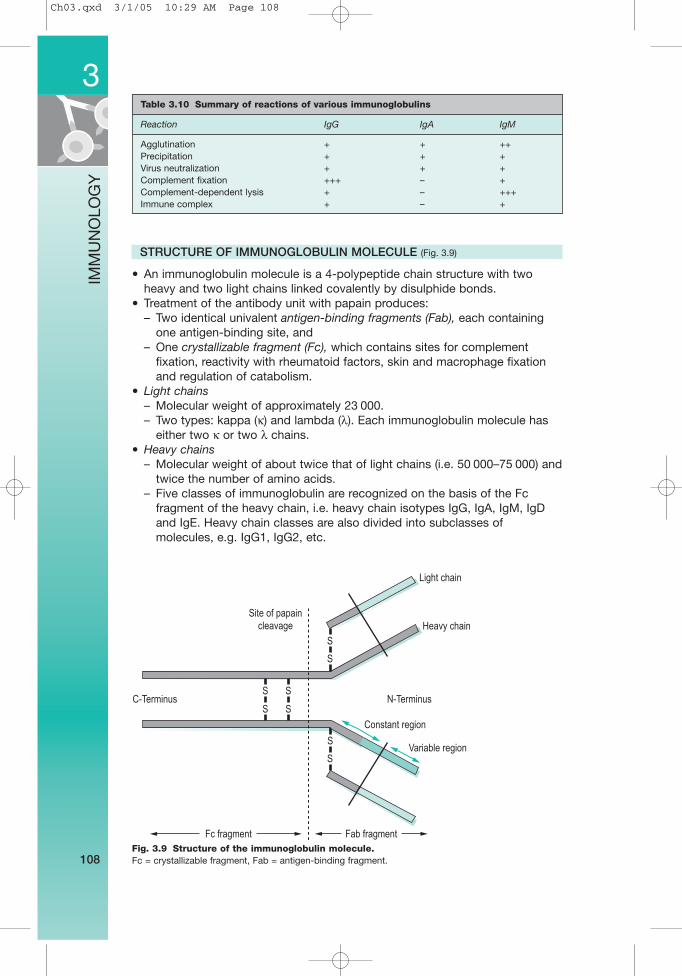
STRUCTURE OF IMMUNOGLOBULIN MOLECULE (Fig. 3.9)
• An immunoglobulin molecule is a 4-polypeptide chain structure with twoheavy and two light chains linked covalently by disulphide bonds.
• Treatment of the antibody unit with papain produces:– Two identical univalent antigen-binding fragments (Fab), each containing
one antigen-binding site, and– One crystallizable fragment (Fc), which contains sites for complement
fixation, reactivity with rheumatoid factors, skin and macrophage fixationand regulation of catabolism.
• Light chains– Molecular weight of approximately 23 000.– Two types: kappa (κ) and lambda (λ). Each immunoglobulin molecule has
either two κ or two λ chains.• Heavy chains
– Molecular weight of about twice that of light chains (i.e. 50 000–75 000) andtwice the number of amino acids.
– Five classes of immunoglobulin are recognized on the basis of the Fcfragment of the heavy chain, i.e. heavy chain isotypes IgG, IgA, IgM, IgDand IgE. Heavy chain classes are also divided into subclasses ofmolecules, e.g. IgG1, IgG2, etc.
3
108
IMM
UN
OLO
GY
Table 3.10 Summary of reactions of various immunoglobulins
Reaction IgG IgA IgM
Agglutination + + ++Precipitation + + +Virus neutralization + + +Complement fixation +++ – +Complement-dependent lysis + – +++Immune complex + – +
Fig. 3.9 Structure of the immunoglobulin molecule. Fc = crystallizable fragment, Fab = antigen-binding fragment.
Site of papain
cleavage
Light chain
Heavy chain
S
S
S
S
S
S
S
S
C-Terminus N-Terminus
Constant region
Variable region
Fab fragmentFc fragment
Ch03.qxd 3/1/05 10:29 AM Page 108
• Both heavy and light chains consist of two regions (Table 3.11):1. A constant region (CH and CL), in which the amino acid sequence of
immunoglobulins of the same class is more or less identical.2. A variable region (VH and VL) where the amino acid sequence varies
considerably from molecule to molecule and contributes to the antigen-binding site.
Development and activation of B cells• Direct B cell/TH cell interaction and cytokines secreted by TH cells are
required for B cells to respond to most antigens.• Stimulation of B cells by protein antigens induces generation of memory
B cells and antibody-secreting plasma cells.• During this clonal expansion and differentiation, the antibody affinity for
antigen may change (affinity maturation), and the biological activities of theantibody can change (isotype class switching).
CLINICAL CONSIDERATIONS (see Table 3.12)
PARAPROTEIN
• A homogeneous band of one immunoglobulin, usually IgG, IgM or IgA. Itspresence implies proliferation of a single clone of cells.
3
109
IMM
UN
OLO
GY
Table 3.11 Antigenic determinants on antibodies
Epitope class Location Comment on epitope
Isotype Constant region 5 human isotopes are IgA, IgD, IgE, IgG, IgM.Each class of Ig heavy chains are identical in all members of a species
Allotype Constant region Vary among individuals of the same species. IgG exhibits the most allotypic difference
Idiotype Variable region Differ among antibodies with different antigen-binding specificities.Monoclonal antibodies have the same idiotype. Anti-idiotypic antibodies will resemble the original antigenic determinant group
Table 3.12 Immunoglobulin products for replacement therapy
Product Indications
Immunoglobulin replacement therapy (pooled from Primary immunodeficiency normal humans)
High-dose immunoglobulin (pooled from normal humans) Immunosuppressive effects used in autoimmunity
Anti-D (pooled from women with high levels of anti-D) Prevention of haemolytic disease of the newborn
Hyperimmune immunoglobulin (pooled from humans Prevention of tetanus, rabies, with high titres of antibodies) varicella zoster and hepatitis B
Antivenom Treatment of snake bite
Monoclonal antibodies (raised against specific human Used as immunosuppressants and cells in mouse hybridomas) cancer treatment
Ch03.qxd 3/1/05 10:29 AM Page 109